Embed Size (px)
Citation preview
CHAPTER THREE
Temporal Patterning of NeuralProgenitors in DrosophilaXin Li, Zhenqing Chen, Claude Desplan1Department of Biology, New York University, New York, New York, USA1Corresponding author: e-mail address: [email protected]
Contents
1.
CurISShttp
Introduction
rent Topics in Developmental Biology, Volume 105 # 2013 Elsevier Inc.N 0070-2153 All rights reserved.://dx.doi.org/10.1016/B978-0-12-396968-2.00003-8
70
1.1 Embryonic and larval ventral nerve cord 71 1.2 Olfactory system 72 1.3 Medulla in the optic lobes 72 1.4 Type II NBs in the central brain 732.
Sequentially Expressed Transcription Factors in NBs Control Birth-Order-Dependent Neuron Fates 73 2.1 Temporal sequence of transcription factors in Drosophila embryonicVNC NBs
73 2.2 Similar or different TF sequence in other systems? 75 2.3 Different TF sequence in medulla NBs 76 2.4 Combinatorial temporal patterning of INPsand type II NBs
77 3. How are Temporal Transitions in NBs Controlled? 783.1
Cross-regulations between the TFs play important roles in the transitions 78 3.2 Differential requirements for cell cycle and asymmetric division 80 3.3 Switching factors involved in TF transitions 804.
Relationship Between Temporal Sequence and NB Competence 81 4.1 Restriction of NB competence 81 4.2 TF sequence and regulation of NB competence 81 4.3 Epigenetic mechanism for loss of NB competence 825.
Feedforward Loops Downstream of the Temporal Sequence ControlNeuron Fates 846.
Progression of the TF Sequence Required for the End of Neurogenesis 85 6.1 Apoptosis of abdominal pNBs 85 6.2 Pros-dependent cell-cycle exit of thoracic and central brain pNBs 86 6.3 Studies in other systems about the end of neurogenesis 877.
Integration of Temporal and Spatial Information Determines Lineages 87 8. Integration of Temporal Identity with Binary Fate Choice 88 9. Conclusions and Future Questions 90 References 9169

70 Xin Li et al.
Abstract
Drosophila has recently become a powerful model system to understand the mecha-nisms of temporal patterning of neural progenitors called neuroblasts (NBs). Two differ-ent temporal sequences of transcription factors (TFs) have been found to be sequentiallyexpressed in NBs of two different systems: the Hunchback, Krüppel, Pdm1/Pdm2, Castor,and Grainyhead sequence in the Drosophila ventral nerve cord; and the Homothorax,Klumpfuss, Eyeless, Sloppy-paired, Dichaete, and Tailless sequence that patternsmedullaNBs. In addition, the intermediate neural progenitors of type II NB lineages are patternedby a different sequence: Dichaete, Grainyhead, and Eyeless. These three examples sug-gest that temporal patterning of neural precursors by sequences of TFs is a commontheme to generate neural diversity. Cross-regulations, including negative feedback reg-ulation and positive feedforward regulation among the temporal factors, can facilitatethe progression of the sequence. However, there are many remaining questions tounderstand themechanism of temporal transitions. The temporal sequence progressionis intimately linked to the progressive restriction of NB competence, and eventuallydetermines the end of neurogenesis. Temporal identity has to be integrated with spatialidentity information, as well as with the Notch-dependent binary fate choices, in order togenerate specific neuron fates.
1. INTRODUCTION
One fundamental question in developmental neurobiology is to under-
stand how to generate the remarkable diversity of neurons and glia present in
adult brains, from a small number of seemingly homogenous neural stem cells
in the embryo. Spatial patterning of neural stem cells that is achieved by various
morphogens and their signaling cascades contributes to the generation of neu-
ral diversity (Bhat, 1999a; Dessaud, McMahon, & Briscoe, 2008). Further-
more, single neural stem cells can generate different neural types in a
stereotyped order: This is achieved by temporal patterning of neural stem cells.
Extensive studies in the vertebrate central nervous system (CNS), especially
cerebral cortex and retina, have revealed that birth order correlates with dis-
tinct neuronal/glial identity (reviewed in Jacob, Maurange, & Gould, 2008;
Livesey & Cepko, 2001; Molyneaux, Arlotta, Menezes, & Macklis, 2007;
Okano & Temple, 2009; Pearson & Doe, 2004). Although both extrinsic
and intrinsic factors are required for the correct specification of tem-
poral identify, isolated neural stem cells cultured in vitro can recapitulate the
sequential generation of different neuron types, thus underscoring a cell-
intrinsic temporal control of stem cells (Gaspard et al., 2008; Naka,
Nakamura, Shimazaki, & Okano, 2008; Shen et al., 2006).
71Temporal Patterning of Neural Progenitors in Drosophila
Neural stem cells are called neuroblasts (NBs) in Drosophila. Drosophila
NBs usually undergo several rounds of Notch-dependent asymmetric divi-
sions to produce a self-renewed NB, and a smaller daughter cell called gan-
glion mother cell (GMC). GMCs generally divide asymmetrically once to
produce two postmitotic progeny with different fates (reviewed in Doe,
2008; Karcavich, 2005; Knoblich, 2010; Lin & Lee, 2012). SeveralDrosoph-
ilaNB lineages in the CNS have been partially or completely characterized.
They generate multiple distinct progeny in a birth-order-dependent manner
(Akiyama-Oda, Hosoya, & Hotta, 1999; Baek & Mann, 2009; Bossing,
Udolph, Doe, & Technau, 1996; Isshiki, Pearson, Holbrook, & Doe, 2001;
Jefferis, Marin, Stocker, & Luo, 2001; Karcavich & Doe, 2005; Lee, Lee, &
Luo, 1999; Lundell & Hirsh, 1998; Pearson & Doe, 2003; Schmid,
Chiba, & Doe, 1999; Schmidt et al., 1997; Skeath & Thor, 2003; Yu,
Chen, Shi, Huang, & Lee, 2009; Yu et al., 2010). Drosophila has recently
become a powerful model system to understand the mechanisms of tem-
poral specification of neurons and there have been several extensive and
excellent reviews on this topic (Brand & Livesey, 2011; Kao & Lee,
2010; Maurange, 2012; Pearson & Doe, 2004). This review will therefore
focus on the most recent advances in temporal patterning of NBs in several
systems of Drosophila that are now well characterized. Below is a brief
introduction of each system to be described.
1.1. Embryonic and larval ventral nerve cordClassic work on the temporal patterning of NBs in theDrosophila embryonic
ventral nerve cord (VNC) has made it undoubtedly the best-characterized
system. The VNC of Drosophila, spanning three thoracic and eight abdom-
inal segments, is the counterpart of the vertebrate spinal cord. During early
embryonic development, NBs delaminate from a neuroectoderm at stereo-
typical positions. Each hemi-segment contains approximately 30 NBs
arranged in seven rows, and each NB is identifiable based on its position
(e.g., NB1-1 is in row1, column1) and molecular markers (Bhat, 1999b;
Doe, 1992; Skeath & Thor, 2003; Technau, Berger, & Urbach, 2006).
The distinction between these NBs is achieved by spatial patterning that will
not be discussed in detail here. The neuronal and glial progeny composition
of each embryonic NB lineage have been determined by early Dil NB label-
ing experiments: A single NB can generate multiple neuron types and glia
(Bossing et al., 1996; Schmid et al., 1999, 1997). At the embryo to larval
transition, some of these NBs commit to apoptosis, while others enter into
72 Xin Li et al.
a quiescence phase. They are then reactivated during the larval stage and
continue to proliferate during larval and pupal stages. These postembryonic
NBs generate �90% of neurons that constitute the adult CNS (Prokop &
Technau, 1991; Truman & Bate, 1988). Relatively less is known about
the postembryonic lineages as compared to embryonic lineages, but several
recent studies have shed some light on the temporal control of larval lineages
(see below).
1.2. Olfactory systemAnother well-characterized system is the olfactory system, including
the antennal lobe, which receives olfactory inputs from olfactory receptor
neurons (ORNs), and the mushroom body, which receives inputs from
projection neurons (PNs) in the antenna lobe. Systematic clonal analysis
of mushroom body neurons using the mosaic analysis with a repressible cell
marker (MARCM) technique has demonstrated that each of the four mush-
room bodyNBs sequentially generates at least three types of neurons, g neu-rons first, followed by a0b0, and then ab neurons (Lee et al., 1999), suggestingbirth-order-dependent neuronal specification. Similarly, the antennal lobe
PNs that are derived from three NBs (an anterodorsal, a lateral, and a ventral
NB) are specified by lineage and birth order to connectwith specific classes of
ORNaxons ( Jefferis et al., 2001). These three lineages have been extensively
characterized (Das, Reichert, & Rodrigues, 2010; Das et al., 2008; Lai,
Awasaki, Ito, & Lee, 2008; Lin, Kao, Yu, Huang, & Lee, 2012; Lin et al.,
2010; Yu et al., 2010). For example, the anterodorsal lineage produces
40 types of PNs in a stereotypic order, while the lateral lineage sequentially
generates 48 pairs of local interneurons paired with distinct PNs.
1.3. Medulla in the optic lobesThe Drosophila optic lobes, composed of lamina, medulla, and lobula
complex, are the processing centers of visual information in the brain.
Among them, the medulla is the largest neuropil. It contains approximately
40,000 neurons, belonging to more than 70 different cell types (Fischbach &
Dittrich, 1989; Morante & Desplan, 2008). These neurons are generated
by NBs derived from a crescent-shaped single-layered neuroepithelium
(NE) in the larval brain called the outer proliferation center (Green,
Hartenstein, & Hartenstein, 1993; White & Kankel, 1978). During devel-
opment, a wave of neurogenesis progresses from the edge of the crescent
toward its center, and sequentially converts NE cells into medulla NBs.
73Temporal Patterning of Neural Progenitors in Drosophila
Therefore, NBs of different ages can be observed together in one snapshot
(Egger, Boone, Stevens, Brand, & Doe, 2007; Yasugi, Umetsu, Murakami,
Sato, & Tabata, 2008). These NBs generate GMCs, which then produce
medulla neurons that form chains below each NB, with the first-born
neurons located in the deepest position and latest-born neurons in the
most superficial position, near the NB (Hasegawa et al., 2011; Morante,
Erclik, & Desplan, 2011). Recently, the developing medulla has emerged
as a new powerful system for studying temporal patterning of NBs (Li
et al., 2013; Suzuki, Kaido, Takayama, & Sato, 2013).
1.4. Type II NBs in the central brainIn contrast to the regular type I NBs that divide asymmetrically to generate a
series of GMCs that each divides once to produce two postmitotic cells,
type II NBs divide asymmetrically to self-renew and to generate a series of
transit amplifying GMCs called intermediate neural progenitors (INPs), with
each INP dividing asymmetrically to generate several GMCs and eventually
giving rise to 6–12 neurons and/or glia (Bayraktar, Boone, Drummond, &
Doe, 2010; Boone & Doe, 2008; Viktorin, Riebli, Popkova, Giangrande, &
Reichert, 2011; Weng, Golden, & Lee, 2010; Zhu, Barshow, Wildonger,
Jan, & Jan, 2011). Type II lineages allow more neurons than type I lineages
to be quickly generated.
2. SEQUENTIALLY EXPRESSED TRANSCRIPTIONFACTORS IN NBs CONTROL BIRTH-
ORDER-DEPENDENT NEURON FATES2.1. Temporal sequence of transcription factorsin Drosophila embryonic VNC NBs
How does a NB generate a stereotypical order of different neuron types
as it ages? Extensive studies in the Drosophila embryonic VNC have led to
the identification of a molecular mechanism of temporal specification,
where a series of transcription factors (TFs) are sequentially and transiently
expressed in NBs as they age. Postmitotic progeny born during each time
window maintains the expression of the TF. This temporal sequence
of TFs includes Hunchback (Hb), Kruppel (Kr), Pdm1/Pdm2 (Pdm), Castor
(Cas), and Grainyhead (Grh) (Baumgardt, Karlsson, Terriente, Diaz-
Benjumea, & Thor, 2009; Brody & Odenwald, 2000; Cui & Doe, 1992;
Grosskortenhaus, Pearson, Marusich, & Doe, 2005; Isshiki et al., 2001;
Kambadur et al., 1998; Mellerick, Kassis, Zhang, & Odenwald, 1992;
74 Xin Li et al.
Romani et al., 1996;Yang,Yeo,Dick,&Chia, 1993). Although differentNBs
in theVNCdelaminate and start differentiation at different times,most of them
follow the same TF sequence. These temporally expressed TFs have been
shown to be both required and sufficient for birth-order-dependent neuronal
specification in several different NB lineages (Baumgardt et al., 2009; Benito-
Sipos et al., 2010; Cleary & Doe, 2006; Grosskortenhaus, Robinson, &
Doe, 2006; Isshiki et al., 2001; Kambadur et al., 1998; Novotny, Eiselt, &
Urban, 2002; Pearson & Doe, 2003; Tran & Doe, 2008). For example, each
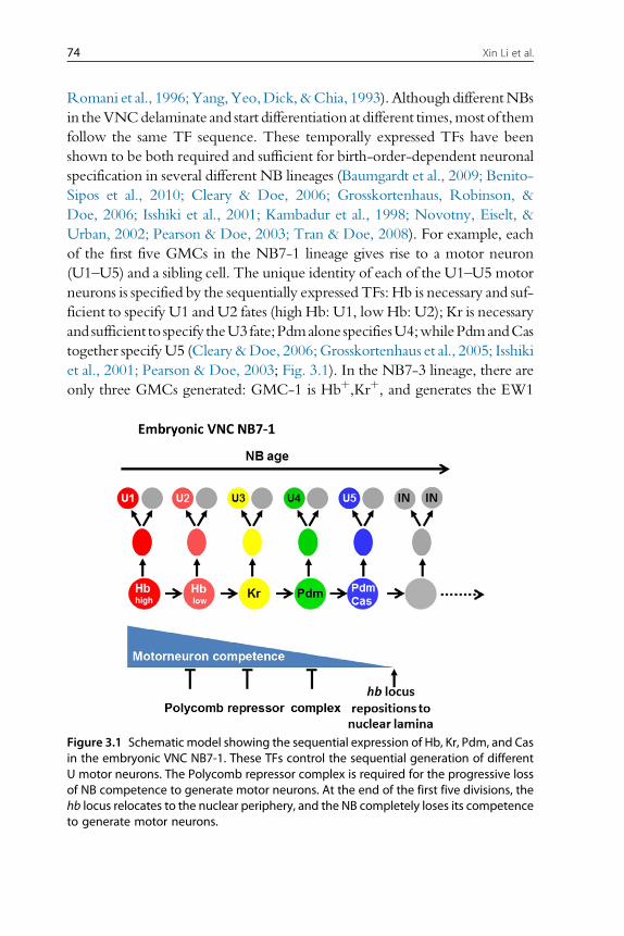
of the first five GMCs in the NB7-1 lineage gives rise to a motor neuron
(U1–U5) and a sibling cell. The unique identity of each of the U1–U5 motor
neurons is specified by the sequentially expressed TFs: Hb is necessary and suf-
ficient to specify U1 andU2 fates (high Hb: U1, lowHb: U2); Kr is necessary
and sufficient to specify theU3fate;Pdmalone specifiesU4;whilePdmandCas
together specifyU5 (Cleary&Doe, 2006;Grosskortenhaus et al., 2005; Isshiki
et al., 2001; Pearson & Doe, 2003; Fig. 3.1). In the NB7-3 lineage, there are
only three GMCs generated: GMC-1 is Hbþ,Krþ, and generates the EW1
Figure 3.1 Schematic model showing the sequential expression of Hb, Kr, Pdm, and Casin the embryonic VNC NB7-1. These TFs control the sequential generation of differentU motor neurons. The Polycomb repressor complex is required for the progressive lossof NB competence to generate motor neurons. At the end of the first five divisions, thehb locus relocates to the nuclear periphery, and the NB completely loses its competenceto generate motor neurons.
75Temporal Patterning of Neural Progenitors in Drosophila
interneuron and the GW motor neuron; GMC-2 is Krþ, and generates the
EW2 interneuron and a sibling that undergoes apoptosis; GMC-3 is Pdmþ,and differentiates directly into the EW3 interneuron (Isshiki et al., 2001;
Karcavich & Doe, 2005; Lundell & Hirsh, 1998). In this case, Hb is necessary
and sufficient for the first-born neuron fates (EW1/GW). Loss of hb results in
a lack of the EW1/GW, while EW2 and EW3 are still present. Prolonged
expression of Hb in the NB leads to additional neurons with EW1/GW fates
at theexpenseofEW2andEW3(Isshiki et al., 2001;Novotnyet al., 2002).Kr is
necessary and sufficient for the second-born neuron fate EW2 (Isshiki
et al., 2001).
There are variations in the function of the temporal TFs: In the NB3-1
lineage, Pdm is not required for specification of the third temporal identity,
but instead to close the preceding Krþ temporal identity window (Tran &
Doe, 2008). The first four GMCs of NB3-1 lineage produce HB9þ,Isletþ
RP motor neurons with a birth order of RP1!RP4!RP3!RP5
(and their non-RP siblings): Hb specifies RP1 and RP4 (high Hb: RP1;
low Hb: RP4); Kr specifies RP3; while Pdm is not required for specifying
RP5, because there is only a modest expansion of Kr expression in NBs in
pdmmutants, with a few extra RP3 neurons produced, but RP5 neurons are
still specified. Similarly, Cas is required for closing the third (RP5) temporal
identity window: In cas mutants, there are ectopic RP5 neurons (Tran &
Doe, 2008).
Thus, a series of TFs sequentially expressed in NBs control the sequential
generation of different neural types in multiple NB lineages. Since different
NBs generate different lineages, these TFs do not specify a certain neuron
type, but control the birth-order-dependent neuronal identity. The birth-
order-dependent temporal identity is integrated with the spatial identity
of the NB within each segment or between different segments, and is trans-
lated into specific cell types. For example, hb controls the first-born cell fates
in multiple lineages, which can be motor neurons, interneurons or glial cells,
depending on the NB lineage (Isshiki et al., 2001).
2.2. Similar or different TF sequence in other systems?Since birth-order-dependent neuronal specification has been widely
observed in various systems, is the same or a similar temporal TF sequence
utilized to pattern neural stem cells of other systems?
In the mushroom body, which is generated by four NBs, each NB
sequentially generates at least three types of neurons. Thus far, no temporal
sequence of TFs that controls the fate of these neurons has been identified in
76 Xin Li et al.
the NB. However, a novel bric-a-brac,tramtrack, broad (BTB)-zinc finger
protein, named Chinmo (chronologically inappropriate morphogenesis),
acts in GMCs or young neurons to control the temporal identity of mush-
room body neurons (Zhu et al., 2006). Although ChinmomRNA is equally
expressed throughout the entire NB lineage, Chinmo protein shows a tem-
poral gradient in the neuronal progeny. It is absent in NBs and is expressed at
its highest levels in the early-born neurons, lower in the next-born neurons,
and undetectable in the latest-born neurons. Reducing or increasing
Chinmo levels causes transformation of neurons toward later or earlier fates,
respectively (Zhu et al., 2006). Recently, microRNAs of the Let-7-com-
plex, the heterochronic miRNAs originally identified in Caenorhabditis
elegans, were found to target chinmo to regulate the temporal identity ofDro-
sophilamushroom body neurons (Wu, Chen, Mercer, & Sokol, 2012). Since
Chinmo protein or Let-7 miRNAs are not detected in NBs, how the
Chinmo gradient in neurons is regulated as the NB ages is not understood.
Postembryonic neuroblasts (pNBs) in the VNC generate 90% of neurons
that constitute the adult CNS. Although a complete temporal TF sequence
has not been identified, recent studies identified two members of the post-
embryonic TF sequence, Cas and Seven-up (Svp) (Maurange, Cheng, &
Gould, 2008; Tsuji, Hasegawa, & Isshiki, 2008). They are required for a
temporal switch of pNBs from generating small Chinmoþ neurons to
instead producing large Br-Cþ (Broad Complex) neurons in many different
lineages. Since the switch from Chinmoþ to Br-Cþ happens later than the
transient expression of Cas or Svp, the switch must be directly controlled by
an unknown member of the TF sequence whose expression is promoted
by cas and svp by feedforward regulation (Maurange et al., 2008). Cas and
Svp are also required for the NBs to eventually end neurogenesis. This is
discussed in detail below.
In the anterodorsal lineage of the antennal lobe, Kr was shown to act in
the NB to define one out of 40 temporal fates of PNs (Kao, Yu, He, Kao, &
Lee, 2012). Loss of Kr from the NB causes a single PN fate to be skipped.
However, loss of Hb, Pdm or Cas does not produce detectable phenotypes
(Kao et al., 2012). It will be interesting to identify more TFs that are tem-
porally expressed in antennal lobe NBs and control the temporal specifica-
tion of a large number of distinct PNs and interneurons.
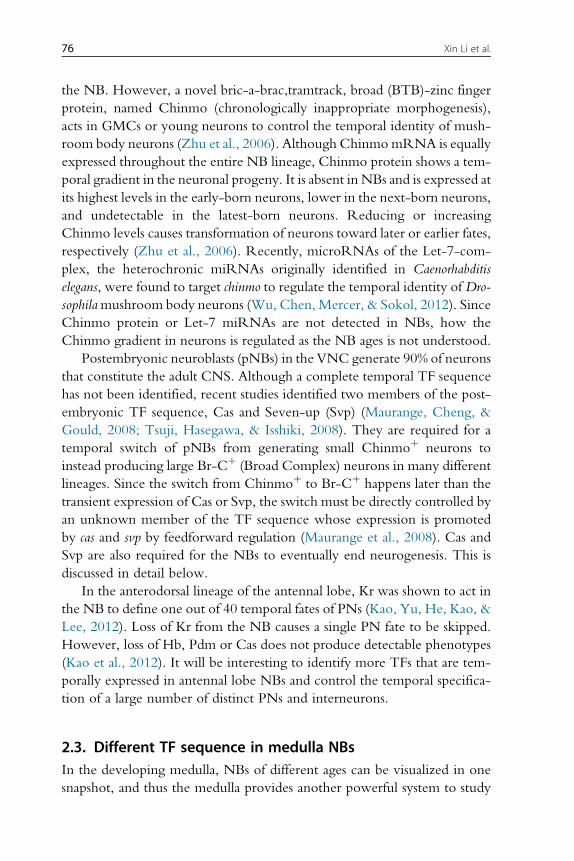
2.3. Different TF sequence in medulla NBsIn the developing medulla, NBs of different ages can be visualized in one
snapshot, and thus the medulla provides another powerful system to study
77Temporal Patterning of Neural Progenitors in Drosophila
temporal patterning of NBs. Recent studies identified a different series of
TFs expressed in medulla NBs: Homothorax (Hth), Klumpfuss (Klu), Eye-
less (Ey), Sloppy paired (Slp), Dichaete (D), and Tailless (Tll) are sequentially
expressed in NBs of increasing ages, with Hth expressed in newly differen-
tiated NBs, Klu, Ey, Slp, and D expressed in increasingly older NBs, and
Tll in the oldest NBs. Hth, Ey, and Slp were shown to control the gener-
ation of specific neuron types born during each timewindow (Li et al., 2013;
Suzuki et al., 2013; Fig. 3.2). This identification of a second TF sequence
that is different from the one in the embryonic VNC suggests that TF
sequence-dependent temporal patterning of NBs is likely to be broadly uti-
lized, and that different TF sequences can be recruited in different systems.
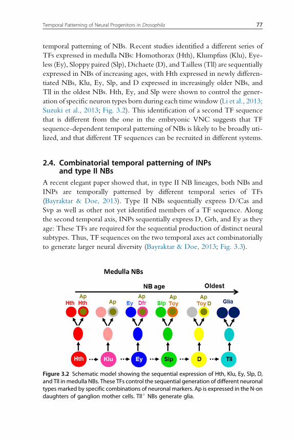
2.4. Combinatorial temporal patterning of INPsand type II NBs
A recent elegant paper showed that, in type II NB lineages, both NBs and
INPs are temporally patterned by different temporal series of TFs
(Bayraktar & Doe, 2013). Type II NBs sequentially express D/Cas and
Svp as well as other not yet identified members of a TF sequence. Along
the second temporal axis, INPs sequentially express D, Grh, and Ey as they
age: These TFs are required for the sequential production of distinct neural
subtypes. Thus, TF sequences on the two temporal axes act combinatorially
to generate larger neural diversity (Bayraktar & Doe, 2013; Fig. 3.3).
Figure 3.2 Schematic model showing the sequential expression of Hth, Klu, Ey, Slp, D,and Tll inmedulla NBs. These TFs control the sequential generation of different neuronaltypes marked by specific combinations of neuronal markers. Ap is expressed in the N-ondaughters of ganglion mother cells. Tllþ NBs generate glia.
Figure 3.3 Schematic model showing combinatorial temporal patterning in type II NBlineages in the central brain. Both NBs and INPs are patterned by distinct temporal seriesof TFs. INPs sequentially express D, Grh, and Ey as they age. These TFs are required forthe sequential production of distinct neuronal subtypes.
78 Xin Li et al.
3. HOW ARE TEMPORAL TRANSITIONS IN NBsCONTROLLED?
3.1. Cross-regulations between the TFs play importantroles in the transitions
In the embryonic VNC, the Hb!Kr!Pdm!Cas!Grh sequence can
be recapitulated in isolated NBs cultured in vitro, suggesting a lineage-
intrinsic mechanism for the TF transitions (Brody & Odenwald, 2000;
Grosskortenhaus et al., 2005). Gain and loss of function studies suggest that
multiple cross-regulations between these temporal TFs exist: one TF acti-
vates the next TF, while repressing the previous TF and the next-plus-one
TF (Fig. 3.4). This, in theory, could allow the TF transitions (Baumgardt
et al., 2009; Brody & Odenwald, 2000; Grosskortenhaus et al., 2006;
Isshiki et al., 2001; Kambadur et al., 1998; Nakajima, Isshiki, Kaneko, &
Ishihara, 2010; Tran & Doe, 2008). However, in most lineages, loss of
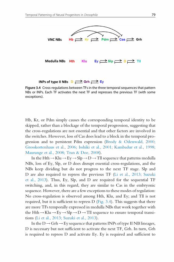
Figure 3.4 Cross-regulations between TFs in the three temporal sequences that patternNBs or INPs. Each TF activates the next TF and represses the previous TF (with someexceptions).
79Temporal Patterning of Neural Progenitors in Drosophila
Hb, Kr, or Pdm simply causes the corresponding temporal identity to be
skipped, rather than a blockage of the temporal progression, suggesting that
the cross-regulations are not essential and that other factors are involved in
the switches. However, loss of Cas does lead to a block in the temporal pro-
gression and to persistent Pdm expression (Brody & Odenwald, 2000;
Grosskortenhaus et al., 2006; Isshiki et al., 2001; Kambadur et al., 1998;
Maurange et al., 2008; Tran & Doe, 2008).
In the Hth!Klu!Ey!Slp!D!Tll sequence that patterns medulla
NBs, loss of Ey, Slp, or D does disrupt essential cross-regulations, and the
NBs keep dividing but do not progress to the next TF stage. Slp and
D are also required to repress the previous TF (Li et al., 2013; Suzuki
et al., 2013). Thus, Ey, Slp, and D are required for the sequential TF
switching, and, in this regard, they are similar to Cas in the embryonic
sequence. However, there are a few exceptions to these modes of regulation:
No cross-regulation is observed among Hth, Klu, and Ey; and Tll is not
required, but it is sufficient to repress D (Fig. 3.4). This suggests that there
are more TFs temporally expressed in medulla NBs that work together with
the Hth!Klu!Ey!Slp!D!Tll sequence to ensure temporal transi-
tions (Li et al., 2013; Suzuki et al., 2013).
In the D!Grh!Ey sequence that patterns INPs of type II NB lineages,
D is necessary but not sufficient to activate the next TF, Grh. In turn, Grh
is required to repress D and activate Ey. Ey is required and sufficient to
80 Xin Li et al.
repress Grh. Thus, a “feedforward activation/feedback repression” model
for D!Grh!Ey cross-regulation appears to control the transitions of
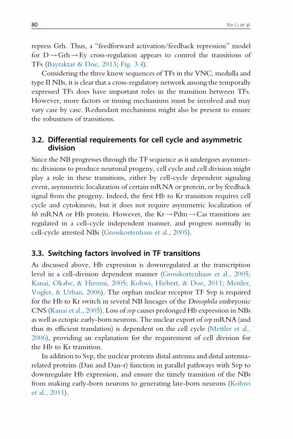
TFs (Bayraktar & Doe, 2013; Fig. 3.4).
Considering the three know sequences of TFs in the VNC, medulla and
type II NBs, it is clear that a cross-regulatory network among the temporally
expressed TFs does have important roles in the transition between TFs.
However, more factors or timing mechanisms must be involved and may
vary case by case. Redundant mechanisms might also be present to ensure
the robustness of transitions.
3.2. Differential requirements for cell cycle and asymmetricdivision
Since the NB progresses through the TF sequence as it undergoes asymmet-
ric divisions to produce neuronal progeny, cell cycle and cell division might
play a role in these transitions, either by cell-cycle dependent signaling
event, asymmetric localization of certain mRNA or protein, or by feedback
signal from the progeny. Indeed, the first Hb to Kr transition requires cell
cycle and cytokinesis, but it does not require asymmetric localization of
hb mRNA or Hb protein. However, the Kr!Pdm!Cas transitions are
regulated in a cell-cycle independent manner, and progress normally in
cell-cycle arrested NBs (Grosskortenhaus et al., 2005).
3.3. Switching factors involved in TF transitionsAs discussed above, Hb expression is downregulated at the transcription
level in a cell-division dependent manner (Grosskortenhaus et al., 2005;
Kanai, Okabe, & Hiromi, 2005; Kohwi, Hiebert, & Doe, 2011; Mettler,
Vogler, & Urban, 2006). The orphan nuclear receptor TF Svp is required
for the Hb to Kr switch in several NB lineages of the Drosophila embryonic
CNS (Kanai et al., 2005). Loss of svp causes prolongedHb expression in NBs
as well as ectopic early-born neurons. The nuclear export of svpmRNA (and
thus its efficient translation) is dependent on the cell cycle (Mettler et al.,
2006), providing an explanation for the requirement of cell division for
the Hb to Kr transition.
In addition to Svp, the nuclear proteins distal antenna and distal antenna-
related proteins (Dan and Dan-r) function in parallel pathways with Svp to
downregulate Hb expression, and ensure the timely transition of the NBs
from making early-born neurons to generating late-born neurons (Kohwi
et al., 2011).
81Temporal Patterning of Neural Progenitors in Drosophila
In the medulla NB TF sequence, both Slp and D are required to turn off
the preceding TF and turn on the next TF, therefore, acting as switching
factors for themselves. Additional transition factors remain to be identified
for other transitions (Li et al., 2013; Suzuki et al., 2013).
4. RELATIONSHIP BETWEEN TEMPORAL SEQUENCEAND NB COMPETENCE
4.1. Restriction of NB competence
Early studies in mammalian neurogenesis have demonstrated that neuralstem cells undergo progressive restriction in their competence to generate
different types of progeny in response to extrinsic signals (Desai &
McConnell, 2000; Livesey & Cepko, 2001). For example, during develop-
ment of the cerebral cortex, neural stem cells generate neurons in the six
cortical layers in an “inside-out” defined order, in which layer 6 and 5
neurons are generated first, followed by layer 4, 3 and 2. The fate of the
progeny also depends on environmental cues to which the cells respond
prior to cell division. Transplantation experiments showed that mid-stage
neural stem cells that are producing layer 4 neurons have the competence
to generate layer 2/3 (later-born) neurons when transplanted into older
brains. However, when these neural stem cells are transplanted into younger
brains in which layer 6 neurons are being generated, they have lost the com-
petence to generate layer 6 neurons. Instead, they produce layer 4 and 5 neu-
rons. Thus, the competence of neural stem cells to generate a given cell type
is progressively lost, although this loss of competence lags behinds the com-
pletion of generation of this neural type (Desai & McConnell, 2000).
4.2. TF sequence and regulation of NB competenceLoss of competence is also observed inDrosophilaNBs, although, in this case,
competence responds to intrinsic TFs rather than extrinsic cues. In the
VNC, providing ectopic Hb to the NB after Hb has been downregulated
is sufficient to generate additional early-born neurons. However, this com-
petence to respond to Hb progressively decreases over time. In the NB7-1
lineage, a pulse of ectopic Hb at the time U1 and U2 are normally born leads
to the generation of several ectopic U1 and U2 neurons. If the pulse of
ectopic Hb is provided later, when U3 and U4 are normally born, only
one ectopic U2 neuron can be generated; and if Hb is given after the fifth
division when the NB switches to generate interneurons, the NB fails to
82 Xin Li et al.
generate ectopic U neurons (Pearson & Doe, 2003). Further studies showed
that NB7-1 has a single early competence window spanning the first five
divisions for responding to Hb, Kr, Pdm, or Cas to make the corresponding
U motor neurons. Then, NBs completely lose the competence to produce
U motor neurons in response to these TFs (Cleary & Doe, 2006; Fig. 3.1).
Similarly in the NB3-1 lineage, when the NB switches to generate interneu-
rons at the fifth division, the competence to generate RP3 motor neurons
in response to Kr is lost (Tran & Doe, 2008). This suggests that NBs can go
sequentially through several competence windows in which they respond
differently to the same temporal TF, allowing the repeated use of the same
TF within one lineage to specify different fates (Cleary & Doe, 2006;
Pearson & Doe, 2003).
The NB competence window can be extended if ectopic Hb is contin-
uously provided before endogenous Hb is downregulated. In this case,
NBs generate many U1/U2 neurons. They also retain their competence
to generate later-born U motor neurons, and the lineage is extended. Thus,
downregulation of Hb expression is required for the gradual loss of NB
competence to respond to Hb (Cleary & Doe, 2006; Grosskortenhaus
et al., 2005; Isshiki et al., 2001). Repression of multiple target genes (includ-
ing Pdm2) by sustained Hb is necessary and sufficient for the maintenance of
NB competence. Indeed, sustained expression of a form of Hb that functions
solely as a transcription activator cannot significantly extend the competence
window (Tran, Miller, & Doe, 2010).
Therefore, loss of NB competence to generate certain neuron fates lags
behind the progression of the TF sequence (e.g., for cell fate specified by
Hb, it is 3 cell divisions later) (Kohwi, Lupton, Lai, Miller, & Doe, 2013;
Pearson & Doe, 2003). However, progression of the TF sequence, that
is, downregulation of Hb relieving its transcription repression on multiple
target genes, including a later temporal TF (Pdm), is required for the NB
to close the current competence window and transit to the next.
4.3. Epigenetic mechanism for loss of NB competenceRecent studies uncovered a close relationship between epigenetic mecha-
nism and loss of NB competence. Mammalian cortical neural stem cells gen-
erate cortical neurons that populate the different layers, and then lose
the competence to generate neurons and switch to producing glia. This is
due to the Polycomb repressor complexes (PRCs) that are required to
suppress transcription of the neural fate TF neurogenin 1 in late-stage
83Temporal Patterning of Neural Progenitors in Drosophila
progenitors (Hirabayashi et al., 2009). PRCs induce repressive chromatin
marks at the neurogenin 1 locus that gradually accumulate over time, and
thus may provide a timer for the loss of competence to make neurons
(Hirabayashi et al., 2009).
In Drosophila, PRCs are also involved in progressively restricting com-
petence for generating motor neurons in NB7-1 and NB3-1: PRC loss
of function extends the competence window to generate the corresponding
motor neuron fate in response to Kr, while PRC gain of function preco-
ciously restricts this competence. In contrast, PRC activity does not affect
the production of interneurons in multiple lineages (Touma, Weckerle, &
Cleary, 2012), suggesting that it is involved specifically in the repression of
multiple target genes involved in motor neuron specification (Fig. 3.1).
Chromosomal architecture has also recently been shown to be involved
in the loss of competence of NB7-1 to respond to Hb, three NB divisions
after hb transcription have stopped. At this stage, the hb gene locus relocates
to the nuclear periphery of the NB, a repressive subnuclear compartment,
preventing further activation of the hb gene by ectopic Hb, which is a
requirement for the specification of early-born neurons (Kohwi et al.,
2013; Fig. 3.1). The timing of the relocation correlates with downregulation
of the Pipsqueak domain nuclear protein, Dan. Prolonging the expression of
Dan can extend the NB competence by preventing relocalization of the
hb locus at the periphery. This study proposed that Dan might competitively
inhibit other Pipsqueak-domain factors, for example, Pipsqueak, a GAGA-
binding factor essential for sequence-specific targeting of PRCs (Huang,
Chang, Yang, Pan, & King, 2002), from binding to and recruiting hb and
other loci to the nuclear lamina (Kohwi et al., 2013). This is consistent with
the model that PRCs are involved in restricting NB competence (Touma
et al., 2012).
In the medulla temporal sequence, NB competence has not yet been
well characterized. Although mis-expressing the first NB TF Hth in all
NBs leads to ectopic neurons with early-born cell fate, the phenotype
becomes less obvious in later part of the lineage, suggesting that the compe-
tence of NBs to generate early-born cell fate in response to the first NB TF
decreases with time. Whether the progressive loss of NB competence
involves similar epigenetic mechanism remains to be studied. Interestingly,
similar to the mammalian cortical neural stem cells, the medulla NBs switch
from neurogenic to gliogenic at their final temporal stage when they express
Tll (Li et al., 2013). Whether and how they lose the competence to generate
neurons at this stage awaits further study.
84 Xin Li et al.
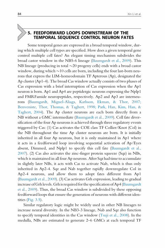
5. FEEDFORWARD LOOPS DOWNSTREAM OF THETEMPORAL SEQUENCE CONTROL NEURON FATES
Some temporal genes are expressed in a broad temporal window, dur-
ing which multiple cell types are specified. How does a given temporal gene
control multiple cell fates? An elegant timing mechanism subdivides the
broad castor window in the NB5-6 lineage (Baumgardt et al., 2009). This
NB lineage (producing in total �20 progeny cells) ends with a broad castor
window, during which�10 cells are born, including the four last-born neu-
rons that express the LIM-homeodomain TF Apterous (Ap), designated the
Ap cluster (Ap1-4). The broad Cas window actually consists of two phases of
Cas expression with a brief interruption of Cas expression when the Ap1
neuron is born. Ap1 and Ap4 are peptidergic neurons expressing the Nplp1
and FMRFamide neuropeptides, respectively. Ap2 and Ap3 are interneu-
rons (Baumgardt, Miguel-Aliaga, Karlsson, Ekman, & Thor, 2007;
Benveniste, Thor, Thomas, & Taghert, 1998; Park, Han, Kim, Han, &
Taghert, 2004). The Ap cluster neurons are each born directly from a
NB without a GMC intermediate (Baumgardt et al., 2009). Cell fate diver-
sification of the four Ap neurons is achieved through three regulatory events
triggered by Cas: (1) Cas activates the COE class TF Collier/Knot (Col) in
the NB throughout the time Ap cluster neurons are born. It is initially
inherited in all four Ap neurons, but it is only maintained in Ap1 where
it acts in a feedforward loop involving sequential activation of Ap/Eyes
absent, Dimmed, and Nplp1 to specify this cell fate (Baumgardt et al.,
2007). (2) Cas also activates the zinc-finger protein squeeze (Sqz) in NBs,
which is maintained in all four Ap neurons. After Sqz had time to accumulate
in slightly later NBs, it acts with Cas to activate Nab, which is thus only
inherited in Ap2-4. Sqz and Nab together rapidly downregulate Col in
Ap2-4 neurons, and allow them to adopt fates different from Ap1
(Baumgardt et al., 2009). (3) Cas activates Grh expression, leading to gradual
increase ofGrh levels.Grh is required for the specificationofAp4 (Baumgardt
et al., 2009). Thus, the broad Cas window is subdivided by these opposing
feedforward loops that ensure the generation of neurons with different iden-
tities (Fig. 3.5).
Similar regulatory logic might be widely used in other NB lineages to
increase neural diversity. In the NB3-3 lineage, Nab and Sqz also function
to specify temporal identities in the Cas window (Tsuji et al., 2008). In the
medulla, NBs are estimated to generate 2–6 GMCs at each temporal TF
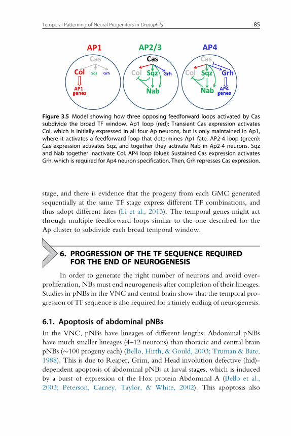
Figure 3.5 Model showing how three opposing feedforward loops activated by Cassubdivide the broad TF window. Ap1 loop (red): Transient Cas expression activatesCol, which is initially expressed in all four Ap neurons, but is only maintained in Ap1,where it activates a feedforward loop that determines Ap1 fate. AP2-4 loop (green):Cas expression activates Sqz, and together they activate Nab in Ap2-4 neurons. Sqzand Nab together inactivate Col. AP4 loop (blue): Sustained Cas expression activatesGrh, which is required for Ap4 neuron specification. Then, Grh represses Cas expression.
85Temporal Patterning of Neural Progenitors in Drosophila
stage, and there is evidence that the progeny from each GMC generated
sequentially at the same TF stage express different TF combinations, and
thus adopt different fates (Li et al., 2013). The temporal genes might act
through multiple feedforward loops similar to the one described for the
Ap cluster to subdivide each broad temporal window.
6. PROGRESSION OF THE TF SEQUENCE REQUIREDFOR THE END OF NEUROGENESIS
In order to generate the right number of neurons and avoid over-
proliferation, NBs must end neurogenesis after completion of their lineages.
Studies in pNBs in the VNC and central brain show that the temporal pro-
gression of TF sequence is also required for a timely ending of neurogenesis.
6.1. Apoptosis of abdominal pNBsIn the VNC, pNBs have lineages of different lengths: Abdominal pNBs
have much smaller lineages (4–12 neurons) than thoracic and central brain
pNBs (�100 progeny each) (Bello, Hirth, & Gould, 2003; Truman & Bate,
1988). This is due to Reaper, Grim, and Head involution defective (hid)-
dependent apoptosis of abdominal pNBs at larval stages, which is induced
by a burst of expression of the Hox protein Abdominal-A (Bello et al.,
2003; Peterson, Carney, Taylor, & White, 2002). This apoptosis also
86 Xin Li et al.
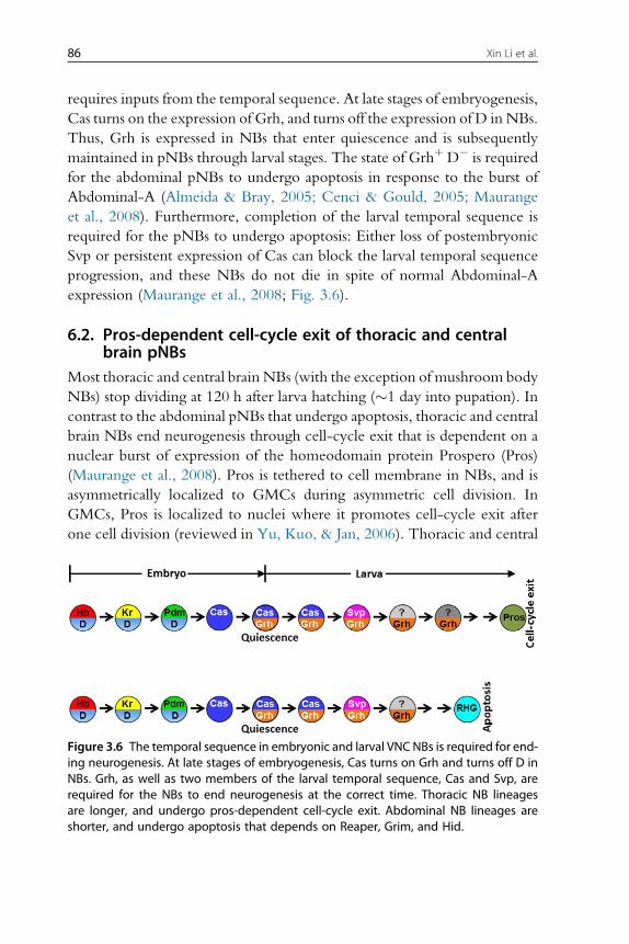
requires inputs from the temporal sequence. At late stages of embryogenesis,
Cas turns on the expression of Grh, and turns off the expression of D in NBs.
Thus, Grh is expressed in NBs that enter quiescence and is subsequently
maintained in pNBs through larval stages. The state of Grhþ D� is required
for the abdominal pNBs to undergo apoptosis in response to the burst of
Abdominal-A (Almeida & Bray, 2005; Cenci & Gould, 2005; Maurange
et al., 2008). Furthermore, completion of the larval temporal sequence is
required for the pNBs to undergo apoptosis: Either loss of postembryonic
Svp or persistent expression of Cas can block the larval temporal sequence
progression, and these NBs do not die in spite of normal Abdominal-A
expression (Maurange et al., 2008; Fig. 3.6).
6.2. Pros-dependent cell-cycle exit of thoracic and centralbrain pNBs
Most thoracic and central brain NBs (with the exception of mushroom body
NBs) stop dividing at 120 h after larva hatching (�1 day into pupation). In
contrast to the abdominal pNBs that undergo apoptosis, thoracic and central
brain NBs end neurogenesis through cell-cycle exit that is dependent on a
nuclear burst of expression of the homeodomain protein Prospero (Pros)
(Maurange et al., 2008). Pros is tethered to cell membrane in NBs, and is
asymmetrically localized to GMCs during asymmetric cell division. In
GMCs, Pros is localized to nuclei where it promotes cell-cycle exit after
one cell division (reviewed in Yu, Kuo, & Jan, 2006). Thoracic and central
Figure 3.6 The temporal sequence in embryonic and larval VNC NBs is required for end-ing neurogenesis. At late stages of embryogenesis, Cas turns on Grh and turns off D inNBs. Grh, as well as two members of the larval temporal sequence, Cas and Svp, arerequired for the NBs to end neurogenesis at the correct time. Thoracic NB lineagesare longer, and undergo pros-dependent cell-cycle exit. Abdominal NB lineages areshorter, and undergo apoptosis that depends on Reaper, Grim, and Hid.
87Temporal Patterning of Neural Progenitors in Drosophila
brain NBs exhibit a burst of expression of nuclear Pros at 120 h. prosmutant
clones contain multiple NB-like cells that do not die at 120 h, and even con-
tinue dividing in the adult brain (Maurange et al., 2008).
Pros-dependent cell-cycle exit of thoracic and central brain NBs also
requires inputs from both embryonic and larval temporal sequences:
Sustained Grh expression in pNBs induced by embryonic Cas is required
to prevent premature cell-cycle exit of NBs; while completion of the larval
temporal sequence is required for the NBs to undergo Pros-dependent cell-
cycle exit at 120 h. Remarkably, stalled temporal sequence caused by either
loss of postembryonic Svp expression or persistent Cas expression prevents
cell-cycle exit and NBs continue to divide even in 7-day-old adults
(Maurange et al., 2008; Fig. 3.6).
6.3. Studies in other systems about the end of neurogenesisIn contrast to VNC and the majority of central brain NBs, mushroom body
NBs that are born at embryonic stages do not have a quiescent stage and con-
tinue proliferating until the end of pupal stage. Tll is required for the
uninterrupted and prolonged proliferation of mushroom body NBs: Loss
of tll causes premature loss of mushroom body NBs in early pupal stage
(Kurusu et al., 2009). Surprisingly, in the medulla NB temporal sequence,
Tll appears to have the opposite function: Tll is the last TF that is expressed
in the oldest NBs. These Tllþ NBs show nuclear localization of Pros, indi-
cating that they undergo Pros-dependent cell-cycle exit at the end of their
life, similar to the thoracic and central brain NBs (Li et al., 2013). Whether
Tll is required or sufficient for ending medulla neurogenesis is currently not
known. If so, it will be interesting to understand how Tll plays completely
opposite roles in the mushroom body and in medulla NBs.
7. INTEGRATION OF TEMPORAL AND SPATIALINFORMATION DETERMINES LINEAGES
Although almost all NBs in the embryonic VNC follow the same
temporal sequence, they generate different lineages depending on their
spatial identity. There are approximately 30 NBs in each hemisegment,
and each NB has an individual fate based on its position and the expression
of specific molecular markers. Intrasegmental specification is achieved by
superimposed activities of segment polarity and dorso-ventral patterning
genes (Technau et al., 2006). In addition to the intrasegmental spatial pat-
terning, homologous NBs in different segments along the antero-posterior
88 Xin Li et al.
axis have slightly different lineages, although they also share significant sim-
ilarities in their lineages and gene expression patterns. Hox genes are critical
players in determining these differences. For example, in Section 6 we dis-
cussed how the abdAHox gene, together with temporal factors, determines
the length of lineages and the end of neurogenesis. Hox genes also work
with temporal genes to control the generation of specific cell types only
found in certain segments. For example, NB5-6 generates the Ap cluster
neurons only in thoracic segments. Late temporal genes cas and grh, and
the thoracic Hox gene Antennapedia (Antp), are required to specify the
Ap cluster neurons (Karlsson, Baumgardt, & Thor, 2010). One of the target
genes for integration of temporal and spatial information is Collier, which
plays important roles in the feedforward loops that specify the Ap cluster
neurons as discussed in Section 5. It will be interesting to further characterize
the exact molecular mechanism of the integration (Karlsson et al., 2010).
Spatial identity also modulates the progression of the temporal sequence
of TFs in NBs. For example, young NB3-3 never expresses Hb, but sequen-
tially expresses Kr, Pdm, Cas, Cas/Grh, and Svp/Grh (Tsuji et al., 2008).
The absence of Hb in the sequence is common to both thoracic and abdom-
inal NB3-3, suggesting that this is modulated by the intrasegmental spatial
patterning genes common to NB3-3 in each segment. Although the tempo-
ral sequence is the same in both thoracic and abdominal NB3-3, the speed of
transitions between TFs is faster in abdominal NB3-3s than in thoracic
NB3-3s. Particularly, the switch from Cas to Svp occurs in late embryo
in abdominal NB3-3, while it occurs in the larval stage for thoracic
NB3-3. This spatial difference in the timing of switching to Svp is also true
for other NB lineages and is regulated by Hox genes. Loss of Antp or mis-
expression of abd-A in thoracic NB3-3 causes precocious Svp expression
during embryogenesis (Tsuji et al., 2008). How the Hox genes modulate
the speed of the temporal progression and how the spatial–temporal infor-
mation is integrated to regulate the NB lineage will be interesting questions
for the future.
8. INTEGRATION OF TEMPORAL IDENTITY WITH BINARYFATE CHOICE
In embryonic NB lineages, GMCs usually divide asymmetrically to
give rise to two progeny with different fates (either two different neuron
types, one neuron and one glia, or one neuron and the other undergoing
programmed cell death, etc.), and this depends on Notch signaling between
89Temporal Patterning of Neural Progenitors in Drosophila
the twoGMC daughter cells (Buescher et al., 1998; Karcavich &Doe, 2005;
Lundell, Lee, Perez, & Chadwell, 2003; Novotny et al., 2002; Schuldt &
Brand, 1999; Skeath & Doe, 1998; Spana & Doe, 1996; Udolph,
Rath, & Chia, 2001). During the asymmetric division of GMCs, Numb,
a repressor of Notch signaling, is asymmetrically localized to one daughter
cell, and this cell adopts a Notch-off (N-off ) fate, while the other cell adopts
a Notch-on (N-on) fate (Buescher et al., 1998; Spana & Doe, 1996). All the
N-on (or N-off ) cells within a lineage are collectively called a hemilineage.
In postembryonic NB lineages, Notch/Numb also function in binary-fate
choices of GMC progeny (Kumar, Bello, & Reichert, 2009; Li et al.,
2013; Lin et al., 2010; Truman, Moats, Altman, Marin, & Williams, 2010).
Notch signaling only differentiates between two alternative fates, but the
actual fates depend on the spatial and temporal identity of theNB. For exam-
ple, Notch has distinct lineage-specific effects in the three antennal lobe
NB lineages: antero-dorsal lineage (N-off: PN fates, N-on: apoptosis); ven-
tral lineage (N-off: apoptosis, N-on: PN fates); and lateral lineage (N-off:
PN fates, N-on: local interneuron fates) (Lin et al., 2010).
How does the Notch-dependent binary fate choice integrate with the
temporal identity of NBs? In the developing medulla, the Notch pathway
regulates the maintenance of the temporal TFs in postmitotic neurons
and the expression of Ap. In the Ey and Slp NB stages, Ey or Slp is only
maintained in the N-off daughter, while the N-on daughter turns on Ap.
At the DNB stage, D is only maintained in theN-on daughter together with
Ap. Although most if not all N-on daughters of medulla GMCs express Ap,
they express different combinations of other TFs and adopt different fates
depending on which NB stage they are born from. For example, the
N-on progeny of EyþGMCs express Ap andDrifter (Dfr), and this is depen-
dent on both Notch signaling and the expression of Ey in NBs. The N-on
progeny of Slpþ GMCs express Ap and twin of eyeless (Toy), and similarly,
this depends on both Notch signaling and Slp expression in NBs. Thus, the
temporal TFs and the Notch pathway together control the expression of
downstream TFs like Drifter and Toy to control neuron fates (Li et al.,
2013; Fig. 3.2).
Studies in the antennal lobe lateral lineage illustrate another interesting
question. In this lineage, PNs and local interneurons are produced as siblings
of each GMC division (N-off: PN, N-on: local interneuron). However,
as there is more diversity of PNs than of local interneurons, the same
local interneuron can be the sibling of different PNs. Thus, it appears that
the tempo of birth-order-dependent fate changes is different between
90 Xin Li et al.
the twoN-off andN-on hemilineages (Lin et al., 2012). How the same tem-
poral identity information regulates the independent pace of temporal
switching of neuronal fates in the two hemilineages is an interesting question
for the future.
9. CONCLUSIONS AND FUTURE QUESTIONS
Currently, two different temporal sequences have been identified in
NBs of two different systems: the Hb!Kr!Pdm!Cas!Grh sequence
in theDrosophilaVNC, and the Hth!Klu!Ey!Slp!D!Tll sequence
that patterns medulla NBs. In addition, the INPs of type II NB lineages are
patterned by a TF sequence D!Grh!Ey. These three examples suggest
that TF-sequence-dependent temporal patterning of neural precursors is a
common theme to generate neural diversity. This suggests that more tem-
poral sequences will be identified in other systems. For example, in the
antennal lobe antero-dorsal lineage, Kr specifies one out of 40 temporal
identities in the NB. Further identification of a complete temporal sequence
will rely on candidate gene approaches, and/or screening based on either
gene expression or mutant phenotypes. Does TF-sequence-dependent tem-
poral patterning of neural precursors also function in vertebrate systems?
There is some evidence suggesting that this might be the case. In the ver-
tebrate retina, one ortholog of hb, Ikaros, specifies early-born cell fates
(Elliott, Jolicoeur, Ramamurthy, & Cayouette, 2008). In mammalian cor-
tical neurogenesis, Foxg1, an ortholog of Slp, functions in cortical progen-
itors to suppress early-born cortical cell fates (Hanashima, Li, Shen, Lai, &
Fishell, 2004). Although the vertebrate systems are much more complex
thanDrosophila, studies in flies have provided important concepts that might
be applicable to vertebrates.
Cross-regulations between temporal TFs are important for temporal
transitions: Feedback negative regulation and feedforward positive regula-
tion among the temporal TFs can facilitate the progression of the sequence.
There are many remaining questions to understand the mechanism of tem-
poral transitions. Even in the best-known systems, there can be missing fac-
tors or timing mechanisms in addition to the identified TF sequence. When
all factors involved and their regulatory relationships are characterized by
genetic analysis, theoretical modeling will provide insights into how the
genetic network precisely times the NB temporal progression.
Loss of NB competence is related to epigenetic changes, such as chro-
matin modifications and chromosome architecture. More studies are needed
91Temporal Patterning of Neural Progenitors in Drosophila
to examine how the expression dynamics of the temporal genes leads to epi-
genetic changes and thus regulates NB competence, and eventually
determines the end of neurogenesis. Such studies are relevant for the devel-
opment of cell replacement therapies using stem cells to treat various
diseases.
Another big challenge is to elucidate how the temporal and spatial iden-
tity of NBs, as well as the Notch-dependent binary fate choices, are inte-
grated to generate specific neuron fates.
Finally, how the temporal sequence evolved is a great question for evo-
lutionary developmental neurobiologists. An interesting observation is that
the Hb!Kr!Pdm!Cas temporal sequence in VNC NBs mimics the
anterior to posterior spatial distribution of Hb!Kr!Pdm!Cas expres-
sion domains at cellular blastoderm in the embryo (Isshiki et al., 2001).
Whether they use the same regulatory logic is not known. The Hth!Klu!Ey!Slp!D!Tll or D!Grh!Ey regulatory cascades have not
been described in other contexts. Evolutionary studies might provide clues
as to how this powerful mechanism has evolved to pattern neural precursors
to generate neural diversity.
REFERENCESAkiyama-Oda, Y., Hosoya, T., & Hotta, Y. (1999). Asymmetric cell division of thoracic
neuroblast 6-4 to bifurcate glial and neuronal lineage in Drosophila. Development,126, 1967–1974.
Almeida, M. S., & Bray, S. J. (2005). Regulation of post-embryonic neuroblasts by Drosoph-ila Grainyhead. Mechanisms of Development, 122, 1282–1293.
Baek, M., & Mann, R. S. (2009). Lineage and birth date specify motor neuron targeting anddendritic architecture in adult Drosophila. The Journal of Neuroscience, 29, 6904–6916.
Baumgardt, M., Karlsson, D., Terriente, J., Diaz-Benjumea, F. J., & Thor, S. (2009). Neu-ronal subtype specification within a lineage by opposing temporal feed-forward loops.Cell, 139, 969–982.
Baumgardt, M.,Miguel-Aliaga, I., Karlsson, D., Ekman, H., & Thor, S. (2007). Specificationof neuronal identities by feedforward combinatorial coding. PLoS Biology, 5, e37.
Bayraktar, O. A., Boone, J. Q., Drummond, M. L., & Doe, C. Q. (2010). Drosophila type IIneuroblast lineages keep Prospero levels low to generate large clones that contribute tothe adult brain central complex. Neural Development, 5, 26.
Bayraktar, O. A., & Doe, C. Q. (2013). Temporal patterning in intermediate progenitorsincreases neural diversity. Nature, 498, 449–455.
Bello, B. C., Hirth, F., & Gould, A. P. (2003). A pulse of the Drosophila Hox proteinAbdominal-A schedules the end of neural proliferation via neuroblast apoptosis. Neuron,37, 209–219.
Benito-Sipos, J., Estacio-Gomez, A., Moris-Sanz, M., Baumgardt, M., Thor, S., & Diaz-Benjumea, F. J. (2010). A genetic cascade involving klumpfuss, nab and castor specifiesthe abdominal leucokinergic neurons in the Drosophila CNS. Development, 137,3327–3336.
92 Xin Li et al.
Benveniste, R. J., Thor, S., Thomas, J. B., & Taghert, P. H. (1998). Cell type-specific reg-ulation of the Drosophila FMRF-NH2 neuropeptide gene by Apterous, a LIMhomeodomain transcription factor. Development, 125, 4757–4765.
Bhat, K. M. (1999a). Segment polarity genes in neuroblast formation and identity specifica-tion during Drosophila neurogenesis. Bioessays, 21, 472–485.
Bhat, K. M. (1999b). Segment polarity genes in neuroblast formation and identity specifica-tion during Drosophila neurogenesis. BioEssays: News and Reviews in Molecular, Cellularand Developmental Biology, 21, 472–485.
Boone, J. Q., & Doe, C. Q. (2008). Identification of Drosophila type II neuroblast lineagescontaining transit amplifying ganglion mother cells. Developmental Neurobiology, 68,1185–1195.
Bossing, T., Udolph, G., Doe, C. Q., & Technau, G. M. (1996). The embryonic centralnervous system lineages of Drosophila melanogaster. I. Neuroblast lineages derived fromthe ventral half of the neuroectoderm. Developmental Biology, 179, 41–64.
Brand, A. H., & Livesey, F. J. (2011). Neural stem cell biology in vertebrates and inverte-brates: More alike than different? Neuron, 70, 719–729.
Brody, T., & Odenwald, W. F. (2000). Programmed transformations in neuroblast geneexpression during Drosophila CNS lineage development. Developmental Biology, 226,34–44.
Buescher, M., Yeo, S. L., Udolph, G., Zavortink, M., Yang, X., Tear, G., et al. (1998).Binary sibling neuronal cell fate decisions in the Drosophila embryonic central nervoussystem are nonstochastic and require inscuteable-mediated asymmetry of ganglionmother cells. Genes & Development, 12, 1858–1870.
Cenci, C., &Gould, A. P. (2005). Drosophila Grainyhead specifies late programmes of neuralproliferation by regulating the mitotic activity and Hox-dependent apoptosis ofneuroblasts. Development, 132, 3835–3845.
Cleary, M. D., & Doe, C. Q. (2006). Regulation of neuroblast competence: Multiple tem-poral identity factors specify distinct neuronal fates within a single early competence win-dow. Genes & Development, 20, 429–434.
Cui, X., & Doe, C. Q. (1992). ming is expressed in neuroblast sublineages and regulates geneexpression in the Drosophila central nervous system. Development, 116, 943–952.
Das, A., Reichert, H., &Rodrigues, V. (2010). Notch regulates the generation of diverse celltypes from the lateral lineage of Drosophila antennal lobe. Journal of Neurogenetics, 24,42–53.
Das, A., Sen, S., Lichtneckert, R., Okada, R., Ito, K., Rodrigues, V., et al. (2008). Drosoph-ila olfactory local interneurons and projection neurons derive from a common neuroblastlineage specified by the empty spiracles gene. Neural Development, 3, 33.
Desai, A. R., & McConnell, S. K. (2000). Progressive restriction in fate potential by neuralprogenitors during cerebral cortical development. Development, 127, 2863–2872.
Dessaud, E., McMahon, A. P., & Briscoe, J. (2008). Pattern formation in the vertebrate neu-ral tube: A sonic hedgehog morphogen-regulated transcriptional network. Development,135, 2489–2503.
Doe, C. Q. (1992). Molecular markers for identified neuroblasts and ganglion mother cells inthe Drosophila central nervous system. Development, 116, 855–863.
Doe, C. Q. (2008). Neural stem cells: Balancing self-renewal with differentiation. Develop-ment, 135, 1575–1587.
Egger, B., Boone, J. Q., Stevens, N. R., Brand, A. H., & Doe, C. Q. (2007). Regulation ofspindle orientation and neural stem cell fate in the Drosophila optic lobe. Neural Devel-opment, 2, 1.
Elliott, J., Jolicoeur, C., Ramamurthy, V., & Cayouette, M. (2008). Ikaros confers early tem-poral competence to mouse retinal progenitor cells. Neuron, 60, 26–39.
93Temporal Patterning of Neural Progenitors in Drosophila
Fischbach, K. F., & Dittrich, A. P. M. (1989). The optic lobe of Drosophila melanogaster. I.A Golgi analysis of wild-type structure. Cell and Tissue Research, 258, 441–475.
Gaspard, N., Bouschet, T., Hourez, R., Dimidschstein, J., Naeije, G., van den Ameele, J.,et al. (2008). An intrinsic mechanism of corticogenesis from embryonic stem cells.Nature, 455, 351–357.
Green, P., Hartenstein, A. Y., & Hartenstein, V. (1993). The embryonic development of theDrosophila visual system. Cell and Tissue Research, 273, 583–598.
Grosskortenhaus, R., Pearson, B. J., Marusich, A., & Doe, C. Q. (2005). Regulation of tem-poral identity transitions in Drosophila neuroblasts. Developmental Cell, 8, 193–202.
Grosskortenhaus, R., Robinson, K. J., & Doe, C. Q. (2006). Pdm and Castor specify late-born motor neuron identity in the NB7-1 lineage.Genes & Development, 20, 2618–2627.
Hanashima, C., Li, S. C., Shen, L., Lai, E., & Fishell, G. (2004). Foxg1 suppresses early cor-tical cell fate. Science, 303, 56–59.
Hasegawa, E., Kitada, Y., Kaido, M., Takayama, R., Awasaki, T., Tabata, T., et al. (2011).Concentric zones, cell migration and neuronal circuits in the Drosophila visual center.Development, 138, 983–993.
Hirabayashi, Y., Suzki, N., Tsuboi, M., Endo, T. A., Toyoda, T., Shinga, J., et al. (2009).Polycomb limits the neurogenic competence of neural precursor cells to promote astro-genic fate transition. Neuron, 63, 600–613.
Huang, D. H., Chang, Y. L., Yang, C. C., Pan, I. C., & King, B. (2002). Pipsqueak encodes afactor essential for sequence-specific targeting of a polycomb group protein complex.Molecular Cell Biology, 22, 6261–6271.
Isshiki, T., Pearson, B., Holbrook, S., & Doe, C. Q. (2001). Drosophila neuroblasts sequen-tially express transcription factors which specify the temporal identity of their neuronalprogeny. Cell, 106, 511–521.
Jacob, J., Maurange, C., & Gould, A. P. (2008). Temporal control of neuronal diversity:Common regulatory principles in insects and vertebrates?Development, 135, 3481–3489.
Jefferis, G. S., Marin, E. C., Stocker, R. F., & Luo, L. (2001). Target neuron prespecificationin the olfactory map of Drosophila. Nature, 414, 204–208.
Kambadur, R., Koizumi, K., Stivers, C., Nagle, J., Poole, S. J., & Odenwald, W. F. (1998).Regulation of POU genes by castor and hunchback establishes layered compartments inthe Drosophila CNS. Genes & Development, 12, 246–260.
Kanai, M. I., Okabe, M., & Hiromi, Y. (2005). Seven-up controls switching of transcriptionfactors that specify temporal identities of Drosophila neuroblasts. Developmental Cell, 8,203–213.
Kao, C. F., & Lee, T. (2010). Birth time/order-dependent neuron type specification.CurrentOpinion in Neurobiology, 20, 14–21.
Kao, C. F., Yu, H. H., He, Y., Kao, J. C., & Lee, T. (2012). Hierarchical deployment offactors regulating temporal fate in a diverse neuronal lineage of the Drosophila centralbrain. Neuron, 73, 677–684.
Karcavich, R. E. (2005). Generating neuronal diversity in the Drosophila central nervoussystem: A view from the ganglion mother cells. Developmental Dynamics, 232, 609–616.
Karcavich, R., & Doe, C. Q. (2005). Drosophila neuroblast 7-3 cell lineage: A model sys-tem for studying programmed cell death, Notch/Numb signaling, and sequential spec-ification of ganglion mother cell identity. The Journal of Comparative Neurology, 481,240–251.
Karlsson, D., Baumgardt, M., & Thor, S. (2010). Segment-specific neuronal subtype spec-ification by the integration of anteroposterior and temporal cues. PLoS Biology, 8,e1000368.
Knoblich, J. A. (2010). Asymmetric cell division: Recent developments and their implica-tions for tumour biology. Nature Reviews. Molecular Cell Biology, 11, 849–860.
94 Xin Li et al.
Kohwi, M., Hiebert, L. S., & Doe, C. Q. (2011). The pipsqueak-domain proteins Distalantenna and Distal antenna-related restrict Hunchback neuroblast expression andearly-born neuronal identity. Development, 138, 1727–1735.
Kohwi, M., Lupton, J. R., Lai, S. L., Miller, M. R., & Doe, C. Q. (2013). Developmentallyregulated subnuclear genome reorganization restricts neural progenitor competence inDrosophila. Cell, 152, 97–108.
Kumar, A., Bello, B., & Reichert, H. (2009). Lineage-specific cell death in postembryonicbrain development of Drosophila. Development, 136, 3433–3442.
Kurusu, M., Maruyama, Y., Adachi, Y., Okabe, M., Suzuki, E., & Furukubo-Tokunaga, K.(2009). A conserved nuclear receptor, Tailless, is required for efficient proliferation andprolonged maintenance of mushroom body progenitors in the Drosophila brain. Devel-opmental Biology, 326, 224–236.
Lai, S. L., Awasaki, T., Ito, K., & Lee, T. (2008). Clonal analysis of Drosophila antennal lobeneurons: Diverse neuronal architectures in the lateral neuroblast lineage. Development,135, 2883–2893.
Lee, T., Lee, A., & Luo, L. (1999). Development of the Drosophila mushroom bodies:Sequential generation of three distinct types of neurons from a neuroblast. Development,126, 4065–4076.
Li, X., Erclik, T., Bertet, C., Chen, Z., Voutev, R., Venkatesh, S., et al. (2013). Temporalpatterning of Drosophila medulla neuroblasts controls neural fates.Nature, 498, 456–462.
Lin, S., Kao, C. F., Yu, H. H., Huang, Y., & Lee, T. (2012). Lineage analysis of Drosophilalateral antennal lobe neurons reveals notch-dependent binary temporal fate decisions.PLoS Biology, 10, e1001425.
Lin, S., Lai, S. L., Yu, H. H., Chihara, T., Luo, L., & Lee, T. (2010). Lineage-specific effectsof Notch/Numb signaling in post-embryonic development of the Drosophila brain.Development, 137, 43–51.
Lin, S., & Lee, T. (2012). Generating neuronal diversity in the Drosophila central nervoussystem. Developmental Dynamics, 241, 57–68.
Livesey, F. J., & Cepko, C. L. (2001). Vertebrate neural cell-fate determination: Lessons fromthe retina. Nature Reviews Neuroscience, 2, 109–118.
Lundell, M. J., & Hirsh, J. (1998). Eagle is required for the specification of serotonin neuronsand other neuroblast 7-3 progeny in the Drosophila CNS. Development, 125, 463–472.
Lundell, M. J., Lee, H. K., Perez, E., & Chadwell, L. (2003). The regulation of apoptosis byNumb/Notch signaling in the serotonin lineage of Drosophila. Development, 130,4109–4121.
Maurange, C. (2012). Temporal specification of neural stem cells: Insights from Drosophilaneuroblasts. Current Topics in Developmental Biology, 98, 199–228.
Maurange, C., Cheng, L., & Gould, A. P. (2008). Temporal transcription factors and theirtargets schedule the end of neural proliferation in Drosophila. Cell, 133, 891–902.
Mellerick, D. M., Kassis, J. A., Zhang, S. D., & Odenwald, W. F. (1992). Castor encodes anovel zinc finger protein required for the development of a subset of CNS neurons inDrosophila. Neuron, 9, 789–803.
Mettler, U., Vogler, G., & Urban, J. (2006). Timing of identity: Spatiotemporal regulation ofhunchback in neuroblast lineages of Drosophila by Seven-up and Prospero.Development,133, 429–437.
Molyneaux, B. J., Arlotta, P., Menezes, J. R., & Macklis, J. D. (2007). Neuronal subtypespecification in the cerebral cortex. Nature Reviews Neuroscience, 8, 427–437.
Morante, J., & Desplan, C. (2008). The color-vision circuit in the medulla of Drosophila.Current Biology, 18, 553–565.
Morante, J., Erclik, T., & Desplan, C. (2011). Cell migration in Drosophila optic lobe neu-rons is controlled by eyeless/Pax6. Development, 138, 687–693.
95Temporal Patterning of Neural Progenitors in Drosophila
Naka, H., Nakamura, S., Shimazaki, T., & Okano, H. (2008). Requirement for COUP-TFIand II in the temporal specification of neural stem cells in CNS development. NatureNeuroscience, 11, 1014–1023.
Nakajima, A., Isshiki, T., Kaneko, K., & Ishihara, S. (2010). Robustness under functionalconstraint: The genetic network for temporal expression in Drosophila neurogenesis.PLoS Computational Biology, 6, e1000760.
Novotny, T., Eiselt, R., & Urban, J. (2002). Hunchback is required for the specification ofthe early sublineage of neuroblast 7-3 in the Drosophila central nervous system. Devel-opment, 129, 1027–1036.
Okano, H., & Temple, S. (2009). Cell types to order: Temporal specification of CNS stemcells. Current Opinion in Neurobiology, 19, 112–119.
Park, D., Han, M., Kim, Y. C., Han, K. A., & Taghert, P. H. (2004). Ap-let neurons –A peptidergic circuit potentially controlling ecdysial behavior in Drosophila. Develop-mental Biology, 269, 95–108.
Pearson, B. J., & Doe, C. Q. (2003). Regulation of neuroblast competence in Drosophila.Nature, 425, 624–628.
Pearson, B. J., & Doe, C. Q. (2004). Specification of temporal identity in the developingnervous system. Annual Review of Cell and Developmental Biology, 20, 619–647.
Peterson, C., Carney, G. E., Taylor, B. J., &White, K. (2002). Reaper is required for neuro-blast apoptosis during Drosophila development. Development, 129, 1467–1476.
Prokop, A., & Technau, G. M. (1991). The origin of postembryonic neuroblasts in the ven-tral nerve cord of Drosophila melanogaster. Development, 111, 79–88.
Romani, S., Jimenez, F., Hoch, M., Patel, N. H., Taubert, H., & Jackle, H. (1996). Kruppel,a Drosophila segmentation gene, participates in the specification of neurons and glialcells. Mechanisms of Development, 60, 95–107.
Schmid, A., Chiba, A., & Doe, C. Q. (1999). Clonal analysis of Drosophila embryonic neu-roblasts: Neural cell types, axon projections and muscle targets. Development, 126,4653–4689.
Schmidt, H., Rickert, C., Bossing, T., Vef, O., Urban, J., & Technau, G. M. (1997). Theembryonic central nervous system lineages of Drosophila melanogaster. II. Neuroblastlineages derived from the dorsal part of the neuroectoderm. Developmental Biology,189, 186–204.
Schuldt, A. J., & Brand, A. H. (1999). Mastermind acts downstream of notch to specify neu-ronal cell fates in the Drosophila central nervous system. Developmental Biology, 205,287–295.
Shen, Q., Wang, Y., Dimos, J. T., Fasano, C. A., Phoenix, T. N., Lemischka, I. R., et al.(2006). The timing of cortical neurogenesis is encoded within lineages of individual pro-genitor cells. Nature Neuroscience, 9, 743–751.
Skeath, J. B., & Doe, C. Q. (1998). Sanpodo and Notch act in opposition to Numb to dis-tinguish sibling neuron fates in the Drosophila CNS. Development, 125, 1857–1865.
Skeath, J. B., & Thor, S. (2003). Genetic control of Drosophila nerve cord development.Current Opinion in Neurobiology, 13, 8–15.
Spana, E. P., & Doe, C. Q. (1996). Numb antagonizes Notch signaling to specify siblingneuron cell fates. Neuron, 17, 21–26.
Suzuki, T., Kaido,M., Takayama,R., & Sato,M. (2013). A temporal mechanism that producesneuronal diversity in the Drosophila visual center. Developmental Biology, 380, 12–24.
Technau, G.M., Berger, C., &Urbach, R. (2006). Generation of cell diversity and segmentalpattern in the embryonic central nervous system of Drosophila. Developmental Dynamics,235, 861–869.
Touma, J. J., Weckerle, F. F., & Cleary, M. D. (2012). Drosophila Polycomb complexesrestrict neuroblast competence to generate motoneurons. Development, 139, 657–666.
96 Xin Li et al.
Tran, K. D., & Doe, C. Q. (2008). Pdm and Castor close successive temporal identity win-dows in the NB3-1 lineage. Development, 135, 3491–3499.
Tran, K. D., Miller, M. R., & Doe, C. Q. (2010). Recombineering Hunchback identifiestwo conserved domains required to maintain neuroblast competence and specifyearly-born neuronal identity. Development, 137, 1421–1430.
Truman, J. W., & Bate, M. (1988). Spatial and temporal patterns of neurogenesis in the cen-tral nervous system of Drosophila melanogaster. Developmental Biology, 125, 145–157.
Truman, J. W., Moats, W., Altman, J., Marin, E. C., & Williams, D. W. (2010). Role ofNotch signaling in establishing the hemilineages of secondary neurons in Drosophilamelanogaster. Development, 137, 53–61.
Tsuji, T., Hasegawa, E., & Isshiki, T. (2008). Neuroblast entry into quiescence is regulatedintrinsically by the combined action of spatial Hox proteins and temporal identity factors.Development, 135, 3859–3869.
Udolph, G., Rath, P., & Chia, W. (2001). A requirement for Notch in the genesis of a subsetof glial cells in the Drosophila embryonic central nervous system which arise throughasymmetric divisions. Development, 128, 1457–1466.
Viktorin, G., Riebli, N., Popkova, A., Giangrande, A., & Reichert, H. (2011). Multipotentneural stem cells generate glial cells of the central complex through transit amplifyingintermediate progenitors in Drosophila brain development. Developmental Biology,356, 553–565.
Weng,M., Golden, K. L., & Lee, C. Y. (2010). dFezf/Earmuff maintains the restricted devel-opmental potential of intermediate neural progenitors in Drosophila. Developmental Cell,18, 126–135.
White, K., & Kankel, D. R. (1978). Patterns of cell division and cell movement in the for-mation of the imaginal nervous system in Drosophila melanogaster. DevelopmentalBiology, 65, 296–321.
Wu, Y. C., Chen, C. H., Mercer, A., & Sokol, N. S. (2012). Let-7-complex microRNAsregulate the temporal identity of Drosophila mushroom body neurons via chinmo.Developmental Cell, 23, 202–209.
Yang, X., Yeo, S., Dick, T., & Chia, W. (1993). The role of a Drosophila POU homeodomain gene in the specification of neural precursor cell identity in the developingembryonic central nervous system. Genes & Development, 7, 504–516.
Yasugi, T., Umetsu, D., Murakami, S., Sato, M., & Tabata, T. (2008). Drosophila optic lobeneuroblasts triggered by a wave of proneural gene expression that is negatively regulatedby JAK/STAT. Development, 135, 1471–1480.
Yu, H. H., Chen, C. H., Shi, L., Huang, Y., & Lee, T. (2009). Twin-spot MARCM toreveal the developmental origin and identity of neurons. Nature Neuroscience, 12,947–953.
Yu, H. H., Kao, C. F., He, Y., Ding, P., Kao, J. C., & Lee, T. (2010). A complete devel-opmental sequence of a Drosophila neuronal lineage as revealed by twin-spot MARCM.PLoS Biology, 8, e1000461.
Yu, F., Kuo, C. T., & Jan, Y. N. (2006). Drosophila neuroblast asymmetric cell division:Recent advances and implications for stem cell biology. Neuron, 51, 13–20.
Zhu, S., Barshow, S., Wildonger, J., Jan, L. Y., & Jan, Y. N. (2011). Ets transcription factorPointed promotes the generation of intermediate neural progenitors in Drosophila larvalbrains. Proceedings of the National Academy of Sciences of the United States of America, 108,20615–20620.
Zhu, S., Lin, S., Kao, C. F., Awasaki, T., Chiang, A. S., & Lee, T. (2006). Gradients of theDrosophila Chinmo BTB-zinc finger protein govern neuronal temporal identity. Cell,127, 409–422.