Embed Size (px)
Citation preview
Studies of the effects of antimicrobial peptides on model
membranes and relevant pathogens
Tânia Martins da Silva
Dissertação de Mestrado em Bioquímica
Universidade do Porto
Faculdade de Ciências
Instituto Ciências Biomédicas Abel Salazar
2011

Tânia Martins da Silva
Studies of the effects of antimicrobial peptides on model
membranes and relevant pathogens
Dissertação de Candidatura ao grau de Mestre em
Bioquímica da Universidade do Porto
Orientador – Doutora Margarida Bastos
Categoria – Professora Associada com Agregação
Afiliação – CIQ(UP), Departamento Química e
Bioquímica, Faculdade de Ciências da Universidade
do Porto
Co-Orientador – Doutora Maria Salomé Gomes
Categoria – Professora Associada
Afiliação – Instituto de Ciências Biomédicas Abel
Salazar e Instituto de Biologia Molecular e Celular
2011

1
CONTENTS
List of symbols and abbreviations…………………………………………………………… 3
Acknowledgements………………………………………………………………………….... 5
Abstract……………………………………………………………………………………….... 7
Resumo……………………………………………………………………………………...…. 9
Thesis Organization…………………………………………………………………………… 10
PART I – INTRODUCTION………………………………………………………………... 13
1. General Introduction…………………………………………………………….……….... 15
2. Pathogens………………………………………………………………………………...… 17
2.1. Leishmania…………………………………………………………………………. 17
2.1.1. Leishmaniasis………………………………………………………………… 17
2.1.2. Life cycle………………………………………..…………………………….. 19
2.1.3. Treatments……………………………………………………………………. 20
2.1.4. Leishmania as a target of AMPs…………………………………………… 21
2.2. Mycobacterium……………………………………………..……………………… 23
2.2.1. Mycobacteria cell wall……………………………………………………….. 23
2.2.2. Mycobacterium avium……………………………………………….………. 24
2.2.3. Treatments…………………………………………………………..……….. 26
2.2.4. Mycobacteria as a target of AMPs…………………………………………. 27
3. Antimicrobial peptides………………………………………………………………..……. 29
3.1. Structural characteristics……………………………………………………..…... 30
3.2. Mechanism of action………………………………………………………………. 31
3.3. Selectivity / Toxicity……………………………………………………………….. 34
3.4. AMPs in the immunity……………………………………………………………... 36
3.5. Resistance to AMPs………………………………………………………………. 37
3.6. AMPs in the clinic: Success, Problems and Challenges……………………… 38
3.7. Cecropin A-melittin derived peptides……………………………………………. 40
3.8. Lactoferrin-derived peptides……………………………………………………… 43
4. Lipids in Biological and Model Membranes……………………………………………... 47
4.1. Lipid Polymorphism…………………………………………………..…………… 47
4.2. AMP interaction with Lipid Membranes as revealed by Model Studies……… 51
PART II – OBJECTIVES…………………………………………………..……………... 53
2
PART III – EXPERIMENTAL METHODS AND RESULTS……………………..…………… 57
III.1 – Antimicrobial activity of lactoferrin-derived peptides against Leishmania... 59
1. Material and Methods………………………………..……………………………………. 61
2. Results……………………………………………..……………………………………….. 67
2.1. Determination of the antimicrobial activity of lactoferrin-derived peptides against L. donovani promastigotes and L. pifanoi amastigotes………………. 67
2.2. Evaluation of Leishmania plasma membrane permeabilization by lactoferrin-derived peptides………………………………………………………. 69
2.3. Effect of lactoferrin-derived peptides on L. pifanoi-infected macrophages….. 74
III.2 – Activity of cecropin A-mellitin-derived and lactoferrin-derived peptides against Mycobacterium avium………………………………………………..……….….. 77
1. Material and Methods…………………………………………..…………………………. 79
2. Results………………………………………………………………………………………. 85
2.1. Cecropin A-melittin-derived peptides…………………………………..……….. 85
2.1.1. Effect on the viability of M. avium in axenic cultures…………………….. 85
2.1.2. Effect on M. avium growing inside macrophages and macrophage toxicity……………………………………………………………………….... 85
2.2. Lactoferrin-derived peptides…………………………………………..…………. 87
2.2.1. Effect on the viability of M. avium in axenic cultures…………………….. 87
2.2.2. Effect on M. avium growing inside macrophages and macrophage toxicity……………………………………………………………….………… 88
III.3 – Characterization of peptide-lipid interaction by X-ray diffraction…………… 93
1. Material and Methods…………………………………………………..…………………. 95
2. Results………………………………………………………..…………………………….. 101
2.1. Cecropin A-melittin-derived peptides………………………………………..….. 101
2.1.1. Phase behavior of POPE membranes…………………………………..… 101
2.2. Lactoferrin-derived peptides……………………………………………..………. 106
2.2.1. Phase behavior of POPE membranes…………………………………..… 106
PART IV – DISCUSSION & CONCLUSIONS……………………………………………... 109
Bibliography………………………………………………..………………………………….. 119
3
LIST OF SYMBOLS AND ABBREVIATIONS
AIDS – Acquired Immune Deficiency Syndrome
AMPs – Antimicrobial Peptides
BMMØ – Bone Marrow derived Macrophages
CA(1-7)M(2-9) or CAM – hybrid of cecropin A (aa 1-7) and melittin (aa 2-9)
CFUs – Colony Forming Units
Di-LFampin – Hybrid of two LFampin265-284 coupled by a special lysine linkage
Di-LFcin– Hybrid of two LFcin17-30 coupled by a special lysine linkage
DMEM – Dulbecco’s Modified Eagle’s Medium
FBS – Fetal Bovine Serum
FITC – Fluorescein isothiocyanate
HBSS – Hank’s Balanced Salt Solution
HIFCS – Heat Inactivated Fetal Calf Serum
HII – Inverted hexagonal phase
HIV – Human Immunodeficiency Virus
hLFcin1-11 – lactoferricin peptide containing amino acids 1-11 from human lactoferrin
IC50 – Concentration that inhibits 50% of parasites growth at the end of 4h
IFN-γ – Interferon gamma
IL-10 – Interleukin 10
IL-12 – Interleukin 12
K6 – Lysine Nε-trimethylated CA(1-7)M(2-9) at a lysine in position 6
K7 – Lysine Nε-trimethylated CA(1-7)M(2-9) at a lysine in position 7
LCCM – L929 cell-conditioned medium
LD50 – Concentration that inhibits 50% of parasites proliferation at the end of 3 or 5 days
for promastigotes and amastigotes, respectively
LF – Lactoferrin
4
LFampin265-284 – lactoferrampin peptide containing amino acids 265-284 from bovine
lactoferrin
LFampin-LFcin – Hybrid of LFampin265-284 and LFcin17-30
LFChimera – Hybrid of LFcin17-30 and LFampin265-284 coupled by a special lysine
linkage
LFChimera-R – Reverse peptide of LFChimera
LFcin17-30 – lactoferricin peptide containing amino acids 17-30 from bovine lactoferrin
LFcin-LFampin – Hybrid of LFcin17-30 and LFampin265-284
LPS – Lipopolysaccharide
Lα – Fluid lamellar phase
Lβ – Gel lamellar phase
MAC – Mycobacterium avium complex
MTT – 3-(4,5-Dimethylthiazol-2-yl)-2,5-diphenyltetrazolium bromide
P:L – Peptide-to-Lipid molar ratio
PBS – Phosphate Buffered Saline
PC – Phosphatidylcholine
PE – Phosphatidylethanolamine
PG – Phosphatidylglicerol
POPE – 1-palmitoyl-2-oleoyl-sn-glycero-3-phosphoethanolamine
SAXD – Small Angle X-ray Diffraction
TB – Tuberculosis
TGF-β – Transforming Growth Factor β
TH – Lipid transition temperature from fluid lamellar to hexagonal phase
TM – Lipid transition temperature from gel lamellar to fluid lamellar phase
TNF-α – Tumor Necrosis Factor α
WAXD – Wide Angle X-ray Diffraction
WHO – World Health Organization

5
ACKNOWLEDGMENTS
First of all I would like to thank my supervisors, Margarida Bastos and Salomé Gomes, for
all the support, patience, for always being available for helping me and for giving me the
opportunity to continue my work. In this year I had the chance not only to grow as a
scientist but also as person, and much of that was due to both of them.
Also thank to all the people that have helped me during this year making possible the
writing of this thesis, especially to:
Guangyue Bai, that although is now very far away her teaching was essential for me.
All the members of the Iron and Innate Immunity group and the Microbiology and
Immunology of Infection group from IBMC, especially to Sílvia Costa, Sandro Gomes,
António Barroso e Tânia Moniz, your help and friendship was fundamental.
All the people that I have met in Centro Investigaciones Biológicas in Madrid, for
making me feel at home even when I was so far away. Especially to the members of
the eukaryotic antibiotic peptides group, Luis Rivas for his wisdom, you taught me a lot,
to María Fernández-Reyes and María Ángeles Abengózar without whom my work
would never be possible and Oscar Santos Calvo, for making me laugh and show me
the amazing city that is Madrid.
Daniela Uhríková (Faculty of Pharmacy, Comenius University, Bratislava, Slovak
Republic) and Sergio Funari (Desy, Hasylab, Germany) for the introduction to the X-ray
world (together with Margarida Bastos) that I completely ignored and now fascinats me.
Jan Bolscher (ACTA, The Netherlands), David Andreu (Universitat Pompeu Fabra,
Barcelona) and Paula Gomes (FCUP, Porto) for providing the peptides.
Finally, but not least, to my parents and my boyfriend, for their infinite patience and love
you are my rocks and the ones that keep my feet on the ground.

6
7
ABSTRACT
Once thought to be a solved problem, the treatment of bacterial infections is currently a
major human health concern. The increasing level of bacterial resistance to the existing
antibiotics, together with the lack of any new effective antibacterial compounds in several
decades, poses a great challenge to the development of new therapies.
Antimicrobial peptides (AMPs) are one potential alternative to fight infectious diseases.
These peptides are present in almost all living organisms, as part of their immune system,
varying in length, sequence and structure. The mode of action of AMPs is still under
debate, but in most cases this is thought to involve membrane disruption (by a variety of
mechanisms) and in some cases they can also have internal targets. Beyond their
capacity of directly killing bacteria and other microorganisms, the possibility of their use
together with conventional antibiotics leading to a synergistic effect has also been
described.
In the present work the effect of AMPs from two families, cecropin A-melittin derivatives
and lactoferrin-derived peptides, were tested for their effects on model membranes, and
on two relevant pathogens: Leishmania and Mycobacterium avium. These are both
intracellular pathogens of macrophages, which frequently act as opportunistic pathogens,
in immunocompromised hosts.
Lactoferrin-derived peptides were found to be leishmanicidal against L. donovani
promastigotes and L. pifanoi amastigotes. In order to investigate the leishmanicidal
mechanism of AMPs the permeabilization of Leishmania plasma membrane was
evaluated by several assays allowing us to conclude that the mechanism of action of
these peptides is based on the permeabilization of the plasma membrane.
Cecropin A-melittin peptides directly inhibited the growth of M. avium in axenic cultures at
high concentrations and they were toxic against macrophages. A Lactoferrin-derived
peptide, lactoferricin, was active against M. avium in axenic cultures and when combined
with ethambutol we observed a synergistic effect against M. avium growing inside
macrophages. At odds, another peptide of the same family, LFChimera, significantly
potentiated the growth of M. avium inside macrophages.
In an attempt to contribute to the understanding of the mechanism of action of
antimicrobial peptides, the interaction of AMPs with model membranes of
phosphatidylethanolamine (PE), the major phospholipid in the bacterial cell membrane,
was evaluated by Small Angle X-ray Diffraction (SAXD). Our results lead us to conclude
that AMPs induce cubic phases in these model systems, which can be seen as a possible
mechanism of their bactericidal action.

8
9
RESUMO
O tratamento de infeções bacterianas constitui atualmente um dos maiores problemas de
saúde humana. Os níveis crescentes de resistência de bactérias patogénicas aos
antibióticos disponíveis, juntamente com a falta de novos fármacos eficazes em várias
décadas, representam um grande desafio no desenvolvimento de novas terapias.
Os péptidos antimicrobianos (PAMs) são uma potencial alternativa no combate às
doenças infeciosas. Estes péptidos estão naturalmente presentes em quase todos os
seres vivos, como parte do seu sistema imunológico, variando no tamanho, sequência e
estrutura. Em geral, é aceite que o seu mecanismo de ação consiste em disrupção
membranar por variados processos, podendo também atuar em alvos intracelulares.
Além da sua capacidade para matar diretamente os microrganismos, os PAMs têm
também a capacidade de atuar em conjunto com antibióticos resultando num efeito
sinergistico.
Neste trabalho foi avaliado o efeito de péptidos de duas famílias de PAMs,
nomeadamente derivados da cecropina A-melitina e derivados da lactoferrina. O seu
efeito foi testado em patogénios relevantes, Leishmania e Mycobacterium avium, ambos
patogénios intracelulares de macrófagos, frequentemente responsáveis por infeções
oportunistas em doentes imunocomprometidos, assim como em membranas modelo.
Péptidos derivados da lactoferrina foram activos contra promastigotas de L. donovani e
amastigotas de L. pifanoi. Para tentar esclarecer o mecanismo de ação destes péptidos,
a permeabilização da membrana plasmática da Leishmania foi avaliada por diferentes
técnicas permitindo-nos concluir que o mecanismo de acção destes péptidos contra
Leishmania consiste na permeabilização da membrana plasmática.
Péptidos derivados da cecropina A-melitina inibiram directamente o crescimento de M.
avium mas apenas em concentrações muito altas, sendo também tóxicos para
macrófagos. No caso dos péptidos derivados da lactoferrina, lactoferricina foi activa
contra M. avium em culturas axénicas e quando em combinação com ethambutol existiu
um efeito sinergistico contra M. avium a crescer dentro de macrófagos. LFChimera por
outro lado potenciou o crescimento deste patogénio no interior dos macrófagos.
Na tentativa de clarificar o mecanismo de ação destes péptidos, a interação de PAMs
com membranas modelo de fosfatidiletanolamina (PE), o principal fosfolípido das
membranas bacterianas, foi avaliada por difração de raios-X (Small Angle X-ray
Diffraction – SAXD). Os nossos resultados mostram que os PAMs induzem a formação
de fases cúbicas nestas membranas modelo, podendo a sua formação ser considerada
como um possível mecanismo de ação bactericida.

10
11
THESIS ORGANIZATION
This thesis is organized into four parts.
Part I comprises the introduction to the thesis theme, where the first section gives a
global overview of the area and subsequent sections develop the more relevant topics.
Part II describes the objectives of this master thesis
Part III encompasses the experimental procedures and the results. This part is divided
into three sections, corresponding to different projects performed during the course of
the thesis. Each section includes its own Materials, Methods, and Results.
Part IV contains the overall Discussion and Conclusions of the work and Bibliography.

12

13
PART I
INTRODUCTION

14
15
1. GENERAL INTRODUCTION
The introduction of the first antibiotics in the clinical practice in the 1940s (Aminov, R.I.
2009) started one of the greatest successes in the history of medicine. These
pharmaceutical compounds saved millions of human lives throughout the last decades
and made people believe that all bacterial infections could be treated and they would
rapidly become a problem of the past. However this belief was proven to be wrong. After a
few years antibiotic-resistant pathogens started to emerge and to disseminate. In
response to that, new antibiotics had to be brought to the clinic and, during several
decades, the number of deaths caused by infections continued to decline. Nowadays we
face a re-emergence of infectious diseases and the appearance of multidrug-resistant
“super-bugs”, the treatment of which is increasingly costly and prone to failure. Examples
of these multidrug-resistant bacteria are: the methicillin-resistant Staphylococcus aureus
(MRSA) that is responsible for a high percentage of hospital-acquired infections and that
is spreading outside the hospital zones (Lohner, K. 2009), multidrug-resistant
Mycobacterium tuberculosis with approximately 440 000 new cases emerging annually,
causing at least 150 000 deaths (WHO 2011a), among many others. This alarming
situation was originated from a combination of factors, such as, the excessive and
inappropriate use of antibiotics in human and animal health (Lohner, K. & Blondelle, S.E.
2005) and the almost complete absence of discovery of new antibiotics in recent years
(only two new classes of antimicrobial compounds were introduced in the market in the
last three decades) (Lohner, K. 2009).
Figure 1 – Publicity poster from the World Health Organization (WHO) for the World Health Day
2011, where the aim was to reach attention for the antimicrobial resistance problem (WHO 2011c).
16
Considering all this there is an urgent need to develop new and effective antimicrobial
therapies. In this context antimicrobial peptides (AMPs) are one potential alternative to
fight infectious diseases because their mode of action makes them less prone to
resistance induction than currently used antibiotics and permits high activity against a vast
range of microorganisms. AMPs are present in almost all living organisms, having broad
functions in innate immunity, including immunomodulation, and providing a first line of
defense against a wide variety of pathogens (Hancock, R.E. & Sahl, H.G. 2006; Lohner, K.
2009). Their mechanism of action is not fully understood, but it is widely accepted that
they act by disrupting the cellular membrane of pathogens, with the possibility of also
acting on intracellular targets. Most of these peptides have multiple modes of action, and
they work in a multiple-hit strategy which increases their efficiency and capacity to evade
potential resistance mechanisms (Nguyen, L.T. et al. 2011). These characteristics may
make AMPs useful in the clinic for a longer time, as compared to conventional antibiotics
(which become obsolete, due to microbial resistance in 1 to 2 decades on average)
(Hancock, R.E. & Sahl, H.G. 2006).
17
2. PATHOGENS
In this work the antimicrobial activity of AMPs was assessed on different pathogens,
namely Leishmania and Mycobacterium avium. In the next sections a brief introduction will
be made to these microorganisms focusing on the epidemiology, structural characteristics,
pathology, on the current treatments available and on the status of AMPs as potential
agents to fight the diseases inflicted by these pathogens.
2.1. LEISHMANIA
Protozoan parasitic diseases, especially malaria, leishmaniasis, and trypanosomiasis, are
considered neglected diseases and remain an unsolved public health problem in certain
areas of the world, with extremely high death rates. The high mortality in developing
countries is often due to the poor sanitary conditions and lack of efficient prophylactic
measures, whereas in developed countries, some of these diseases that were eradicated
are re-appearing especially as opportunistic diseases in immunocompromised hosts
(Piscopo, T.V. & Mallia Azzopardi, C. 2007; Zucca, M. & Savoia, D. 2011).
Leishmania is a genus of protozoan parasites that are transmitted to the mammalian host
(e.g. canines, rodents and humans) by the bite of phlebotomine sand flies, and are the
causative agents of a set of clinical diseases collectively known as leishmaniasis. The
World Health Organization estimates that 350 million people are at risk of contracting
leishmaniasis and approximately 2 million new cases occur per year. Visceral infections,
caused by L. donovani and L. infantum, are highly fatal if not treated, and are responsible
for 50 000 deaths annually, a rate surpassed among parasitic diseases only by malaria
(WHO 2010).
Leishmaniasis is endemic in more than 60 countries worldwide, mostly found in tropical
and subtropical regions of the world. This disease is also endemic to all European
Mediterranean countries including Portugal, mostly as canine leishmaniasis, in humans
the opportunistic co-infection with HIV is of special relevance (WHO 2010).
2.1.1. LEISHMANIASIS
Clinical manifestations of leishmaniasis differ widely depending on the Leishmania
species (there are at least 20 species) as well as on host factors such as immunity status
(Goto, H. & Lindoso, J.L. 2010). The interplay between Leishmania and the mammalian
host response is manifest not only in terms of clinical outcome but also in the rate of

18
spontaneous healing and recurrent disease. Three main types of disease patterns occur:
cutaneous, mucocutaneous and visceral infections (figure 2) (Piscopo, T.V. & Mallia
Azzopardi, C. 2007; Zucca, M. & Savoia, D. 2011).
Figure 2 – Clinical forms of leishmaniasis. A) Cutaneous, B) mucocutaneous and C) visceral
leishmaniasis (WHO 2011b).
Cutaneous leishmaniasis can potentially be associated with all Leishmania species,
being the most common form of the disease characterized by the appearance of ulcers at
the site of the bite on exposed parts of the body, such as the face, arms and legs.
Normally these cutaneous forms of the disease are benign and the lesions spontaneously
heal leaving scars. However there are some species of Leishmania that cause more
complicated outcomes, for instance mucocutaneous leishmaniasis (WHO 2010). This
form of the disease is caused by metastasis to the mucosal tissues of the mouth and
upper respiratory tract by lymphatic and haematogenous dissemination potentially
evolving to disfiguring lesions. Some Leishmania species that normally do not cause this
type of leishmaniasis, have been reported to induce similar lesions in immunosuppressed
patients (Goto, H. & Lindoso, J.L. 2010; WHO 2010). Visceral leishmaniasis is the most
serious and complicated form of the disease and fatal if not treated. The infection is due to
the invasion by the parasites of mononuclear phagocytic cells of the liver, spleen, bone
marrow and intestinal mucosa. Aspects such as malnutrition, genetic factors and co-
infections that lead to immunosuppression are risk factors to develop the disease
(Piscopo, T.V. & Mallia Azzopardi, C. 2007; WHO 2010).
19
2.1.2. LIFE CYCLE
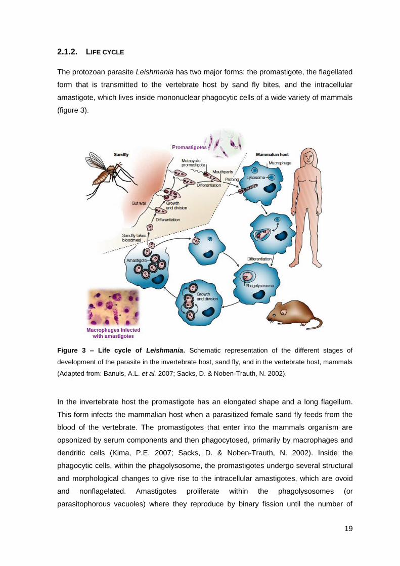
The protozoan parasite Leishmania has two major forms: the promastigote, the flagellated
form that is transmitted to the vertebrate host by sand fly bites, and the intracellular
amastigote, which lives inside mononuclear phagocytic cells of a wide variety of mammals
(figure 3).
Figure 3 – Life cycle of Leishmania. Schematic representation of the different stages of
development of the parasite in the invertebrate host, sand fly, and in the vertebrate host, mammals
(Adapted from: Banuls, A.L. et al. 2007; Sacks, D. & Noben-Trauth, N. 2002).
In the invertebrate host the promastigote has an elongated shape and a long flagellum.
This form infects the mammalian host when a parasitized female sand fly feeds from the
blood of the vertebrate. The promastigotes that enter into the mammals organism are
opsonized by serum components and then phagocytosed, primarily by macrophages and
dendritic cells (Kima, P.E. 2007; Sacks, D. & Noben-Trauth, N. 2002). Inside the
phagocytic cells, within the phagolysosome, the promastigotes undergo several structural
and morphological changes to give rise to the intracellular amastigotes, which are ovoid
and nonflagelated. Amastigotes proliferate within the phagolysosomes (or
parasitophorous vacuoles) where they reproduce by binary fission until the number of
20
parasites increases enough for the cell to burst and free the amastigotes to infect other
cells and continue their life cycles. The parasites could then go back to the invertebrate
host when a sand fly ingests blood that contains mononuclear phagocytic cells infected
with amastigotes (Banuls, A.L. et al. 2007; Kamhawi, S. 2006; Olivier, M. et al. 2005).
One of the most remarkable accomplishments of Leishmania is that they successfully live
inside mammalian cells that are responsible for killing pathogens, the macrophages. For
that Leishmania had to evolve a range of sophisticated mechanisms to subvert host
surveillance by altering the macrophage signal transduction machinery, thereby
modulating the macrophage environment in its favor. Some examples of these
mechanism are the production of lipophosphoglycan at the surface of the parasites that
confers resistance to the lytic action of the complement system, allowing the infection of
macrophages and creating appropriate conditions for the promastigote-to-amastigote
differentiation (Lodge, R. & Descoteaux, A. 2005). Other examples include preventing the
activation of macrophages defense mechanisms such as the production of oxygen and
nitrogen reactive species and also inhibition of an adequate and protective T cell response
by inhibiting the production of pro-inflammatory signals such as IL-12 and promoting the
release of anti-inflammatory cytokines, like IL-10 and TGF-β. (Kima, P.E. 2007; Olivier, M.
et al. 2005).
2.1.3. TREATMENTS
Drug treatment for leishmaniasis exists since the 20th Century, but only a limited range of
drugs are available. The efficacy of these drugs is affected by a combination of factors,
such as the differences in the sensitivity of Leishmania species to the drugs and their
pharmacokinetic properties, and the immune status of the patient (Goto, H. & Lindoso, J.L.
2010).
The current treatment of leishmaniasis consists in the administration of pentavalent
antimonials as a first option. As second line drugs there are some alternatives such as
amphotericin B, which is effective in antimonials-resistant cases, miltefosine which was
the first orally active drug made available against leishmaniasis, pentamidine that can also
be used in the treatment of resistant cases of visceral leishmaniasis, as well as
paromomycin (Goto, H. & Lindoso, J.L. 2010; Piscopo, T.V. & Mallia Azzopardi, C. 2007;
WHO 2010).
Parasite resistance to these conventional treatments, lack of efficacy, toxicity and high
costs associated with them lead to a clear need to improve the existing drugs and to
21
identify new targets and in that way develop new therapies (Piscopo, T.V. & Mallia
Azzopardi, C. 2007). However, the highest prevalence of parasitic diseases such as
leishmaniasis occurs in the poorest areas of the world, and from the total investment in
health research in the world, the work related with malaria, leishmaniasis and
trypanosomiasis accounts for only about 0.1% whereas the contribution of these diseases
to total burden is 5%. Nevertheless in the last years the search for new antiparasitic
treatments has received new impulse thanks to new technical and political developments,
and the appearance of new therapies, such as protease and topoisomerase inhibitors,
RNA interference- based approaches, nano-drug delivery and proteomics, as well as
antimicrobial peptides (Zucca, M. & Savoia, D. 2011).
2.1.4. LEISHMANIA AS A TARGET OF AMPS
In what concerns AMPs, protozoans have received much less attention as a target than
pathogens such as bacteria or fungi, although a variety of in vivo and in vitro antiparasitic
assays suggest that these compounds could represent a powerful tool for the
development of new therapies as well as to complement current ones (Rivas, L. et al.
2009).
Following the discovery of AMPs, magainins and cecropins were the first antimicrobial
peptides to display antiparasitic activities along with hybrids of cecropin A-melittin (Andreu,
D. et al. 1992; Boman, H.G. et al. 1989; Mor, A. 2009). Histatin 5, a human salivary AMP,
is also active against Leishmania, not by acting in the plasma membrane, as usual for
AMPs, but by decreasing the mitochondrial ATP synthesis leading to cell death (Luque-
Ortega, J.R. et al. 2008).
Although Leishmania are eukaryotic pathogens, their cytoplasmic membranes have higher
levels of negative phospholipids, such as phosphatidylserine and phosphatidylinositol than
higher eukaryotes, conferring them more susceptibility to the action of AMPs than
mammal cells (Wassef, M.K. et al. 1985). Leishmania is an interesting model as a target
for AMPs primarily due to the absence of barriers external to the plasma membrane
contrary to what happens in bacteria. The promastigote form of the parasite is endowed
with an anionic glycocalix, essentially composed of lipophosphoglycan (LPG), an anionic
oligosaccharide, anchored to the membrane through glycosylinositolphospholipids (GIPLs)
(figure 4). Abundant proteolytic activity also exists at the surface, due to the presence and
activity of GPI-proteins and proteophosphoglycans (PPGs) (figure 4) (Rivas, L. et al.
2009). These structures present at the surface of the membrane have some duality in
what concerns resistance and/ or susceptibility to AMPs. The presence of a negatively
22
charged glycocalix can favor peptide interaction however they can also delay the
interaction of the peptides with the cytoplasmic membrane diminishing their efficacy; the
proteolytic activity at the surface of the membrane, can also confer resistance to AMPs
due to increased possibility of peptide degradation. A second important characteristic of
Leishmania cells is the fact that endo- and exocytosis are confined to a special area
accounting for about 2% of the plasma membrane known as the flagellar pocket (deep
invagination of the plasma membrane that is located at the base of the flagellum devoid of
attached microtubules) (Morgan, G.W. et al. 2002; Overath, P. et al. 1997). Theoretically,
this may somehow limit the repairing capacity of the membrane upon exposure to AMPs
(Luque-Ortega, J.R. & Rivas, L. 2010).
Although amastigotes can appear more vulnerable to AMPs due to the much less
developed glycocalix (figure 4), their location inside phagocytic cells and the substantial
changes in the membrane composition adopted by these parasites along their life cycle,
can be barriers to the action of AMPs (Luque-Ortega, J.R. & Rivas, L. 2010).
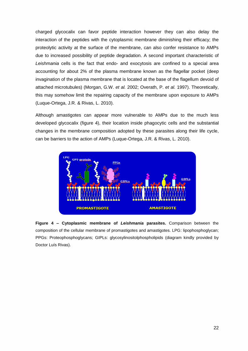
Figure 4 – Cytoplasmic membrane of Leishmania parasites. Comparison between the
composition of the cellular membrane of promastigotes and amastigotes. LPG: lipophosphoglycan;
PPGs: Proteophosphoglycans; GIPLs: glycosylinositolphospholipids (diagram kindly provided by
Doctor Luís Rivas).
23
2.2. MYCOBACTERIUM
Tuberculosis (TB) remains today one of the most deadly human diseases. WHO
estimates that one-third of the world's population is currently infected with the TB bacillus
with 5-10% of people developing the disease. In 2009 approximately 2 million people died
of TB. The incidence of tuberculosis in the United States had been declining since the turn
of the 20th century, however since 1985 it has been rising mainly due to the AIDS
epidemic, the lack of efficacy of the anti-tuberculosis vaccine and the emergence of
multidrug-resistant strains (Clemens, D.L. & Horwitz, M.A. 1995) The genus
Mycobacterium includes not only Mycobacterium tuberculosis, the causative agent for TB
discovered in 1882 by Robert Koch (Koul, A. et al. 2011), but other pathogenic species
such as M. leprae and M. ulcerans, and non-pathogenic species, usually referred to as
nontuberculous mycobacteria (NTM). More than 130 species of NTM are known and most
of them are typically environmental organisms present in water, soil, dust and plants.
Contact with these contaminated environments may be responsible for infection in
humans and animals with the possibility of transmission from human to human being
almost null (Pieters, J. 2001; Tortoli, E. 2006; Tortoli, E. 2009). The large majority of NTM
are nonpathogenic for healthy individuals, but they can act as opportunists in
immunocompromised patients, especially in AIDS patients. The consequences of
mycobacterial infection depend on the virulence of the infecting Mycobacterium and the
resistance of the host. In humans the outcome can range from relatively mild and transient
symptoms, mostly caused by pulmonary infections, to a widely disseminated disease
(Pieters, J. 2001; Tortoli, E. 2009).
2.2.1. MYCOBACTERIA CELL WALL
Mycobacteria cells are irregular rods, aerobic, and unable to form spores (Prescott, L.M.
et al. 2002). Mycobacterium species are characterized by an extremely complex and
highly impermeable cell wall, composed of mycolic acids, glycolipids, lipoglycans,
polysaccharides and pore forming proteins (figure 5). This unique cell wall contributes to
the capacity of the pathogen to survive inside the host and to resist the chemotherapy
(Nigou, J. et al. 2003).
The cell wall skeleton determines the size and shape of mycobacteria, and contains three
different covalently linked layers, peptidoglycan, arabinogalactan and mycolic acids
(Guenin-Mace, L. et al. 2009). The covalent linkage of the mycolic acids results in a
hydrophobic layer of extremely low fluidity and high impermeability. Also bound to the
mycolic acids by hydrophobic links, in the external side, are various free lipids such as
24
phenolic glycolipids, sulpholipids, among others. The outer layer of the cell wall that some
authors refer to as capsule is mainly composed of polysaccharides such as glucan and
arabinomannan (Abdallah, A.M. et al. 2007).
The presence of mycolic acids and other lipids outside the peptidoglycan layer makes the
mycobacteria acid-fast. This means these bacteria are not stained by the Gram stain, and
have to be stained with a harsher treatment, the Ziehl-Neelsen method (Prescott, L.M. et
al. 2002).
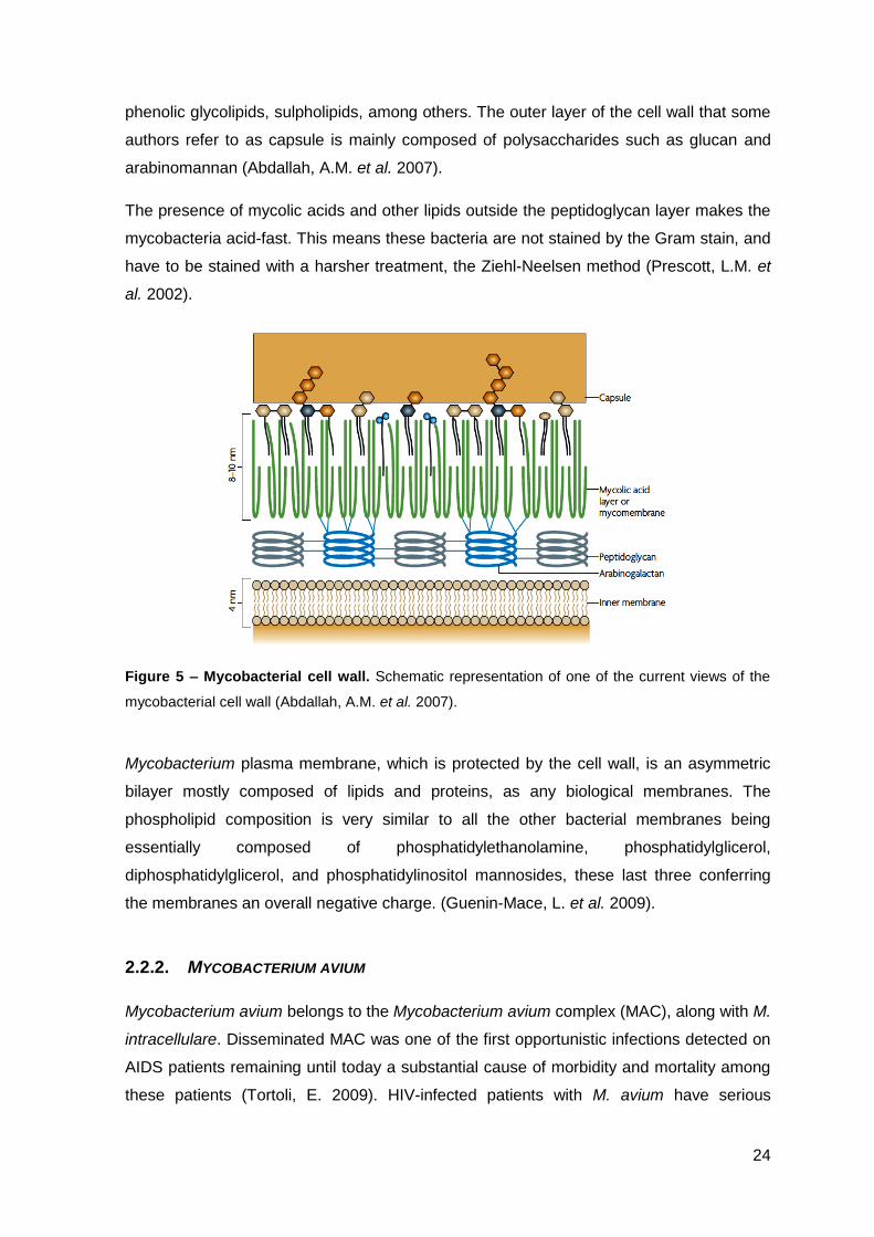
Figure 5 – Mycobacterial cell wall. Schematic representation of one of the current views of the
mycobacterial cell wall (Abdallah, A.M. et al. 2007).
Mycobacterium plasma membrane, which is protected by the cell wall, is an asymmetric
bilayer mostly composed of lipids and proteins, as any biological membranes. The
phospholipid composition is very similar to all the other bacterial membranes being
essentially composed of phosphatidylethanolamine, phosphatidylglicerol,
diphosphatidylglicerol, and phosphatidylinositol mannosides, these last three conferring
the membranes an overall negative charge. (Guenin-Mace, L. et al. 2009).
2.2.2. MYCOBACTERIUM AVIUM
Mycobacterium avium belongs to the Mycobacterium avium complex (MAC), along with M.
intracellulare. Disseminated MAC was one of the first opportunistic infections detected on
AIDS patients remaining until today a substantial cause of morbidity and mortality among
these patients (Tortoli, E. 2009). HIV-infected patients with M. avium have serious
25
complications associated with debilitating symptoms and shortened survival. In 1994, in
the United States, it was estimated that 37 000 people were infected with disseminated
Mycobacterium avium complex, a greater number than the people infected by TB in this
year also in the same country (Horsburgh, C.R. et al. 2001; Reed, C. et al. 2006).
Infection of AIDS patients by M. avium occurs in advanced stages of the disease, when
the levels of CD4+ T cells are very low. M. avium can also infect patients with other
debilitating diseases, especially restrictive and obstructive pulmonary diseases, that
compromise the immune system, such as chronic obstructive pulmonary disease,
emphysema, chronic bronchitis and children with lymphadenitis (Appelberg, R. 2006b).
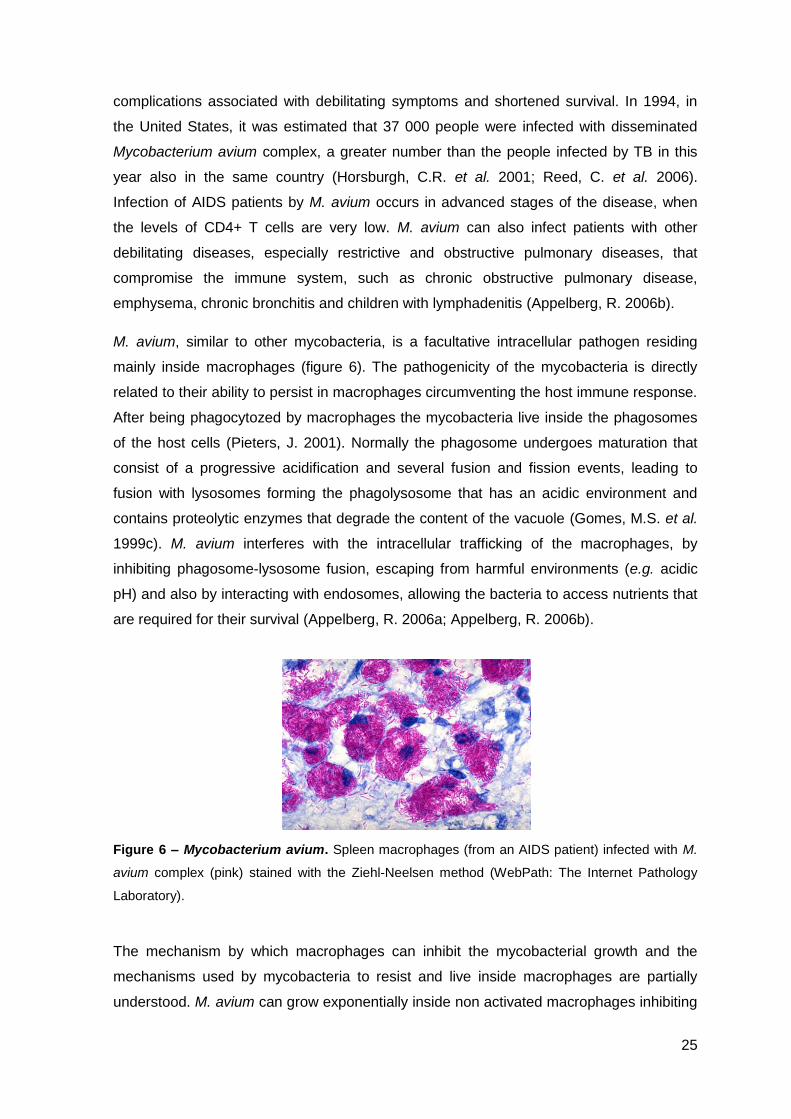
M. avium, similar to other mycobacteria, is a facultative intracellular pathogen residing
mainly inside macrophages (figure 6). The pathogenicity of the mycobacteria is directly
related to their ability to persist in macrophages circumventing the host immune response.
After being phagocytozed by macrophages the mycobacteria live inside the phagosomes
of the host cells (Pieters, J. 2001). Normally the phagosome undergoes maturation that
consist of a progressive acidification and several fusion and fission events, leading to
fusion with lysosomes forming the phagolysosome that has an acidic environment and
contains proteolytic enzymes that degrade the content of the vacuole (Gomes, M.S. et al.
1999c). M. avium interferes with the intracellular trafficking of the macrophages, by
inhibiting phagosome-lysosome fusion, escaping from harmful environments (e.g. acidic
pH) and also by interacting with endosomes, allowing the bacteria to access nutrients that
are required for their survival (Appelberg, R. 2006a; Appelberg, R. 2006b).
Figure 6 – Mycobacterium avium. Spleen macrophages (from an AIDS patient) infected with M.
avium complex (pink) stained with the Ziehl-Neelsen method (WebPath: The Internet Pathology
Laboratory).
The mechanism by which macrophages can inhibit the mycobacterial growth and the
mechanisms used by mycobacteria to resist and live inside macrophages are partially
understood. M. avium can grow exponentially inside non activated macrophages inhibiting
26
the production of superoxide, however this growth is more restrictive if the macrophages
are activated with IFN-γ and TNF-α, independently of the production of nitrogen and
oxygen reactive species (Gomes, M.S. & Appelberg, R. 2002; Gomes, M.S. et al. 1999b).
Also mechanisms that lead to restriction of access to nutrients by mycobacteria and
macrophage death pathways may contribute to the elimination of the pathogen
(Appelberg, R. 2006a; Behar, S.M. et al. 2010).
2.2.3. TREATMENTS
Infections by mycobacteria are very difficult to treat due to a combination of factors, such
as: i) the poor action of the antibiotics available, mainly due to the highly impermeable cell
wall; ii) the fact that these are intracellular bacteria; iii) the long duration of the treatments,
that much of the time the patients do not follow to the end and iv) the antibiotic resistance
that is increasing worldwide.
The basic treatment of all pathogenic mycobacteria consists of a combination of different
antibiotics taken for several months. In the case of tuberculosis the treatment varies
according to the susceptibility of the isolated strain to the available drugs, going from the
administration of antibiotics such as isoniazid, rifampicin, pyrazinamide and ethambutol to
fluoroquinolone or second-line injectable drugs, such as amikacin, kanamycin and
capreomycin, that are all ineffective against extensively-drug resistant TB (Koul, A. et al.
2011).
In the case of Mycobacterium avium current therapy consists of ethambutol with a
macrolide that can be azithromycin or clarithromycin, and rifamycin derivatives (rifampicin
and rifabutin) taken for six months to one year with an overall clinical success rate of no
more than 60% in AIDS patients (Deshpande, D. et al. 2010). The administration of an
effective antiretroviral therapy in immunocompromised patients reduces the number of
people at risk of developing disseminated MAC by restoring the immune function of the
patients essentially by elevating the number of CD4+ T cells (Horsburgh, C.R. et al. 2001).
The global control of mycobacterial infections will be achieved with new antimycobacterial
drugs that follow some criteria: shorten treatment duration, target multidrug-resistant
strains, a more simple treatment by reducing the daily pill burden and the dosing
frequency, and the possibility of co-administration with anti-HIV drugs (Koul, A. et al.
2011).
Some alternative therapies consist in the combination of antibiotics with adjunctive
immunomodulators – adjunctive immunotherapy – such as picolinic acid, that have shown
27
direct antimicrobial activity against both extracellular and intramacrophagic MAC
organisms, alone and in combination with conventional antibiotics (Cai, S. et al. 2006).
The observation that the growth of M. avium inside macrophages is directly proportional to
the amount of iron available, and the fact that AIDS patients have increased iron
deposition in different tissues favoring the growth of M. avium, makes the use of iron
chelators, that deprives the mycobacteria from an essential nutrient for their survival, a
promising road for the treatment of this disease. Recent studies have shown the efficacy
of some iron chelators both in vitro and in animal models infected with M. avium
(Fernandes, S.S. et al. 2010; Gomes, M.S. et al. 2001; Gomes, M.S. et al. 1999a).
2.2.4. MYCOBACTERIA AS A TARGET OF AMPS
Antimicrobial peptides, such as cathelicidins and defensins, like human neutrophil peptide
(HNP) and human β-defensin 2 (HBD-2), have also shown activity against M. tuberculosis
and M. avium (Jena, P. et al. 2011; Mendez-Samperio, P. 2008; Ogata, K. et al. 1992).
The combination of HNPs with anti-tuberculosis drugs, like isoniazid and rifampicin,
against intracellular mycobacteria resulted in a significant reduction in the mycobacterial
load (Kalita, A. et al. 2004; Sharma, S. et al. 2000). This effect could be due to increased
permeability of both mycobacterial cell wall and plasmatic membrane by AMPs increasing
the access of the antibiotics to intracellular targets like DNA (Sharma, S. et al. 1999). By
potentiating the effect of antibiotics, AMPs can allow the reduction of the therapeutic
dosage of drugs to approximately half and also reduce the time of treatment (Kalita, A. et
al. 2004).

28
29
3. ANTIMICROBIAL PEPTIDES (AMPS)
Considering the potential of antimicrobial peptides as new alternative therapies to fight
infectious diseases, in the next sections, several aspects concerning AMPs will be
addressed, namely: their structural characteristics, mechanisms of action, role in the
immunity, and the problems, successes and challenges of introducing AMPs in the clinical
practice.
The term “Antimicrobial peptide” refers to a large number of peptides first characterized on
the basis of their activity against bacteria and fungi. They constitute a primitive immune
defense mechanism and they were found, first in the 1980s, on a variety of eukaryotic
organisms, from insects to humans (Nguyen, L.T. et al. 2011; Reddy, K.V. et al. 2004).
Some of them were first isolated due to their antimicrobial activity, whereas others were
discovered for various unrelated functions before their potential as antimicrobial peptides
was recognized (Wiesner, J. & Vilcinskas, A. 2010). These peptides are multifunctional
and act in concert with other immune mechanisms and have evolved in nature throughout
centuries to protect their hosts against diverse pathogens, such as bacteria, fungi, virus,
protozoa and even cancer cells (Nguyen, L.T. et al. 2011). So far, more than 1500
peptides from different sources have been reported in The Antimicrobial Peptide
Database – APD (http://aps.unmc.edu/AP/main.php) and the majority of them are
classified as antibacterial peptides. Virtually all human tissues can produce AMPs, either
constitutively or induced by inflammatory stimulus, more frequently in body sites exposed
to pathogens, such as the skin or mucosa, or in some blood cell types, such as
neutrophils, eosinophils and platelets (Wiesner, J. & Vilcinskas, A. 2010).
Natural AMPs are gene-encoded and ribosome-synthesized peptides that derive from
precursor peptides through one or more proteolytic activation steps. De novo design of
synthetic peptides was introduced in the 1990s with the use of high-throughput
combinatorial library screening, structure based modeling and predictive algorithms to
determine the optimal active peptide. Although this approach produced interesting results,
the need for a high initial investment in technology compared to traditional methods has
delayed the widespread use of this approach. On the other hand, rational design of
peptides using existent sequences as templates that are modified to obtain peptides with
the desired properties (antimicrobial activity and selectivity) and the discovery of AMPs
through the search within natural proteins (e.g. lactoferrin) have also led to the successful
production of more potent antimicrobial peptides (Nguyen, L.T. et al. 2011).
30
3.1. STRUCTURAL CHARACTERISTICS
Antimicrobial peptides vary widely in many aspects, such as length, sequence, structure
and source, but there are some common traits for most of them, that are important for
their antimicrobial activity. They can be defined as short (<50 amino-acids), with a positive
net charge (generally +2 to +9, due to the existence of lysine and arginine residues) that
allows them to interact with negatively charged membranes, such as bacterial membranes,
although anionic antimicrobial peptides have also been described. They also have a
substantial proportion of hydrophobic residues. AMPs have a random structure in solution
but when in a membranous environment they adopt an amphipatic structure, allowing
them to bind more efficiently to phospholipid membranes (Shai, Y. et al. 2006). The full
classification of AMPs is difficult due to their huge diversity but almost all authors divide
and classify the peptides based on their secondary structure. One of those classifications
divides AMPs into three groups (table 1 and figure 7): α-helical peptides, β-sheet
peptides and extended peptides which do not fold into regular secondary structures and
are often composed of a high number of certain amino-acids, like histidine, tryptophan and
arginine (Nguyen, L.T. et al. 2011).
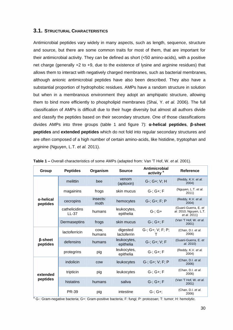
Table 1 – Overall characteristics of some AMPs (adapted from: Van 'T Hof, W. et al. 2001).
Group Peptides Organism Source Antimicrobial
activity a
Reference
α-helical peptides
melittin bee venom
(apitoxin) G-; G+; V; H
(Reddy, K.V. et al. 2004)
magainins frogs skin mucus G-; G+; F (Nguyen, L.T. et al.
2011)
cecropins insects:
moth hemocytes G-; G+; F; P
(Reddy, K.V. et al. 2004)
cathelicidins LL-37
humans leukocytes,
epithelia G-; G+
(Guaní-Guerra, E. et al. 2010; Nguyen, L.T.
et al. 2011)
Dermaseptins frogs skin mucus G-; G+; F (Van 'T Hof, W. et al.
2001)
β-sheet peptides
lactoferricin cow,
humans digested
lactoferrin G-; G+; V; F; P;
T (Chan, D.I. et al.
2006)
defensins humans leukocytes,
epithelia G-; G+; V; F
(Guaní-Guerra, E. et al. 2010)
protegrins pig leukocytes,
epithelia G-; G+; F
(Reddy, K.V. et al. 2004)
extended peptides
indolicin cow leukocytes G-; G+; V; F; P (Chan, D.I. et al.
2006)
tripticin pig leukocytes G-; G+; F (Chan, D.I. et al.
2006)
histatins humans saliva G-; G+; F (Van 'T Hof, W. et al.
2001)
PR-39 pig intestine G-; G+; (Chan, D.I. et al.
2006)
a G-: Gram-negative bacteria; G+: Gram-positive bacteria; F: fungi; P: protozoan; T: tumor; H: hemolytic.
31
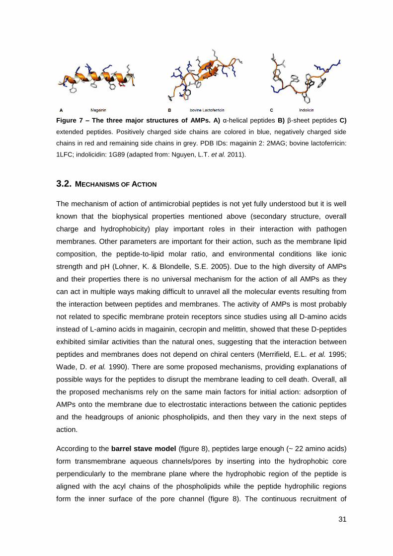
Figure 7 – The three major structures of AMPs. A) α-helical peptides B) β-sheet peptides C)
extended peptides. Positively charged side chains are colored in blue, negatively charged side
chains in red and remaining side chains in grey. PDB IDs: magainin 2: 2MAG; bovine lactoferricin:
1LFC; indolicidin: 1G89 (adapted from: Nguyen, L.T. et al. 2011).
3.2. MECHANISMS OF ACTION
The mechanism of action of antimicrobial peptides is not yet fully understood but it is well
known that the biophysical properties mentioned above (secondary structure, overall
charge and hydrophobicity) play important roles in their interaction with pathogen
membranes. Other parameters are important for their action, such as the membrane lipid
composition, the peptide-to-lipid molar ratio, and environmental conditions like ionic
strength and pH (Lohner, K. & Blondelle, S.E. 2005). Due to the high diversity of AMPs
and their properties there is no universal mechanism for the action of all AMPs as they
can act in multiple ways making difficult to unravel all the molecular events resulting from
the interaction between peptides and membranes. The activity of AMPs is most probably
not related to specific membrane protein receptors since studies using all D-amino acids
instead of L-amino acids in magainin, cecropin and melittin, showed that these D-peptides
exhibited similar activities than the natural ones, suggesting that the interaction between
peptides and membranes does not depend on chiral centers (Merrifield, E.L. et al. 1995;
Wade, D. et al. 1990). There are some proposed mechanisms, providing explanations of
possible ways for the peptides to disrupt the membrane leading to cell death. Overall, all
the proposed mechanisms rely on the same main factors for initial action: adsorption of
AMPs onto the membrane due to electrostatic interactions between the cationic peptides
and the headgroups of anionic phospholipids, and then they vary in the next steps of
action.
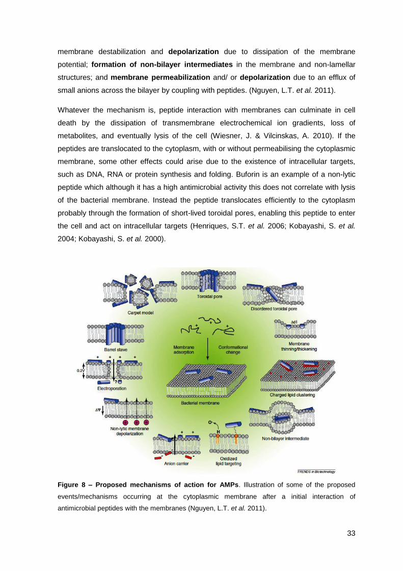
According to the barrel stave model (figure 8), peptides large enough (~ 22 amino acids)
form transmembrane aqueous channels/pores by inserting into the hydrophobic core
perpendicularly to the membrane plane where the hydrophobic region of the peptide is
aligned with the acyl chains of the phospholipids while the peptide hydrophilic regions
form the inner surface of the pore channel (figure 8). The continuous recruitment of
32
peptides to the membrane increases the pore size leading to leakage of the cells contents,
membrane depolarization and thereby cell death (Nguyen, L.T. et al. 2011). The peptides
arrangement in the membrane causes high repulsion due to their charges and this can
culminate in the disintegration of the pore. This model is only possible for peptides with
not too high charge and it is believed that the number of peptides acting by this
mechanism is very low. An example is alamethicin from the fungus Trichoderma viridis,
and perforin, produced by mammalian killer lymphocytes and complement component C9
(Wiesner, J. & Vilcinskas, A. 2010).
The carpet model (figure 8) proposes that the membrane surface is covered by the
peptides, aligned parallel to the bilayer surface, in a “carpet like” manner, diminishing the
fluidity of the membrane. When the concentration of the peptide reaches a threshold value
the integrity of the membrane is lost leading to membrane micellization, toroidal pores or
aggregated channels formation (see below) and cell death (figure 8) (Lohner, K. 2009).
The formation of toroidal or wormhole pores (figure 8) is the best characterized and the
most consistent mechanism of action of AMPs. This model can be seen as representing
one of the possible final stages of the previous carpet model, where the formation of pores
leads to disruption of the membranes. In fact, these two models have been unified in the
so called “Shai-Matzusaki-Huang” model that proposes that peptides bind parallel to the
membrane with the apolar amino acids penetrating partly into the hydrophobic core, while
the cationic residues interact with the headgroups of anionic phospholipids, inducing
membrane thinning and a curvature strain. To release this strain the orientation of the
peptides changes from parallel to perpendicular, inducing bending of the membrane
interface towards the hydrophobic interior, maintaining the contact between the peptides
and the charged headgroups from the phospholipids. These events lead to the formation
of toroidal pores composed by the peptides and the lipid headgroups (figure 8). Upon
disintegration of the pores some peptides can be translocated to the inner leaflet of the
membranes reaching the cytoplasm or the membrane can be disrupted due to
depolarization or micellization, resulting in cell death (Nicolas, P. 2009; Rivas, L. et al.
2009).
Other mechanisms of action have recently been suggested that do not imply the
recruitment of a high concentration of peptides to the membrane, and they do not
necessarily exclude each other (or even the models explained above) (figure 8). Some
examples are, lipid segregation, where the lipids can move laterally in the membrane
and thus form domains rich in anionic lipids (induced by the presence of the positively
charged peptide), and this can induce small leakage of intracellular contents and/ or
33
membrane destabilization and depolarization due to dissipation of the membrane
potential; formation of non-bilayer intermediates in the membrane and non-lamellar
structures; and membrane permeabilization and/ or depolarization due to an efflux of
small anions across the bilayer by coupling with peptides. (Nguyen, L.T. et al. 2011).
Whatever the mechanism is, peptide interaction with membranes can culminate in cell
death by the dissipation of transmembrane electrochemical ion gradients, loss of
metabolites, and eventually lysis of the cell (Wiesner, J. & Vilcinskas, A. 2010). If the
peptides are translocated to the cytoplasm, with or without permeabilising the cytoplasmic
membrane, some other effects could arise due to the existence of intracellular targets,
such as DNA, RNA or protein synthesis and folding. Buforin is an example of a non-lytic
peptide which although it has a high antimicrobial activity this does not correlate with lysis
of the bacterial membrane. Instead the peptide translocates efficiently to the cytoplasm
probably through the formation of short-lived toroidal pores, enabling this peptide to enter
the cell and act on intracellular targets (Henriques, S.T. et al. 2006; Kobayashi, S. et al.
2004; Kobayashi, S. et al. 2000).
Figure 8 – Proposed mechanisms of action for AMPs. Illustration of some of the proposed
events/mechanisms occurring at the cytoplasmic membrane after a initial interaction of
antimicrobial peptides with the membranes (Nguyen, L.T. et al. 2011).
34
AMPs can also be cytotoxic against cancerous cells promoting apoptosis (e.g. lactoferrin
proved to inhibit the tumor growth and metastasis in a rodent model with breast cancer,
while lactoferrricin causes apoptosis in Jurkat T-leukemia cells) (Iigo, M. et al. 2009;
Mader, J.S. et al. 2007; Wiesner, J. & Vilcinskas, A. 2010). It is believed that this effect is
first due to lysis of the cellular membrane of the cancerous cells, that are recognized by
some AMPs due to the unusually high amounts of negatively charged gangliosides and
phosphatidylserine (PS) in the surface of these cells, a negative membrane potential and
a higher membrane fluidity, allowing the peptides to insert into the membrane more easily
(Hoskin, D.W. & Ramamoorthy, A. 2008). Afterwards, mitochondrial membranes can also
be disrupted due to entrance of the peptide into the cell, apoptosis pathways can be
activated and angiogenesis inhibited (Gifford, J.L. et al. 2005; Guaní-Guerra, E. et al.
2010).
3.3. SELECTIVITY / TOXICITY
Independently of how AMPs exert their activity against pathogens the peptides have to
interact with the cellular membrane resulting either in disruption or traversing this barrier.
This interaction must be as selective as possible regarding the distinction between
mammalian cells (eukaryotic cells) and pathogen cells, such as bacteria (prokaryotic cells).
Cytoplasmic membranes of mammalian cells are predominantly constituted by
phospholipids and cholesterol. Phospholipids are asymmetrically distributed between the
inner and the outer leaflet of the bilayer exposing predominantly zwitterionic
phosphatidylcholine (PC) and sphingomyelin to the extracellular side. On the other hand,
cytoplasmic bacterial membranes do not have cholesterol and are mainly composed of
zwitterionic phosphotidylethanolamine (PE) and negatively charged phosphatidylglicerol
(PG), diphosphatidylglicerol and cardiolipin, conferring an overall negative charge to the
outer side of the cytoplasmatic membrane (Lohner, K. 2009; Lohner, K. & Blondelle, S.E.
2005). The main principle of peptide interaction with membranes relies on electrostatic
interactions between cationic peptides and negatively charged membranes and thus
considering the composition of both cell types, bacteria are a preferential target for AMPs.
However, before reaching the cytoplasmic membrane of the bacteria, peptides have to
overcome other barriers. In the case of Gram-positive bacteria, a thick peptidoglycan layer
embedded with teichoic and lipoteichoic acids (which are negatively charged) is present
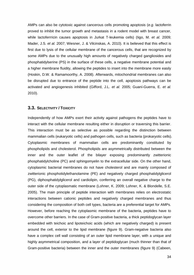
around the cell, exterior to the lipid membrane (figure 9). Gram-negative bacteria also
have a complex cell wall consisting of an outer lipid membrane layer, with a unique and
highly asymmetrical composition, and a layer of peptidoglycan (much thinner than that of
Gram-positive bacteria) between the inner and the outer membranes (figure 9) (Cabeen,
35
M.T. & Jacobs-Wagner, C. 2005). In Gram-negatives outer membrane,
lipopolysaccharides (LPS – negatively charged) are located exclusively in the outer leaflet
and phospholipids are confined to the inner leaflet (figure 9) (Lohner, K. 2009; Lohner, K.
& Blondelle, S.E. 2005). In the case of Mycobacteria, an even thicker and more complex
cell wall is also present (see section 2.2.1). Even so, in normal conditions the overall
charge of a bacterial cell wall is negative, increasing the chances of interaction with
antimicrobial peptides. (Guenin-Mace, L. et al. 2009; Prescott, L.M. et al. 2002).
Figure 9 – Bacteria cell wall. Schematic representation of the cell wall of a) Gram-positive and b)
Gram-negative bacteria (Cabeen, M.T. & Jacobs-Wagner, C. 2005).
The membrane potential can also contribute for the selectivity of AMPs since in
eukaryotes it is less negative than in prokaryotes (Van 'T Hof, W. et al. 2001).
Antimicrobial peptides can also kill eukaryotic pathogens, such as fungi and protozoa,
raising the question of what are the features of the pathogens, either eukaryotes or
prokaryotes that allows some AMPs to discriminate them from mammal cells. These
specificity seems to rely on charge, since pathogens seems to have a higher percentage
of anionic phospholipids than mammal cells, thus explaining the specificity of the cationic
AMPs (Lohner, K. 2009; Lohner, K. & Blondelle, S.E. 2005). However some peptides do
not distinguish host and pathogen cells being able to disrupt mammalian cells (e.g.
melittin) and thus cannot be used in the clinic. The reasons behind this activity are not
completely understood and are most probably due to the properties of the peptides, like a
36
high positive charge, too large hydrophobic surfaces, between others (Nguyen, L.T. et al.
2011). In the evaluation of the activity of AMPs the cytotoxicity of these peptides towards
mammalian cells should always be addressed. There are many different cytotoxicity
assays that can be performed but the outcome of each of them strongly depends on many
factors such as the origin and life storage of the mammal cells, the peptide-to-cell ratio,
the medium used, between others, so the results must be carefully analyzed (Van 'T Hof,
W. et al. 2001). Based on the results of cytotoxicity assays together with the antimicrobial
activity of AMPs, the selectivity index, defined as the cytotoxicity activity divided by the
antimicrobial activity, can be determined being a useful tool to predict the potential of a
given AMP as a therapeutic agent. However, the characteristics of these peptides and the
prediction of their action, either antimicrobial activity or cytotoxicity, is not straightforward
and so they should be analyzed on a case-by-case basis.
3.4. AMPS IN THE IMMUNITY
Besides the role of AMPs as endogenous antibiotics (directly killing the pathogens), they
can also participate in multiple aspects of immunity and for that they can also be called
host defense peptides (HDPs). Combination of these different but complementary
functions is essential for the effective control of infections in the host organism. AMPs
seem to actively participate in the immune system either by immunomodulation,
neutralization of endotoxins, enhancement of phagocytosis, induction of both
angiogenesis and wound repair, leukocyte chemotaxis and synergism with cytokines
(Yang, D. et al. 2002; Zaiou, M. 2007). Thus, in response to an infection, antimicrobial
peptides can promote bacterial clearance not only through direct killing but also through
the establishment of immune cell circuits (Auvynet, C. & Rosenstein, Y. 2009; Guaní-
Guerra, E. et al. 2010).
As a consequence of the actions stated above these peptides are also involved in
autoimmunity. The abnormal concentration, processing or signaling of AMPs is associated
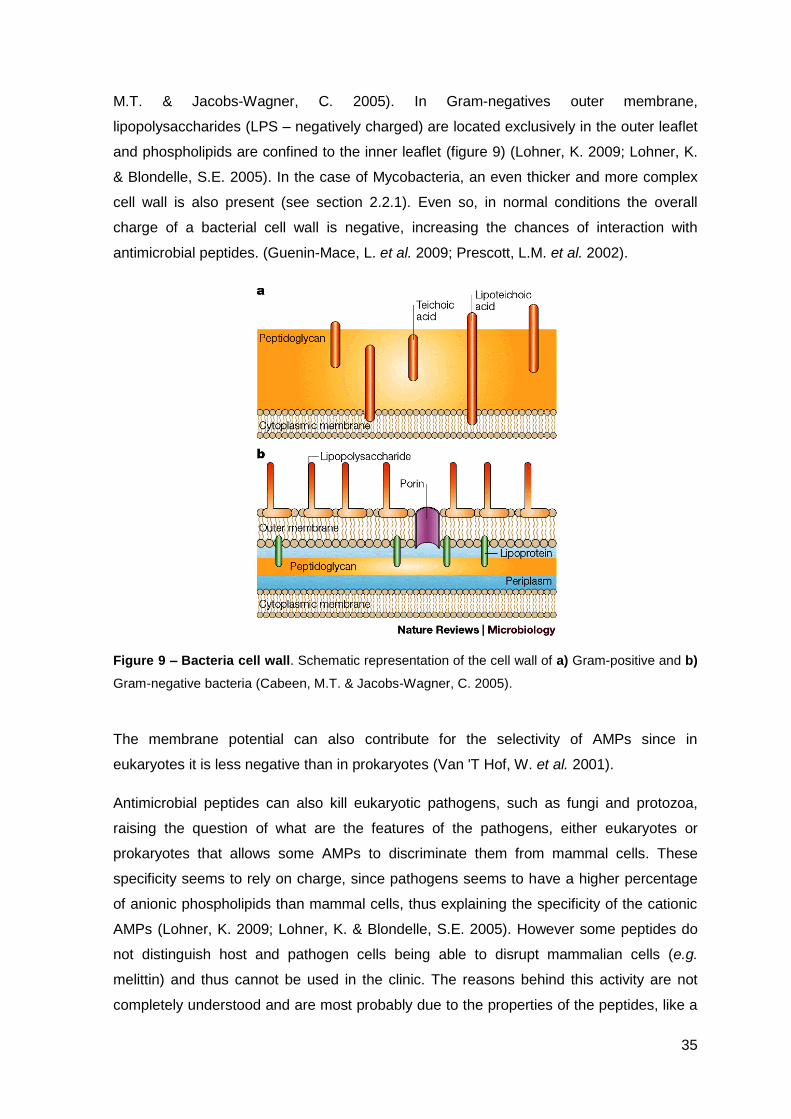
with a growing list of autoimmune diseases (figure 10), in which these peptides usually act,
not as a single direct cause but as an important factor that influences the outcome of the
diseases (Guaní-Guerra, E. et al. 2010).
37
Figure 10 – Role of AMPs in the immune system. Exemplification of some of the immune
processes and inflammatory diseases where AMPs are involved (Guaní-Guerra, E. et al. 2010).
3.5. RESISTANCE TO AMPS
Induction of resistance is less likely for AMPs when compared to conventional antibiotics,
due to the fast kinetics of the antimicrobial process and to the nature of the target –
plasma membrane – since it would require an overall reorganization of the cell membrane
structure, namely the phospholipid composition, affecting simultaneously the pleiade of
transport systems and the enzymes embedded in the phospholipid matrix. Although in
principle acquiring resistance to AMPs is more difficult, it has been pointed that it might
have more severe effects if it led to cross-resistance to innate human antimicrobial
peptides (Hancock, R.E. & Sahl, H.G. 2006).
The most common mechanisms of bacterial resistance to AMPs described so far consist
in the modification of the bacterial envelope leading to charge reduction and the
proteolytic cleavage of peptides (Wiesner, J. & Vilcinskas, A. 2010). These mechanisms
include the production of secreted proteins or cell-surface proteins that irreversibly binds
or cleaves AMPs and glycopolymeric matrices that trap the peptides preventing their
access to the bacterial cytoplasmic membrane (figure 11). Electrostatic repulsion of AMPs
can also arise from several modifications such as substitutions of lipid A of LPS by
aminoarabinose and ethanolamine, modification of phosphatidylglicerol by ligation of a L-
38
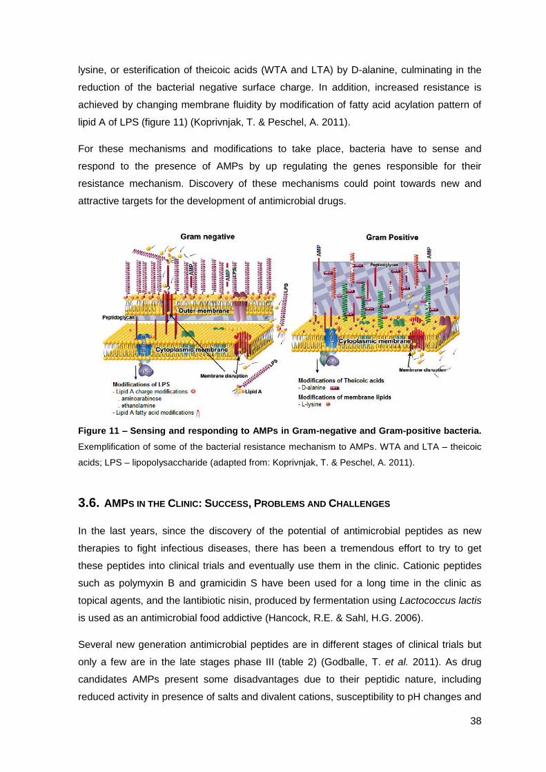
lysine, or esterification of theicoic acids (WTA and LTA) by D-alanine, culminating in the
reduction of the bacterial negative surface charge. In addition, increased resistance is
achieved by changing membrane fluidity by modification of fatty acid acylation pattern of
lipid A of LPS (figure 11) (Koprivnjak, T. & Peschel, A. 2011).
For these mechanisms and modifications to take place, bacteria have to sense and
respond to the presence of AMPs by up regulating the genes responsible for their
resistance mechanism. Discovery of these mechanisms could point towards new and
attractive targets for the development of antimicrobial drugs.
Figure 11 – Sensing and responding to AMPs in Gram-negative and Gram-positive bacteria.
Exemplification of some of the bacterial resistance mechanism to AMPs. WTA and LTA – theicoic
acids; LPS – lipopolysaccharide (adapted from: Koprivnjak, T. & Peschel, A. 2011).
3.6. AMPS IN THE CLINIC: SUCCESS, PROBLEMS AND CHALLENGES
In the last years, since the discovery of the potential of antimicrobial peptides as new
therapies to fight infectious diseases, there has been a tremendous effort to try to get
these peptides into clinical trials and eventually use them in the clinic. Cationic peptides
such as polymyxin B and gramicidin S have been used for a long time in the clinic as
topical agents, and the lantibiotic nisin, produced by fermentation using Lactococcus lactis
is used as an antimicrobial food addictive (Hancock, R.E. & Sahl, H.G. 2006).
Several new generation antimicrobial peptides are in different stages of clinical trials but
only a few are in the late stages phase III (table 2) (Godballe, T. et al. 2011). As drug
candidates AMPs present some disadvantages due to their peptidic nature, including
reduced activity in presence of salts and divalent cations, susceptibility to pH changes and
39
to protease and other plasma components’ activity, resulting in low metabolic stability and
bioavailability, and reduced in vivo half-lives (Rotem, S. & Mor, A. 2009). The root of
administration is also a problem since they would not be reabsorbed from the intestinal
tract if administered orally, and if injected they could trigger immune responses that
neutralize the active component or induce allergic reactions. So they should be
administered as topical drugs for the treatment of skin and wound infections, limiting their
applicability (Wiesner, J. & Vilcinskas, A. 2010). Other safety considerations should be
taken into account. They can cross-react with receptors for neuropeptides and peptide
hormones, and the rapidly degradation of AMPs could lead to unwanted levels of amino
acids for which some patients are sensitive, like glutamate (Chinese restaurant syndrome)
and phenylalanine (phenylketonuria) (Van 'T Hof, W. et al. 2001).
The high cost of production associated with peptide synthesis is another drawback in the
clinical application of these peptides. If the peptides have to be applied as topical
formulations, the amount of peptide applied needs to be much higher and therefore the
costs will be higher as well. Even when AMPs successfully finished several phase III trials,
in most of the cases, they were not able to demonstrate superior protection when
compared to traditional therapies (Godballe, T. et al. 2011).
Several approaches have been adopted to try to overcome these problems, keeping in
mind that the requirements for antimicrobial activity do not rely on a defined secondary
structure or even in a consensus sequence, which was not identified when several
sequences of natural AMPs deposited in the Antimicrobial Sequence Database – AMSDb
(http://www.bbcm.univ.trieste.it/~tossi/amsdb.html) were analyzed (Rotem, S. & Mor, A.
2009). The only common trait to all of them, as mentioned above (3.1) is the occurrence of
both hydrophobic and positively charged amino acids. In this context some solutions have
been proposed: i) the use of D-amino acids (rather than L-amino acids) which are
resistant to proteases but have higher costs when compared with L-amino acids; ii) the
use of nonpeptidic backbones (peptidomimetics); iii) formulation to improve stability, for
example in liposomes; iv) pro-drug molecules, among others (Hancock, R.E. & Sahl, H.G.
2006).
Peptidomimetics (mimics of AMPs) deserves a special word, since this approach
conserves the important features mentioned before (hydrophobicity and charge), and the
antimicrobial activity, trying to solve some of the problems associated with natural
peptides (costs, susceptibility to proteases, etc.) (Godballe, T. et al. 2011).
Peptidomimetics refers to any oligomeric sequence designed to mimic a peptide structure
40
and/ or function but whose backbone is not solely based on α-amino acids (Rotem, S. &
Mor, A. 2009).
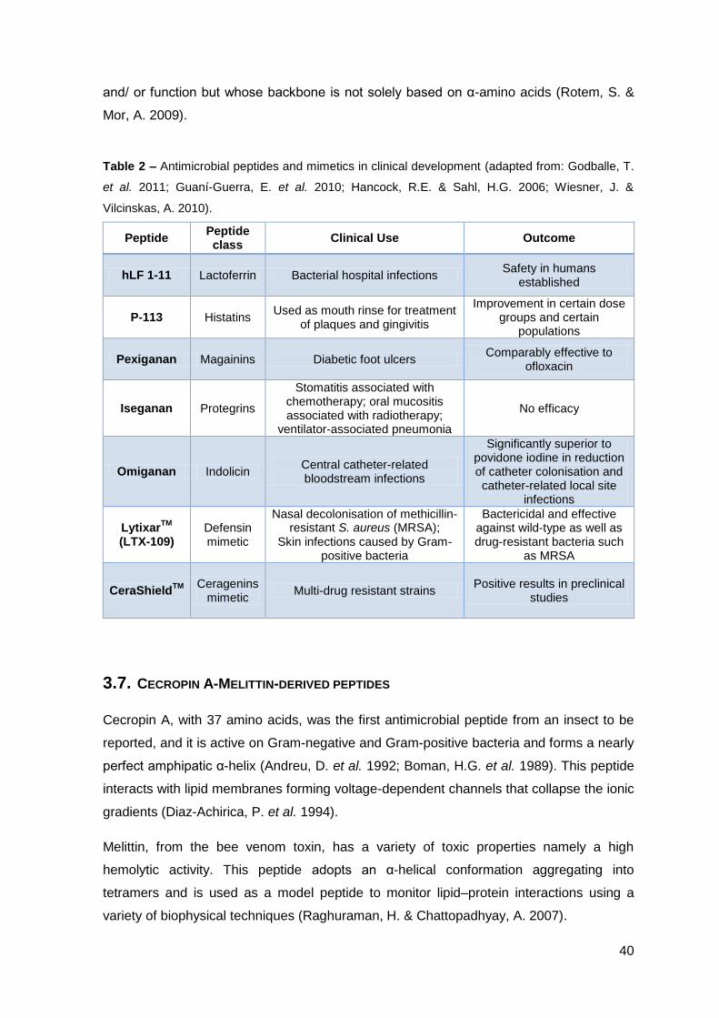
Table 2 – Antimicrobial peptides and mimetics in clinical development (adapted from: Godballe, T.
et al. 2011; Guaní-Guerra, E. et al. 2010; Hancock, R.E. & Sahl, H.G. 2006; Wiesner, J. &
Vilcinskas, A. 2010).
Peptide Peptide class
Clinical Use Outcome
hLF 1-11 Lactoferrin Bacterial hospital infections Safety in humans
established
P-113 Histatins Used as mouth rinse for treatment
of plaques and gingivitis
Improvement in certain dose groups and certain
populations
Pexiganan Magainins Diabetic foot ulcers Comparably effective to
ofloxacin
Iseganan Protegrins
Stomatitis associated with chemotherapy; oral mucositis associated with radiotherapy;
ventilator-associated pneumonia
No efficacy
Omiganan Indolicin Central catheter-related bloodstream infections
Significantly superior to povidone iodine in reduction of catheter colonisation and catheter-related local site
infections
LytixarTM
(LTX-109) Defensin mimetic
Nasal decolonisation of methicillin-resistant S. aureus (MRSA);
Skin infections caused by Gram-positive bacteria
Bactericidal and effective against wild-type as well as drug-resistant bacteria such
as MRSA
CeraShieldTM
Ceragenins
mimetic Multi-drug resistant strains
Positive results in preclinical studies
3.7. CECROPIN A-MELITTIN-DERIVED PEPTIDES
Cecropin A, with 37 amino acids, was the first antimicrobial peptide from an insect to be
reported, and it is active on Gram-negative and Gram-positive bacteria and forms a nearly
perfect amphipatic α-helix (Andreu, D. et al. 1992; Boman, H.G. et al. 1989). This peptide
interacts with lipid membranes forming voltage-dependent channels that collapse the ionic
gradients (Diaz-Achirica, P. et al. 1994).
Melittin, from the bee venom toxin, has a variety of toxic properties namely a high
hemolytic activity. This peptide adopts an α-helical conformation aggregating into
tetramers and is used as a model peptide to monitor lipid–protein interactions using a
variety of biophysical techniques (Raghuraman, H. & Chattopadhyay, A. 2007).
41
These two peptides, cecropin A and melittin, are composed by hydrophilic and
hydrophobic domains separated by a flexible hinge region, and therefore they adopt an α-
helix – hinge – α-helix conformation, the only difference is the reverse polarity. The
hydrophobic region of cecropin A is localized on the C-terminal whereas in melittin is
localized on the N-terminal (Boman, H.G. et al. 1989). In an attempt to obtain antimicrobial
peptides with strong bactericidal activity and lower hemolytic properties, H.G. Boman, et al
in 1989 synthesized for the first time cecropin A-melittin hybrids, and they found that
these hybrids had better antimicrobial properties than the parental compounds (Boman,
H.G. et al. 1989). In particular a hybrid formed by the first 8 amino acids of the cationic
region of cecropin A and the first 18 amino acids from the hydrophobic and nonhemolytic
region of mellitin (CA(1-8)M(1-18)) exhibited a wider spectrum of activity and improved
potency relative to cecropin A without the cytotoxic effects of melittin (Boman, H.G. et al.
1989). In the continuation of this work, D. Andreu et al in 1992 synthesized shorter hybrids
(15 aa) that retained a significant activity when compared to the larger versions of the
hybrid, especially CA(1-7)M(2-9) (Andreu, D. et al. 1992).
The mechanism of action of these hybrids is thought to be membrane disruption due to
the formation of ionic channels (larger hybrids) or disintegration of the membrane due to a
detergent-like action (shorter hybrids) or even the formation of toroidal pores (Abrunhosa,
F. et al. 2005; Andreu, D. et al. 1992; Bastos, M. et al. 2008; Diaz-Achirica, P. et al. 1994).
D-enantiomers of all the hybrids were synthesized and tested whether they required
interaction with receptors to exert their antimicrobial activity. The antimicrobial activity of
the D-enantiomers was quantitatively equivalent to that of the L-enantiomers. This is
interpreted to mean that the peptides do not act by tight interactions with chiral receptors,
enzymes or lipids in the lysis and killing of the pathogens (Merrifield, E.L. et al. 1995;
Wade, D. et al. 1990).
Although the aim of the synthesis of these hybrids was the reduction of hemolytic activity
together with a potent antimicrobial activity, the first goal was not successfully achieved.
The hybrids CA(1-8)M(1-18) and CA(1-7)M(2-9), especially the last one, are able to
interact and disrupt zwitterionic model membranes (composed by phosphatidylcholine -
PC), a model for erythrocytes, and toxicity towards erythrocytes was also described for
them (Abrunhosa, F. et al. 2005; Fernandez-Reyes, M. et al. 2010). In an attempt to
preserve the high antimicrobial activity of CA(1-7)M(2-9) but decreasing the hemolytic
activity, some additional modifications where performed in this hybrid peptide.
Peptide optimization by residue-specific modifications is one of the most effective and
used strategies to obtain more active peptides with lower hemolytic activity. There are
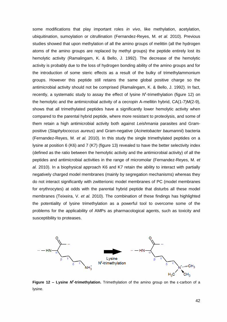
42
some modifications that play important roles in vivo, like methylation, acetylation,
ubiquitination, sumoylation or citrullination (Fernandez-Reyes, M. et al. 2010). Previous
studies showed that upon methylation of all the amino groups of melittin (all the hydrogen
atoms of the amino groups are replaced by methyl groups) the peptide entirely lost its
hemolytic activity (Ramalingam, K. & Bello, J. 1992). The decrease of the hemolytic
activity is probably due to the loss of hydrogen bonding ability of the amino groups and for
the introduction of some steric effects as a result of the bulky of trimethylammonium
groups. However this peptide still retains the same global positive charge so the
antimicrobial activity should not be comprised (Ramalingam, K. & Bello, J. 1992). In fact,
recently, a systematic study to assay the effect of lysine Nε-trimethylation (figure 12) on
the hemolytic and the antimicrobial activity of a cecropin A-mellitin hybrid, CA(1-7)M(2-9),
shows that all trimethylated peptides have a significantly lower hemolytic activity when
compared to the parental hybrid peptide, where more resistant to proteolysis, and some of
them retain a high antimicrobial activity both against Leishmania parasites and Gram-
positive (Staphylococcus aureus) and Gram-negative (Acinetobacter baumannii) bacteria
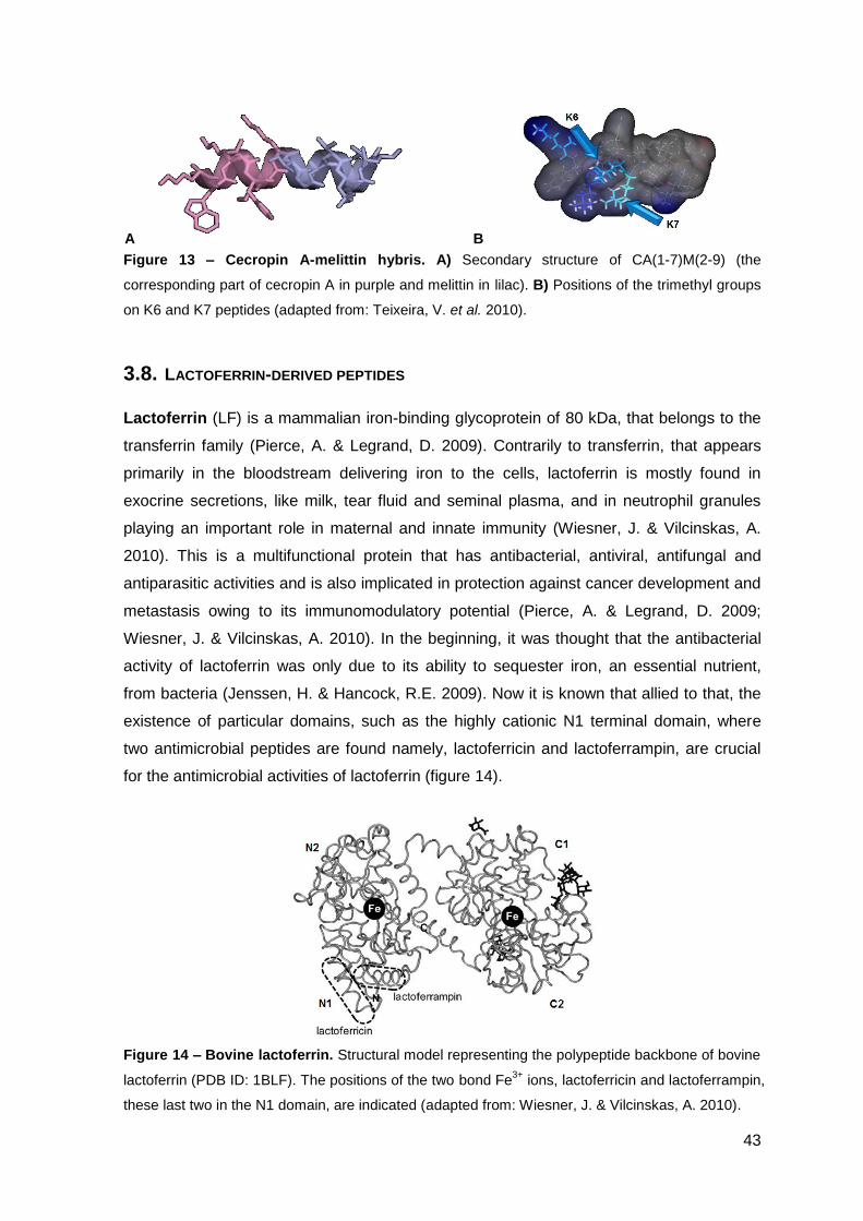
(Fernandez-Reyes, M. et al. 2010). In this study the single trimethylated peptides on a
lysine at position 6 (K6) and 7 (K7) (figure 13) revealed to have the better selectivity index
(defined as the ratio between the hemolytic activity and the antimicrobial activity) of all the
peptides and antimicrobial activities in the range of micromolar (Fernandez-Reyes, M. et
al. 2010). In a biophysical approach K6 and K7 retain the ability to interact with partially
negatively charged model membranes (mainly by segregation mechanisms) whereas they
do not interact significantly with zwitterionic model membranes of PC (model membranes
for erythrocytes) at odds with the parental hybrid peptide that disturbs all these model
membranes (Teixeira, V. et al. 2010). The combination of these findings has highlighted
the potentiality of lysine trimethylation as a powerful tool to overcome some of the
problems for the applicability of AMPs as pharmacological agents, such as toxicity and
susceptibility to proteases.
Figure 12 – Lysine Nε-trimethylation. Trimethylation of the amino group on the ε-carbon of a
lysine.
43
Figure 13 – Cecropin A-melittin hybris. A) Secondary structure of CA(1-7)M(2-9) (the
corresponding part of cecropin A in purple and melittin in lilac). B) Positions of the trimethyl groups
on K6 and K7 peptides (adapted from: Teixeira, V. et al. 2010).
3.8. LACTOFERRIN-DERIVED PEPTIDES
Lactoferrin (LF) is a mammalian iron-binding glycoprotein of 80 kDa, that belongs to the
transferrin family (Pierce, A. & Legrand, D. 2009). Contrarily to transferrin, that appears
primarily in the bloodstream delivering iron to the cells, lactoferrin is mostly found in
exocrine secretions, like milk, tear fluid and seminal plasma, and in neutrophil granules
playing an important role in maternal and innate immunity (Wiesner, J. & Vilcinskas, A.
2010). This is a multifunctional protein that has antibacterial, antiviral, antifungal and
antiparasitic activities and is also implicated in protection against cancer development and
metastasis owing to its immunomodulatory potential (Pierce, A. & Legrand, D. 2009;
Wiesner, J. & Vilcinskas, A. 2010). In the beginning, it was thought that the antibacterial
activity of lactoferrin was only due to its ability to sequester iron, an essential nutrient,
from bacteria (Jenssen, H. & Hancock, R.E. 2009). Now it is known that allied to that, the
existence of particular domains, such as the highly cationic N1 terminal domain, where
two antimicrobial peptides are found namely, lactoferricin and lactoferrampin, are crucial
for the antimicrobial activities of lactoferrin (figure 14).
Figure 14 – Bovine lactoferrin. Structural model representing the polypeptide backbone of bovine
lactoferrin (PDB ID: 1BLF). The positions of the two bond Fe3+
ions, lactoferricin and lactoferrampin,
these last two in the N1 domain, are indicated (adapted from: Wiesner, J. & Vilcinskas, A. 2010).
A B
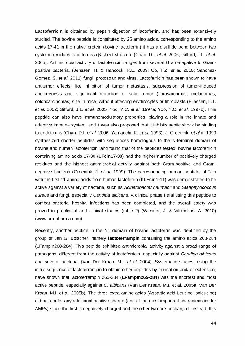
44
Lactoferricin is obtained by pepsin digestion of lactoferrin, and has been extensively
studied. The bovine peptide is constituted by 25 amino acids, corresponding to the amino
acids 17-41 in the native protein (bovine lactoferrin) it has a disulfide bond between two
cysteine residues, and forms a β-sheet structure (Chan, D.I. et al. 2006; Gifford, J.L. et al.
2005). Antimicrobial activity of lactoferricin ranges from several Gram-negative to Gram-
positive bacteria, (Jenssen, H. & Hancock, R.E. 2009; Oo, T.Z. et al. 2010; Sanchez-
Gomez, S. et al. 2011) fungi, protozoan and virus. Lactoferricin has been shown to have
antitumor effects, like inhibition of tumor metastasis, suppression of tumor-induced
angiogenesis and significant reduction of solid tumor (fibrosarcomas, melanomas,
coloncarcinomas) size in mice, without affecting erythrocytes or fibroblasts (Eliassen, L.T.
et al. 2002; Gifford, J.L. et al. 2005; Yoo, Y.C. et al. 1997a; Yoo, Y.C. et al. 1997b). This
peptide can also have immunomodulatory properties, playing a role in the innate and
adaptive immune system, and it was also proposed that it inhibits septic shock by binding
to endotoxins (Chan, D.I. et al. 2006; Yamauchi, K. et al. 1993). J. Groenink, et al in 1999
synthesized shorter peptides with sequences homologous to the N-terminal domain of
bovine and human lactoferricin, and found that of the peptides tested, bovine lactoferricin
containing amino acids 17-30 (LFcin17-30) had the higher number of positively charged
residues and the highest antimicrobial activity against both Gram-positive and Gram-
negative bacteria (Groenink, J. et al. 1999). The corresponding human peptide, hLFcin
with the first 11 amino acids from human lactoferrin (hLFcin1-11) was demonstrated to be
active against a variety of bacteria, such as Acinetobacter baumanii and Stahphylococcus
aureus and fungi, especially Candida albicans. A clinical phase I trial using this peptide to
combat bacterial hospital infections has been completed, and the overall safety was
proved in preclinical and clinical studies (table 2) (Wiesner, J. & Vilcinskas, A. 2010)
(www.am-pharma.com).
Recently, another peptide in the N1 domain of bovine lactoferrin was identified by the
group of Jan G. Bolscher, namely lactoferrampin containing the amino acids 268-284
(LFampin268-284). This peptide exhibited antimicrobial activity against a broad range of
pathogens, different from the activity of lactoferricin, especially against Candida albicans
and several bacteria, (Van Der Kraan, M.I. et al. 2004). Systematic studies, using the
initial sequence of lactoferrampin to obtain other peptides by truncation and/ or extension,
have shown that lactoferrampin 265-284 (LFampin265-284) was the shortest and most
active peptide, especially against C. albicans (Van Der Kraan, M.I. et al. 2005a; Van Der
Kraan, M.I. et al. 2005b). The three extra amino acids (Aspartic acid-Leucine-Isoleucine)
did not confer any additional positive charge (one of the most important characteristics for
AMPs) since the first is negatively charged and the other two are uncharged. Instead, this
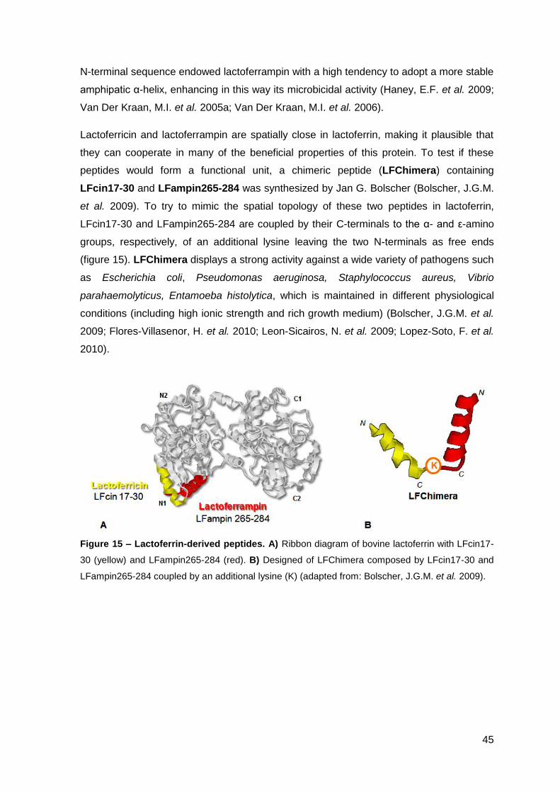
45
N-terminal sequence endowed lactoferrampin with a high tendency to adopt a more stable
amphipatic α-helix, enhancing in this way its microbicidal activity (Haney, E.F. et al. 2009;
Van Der Kraan, M.I. et al. 2005a; Van Der Kraan, M.I. et al. 2006).
Lactoferricin and lactoferrampin are spatially close in lactoferrin, making it plausible that
they can cooperate in many of the beneficial properties of this protein. To test if these
peptides would form a functional unit, a chimeric peptide (LFChimera) containing
LFcin17-30 and LFampin265-284 was synthesized by Jan G. Bolscher (Bolscher, J.G.M.
et al. 2009). To try to mimic the spatial topology of these two peptides in lactoferrin,
LFcin17-30 and LFampin265-284 are coupled by their C-terminals to the α- and ε-amino
groups, respectively, of an additional lysine leaving the two N-terminals as free ends
(figure 15). LFChimera displays a strong activity against a wide variety of pathogens such
as Escherichia coli, Pseudomonas aeruginosa, Staphylococcus aureus, Vibrio
parahaemolyticus, Entamoeba histolytica, which is maintained in different physiological
conditions (including high ionic strength and rich growth medium) (Bolscher, J.G.M. et al.
2009; Flores-Villasenor, H. et al. 2010; Leon-Sicairos, N. et al. 2009; Lopez-Soto, F. et al.
2010).
Figure 15 – Lactoferrin-derived peptides. A) Ribbon diagram of bovine lactoferrin with LFcin17-
30 (yellow) and LFampin265-284 (red). B) Designed of LFChimera composed by LFcin17-30 and
LFampin265-284 coupled by an additional lysine (K) (adapted from: Bolscher, J.G.M. et al. 2009).

46
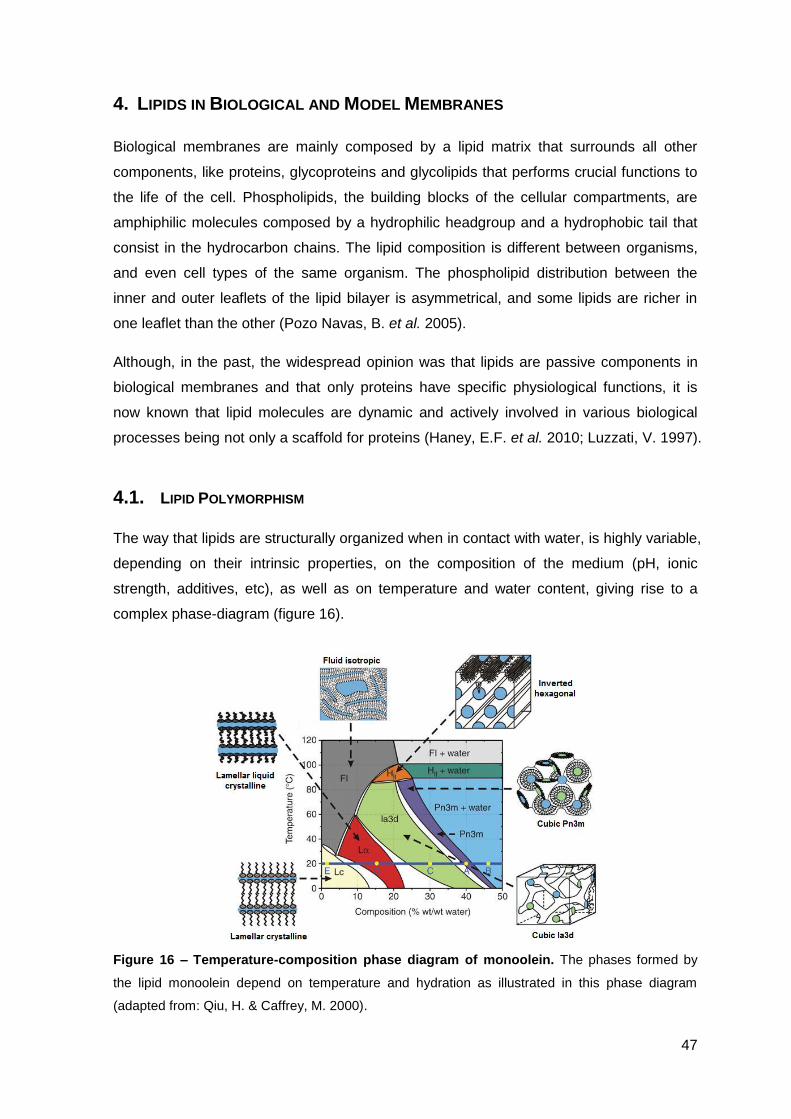
47
4. LIPIDS IN BIOLOGICAL AND MODEL MEMBRANES
Biological membranes are mainly composed by a lipid matrix that surrounds all other
components, like proteins, glycoproteins and glycolipids that performs crucial functions to
the life of the cell. Phospholipids, the building blocks of the cellular compartments, are
amphiphilic molecules composed by a hydrophilic headgroup and a hydrophobic tail that
consist in the hydrocarbon chains. The lipid composition is different between organisms,
and even cell types of the same organism. The phospholipid distribution between the
inner and outer leaflets of the lipid bilayer is asymmetrical, and some lipids are richer in
one leaflet than the other (Pozo Navas, B. et al. 2005).
Although, in the past, the widespread opinion was that lipids are passive components in
biological membranes and that only proteins have specific physiological functions, it is
now known that lipid molecules are dynamic and actively involved in various biological
processes being not only a scaffold for proteins (Haney, E.F. et al. 2010; Luzzati, V. 1997).
4.1. LIPID POLYMORPHISM
The way that lipids are structurally organized when in contact with water, is highly variable,
depending on their intrinsic properties, on the composition of the medium (pH, ionic
strength, additives, etc), as well as on temperature and water content, giving rise to a
complex phase-diagram (figure 16).
Figure 16 – Temperature-composition phase diagram of monoolein. The phases formed by
the lipid monoolein depend on temperature and hydration as illustrated in this phase diagram
(adapted from: Qiu, H. & Caffrey, M. 2000).
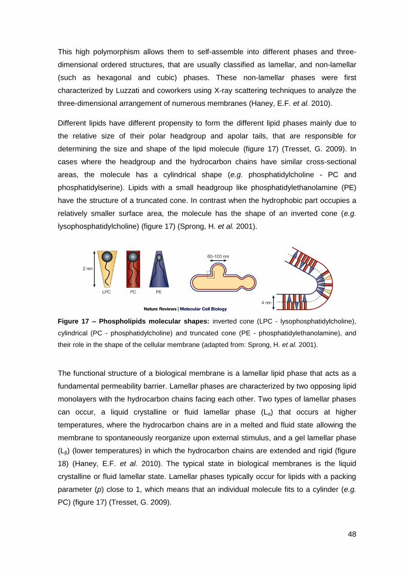
48
This high polymorphism allows them to self-assemble into different phases and three-
dimensional ordered structures, that are usually classified as lamellar, and non-lamellar
(such as hexagonal and cubic) phases. These non-lamellar phases were first
characterized by Luzzati and coworkers using X-ray scattering techniques to analyze the
three-dimensional arrangement of numerous membranes (Haney, E.F. et al. 2010).
Different lipids have different propensity to form the different lipid phases mainly due to
the relative size of their polar headgroup and apolar tails, that are responsible for
determining the size and shape of the lipid molecule (figure 17) (Tresset, G. 2009). In
cases where the headgroup and the hydrocarbon chains have similar cross-sectional
areas, the molecule has a cylindrical shape (e.g. phosphatidylcholine - PC and
phosphatidylserine). Lipids with a small headgroup like phosphatidylethanolamine (PE)
have the structure of a truncated cone. In contrast when the hydrophobic part occupies a
relatively smaller surface area, the molecule has the shape of an inverted cone (e.g.
lysophosphatidylcholine) (figure 17) (Sprong, H. et al. 2001).
Figure 17 – Phospholipids molecular shapes: inverted cone (LPC - lysophosphatidylcholine),
cylindrical (PC - phosphatidylcholine) and truncated cone (PE - phosphatidylethanolamine), and
their role in the shape of the cellular membrane (adapted from: Sprong, H. et al. 2001).
The functional structure of a biological membrane is a lamellar lipid phase that acts as a
fundamental permeability barrier. Lamellar phases are characterized by two opposing lipid
monolayers with the hydrocarbon chains facing each other. Two types of lamellar phases
can occur, a liquid crystalline or fluid lamellar phase (Lα) that occurs at higher
temperatures, where the hydrocarbon chains are in a melted and fluid state allowing the
membrane to spontaneously reorganize upon external stimulus, and a gel lamellar phase
(Lβ) (lower temperatures) in which the hydrocarbon chains are extended and rigid (figure
18) (Haney, E.F. et al. 2010). The typical state in biological membranes is the liquid
crystalline or fluid lamellar state. Lamellar phases typically occur for lipids with a packing
parameter (p) close to 1, which means that an individual molecule fits to a cylinder (e.g.
PC) (figure 17) (Tresset, G. 2009).
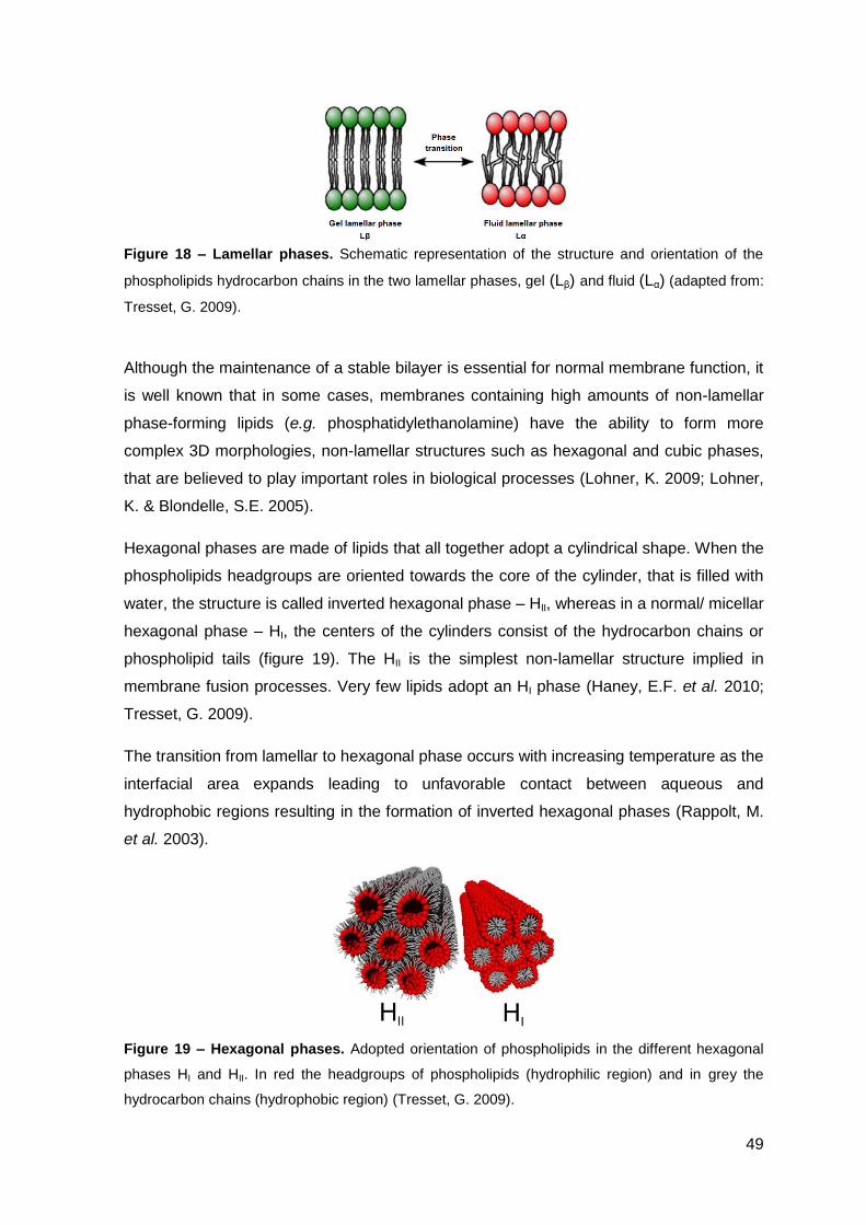
49
Figure 18 – Lamellar phases. Schematic representation of the structure and orientation of the
phospholipids hydrocarbon chains in the two lamellar phases, gel (Lβ) and fluid (Lα) (adapted from:
Tresset, G. 2009).
Although the maintenance of a stable bilayer is essential for normal membrane function, it
is well known that in some cases, membranes containing high amounts of non-lamellar
phase-forming lipids (e.g. phosphatidylethanolamine) have the ability to form more
complex 3D morphologies, non-lamellar structures such as hexagonal and cubic phases,
that are believed to play important roles in biological processes (Lohner, K. 2009; Lohner,
K. & Blondelle, S.E. 2005).
Hexagonal phases are made of lipids that all together adopt a cylindrical shape. When the
phospholipids headgroups are oriented towards the core of the cylinder, that is filled with
water, the structure is called inverted hexagonal phase – HII, whereas in a normal/ micellar
hexagonal phase – HI, the centers of the cylinders consist of the hydrocarbon chains or
phospholipid tails (figure 19). The HII is the simplest non-lamellar structure implied in
membrane fusion processes. Very few lipids adopt an HI phase (Haney, E.F. et al. 2010;
Tresset, G. 2009).
The transition from lamellar to hexagonal phase occurs with increasing temperature as the
interfacial area expands leading to unfavorable contact between aqueous and
hydrophobic regions resulting in the formation of inverted hexagonal phases (Rappolt, M.
et al. 2003).
Figure 19 – Hexagonal phases. Adopted orientation of phospholipids in the different hexagonal
phases HI and HII. In red the headgroups of phospholipids (hydrophilic region) and in grey the
hydrocarbon chains (hydrophobic region) (Tresset, G. 2009).
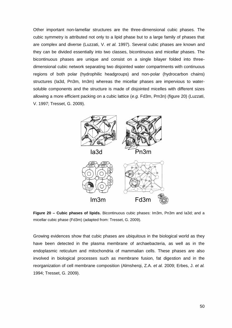
50
Other important non-lamellar structures are the three-dimensional cubic phases. The
cubic symmetry is attributed not only to a lipid phase but to a large family of phases that
are complex and diverse (Luzzati, V. et al. 1997). Several cubic phases are known and
they can be divided essentially into two classes, bicontinuous and micellar phases. The
bicontinuous phases are unique and consist on a single bilayer folded into three-
dimensional cubic network separating two disjointed water compartments with continuous
regions of both polar (hydrophilic headgroups) and non-polar (hydrocarbon chains)
structures (Ia3d, Pn3m, Im3m) whereas the micellar phases are impervious to water-
soluble components and the structure is made of disjointed micelles with different sizes
allowing a more efficient packing on a cubic lattice (e.g. Fd3m, Pm3n) (figure 20) (Luzzati,
V. 1997; Tresset, G. 2009).
Figure 20 – Cubic phases of lipids. Bicontinuous cubic phases: Im3m, Pn3m and Ia3d; and a
micellar cubic phase (Fd3m) (adapted from: Tresset, G. 2009).
Growing evidences show that cubic phases are ubiquitous in the biological world as they
have been detected in the plasma membrane of archaebacteria, as well as in the
endoplasmic reticulum and mitochondria of mammalian cells. These phases are also
involved in biological processes such as membrane fusion, fat digestion and in the
reorganization of cell membrane composition (Almsherqi, Z.A. et al. 2009; Erbes, J. et al.
1994; Tresset, G. 2009).
51
4.2. AMP INTERACTION WITH LIPID MEMBRANES AS REVEALED BY MODEL STUDIES
Regarding the interaction of antimicrobial peptides with membranes it is crucial to
understand if AMPs are capable of altering the phospholipid phase behavior and in that
case to correlate these possible changes in lipid polymorphism with models for the
mechanism of action of AMPs. Using model membranes, namely liposomes (concentric
bilayer vesicles) with different phospholipid compositions, and analyzing them by using
different techniques (e.g. calorimetry, X-ray diffraction, spectroscopy), can give the
desired information that enables the understanding of these changes and how they affect
the overall structure and stability of the membrane. In this way the mechanism of action of
AMPs could be clarified, and lead to a fine-tuning of new peptides in order to obtain a
better therapeutic agent. The induction of cubic phases by AMPs, mostly by lowering the
lamellar to non-lamellar phase boundary of lipid membranes, could mean that the peptide
induces membrane disruption. Without disruption of the membrane, the formation of non-
lamellar phases can also alter the lipid domains in the cellular membrane that are
important for many biological processes such endocytosis (induced by lipid rafts) or even
the activity of membranar proteins that most of the time depend on these domains
culminating into impairment of the membrane function (Lohner, K. 2009; Lohner, K. &
Blondelle, S.E. 2005).
Model membranes can be composed of different phospholipids, and as such their
characteristics can be fine-tunned to the desired purpose of study. The compositions most
widely used for the study of AMPs are: zwitterionic membranes of PC that mimics the
erythrocytes membranes; the combination of PC and PG or PE with PG to try to mimic the
cytoplasmic bacterial membrane that has a global negative charge. Recently much
emphasis has been put on the use of POPE as the major class of phospholipids in the
bacterial membrane whereas the amount of PC is more important in fungus. These are
very simplified models of more complex structures, where the main aspect relies on
changing the charge balance. Nevertheless, it is more and more clear that the AMPs are
sensitive to the actual membrane composition, and thus efforts are also being made in
biophysical studies to vary model membrane composition. More elaborated models can
be used by the introduction of sphingomyelin and sterols for eukaryotic membranes and
cardiolipin for prokaryotic membranes, or even by using membrane extracts of the
bacteria. It should nevertheless be stressed that the main paradigm of model studies is to
“keep it simple”, in order to enable discriminate analysis of interaction effects.

52

53
PART II
OBJECTIVES

54
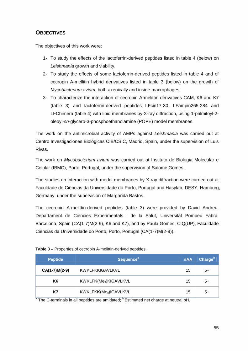
55
OBJECTIVES
The objectives of this work were:
1- To study the effects of the lactoferrin-derived peptides listed in table 4 (below) on
Leishmania growth and viability.
2- To study the effects of some lactoferrin-derived peptides listed in table 4 and of
cecropin A-mellitin hybrid derivatives listed in table 3 (below) on the growth of
Mycobacterium avium, both axenically and inside macrophages.
3- To characterize the interaction of cecropin A-melittin derivatives CAM, K6 and K7
(table 3) and lactoferrin-derived peptides LFcin17-30, LFampin265-284 and
LFChimera (table 4) with lipid membranes by X-ray diffraction, using 1-palmitoyl-2-
oleoyl-sn-glycero-3-phosphoethanolamine (POPE) model membranes.
The work on the antimicrobial activity of AMPs against Leishmania was carried out at
Centro Investigaciones Biológicas CIB/CSIC, Madrid, Spain, under the supervision of Luis
Rivas.
The work on Mycobacterium avium was carried out at Instituto de Biologia Molecular e
Celular (IBMC), Porto, Portugal, under the supervision of Salomé Gomes.
The studies on interaction with model membranes by X-ray diffraction were carried out at
Faculdade de Ciências da Universidade do Porto, Portugal and Hasylab, DESY, Hamburg,
Germany, under the supervision of Margarida Bastos.
The cecropin A-melittin-derived peptides (table 3) were provided by David Andreu,
Departament de Ciències Experimentals i de la Salut, Universitat Pompeu Fabra,
Barcelona, Spain (CA(1-7)M(2-9), K6 and K7), and by Paula Gomes, CIQ(UP), Faculdade
Ciências da Universidade do Porto, Porto, Portugal (CA(1-7)M(2-9)).
Table 3 – Properties of cecropin A-melittin-derived peptides.
Peptide Sequencea #AA Charge
b
CA(1-7)M(2-9) KWKLFKKIGAVLKVL 15 5+
K6 KWKLFK(Me3)KIGAVLKVL 15 5+
K7 KWKLFKK(Me3)IGAVLKVL 15 5+
a The C-terminals in all peptides are amidated;
b Estimated net charge at neutral pH.
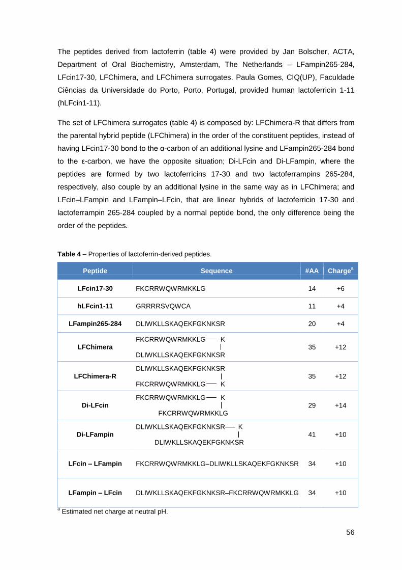
56
The peptides derived from lactoferrin (table 4) were provided by Jan Bolscher, ACTA,
Department of Oral Biochemistry, Amsterdam, The Netherlands – LFampin265-284,
LFcin17-30, LFChimera, and LFChimera surrogates. Paula Gomes, CIQ(UP), Faculdade
Ciências da Universidade do Porto, Porto, Portugal, provided human lactoferricin 1-11
(hLFcin1-11).
The set of LFChimera surrogates (table 4) is composed by: LFChimera-R that differs from
the parental hybrid peptide (LFChimera) in the order of the constituent peptides, instead of
having LFcin17-30 bond to the α-carbon of an additional lysine and LFampin265-284 bond
to the ε-carbon, we have the opposite situation; Di-LFcin and Di-LFampin, where the
peptides are formed by two lactoferricins 17-30 and two lactoferrampins 265-284,
respectively, also couple by an additional lysine in the same way as in LFChimera; and
LFcin–LFampin and LFampin–LFcin, that are linear hybrids of lactoferricin 17-30 and
lactoferrampin 265-284 coupled by a normal peptide bond, the only difference being the
order of the peptides.
Table 4 – Properties of lactoferrin-derived peptides.
Peptide Sequence #AA Chargea
LFcin17-30 FKCRRWQWRMKKLG 14 +6
hLFcin1-11 GRRRRSVQWCA 11 +4
LFampin265-284 DLIWKLLSKAQEKFGKNKSR 20 +4
LFChimera FKCRRWQWRMKKLG K DLIWKLLSKAQEKFGKNKSR
35 +12
LFChimera-R DLIWKLLSKAQEKFGKNKSR FKCRRWQWRMKKLG K
35 +12
Di-LFcin FKCRRWQWRMKKLG K FKCRRWQWRMKKLG
29 +14
Di-LFampin DLIWKLLSKAQEKFGKNKSR K DLIWKLLSKAQEKFGKNKSR
41 +10
LFcin – LFampin FKCRRWQWRMKKLG–DLIWKLLSKAQEKFGKNKSR 34 +10
LFampin – LFcin DLIWKLLSKAQEKFGKNKSR–FKCRRWQWRMKKLG 34 +10
a Estimated net charge at neutral pH.
57
PART III
EXPERIMENTAL METHODS AND
RESULTS

58
59
III.1 – Antimicrobial activity of
lactoferrin-derived peptides against
Leishmania

60
61
1. MATERIAL AND METHODS
Peptides
The lactoferrin-derived peptides tested for their antimicrobial activity against Leishmania
were lactoferricin (LFcin17-30), lactoferrampin (LFampin265-284), LFChimera, and
LFChimera surrogates described on table 4 of page 56.
Leishmania donovani promastigotes
Promastigotes of strain MHOM/SD/00/1S-2D (WT TURCO) of L. donovani were grown at
26 ºC in RPMI 1640 (Gibco) pH=6.8, supplemented with 10% heat-inactivated fetal calf
serum (HIFCS), 25 mM HEPES, 2 mM L-glutamine, 10 μg/mL gentamicin and 20 units/mL
unicilin.
Promastigotes of the WT TURCO-derived 3-Luc strain of L. donovani, which express a
cytoplasmic form of luciferase (Luque-Ortega, J.R. et al. 2001), were grown in the same
conditions as the wild-type strain, except for the addition of 30 μg/mL geneticin to the
medium.
Leishmania pifanoi amastigotes
Amastigotes MHOM/VE/60/Ltrod of L. pifanoi were grown at 32 ºC in M199 pH=7.2,
supplemented with 20% HIFCS, 5% trypticase, 13.8 mM glucose, 76 μM hemin and 48
μg/mL gentamicin.
Leishmania harvesting
The parasites were collected at late exponential growth phase by centrifugation (2000 rpm,
10 min, 4 ºC). The supernatant was discarded carefully and the cells were washed twice
with Hank’s Balanced Salt Solution (HBSS: 137 mM NaCl, 5.3 mM KCl, 0.4 mM KH2PO4,
4.2 mM NaHCO3 and 0.4 mM Na2HPO4, pH=7.2, supplemented with 10 mM D-glucose) at
4ºC. Cells were resuspended in the same buffer at final density of 4x10⁷ cells/mL or 2x10⁷
cells/mL, and kept on ice until use.
62
Determination of antimicrobial activities
Principles
The antimicrobial activity of the different peptides was determined by colorimetric
measurement of the reduction of MTT (3-(4,5-Dimethylthiazol-2-yl)-2,5-
diphenyltetrazolium bromide) to blue insoluble formazan by mitochondrial reductases.
When cell viability is impaired, this leads to a decrease in formazan formation; by the
same rule, when tested on parasite proliferation, the amount of formazan is proportional to
the number of viable cells.
This method can be applied to assess the impact of an antimicrobial peptide on cell
viability (short-term effect) and on the cell proliferation (long-term effect), using the same
sample, and determine parameters like the IC50 (the concentration of AMP that inhibits
50% of MTT reduction at the end of 4h) and the LD50 (lethal concentration that causes
50% inhibition of parasite proliferation at the end of 3 or 5 days, for promastigotes and
amastigotes, respectively) (Luque-Ortega, J.R. & Rivas, L. 2010).
Method
Parasite suspension (4x10⁷ cells/mL) was distributed (60 μL/well) in a sterile 96-well
microplate; 60 μL of the corresponding peptide solution (all the solutions and suspensions
in HBSS + 10 mM glucose) was added to the respective well, and incubated for 4h at 26
ºC or 32 ºC, for promastigotes and amastigotes, respectively.
Long-term effect:
- For promastigotes, after 4h of incubation 20 μL of each well were transferred into a
new 96-well microplate with 130 μL/well of RPMI 1640 without phenol red + 10%
HIFCS, and incubated at 26 ºC, for 3 days, to allow parasite proliferation. At the end of
the incubation, 50 μL of 2 mg/mL MTT solution was added to each well and further
incubated at 26 ºC (protected from light) until colour development. Finally, to stop the
reaction and to dissolve the blue formazan crystals, 50 μL of SDS 10% was added to
each well. Absorbance was read at 600 nm in a microplate reader (Bio-Rad model 680)
fitted with a 595 nm filter.
- For amastigotes, in the end of the incubation, 20 μL of each well were transferred to a
new 96-well microplate with 130 μL/well of M199 + 10% HIFCS, and incubated at 32 ºC,
for 5 days, to allow parasite proliferation. Afterwards, the content of each well was
transfer into 1.5 mL microcentrifuge tubes, and wells were additionally washed with 100
μL of HBSS. The tubes were centrifuged and washed again with 1 mL of HBSS. The
63
pellet was resuspended in 100 μL of 0.5 mg/mL MTT solution, and the whole content of
the tubes was transferred into a new 96-well microplate and MTT reduction carried out
as described previously.
For the short-term assay, after incubation with the respective peptide, 100 μL of MTT (1
mg/mL) solution were added to each well of the plate incubated for 4h, and proceeded as
described before.
The parameters IC50 and LD50 were calculated using the SigmaPlot® software, and the
results represented as the media of at least two independent experiments (conditions
tested in triplicates) with the corresponding standard deviation (SD) indicated.
Real-time monitoring of in vivo changes in intracellular levels of ATP in L. donovani
promastigotes
Principles
A strain of L. donovani promastigote, 3-Luc that express a cytoplasmic form of luciferase
was used to monitor the levels of intracellular ATP. This assay is based on the reaction,
catalyzed by luciferase that converts D-luciferin into D-oxoluciferin in the presence of O2,
with consumption of one molecule of ATP and emission of one foton. To overcome the
low permeability of the cellular membrane to D-luciferin at physiological pH, a caged ester
analogue of luciferin, DMNPE-D-luciferin (D-luciferin, 1-(4,5-dimethoxy-2-nitrophenyl)
ethyl ester), free-permeable to the membrane, was used. Once inside the cytoplasm D-
luciferin is released by intracellular esterases. In this case, luminescence is directly
related to the cytoplasmic levels of free-ATP, the limiting substrate for the reaction
(Luque-Ortega, J.R. et al. 2003).
When the cells are intact, and in the presence of luciferase substrate, cells emit light.
However in the presence of a peptide that induces membrane damages, the loss of ionic
gradient or, for larger lesions, leakage of ATP, with subsequent depletion in the cytoplasm,
will lead to the decrease of the light emitted (Luque-Ortega, J.R. et al. 2003).
Method
Parasites, L. donovani 3-Luc promastigotes, were resuspended at 2x10⁷ cells/mL in HBSS
+ 10 mM glucose, incubated at 26 ºC with DMNPE-D-luciferin (25 μM, final concentration),
and 90 μL of the suspension was distributed into a black 96-well microplate. When
luminescence reached a plateau, 10 μL of the respective peptide was added (t = 0) and
64
the decay on the luminescence was monitored in a BMG Polarstar Galaxy microwell
reader (Öffenburg, Germany) equipped with luminescence setting. Luminescence data
were referred as percentage of luminescence corrected with the corresponding values of
untreated promastigotes.
Plasma membrane permeabilization of L. donovani promastigotes
Principles
The presence of a peptide may induce lesions into the cytoplasmic membrane large
enough to allow entrance of the cationic vital dye, Sytox Green (MW = 600 Da), to which
intact cells are impermeable. Once the dye gains access to the intracellular space, it binds
to nucleic acids with high-affinity, close to irreversibility in practical terms. This enhances
its fluorescence more than 500-fold (Luque-Ortega, J.R. & Rivas, L. 2010). So the
increase in fluorescence stands for the severity of the damage to the parasites
membranes.
Method
Two different assays were performed to assess the entrance of the vital dye:
- First, the parasites L. donovani promastigotes (90 μL at 2x10⁷ cells/mL) were incubated
with Sytox Green (1 μM, final concentration) in HBSS + 10 mM glucose and
fluorescence changes were monitored at λex = 504 nm and λem = 524 nm in a BMG
Polarstar Galaxy microwell reader (Öffenburg, Germany), immediately after adding 10
μL of the corresponding peptide solution (t = 0). The results were expressed as
percentage relative to full permeabilized cells achieved by the addition of 0.1% Triton
X-100.
- Second, to assess the incorporation of the dye into individual parasites, L. donovani
promastigotes and antimicrobial peptides were incubated at the same conditions as
described above, except that the Sytox Green was added only after 4h of incubation at
26 ºC, and the fluorescence was measured by cytofluorometry in a Beckman-Coulter
FC-500 Analyzer. Maximal permeation was achieved by 15 μM of digitonin in order to
preserve parasite shape.
65
AMPs or CPPs: Confocal Microscopy
To evaluate whether the peptides enter the cells without permeabilizing the membrane
and extrapolate if these peptides had an internal target to account for their lethal action, in
other words, whether they behaved or not as Cell-Penetrating Peptides (CPPs) L.
donovani promastigotes and L. pifanoi amastigotes (4x10⁷ cells/mL) were incubated with
the peptides labeled with FITC (fluorescein isothiocyanate) in HBSS + 10 mM glucose for
4h at 26 ºC and 32 ºC for promastigotes and amastigotes, respectively. After 4h of
incubation propidium iodide, PI, (50 μg/mL, final concentration) was added to the cells. A
15 μL drop of each sample was placed in microscope slides previously coated with poly-
lysine (to favour parasite attachment and immobilization) and parasites were observed
and photographed in a Confocal Laser Scanning Microscope Leica TCS – SP2 ABOS.
Ultrastructural alterations: Transmission Electron Microscopy
L. donovani promastigotes or L. pifanoi amastigotes suspension (4x10⁷ cells/mL) in HBSS
+ 10 mM glucose were incubated with the peptides for 4h at 26 ºC or 32 ºC, for
promastigotes and amastigotes, respectively. After the incubation, cells were washed with
PBS and resuspended in a 3% glutaraldehyde solution in PBS for 1-2h, for cell fixation,
and then incubated with 2.5% osmium tetroxide (OsO4) for about the same time (1-2h),
with PBS washes between steps. Cells were dehydrated by successive incubations with
increasing concentrations of ethanol: 30% and 50% for 30 min; 70% overnight; 90% and
100% for 30 min. At the end, parasites were incubated with propylene oxide and
afterwards, propylene oxide was successively replaced by a mix of resins 3A / 7B (resin A:
6.88 mL of Epon-812 epoxy resin + 11.11 mL of dodecenyl succinic anhydride (DDSA) –
resin B: 22 mL of Epon-812 epoxy resin + 19.8 mL of methyl -5-norbornene-2,3-
dicarboxylic anhydride (NMA)). The protocol consisted in successive incubations with
different ratios of a mixture between propylene oxide and the resins: 3:1 and 1:1 for 30
min, 1:3 overnight and 0:1 for 30 min. The inclusion of cells into the resins was completed
with a polymerization step (70ºC for 48h), catalyzed by the addition of 1.8% 2,4,6-
tri(dimethylamino methyl) phenol (DMP30) as accelerator. The samples were distributed
into electron microscopy capsules (Size 3, Agar Scientific, Stanted, England) that were
trimmed and sliced with an ultramicrotome LKB. Sections were placed into formvard-
carbon-coated copper grids and stained with 25 mM lead citrate. The samples were
observed and photographed in a transmission electron microscope JEOL-1230.
66
Colocalization of antimicrobial peptides and L. pifanoi amastigotes in peritoneal
macrophages: Confocal microscopy
Three days (day 0) prior to macrophage harvesting, BALB/c mice were intraperitoneally
(i.p) injected with 1 mL of 10% thioglycolate. On the 3rd day, mice were sacrificed by
cervical dislocation, and macrophages were obtained by washing the peritoneum twice
with warm PBS. 100 000 cells/well were seeded in a Lab-Tek 16-well chamber slide
system, with RPMI 1640 + 10% HIFCS. Macrophages were incubated for 2-3h at 37 ºC to
allow cell attachment to the glass bottom of the chamber slide.
When the macrophages were well attached to the coverslides, the medium was replaced
with a suspension of L. pifanoi amastigotes in fresh medium, at a final ratio
amastigote:macrophage of 3:1 and incubated for at least 4h at 32 ºC. Next, non
phagocytized parasites were removed by washing each well twice with warm PBS, and
substituted for new media (RPMI 1640 + 10% HIFCS) with or without peptides at different
concentrations. To ascertain whether FITC-labeled peptides, have any privileged
subcellular accumulation inside the infected macrophages, including the parasitophorus
vacuole where the Leishmania amastigotes grow, macrophages, previously seeded in a
Lab-Tek 16-well chamber slide system, were incubated with the labeled peptides
overnight. After that, wells were washed twice with warm PBS. The plastic wells, and any
trace of silicone gasket, were removed from the slide carefully according to the
manufacturer instructions. Before covering the slide with a coverslide, a drop of PBS was
placed in the middle of each preparation to avoid cell drying. Finally macrophages were
observed and photographed in a Confocal Laser Scanning Microscope Leica TCS – SP5
ABOS.
Determination of macrophage viability
To measure macrophage viability in the presence of different peptide concentrations,
mouse peritoneal macrophages were seeded (100 000 cells/well) into a 96-well plate.
After adherence to the plate, macrophages were incubated, for 4h in 100 μL of HBSS + 10
mM glucose with or without peptides at different concentrations. After peptide incubation,
100 μL of 1 mg/mL MTT solution was added to each well, and MTT reduction was
measured as described before.
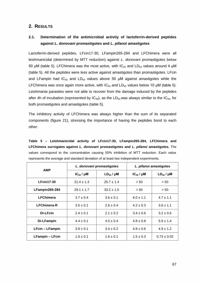
67
2. RESULTS
2.1. Determination of the antimicrobial activity of lactoferrin-derived peptides
against L. donovani promastigotes and L. pifanoi amastigotes
Lactoferrin-derived peptides, LFcin17-30, LFampin265-284 and LFChimera were all
leishmanicidal (determined by MTT reduction) against L. donovani promastigotes below
50 μM (table 5). LFChimera was the most active, with IC50 and LD50 values around 4 μM
(table 5). All the peptides were less active against amastigotes than promastigotes. LFcin
and LFampin had IC50 and LD50 values above 50 μM against amastigotes while the
LFChimera was once again more active, with IC50 and LD50 values below 10 μM (table 5).
Leishmania parasites were not able to recover from the damage induced by the peptides
after 4h of incubation (represented by IC50), as the LD50 was always similar to the IC50, for
both promastigotes and amastigotes (table 5).
The inhibitory activity of LFChimera was always higher than the sum of its separated
components (figure 21), stressing the importance of having the peptides bond to each
other.
Table 5 – Leishmanicidal activity of LFcin17-30, LFampin265-284, LFChimera and
LFChimera surrogates against L. donovani promastigotes and L. pifanoi amastigotes. The
values correspond to the concentration causing 50% inhibition of MTT reduction. Each value
represents the average and standard deviation of at least two independent experiments.
AMP L. donovani promastigotes L. pifanoi amastigotes
IC50 / μM LD50 / μM IC50 / μM LD50 / μM
LFcin17-30 21.4 ± 1.3 25.7 ± 1.4 > 50 > 50
LFampin265-284 29.1 ± 1.7 33.2 ± 1.5 > 50 > 50
LFChimera 3.7 ± 0.4 3.6 ± 0.1 6.0 ± 1.1 4.7 ± 1.1
LFChimera-R 3.5 ± 0.1 2.6 ± 0.4 4.2 ± 0.3 3.6 ± 1.1
Di-LFcin 2.4 ± 0.1 2.1 ± 0.2 3.4 ± 0.6 3.2 ± 0.6
Di-LFampin 4.4 ± 0.1 4.0 ± 0.4 4.8 ± 0.8 5.9 ± 1.4
LFcin – LFampin 3.9 ± 0.1 3.4 ± 0.2 4.8 ± 0.6 4.9 ± 1.2
LFampin – LFcin 1.6 ± 0.1 1.6 ± 0.1 1.5 ± 0.3 0.73 ± 0.02
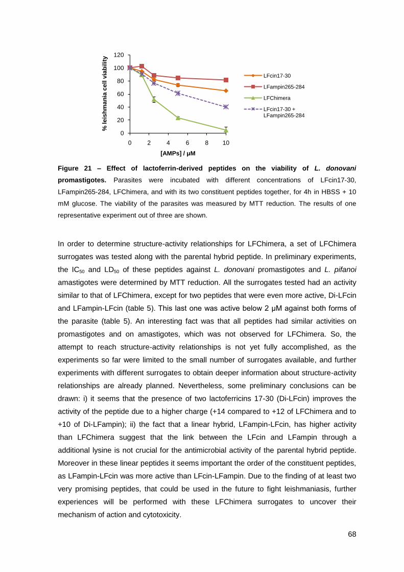
68
0
20
40
60
80
100
120
0 2 4 6 8 10
% l
eis
hm
an
ia c
ell
via
bil
ity
[AMPs] / μM
LFcin17-30
LFampin265-284
LFChimera
LFcin17-30 + LFampin265-284
Figure 21 – Effect of lactoferrin-derived peptides on the viability of L. donovani
promastigotes. Parasites were incubated with different concentrations of LFcin17-30,
LFampin265-284, LFChimera, and with its two constituent peptides together, for 4h in HBSS + 10
mM glucose. The viability of the parasites was measured by MTT reduction. The results of one
representative experiment out of three are shown.
In order to determine structure-activity relationships for LFChimera, a set of LFChimera
surrogates was tested along with the parental hybrid peptide. In preliminary experiments,
the IC50 and LD50 of these peptides against L. donovani promastigotes and L. pifanoi
amastigotes were determined by MTT reduction. All the surrogates tested had an activity
similar to that of LFChimera, except for two peptides that were even more active, Di-LFcin
and LFampin-LFcin (table 5). This last one was active below 2 μM against both forms of
the parasite (table 5). An interesting fact was that all peptides had similar activities on
promastigotes and on amastigotes, which was not observed for LFChimera. So, the
attempt to reach structure-activity relationships is not yet fully accomplished, as the
experiments so far were limited to the small number of surrogates available, and further
experiments with different surrogates to obtain deeper information about structure-activity
relationships are already planned. Nevertheless, some preliminary conclusions can be
drawn: i) it seems that the presence of two lactoferricins 17-30 (Di-LFcin) improves the
activity of the peptide due to a higher charge (+14 compared to +12 of LFChimera and to
+10 of Di-LFampin); ii) the fact that a linear hybrid, LFampin-LFcin, has higher activity
than LFChimera suggest that the link between the LFcin and LFampin through a
additional lysine is not crucial for the antimicrobial activity of the parental hybrid peptide.
Moreover in these linear peptides it seems important the order of the constituent peptides,
as LFampin-LFcin was more active than LFcin-LFampin. Due to the finding of at least two
very promising peptides, that could be used in the future to fight leishmaniasis, further
experiences will be performed with these LFChimera surrogates to uncover their
mechanism of action and cytotoxicity.
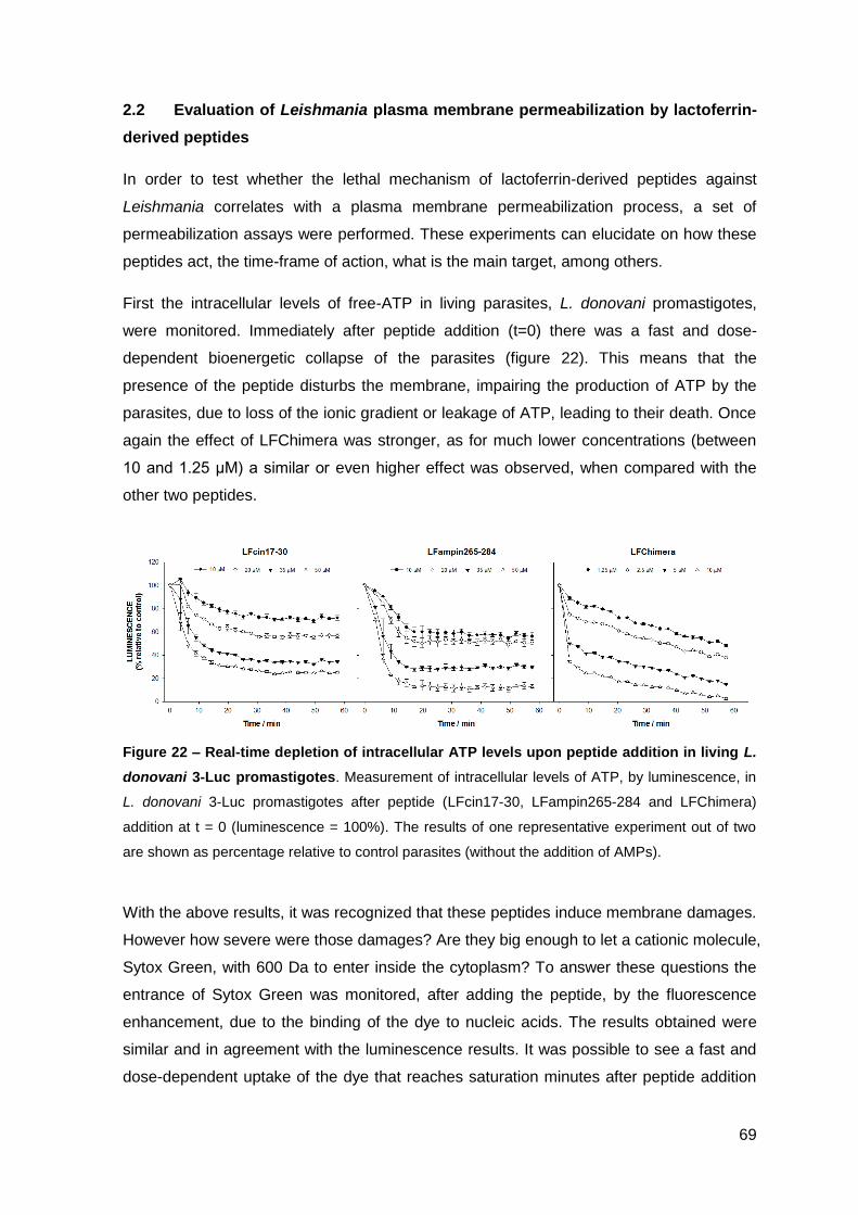
69
2.2 Evaluation of Leishmania plasma membrane permeabilization by lactoferrin-
derived peptides
In order to test whether the lethal mechanism of lactoferrin-derived peptides against
Leishmania correlates with a plasma membrane permeabilization process, a set of
permeabilization assays were performed. These experiments can elucidate on how these
peptides act, the time-frame of action, what is the main target, among others.
First the intracellular levels of free-ATP in living parasites, L. donovani promastigotes,
were monitored. Immediately after peptide addition (t=0) there was a fast and dose-
dependent bioenergetic collapse of the parasites (figure 22). This means that the
presence of the peptide disturbs the membrane, impairing the production of ATP by the
parasites, due to loss of the ionic gradient or leakage of ATP, leading to their death. Once
again the effect of LFChimera was stronger, as for much lower concentrations (between
10 and 1.25 μM) a similar or even higher effect was observed, when compared with the
other two peptides.
Figure 22 – Real-time depletion of intracellular ATP levels upon peptide addition in living L.
donovani 3-Luc promastigotes. Measurement of intracellular levels of ATP, by luminescence, in
L. donovani 3-Luc promastigotes after peptide (LFcin17-30, LFampin265-284 and LFChimera)
addition at t = 0 (luminescence = 100%). The results of one representative experiment out of two
are shown as percentage relative to control parasites (without the addition of AMPs).
With the above results, it was recognized that these peptides induce membrane damages.
However how severe were those damages? Are they big enough to let a cationic molecule,
Sytox Green, with 600 Da to enter inside the cytoplasm? To answer these questions the
entrance of Sytox Green was monitored, after adding the peptide, by the fluorescence
enhancement, due to the binding of the dye to nucleic acids. The results obtained were
similar and in agreement with the luminescence results. It was possible to see a fast and
dose-dependent uptake of the dye that reaches saturation minutes after peptide addition
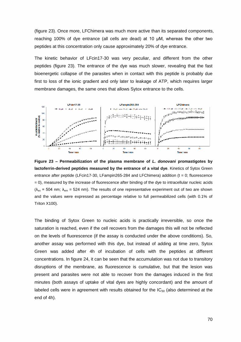
70
(figure 23). Once more, LFChimera was much more active than its separated components,
reaching 100% of dye entrance (all cells are dead) at 10 μM, whereas the other two
peptides at this concentration only cause approximately 20% of dye entrance.
The kinetic behavior of LFcin17-30 was very peculiar, and different from the other
peptides (figure 23). The entrance of the dye was much slower, revealing that the fast
bioenergetic collapse of the parasites when in contact with this peptide is probably due
first to loss of the ionic gradient and only later to leakage of ATP, which requires larger
membrane damages, the same ones that allows Sytox entrance to the cells.
Figure 23 – Permeabilization of the plasma membrane of L. donovani promastigotes by
lactoferrin-derived peptides measured by the entrance of a vital dye. Kinetics of Sytox Green
entrance after peptide (LFcin17-30, LFampin265-284 and LFChimera) addition (t = 0; fluorescence
= 0), measured by the increase of fluorescence after binding of the dye to intracellular nucleic acids
(λex = 504 nm; λem = 524 nm). The results of one representative experiment out of two are shown
and the values were expressed as percentage relative to full permeabilized cells (with 0.1% of
Triton X100).
The binding of Sytox Green to nucleic acids is practically irreversible, so once the
saturation is reached, even if the cell recovers from the damages this will not be reflected
on the levels of fluorescence (if the assay is conducted under the above conditions). So,
another assay was performed with this dye, but instead of adding at time zero, Sytox
Green was added after 4h of incubation of cells with the peptides at different
concentrations. In figure 24, it can be seen that the accumulation was not due to transitory
disruptions of the membrane, as fluorescence is cumulative, but that the lesion was
present and parasites were not able to recover from the damages induced in the first
minutes (both assays of uptake of vital dyes are highly concordant) and the amount of
labeled cells were in agreement with results obtained for the IC50 (also determined at the
end of 4h).
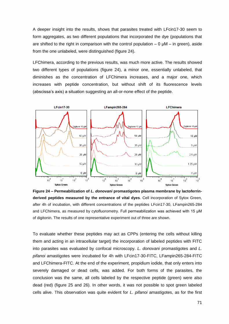
71
A deeper insight into the results, shows that parasites treated with LFcin17-30 seem to
form aggregates, as two different populations that incorporated the dye (populations that
are shifted to the right in comparison with the control population – 0 μM – in green), aside
from the one unlabeled, were distinguished (figure 24).
LFChimera, according to the previous results, was much more active. The results showed
two different types of populations (figure 24), a minor one, essentially unlabeled, that
diminishes as the concentration of LFChimera increases, and a major one, which
increases with peptide concentration, but without shift of its fluorescence levels
(abscissa’s axis) a situation suggesting an all-or-none effect of the peptide.
Figure 24 – Permeabilization of L. donovani promastigotes plasma membrane by lactoferrin-
derived peptides measured by the entrance of vital dyes. Cell incorporation of Sytox Green,
after 4h of incubation, with different concentrations of the peptides LFcin17-30, LFampin265-284
and LFChimera, as measured by cytofluorometry. Full permeabilization was achieved with 15 μM
of digitonin. The results of one representative experiment out of three are shown.
To evaluate whether these peptides may act as CPPs (entering the cells without killing
them and acting in an intracellular target) the incorporation of labeled peptides with FITC
into parasites was evaluated by confocal microscopy. L. donovani promastigotes and L.
pifanoi amastigotes were incubated for 4h with LFcin17-30-FITC, LFampin265-284-FITC
and LFChimera-FITC. At the end of the experiment, propidium iodide, that only enters into
severely damaged or dead cells, was added. For both forms of the parasites, the
conclusion was the same, all cells labeled by the respective peptide (green) were also
dead (red) (figure 25 and 26). In other words, it was not possible to spot green labeled
cells alive. This observation was quite evident for L. pifanoi amastigotes, as for the first
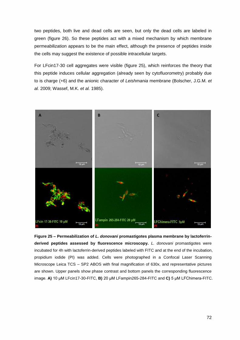
72
two peptides, both live and dead cells are seen, but only the dead cells are labeled in
green (figure 26). So these peptides act with a mixed mechanism by which membrane
permeabilization appears to be the main effect, although the presence of peptides inside
the cells may suggest the existence of possible intracellular targets.
For LFcin17-30 cell aggregates were visible (figure 25), which reinforces the theory that
this peptide induces cellular aggregation (already seen by cytofluorometry) probably due
to is charge (+6) and the anionic character of Leishmania membrane (Bolscher, J.G.M. et
al. 2009; Wassef, M.K. et al. 1985).
Figure 25 – Permeabilization of L. donovani promastigotes plasma membrane by lactoferrin-
derived peptides assessed by fluorescence microscopy. L. donovani promastigotes were
incubated for 4h with lactoferrin-derived peptides labeled with FITC and at the end of the incubation,
propidium iodide (PI) was added. Cells were photographed in a Confocal Laser Scanning
Microscope Leica TCS – SP2 ABOS with final magnification of 630x, and representative pictures
are shown. Upper panels show phase contrast and bottom panels the corresponding fluorescence
image. A) 10 μM LFcin17-30-FITC, B) 20 μM LFampin265-284-FITC and C) 5 μM LFChimera-FITC.
A B C
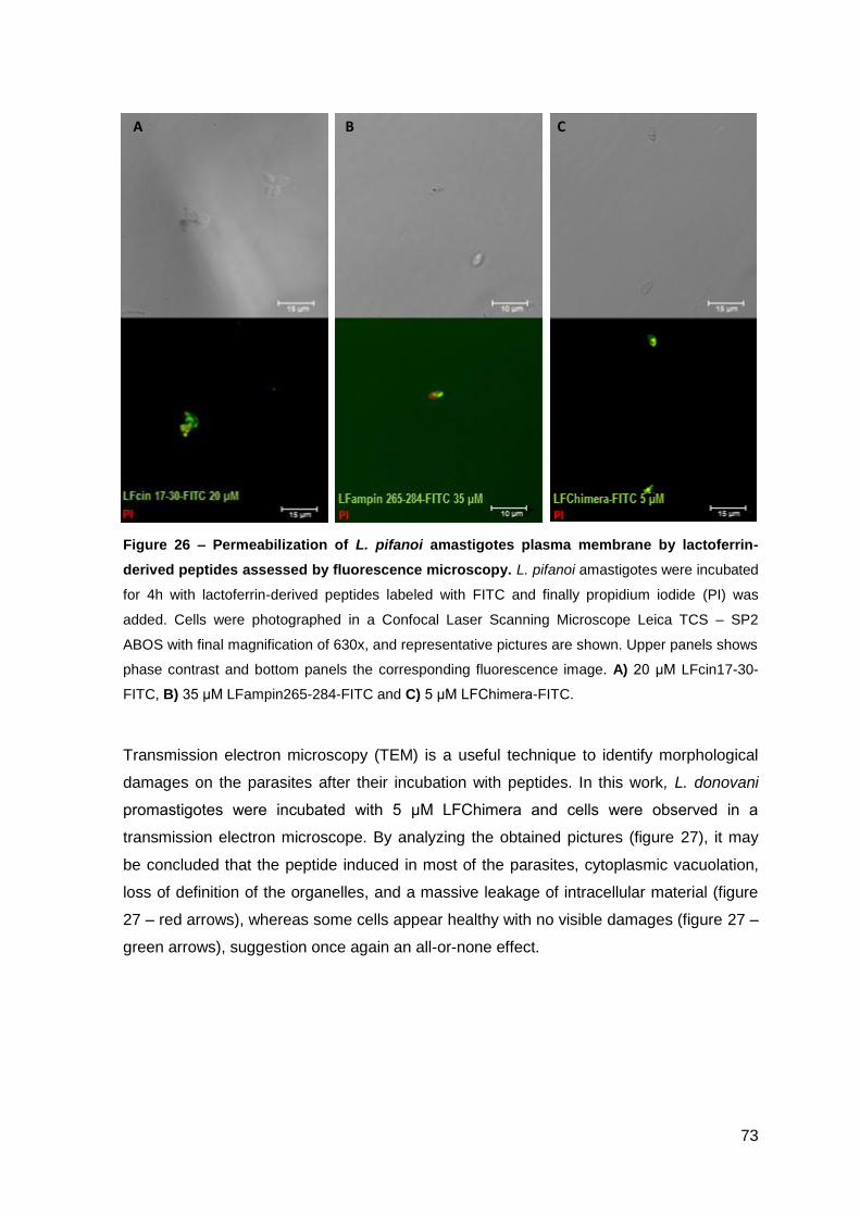
73
Figure 26 – Permeabilization of L. pifanoi amastigotes plasma membrane by lactoferrin-
derived peptides assessed by fluorescence microscopy. L. pifanoi amastigotes were incubated
for 4h with lactoferrin-derived peptides labeled with FITC and finally propidium iodide (PI) was
added. Cells were photographed in a Confocal Laser Scanning Microscope Leica TCS – SP2
ABOS with final magnification of 630x, and representative pictures are shown. Upper panels shows
phase contrast and bottom panels the corresponding fluorescence image. A) 20 μM LFcin17-30-
FITC, B) 35 μM LFampin265-284-FITC and C) 5 μM LFChimera-FITC.
Transmission electron microscopy (TEM) is a useful technique to identify morphological
damages on the parasites after their incubation with peptides. In this work, L. donovani
promastigotes were incubated with 5 μM LFChimera and cells were observed in a
transmission electron microscope. By analyzing the obtained pictures (figure 27), it may
be concluded that the peptide induced in most of the parasites, cytoplasmic vacuolation,
loss of definition of the organelles, and a massive leakage of intracellular material (figure
27 – red arrows), whereas some cells appear healthy with no visible damages (figure 27 –
green arrows), suggestion once again an all-or-none effect.
A B C
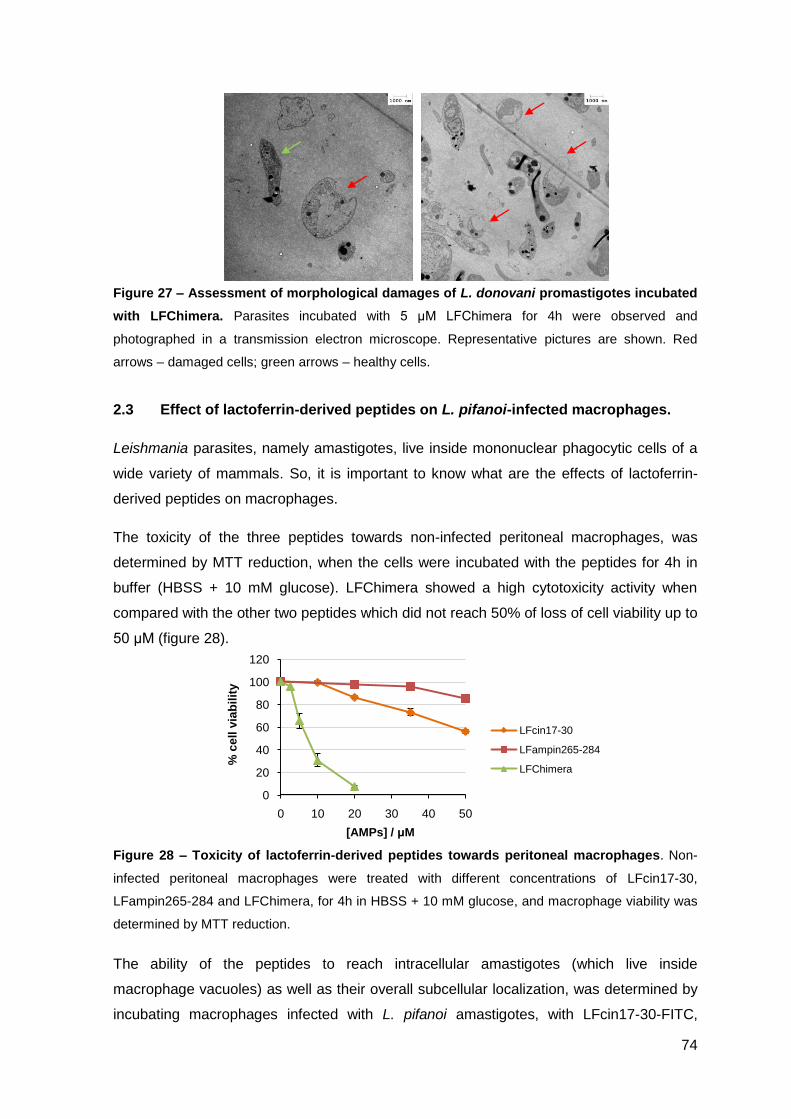
74
0
20
40
60
80
100
120
0 10 20 30 40 50
% c
ell
via
bil
ity
[AMPs] / μM
LFcin17-30
LFampin265-284
LFChimera
Figure 27 – Assessment of morphological damages of L. donovani promastigotes incubated
with LFChimera. Parasites incubated with 5 μM LFChimera for 4h were observed and
photographed in a transmission electron microscope. Representative pictures are shown. Red
arrows – damaged cells; green arrows – healthy cells.
2.3 Effect of lactoferrin-derived peptides on L. pifanoi-infected macrophages.
Leishmania parasites, namely amastigotes, live inside mononuclear phagocytic cells of a
wide variety of mammals. So, it is important to know what are the effects of lactoferrin-
derived peptides on macrophages.
The toxicity of the three peptides towards non-infected peritoneal macrophages, was
determined by MTT reduction, when the cells were incubated with the peptides for 4h in
buffer (HBSS + 10 mM glucose). LFChimera showed a high cytotoxicity activity when
compared with the other two peptides which did not reach 50% of loss of cell viability up to
50 μM (figure 28).
Figure 28 – Toxicity of lactoferrin-derived peptides towards peritoneal macrophages. Non-
infected peritoneal macrophages were treated with different concentrations of LFcin17-30,
LFampin265-284 and LFChimera, for 4h in HBSS + 10 mM glucose, and macrophage viability was
determined by MTT reduction.
The ability of the peptides to reach intracellular amastigotes (which live inside
macrophage vacuoles) as well as their overall subcellular localization, was determined by
incubating macrophages infected with L. pifanoi amastigotes, with LFcin17-30-FITC,
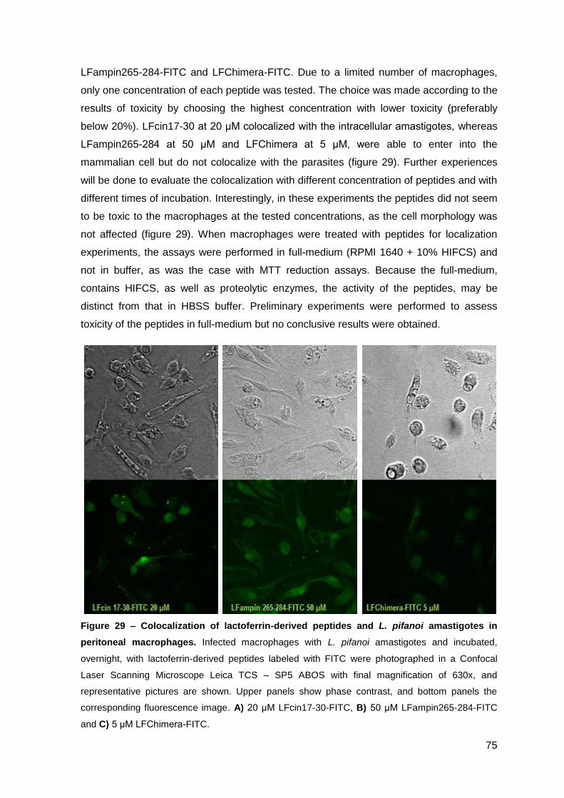
75
LFampin265-284-FITC and LFChimera-FITC. Due to a limited number of macrophages,
only one concentration of each peptide was tested. The choice was made according to the
results of toxicity by choosing the highest concentration with lower toxicity (preferably
below 20%). LFcin17-30 at 20 μM colocalized with the intracellular amastigotes, whereas
LFampin265-284 at 50 μM and LFChimera at 5 μM, were able to enter into the
mammalian cell but do not colocalize with the parasites (figure 29). Further experiences
will be done to evaluate the colocalization with different concentration of peptides and with
different times of incubation. Interestingly, in these experiments the peptides did not seem
to be toxic to the macrophages at the tested concentrations, as the cell morphology was
not affected (figure 29). When macrophages were treated with peptides for localization
experiments, the assays were performed in full-medium (RPMI 1640 + 10% HIFCS) and
not in buffer, as was the case with MTT reduction assays. Because the full-medium,
contains HIFCS, as well as proteolytic enzymes, the activity of the peptides, may be
distinct from that in HBSS buffer. Preliminary experiments were performed to assess
toxicity of the peptides in full-medium but no conclusive results were obtained.
Figure 29 – Colocalization of lactoferrin-derived peptides and L. pifanoi amastigotes in
peritoneal macrophages. Infected macrophages with L. pifanoi amastigotes and incubated,
overnight, with lactoferrin-derived peptides labeled with FITC were photographed in a Confocal
Laser Scanning Microscope Leica TCS – SP5 ABOS with final magnification of 630x, and
representative pictures are shown. Upper panels show phase contrast, and bottom panels the
corresponding fluorescence image. A) 20 μM LFcin17-30-FITC, B) 50 μM LFampin265-284-FITC
and C) 5 μM LFChimera-FITC.

76
77
III.2 – Activity of cecropin A-mellitin-
derived and lactoferrin-derived
peptides against Mycobacterium
avium

78
79
1. MATERIAL AND METHODS
Peptides
In the evaluation of the activity against Mycobacterium avium, the following peptides were
used: cecropin A-melittin derivatives CA(1-7)M(2-9) or CAM, K6 and K7 (described in
table 3, page 55) and lactoferrin-derived peptides hLF1-11, LFcin17-30, LFampin265-284
and LFChimera (described in table 4, page 56).
Bacteria
M. avium strain 2447, smooth transparent variant (SmT) was isolated from an AIDS
patient and provided to us by Dr. F. Portaels (Institute of Tropical Medicine, Antwerp,
Belgium).
Mycobacteria were grown to mid-log phase in Middlebrook 7H9 medium (Difco, Sparks,
MD) containing 0.05% of Tween 80 (Sigma, St. Louis, MO) and 10% of ADC supplement
(Albumin-Dextrose-Catalase) at 37 ºC. Bacteria were harvested by centrifugation, washed
twice with saline containing 0.04% Tween 80, resuspended in the same solution and
briefly sonicated in order to disrupt bacterial clumps. The suspension was stored in
aliquots at -80 ºC until use. Just before use, an aliquot was quickly thawed and diluted to
the appropriate concentration.
Effect of antimicrobial peptides on the viability of M. avium in axenic cultures
Principles
To determine the direct effect of the antimicrobial peptides on M. avium 2447 SmT in
axenic cultures (bacteria growing in liquid medium), three different methods were used.
First the bacterial growth was monitored by optical density at 600 nm. When they were in
the exponential phase of growth, the viability of M. avium was quantified by resazurin
reduction and by colony forming units (CFUs).
Resazurin is a blue oxidation-reduction indicator (Palomino, J.C. et al. 2002) which is non-
fluorescent until it is reduced to the pink colored and highly fluorescent resorufin.
Resazurin reduction can be used to measure cellular viability, as only viable cells can
reduce this dye and the level of resazurin reduction is proportional to the number and
viability of the cells present. This is a simple and non-destructive assay, that can be
80
applied both to adherent and non-adherent cells, and it complements and gives
comparable data with the colony forming units (CFUs) assay (Anoopkumar-Dukie, S. et al.
2005).
Method
M. avium 2447 SmT was grown in Middlebrook 7H9 medium to exponential phase. 107
CFU of bacteria were seeded per well into 96-wells flat bottom plates and incubated with
the peptides in a final volume of 200 μL. Each peptide concentration was tested in
triplicates. The plates were incubated at 37 ºC in a humid atmosphere. At the 6th day of
culture, 10 μL of 2.5 mM resazurin was added to each well and after 24h of incubation the
fluorescence was measured in a Gemini XM plate fluorometer (Molecular Devices) (λex =
560 nm; λem = 590 nm). The results were expressed as percentage of the fluorescence
obtained in control wells without peptides.
% mycobacterial cell viability = relative fluorescence intensity in experimental wells
relative fluorescence intensity in control wells x 100
In some of the assays, after 7 days of incubation, the viability of the bacteria was also
measured by a colony forming units (CFUs) assay (using the same wells as the resazurin
assay). The bacterial suspension in each well was serially diluted in water containing
0.04% Tween 80. The dilutions were plated in Middlebrook 7H10 agar medium (Difco,
Sparks, MD), supplemented with OADC (Oleic acid-Albumin-Dextrose-Catalase) and the
number of colonies was counted after 7 days at 37 ºC. The difference, in terms of log10
CFU/mL between the first and the last day of incubation was designated “log increase”.
log increase CFU/mL = log10 CFU/mL (day 7) – log10 CFU/mL (day 0)
Collection, infection and treatment of Bone Marrow derived Macrophages (BMMØ)
Macrophages were derived from the bone marrow of BALB/c INF-γ knock-out mice. Each
femur and tibia was flushed with 5 mL of Hank’s Balanced Salt Solution (HBSS, Gibco,
Paisley, U.K.). The cell suspension was centrifuged and resuspended in Dulbecco’s
Modified Eagle’s Medium (DMEM, Gibco, Paisley, U.K.) supplemented with 10 mM
glutamine, 10 mM HEPES, 1 mM sodium pyruvate, 10% Fetal Bovine Serum (FBS, Gibco,
Paisley, U.K.) and 10% of L929 cell conditioned medium (LCCM) as a source of
Macrophage Colony Stimulating Factor (M-CSF).
81
Cells were cultured overnight at 37 ºC in a 7% CO2 atmosphere to remove fibroblasts.
Non-adherent cells were collected with cold HBSS medium, washed and seeded at the
concentration of 5x10⁵ cells/well in 24-wells culture plates (Nunc, Gibco). In some cases
LPS-free round glass coverslips were placed at the bottom of culture wells, so that
macrophages could adhere to this coverslip and later be observed by confocal microscopy.
The plates were incubated at 37 ºC in a 7% CO2 atmosphere. Four days after seeding,
10% of LCCM was added to the culture medium and on the 7th day, the medium was
renewed. At day 10 of culture, when the cells are fully differentiated into macrophages,
each well was incubated with 106 CFU of M avium 2447 SmT in DMEM, for 4h at 37 ºC in
a 7% CO2 atmosphere. After incubation, cells were washed several times with warm
HBSS to remove non-internalized bacteria, and re-incubated with new medium, DMEM
with 10% FBS and 10% LCCM, with or without different concentrations of peptides alone
or in combination with different antibiotics. Each condition was tested in triplicates.
After 4 or 7 days of infection, the intracellular growth of M. avium 2447 SmT was
evaluated by colony forming units (CFUs). Macrophages were lysed, using 0.1% saponin.
The bacterial suspensions were serially diluted in water containing 0.04% Tween 80 and
plated in Middlebrook 7H10 agar medium supplemented with OADC. The number of
colonies was counted after 7 days at 37 ºC. The difference, in terms of log10 CFU/well
between the first and the last day of culture was designated “log increase”.
log increase CFU/well = log10 CFU/well (day 4 or 7) – log10 CFU/well (day 0)
Determination of macrophage viability
Principles
The viability of infected macrophages was determined by resazurin reduction (see above)
and by the use of VibrantTM Cytotoxicity Assay Kit V-23111 (Molecular Probes) (both
assays can be performed using the same wells).
The cytotoxicity assay kit monitors the release of the cytosolic enzyme glucose 6-
phosphate dehydrogenase (G6PD) from damaged cells into the surrounding medium. This
enzyme is detected in cell culture supernatants by a two-step enzymatic process that
leads to the reduction of resazurin (figure 30), and the resulting fluorescence signal is
proportional to the amount of G6PD released which in turn correlates with the number of
dead cells in the sample.
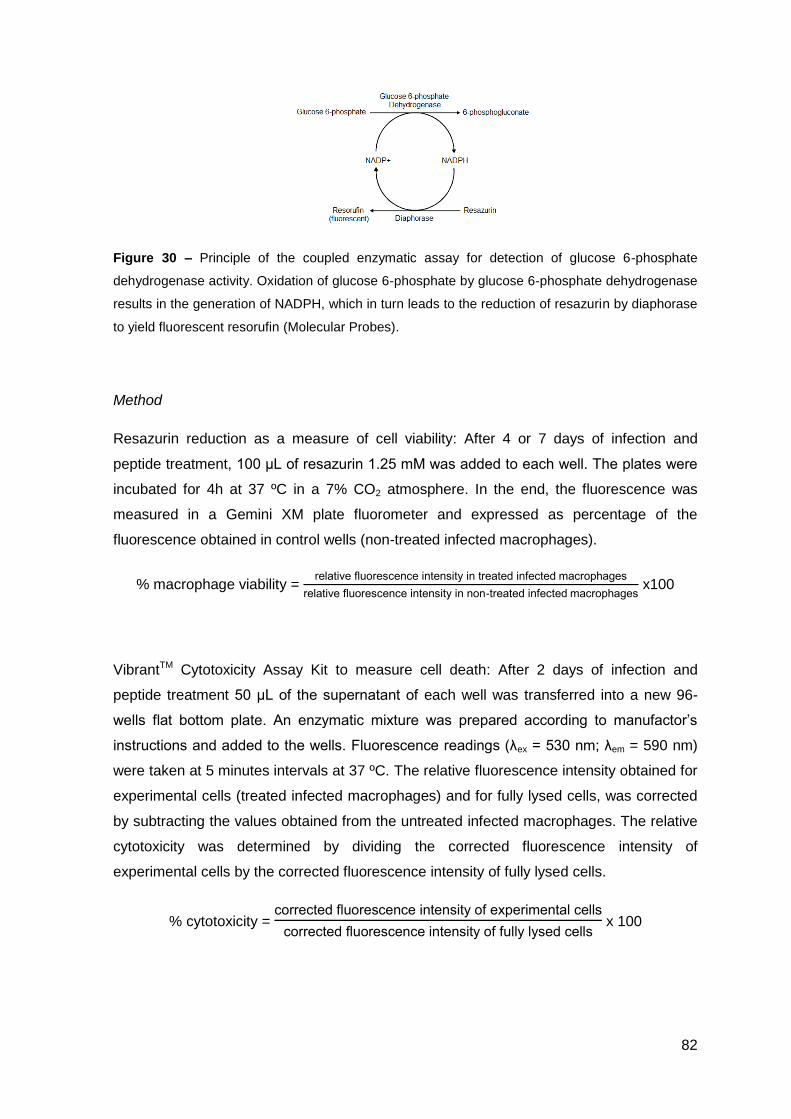
82
Figure 30 – Principle of the coupled enzymatic assay for detection of glucose 6-phosphate
dehydrogenase activity. Oxidation of glucose 6-phosphate by glucose 6-phosphate dehydrogenase
results in the generation of NADPH, which in turn leads to the reduction of resazurin by diaphorase
to yield fluorescent resorufin (Molecular Probes).
Method
Resazurin reduction as a measure of cell viability: After 4 or 7 days of infection and
peptide treatment, 100 μL of resazurin 1.25 mM was added to each well. The plates were
incubated for 4h at 37 ºC in a 7% CO2 atmosphere. In the end, the fluorescence was
measured in a Gemini XM plate fluorometer and expressed as percentage of the
fluorescence obtained in control wells (non-treated infected macrophages).
% macrophage viability = relative fluorescence intensity in treated infected macrophages
relative fluorescence intensity in non-treated infected macrophages x100
VibrantTM Cytotoxicity Assay Kit to measure cell death: After 2 days of infection and
peptide treatment 50 μL of the supernatant of each well was transferred into a new 96-
wells flat bottom plate. An enzymatic mixture was prepared according to manufactor’s
instructions and added to the wells. Fluorescence readings (λex = 530 nm; λem = 590 nm)
were taken at 5 minutes intervals at 37 ºC. The relative fluorescence intensity obtained for
experimental cells (treated infected macrophages) and for fully lysed cells, was corrected
by subtracting the values obtained from the untreated infected macrophages. The relative
cytotoxicity was determined by dividing the corrected fluorescence intensity of
experimental cells by the corrected fluorescence intensity of fully lysed cells.
% cytotoxicity = corrected fluorescence intensity of experimental cells
corrected fluorescence intensity of fully lysed cells x 100
83
Confocal Microscopy
At day 3rd of infection, macrophages that were cultured in glass coverslips, were washed
with warm PBS and fixed with 4% paraformaldehyde (Merck) in PBS supplemented with
120 mM sucrose for 30 min at room temperature. For fluorescence quenching, cells were
incubated with PBS containing 50 mM of NH4Cl, for 10 min. Cells were washed again with
PBS and incubated with Auramine O stain kit (Polysciences, Inc.). Briefly, cells were
incubated with a few drops of Auramine O for 15 minutes (without letting the surface dry
and protected from light), then with fluorescent decolorizer, for 30-60 seconds, and finally
with potassium permanganate for 2 minutes. Between incubations cells were washed with
distilled water. Slides were mounted using Mowiol (Polyscience) and sealed with nail
polish. Macrophages were observed and photographed in a Laser Scanning Confocal
Microscope Leica SP2 AOBS SE (Leica Microsystems, Germany).

84
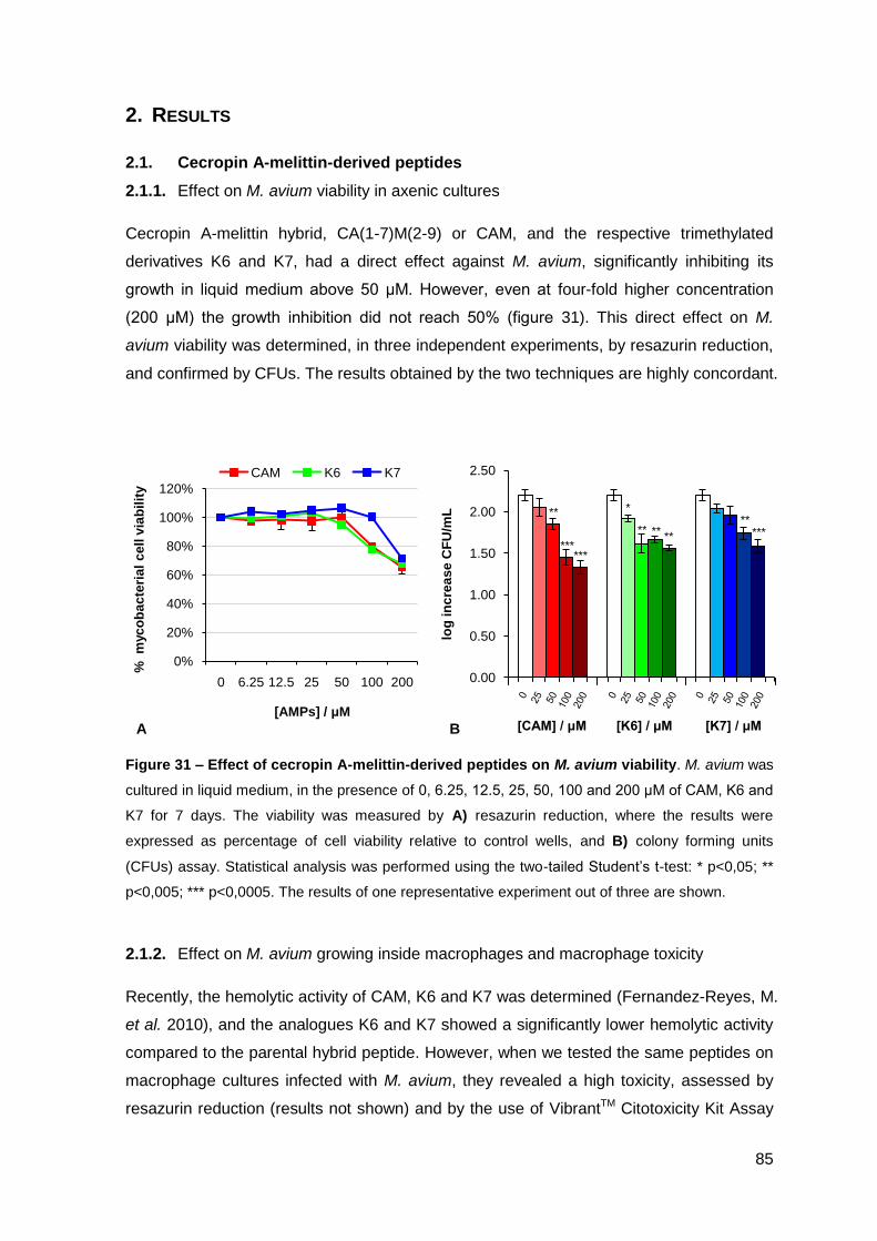
85
0%
20%
40%
60%
80%
100%
120%
0 6.25 12.5 25 50 100 200
%
myc
ob
ac
teri
al
ce
ll v
iab
ilit
y
[AMPs] / μM
CAM K6 K7
0.00
0.50
1.00
1.50
2.00
2.50
1 2 3
log
in
cre
as
e C
FU
/mL **
******
** ** **
***
***
2. RESULTS
2.1. Cecropin A-melittin-derived peptides
2.1.1. Effect on M. avium viability in axenic cultures
Cecropin A-melittin hybrid, CA(1-7)M(2-9) or CAM, and the respective trimethylated
derivatives K6 and K7, had a direct effect against M. avium, significantly inhibiting its
growth in liquid medium above 50 μM. However, even at four-fold higher concentration
(200 μM) the growth inhibition did not reach 50% (figure 31). This direct effect on M.
avium viability was determined, in three independent experiments, by resazurin reduction,
and confirmed by CFUs. The results obtained by the two techniques are highly concordant.
Figure 31 – Effect of cecropin A-melittin-derived peptides on M. avium viability. M. avium was
cultured in liquid medium, in the presence of 0, 6.25, 12.5, 25, 50, 100 and 200 μM of CAM, K6 and
K7 for 7 days. The viability was measured by A) resazurin reduction, where the results were
expressed as percentage of cell viability relative to control wells, and B) colony forming units
(CFUs) assay. Statistical analysis was performed using the two-tailed Student’s t-test: * p<0,05; **
p<0,005; *** p<0,0005. The results of one representative experiment out of three are shown.
2.1.2. Effect on M. avium growing inside macrophages and macrophage toxicity
Recently, the hemolytic activity of CAM, K6 and K7 was determined (Fernandez-Reyes, M.
et al. 2010), and the analogues K6 and K7 showed a significantly lower hemolytic activity
compared to the parental hybrid peptide. However, when we tested the same peptides on
macrophage cultures infected with M. avium, they revealed a high toxicity, assessed by
resazurin reduction (results not shown) and by the use of VibrantTM Citotoxicity Kit Assay
A B [CAM] / μM [K6] / μM [K7] / μM
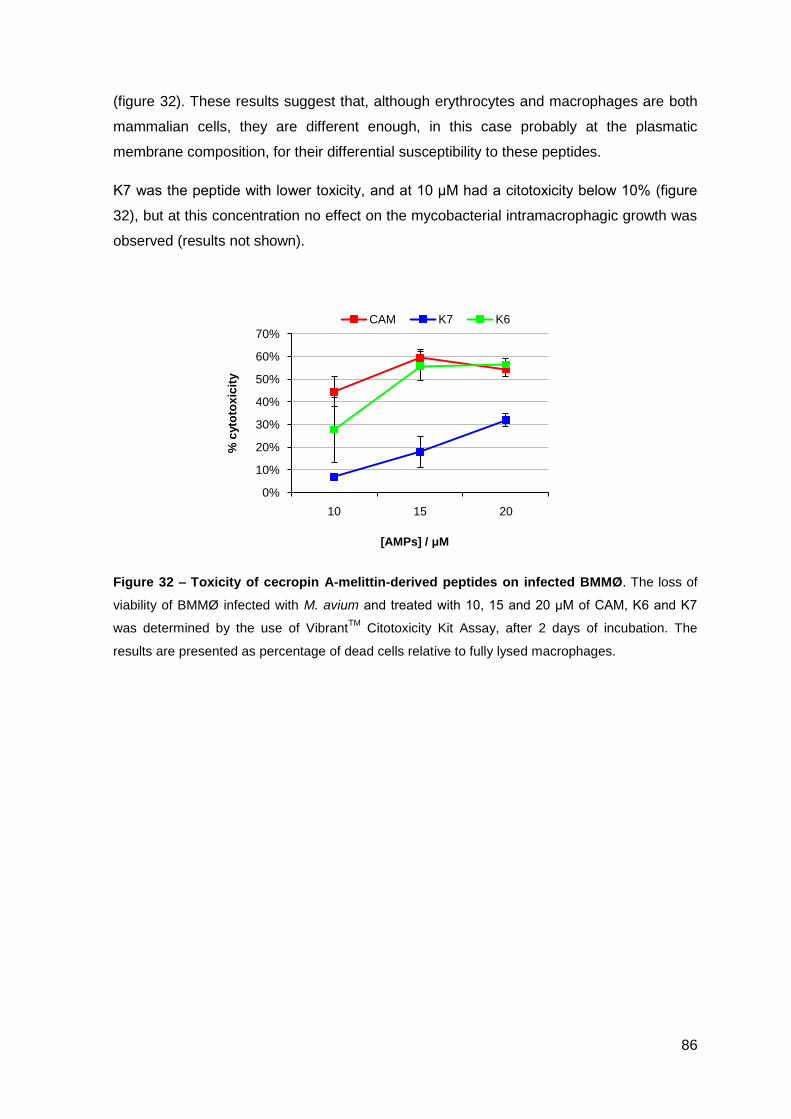
86
0%
10%
20%
30%
40%
50%
60%
70%
10 15 20
% c
yto
tox
icit
y
[AMPs] / μM
CAM K7 K6
(figure 32). These results suggest that, although erythrocytes and macrophages are both
mammalian cells, they are different enough, in this case probably at the plasmatic
membrane composition, for their differential susceptibility to these peptides.
K7 was the peptide with lower toxicity, and at 10 μM had a citotoxicity below 10% (figure
32), but at this concentration no effect on the mycobacterial intramacrophagic growth was
observed (results not shown).
Figure 32 – Toxicity of cecropin A-melittin-derived peptides on infected BMMØ. The loss of
viability of BMMØ infected with M. avium and treated with 10, 15 and 20 μM of CAM, K6 and K7
was determined by the use of VibrantTM
Citotoxicity Kit Assay, after 2 days of incubation. The
results are presented as percentage of dead cells relative to fully lysed macrophages.
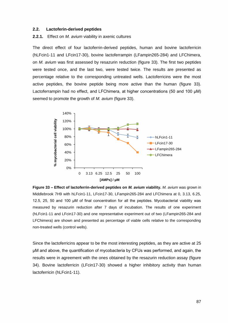
87
0%
20%
40%
60%
80%
100%
120%
140%
0 3.13 6.25 12.5 25 50 100
% m
yc
ob
ac
teri
al
ce
ll v
iab
ilit
y
[AMPs] / μM
hLFcin1-11
LFcin17-30
LFampin265-284
LFChimera
2.2. Lactoferin-derived peptides
2.2.1. Effect on M. avium viability in axenic cultures
The direct effect of four lactoferrin-derived peptides, human and bovine lactoferricin
(hLFcin1-11 and LFcin17-30), bovine lactoferrampin (LFampin265-284) and LFChimera,
on M. avium was first assessed by resazurin reduction (figure 33). The first two peptides
were tested once, and the last two, were tested twice. The results are presented as
percentage relative to the corresponding untreated wells. Lactoferricins were the most
active peptides, the bovine peptide being more active than the human (figure 33).
Lactoferrampin had no effect, and LFChimera, at higher concentrations (50 and 100 μM)
seemed to promote the growth of M. avium (figure 33).
Figure 33 – Effect of lactoferrin-derived peptides on M. avium viability. M. avium was grown in
Middlebrook 7H9 with hLFcin1-11, LFcin17-30, LFampin265-284 and LFChimera at 0, 3.13, 6.25,
12.5, 25, 50 and 100 μM of final concentration for all the peptides. Mycobacterial viability was
measured by resazurin reduction after 7 days of incubation. The results of one experiment
(hLFcin1-11 and LFcin17-30) and one representative experiment out of two (LFampin265-284 and
LFChimera) are shown and presented as percentage of viable cells relative to the corresponding
non-treated wells (control wells).
Since the lactoferricins appear to be the most interesting peptides, as they are active at 25
μM and above, the quantification of mycobacteria by CFUs was performed, and again, the
results were in agreement with the ones obtained by the resazurin reduction assay (figure
34). Bovine lactoferricin (LFcin17-30) showed a higher inhibitory activity than human
lactoferricin (hLFcin1-11).
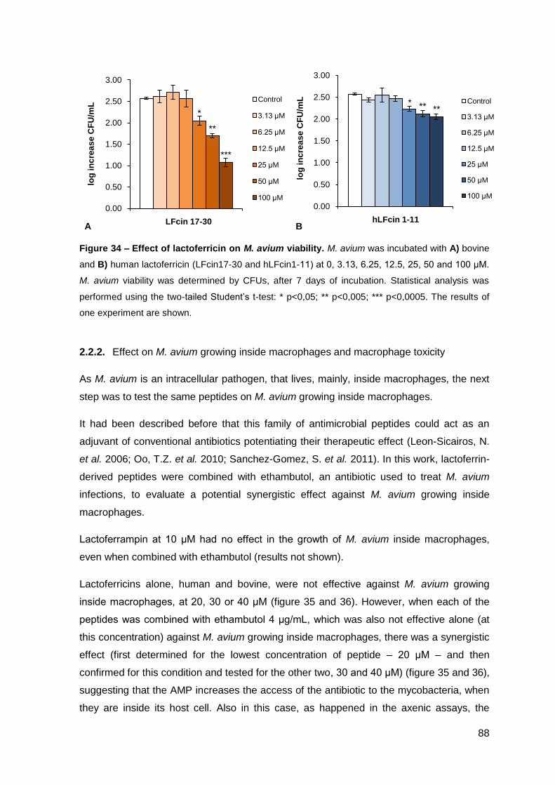
88
0.00
0.50
1.00
1.50
2.00
2.50
3.00
bLFcin 17-30
log
in
cre
as
e C
FU
/mL
Control
3.13 μM
6.25 μM
12.5 μM
25 μM
50 μM
100 μM
**
*
***
0.00
0.50
1.00
1.50
2.00
2.50
3.00
hLFcin 1-11
log
in
cre
as
e C
FU
/mL Control
3.13 μM
6.25 μM
12.5 μM
25 μM
50 μM
100 μM
* ** **
Figure 34 – Effect of lactoferricin on M. avium viability. M. avium was incubated with A) bovine
and B) human lactoferricin (LFcin17-30 and hLFcin1-11) at 0, 3.13, 6.25, 12.5, 25, 50 and 100 μM.
M. avium viability was determined by CFUs, after 7 days of incubation. Statistical analysis was
performed using the two-tailed Student’s t-test: * p<0,05; ** p<0,005; *** p<0,0005. The results of
one experiment are shown.
2.2.2. Effect on M. avium growing inside macrophages and macrophage toxicity
As M. avium is an intracellular pathogen, that lives, mainly, inside macrophages, the next
step was to test the same peptides on M. avium growing inside macrophages.
It had been described before that this family of antimicrobial peptides could act as an
adjuvant of conventional antibiotics potentiating their therapeutic effect (Leon-Sicairos, N.
et al. 2006; Oo, T.Z. et al. 2010; Sanchez-Gomez, S. et al. 2011). In this work, lactoferrin-
derived peptides were combined with ethambutol, an antibiotic used to treat M. avium
infections, to evaluate a potential synergistic effect against M. avium growing inside
macrophages.
Lactoferrampin at 10 μM had no effect in the growth of M. avium inside macrophages,
even when combined with ethambutol (results not shown).
Lactoferricins alone, human and bovine, were not effective against M. avium growing
inside macrophages, at 20, 30 or 40 μM (figure 35 and 36). However, when each of the
peptides was combined with ethambutol 4 μg/mL, which was also not effective alone (at
this concentration) against M. avium growing inside macrophages, there was a synergistic
effect (first determined for the lowest concentration of peptide – 20 μM – and then
confirmed for this condition and tested for the other two, 30 and 40 μM) (figure 35 and 36),
suggesting that the AMP increases the access of the antibiotic to the mycobacteria, when
they are inside its host cell. Also in this case, as happened in the axenic assays, the
A B
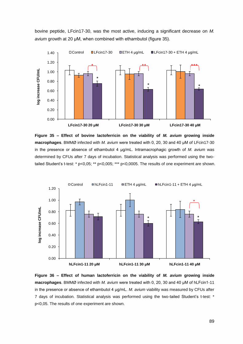
89
0.00
0.20
0.40
0.60
0.80
1.00
1.20
1.40
LFcin17-30 20 μM LFcin17-30 30 μM LFcin17-30 40 μM
log
in
cre
as
e C
FU
/mL
Control LFcin17-30 ETH 4 μg/mL LFcin17-30 + ETH 4 μg/mL
*
**
* ** ***
bovine peptide, LFcin17-30, was the most active, inducing a significant decrease on M.
avium growth at 20 μM, when combined with ethambutol (figure 35).
Figure 35 – Effect of bovine lactoferricin on the viability of M. avium growing inside
macrophages. BMMØ infected with M. avium were treated with 0, 20, 30 and 40 μM of LFcin17-30
in the presence or absence of ethambutol 4 μg/mL. Intramacrophagic growth of M. avium was
determined by CFUs after 7 days of incubation. Statistical analysis was performed using the two-
tailed Student’s t-test: * p<0,05; ** p<0,005; *** p<0,0005. The results of one experiment are shown.
Figure 36 – Effect of human lactoferricin on the viability of M. avium growing inside
macrophages. BMMØ infected with M. avium were treated with 0, 20, 30 and 40 μM of hLFcin1-11
in the presence or absence of ethambutol 4 μg/mL. M. avium viability was measured by CFUs after
7 days of incubation. Statistical analysis was performed using the two-tailed Student’s t-test: *
p<0,05. The results of one experiment are shown.
0.00
0.20
0.40
0.60
0.80
1.00
1.20
hLFcin1-11 20 μM hLFcin1-11 30 μM hLFcin1-11 40 μM
log
in
cre
as
e C
FU
/mL
Control hLFcin1-11 ETH 4 μg/mL hLFcin1-11 + ETH 4 μg/mL
*
*
*
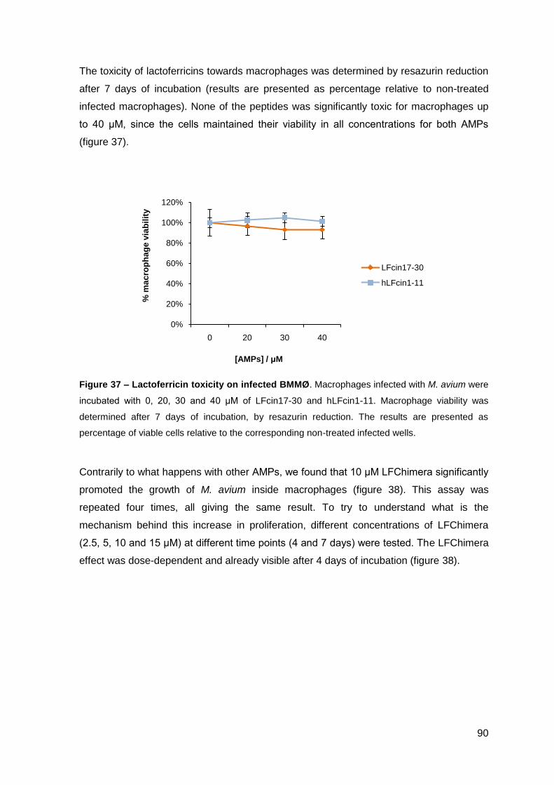
90
0%
20%
40%
60%
80%
100%
120%
0 20 30 40
% m
ac
rop
ha
ge
via
bil
ity
[AMPs] / μM
LFcin17-30
hLFcin1-11
The toxicity of lactoferricins towards macrophages was determined by resazurin reduction
after 7 days of incubation (results are presented as percentage relative to non-treated
infected macrophages). None of the peptides was significantly toxic for macrophages up
to 40 μM, since the cells maintained their viability in all concentrations for both AMPs
(figure 37).
Figure 37 – Lactoferricin toxicity on infected BMMØ. Macrophages infected with M. avium were
incubated with 0, 20, 30 and 40 μM of LFcin17-30 and hLFcin1-11. Macrophage viability was
determined after 7 days of incubation, by resazurin reduction. The results are presented as
percentage of viable cells relative to the corresponding non-treated infected wells.
Contrarily to what happens with other AMPs, we found that 10 μM LFChimera significantly
promoted the growth of M. avium inside macrophages (figure 38). This assay was
repeated four times, all giving the same result. To try to understand what is the
mechanism behind this increase in proliferation, different concentrations of LFChimera
(2.5, 5, 10 and 15 μM) at different time points (4 and 7 days) were tested. The LFChimera
effect was dose-dependent and already visible after 4 days of incubation (figure 38).
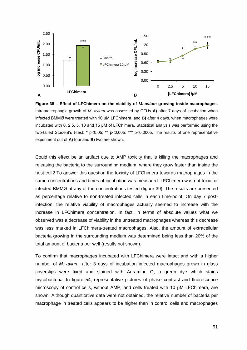
91
0.00
0.30
0.60
0.90
1.20
1.50
0 2.5 5 10 15
log
in
cre
as
e C
FU
/mL
[LFChimera] /μM
*
*****
0.00
0.50
1.00
1.50
2.00
2.50
LFChimera
log
in
cre
as
e C
FU
/mL
Control
LFChimera 10 μM
***
Figure 38 – Effect of LFChimera on the viability of M. avium growing inside macrophages.
Intramacrophagic growth of M. avium was assessed by CFUs A) after 7 days of incubation when
infected BMMØ were treated with 10 μM LFChimera, and B) after 4 days, when macrophages were
incubated with 0, 2.5, 5, 10 and 15 μM of LFChimera. Statistical analysis was performed using the
two-tailed Student’s t-test: * p<0,05; ** p<0,005; *** p<0,0005. The results of one representative
experiment out of A) four and B) two are shown.
Could this effect be an artifact due to AMP toxicity that is killing the macrophages and
releasing the bacteria to the surrounding medium, where they grow faster than inside the
host cell? To answer this question the toxicity of LFChimera towards macrophages in the
same concentrations and times of incubation was measured. LFChimera was not toxic for
infected BMMØ at any of the concentrations tested (figure 39). The results are presented
as percentage relative to non-treated infected cells in each time-point. On day 7 post-
infection, the relative viability of macrophages actually seemed to increase with the
increase in LFChimera concentration. In fact, in terms of absolute values what we
observed was a decrease of viability in the untreated macrophages whereas this decrease
was less marked in LFChimera-treated macrophages. Also, the amount of extracellular
bacteria growing in the surrounding medium was determined being less than 20% of the
total amount of bacteria per well (results not shown).
To confirm that macrophages incubated with LFChimera were intact and with a higher
number of M. avium, after 3 days of incubation infected macrophages grown in glass
coverslips were fixed and stained with Auramine O, a green dye which stains
mycobacteria. In figure 54, representative pictures of phase contrast and fluorescence
microscopy of control cells, without AMP, and cells treated with 10 μM LFChimera, are
shown. Although quantitative data were not obtained, the relative number of bacteria per
macrophage in treated cells appears to be higher than in control cells and macrophages
A B
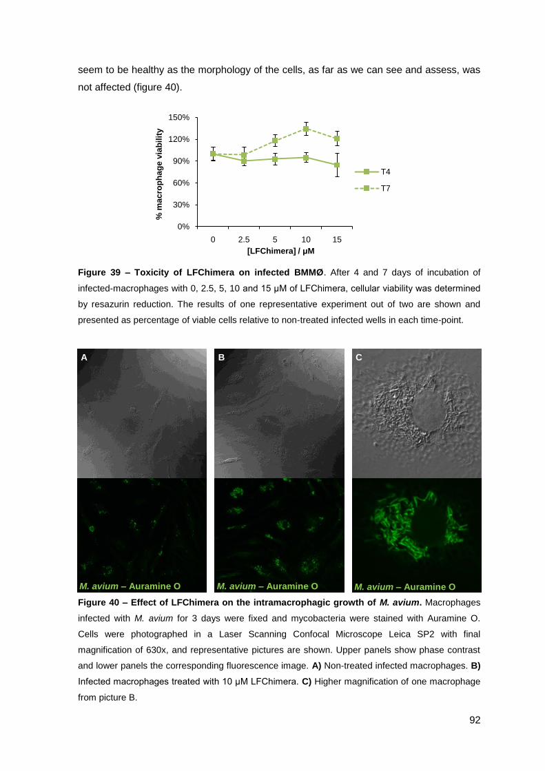
92
0%
30%
60%
90%
120%
150%
0 2.5 5 10 15
% m
ac
rop
ha
ge
via
bil
ity
[LFChimera] / μM
T4
T7
seem to be healthy as the morphology of the cells, as far as we can see and assess, was
not affected (figure 40).
Figure 39 – Toxicity of LFChimera on infected BMMØ. After 4 and 7 days of incubation of
infected-macrophages with 0, 2.5, 5, 10 and 15 μM of LFChimera, cellular viability was determined
by resazurin reduction. The results of one representative experiment out of two are shown and
presented as percentage of viable cells relative to non-treated infected wells in each time-point.
Figure 40 – Effect of LFChimera on the intramacrophagic growth of M. avium. Macrophages
infected with M. avium for 3 days were fixed and mycobacteria were stained with Auramine O.
Cells were photographed in a Laser Scanning Confocal Microscope Leica SP2 with final
magnification of 630x, and representative pictures are shown. Upper panels show phase contrast
and lower panels the corresponding fluorescence image. A) Non-treated infected macrophages. B)
Infected macrophages treated with 10 μM LFChimera. C) Higher magnification of one macrophage
from picture B.
M. avium – Auramine O M. avium – Auramine O M. avium – Auramine O
A B C
93
III.3 – Characterization of peptide-lipid
interaction by X-ray diffraction

94
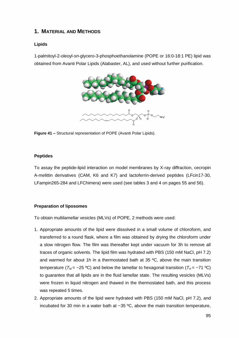
95
1. MATERIAL AND METHODS
Lipids
1-palmitoyl-2-oleoyl-sn-glycero-3-phosphoethanolamine (POPE or 16:0-18:1 PE) lipid was
obtained from Avanti Polar Lipids (Alabaster, AL), and used without further purification.
Figure 41 – Structural representation of POPE (Avanti Polar Lipids).
Peptides
To assay the peptide-lipid interaction on model membranes by X-ray diffraction, cecropin
A-melittin derivatives (CAM, K6 and K7) and lactoferrin-derived peptides (LFcin17-30,
LFampin265-284 and LFChimera) were used (see tables 3 and 4 on pages 55 and 56).
Preparation of liposomes
To obtain multilamellar vesicles (MLVs) of POPE, 2 methods were used:
1. Appropriate amounts of the lipid were dissolved in a small volume of chloroform, and
transferred to a round flask, where a film was obtained by drying the chloroform under
a slow nitrogen flow. The film was thereafter kept under vacuum for 3h to remove all
traces of organic solvents. The lipid film was hydrated with PBS (150 mM NaCl, pH 7.2)
and warmed for about 1h in a thermostated bath at 35 ºC, above the main transition
temperature (TM = ~25 ºC) and below the lamellar to hexagonal transition (TH = ~71 ºC)
to guarantee that all lipids are in the fluid lamellar state. The resulting vesicles (MLVs)
were frozen in liquid nitrogen and thawed in the thermostated bath, and this process
was repeated 5 times.
2. Appropriate amounts of the lipid were hydrated with PBS (150 mM NaCl, pH 7.2), and
incubated for 30 min in a water bath at ~35 ºC, above the main transition temperature,
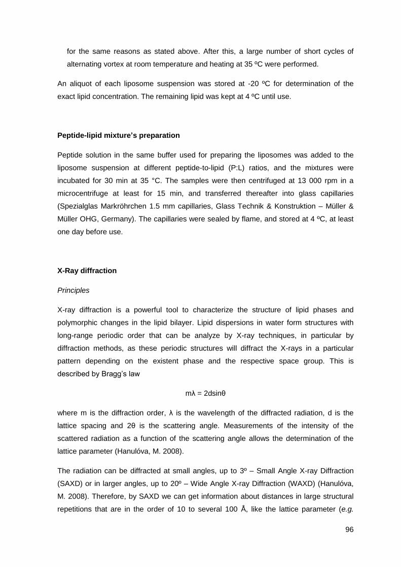
96
for the same reasons as stated above. After this, a large number of short cycles of
alternating vortex at room temperature and heating at 35 ºC were performed.
An aliquot of each liposome suspension was stored at -20 ºC for determination of the
exact lipid concentration. The remaining lipid was kept at 4 ºC until use.
Peptide-lipid mixture’s preparation
Peptide solution in the same buffer used for preparing the liposomes was added to the
liposome suspension at different peptide-to-lipid (P:L) ratios, and the mixtures were
incubated for 30 min at 35 °C. The samples were then centrifuged at 13 000 rpm in a
microcentrifuge at least for 15 min, and transferred thereafter into glass capillaries
(Spezialglas Markröhrchen 1.5 mm capillaries, Glass Technik & Konstruktion – Müller &
Müller OHG, Germany). The capillaries were sealed by flame, and stored at 4 ºC, at least
one day before use.
X-Ray diffraction
Principles
X-ray diffraction is a powerful tool to characterize the structure of lipid phases and
polymorphic changes in the lipid bilayer. Lipid dispersions in water form structures with
long-range periodic order that can be analyze by X-ray techniques, in particular by
diffraction methods, as these periodic structures will diffract the X-rays in a particular
pattern depending on the existent phase and the respective space group. This is
described by Bragg’s law
mλ = 2dsinθ
where m is the diffraction order, λ is the wavelength of the diffracted radiation, d is the
lattice spacing and 2θ is the scattering angle. Measurements of the intensity of the
scattered radiation as a function of the scattering angle allows the determination of the
lattice parameter (Hanulóva, M. 2008).
The radiation can be diffracted at small angles, up to 3º – Small Angle X-ray Diffraction
(SAXD) or in larger angles, up to 20º – Wide Angle X-ray Diffraction (WAXD) (Hanulóva,
M. 2008). Therefore, by SAXD we can get information about distances in large structural
repetitions that are in the order of 10 to several 100 Å, like the lattice parameter (e.g.
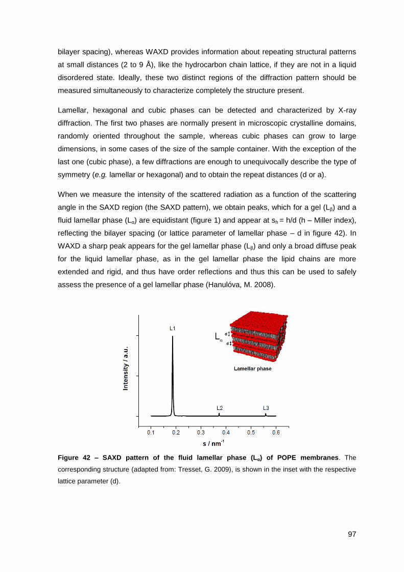
97
bilayer spacing), whereas WAXD provides information about repeating structural patterns
at small distances (2 to 9 Å), like the hydrocarbon chain lattice, if they are not in a liquid
disordered state. Ideally, these two distinct regions of the diffraction pattern should be
measured simultaneously to characterize completely the structure present.
Lamellar, hexagonal and cubic phases can be detected and characterized by X-ray
diffraction. The first two phases are normally present in microscopic crystalline domains,
randomly oriented throughout the sample, whereas cubic phases can grow to large
dimensions, in some cases of the size of the sample container. With the exception of the
last one (cubic phase), a few diffractions are enough to unequivocally describe the type of
symmetry (e.g. lamellar or hexagonal) and to obtain the repeat distances (d or a).
When we measure the intensity of the scattered radiation as a function of the scattering
angle in the SAXD region (the SAXD pattern), we obtain peaks, which for a gel (Lβ) and a
fluid lamellar phase (Lα) are equidistant (figure 1) and appear at sh = h/d (h – Miller index),
reflecting the bilayer spacing (or lattice parameter of lamellar phase – d in figure 42). In
WAXD a sharp peak appears for the gel lamellar phase (Lβ) and only a broad diffuse peak
for the liquid lamellar phase, as in the gel lamellar phase the lipid chains are more
extended and rigid, and thus have order reflections and thus this can be used to safely
assess the presence of a gel lamellar phase (Hanulóva, M. 2008).
Figure 42 – SAXD pattern of the fluid lamellar phase (Lα) of POPE membranes. The
corresponding structure (adapted from: Tresset, G. 2009), is shown in the inset with the respective
lattice parameter (d).
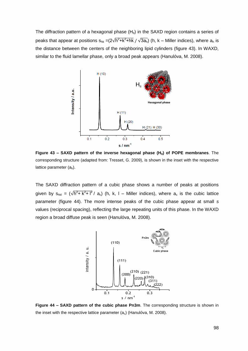
98
The diffraction pattern of a hexagonal phase (HII) in the SAXD region contains a series of
peaks that appear at positions shk =(2 h2+k2+hk 3ah) (h, k – Miller indices), where ah is
the distance between the centers of the neighboring lipid cylinders (figure 43). In WAXD,
similar to the fluid lamellar phase, only a broad peak appears (Hanulóva, M. 2008).
Figure 43 – SAXD pattern of the inverse hexagonal phase (HII) of POPE membranes. The
corresponding structure (adapted from: Tresset, G. 2009), is shown in the inset with the respective
lattice parameter (ah).
The SAXD diffraction pattern of a cubic phase shows a number of peaks at positions
given by shkl = ( h2+ k2+ l2 / ac) (h, k, l – Miller indices), where ac is the cubic lattice
parameter (figure 44). The more intense peaks of the cubic phase appear at small s
values (reciprocal spacing), reflecting the large repeating units of this phase. In the WAXD
region a broad diffuse peak is seen (Hanulóva, M. 2008).
Figure 44 – SAXD pattern of the cubic phase Pn3m. The corresponding structure is shown in
the inset with the respective lattice parameter (ac) (Hanulóva, M. 2008).
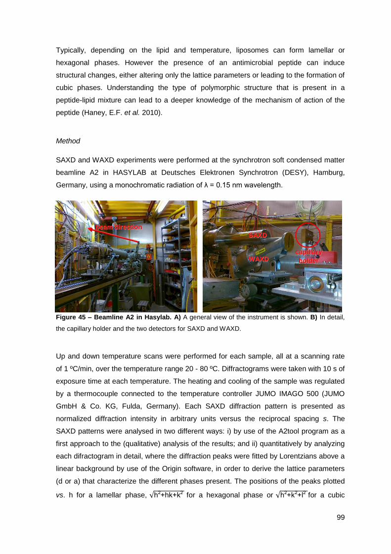
99
Typically, depending on the lipid and temperature, liposomes can form lamellar or
hexagonal phases. However the presence of an antimicrobial peptide can induce
structural changes, either altering only the lattice parameters or leading to the formation of
cubic phases. Understanding the type of polymorphic structure that is present in a
peptide-lipid mixture can lead to a deeper knowledge of the mechanism of action of the
peptide (Haney, E.F. et al. 2010).
Method
SAXD and WAXD experiments were performed at the synchrotron soft condensed matter
beamline A2 in HASYLAB at Deutsches Elektronen Synchrotron (DESY), Hamburg,
Germany, using a monochromatic radiation of λ = 0.15 nm wavelength.
Figure 45 – Beamline A2 in Hasylab. A) A general view of the instrument is shown. B) In detail,
the capillary holder and the two detectors for SAXD and WAXD.
Up and down temperature scans were performed for each sample, all at a scanning rate
of 1 ºC/min, over the temperature range 20 - 80 ºC. Diffractograms were taken with 10 s of
exposure time at each temperature. The heating and cooling of the sample was regulated
by a thermocouple connected to the temperature controller JUMO IMAGO 500 (JUMO
GmbH & Co. KG, Fulda, Germany). Each SAXD diffraction pattern is presented as
normalized diffraction intensity in arbitrary units versus the reciprocal spacing s. The
SAXD patterns were analysed in two different ways: i) by use of the A2tool program as a
first approach to the (qualitative) analysis of the results; and ii) quantitatively by analyzing
each difractogram in detail, where the diffraction peaks were fitted by Lorentzians above a
linear background by use of the Origin software, in order to derive the lattice parameters
(d or a) that characterize the different phases present. The positions of the peaks plotted
vs. h for a lamellar phase, h2+hk+k2 for a hexagonal phase or h2+k2+l2 for a cubic
bbeeaamm ddiirreeccttiioonn
ccaappiillllaarryy
hhoollddeerr
SSAAXXDD
WWAAXXDD
100
phase (h, k, l – Miller indices), should fall on a straight line, passing through the origin
(0,0), and the lattice parameter d, ah or ac, for lamellar, hexagonal and cubic phase,
respectively, is given by the inverse of the slope. WAXD measurements were used to
verify the existence of the gel phase (Lβ) (not shown).
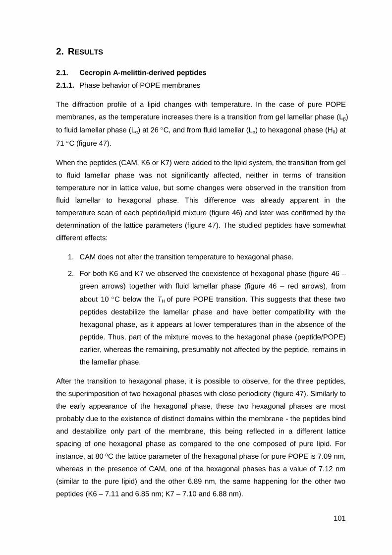
101
2. RESULTS
2.1. Cecropin A-melittin-derived peptides
2.1.1. Phase behavior of POPE membranes
The diffraction profile of a lipid changes with temperature. In the case of pure POPE
membranes, as the temperature increases there is a transition from gel lamellar phase (Lβ)
to fluid lamellar phase (Lα) at 26 C, and from fluid lamellar (Lα) to hexagonal phase (HII) at
71 C (figure 47).
When the peptides (CAM, K6 or K7) were added to the lipid system, the transition from gel
to fluid lamellar phase was not significantly affected, neither in terms of transition
temperature nor in lattice value, but some changes were observed in the transition from
fluid lamellar to hexagonal phase. This difference was already apparent in the
temperature scan of each peptide/lipid mixture (figure 46) and later was confirmed by the
determination of the lattice parameters (figure 47). The studied peptides have somewhat
different effects:
1. CAM does not alter the transition temperature to hexagonal phase.
2. For both K6 and K7 we observed the coexistence of hexagonal phase (figure 46 –
green arrows) together with fluid lamellar phase (figure 46 – red arrows), from
about 10 C below the TH of pure POPE transition. This suggests that these two
peptides destabilize the lamellar phase and have better compatibility with the
hexagonal phase, as it appears at lower temperatures than in the absence of the
peptide. Thus, part of the mixture moves to the hexagonal phase (peptide/POPE)
earlier, whereas the remaining, presumably not affected by the peptide, remains in
the lamellar phase.
After the transition to hexagonal phase, it is possible to observe, for the three peptides,
the superimposition of two hexagonal phases with close periodicity (figure 47). Similarly to
the early appearance of the hexagonal phase, these two hexagonal phases are most
probably due to the existence of distinct domains within the membrane - the peptides bind
and destabilize only part of the membrane, this being reflected in a different lattice
spacing of one hexagonal phase as compared to the one composed of pure lipid. For
instance, at 80 ºC the lattice parameter of the hexagonal phase for pure POPE is 7.09 nm,
whereas in the presence of CAM, one of the hexagonal phases has a value of 7.12 nm
(similar to the pure lipid) and the other 6.89 nm, the same happening for the other two
peptides (K6 – 7.11 and 6.85 nm; K7 – 7.10 and 6.88 nm).
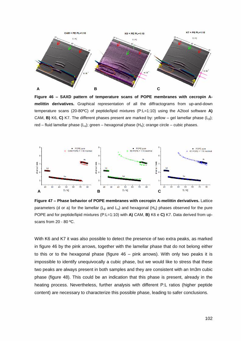
102
Figure 46 – SAXD pattern of temperature scans of POPE membranes with cecropin A-
melittin derivatives. Graphical representation of all the diffractograms from up-and-down
temperature scans (20-80ºC) of peptide/lipid mixtures (P:L=1:10) using the A2tool software A)
CAM, B) K6, C) K7. The different phases present are marked by: yellow – gel lamellar phase (Lβ);
red – fluid lamellar phase (Lα); green – hexagonal phase (HII); orange circle – cubic phases.
Figure 47 – Phase behavior of POPE membranes with cecropin A-melittin derivatives. Lattice
parameters (d or a) for the lamellar (Lβ and Lα) and hexagonal (HII) phases observed for the pure
POPE and for peptide/lipid mixtures (P:L=1:10) with A) CAM, B) K6 e C) K7. Data derived from up-
scans from 20 - 80 ºC.
With K6 and K7 it was also possible to detect the presence of two extra peaks, as marked
in figure 46 by the pink arrows, together with the lamellar phase that do not belong either
to this or to the hexagonal phase (figure 46 – pink arrows). With only two peaks it is
impossible to identify unequivocally a cubic phase, but we would like to stress that these
two peaks are always present in both samples and they are consistent with an Im3m cubic
phase (figure 48). This could be an indication that this phase is present, already in the
heating process. Nevertheless, further analysis with different P:L ratios (higher peptide
content) are necessary to characterize this possible phase, leading to safer conclusions.
A B C
A B C
103
y = 0.0932xR² = 1
0.00
0.10
0.20
0.30
0.40
0 1 2 3 4s
/ n
m-1
(h2+k2+l2)1/2
Im3m
Figure 48 – Evidence for a cubic Im3m phase induced by K6 and K7 in POPE membranes. A
plot of s = f ( h2+k2+l2 ) for the observed reflections at 23 ºC that do not fit with lamellar or
hexagonal phase.
In the cooling process, the temperature scan indicated, with all the peptides, the presence
of structures of higher symmetry (several peaks observed at small s values) (figure 46 –
orange circle), most probably a cubic phase. Further analysis was thus performed with
these down-scans, to try to determine what was the phase present. At this moment, we
chose to analyze in detail for all peptides the diffraction patterns at one temperature,
namely at 50 ºC (on cooling) (tables 6-8, figure 49). The diffraction patterns are
compatible with the coexistence of two bicontinuous cubic phases, of Im3m and Pn3m
symmetry, and a fluid lamellar phase. It should be stressed that in some cases, due to
very few reflections (less than 4-5) it is not possible to make a safe assignment to a
particular lattice. For the parental hybrid peptide, CAM, the Pn3m phase is better defined
as compared with the Im3m phase, as more peaks are observed for the first one. With the
trimethylated peptides (K6 and K7) more peaks corresponding to an Im3m phase are
visible, especially at higher s values. The lattice parameters determined have similar
values between the same phases present in the three peptide/lipid mixtures (tables 6-8).
For example the fluid lamellar phase (Lα) present in each of the three peptide/lipid
mixtures have similar values between them (d CAM = 5.24 nm; d K6 = 5.28 nm; d K7 = 5.23
nm).
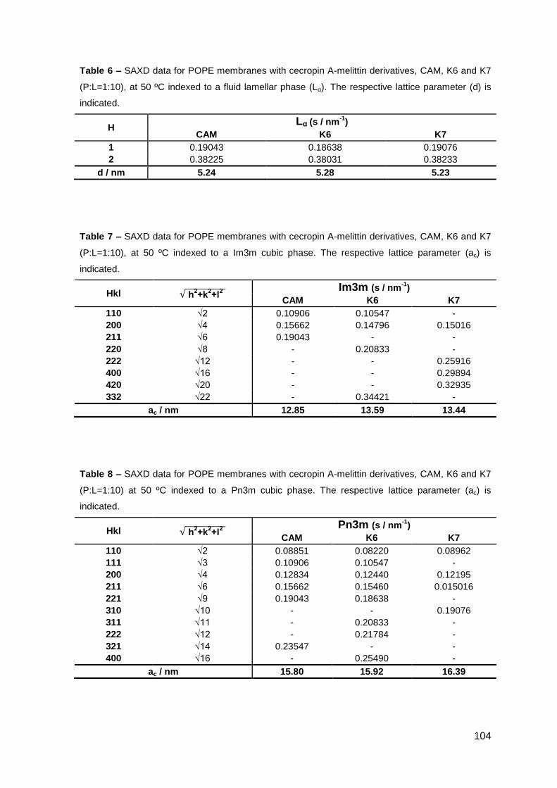
104
Table 6 – SAXD data for POPE membranes with cecropin A-melittin derivatives, CAM, K6 and K7
(P:L=1:10), at 50 ºC indexed to a fluid lamellar phase (Lα). The respective lattice parameter (d) is
indicated.
H Lα (s / nm
-1)
CAM K6 K7
1 0.19043 0.18638 0.19076
2 0.38225 0.38031 0.38233
d / nm 5.24 5.28 5.23
Table 7 – SAXD data for POPE membranes with cecropin A-melittin derivatives, CAM, K6 and K7
(P:L=1:10), at 50 ºC indexed to a Im3m cubic phase. The respective lattice parameter (ac) is
indicated.
Hkl h
l
Im3m (s / nm-1
) CAM K6 K7
110 √2 0.10906 0.10547 -
200 √4 0.15662 0.14796 0.15016
211 √6 0.19043 - -
220 √8 - 0.20833 -
222 √12 - - 0.25916
400 √16 - - 0.29894
420 √20 - - 0.32935
332 √22 - 0.34421 -
ac / nm 12.85 13.59 13.44
Table 8 – SAXD data for POPE membranes with cecropin A-melittin derivatives, CAM, K6 and K7
(P:L=1:10) at 50 ºC indexed to a Pn3m cubic phase. The respective lattice parameter (ac) is
indicated.
Hkl h
l
Pn3m (s / nm-1
) CAM K6 K7
110 √2 0.08851 0.08220 0.08962
111 √3 0.10906 0.10547 -
200 √4 0.12834 0.12440 0.12195
211 √6 0.15662 0.15460 0.015016
221 √9 0.19043 0.18638 -
310 √10 - - 0.19076
311 √11 - 0.20833 -
222 √12 - 0.21784 -
321 √14 0.23547 - -
400 √16 - 0.25490 -
ac / nm 15.80 15.92 16.39
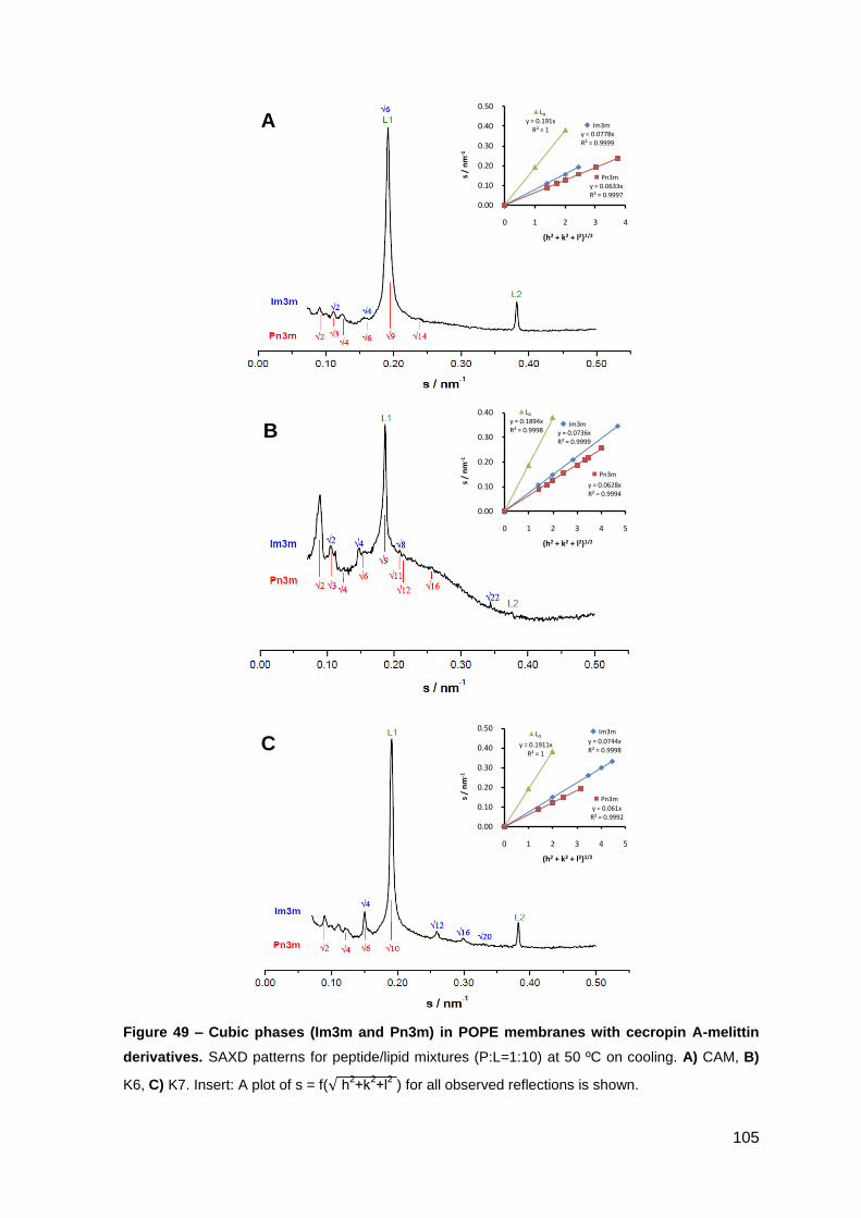
105
Figure 49 – Cubic phases (Im3m and Pn3m) in POPE membranes with cecropin A-melittin
derivatives. SAXD patterns for peptide/lipid mixtures (P:L=1:10) at 50 ºC on cooling. A) CAM, B)
K6, C) K7. Insert: A plot of s = f( h2+k
2+l
2 ) for all observed reflections is shown.
A
B
C
y = 0.191xR² = 1
y = 0.0778xR² = 0.9999
y = 0.0633xR² = 0.9997
0.00
0.10
0.20
0.30
0.40
0.50
0 1 2 3 4
s /
nm
-1
(h2 + k2 + l2)1/2
▲Lα
Pn3m
Im3m
y = 0.1894xR² = 0.9998 y = 0.0736x
R² = 0.9999
y = 0.0628xR² = 0.9994
0.00
0.10
0.20
0.30
0.40
0 1 2 3 4 5
s /
nm
-1
(h2 + k2 + l2)1/2
▲Lα
Pn3m
Im3m
y = 0.1911xR² = 1
y = 0.0744xR² = 0.9998
y = 0.061xR² = 0.9992
0.00
0.10
0.20
0.30
0.40
0.50
0 1 2 3 4 5
s /
nm
-1
(h2 + k2 + l2)1/2
▲Lα
Pn3m
Im3m
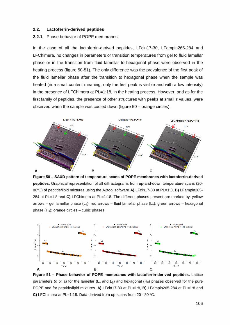
106
A B C
2.2. Lactoferrin-derived peptides
2.2.1. Phase behavior of POPE membranes
In the case of all the lactoferrin-derived peptides, LFcin17-30, LFampin265-284 and
LFChimera, no changes in parameters or transition temperatures from gel to fluid lamellar
phase or in the transition from fluid lamellar to hexagonal phase were observed in the
heating process (figure 50-51). The only difference was the prevalence of the first peak of
the fluid lamellar phase after the transition to hexagonal phase when the sample was
heated (in a small content meaning, only the first peak is visible and with a low intensity)
in the presence of LFChimera at PL=1:18, in the heating process. However, and as for the
first family of peptides, the presence of other structures with peaks at small s values, were
observed when the sample was cooled down (figure 50 – orange circles).
Figure 50 – SAXD pattern of temperature scans of POPE membranes with lactoferrin-derived
peptides. Graphical representation of all diffractograms from up-and-down temperature scans (20-
80ºC) of peptide/lipid mixtures using the A2tool software A) LFcin17-30 at PL=1:8, B) LFampin265-
284 at PL=1:8 and C) LFChimera at PL=1:18. The different phases present are marked by: yellow
arrows – gel lamellar phase (Lβ); red arrows – fluid lamellar phase (Lα); green arrows – hexagonal
phase (HII); orange circles – cubic phases.
Figure 51 – Phase behavior of POPE membranes with lactoferrin-derived peptides. Lattice
parameters (d or a) for the lamellar (Lα and Lβ) and hexagonal (HII) phases observed for the pure
POPE and for peptide/lipid mixtures. A) LFcin17-30 at PL=1:8, B) LFampin265-284 at PL=1:8 and
C) LFChimera at PL=1:18. Data derived from up-scans from 20 - 80 ºC.
A B C
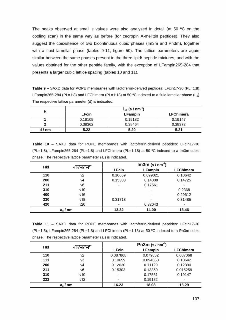
107
The peaks observed at small s values were also analyzed in detail (at 50 ºC on the
cooling scan) in the same way as before (for cecropin A-melittin peptides). They also
suggest the coexistence of two bicontinuous cubic phases (Im3m and Pn3m), together
with a fluid lamellar phase (tables 9-11; figure 50). The lattice parameters are again
similar between the same phases present in the three lipid/ peptide mixtures, and with the
values obtained for the other peptide family, with the exception of LFampin265-284 that
presents a larger cubic lattice spacing (tables 10 and 11).
Table 9 – SAXD data for POPE membranes with lactoferrin-derived peptides: LFcin17-30 (PL=1:8),
LFampin265-284 (PL=1:8) and LFChimera (PL=1:18) at 50 ºC indexed to a fluid lamellar phase (Lα).
The respective lattice parameter (d) is indicated.
H Lα (s / nm
-1)
LFcin LFampin LFChimera
1 0.19105 0.19182 0.19147
2 0.38362 0.38464 0.38372
d / nm 5.22 5.20 5.21
Table 10 – SAXD data for POPE membranes with lactoferrin-derived peptides: LFcin17-30
(PL=1:8), LFampin265-284 (PL=1:8) and LFChimera (PL=1:18) at 50 ºC indexed to a Im3m cubic
phase. The respective lattice parameter (ac) is indicated.
Hkl h
l
Im3m (s / nm-1
) LFcin LFampin LFChimera
110 √2 0.10659 0.099021 0.10642
200 √4 0.15303 0.14008 0.14725
211 √6 - 0.17561 -
310 √10 - - 0.2368
400 √16 - - 0.29612
330 √18 0.31718 - 0.31485
420 √20 - 0.32043 -
ac / nm 13.32 14.00 13.46
Table 11 – SAXD data for POPE membranes with lactoferrin-derived peptides: LFcin17-30
(PL=1:8), LFampin265-284 (PL=1:8) and LFChimera (PL=1:18) at 50 ºC indexed to a Pn3m cubic
phase. The respective lattice parameter (ac) is indicated.
Hkl h
l
Pn3m (s / nm-1
) LFcin LFampin LFChimera
110 √2 0.087868 0.079632 0.087068
111 √3 0.10659 0.094663 0.10642
200 √4 0.12030 0.11129 0.12390
211 √6 0.15303 0.13350 0.015259
310 √10 - 0.17561 0.19147
222 √12 - 0.19182 -
ac / nm 16.23 18.08 16.29
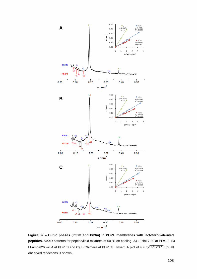
108
y = 0.1918xR² = 1
y = 0.0743xR² = 0.9999
y = 0.0614xR² = 0.9994
0.00
0.10
0.20
0.30
0.40
0.50
0 1 2 3 4 5
s /
nm
-1
(h2 + k2 + l2)1/2
▲Lα
Pn3m
Im3m
y = 0.1922xR² = 1
y = 0.0714xR² = 0.9998
y = 0.0553xR² = 0.9997
0.00
0.10
0.20
0.30
0.40
0.50
0 1 2 3 4 5
s /
nm
-1
(h2 + k2 + l2)1/2
▲Lα
Pn3m
Im3m
y = 0.1917xR² = 1
y = 0.0751xR² = 0.9998
y = 0.0616xR² = 0.999
0.00
0.10
0.20
0.30
0.40
0.50
0 1 2 3 4 5
s /
nm
-1
(h2 + k2 + l2)1/2
▲Lα
Pn3m
Im3m
Figure 52 – Cubic phases (Im3m and Pn3m) in POPE membranes with lactoferrin-derived
peptides. SAXD patterns for peptide/lipid mixtures at 50 ºC on cooling. A) LFcin17-30 at PL=1:8, B)
LFampin265-284 at PL=1:8 and C) LFChimera at PL=1:18. Insert: A plot of s = f( h2+k
2+l
2 ) for all
observed reflections is shown.
A
B
C

109
PART IV
DISCUSSION & CONCLUSIONS

110

111
DISCUSSION & CONCLUSIONS
In this work we used microbiological and biophysical techniques to study the effects of
cecropin A-melittin derived and lactoferrin-derived peptides, on different pathogens and on
model membranes.
The antimicrobial activity of cecropin A-melittin derivatives was assessed against
Mycobacterium avium in axenic cultures (bacteria growing in liquid medium) and in
cultures of macrophages, the cell inside which mycobacteria usually grow. CAM, K6 and
K7 directly inhibit M. avium growth above 50 μM but even at 200 μM the growth inhibition
did not reach 50%. This lack of significant effect suggests that these peptides are not able
to crossover the highly impermeable and complex mycobacterial cell wall. In addition,
these peptides are toxic towards macrophages at much lower concentrations, the
cytotoxicity of CAM and K6 at 20 μM overpassing 50%. For concentrations at which the
toxicity does not reach 10%, no effect on the viability of M. avium growing inside
macrophages was seen.
All these results discourage the use of these particular peptides as therapeutic agents
against mycobacterial infections, as their activity was very low and they cannot be used
on macrophages, the host cell of these bacteria. Nevertheless the results are interesting
in some points of view. For instance, recent studies showed that the hemolytic activity of
the trimethylated peptides K6 and K7 was very low (Fernandez-Reyes, M. et al. 2010),
which was seen as a promising characteristic in terms of therapeutic utility. However, our
results showing that the peptides were toxic towards macrophages, indicate that although
erythrocytes and macrophages are both mammalian cells, there must be crucial
differences in their cellular membranes. This suggests that the use of erythrocytes only as
target cells for determination of AMP toxicity may be misleading. The differential effects of
cecropin A-mellitin peptides on different cell types could be more clarified with biophysical
studies using model membranes mimicking these different cells. Until now the most
common model membrane of choice for mammalian cells is solely composed by
phospatidylcholine (PC) the main lipid of the cellular membrane of erythrocytes. The lipid
composition of the cytoplasmic membrane of macrophages must be very different from
the membrane of erythrocytes, containing other phospholipids and possibly changing the
relative amounts, and this could explain the differences in behavior that we encountered.
However the knowledge about the lipid composition of the macrophage cytoplasmic
membrane is poor and so no model membranes more specific for these cells are known
up to now. The use of lipidomics, for instance, could be useful to try to uncover this
composition, and be subsequently used to make model membranes, leading to a deeper
112
knowledge on how these peptides, and others, interact with these types of cells, and in
that way refine the applicability of the biophysical techniques as screening methods for the
antimicrobial activity of the AMPs. We hope to use this in the future.
We have also investigated the effects of several lactoferrin-derived peptides on two
intramacrophagic pathogens, Leishmania and Mycobacterium avium.
In this study, we found that Lactoferricin (LFcin17-30), lactoferrampin (LFampin265-284)
and LFChimera were active against L. donovani promastigotes, and LFChimera is of
special interest due to its high activity both against L. donovani promastigotes and L.
pifanoi amastigotes. Our results indicate that the leishmanicidal mechanism of these
AMPs consists on targeting the plasma membrane, leading to its permeabilization that
culminates into bioenergetic collapse of the parasites due to loss of the ionic gradient and
leakage of ATP, and also leakage of the cytoplasmic content, ending in parasitic death.
The mechanism of action of LFcin17-30 was also very interesting and peculiar, as it
appears to induce cellular aggregation. The fact that this peptide was able to reach
intracellular amastigotes when they were inside its host cell strongly suggests that it will
also be able to exert its leishmanicidal effect inside macrophages. However, the effect of
this peptide on the parasitic load inside macrophages was not determined. Future
experiments will be performed to address this question.
The activity of LFChimera against L. donovani promastigotes was much higher than the
sum of its separated components, revealing that having the two peptides, LFcin17-30 and
LFampin265-284, bond to each other is important for the antimicrobial activity. To try to
understand what are the special features in LFChimera that make this peptide so active, a
set of surrogates that varied some of the characteristics of this peptide, such as charge,
order of the constituent peptides (LFcin17-30 and LFampin265-284), and the bond
between them, were also tested. The activity against Leishmania parasites of all the
peptides was equal or higher than the parental peptide. Two of these peptides, Di-LFcin
and especially LFampin-LFcin, were found to have higher activity than LFChimera,
against both forms of the parasite, promastigotes and amastigotes. This is in contrast with
the LFChimera, which was less active in amastigotes as compared to promastigotes.
These results suggest that the charge (higher in Di-LFcin in comparison to LFChimera) is
important for the activity of AMPs and that the bond between the peptides appears to play
an important role too, as in the most potent surrogate (LFampin-LFcin) instead of having
the peptides linked by an additional lysine in a special linkage through the side chain (like
in LFChimera), they are linked trough a normal peptide bond. So, the attempt to reach
structure-activity relationships is not yet fully accomplished, as the experiments so far
113
were limited to the small number of surrogates available, and so, further studies will be
done with different surrogates to obtain deeper information about structure-activity
relationships. Nevertheless we should stress that at least two very promising peptides
were found, with high antimicrobial activity that makes them good candidates to fight
leishmaniasis. Further studies should be done in order to understand what are the
antimicrobial mechanisms of these peptides. Also of importance, their toxicity towards
mammalian cells, such as macrophages and erythrocytes, should be carefully evaluated.
Lactoferrin-derived peptides had variable impact on the growth of M. avium.
Lactoferrampin had no effect against M. avium either in axenic cultures or growing inside
macrophages. Lactoferricins (human, hLFcin1-11, and bovine, LFcin17-30, peptide)
significantly inhibited the growth of M. avium axenically at and above 25 μM. Although
they did not inhibit the growth of M. avium inside macrophages up to 40 μM, they showed
a synergistic effect with ethambutol in this model. These results suggest that the peptide
is increasing the access of the antibiotic to the mycobacteria that are growing inside the
phagosome of the macrophage. This synergistic effect of lactoferricin with conventional
antibiotics was already shown for several other pathogens (Gifford, J.L. et al. 2005;
Jenssen, H. & Hancock, R.E. 2009; Leon-Sicairos, N. et al. 2006; Oo, T.Z. et al. 2010;
Sanchez-Gomez, S. et al. 2011). It is thought that this peptide disturbs the membrane, in
this case the cytoplasmic membrane of the macrophage and/ or the phagosome
membrane, probably by forming transient pores increasing the permeability of the
membrane not only to the peptide itself but also to compounds that are close to these
pores, such as the antibiotic. The combined efficacy could also be due to a more localized
effect on the mycobacteria cell wall. The peptide could induce dissipation of the proton-
motive force resulting in decreased activity of ATP-dependent multidrug efflux pumps,
allowing in that way the entrance of the antibiotic (Gifford, J.L. et al. 2005). Nevertheless,
whatever the mechanisms are, the combination of lactoferricin with antibiotics like
ethambutol, for the treatment of mycobacterial infections, as for others, has many
advantages such as the decrease in the antibiotic dosages and in the duration of the
treatment, and more important the decreased ability of the bacteria to acquire resistance
to both AMPs and antibiotics.
LFChimera, contrary to the effect observed on Leishmania and on other pathogens
(Bolscher, J.G.M. et al. 2009; Flores-Villasenor, H. et al. 2010; Leon-Sicairos, N. et al.
2009; Lopez-Soto, F. et al. 2010), did not inhibit the growth of M. avium. In fact the
number of M. avium growing inside macrophages increased almost two-fold in the
presence of LFChimera at 10 μM. This result was found to be dose-dependent and
statistically significant as early as 4 days after infection, with no apparent impact on
114
macrophage viability. Indeed the macrophage viability after 7 days of infection and
incubation with LFChimera was maintained at somewhat higher levels than in untreated
macrophages. These unexpected results raise the question of the mechanisms underlying
the increased growth of M. avium. We can try to propose some explanations. This peptide
could be: i) inhibiting a mechanism that normally contributes for bacterial growth restriction
(like production of reactive oxygen and/ or nitrogen species); ii) facilitating the access of
the bacteria to some critical nutrient or growth factor; iii) interfering with lipid metabolism
and iron availability; iv) interfering with the characteristics of the membranes of
mycobacteria-containing phagosomes, since AMPs act on membranes, and so many
more. Understanding the processes driving this effect could help to clarify the
mechanisms by which macrophages actually control the intracellular growth of
mycobacteria, leading to the discovery of possible new targets for antimicrobial agents.
Also somewhat conflicting are other results revealing that LFChimera was toxic towards
peritoneal macrophages infected with L. pifanoi amastigotes. This difference in toxicity
between mycobacteria-infected BMMØ and leishmania-infected-peritoneal macrophages
could result from:
- the peptide-to-cell ratio. Although the concentrations of peptide was the same in both
assays the amount of peritoneal cells was 5 times less than the amount of bone
marrow derived macrophages (BMMØ) used in the mycobacterial infections, so the
number of peptide molecules per cell is much higher in the case of peritoneal
macrophages.
- the type of medium. The peritoneal macrophages were kept in HBSS buffer
supplemented with D-glucose 10 mM, whereas BMMØ were cultured in a complete
medium with 10% of fetal bovine serum that can potentially decrease the toxicity of the
peptide.
- peritoneal macrophages and BMMØ are primary macrophages but the first ones are
more differentiated, with less capacity to maintain viability in culture, potentially being
more susceptible to the activity of the peptides.
So the comparison of results obtained with different techniques and different cells should
be done very carefully and every variable should be considered before taking any
definitive conclusion.
In an attempt to shed some light on the mechanism of action of these families of peptides,
the interaction between AMPs and simple model membranes was characterized in
this work by a biophysical technique, SAXD.
115
The choice of the lipid composition of the model membranes is important when we want to
use biophysical results to help interpret the biological action. As such, we have used
mainly model membranes of phosphatidylethanolamine (PE), as it is the most abundant
phospholipid of the bacterial membranes. Studies were also performed with lipid mixtures
composed of PE and phosphatidylglicerol (PG), as PG is also one of the major
phospholipids in the bacteria cell membrane, being one of the main responsible for its
characteristic negative charge. Nevertheless, these results are still being analyzed.
Studying the phase behavior of the model membranes in the absence and presence of
AMPs can provide information about their mechanism of action. By SAXD we can retrieve
this structural information by studying the lamellar/non-lamellar phase transitions and the
changes induced by the presence of the peptide. In the last years growing evidence is
showing that AMPs are able to induce or favor the formation of non-lamellar phases,
namelly three dimensional cubic phases, as part of their mechanism of action (Bastos, M.
et al. 2011; Haney, E.F. et al. 2010; Zweytick, D. et al. 2008). The formation of such cubic
phases, could indicate that the peptides act by promoting an extensive curvature of the
bilayer, leading eventually to disruption of the membrane and leakage of the cell content
and thereby cell death. Also the existence of cubic phase could indicate that the peptide is
inducing cubic domains in the membranes compatible with the formation of toroidal pores,
a proposed mechanism for the action of AMPs.
All the peptides analyzed induce the formation of bicontinuous cubic phases in POPE
liposomes (Pn3m and Im3m) on the cooling process. However each family of peptides
affects differently the transition temperatures from lamellar (Lα) to hexagonal phase (HII),
in the heating process. In the cecropin A-melittin family all peptides induce changes in the
hexagonal phase, either by the existence of two hexagonal phases superimposed or, in
the case of the trimethylated peptides (K6 and K7), by the early appearance of the
hexagonal phase (10ºC bellow the temperature observed for the pure lipid system),
whereas in the case of lactoferrin-derived peptides no changes were observed in this
transition (Lα → HII).
The main conclusion at this point is thus that the interaction strongly depends on the
peptides, and even in the same peptide family quite different patterns of interaction can be
observed (e.g. trimethylated peptides, K6 and K7). It should be noted that the behavior
also depends on lipid composition – in a recent study (Bastos, M. et al. 2011) we found
that lactoferrampin (LFampin265-284) induces the formation of a micellar cubic phase
(Pm3n) in model membranes of PC/PG, whereas in the case of PE membranes the same
peptide induces the formation of bicontinuous cubic phases. So the interaction between
AMPs and lipid membranes is not as unspecific as it was thought some years ago, the
116
details of the interaction go beyond the plain existence of charge at the surface of the
bilayer. Although this is work still in progress, we can clearly say at this point that the use
of biophysical techniques can help to define the importance of a lipid composition in the
overall mechanism of action of antimicrobial peptides and predict their antimicrobial
activity.
To follow this work, further experiments with lactoferrin-derived peptides are warranted.
The potential of LFcin17-30, as well as lactoferricin variants (to be designed de novo) for
synergism with other antibiotics, such as rifampicin and clarithromycin, will be assessed
against M. avium growing inside macrophages.
The mechanism by which LFChimera promotes the growth of M. avium inside
macrophages will be assessed by:
- infecting macrophages with different intracellular pathogens in order to determine if the
effect observed for M. avium is exclusive for this bacteria;
- activating macrophages with LPS and IFN-γ, for example, and evaluate the effect of
LFChimera on the levels of macrophage antimicrobial molecules (e.g. NO);
- infecting phox- and iNOS-deficient macrophages with M. avium and treating with
LFChimera to ask whether the growth-promoting effect of the peptide depends in any
way on oxygen and nitrogen reactive species;
- characterizing LFChimera’s distribution inside non-infected and infected macrophages
and the ability of the peptide to reach M. avium containing phagosomes by confocal
microscopy using fluorescence-labeled LFChimera;
- determining the impact of LFChimera on the degree of maturation of M. avium-
containing phagosomes by confocal microscopy, using appropriate fluorescent probes
and molecular markers;
- assessing the integrity of phagosomal and other macrophage membranes when
treated with LFChimera by electron microscopy;
- determining the effect of LFChimera surrogates on M. avium growing inside
macrophages, trying to determine which features of LFChimera are important for the
mechanism of promoting the intracellular growth of M. avium.
The interaction of these peptides with simple model membranes prepared with different
combinations of PC, PG, PE, LPS, phosphatidylserine and cardiolipin, will be assess by
Differential Scanning Calorimetry (DSC) and by X-ray diffraction (SAXD and WAXD).
More complex preparations of model membranes with extracts of bacteria and extracts of
117
macrophage membranes will also be used, trying to show distinctive patterns
characteristic of each peptide/ cell interaction.
As a final note we would like to stress that although no immediate or straightforward
correlation is apparent between the microbiological and biophysical results, it has been
the aim of the groups of Salomé Gomes and Margarida Bastos to perform these studies in
parallel, as we have been getting input from the results in one area to lead to changes in
procedure or way of “reasoning” in the other. The biophysical insight can help
understanding some features of microbiological action, and conversely the later results
direct the way that future biophysical experiments are designed.

118
119
BIBLIOGRAPHY
ABDALLAH, A.M., GEY VAN PITTIUS, N.C., CHAMPION, P.A., COX, J., LUIRINK, J.,
VANDENBROUCKE-GRAULS, C.M., APPELMELK, B.J. & BITTER, W. 2007.
Type VII secretion - mycobacteria show the way. Nat Rev Microbiol. 5(11): 883-
891.
ABRUNHOSA, F., FARIA, S., GOMES, P., TOMAZ, I., PESSOA, J.C., ANDREU, D. &
BASTOS, M. 2005. Interaction and Lipid-Induced Conformation of Two
Cecropin−Melittin Hybrid Peptides Depend on Peptide and Membrane
Composition. J Phys Chem B. 109(36): 17311-17319.
ALMSHERQI, Z.A., LANDH, T., KOHLWEIN, S.D. & DENG, Y. 2009. Chapter 6: cubic
membranes the missing dimension of cell membrane organization. Int Rev Cell
Mol Biol. 274: 275-342.
AMINOV, R.I. 2009. The role of antibiotics and antibiotic resistance in nature. Environ
Microbiol. 11(12): 2970-2988.
ANDREU, D., UBACH, J., BOMAN, A., WAHLIN, B., WADE, D., MERRIFIELD, R.B. &
BOMAN, H.G. 1992. Shortened cecropin A-melittin hybrids. Significant size
reduction retains potent antibiotic activity. FEBS Lett. 296(2): 190-194.
ANOOPKUMAR-DUKIE, S., CAREY, J.B., CONERE, T., O'SULLIVAN, E., VAN PELT,
F.N. & ALLSHIRE, A. 2005. Resazurin assay of radiation response in cultured
cells. Br J Radiol. 78(934): 945-947.
APPELBERG, R. 2006a. Macrophage nutriprive antimicrobial mechanisms. J Leukoc Biol.
79(6): 1117-1128.
APPELBERG, R. 2006b. Pathogenesis of Mycobacterium avium infection. Immunol Res.
35(3): 179-190.
AUVYNET, C. & ROSENSTEIN, Y. 2009. Multifunctional host defense peptides:
Antimicrobial peptides, the small yet big players in innate and adaptive immunity.
FEBS J. 276(22): 6497-6508.
Avanti Polar Lipids. 1-palmitoyl-2-oleoyl-sn-glycero-3-phosphoethanolamine. Available on:
http://www.avantilipids.com/index.php?option=com_content&view=article&id=571&
Itemid=229&catnumber=850757. Access on: 27 July 2011.
120
BANULS, A.L., HIDE, M. & PRUGNOLLE, F. 2007. Leishmania and the leishmaniases: a
parasite genetic update and advances in taxonomy, epidemiology and
pathogenicity in humans. Adv Parasitol. 64: 1-109.
BASTOS, M., BAI, G., GOMES, P., ANDREU, D., GOORMAGHTIGH, E. & PRIETO, M.
2008. Energetics and partition of two cecropin-melittin hybrid peptides to model
membranes of different composition. Biophys J. 94(6): 2128-2141.
BASTOS, M., SILVA, T., TEIXEIRA, V., NAZMI, K., BOLSCHER, J.G.M., FUNARI, S.S. &
UHRÍKOVÁ, D. 2011. Lactoferrin-Derived Antimicrobial Peptide Induces a Micellar
Cubic Phase in a Model Membrane System. Biophys J. 101(3): L20-L22.
BEHAR, S.M., DIVANGAHI, M. & REMOLD, H.G. 2010. Evasion of innate immunity by
Mycobacterium tuberculosis: is death an exit strategy? Nat Rev Microbiol. 8(9):
668-674.
BOLSCHER, J.G.M., ADÃO, R., NAZMI, K., VAN DEN KEYBUS, P.A.M., VAN 'T HOF, W.,
NIEUW AMERONGEN, A.V., BASTOS, M. & VEERMAN, E.C.I. 2009. Bactericidal
activity of LFchimera is stronger and less sensitive to ionic strength than its
constituent lactoferricin and lactoferrampin peptides. Biochimie. 91(1): 123-132.
BOMAN, H.G., WADE, D., BOMAN, I.A., WAHLIN, B. & MERRIFIELD, R.B. 1989.
Antibacterial and antimalarial properties of peptides that are cecropin-melittin
hybrids. FEBS Lett. 259(1): 103-106.
CABEEN, M.T. & JACOBS-WAGNER, C. 2005. Bacterial cell shape. Nat Rev Microbiol.
3(8): 601-610.
CAI, S., SATO, K., SHIMIZU, T., YAMABE, S., HIRAKI, M., SANO, C. & TOMIOKA, H.
2006. Antimicrobial activity of picolinic acid against extracellular and intracellular
Mycobacterium avium complex and its combined activity with clarithromycin,
rifampicin and fluoroquinolones. J Antimicrob Chemother. 57(1): 85-93.
CHAN, D.I., PRENNER, E.J. & VOGEL, H.J. 2006. Tryptophan- and arginine-rich
antimicrobial peptides: structures and mechanisms of action. Biochim Biophys
Acta. 1758(9): 1184-1202.
CLEMENS, D.L. & HORWITZ, M.A. 1995. Characterization of the Mycobacterium
tuberculosis phagosome and evidence that phagosomal maturation is inhibited. J
Exp Med. 181(1): 257-270.
121
DESHPANDE, D., SRIVASTAVA, S., MEEK, C., LEFF, R. & GUMBO, T. 2010.
Ethambutol optimal clinical dose and susceptibility breakpoint identification by use
of a novel pharmacokinetic-pharmacodynamic model of disseminated intracellular
Mycobacterium avium. Antimicrob Agents Chemother. 54(5): 1728-1733.
DIAZ-ACHIRICA, P., PRIETO, S., UBACH, J., ANDREU, D., RIAL, E. & RIVAS, L. 1994.
Permeabilization of the mitochondrial inner membrane by short cecropin-A-melittin
hybrid peptides. Eur J Biochem. 224(1): 257-263.
ELIASSEN, L.T., BERGE, G., SVEINBJORNSSON, B., SVENDSEN, J.S., VORLAND,
L.H. & REKDAL, O. 2002. Evidence for a direct antitumor mechanism of action of
bovine lactoferricin. Anticancer Res. 22(5): 2703-2710.
ERBES, J., CZESLIK, C., HAHN, W., WINTER, R., RAPPOLT, M. & RAPP, G. 1994. On
the existence of bicontinuous cubic phases in dioleoylphosphatidylethanolamine.
Berichte der Bunsengesellschaft für physikalische Chemie. 98(10): 1287-1293.
FERNANDES, S.S., NUNES, A., GOMES, A.R., DE CASTRO, B., HIDER, R.C., RANGEL,
M., APPELBERG, R. & GOMES, M.S. 2010. Identification of a new hexadentate
iron chelator capable of restricting the intramacrophagic growth of Mycobacterium
avium. Microbes Infect. 12(4): 287-294.
FERNANDEZ-REYES, M., DIAZ, D., DE LA TORRE, B.G., CABRALES-RICO, A.,
VALLES-MIRET, M., JIMENEZ-BARBERO, J., ANDREU, D. & RIVAS, L. 2010.
Lysine N(epsilon)-trimethylation, a tool for improving the selectivity of antimicrobial
peptides. J Med Chem. 53(15): 5587-5596.
FLORES-VILLASENOR, H., CANIZALEZ-ROMAN, A., REYES-LOPEZ, M., NAZMI, K.,
DE LA GARZA, M., ZAZUETA-BELTRAN, J., LEON-SICAIROS, N. & BOLSCHER,
J.G. 2010. Bactericidal effect of bovine lactoferrin, LFcin, LFampin and LFchimera
on antibiotic-resistant Staphylococcus aureus and Escherichia coli. Biometals.
23(3): 569-578.
GIFFORD, J.L., HUNTER, H.N. & VOGEL, H.J. 2005. Lactoferricin: a lactoferrin-derived
peptide with antimicrobial, antiviral, antitumor and immunological properties. Cell
Mol Life Sci. 62(22): 2588-2598.
GODBALLE, T., NILSSON, L.L., PETERSEN, P.D. & JENSSEN, H. 2011. Antimicrobial β-
Peptides and α-Peptoids. Chem Biol Drug Des. 77(2): 107-116.
122
GOMES, M.S. & APPELBERG, R. 2002. NRAMP1- or cytokine-induced bacteriostasis of
Mycobacterium avium by mouse macrophages is independent of the respiratory
burst. Microbiology. 148(10): 3155-3160.
GOMES, M.S., BOELAERT, J.R. & APPELBERG, R. 2001. Role of iron in experimental
Mycobacterium avium infection. J Clin Virol. 20(3): 117-122.
GOMES, M.S., DOM, G., PEDROSA, J., BOELAERT, J.R. & APPELBERG, R. 1999a.
Effects of iron deprivation on Mycobacterium avium growth. Tuber Lung Dis. 79(5):
321-328.
GOMES, M.S., FLÓRIDO, M., PAIS, T.F. & APPELBERG, R. 1999b. Improved Clearance
of Mycobacterium avium Upon Disruption of the Inducible Nitric Oxide Synthase
Gene. J Immunol. 162(11): 6734-6739.
GOMES, M.S., PAUL, S., MOREIRA, A.L., APPELBERG, R., RABINOVITCH, M. &
KAPLAN, G. 1999c. Survival of Mycobacterium avium and Mycobacterium
tuberculosis in Acidified Vacuoles of Murine Macrophages. Infect. Immun. 67(7):
3199-3206.
GOTO, H. & LINDOSO, J.L. 2010. Current diagnosis and treatment of cutaneous and
mucocutaneous leishmaniasis. Expert Rev Anti Infect Ther. 8(4): 419-433.
GROENINK, J., WALGREEN-WETERINGS, E., VAN 'T HOF, W., VEERMAN, E.C.I. &
NIEUW AMERONGEN, A.V. 1999. Cationic amphipathic peptides, derived from
bovine and human lactoferrins, with antimicrobial activity against oral pathogens.
FEMS Microbiology Letters. 179(2): 217-222.
GUANÍ-GUERRA, E., SANTOS-MENDOZA, T., LUGO-REYES, S.O. & TERÁN, L.M.
2010. Antimicrobial peptides: General overview and clinical implications in human
health and disease. Clin Immunol. 135(1): 1-11.
GUENIN-MACE, L., SIMEONE, R. & DEMANGEL, C. 2009. Lipids of pathogenic
Mycobacteria: contributions to virulence and host immune suppression.
Transbound Emerg Dis. 56(6-7): 255-268.
HANCOCK, R.E. & SAHL, H.G. 2006. Antimicrobial and host-defense peptides as new
anti-infective therapeutic strategies. Nat Biotechnol. 24(12): 1551-1557.
123
HANEY, E.F., NATHOO, S., VOGEL, H.J. & PRENNER, E.J. 2010. Induction of non-
lamellar lipid phases by antimicrobial peptides: a potential link to mode of action.
Chem Phys Lipids. 163(1): 82-93.
HANEY, E.F., NAZMI, K., LAU, F., BOLSCHER, J.G. & VOGEL, H.J. 2009. Novel
lactoferrampin antimicrobial peptides derived from human lactoferrin. Biochimie.
91(1): 141-154.
HANULÓVA, M. 2008. Interaction of antimicrobial peptides with lipid membranes.
Dissertation for Doctoral degree. Departments Physik, Universität Hamburg.
Hamburg.
HENRIQUES, S.T., MELO, M.N. & CASTANHO, M.A. 2006. Cell-penetrating peptides and
antimicrobial peptides: how different are they? Biochem J. 399(1): 1-7.
HORSBURGH, C.R., GETTINGS, J., ALEXANDER, L.N. & LENNOX, J.L. 2001.
Disseminated Mycobacterium avium Complex Disease among Patients Infected
with Human Immunodeficiency Virus, 1985–2000. Clin Infect Dis. 33(11): 1938-
1943.
HOSKIN, D.W. & RAMAMOORTHY, A. 2008. Studies on anticancer activities of
antimicrobial peptides. Biochim Biophys Acta. 1778(2): 357-375.
IIGO, M., ALEXANDER, D.B., LONG, N., XU, J., FUKAMACHI, K., FUTAKUCHI, M.,
TAKASE, M. & TSUDA, H. 2009. Anticarcinogenesis pathways activated by bovine
lactoferrin in the murine small intestine. Biochimie. 91(1): 86-101.
JENA, P., MISHRA, B., LEIPPE, M., HASILIK, A., GRIFFITHS, G. & SONAWANE, A.
2011. Membrane-active antimicrobial peptides and human placental lysosomal
extracts are highly active against mycobacteria. Peptides. 32(5): 881-887.
JENSSEN, H. & HANCOCK, R.E. 2009. Antimicrobial properties of lactoferrin. Biochimie.
91(1): 19-29.
KALITA, A., VERMA, I. & KHULLER, G.K. 2004. Role of Human Neutrophil Peptide-1 as a
Possible Adjunct to Antituberculosis Chemotherapy. J Infect Dis. 190(8): 1476-
1480.
KAMHAWI, S. 2006. Phlebotomine sand flies and Leishmania parasites: friends or foes?
Trends Parasitol. 22(9): 439-445.
124
KIMA, P.E. 2007. The amastigote forms of Leishmania are experts at exploiting host cell
processes to establish infection and persist. Int J Parasitol. 37(10): 1087-1096.
KOBAYASHI, S., CHIKUSHI, A., TOUGU, S., IMURA, Y., NISHIDA, M., YANO, Y. &
MATSUZAKI, K. 2004. Membrane Translocation Mechanism of the Antimicrobial
Peptide Buforin 2. Biochemistry. 43(49): 15610-15616.
KOBAYASHI, S., TAKESHIMA, K., PARK, C.B., KIM, S.C. & MATSUZAKI, K. 2000.
Interactions of the novel antimicrobial peptide buforin 2 with lipid bilayers: proline
as a translocation promoting factor. Biochemistry. 39(29): 8648-8654.
KOPRIVNJAK, T. & PESCHEL, A. 2011. Bacterial resistance mechanisms against host
defense peptides. Cell Mol Life Sci. 68(13): 2243-2254.
KOUL, A., ARNOULT, E., LOUNIS, N., GUILLEMONT, J. & ANDRIES, K. 2011. The
challenge of new drug discovery for tuberculosis. Nature. 469(7331): 483-490.
LEON-SICAIROS, N., CANIZALEZ-ROMAN, A., DE LA GARZA, M., REYES-LOPEZ, M.,
ZAZUETA-BELTRAN, J., NAZMI, K., GOMEZ-GIL, B. & BOLSCHER, J.G. 2009.
Bactericidal effect of lactoferrin and lactoferrin chimera against halophilic Vibrio
parahaemolyticus. Biochimie. 91(1): 133-140.
LEON-SICAIROS, N., REYES-LOPEZ, M., ORDAZ-PICHARDO, C. & DE LA GARZA, M.
2006. Microbicidal action of lactoferrin and lactoferricin and their synergistic effect
with metronidazole in Entamoeba histolytica. Biochem Cell Biol. 84(3): 327-336.
LODGE, R. & DESCOTEAUX, A. 2005. Modulation of phagolysosome biogenesis by the
lipophosphoglycan of Leishmania. Clin Immunol. 114(3): 256-265.
LOHNER, K. 2009. New strategies for novel antibiotics: peptides targeting bacterial cell
membranes. Gen Physiol Biophys. 28(2): 105-116.
LOHNER, K. & BLONDELLE, S.E. 2005. Molecular mechanisms of membrane
perturbation by antimicrobial peptides and the use of biophysical studies in the
design of novel peptide antibiotics. Comb Chem High Throughput Screen. 8(3):
241-256.
LOPEZ-SOTO, F., LEON-SICAIROS, N., NAZMI, K., BOLSCHER, J.G. & DE LA GARZA,
M. 2010. Microbicidal effect of the lactoferrin peptides lactoferricin17-30,
125
lactoferrampin265-284, and lactoferrin chimera on the parasite Entamoeba
histolytica. Biometals. 23(3): 563-568.
LUQUE-ORTEGA, J.R. & RIVAS, L. 2010. Characterization of the leishmanicidal activity
of antimicrobial peptides. Methods Mol Biol. 618: 393-420.
LUQUE-ORTEGA, J.R., RIVERO-LEZCANO, O.M., CROFT, S.L. & RIVAS, L. 2001. In
Vivo Monitoring of Intracellular ATP Levels in Leishmania donovani Promastigotes
as a Rapid Method To Screen Drugs Targeting Bioenergetic Metabolism.
Antimicrob Agents Chemother. 45(4): 1121-1125.
LUQUE-ORTEGA, J.R., SAUGAR, J.M., CHIVA, C., ANDREU, D. & RIVAS, L. 2003.
Identification of new leishmanicidal peptide lead structures by automated real-time
monitoring of changes in intracellular ATP. Biochem. J. 375(1): 221-230.
LUQUE-ORTEGA, J.R., VAN'T HOF, W., VEERMAN, E.C., SAUGAR, J.M. & RIVAS, L.
2008. Human antimicrobial peptide histatin 5 is a cell-penetrating peptide targeting
mitochondrial ATP synthesis in Leishmania. FASEB J. 22(6): 1817-1828.
LUZZATI, V. 1997. Biological significance of lipid polymorphism: the cubic phases. Curr
Opin Struct Biol. 7(5): 661-668.
LUZZATI, V., DELACROIX, H., GULIK, A., GULIK-KRZYWICKI, T., MARIANI, P. &
VARGAS, R. 1997. Chapter 1 The Cubic Phases of Lipids. Curr Top Membr. 44:
3-14, 14a, 14b, 15-24.
MADER, J.S., RICHARDSON, A., SALSMAN, J., TOP, D., DE ANTUENO, R., DUNCAN,
R. & HOSKIN, D.W. 2007. Bovine lactoferricin causes apoptosis in Jurkat T-
leukemia cells by sequential permeabilization of the cell membrane and targeting
of mitochondria. Exp Cell Res. 313(12): 2634-2650.
MENDEZ-SAMPERIO, P. 2008. Role of antimicrobial peptides in host defense against
mycobacterial infections. Peptides. 29(10): 1836-1841.
MERRIFIELD, E.L., MITCHELL, S.A., UBACH, J., BOMAN, H.G., ANDREU, D. &
MERRIFIELD, R.B. 1995. D-enantiomers of 15-residue cecropin A-melittin hybrids.
Int J Pept Protein Res. 46(3-4): 214-220.
126
Molecular Probes. Vybrant(TM) Cytotoxicity Assay Kit (V-23111). Product information.
Available on: http://probes.invitrogen.com/media/pis/mp23111.pdf. Access on:
21July 2011.
MOR, A. 2009. Multifunctional host defense peptides: antiparasitic activities. FEBS J.
276(22): 6474-6482.
MORGAN, G.W., HALL, B.S., DENNY, P.W., CARRINGTON, M. & FIELD, M.C. 2002.
The kinetoplastida endocytic apparatus. Part I: a dynamic system for nutrition and
evasion of host defences. Trends Parasitol. 18(11): 491-496.
NGUYEN, L.T., HANEY, E.F. & VOGEL, H.J. 2011. The expanding scope of antimicrobial
peptide structures and their modes of action. Trends Biotechnol. In Press,
Corrected Proof.
NICOLAS, P. 2009. Multifunctional host defense peptides: intracellular-targeting
antimicrobial peptides. FEBS J. 276(22): 6483-6496.
NIGOU, J., GILLERON, M. & PUZO, G. 2003. Lipoarabinomannans: from structure to
biosynthesis. Biochimie. 85(1-2): 153-166.
OGATA, K., LINZER, B.A., ZUBERI, R.I., GANZ, T., LEHRER, R.I. & CATANZARO, A.
1992. Activity of defensins from human neutrophilic granulocytes against
Mycobacterium avium-Mycobacterium intracellulare. Infect. Immun. 60(11): 4720-
4725.
OLIVIER, M., GREGORY, D.J. & FORGET, G. 2005. Subversion Mechanisms by Which
Leishmania Parasites Can Escape the Host Immune Response: a Signaling Point
of View. Clin. Microbiol. Rev. 18(2): 293-305.
OO, T.Z., COLE, N., GARTHWAITE, L., WILLCOX, M.D. & ZHU, H. 2010. Evaluation of
synergistic activity of bovine lactoferricin with antibiotics in corneal infection. J
Antimicrob Chemother. 65(6): 1243-1251.
OVERATH, P., STIERHOF, Y.-D. & WIESE, M. 1997. Endocytosis and secretion in
trypanosomatid parasites - Tumultuous traffic in a pocket. Trends Cell Biol. 7(1):
27-33.
PALOMINO, J.C., MARTIN, A., CAMACHO, M., GUERRA, H., SWINGS, J. & PORTAELS,
F. 2002. Resazurin microtiter assay plate: simple and inexpensive method for
127
detection of drug resistance in Mycobacterium tuberculosis. Antimicrob Agents
Chemother. 46(8): 2720-2722.
PIERCE, A. & LEGRAND, D. 2009. Advances in lactoferrin research. Biochimie. 91(1): 1-
2.
PIETERS, J. 2001. Entry and survival of pathogenic mycobacteria in macrophages.
Microbes Infect. 3(3): 249-255.
PISCOPO, T.V. & MALLIA AZZOPARDI, C. 2007. Leishmaniasis. Postgrad Med J.
83(976): 649-657.
Polysciences Inc. Auramine O Stain Kit. Techinical Data Sheet. Available on:
http://www.polysciences.com/SiteData/poly/Assets/DataSheets/751.pdf. Access on:
21 July 2011.
POZO NAVAS, B., LOHNER, K., DEUTSCH, G., SEVCSIK, E., RISKE, K.A., DIMOVA, R.,
GARIDEL, P. & PABST, G. 2005. Composition dependence of vesicle morphology
and mixing properties in a bacterial model membrane system. Biochim Biophys
Acta. 1716(1): 40-48.
PRESCOTT, L.M., HARLEY, J.P. & KLEIN, D.A. 2002. Microbiology. Boston, McGraw
Hill.
QIU, H. & CAFFREY, M. 2000. The phase diagram of the monoolein/water system:
metastability and equilibrium aspects. Biomaterials. 21(3): 223-234.
RAGHURAMAN, H. & CHATTOPADHYAY, A. 2007. Melittin: a membrane-active peptide
with diverse functions. Biosci Rep. 27(4-5): 189-223.
RAMALINGAM, K. & BELLO, J. 1992. Effect of permethylation on the haemolytic activity
of melittin. Biochem J. 284 ( Pt 3): 663-665.
RAPPOLT, M., HICKEL, A., BRINGEZU, F. & LOHNER, K. 2003. Mechanism of the
lamellar/inverse hexagonal phase transition examined by high resolution x-ray
diffraction. Biophys J. 84(5): 3111-3122.
REDDY, K.V., YEDERY, R.D. & ARANHA, C. 2004. Antimicrobial peptides: premises and
promises. Int J Antimicrob Agents. 24(6): 536-547.
128
REED, C., VON REYN, C.F., CHAMBLEE, S., ELLERBROCK, T.V., JOHNSON, J.W.,
MARSH, B.J., JOHNSON, L.S., TRENSCHEL, R.J. & HORSBURGH, C.R. 2006.
Environmental Risk Factors for Infection with Mycobacterium avium Complex. Am
J Epidemiol. 164(1): 32-40.
RIVAS, L., LUQUE-ORTEGA, J.R. & ANDREU, D. 2009. Amphibian antimicrobial
peptides and Protozoa: Lessons from parasites. Biochim Biophys Acta. 1788(8):
1570-1581.
ROTEM, S. & MOR, A. 2009. Antimicrobial peptide mimics for improved therapeutic
properties. Biochim Biophys Acta. 1788(8): 1582-1592.
SACKS, D. & NOBEN-TRAUTH, N. 2002. The immunology of susceptibility and
resistance to leishmania major in mice. Nat Rev Immunol. 2(11): 845.
SANCHEZ-GOMEZ, S., JAPELJ, B., JERALA, R., MORIYON, I., FERNANDEZ ALONSO,
M., LEIVA, J., BLONDELLE, S.E., ANDRA, J., BRANDENBURG, K., LOHNER, K.
& MARTINEZ DE TEJADA, G. 2011. Structural Features Governing the Activity of
Lactoferricin-Derived Peptides That Act in Synergy with Antibiotics against
Pseudomonas aeruginosa In Vitro and In Vivo. Antimicrob Agents Chemother.
55(1): 218-228.
SHAI, Y., MAKOVITZKY, A. & AVRAHAMI, D. 2006. Host defense peptides and
lipopeptides: modes of action and potential candidates for the treatment of
bacterial and fungal infections. Curr Protein Pept Sci. 7(6): 479-486.
SHARMA, S., VERMA, I. & KHULLER, G.K. 1999. Biochemical interaction of human
neutrophil peptide-1 with Mycobacterium tuberculosis H37Ra. Arch Microbiol.
171(5): 338-342.
SHARMA, S., VERMA, I. & KHULLER, G.K. 2000. Antibacterial activity of human
neutrophil peptide-1 against Mycobacterium tuberculosis H37Rv: in vitro and ex
vivo study. Eur Respir J. 16(1): 112-117.
SPRONG, H., VAN DER SLUIJS, P. & VAN MEER, G. 2001. How proteins move lipids
and lipids move proteins. Nat Rev Mol Cell Biol. 2(7): 504-513.
TEIXEIRA, V., FEIO, M.J., RIVAS, L., DE LA TORRE, B.G., ANDREU, D., COUTINHO, A.
& BASTOS, M. 2010. Influence of Lysine Nε-Trimethylation and Lipid Composition
129
on the Membrane Activity of the Cecropin A-Melittin Hybrid Peptide
CA(1−7)M(2−9). J Phys Chem B. 114(49): 16198-16208.
TORTOLI, E. 2006. The new mycobacteria: an update. FEMS Immunol Med Microbiol.
48(2): 159-178.
TORTOLI, E. 2009. Clinical manifestations of nontuberculous mycobacteria infections.
Clin Microbiol Infect. 15(10): 906-910.
TRESSET, G. 2009. The multiple faces of self-assembled lipidic systems. PMC Biophys.
2(1): 3.
VAN 'T HOF, W., VEERMAN, E.C., HELMERHORST, E.J. & AMERONGEN, A.V. 2001.
Antimicrobial peptides: properties and applicability. Biol Chem. 382(4): 597-619.
VAN DER KRAAN, M.I., GROENINK, J., NAZMI, K., VEERMAN, E.C., BOLSCHER, J.G.
& NIEUW AMERONGEN, A.V. 2004. Lactoferrampin: a novel antimicrobial peptide
in the N1-domain of bovine lactoferrin. Peptides. 25(2): 177-183.
VAN DER KRAAN, M.I., NAZMI, K., TEEKEN, A., GROENINK, J., VAN 'T HOF, W.,
VEERMAN, E.C., BOLSCHER, J.G. & NIEUW AMERONGEN, A.V. 2005a.
Lactoferrampin, an antimicrobial peptide of bovine lactoferrin, exerts its
candidacidal activity by a cluster of positively charged residues at the C-terminus
in combination with a helix-facilitating N-terminal part. Biol Chem. 386(2): 137-142.
VAN DER KRAAN, M.I., NAZMI, K., VAN 'T HOF, W., AMERONGEN, A.V., VEERMAN,
E.C. & BOLSCHER, J.G. 2006. Distinct bactericidal activities of bovine lactoferrin
peptides LFampin 268-284 and LFampin 265-284: Asp-Leu-Ile makes a difference.
Biochem Cell Biol. 84(3): 358-362.
VAN DER KRAAN, M.I., VAN MARLE, J., NAZMI, K., GROENINK, J., VAN 'T HOF, W.,
VEERMAN, E.C., BOLSCHER, J.G. & NIEUW AMERONGEN, A.V. 2005b.
Ultrastructural effects of antimicrobial peptides from bovine lactoferrin on the
membranes of Candida albicans and Escherichia coli. Peptides. 26(9): 1537-1542.
WADE, D., BOMAN, A., WAHLIN, B., DRAIN, C.M., ANDREU, D., BOMAN, H.G. &
MERRIFIELD, R.B. 1990. All-D amino acid-containing channel-forming antibiotic
peptides. Proc Natl Acad Sci U S A. 87(12): 4761-4765.
130
WASSEF, M.K., FIORETTI, T.B. & DWYER, D.M. 1985. Lipid analyses of isolated surface
membranes of Leishmania donovani promastigotes. Lipids. 20(2): 108-115.
WebPath: The Internet Pathology Laboratory. Special Stains in Histology - Mycobacterium
avium complex, Ziehl-Neelsen acid fast stain. Available on:
http://library.med.utah.edu/WebPath/HISTHTML/STAINS/STAIN018.html. Access
on: 25 August 2011
WHO. Combat Antimicrobial Resistance. Fact Sheet. Available on
http://www.who.int/world-health-day/2011/WHD201_FS_EN.pdf. Access on: 27
July 2011.
WHO. Leishmaniasis. Available on: http://www.who.int/leishmaniasis/en/. Access on: 27
July 2011.
WHO. World Health Day – 7 April 2011. Slides on antimicrobial resistance. Available on:
http://www.who.int/world-health-day/2011/WHD_AMR.pps. Access on: 27 July
2011.
WHO, E.C.O.T.C.O.T.L. 2010. Control of the leishmaniases : report of a meeting of the
WHO Expert Committee on the Control of Leishmaniases, Geneva, 22-26 March
2010, Geneva, World Health Organization.
WIESNER, J. & VILCINSKAS, A. 2010. Antimicrobial peptides: The ancient arm of the
human immune system. Virulence. 1(5): 440-464.
YAMAUCHI, K., TOMITA, M., GIEHL, T.J. & ELLISON III, R.T. 1993. Antibacterial activity
of lactoferrin and a pepsin-derived lactoferrin peptide fragment. Infect Immun.
61(2): 719-728.
YANG, D., BIRAGYN, A., KWAK, L.W. & OPPENHEIM, J.J. 2002. Mammalian defensins
in immunity: more than just microbicidal. Trends Immunol. 23(6): 291-296.
YOO, Y.C., WATANABE, R., KOIKE, Y., MITOBE, M., SHIMAZAKI, K., WATANABE, S. &
AZUMA, I. 1997a. Apoptosis in human leukemic cells induced by lactoferricin, a
bovine milk protein-derived peptide: involvement of reactive oxygen species.
Biochem Biophys Res Commun. 237(3): 624-628.
131
YOO, Y.C., WATANABE, S., WATANABE, R., HATA, K., SHIMAZAKI, K. & AZUMA, I.
1997b. Bovine lactoferrin and lactoferricin, a peptide derived from bovine
lactoferrin, inhibit tumor metastasis in mice. Jpn J Cancer Res. 88(2): 184-190.
ZAIOU, M. 2007. Multifunctional antimicrobial peptides: therapeutic targets in several
human diseases. J Mol Med (Berl). 85(4): 317-329.
ZUCCA, M. & SAVOIA, D. 2011. Current developments in the therapy of protozoan
infections. Open Med Chem J. 5: 4-10.
ZWEYTICK, D., TUMER, S., BLONDELLE, S.E. & LOHNER, K. 2008. Membrane
curvature stress and antibacterial activity of lactoferricin derivatives. Biochem
Biophys Res Commun. 369(2): 395-400.