Embed Size (px)
Citation preview

Review ArticleEpithelial Antimicrobial Peptides: Guardian of the Oral Cavity
Mayank Hans1 and Veenu Madaan Hans2
1 Department of Dentistry, ESIC Medical College and Hospital, Faridabad, Haryana 121001, India2Department of Periodontics, Faculty of Dental Sciences, SGT University, Gurgaon, Haryana 122505, India
Correspondence should be addressed to Veenu Madaan Hans; [email protected]
Received 18 June 2014; Revised 18 August 2014; Accepted 3 September 2014; Published 11 November 2014
Academic Editor: Hubert Vaudry
Copyright © 2014 M. Hans and V. Madaan Hans. This is an open access article distributed under the Creative CommonsAttribution License, which permits unrestricted use, distribution, and reproduction in any medium, provided the original work isproperly cited.
Gingival epithelium provides first line of defence from the microorganisms present in dental plaque. It not only provides amechanical barrier but also has an active immune function too. Gingival epithelial cells participate in innate immunity by producinga range of antimicrobial peptides to protect the host against oral pathogens.These epithelial antimicrobial peptides (EAPs) includethe 𝛽-defensin family, cathelicidin (LL-37), calprotectin, and adrenomedullin. While some are constitutively expressed in gingivalepithelial cells, others are induced upon exposure to microbial insults. It is likely that these EAPs have a role in determining theinitiation and progression of oral diseases. EAPs are broad spectrumantimicrobials with a different but overlapping range of activity.Apart from antimicrobial activity, they participate in several other crucial roles in host tissues. Some of these, for instance, 𝛽-defensins, are chemotactic to immune cells. Others, such as calprotectin are important for wound healing and cell proliferation.Adrenomedullin, a multifunctional peptide, has its biological action in a wide range of tissues. Not only is it a potent vasodilator butalso it has several endocrine effects. Knowing in detail the various bioactions of these EAPsmay provide us with useful informationregarding their utility as therapeutic agents.
1. Introduction
Oral cavity is a diverse ecosystem where several microbes,commensal and pathogenic, interact and proliferate in themoist and warm environment. It is perhaps the only ecosys-tem of the human body which is dynamic, open, and close atthe same time. The uniqueness of the oral cavity also lies inthe presence of specialized interface which in the hard tissue(tooth) extrudes through the underlying soft tissue (gingivalepithelium) [1]. The gingival epithelium constitutes oral epi-thelium, sulcular epithelium, and junctional epithelium.Since the tooth surface is invariably covered by a layer ofmicrobial biofilm termed “dental plaque,” the surroundingepithelium is continuously exposed to microorganisms andtheir products [2]. The role of gingival epithelium has beenunder the scanner because of this close interaction. Previ-ously considered to be an innocent bystander, its active rolein host microbial interaction has been elucidated with recentresearch [3].
Gingival epithelium works in many ways for the pro-tection of underlying connective tissue. First, it provides
a physical barrier which does not allow microbes to invadewhile permitting selective transaction with oral environ-ment. Second, upon interaction with microbes it secretescytokines as well as chemokines to activate the influx of neu-trophils and other immune cells into the sulcular area [4].Third, the gingival epithelium has been found to be asource of antimicrobial products termed epithelial antimicro-bial peptides (EAP). These EAPs are considered to be impor-tant for the innate immunity of the host. These peptides areknown to possess a wide spectrum of antimicrobial activityacting against Gram-positive and Gram-negative bacterialspecies as well as yeast and some viruses [5]. Thereby,EAPs have the potential to prevent various oral diseases ofmicrobial origin such as periodontitis, dental caries, candidi-asis, and herpetic gingivostomatitis.
Since gingival epithelial cells are in close proximity withmicrobes and their virulence factors, the response of epithe-lium to these insults determines the health of the gingi-val sulcus. Several families of antimicrobial peptides havebeen identified in oral cavity which includes 𝛼-defensins,𝛽-defensins, calprotectin, adrenomedullin, histatins, and
Hindawi Publishing CorporationInternational Journal of PeptidesVolume 2014, Article ID 370297, 13 pageshttp://dx.doi.org/10.1155/2014/370297
2 International Journal of Peptides
cathelicidin. Of these 𝛼-defensins are of nonepithelial origin,with their major source being the neutrophils migratinginto the gingival sulcus as primary host response, whereashistatins are secreted in saliva, produced by parotid andsubmandibular salivary gland duct cells [6, 7]. The EAPswork in tandemwith other salivary antimicrobial agents suchas histatins, immunoglobulins, and lysozyme to strengthenfirst line of host defence. These antimicrobial peptides areexpressed in oral epithelium constitutively and are inducible;thus, they have both functions, of surveillance and of hostdefence [8].
2. Epithelial Antimicrobial Peptides inOral Cavity
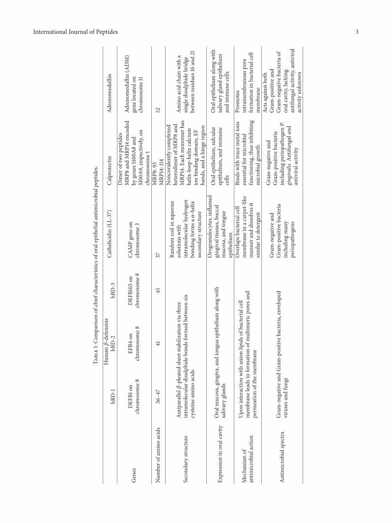
Several antimicrobial peptides produced by epithelial cellsare widespread in animal kingdom ranging from Cecropinsin insects to Magainins in frogs and Bactenecins in cattle[9–11]. All these antimicrobial peptides although differentin structure and amino acid composition share a commontrait of being broad spectrum antibiotics and essentially thefirst line of defence for host. First mammalian antimicrobialpeptide identified in oral epithelium was the lingual antimi-crobial peptide (LAP) by the works of Schonwetter et al. onbovine tongue [12]. LAP was found to have antibiotic activityagainst Gram-positive and Gram-negative bacteria as well asantifungal properties [12]. After LAP, several more EAPs havebeen identified in mammals including humans. The EAPsidentified in humans are 𝛽-defensin family, cathelicidin (LL-37), calprotectin, and adrenomedullin. The major character-istics of these EAPs are compared in Table 1.
3. The 𝛽-Defensin Family
Beta-defensins are universally expressed in all human epithe-lial cells [13]. In the oral cavity, they have been found in oralmucosa, gingiva, and tongue epithelium along with salivaryglands [14]. They were first described in bovine trachealpseudostratified epithelium [15]. Beta-defensins share certainstructural similarities with𝛼-defensins although they are a bitlarger than 𝛼-defensins. However, both 𝛼- and 𝛽-defensinsare cationic in nature because of presence of arginine andlysine residue in their structure [16]. Out of several 𝛽-defensins (hBDs) known, four have been characterized elabo-rately and termed as hBD-1, hBD-2, hBD-3, and hBD-4. Outof this only hBD-1, hBD-2, and hBD-3 are expressed in theoral cavity [17]. hBD-1 and hBD-2, found in the suprabasallayer of epithelium, can be localized to differentiated epithe-lial cells, whereas hBD-3 is expressed in undifferentiatedepithelial cells found in the basal layer of epithelium [18, 19].Apart from hBD-1, which is constitutively expressed, therest of the 𝛽-defensins are inducible upon stimulation withmicrobes or proinflammatory cytokines [20]. Recentlyanother 𝛽-defensin hBD-9 has been identified and localizedin gingival epithelium [21].
3.1. Genes Encoding 𝛽-Defensin. The first human 𝛽-defensinwas isolated from hemofiltrate passing through kidney [22].Thegene encoding for hBD-1 is present on chromosome8 and
is termed DEFB1. It lies in close proximity to the neutrophil𝛼-defensin gene (DEFA 1), arranged approximately 100–150kilobases apart. DEFB1 has two exons and one intron whichis translated into a hBD-1 propeptide [23]. This propeptideundergoes posttranslational modifications and forms severalhBD-1 mature peptides upon cleavage which are about 36–47amino acids long [24].
The second human 𝛽-defensin was first isolated in thepsoriatic skin keratinocytes [25]. hBD-2 is encoded via geneEFB4 which is also located on chromosome 8 in close prox-imity to DEFB1. Similar to DEFB1, it has two exons and oneintronwhich encodes a signal peptidewhich is 23 amino acidslong and a mature peptide which is 41 amino acids long [26].
DEFB103, which is the gene for hBD-3, has been localizedon chromosome 8 above the hBD-2 gene. The translationalproduct of this gene consists of a propeptide with a signalpeptide domain and a mature peptide of 22 and 45 aminoacids in length, respectively. The amino acid sequence ofhBD-3 shares some amount of similarity with hBD-2 [27].
3.2. Induction of Genetic Expression. Human 𝛽-defensin-1 isexpressed at a low level within gingival and buccal epithelium,dental pulp, and salivary gland tissue [5]. It has been observedthat hBD-1 is not regulated in response to infection or otherstimuli, which means that it is not inducible. Otherwiseexpressed at a very low level, hBD-2 and hBD-3 are, on theother hand, inducible in response tomicrobes and their prod-ucts [28]. Their amplified expression in response to infectionhas already been confirmed in gingivitis and periodontitis[29, 30]. IL-1𝛽, tumor necrosis factor (TNF)-𝛼, and IL-17 havebeen implicated in the induction of hBD-2 and hBD-3 expres-sion in epithelial cells [31]. Their induction via cytokinesconfirms their role in innate immunity. IL-17 also induces 𝛽-defensin expression against the colonization of Candida inoral cavity [32]. Thus, 𝛽-defensins expressed within the oralcavity not only check an overgrowth of commensal micro-organisms but also prevent colonization of pathogens.
3.3. Structure. The structure of 𝛽-defensins is characterizedby an antiparallel 𝛽-pleated sheet stabilization via threeintramolecular disulphide bonds formedbetween six cysteineamino acids. Also, hBD-2 and hBD-3 contain an 𝛼-helicaldomain at their N terminus. Since 𝛽-defensin peptide con-tains both hydrophilic and hydrophobic domains in theirstructure, they are amphiphatic in nature [33]. The aminoacid sequences of these defensins and other oral EAPs areprovided in Table 2.
3.4. Antimicrobial Activity. Thesalivary concentrationof hBD-1and hBD-2 varies in a range of nondetectable to 39 ng/mL and33 ng/mL, respectively [34]. On the other hand, hBD-3 existsat a concentration range of 0.31 𝜇g/mL in saliva [35]. Severalin vitro assays have revealed 𝛽-defensins to be active againstbroad range ofmicrobes including Gram-positive andGram-negative bacteria, enveloped viruses, and fungi [36–39].Theyare also efficacious against periodontal pathogens such asAggregatibacter actinomycetemcomitans and Fusobacteriumnucleatum [37]. A recent study demonstrated extermination
International Journal of Peptides 3
Table1:Com
paris
onof
chiefcharacteristicso
foralepithelialantim
icrobialpeptides.
Hum
an𝛽-defensin
sCa
thelicidin
(LL-37)
Calprotectin
Adreno
medullin
hBD-1
hBD-2
hBD-3
Genes
DEF
B1on
chromosom
e8EF
B4on
chromosom
e8DEF
B103
onchromosom
e8CA
MPgene
onchromosom
e3
Dim
erof
twopeptides
MRP
8andMRP
14encoded
bygenesS
100A
8and
S100A9,respectiv
ely,on
chromosom
e1
Adreno
medullin
(ADM)
gene
locatedon
chromosom
e11
Num
bero
faminoacids
36–4
741
4537
MRP
8:93
MRP
14:114
52
Second
arystr
ucture
Antiparallel𝛽-pleated
sheetstabilizationviathree
intram
olecular
disulphide
bond
sformed
betweensix
cyste
inea
minoacids
Rand
omcoilin
aqueou
ssolutio
nswith
intram
olecular
hydrogen
bond
ingform
sa𝛼-helix
second
arystr
ucture
Non
covalentlycomplexed
heterodimer
ofMRP
8and
MRP
14.E
achmon
omer
has
helix-lo
op-helixcalcium
ionbind
ingdo
main,
EFhand
s,andah
inge
region
Aminoacid
chainwith
asin
gled
isulphide
bridge
betweenresid
ues16and21
Expressio
nin
oralcavity
Oralm
ucosa,ging
iva,andtong
ueepith
elium
alon
gwith
salivaryglands
Orogranulocytes,infl
amed
ging
ivaltissues,buccal
mucosa,andtong
ueepith
elium
Oralepithelium,sulcular
epith
elium,and
immun
ecells
Oralepithelium
alon
gwith
salivaryglandepith
elium
andim
mun
ecells
Mechanism
ofantim
icrobialactio
n
Upo
ninteractionwith
anionlip
idso
fbacteria
lcell
mem
braneleads
toform
ationof
multim
ericpo
resa
ndperm
eatio
nof
them
embrane
Overla
psbacterialcell
mem
braneinac
arpet-like
mannera
nddissolvesit
similartodetergent
Bind
swith
tracem
etalions
essentialfor
microbial
functio
ning
,thu
sinh
ibiting
microbialgrow
th
Prom
otes
intram
embranou
spore
form
ationin
bacterialcell
mem
brane
Antim
icrobialspectra
Gram-negativea
ndGram-positive
bacteria,envelo
ped
virusesa
ndfung
i
Gram-negativea
ndGram-positive
bacteria
inclu
ding
many
perio
pathogens
Gram-negativea
ndGram-positive
bacteria
inclu
ding
perio
pathogen
P.gingivalis.
Antifu
ngaland
antiv
iralactivity
Actsagainstb
oth
Gram-positive
and
Gram-negativeb
acteria
oforalcavity,lacking
antifun
galactivity,antivira
lactiv
ityun
know
n

4 International Journal of Peptides
Table 2: Amino acid sequences of oral epithelial antimicrobial peptides.
Human 𝛽-defensinshBD-1 DHYNCVSSGG QCLYSACPIF TKIQGTCYRG KAKCCKhBD-2 GIGDPVTCLK SGAICHPVFC PRRYKQIGTC GLPGTKCCKK PhBD-3 GIINTLQKYY CRVRGGRCAV LSCLPKEEQI GKCSTRGRKC CRRKK
Human cathelicidin(LL-37) LLGDFFRKSK EKIGKEFKRI VQRIKDFLRN LVPRTES
Calprotectin
MRP8 MLTELEKALN SIIDVYHKYS LIKGNFHAVY RDDLKKLLET ECPQYIRKKG ADVWFKELDINTDGAVNFQE FLILVIKMGV AAHKKSHEES HKE
MRP14 MTCKMSQLER NIETIINTFH QYSVKLGHPD TLNQGEFKEL VRKDLQNFLK KENKNEKVIEHIMEDLDTNA DKQLSFFEFI MLMARLTWAS HEKMHEGDEG PGHHHKPGLG EGTP
Adrenomedullin YRQSMNNFQG LRSFGCRFGT CTVQKLAHQI YQFTDKDKDN VAPRSKISPQ GY
of F. nucleatumwith in gingival epithelial cells via hBD-2 andhBD-3 [40].Their spectrum of activity also includes Candidaalbicans along with other Candida spp. in oral cavity [38]. Inan experiment carried out to assess the antimicrobial activityof hBD-1, hBD-2, and hBD-3 against periodontopathic andcariogenic bacteria, it was suggested that Gram-negativebacteria except F. nucleatum were less susceptible to antimi-crobial peptides than Gram-positive organisms. Except forhBD-1, all peptides demonstrated 100% bactericidal activitywith concentration of >10mg/L of peptides. hBD-1 andhBD-2 were significantly less effective than hBD-3 in theirantimicrobial activity. The minimum inhibitory concentra-tion (MIC) for hBD-3 was around 100–200mg/L for A. acti-nomycetemcomitans, P. gingivalis, and Prevotella intermedia,whereas it was 12.5mg/L for F. nucleatum [41]. In anotherstudy, the MIC of hBD-2 and hBD-3 was found to bein a range of 3.9 to >250𝜇g/mL and 1.4 to >250𝜇g/mL,respectively, againstA. actinomycetemcomitans, F. nucleatum,P. gingivalis, Peptostreptococcus micros, Streptococcus mutans,S. sanguis, and Candida spp. [36].
It has also been observed that certain periodontal patho-gens have developed resistance against 𝛽-defensins, therebyenhancing their pathogenicity. These pathogens include P.gingivalis and Treponema denticola [42, 43]. It is assumed thatT. denticola interacts with the signal transduction pathwayto suppress the expression of 𝛽-defensins [43]. Also, recentdata demonstrates that hBD-2 and hBD-3 expression in adultgingival epithelial cells inactivates human immunodeficiencyvirus (HIV) suggesting an important role of 𝛽-defensins inantiviral defence [39].
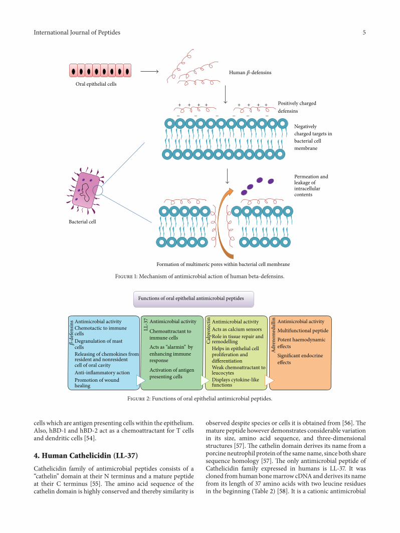
3.5. Mechanism of Antimicrobial Action. 𝛽-defensins belongto a group of cationic antimicrobial peptides (CAPs), knownto target the bacterial cell membrane. Since 𝛽-defensins arepositively charged, they bind to the negatively charged siteson the bacterial cell membrane. In Gram-negative bacteria,the target is lipopolysaccharide (LPS), whereas in Gram-positive bacteria it is teichoic acid. Apart from these, mem-brane rich phospholipids (phosphatidyl glycerol) which arecommon to both Gram-positive and Gram-negative bacteriaare also targeted by 𝛽-defensins. As the eukaryotic cell
membrane is rich in phosphatidylcholine rich phospholipidswhich are zwitterionic in nature, they are exempted fromthe action of 𝛽-defensins [8]. It has been hypothesized thatdefensins upon interaction with anion lipids of bacterialcell membrane leads to formation of multimeric pores andpermeation of the membrane (Figure 1). This permeationleads to the loss of vital contents of the bacterial cell leadingto cell death [44].
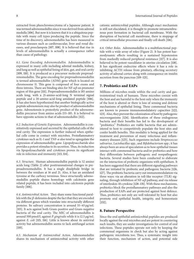
3.6. Other Roles. Various roles of 𝛽-defensins are summa-rized in Figure 2. Since 𝛽-defensins exhibit similarities tocytokines structurally, they show potent chemotactic activityto a range of immune cells [45, 46]. This activity takesplace at a much lower level of 𝛽-defensin concentrationthan required for its antimicrobial activity. hBD-1 activatesimmature dendritic cells andmemoryT cells, hBD-2 is able torecruit mast cells and neutrophils, and hBD-3 is chemotacticfor neutrophils, dendritic cells, mast cells, and monocytes.hBD-2 and hBD-3 also induce degranulation of mast cells[47–49]. Recently, it has been demonstrated that 𝛽-defensinsare able to bind to CXCR 4 receptors on T cells, leading tointernalization of receptor. This receptor is crucial for HIV-1 infection of T cell, thereby its internalization provides adefence against HIV-1 infection [39].
Beta-defensins have a role in inducing the resident aswell as nonresident cells to release chemokines. They inducethe release of several chemokines from keratinocytes suchas monocyte chemotactic protein-1, interferon-𝛾 inducibleprotein-10, and macrophage inflammatory protein 3𝛼 [50].Beta-defensins are known to inhibit production of chemo-kines and cytokines such as TNF-𝛼 and IL-6, thereby exhibit-ing a role in anti-inflammatory activities and suppression ofimmune response [51, 52].They also have a role in promotionof wound healing, where the growth factors secreted byvarious immune cells in the area of injury can stimulate ker-atinocytes to release hBD-3. These growth factors includeinsulin-like growth factor- (IGF-) 1, transforming growthfactor- (TGF-) 𝛼, and epidermal growth factor (EGF) [53].Also, 𝛽-defensins act as a connecting link between innateand adaptive immunity. Epithelial cells release 𝛽-defensinsalong with chemokines and cytokines, to signal Langerhans
International Journal of Peptides 5
Oral epithelial cells
Bacterial cell
Positively charged defensins
Negatively charged targets in bacterial cell membrane
Permeation and leakage of intracellular contents
Formation of multimeric pores within bacterial cell membrane
Human 𝛽-defensins
++++++++
−−−−−−
Figure 1: Mechanism of antimicrobial action of human beta-defensins.
Antimicrobial activityChemotactic to immunecellsDegranulation of mastcellsReleasing of chemokines fromresident and nonresidentcell of oral cavityAnti-inflammatory actionPromotion of woundhealing
Functions of oral epithelial antimicrobial peptides
LL-37 Antimicrobial activity
Chemoattractant toimmune cells
Acts as “alarmin” byenhancing immuneresponse
Activation of antigenpresenting cells
Calp
rote
ctin Antimicrobial activityActs as calcium sensorsRole in tissue repair andremodellingHelps in epithelial cellproliferation anddifferentiationWeak chemoattractant toleucocytesDisplays cytokine-likefunctions
Adre
nom
edul
lin Antimicrobial activity
Multifunctional peptide
Potent haemodynamiceffects
Significant endocrineeffects
𝛽-d
efen
sins
Figure 2: Functions of oral epithelial antimicrobial peptides.
cells which are antigen presenting cells within the epithelium.Also, hBD-1 and hBD-2 act as a chemoattractant for T cellsand dendritic cells [54].
4. Human Cathelicidin (LL-37)
Cathelicidin family of antimicrobial peptides consists of a“cathelin” domain at their N terminus and a mature peptideat their C terminus [55]. The amino acid sequence of thecathelin domain is highly conserved and thereby similarity is
observed despite species or cells it is obtained from [56]. Themature peptide however demonstrates considerable variationin its size, amino acid sequence, and three-dimensionalstructures [57]. The cathelin domain derives its name from aporcine neutrophil protein of the samename, since both sharesequence homology [57]. The only antimicrobial peptide ofCathelicidin family expressed in humans is LL-37. It wascloned fromhuman bonemarrow cDNAandderives its namefrom its length of 37 amino acids with two leucine residuesin the beginning (Table 2) [58]. It is a cationic antimicrobial

6 International Journal of Peptides
peptide of 18 Kda size, therefore also termed hCAP 18 [59].It is expressed in epithelial cells lining the respiratory, gas-trointestinal, and urogenital tract as well as oral cavity,although its main source in oral cavity is from neutrophilicgranules and to a lesser extent from epithelial cells [18]. Inoral cavity, LL-37 is expressed in inflamed gingival tissues,buccal mucosa, and tongue epithelium [60]. It has also beenidentified in saliva and GCF [61, 62]. The concentration ofLL-37 is found to increase with increasing depth of gingivalsulcus [63]. It has also been proposed that LL-37 detectedin gingival epithelium may be the product of neutrophilmigration through gingival epithelium rather than epithelialcells themselves [18].
4.1. Gene Encoding LL-37. Thegene encoding LL-37 is locatedon chromosome 3 at location 3p21.3. It has been namedcathelicidin antimicrobial peptide (CAMP) gene. LL-37 genehas four exons and three introns. First three exons encodefor the signal sequence and cathelin region of the peptidewhile the fourth exon translates into mature peptide [54].In intron and promoter region of LL-37, there exist bindingsites for acute phase response factors, which establish theupregulation of LL-37 in inflammation [64].
4.2. Induction of Gene Expression. LL-37 expression in vari-ous cell types has been found to be upregulated on exposureto growth factors, differentiating agents, and microorgan-isms. Insulin-like growth factor-1 which is known to promotewound healing upregulates LL-37 expression [65]. Also,vitamin D which is a differentiating agent has been found toamplify LL-37 activity [66]. Increased level of LL-37 in gingi-val tissues in response to inflammation correlates positivelywith depth of gingival crevice [63]. In a comparative study,levels of LL-37 in GCF were found to be significantly elevatedin chronic periodontitis patients than in gingivitis patientsand healthy volunteers [67].
4.3. Structure. The cathelicidin gene in humans is translatedinto an inactive precursor protein termed as hCAP-18. Uponposttranslational processing an active C terminus peptidewith 37 amino acids is released from precursor protein. Thiscleavage is carried out via proteolytic enzyme elastase orproteinase-3 [68, 69]. This peptide has a net positive chargeat physiologic pH and more than 50% of its residues arehydrophilic in nature. Structurally, it exists as a random coilin aqueous solutions. Many of its amino acids form intra-molecular hydrogen bonds, acquiring an 𝛼-helix secondarystructure. It is supposed that antibacterial activity of LL-37 iscorrelated with 𝛼-helicity [70].
4.4. Antimicrobial Activity. Found in saliva in a concentrationof 0.14–3 𝜇g/mL, LL-37 is active against both Gram-negativeand Gram-positive bacteria including established perio-pathogens P. gingivalis andA. actinomycetemcomitans [71]. ItsMICwas 30–60 𝜇g/mL and 125 𝜇g/mL againstA. actinomyce-temcomitans and P. gingivalis, respectively [37]. LL-37 main-tains its antimicrobial activity even in presence of gingipainswith some salivary components providing it with protection[72]. It is ascertained that LL-37 inhibits the inflammatory
response in gingival fibroblasts toP. gingivalis and its products[73]. LL-37 binds directly to bacterial lipopolysaccharide [59].Morbus Kostmann syndrome is a genetic form of periodontaldiseasewhichwas shown to have a near absence of LL-37 [74].This suggests the importance of LL-37 as an antimicrobialpeptide in oral cavity.
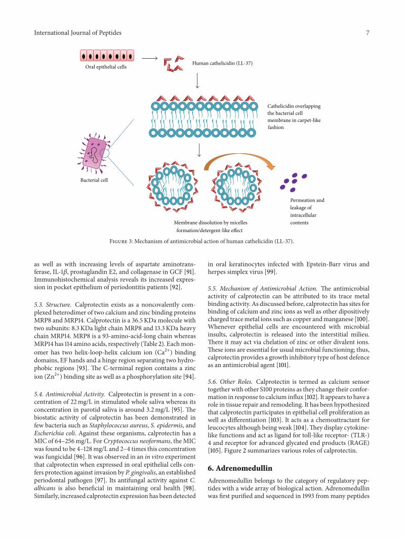
4.5. Mechanism of Antimicrobial Action. LL-37 belongs to thecategory of amphiphatic 𝛼-helical antimicrobial peptides. Ithas been proposed that these peptides do not function viaformation of transmembrane pores within the lipid biolayersof microbial cell membrane as is the case with defensins.Although the exact mechanism of action of Cathelicidins isnot clear, some members of this family overlap the bacterialcell membrane in a carpet-like manner and dissolve it similarto a detergent by micelles formation [75] (Figure 3).
4.6. Other Roles. LL-37 acts as a chemoattractant and causesinflux of neutrophils, monocytes, and T cells to the site ofinflammation [76]. Some researchers believe that it acts asan “alarmin” rather than an antimicrobial by enhancing theimmune response leading to activation of antigen presentingcells [77]. The major roles of LL-37 are described in Figure 2.
5. Calprotectin
Calprotectin, also known as calgranulin, is a heterodimer oftwo anionic peptidesMRP8 andMRP14 [78]. Several nomen-clatures have been proposed for these individual peptidesas well as calprotectin dimer but controversy regardingnomenclature still exists. These belong to S100 family of cal-cium binding proteins [79]. Calprotectin has been found tobe constitutively expressed in cells of immune function suchas neutrophils, monocytes, macrophages, and epithelial cells[80–82]. Its level increases in plasma, saliva, and synovialfluid during infectious and inflammatory diseases [83–85].Also, it is highly responsive to any kind of stress in epidermalcells [86].
5.1. Gene Encoding for Calprotectin. The HGNC approvednames of genes encoding for MRP8 and MRP14 are S100A8and S100A9, respectively. The genes are from a closelylocated cluster of thirteen genes on chromosome 1 at location1q21 [87]. The organization of these genes is evolutionarilyconserved. Both of these peptides have three exons separatedby two introns. Exon 1 is untranslated, whereas exons 2 and 3encode into an N-terminal and a C-terminal EF hand motif,respectively [88].
5.2. Induction of Genetic Expression. Calprotectin expressionis upregulated in epithelial cells upon induction of stressand exposure to ultraviolet radiation and in wound healing[86]. Also, its level heightens via exposure to complementfactor C5a and proinflammatory cytokines such as TNF-𝛼,IL-1𝛽, and IL-6 [89]. Calprotectin levels have been found topositively correlate with severity of periodontitis in gingivalcrevicular fluid (GCF) [90].The origin of calprotectin inGCFis from sulcular epithelium as well as immune cells. Cal-protectin expression amplifies with increased pocket depth
International Journal of Peptides 7
Oral epithelial cells
Bacterial cell
Cathelicidin overlapping the bacterial cell membrane in carpet-like fashion
Permeation and leakage of intracellular contents Membrane dissolution by micelles
formation/detergent-like effect
Human cathelicidin (LL-37)
Figure 3: Mechanism of antimicrobial action of human cathelicidin (LL-37).
as well as with increasing levels of aspartate aminotrans-ferase, IL-1𝛽, prostaglandin E2, and collagenase in GCF [91].Immunohistochemical analysis reveals its increased expres-sion in pocket epithelium of periodontitis patients [92].
5.3. Structure. Calprotectin exists as a noncovalently com-plexed heterodimer of two calcium and zinc binding proteinsMRP8 and MRP14. Calprotectin is a 36.5 KDa molecule withtwo subunits: 8.3 KDa light chain MRP8 and 13.3 KDa heavychain MRP14. MRP8 is a 93-amino-acid-long chain whereasMRP14 has 114 amino acids, respectively (Table 2). Eachmon-omer has two helix-loop-helix calcium ion (Ca2+) bindingdomains, EF hands and a hinge region separating two hydro-phobic regions [93]. The C-terminal region contains a zincion (Zn2+) binding site as well as a phosphorylation site [94].
5.4. Antimicrobial Activity. Calprotectin is present in a con-centration of 22mg/L in stimulated whole saliva whereas itsconcentration in parotid saliva is around 3.2mg/L [95]. Thebiostatic activity of calprotectin has been demonstrated infew bacteria such as Staphylococcus aureus, S. epidermis, andEscherichia coli. Against these organisms, calprotectin has aMIC of 64–256mg/L. For Cryptococcus neoformans, the MICwas found to be 4–128mg/L and 2–4 times this concentrationwas fungicidal [96]. It was observed in an in vitro experimentthat calprotectin when expressed in oral epithelial cells con-fers protection against invasion by P. gingivalis, an establishedperiodontal pathogen [97]. Its antifungal activity against C.albicans is also beneficial in maintaining oral health [98].Similarly, increased calprotectin expression has been detected
in oral keratinocytes infected with Epstein-Barr virus andherpes simplex virus [99].
5.5. Mechanism of Antimicrobial Action. The antimicrobialactivity of calprotectin can be attributed to its trace metalbinding activity. As discussed before, calprotectin has sites forbinding of calcium and zinc ions as well as other dipositivelycharged tracemetal ions such as copper andmanganese [100].Whenever epithelial cells are encountered with microbialinsults, calprotectin is released into the interstitial milieu.There it may act via chelation of zinc or other divalent ions.These ions are essential for usual microbial functioning; thus,calprotectin provides a growth inhibitory type of host defenceas an antimicrobial agent [101].
5.6. Other Roles. Calprotectin is termed as calcium sensortogether with other S100 proteins as they change their confor-mation in response to calcium influx [102]. It appears to have arole in tissue repair and remodeling. It has been hypothesizedthat calprotectin participates in epithelial cell proliferation aswell as differentiation [103]. It acts as a chemoattractant forleucocytes although being weak [104].They display cytokine-like functions and act as ligand for toll-like receptor- (TLR-)4 and receptor for advanced glycated end products (RAGE)[105]. Figure 2 summarizes various roles of calprotectin.
6. Adrenomedullin
Adrenomedullin belongs to the category of regulatory pep-tides with a wide array of biological action. Adrenomedullinwas first purified and sequenced in 1993 from many peptides
8 International Journal of Peptides
extracted from pheochromocytoma of a Japanese patient. Itwas termed adrenomedullin since it was derived fromadrenalmedulla [106]. But now it is known that it is a ubiquitous pep-tide with many cell types producing the peptide. Since thetime of its discovery, adrenomedullin has been measured invarious diseases such as cardiovascular, liver and renal dis-eases, and preeclampsia [107, 108]. It is believed that rise inlevels of adrenomedullin is actually a consequence ratherthan cause of pathology.
6.1. Gene Encoding Adrenomedullin. Adrenomedullin isexpressed in many cells including adrenal medulla, kidney,and lung aswell as epithelial lining of skin, gut, and oral cavity[109, 110]. It is produced as a precursor molecule preproad-renomedullin. The gene encoding for preproadrenomedullinis termed adrenomedullin (ADM) gene which is located onchromosome 11. This gene is composed of four exons andthree introns. There are binding sites for NF-𝜅𝛽 on promoterregion of this gene [111]. Preproadrenomedullin is 185 aminoacids long, with a 21-amino-acid-long N terminal signalpeptide and a 20-amino-acid-long amidated peptide [106].It has also been hypothesized that another biologically activepeptide adrenotensinmay also be product of adrenomedullingene. Adrenotensin is proteolytic product of the adrenome-dullin precursor from amino acids 153–185. It is believed tohave opposite actions to that of adrenomedullin [112].
6.2. Induction of Genetic Expression. Adrenomedullin is con-stitutively expressed and secreted by the epithelial cells of theoral cavity. The expression is further induced when epithe-lial cells come in contact with microbes. Proinflammatorycytokines such as IL-1 and TNF-𝛼 also tend to upregulate theexpression of adrenomedullin gene. Lipopolysaccharide alsoprovides a potent stimulus to its secretion.Thus, its inductionvia lipopolysaccharide and cytokines proves its significantrole in infection and immunity [113, 114].
6.3. Structure. Human adrenomedullin peptide is 52 aminoacids long (Table 2) after posttransnational changes in pre-proadrenomedullin. It has a single disulphide bridge inbetween the residues at 16 and 21. Also, it has an amidatedtyrosine at the carboxy terminus. Since structurally adreno-medullin peptide shares homology with calcitonin generelated peptide, it has been included into calcitonin peptidefamily [106].
6.4. Antimicrobial Action. They share some functional paral-lels with the𝛽-defensins despite the fact that they are encodedvia different genes which translate into structurally differentproteins. Its salivary concentration is around 55–65 pg/mL[115]. It acts against both Gram-positive and Gram-negativebacteria of the oral cavity. The MIC of adrenomedullin isaround 500 pmol/L against P. gingivalis while it is 12.5 𝜇g/mLagainst E. coli [115, 116]. Little is known about its antiviralactivity but adrenomedullin seems to lack antifungal action[110].
6.5. Mechanism of Antimicrobial Action. Adrenomedullinshares its mechanism of antimicrobial activity with other
cationic antimicrobial peptides. Although exact mechanismis still not elucidated, it is thought to promote intramembra-nous pore formation in bacterial cell membrane. With thedisruption of bacterial cell membrane, there is stoppage ofcritical intracellular processes and finally cell death [116].
6.6. Other Roles. Adrenomedullin is a multifunctional pep-tide with a wide array of roles (Figure 2). It has potent hae-modynamic effects resulting in a sustained hypotensionfrom markedly reduced peripheral resistance [117]. It is alsobelieved to be potent vasodilator in uterine circulation [118].It has significant endocrine effects where it has a role ininhibiting ACTH release from pituitary, effecting secretoryactivity of adrenal cortex along with consequence on insulinsecretion from the pancreas [119–121].
7. Probiotics and EAPs
Millions of microbes reside within the oral cavity and gas-trointestinal track of humans. These microbes coexist withthe host and are harmful to the host only when the immunityof the host is altered or there is loss of sensing and defensemechanisms of epithelial lining. These commensal bacteriaare known to possess immunomodulatory capacities [122,123]. Also, they prevent the colonization of host by pathogenicmicroorganisms [124]. Identification of these endogenousbacteria and their benefits has led to the development of“probiotics.” Probiotics are viable bacteria that are admin-istered to host to competitively populate the host sites andconfer health benefits. This modality is being applied for thetreatment and prevention of many infectious and inflam-matory diseases [125]. Such organisms include Streptococcussalivarius, Lactobacillus spp., and Bifidobacterium spp.; it hasalways been an area of speculation as to how epithelial tissuesinteract with commensal bacteria and/or probiotics and howthey differentiate between pathogenic and nonpathogenicbacteria. Several studies have been conducted to elaborateon the interaction of probiotic organisms with epithelium. Ithas been suggested that there are different signaling pathwaysthat are initiated by probiotic and pathogenic bacteria [126,127].The probiotic bacteria carry out immunomodulation viathree ways: via an alteration in toll-like receptor (TLR) sig-naling, through inhibition of NF-𝜅𝛽 pathway, and via releaseof interleukin-10 cytokine [128–131].With these mechanisms,probiotics block the proinflammatory pathways and also theproduction of EAPs and are protected against host defence.Thus, probiotics not only are well tolerated by host but alsopromote oral epithelial health, integrity, and homeostasis[132].
8. Future Perspective
Since the oral epithelial antimicrobial peptides are producedlocally against the oral microbes and are potent in counteringsuch insults, they are under investigations for control of oralinfections. These peptides operate not only by keeping thecommensal organisms in check but also by acting againstpathogenic microbiota too. Thus, a systematic insight intotheir function, mechanism of action, and potential side
International Journal of Peptides 9
effects is essential to develop them into therapeutic agentsto counter oral infections such as periodontitis. The levelsof these antimicrobial peptides are found to increase locallyin periodontitis, and their external application may provideprotection from progression of disease. Oral EAPs can bedeveloped in a variety of ways for their therapeutic benefits.These can be produced in form of gels, mouthwashes, andgum paints for local application over periodontal tissues tonot only prevent the development of periodontal disease butalso reverse the existing disease. EAPs can also be developedas local drug delivery agents within the periodontal pocketsto curtail the effects of periopathogenic bacteria. Becauseof their antiviral and antifungal effects, EAPs can be usedin immunocompromised individuals against opportunisticinfections such as candidiasis and herpetic gingivostomatitis.The activity of 𝛽-defensins against HIV holds potential fordrugs that are more efficacious with lesser side effects thanexisting drugs for AIDS. Iseganan hydrochloride, a syn-thetic cathelicidin, is under investigation for prevention ofulcerative oral mucositis and has shown promising results[133]. Iseganan hydrochloride is the salt of 17-amino-acid-long synthetic protegrin-1, an 18-amino-acid antimicrobialpeptide isolated from porcine leucocyte. Iseganan HCl isavailable as oral solution by the name of IB-367 rinse and is abroad spectrum antimicrobial and acts rapidly by disruptingcell membranes of microorganisms including bacteria, fungi,and viruses [134].
Epithelial cells are believed to behave differently whenexposed to commensal and pathogenic bacteria. This couldmean that antimicrobial peptide secreted from epithelialcells may have differential action against commensals andpathogens. In view of the fact that these antimicrobial pep-tides are broad spectrum and rapidly acting, they providelittle chance of development of resistance in microbes againstthem. This property could be fruitful in developing noveltherapeutic agents that lack resistance compared to conven-tional antibiotics. Finally, these antimicrobial peptides couldbe developed as biomarkers for oral disease diagnosis andprognosis. Therefore, there remains a whole unexploredworld of therapeutic benefits of epithelial antimicrobial pep-tides which needs to be ventured into.
Conflict of Interests
The authors declare that there is no conflict of interestsregarding the publication of this paper.
References
[1] M.T. Pollanen, J. I. Salonen, andV.-J. Uitto, “Structure and func-tion of the tooth-epithelial interface in health and disease,” Peri-odontology 2000, vol. 31, no. 1, pp. 12–31, 2003.
[2] H. E. Schroeder andM. A. Listgarten, “The gingival tissues:Thearchitecture of periodontal protection,” Periodontology 2000,vol. 14, no. 1, pp. 91–120, 1997.
[3] M. S. Tonetti, “Molecular factors associated with compartmen-talization of gingival immune responses and transepithelialneutrophil migration,” Journal of Periodontal Research, vol. 32,no. 1, pp. 104–109, 1997.
[4] B. A. Dale, “Periodontal epithelium: A newly recognized role inhealth and disease,” Periodontology 2000, vol. 30, no. 1, pp. 70–78, 2002.
[5] G. Diamond, N. Beckloff, and L. K. Ryan, “Host defense pep-tides in the oral cavity and the lung: similarities and differences,”Journal of Dental Research, vol. 87, no. 10, pp. 915–927, 2008.
[6] T. Ganz, M. E. Selsted, D. Szklarek et al., “Defensins. Naturalpeptide antibiotics of human neutrophils,”The Journal of Clini-cal Investigation, vol. 76, no. 4, pp. 1427–1435, 1985.
[7] F. G. Oppenheim, T. Xu, F. M. McMillian et al., “Histatins,a novel family of histidine-rich proteins in human parotidsecretion. Isolation, characterization, primary structure, andfungistatic effects on Candida albicans,” Journal of BiologicalChemistry, vol. 263, no. 16, pp. 7472–7477, 1988.
[8] A. Weinberg, S. Krisanaprakornkit, and B. A. Dale, “Epithelialantimicrobial peptides: review and significance for oral applica-tions,” Critical Reviews in Oral Biology and Medicine, vol. 9, no.4, pp. 399–414, 1998.
[9] H. G. Boman, “Cecropins : antibacterial peptides from insectsand pigs,” in Phylogenetic Perspectives in Immunity: The Insect-Host Defense, J. Hoffmann, S. Natori, and C. Janeway, Eds., pp.24–37, Landes Biomed, Austin, Tex, USA, 1994.
[10] M. Zasloff, “Magainins, a class of antimicrobial peptides fromXenopus skin: isolation, characterization of two active forms,and partial cDNA sequence of a precursor,” Proceedings of theNational Academy of Sciences of the United States of America,vol. 84, no. 15, pp. 5449–5453, 1987.
[11] D. Romeo, B. Skerlavaj, M. Bolognesi, and R. Gennaro, “Struc-ture and bactericidal activity of an antibiotic dodecapeptidepurified from bovine neutrophils,” Journal of Biological Chem-istry, vol. 263, no. 20, pp. 9573–9575, 1988.
[12] B. S. Schonwetter, E. D. Stolzenberg, andM. A. Zasloff, “Epithe-lial antibiotics induced at sites of inflammation,” Science, vol.267, no. 5204, pp. 1645–1648, 1995.
[13] B. A. Dale and S. Krisanaprakornkit, “Defensin antimicrobialpeptides in the oral cavity,” Journal of Oral Pathology andMedicine, vol. 30, no. 6, pp. 321–327, 2001.
[14] M. Mathews, H. P. Jia, J. M. Guthmiller et al., “Production of 𝛽-defensin antimicrobial peptides by the oral mucosa and salivaryglands,” Infection and Immunity, vol. 67, no. 6, pp. 2740–2745,1999.
[15] G. Diamond,M. Zasloff,H. Eck,M. Brasseur,W. LeeMaloy, andC. L. Bevins, “Tracheal antimicrobial peptide, a cysteine-richpeptide from mammalian tracheal mucosa: peptide isolationand cloning of a cDNA,” Proceedings of the National Academy ofSciences of the United States of America, vol. 88, no. 9, pp. 3952–3956, 1991.
[16] B. L. Kagan, T. Ganz, and R. I. Lehrer, “Defensins: a family ofantimicrobial and cytotoxic peptides,” Toxicology, vol. 87, no. 1–3, pp. 131–149, 1994.
[17] Y. Abiko,M. Saitoh,M. Nishimura, M. Yamazaki, D. Sawamura,and T. Kaku, “Role of 𝛽-defensins in oral epithelial health anddisease,”Medical Molecular Morphology, vol. 40, no. 4, pp. 179–184, 2007.
[18] B. A. Dale, J. R. Kimball, S. Krisanaprakornkit et al., “Localizedantimicrobial peptide expression in human gingiva,” Journal ofPeriodontal Research, vol. 36, no. 5, pp. 285–294, 2001.
[19] Q. Lu, L. P. Samaranayake, R. P. Darveau, and L. Jin, “Expressionof human 𝛽-defensin-3 in gingival epithelia,” Journal of Peri-odontal Research, vol. 40, no. 6, pp. 474–481, 2005.
10 International Journal of Peptides
[20] S. Krisanaprakornkit, A. Weinberg, C. N. Perez, and B. A. Dale,“Expression of the peptide antibiotic human 𝛽-defensin 1 incultured gingival epithelial cells and gingival tissue,” Infectionand Immunity, vol. 66, no. 9, pp. 4222–4228, 1998.
[21] P. Premratanachai, S. Joly, G. K. Johnson, P. B. McCray Jr., H. P.Jia, and J. M. Guthmiller, “Expression and regulation of novelhuman 𝛽-defensins in gingival keratinocytes,” Oral Microbiol-ogy and Immunology, vol. 19, no. 2, pp. 111–117, 2004.
[22] K. W. Bensch, M. Raida, H. J. Magert, P. Schulz-Knappe, andW. G. Forssmann, “hBD-1: a novel 𝛽-defensin from humanplasma,” FEBS Letters, vol. 368, no. 2, pp. 331–335, 1995.
[23] L. Liu, C. Zhao, H. H. Q. Heng, and T. Ganz, “The human𝛽-defensin-1 and 𝛼-defensins are encoded by adjacent genes:two peptide families with differing disulfide topology share acommon ancestry,” Genomics, vol. 43, no. 3, pp. 316–320, 1997.
[24] T. Ganz, “Defensins: antimicrobial peptides of innate immu-nity,” Nature Reviews Immunology, vol. 3, no. 9, pp. 710–720,2003.
[25] J. Harder, J. Bartels, E. Christophers, and J. M. Schroder, “Apeptide antibiotic from human skin,” Nature, vol. 387, no. 6636,p. 861, 1997.
[26] J. Harder, R. Siebert, Y. Zhang et al., “Mapping of the geneencoding human 𝛽-defensin-2 (DEFB2) to chromosome region8p22-p23.1,” Genomics, vol. 46, no. 3, pp. 472–475, 1997.
[27] H. P. Jia, B. C. Schutte, A. Schudy et al., “Discovery of newhuman 𝛽-defensins using a genomics-based approach,” Gene,vol. 263, no. 1-2, pp. 211–218, 2001.
[28] J. Harder, J. Bartels, E. Christophers, and J.-M. Schroder, “Iso-lation and characterization of human betadefensin-3, a novelhuman inducible peptide antibiotic,” The Journal of BiologicalChemistry, vol. 276, no. 8, pp. 5707–5713, 2001.
[29] S. Offenbacher, S. P. Barros, D. W. Paquette et al., “Gingivaltranscriptome patterns during induction and resolution ofexperimental gingivitis in humans,” Journal of Periodontology,vol. 80, no. 12, pp. 1963–1982, 2009.
[30] Q. Lu, L. Jin, R. P. Darveau, and L. P. Samaranayake, “Expressionof human 𝛽-defensins-1 and -2 peptides in unresolved chronicperiodontitis,” Journal of Periodontal Research, vol. 39, no. 4, pp.221–227, 2004.
[31] G. Diamond, N. Beckloff, A. Weinberg, and K. O. Kisich, “Theroles of antimicrobial peptides in innate host defense,” CurrentPharmaceutical Design, vol. 15, no. 21, pp. 2377–2392, 2009.
[32] H. R. Conti, F. Shen, N. Nayyar et al., “Th17 cells and IL-17receptor signaling are essential formucosal host defense againstoral candidiasis,” Journal of ExperimentalMedicine, vol. 206, no.2, pp. 299–311, 2009.
[33] M. E. Selsted, Y.-Q. Tang, W. L. Morris et al., “Purification,primary structures, and antibacterial activities of 𝛽-defensins, anew family of antimicrobial peptides from bovine neutrophils,”The Journal of Biological Chemistry, vol. 268, no. 9, pp. 6641–6648, 1993.
[34] M. S. Gardner, M. D. Rowland, A. Y. Siu, J. L. Bundy, D. K.Wagener, and J. L. Stephenson Jr., “Comprehensive defensinassay for Saliva,” Analytical Chemistry, vol. 81, no. 2, pp. 557–566, 2009.
[35] R. Tao, R. J. Jurevic, K. K. Coulton et al., “Salivary antimicrobialpeptide expression and dental caries experience in children,”Antimicrobial Agents andChemotherapy, vol. 49, no. 9, pp. 3883–3888, 2005.
[36] S. Joly, C. Maze, P. B. McCray Jr., and J. M. Guthmiller, “Human𝛽- defensins 2 and demonstrate strain selective activity againstoral microorganisms,” Journal of Clinical Microbiology, vol. 42,no. 3, pp. 1024–1029, 2004.
[37] S. Ji, J. Hyun, E. Park, B.-L. Lee, K.-K. Kim, and Y. Choi, “Sus-ceptibility of various oral bacteria to antimicrobial peptides andto phagocytosis by neutrophils,” Journal of Periodontal Research,vol. 42, no. 5, pp. 410–419, 2007.
[38] Z. Feng, B. Jiang, J. Chandra, M. Ghannoum, S. Nelson, and A.Weinberg, “Human beta-defensins: differential activity againstcandidal species and regulation by Candida albicans,” Journal ofDental Research, vol. 84, no. 5, pp. 445–450, 2005.
[39] Z. Feng, G. R. Dubyak, M. M. Lederman, and A. Weinberg,“Cutting edge: human 𝛽 defensin 3—a novel antagonist of theHIV-1 coreceptor CXCR4,”The Journal of Immunology, vol. 177,no. 2, pp. 782–786, 2006.
[40] S. Ji, J. E. Shin, Y. C. Kim, andY. Choi, “Intracellular degradationof Fusobacterium nucleatum in human gingival epithelial cells,”Molecules and Cells, vol. 30, no. 6, pp. 519–526, 2010.
[41] K. Ouhara, H. Komatsuzawa, S. Yamada et al., “Susceptibilitiesof periodontopathogenic and cariogenic bacteria to antibac-terial peptides, 𝛽-defensins and LL37, produced by humanepithelial cells,” Journal of Antimicrobial Chemotherapy, vol. 55,no. 6, pp. 888–896, 2005.
[42] C. E. Shelburne, W. A. Coulter, D. Olguin, M. S. Lantz, andD. E. Lopatin, “Induction of 𝛽-defensin resistance in the oralanaerobe Porphyromonas gingivalis,” Antimicrobial Agents andChemotherapy, vol. 49, no. 1, pp. 183–187, 2005.
[43] J. E. Shin and Y. Choi, “Treponema denticola suppresses expres-sion of human 𝛽-defensin-2 in gingival epithelial cells throughinhibition of TNF𝛼 production and TLR2 activation,”Moleculesand cells, vol. 29, no. 4, pp. 407–412, 2010.
[44] Y. Agawa, S. Lee, S. Ono et al., “Interaction with phospholipidbilayers, ion channel formation, and antimicrobial activity ofbasic amphipathic 𝛼-helical model peptides of various chainlengths,” Journal of Biological Chemistry, vol. 266, no. 30, pp.20218–20222, 1991.
[45] A. M. Cole, T. Ganz, A. M. Liese, M. D. Burdick, L. Liu, andR. M. Strieter, “Cutting edge: IFN-inducible ELR- CXC chemo-kines display defensin-like antimicrobial activity,” Journal ofImmunology, vol. 167, no. 2, pp. 623–627, 2001.
[46] D. Yang, A. Biragyn, L. W. Kwak, and J. J. Oppenheim, “Mam-malian defensins in immunity: more than just microbicidal,”Trends in Immunology, vol. 23, no. 6, pp. 291–296, 2002.
[47] D. Yang, O. Chertov, S. N. Bykovskaia et al., “𝛽-defensins:linking innate and adaptive immunity through dendritic and Tcell CCR6,” Science, vol. 286, no. 5439, pp. 525–528, 1999.
[48] F. Niyonsaba, H. Ogawa, and I. Nagaoka, “Human 𝛽-defensin-2 functions as a chemotactic agent for tumour necrosis factor-𝛼-treated human neutrophils,” Immunology, vol. 111, no. 3, pp.273–281, 2004.
[49] F. Niyonsaba, A. Someya, M. Hirata, H. Ogawa, and I. Nagaoka,“Evaluation of the effects of peptide antibiotics human 𝛽-defensins-1/-2 and LL-37 on histamine release and prostaglan-din D
2production frommast cells,” European Journal of Immu-
nology, vol. 31, no. 4, pp. 1066–1075, 2001.[50] F. Niyonsaba, H. Ushio, N. Nakano et al., “Antimicrobial pep-
tides human 𝛽-defensins stimulate epidermal keratinocytemigration, proliferation and production of proinflammatorycytokines and chemokines,” Journal of Investigative Dermatol-ogy, vol. 127, no. 3, pp. 594–604, 2007.
International Journal of Peptides 11
[51] K. G. Kohlgraf, L. C. Pingel, D. E. Dietrich, and K. A. Brog-den, “Defensins as anti-inflammatory compounds andmucosaladjuvants,” Future Microbiology, vol. 5, no. 1, pp. 99–113, 2010.
[52] F. Semple, S. Webb, H.-N. Li et al., “Human 𝛽-defensin 3 hasimmunosuppressive activity in vitro and in vivo,” EuropeanJournal of Immunology, vol. 40, no. 4, pp. 1073–1078, 2010.
[53] O. E. Sorensen, J. B. Cowland, K. Theilgaard-Monch, L. Liu,T. Ganz, and N. Borregaard, “Wound healing and expressionof antimicrobial peptides/polypeptides in human keratinocytes,a consequence of common growth factors,” The Journal ofImmunology, vol. 170, no. 11, pp. 5583–5589, 2003.
[54] D. Yang, A. Biragyn, D. M. Hoover, J. Lubkowski, and J. J.Oppenheim, “Multiple roles of antimicrobial defensins, cathe-licidins, and eosinophil-derived neurotoxin in host defense,”Annual Review of Immunology, vol. 22, pp. 181–215, 2004.
[55] B. Ramanathan, E. G. Davis, C. R. Ross, and F. Blecha, “Cathe-licidins: microbicidal activity, mechanisms of action, and rolesin innate immunity,” Microbes and Infection, vol. 4, no. 3, pp.361–372, 2002.
[56] M. Zaiou and R. L. Gallo, “Cathelicidins, essential gene-encoded mammalian antibiotics,” Journal of Molecular Medi-cine, vol. 80, no. 9, pp. 549–561, 2002.
[57] R. Gennaro and M. Zanetti, “Structural features and biologicalactivities of the cathelicidin-derived antimicrobial peptides,”Biopolymers, vol. 55, pp. 31–49, 2000.
[58] G. H. Gudmundsson, B. Agerberth, J. Odeberg, T. Bergman,B. Olsson, and R. Salcedo, “The human gene FALL39 andprocessing of the cathelin precursor to the antibacterial peptideLL-37 in granulocytes,” European Journal of Biochemistry, vol.238, no. 2, pp. 325–332, 1996.
[59] J. W. Larrick, M. Hirata, R. F. Balint, J. Lee, J. Zhong, and S. C.Wright, “Human CAP18: a novel antimicrobial lipopolysaccha-ride-binding protein,” Infection and Immunity, vol. 63, no. 4, pp.1291–1297, 1995.
[60] M. F. Nilsson, B. Sandstedt, O. Sørensen, G. Weber, N. Bor-regaard, and M. Stahle-Backdahl, “The human cationic anti-microbial protein (hCAP18), a peptide antibiotic, is widelyexpressed in human squamous epithelia and colocalizes withinterleukin-6,” Infection and Immunity, vol. 67, no. 5, pp. 2561–2566, 1999.
[61] M. Murakami, T. Ohtake, R. A. Dorschner, and R. L. Gallo,“Cathelicidin antimicrobial peptides are expressed in salivaryglands and saliva,” Journal of Dental Research, vol. 81, no. 12, pp.845–850, 2002.
[62] O. Turkoglu, G. Emingil, N. Kutukculer, andG. Atilla, “Gingivalcrevicular fluid levels of cathelicidin ll-37 and interleukin-18 inpatients with chronic periodontitis,” Journal of Periodontology,vol. 80, no. 6, pp. 969–976, 2009.
[63] I. Hosokawa, Y. Hosokawa, H. Komatsuzawa et al., “Innateimmune peptide LL-37 displays distinct expression patternfrom beta-defensins in inflamed gingival tissue,” Clinical andExperimental Immunology, vol. 146, no. 2, pp. 218–225, 2006.
[64] M. Zanetti, R. Gennaro, M. Scocchi, and B. Skerlavaj, “Struc-ture and biology of cathelicidins,” Advances in ExperimentalMedicine and Biology, vol. 479, pp. 203–218, 2000.
[65] O. E. Sorensen, J. B. Cowland, K. Theilgaard-Monch, L. Liu, T.Ganz, and N. Borregaard, “Wound healing and expression ofantimicrobial peptides/polypeptides in human keratinocytes, aconsequence of common growth factors,” Journal of Immunol-ogy, vol. 170, no. 11, pp. 5583–5589, 2003.
[66] G. Weber, J. D. Heilborn, C. I. C. Jimenez, A. Hammarsjo, H.Torma, and M. Stahle, “Vitamin D induces the antimicrobialprotein hCAP18 in human skin,” Journal of Investigative Derma-tology, vol. 124, no. 5, pp. 1080–1082, 2005.
[67] O. Turkoglu, G. Emingil, N. Kutukcxuler, and G. Atilla, “Gingi-val crevicular fluid levels of cathelicidin ll-37 and interleukin-18in patients with chronic periodontitis,” Journal of Periodontol-ogy, vol. 80, no. 6, pp. 969–976, 2009.
[68] A. Panyutich, J. Shi, P. L. Boutz, C. Zhao, and T. Ganz, “Porcinepolymorphonuclear leukocytes generate extracellular microbi-cidal activity by elastase-mediated activation of secreted pro-protegrins,” Infection and Immunity, vol. 65, no. 3, pp. 978–985,1997.
[69] O. E. Sorensen, P. Follin, A. H. Johnsen et al., “Human catheli-cidin, hCAP-18, is processed to the antimicrobial peptide LL-37by extracellular cleavage with proteinase 3,” Blood, vol. 97, no.12, pp. 3951–3959, 2001.
[70] V. Nizet and R. L. Gallo, “Cathelicidins and innate defenseagainst invasive bacterial infection,” Scandinavian Journal ofInfectious Diseases, vol. 35, no. 9, pp. 670–676, 2003.
[71] G. Bachrach, G. Chaushu, M. Zigmond et al., “Salivary LL-37 secretion in individuals with down syndrome is normal,”Journal of Dental Research, vol. 85, no. 10, pp. 933–936, 2006.
[72] M. Gutner, S. Chaushu, D. Balter, and G. Bachrach, “Salivaenables the antimicrobial activity of LL-37 in the presence ofproteases of Porphyromonas gingivalis,” Infection and Immunity,vol. 77, no. 12, pp. 5558–5563, 2009.
[73] M. Inomata, T. Into, and Y. Murakami, “Suppressive effect ofthe antimicrobial peptide LL-37 on expression of IL-6, IL-8and CXCL10 induced by Porphyromonas gingivalis cells andextracts in human gingival fibroblasts,”European Journal of OralSciences, vol. 118, no. 6, pp. 574–581, 2010.
[74] K. Putsep, G. Carlsson, H. G. Boman, and M. Andersson,“Deficiency of antibacterial peptides in patients with morbusKostmann: an observation study,”TheLancet, vol. 360, no. 9340,pp. 1144–1149, 2002.
[75] Z. Oren and Y. Shai, “Selective lysis of bacteria but not mam-malian cells by diastereomers of melittin: structure-functionstudy,” Biochemistry, vol. 36, no. 7, pp. 1826–1835, 1997.
[76] O.Chertov,D. F.Michiel, L. Xu et al., “Identification of defensin-1, defensin-2, and CAP37/azurocidin as T-cell chemoattractantproteins released from interleukin-8-stimulated neutrophils,”Journal of Biological Chemistry, vol. 271, no. 6, pp. 2935–2940,1996.
[77] D. Yang and J. J. Oppenheim, “Alarmins and antimicrobialimmunity,”Medical Mycology, vol. 47, pp. S146–S153, 2009.
[78] C. Propper, X. Huang, J. Roth, C. Sorg, and W. Nacken,“Analysis of the MRP8-MRP14 protein-protein interaction bythe two- hybrid system suggests a prominent role of the C-terminal domain of S100 proteins in dimer formation,” Journalof Biological Chemistry, vol. 274, no. 1, pp. 183–188, 1999.
[79] J. R. Dorin, M. Novak, R. E. Hill, D. J. Brock, D. S. Secher, andV. van Heyningen, “A clue to the basic defect in cystic fibrosisfrom cloning the CF antigen gene,”Nature, vol. 326, no. 6113, pp.614–617, 1987.
[80] I. Dale, P. Brandtzaeg, M. K. Fagerhol, and H. Scott, “Distribu-tion of a new myelomonocytic antigen (L1) in human periph-eral blood leukocytes. Immunofluorescence and immunoper-oxidase staining features in comparison with lysozyme andlactoferrin,”The American Journal of Clinical Pathology, vol. 84,no. 1, pp. 24–34, 1985.
12 International Journal of Peptides
[81] K. Odink, N. Cerletti, J. Bruggen et al., “Two calcium-bindingproteins in infiltrate macrophages of rheumatoid arthritis,”Nature, vol. 330, no. 6143, pp. 80–82, 1987.
[82] P. Brandtzaeg, I. Dale, and M. K. Fagerhol, “Distribution of aformalin-resistant myelomonocytic antigen (L1) in human tis-sues. II. Normal and aberrant occurrence in various epithelia,”The American Journal of Clinical Pathology, vol. 87, no. 6, pp.700–707, 1987.
[83] J. Sander, M. K. Fagerhol, J. S. Bakken, and I. Dale, “Plasmalevels of the leucocyte L1 protein in febrile conditions: relationto aetiology, number of leucocytes in blood, blood sedimenta-tion reaction and C-reactive protein,” Scandinavian Journal ofClinical & Laboratory Investigation, vol. 44, no. 4, pp. 357–362,1984.
[84] M. Cuida, A.-K. Halse, A. C. Johannessen, T. Tynning, and R.Jonsson, “Indicators of salivary gland inflammation in primarySjogren’s syndrome,” European Journal of Oral Sciences, vol. 105,no. 3, pp. 228–233, 1997.
[85] H. B. Hammer, T. K. Kvien, A. Glennas, and K. Melby, “Alongitudinal study of calprotectin as an inflammatory markerin patients with reactive arthritis,” Clinical and ExperimentalRheumatology, vol. 13, no. 1, pp. 59–64, 1995.
[86] C. Marionnet, F. Bernerd, A. Dumas et al., “Modulation ofgene expression induced in human epidermis by environmentalstress in vivo,” Journal of Investigative Dermatology, vol. 121, no.6, pp. 1447–1458, 2003.
[87] S. G. Gregory, K. F. Barlow, K. E. McLay et al., “The DNAsequence and biological annotation of human chromosome 1,”Nature, vol. 441, pp. 315–321, 2006.
[88] C. Kerkhoff,M. Klempt, and C. Sorg, “Novel insights into struc-ture and function of MRP8 (S100A8) and MRP14 (S100A9),”Biochimica et Biophysica Acta—Molecular Cell Research, vol.1448, no. 2, pp. 200–211, 1998.
[89] D. Benet Bosco Dhas, B. Vishnu Bhat, and D. Bahubali Gane,“Role of calprotectin in infection and inflammation,” CurrentPediatric Research, vol. 16, no. 2, pp. 83–94, 2012.
[90] J.-I. Kido, T. Nakamura, R. Kido et al., “Calprotectin in gingivalcrevicular fluid correlates with clinical and biochemical mark-ers of periodontal disease,” Journal of Clinical Periodontology,vol. 26, no. 10, pp. 653–657, 1999.
[91] T. Nakamura, J.-I. Kido, R. Kido et al., “The association ofcalprotectin level in gingival crevicular fluid with gingival indexand the activities of collagenase and aspartate aminotransferasein adult periodontitis patients,” Journal of Periodontology, vol.71, no. 3, pp. 361–367, 2000.
[92] R. S. Gomez, P. Langer, M. Pelka, P. von den Driesch, A. C.Johannessen, andM. Simon Jr., “Variational expression of func-tionally different macrophage markers (27E10, 25F9, RM3/1) innormal gingiva and inflammatory periodontal disease,” Journalof Clinical Periodontology, vol. 22, no. 5, pp. 341–346, 1995.
[93] P. A. Hessian, J. Edgeworth, and N. Hogg, “MRP-8 and MRP-14, two abundant Ca(2+)-binding proteins of neutrophils andmonocytes,” Journal of Leukocyte Biology, vol. 53, no. 2, pp. 197–204, 1993.
[94] C. W. Heizmann, G. Fritz, and B. W. Schafer, “S100 proteins:structure, functions and pathology,” Frontiers in Bioscience, vol.7, pp. d1356–d1368, 2002.
[95] M. Cuida, J. G. Brun, T. Tynning, and R. Jonsson, “Calprotectinlevels in oral fluids: the importance of collection site,” EuropeanJournal of Oral Sciences, vol. 103, no. 1, pp. 8–10, 1995.
[96] M. Steinbakk, C.-F. Naess-Andresen, E. Lingaas, I. Dale, P.Brandtzaeg, and M. K. Fagerhol, “Antimicrobial actions ofcalcium binding leucocyte L1 protein, calprotectin,”The Lancet,vol. 336, no. 8718, pp. 763–765, 1990.
[97] K. Nisapakultorn, K. F. Ross, andM. C. Herzberg, “Calprotectinexpression in vitro by oral epithelial cells confers resistance toinfection by Porphyromonas gingivalis,” Infection and Immu-nity, vol. 69, no. 7, pp. 4242–4247, 2001.
[98] A. R. K.Murthy, R. I. Lehrer, S. S. L. Harwig, andK. T.Miyasaki,“In vitro candidastatic properties of the human neutrophilcalprotectin complex,” Journal of Immunology, vol. 151, no. 11,pp. 6291–6301, 1993.
[99] L. R. Eversole, K. T. Miyasaki, and R. E. Christensen, “Ker-atinocyte expression of calprotectin in oral inflammatorymucosal diseases,” Journal of Oral Pathology and Medicine, vol.22, no. 7, pp. 303–307, 1993.
[100] S. M. Damo, T. E. Kehl-Fie, N. Sugitani et al., “Molecularbasis for manganese sequestration by calprotectin and roles inthe innate immune response to invading bacterial pathogens,”Proceedings of the National Academy of Sciences of the UnitedStates of America, vol. 110, no. 10, pp. 3841–3846, 2013.
[101] P. Brandtzaeg, T.-O. Gabrielsen, I. Dale, F.Muller,M. Steinbakk,and M. K. Fagerhol, “The leucocyte protein L1 (calprotectin):a putative nonspecific defence factor at epithelial surfaces,”Advances in Experimental Medicine and Biology, vol. 371, pp.201–206, 1995.
[102] D. B. Zimmer, J. O. Eubanks, D. Ramakrishnan, and M. F.Criscitiello, “Evolution of the S100 family of calcium sensorproteins,” Cell Calcium, vol. 53, no. 3, pp. 170–179, 2013.
[103] C. Kerkhoff, A. Voss, T. E. Scholzen,M.M. Averill, K. S. Zanker,and K. E. Bornfeldt, “Novel insights into the role of S100A8/A9in skin biology,” Experimental Dermatology, vol. 21, no. 11, pp.822–826, 2012.
[104] W. Nacken, J. Roth, C. Sorg, and C. Kerkhoff, “S100A9/S100A8:myeloid representatives of the S100 protein family as prominentplayers in innate immunity,” Microscopy Research and Tech-nique, vol. 60, no. 6, pp. 569–580, 2003.
[105] A. Gonzalez-Lopez, A. Aguirre, I. Lopez-Alonso et al., “MMP-8 deficiency increases TLR/RAGE ligands S100A8 and S100A9and exacerbates lung inflammation during endotoxemia,” PLoSONE, vol. 7, no. 6, Article ID e39940, 2012.
[106] K. Kitamura, K. Kangawa, M. Kawamoto et al., “Adrenomedul-lin: a novel hypotensive peptide isolated fromhumanpheochro-mocytoma,” Biochemical and Biophysical Research Communica-tions, vol. 192, no. 2, pp. 553–560, 1993.
[107] B. Cheung and R. Leung, “Elevated plasma levels of humanadrenomedullin in cardiovascular, respiratory, hepatic andrenal disorders,” Clinical Science, vol. 92, no. 1, pp. 59–62, 1997.
[108] T. Hata, K. Miyazaki, and K. Matsui, “Decreased circulatingadrenomedullin in preeclampsia,”TheLancet, vol. 350, no. 9091,p. 1600, 1997.
[109] K. Kitamura, J. Sakata, K. Kangawa, M. Kojima, H. Matsuo, andT. Eto, “Cloning and characterization of cDNA encoding a pre-cursor for human adrenomedullin,”Biochemical andBiophysicalResearch Communications, vol. 194, no. 2, pp. 720–725, 1993.
[110] R. P. Allaker, C. Zihni, and S. Kapas, “An investigation into theantimicrobial effects of adrenomedullin on members of theskin, oral, respiratory tract and gutmicroflora,”FEMS Immunol-ogy and Medical Microbiology, vol. 23, no. 4, pp. 289–293, 1999.
[111] T. Ishimitsu, M. Kojima, K. Kangawa et al., “Genomic structureof human adrenomedullin gene,” Biochemical and BiophysicalResearch Communications, vol. 203, no. 1, pp. 631–639, 1994.
International Journal of Peptides 13
[112] B. Gumusel, J.-K. Chang, A. Hyman, and H. Lippton, “Adre-notensin: an ADM gene product with the opposite effects ofADM,” Life Sciences, vol. 57, no. 8, pp. PL87–PL90, 1995.
[113] S. Kapas, A. Bansal, V. Bhargava et al., “Adrenomedullin expres-sion in pathogen-challenged oral epithelial cells,” Peptides, vol.22, no. 9, pp. 1485–1489, 2001.
[114] S. Kapas, M. Luisa Tenchini, and P. M. Farthing, “Regulationof adrenomedullin secretion in cultured human skin and oralkeratinocytes,” Journal of Investigative Dermatology, vol. 117, no.2, pp. 353–359, 2001.
[115] S. Kapas, K. Pahal, A. T. Cruchley, E. Hagi-Pavli, and J. P. Hin-son, “Expression of adrenomedullin and its receptors in humansalivary tissue,” Journal of Dental Research, vol. 83, no. 4, pp.333–337, 2004.
[116] R. P. Allaker, P. W. Grosvenor, D. C. McAnerney et al., “Mech-anisms of adrenomedullin antimicrobial action,” Peptides, vol.27, no. 4, pp. 661–666, 2006.
[117] H. He, H. Bessho, Y. Fujisawa et al., “Effects of a synthetic ratadrenomedullin on regional hemodynamics in rats,” EuropeanJournal of Pharmacology, vol. 273, no. 3, pp. 209–214, 1995.
[118] A. Friedman, L. Todd, R. S. Baker, and K. E. Clark, “Uterinevascular effects of adrenomedullin,” Journal of the Society forGynecologic Investigation, vol. 5, p. F449, 1998.
[119] W. K. Samson, T. Murphy, and D. A. Schell, “A novel vasoac-tive peptide, adrenomedullin, inhibits pituitary adrenocorti-cotropin release,” Endocrinology, vol. 136, no. 5, pp. 2349–2352,1995.
[120] G. G. Nussdorfer, “Paracrine control of adrenal cortical func-tion by medullary chromaffin cells,” Pharmacological Reviews,vol. 48, no. 4, pp. 495–530, 1996.
[121] A. Martınez, C. Weaver, J. Lopez et al., “Regulation of insulinsecretion and blood glucose metabolism by adrenomedullin,”Endocrinology, vol. 137, no. 6, pp. 2626–2632, 1996.
[122] J. J. Cebra, “Influences of microbiota on intestinal immune sys-tem development,” The American Journal of Clinical Nutrition,vol. 69, no. 5, pp. 1046S–1051S, 1999.
[123] L. V. Hooper,M.H.Wong, A.Thelin, L. Hansson, P. G. Falk, andJ. I. Gordon, “Molecular analysis of commensal host-microbialrelationships in the intestine,” Science, vol. 291, no. 5505, pp. 881–884, 2001.
[124] S. K. Mazmanian, H. L. Cui, A. O. Tzianabos, and D. L. Kasper,“An immunomodulatorymolecule of symbiotic bacteria directsmaturation of the host immune system,” Cell, vol. 122, no. 1, pp.107–118, 2005.
[125] G. Reid, J. Jass, M. T. Sebulsky, and J. K. McCormick, “Potentialuses of probiotics in clinical practice,” Clinical MicrobiologyReviews, vol. 16, no. 4, pp. 658–672, 2003.
[126] W. O. Chung and B. A. Dale, “Innate immune response oforal and foreskin keratinocytes: utilization of different signalingpathways by various bacterial species,” Infection and Immunity,vol. 72, no. 1, pp. 352–358, 2004.
[127] Y. Hasegawa, J. J. Mans, S. Mao et al., “Gingival epithelial celltranscriptional responses to commensal and opportunistic oralmicrobial species,” Infection and Immunity, vol. 75, no. 5, pp.2540–2547, 2007.
[128] A. S. Neish, A. T. Gewirtz, H. Zeng et al., “Prokaryotic regula-tion of epithelial responses by inhibition of I𝜅B-𝛼 ubiquitina-tion,” Science, vol. 289, no. 5484, pp. 1560–1563, 2000.
[129] S. Rakoff-Nahoum, J. Paglino, F. Eslami-Varzaneh, S. Edberg,and R. Medzhitov, “Recognition of commensal microflora bytoll-like receptors is required for intestinal homeostasis,” Cell,vol. 118, no. 2, pp. 229–241, 2004.
[130] M.-T. Tien, S. E. Girardin, B. Regnault et al., “Antiinflammatoryeffect of Lactobacillus casei on Shigella -infected human intesti-nal epithelial cells,” Journal of Immunology, vol. 176, no. 6, pp.1228–1237, 2006.
[131] C. Grangette, S. Nutten, E. Palumbo et al., “Enhanced antiin-flammatory capacity of a Lactobacillus plantarum mutant syn-thesizing modified teichoic acids,” Proceedings of the NationalAcademy of Sciences of the United States of America, vol. 102, no.29, pp. 10321–10326, 2005.
[132] C. Cosseau, D. A. Devine, E. Dullaghan et al., “The commensalStreptococcus salivarius K12 downregulates the innate immuneresponses of human epithelial cells and promotes host-microbehomeostasis,” Infection and Immunity, vol. 76, no. 9, pp. 4163–4175, 2008.
[133] F. J. Giles, C. B. Miller, D. D. Hurd et al., “A phase III, ran-domized, double-blind, placebo-controlled, multinational trialof Iseganan for the prevention of oral mucositis in patientsreceiving stomatotoxic chemotherapy (PROMPT-CT Trial),”Leukemia & Lymphoma, vol. 44, no. 7, pp. 1165–1172, 2003.
[134] S. Elad, J. B. Epstein, J. Raber-Durlacher, P. Donnelly, and J.Strahilevitz, “The antimicrobial effect of Iseganan HCl oralsolution in patients receiving stomatotoxic chemotherapy:analysis from a multicenter, double-blind, placebo-controlled,randomized, phase III clinical trial,” Journal of Oral Pathology& Medicine, vol. 41, no. 3, pp. 229–234, 2012.

Submit your manuscripts athttp://www.hindawi.com
Hindawi Publishing Corporationhttp://www.hindawi.com Volume 2014
Anatomy Research International
PeptidesInternational Journal of
Hindawi Publishing Corporationhttp://www.hindawi.com Volume 2014
Hindawi Publishing Corporation http://www.hindawi.com
International Journal of
Volume 2014
Zoology
Hindawi Publishing Corporationhttp://www.hindawi.com Volume 2014
Molecular Biology International
GenomicsInternational Journal of
Hindawi Publishing Corporationhttp://www.hindawi.com Volume 2014
The Scientific World JournalHindawi Publishing Corporation http://www.hindawi.com Volume 2014
Hindawi Publishing Corporationhttp://www.hindawi.com Volume 2014
BioinformaticsAdvances in
Marine BiologyJournal of
Hindawi Publishing Corporationhttp://www.hindawi.com Volume 2014
Hindawi Publishing Corporationhttp://www.hindawi.com Volume 2014
Signal TransductionJournal of
Hindawi Publishing Corporationhttp://www.hindawi.com Volume 2014
BioMed Research International
Evolutionary BiologyInternational Journal of
Hindawi Publishing Corporationhttp://www.hindawi.com Volume 2014
Hindawi Publishing Corporationhttp://www.hindawi.com Volume 2014
Biochemistry Research International
ArchaeaHindawi Publishing Corporationhttp://www.hindawi.com Volume 2014
Hindawi Publishing Corporationhttp://www.hindawi.com Volume 2014
Genetics Research International
Hindawi Publishing Corporationhttp://www.hindawi.com Volume 2014
Advances in
Virolog y
Hindawi Publishing Corporationhttp://www.hindawi.com
Nucleic AcidsJournal of
Volume 2014
Stem CellsInternational
Hindawi Publishing Corporationhttp://www.hindawi.com Volume 2014
Hindawi Publishing Corporationhttp://www.hindawi.com Volume 2014
Enzyme Research
Hindawi Publishing Corporationhttp://www.hindawi.com Volume 2014
International Journal of
Microbiology