Embed Size (px)
Citation preview

STUDIA UNIVERSITATIS BABE Ş-BOLYAI, BIOLOGIA
Volume printed with the financial support given fro m A type grants accomplished by the Faculty of Biology and Geology
Referees: Professor Aharon Oren, Ph.D.
Professor Constantin Crăciun, Ph.D. Professor Octavian Popescu, Ph.D. Professor Corneliu Tarba, Ph. D. Associate Professor Alexandru Crisan, Ph.D. Associate Professor Cristina Dobrotă, Ph.D. Senior Researcher Constantin Deliu, Ph.D. Senior Researcher Martin Keul, Ph.D. Senior Researcher Vasile Muntean, Ph.D.
EDITORIAL BOARD:
EDITOR-IN-CHIEF: Professor Mihail Drăgan-Bularda, Ph.D.
BOARD OF SUBJECT EDITORS:
Professor Octavian Popescu , Ph.D., (Genetics) Associate Member of the Romanian Academy, Babeş-Bolyai University, Cluj-Napoca Professor Leontin Ştefan Péterfi , Ph.D., (Botany, Algaeology) Associate Member of the Romanian Academy, Babeş-Bolyai University, Cluj-Napoca Senior Researcher Dan Munteanu , Ph.D., (Vertebrate Zoology) Associate Member of the Romanian Academy, Romanian Academy, Cluj-Napoca Senior Researcher Anca Simu , Ph.D., (Citology, Cellular Pathology) Associate Member of the Romanian Academy, Institute of Citology and Cellular Pathology, Bucharest Senior Researcher Gheorghe Racovi Ńă, Ph.D., (Ecology, Speleology) Institute of Speology „Emil RacoviŃă”, Cluj-Napoca Professor Nicolae Drago ş, Ph.D., (Cell and Molecular Biology) Babeş-Bolyai University, Cluj-Napoca Professor Corneliu Tarba , Ph.D., (Animal Physiology, Biophysics) Babeş-Bolyai University, Cluj-Napoca Associate Professor László Rakosy , Ph.D., (Invertebrate Zoology) Babeş-Bolyai University, Cluj-Napoca
INTERNATIONAL EDITORS: Professor László Gallé , Ph.D., (Ecology) Member of the Hungarian Academy, University of Szeged, Szeged, Hungary Professor Michael Moustakas , Ph.D., (Plant Biology) Aristotle University, Thessaloniki, Greece Professor Aharon Oren , Ph. D., (Microbial Ecology) Alexander Silberman Institute of Life Sciences, Jerusalem, Israel Professor Helga Stan-Lötter , Ph.D., (Microbiology) University of Salzburg, Salzburg, Austria David B. Hicks , Ph. D., (Biophysics, Molecular Biology) Mount Sinai School of Medicine, New York City, U.S.A. Cassian Sitaru , Ph. D., (Immunology) University of Lubeck, Lubeck, Germany Dimitry Y. Sorokin , Ph. D., (Environmental Microbiology) “Winogradsky” Institute of Microbiology, Moscow, Russia Miklos Szekeres , Ph. D., (Plant Physiology) Institute of Plant Biology, Szeged, Hungary
SECRETARY OF THE EDITORIAL BOARD: Lecturer Horia Banciu, Ph.D.
(Babeş-Bolyai University, Cluj-Napoca)
ANUL LII 2007
S T U D I A
UNIVERSITATIS BABEŞ–BOLYAI
BIOLOGIA
1
Editorial Office: 400015 – Cluj–Napoca B. P. Hasdeu no. 51, Phone: 0264-405352
SUMAR - SOMMAIRE - CONTENTS - INHALT
I. DUMA, Pellenes seriatus (Thorell, 1875) (Araneae: Salticidae) New for Romania .............................3
V. BERCEA, B. DRUGĂ, C. VASILESCU, N. DRAGOŞ, The Kinetics of Plastoquinone Re-Oxidation in Darkness, in the Chloroplasts Isolated from the Green Alga Mougeotia sp., strain AICB 560 ...................................................................................................7
V. BERCEA, C. VASILESCU, B. DRUGĂ, N. DRAGOŞ, Chlororespiration Study on Isolated Chloroplasts from the Green Alga Mougeotia sp., strain AICB 560 ................................................15
L. MOMEU, K.W. BATTES, F. PRICOPE, A. AVRAM, K.P. BATTES, M. CÎMPEAN, D. URECHE, I. STOICA, Preliminary Data on Algal, Macroinvertebrate and Fish Communities from the Arieş Catchment Area, Transylvania, Romania...........................................25
H. BANCIU, F. OLARU, V. HENGST, M. BANCIU, I. PETRESCU, A. MOCANU, C. TARBA, T. YUPSANIS, M. TOMOAIA-COTISEL, Partial Biochemical Characterization of Storage Protein from Aleurone Cells of Barley (Hordeum vulgare l.).........................................37
C. PUICA, C. CRACIUN, M. RUSU, I. ROMAN, M. CRISTESCU, Ultrastructural Studies concerning the Reactivity of the Hipothalamic-Pituitary Axis following L-Monosodium Glutamate Administration in Juvenile Rabbits ..................................................................................47
I. ROMAN, C. PUICA, M. A. RUSU, M. BORSA AVRAM, The Effect of In Vitro CCl4 Action as well as Some Bioprotective Substances upon the Wistar Rat Liver.....................................................63
M. CURTICĂPEAN, M. DRĂGAN-BULARDA, The Microbial Distribution from Water and Sediment of TarniŃa Dam Reservoir................................................................................67
M. CURTICĂPEAN, M. DRĂGAN-BULARDA, The Qualitative Enzymatic Activity of Gilău and TarniŃa Dam Reservoir Sediment..........................................................................79
V. MUNTEANU, Bacterial and Enzymatic Indicators of Water and Sediment Pollution in the Arieş River.......................................................................................................................87
M. N. FILIMON, M. DRĂGAN-BULARDA, The Quantitative Variations of Some Ecophysiological Groups of Bacteria from the Sediments of the Crişul Alb River.........................93
M. N. FILIMON, M. DRĂGAN-BULARDA, Seasonal Pollution caused by Sewage Waste Waters in the Crişul Alb River during 2005.........................................................................103
STUDIA UNIVERSITATIS BABEŞ – BOLYAI, BIOLOGIA, LII, 1, 2007 (p.3-6)
=== SHORT COMMUNICATION ===
PELLENES SERIATUS (THORELL, 1875) (ARANEAE: SALTICIDAE) NEW FOR ROMANIA
IOAN DUMA1
SUMMARY. In this paper Pellenes seriatus (Thorell, 1875) is presented and illustrated for the first time for the Romanian fauna. The new illustrations contribute to a better knowledge about the morphological characterization of the species. The currently known distribution of this species in Romania is also given. KEYWORDS: Banat Region, Pellenes seriatus, tripunctatus group
Introduction
According to Platnick (2007) and Logunov and Marusik (1994), the species is known from Greece, Bulgaria, Russia, and Central Asia. In Fauna Europaea (Helsdingen, 2007) the species is also cited in Ukraine and Italy.
According to Logunov et al. (1999), along with Pellenes sibiricus Logunov et Marusik, 1994 and Pellenes tripunctatus (Walckenaer, 1802), this species belongs to the tripunctatus group, and was often misidentified as Pellenes tripunctatus.
With this new finding the number of jumping spiders species known in Romania rises from 75 as given by Weiss and Urak (2000) to 77 (including the new findings of Pseudeuophrys lanigera (Simon, 1871), presented by the author at the Symposium Internationale Entomofaunisticum Europae Centralis- XX).
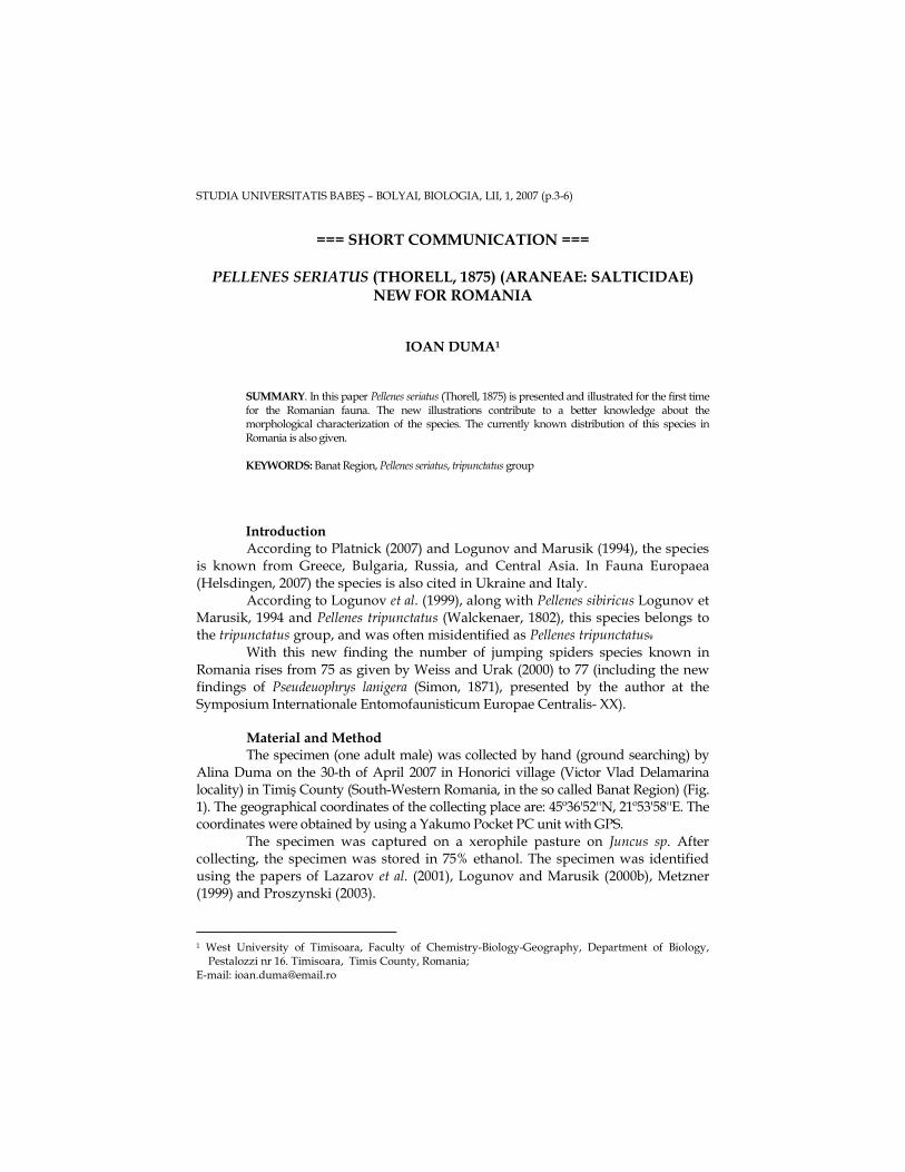
Material and Method The specimen (one adult male) was collected by hand (ground searching) by
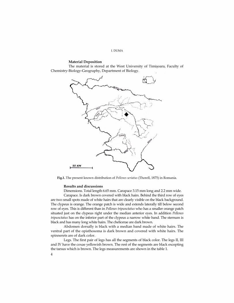
Alina Duma on the 30-th of April 2007 in Honorici village (Victor Vlad Delamarina locality) in Timiş County (South-Western Romania, in the so called Banat Region) (Fig. 1). The geographical coordinates of the collecting place are: 45º36'52''N, 21º53'58''E. The coordinates were obtained by using a Yakumo Pocket PC unit with GPS.
The specimen was captured on a xerophile pasture on Juncus sp. After collecting, the specimen was stored in 75% ethanol. The specimen was identified using the papers of Lazarov et al. (2001), Logunov and Marusik (2000b), Metzner (1999) and Proszynski (2003).
1 West University of Timisoara, Faculty of Chemistry-Biology-Geography, Department of Biology,
Pestalozzi nr 16. Timisoara, Timis County, Romania; E-mail: [email protected]
I. DUMA
4
Material Deposition The material is stored at the West University of Timişoara, Faculty of
Chemistry-Biology-Geography, Department of Biology.
Fig.1. The present known distribution of Pellenes seriatus (Thorell, 1875) in Romania.
Results and discussions
Dimensions. Total length 6.65 mm. Carapace 3.15 mm long and 2.2 mm wide. Carapace. Is dark brown covered with black hairs. Behind the third row of eyes
are two small spots made of white hairs that are clearly visible on the black background. The clypeus is orange. The orange patch is wide and extends laterally till below second row of eyes. This is different than in Pellenes tripunctatus who has a smaller orange patch situated just on the clypeus right under the median anterior eyes. In addition Pellenes tripunctatus has on the inferior part of the clypeus a narrow white band. The sternum is black and has many long white hairs. The chelicerae are dark brown.
Abdomen dorsally is black with a median band made of white hairs. The ventral part of the opisthosoma is dark brown and covered with white hairs. The spinnerets are of dark color.
Legs. The first pair of legs has all the segments of black color. The legs II, III and IV have the coxae yellowish brown. The rest of the segments are black excepting the tarsus which is brown. The legs measurements are shown in the table 1.
A NEW SALTICID SPECIES FOR ROMANIA
5
Table 1. The legs measurements (mm) of the Pellenes seriatus male
Leg Femora Patella Tibia Metatarsus Tarsus Total
I 2,30 1,45 1,70 1,10 0,75 7,30 II 1,40 0,90 0,95 0,65 0,55 4,45 III 1,85 1,00 1,05 0,95 0,60 5,45 IV 1,65 0,75 1,05 0,90 0,70 5,05
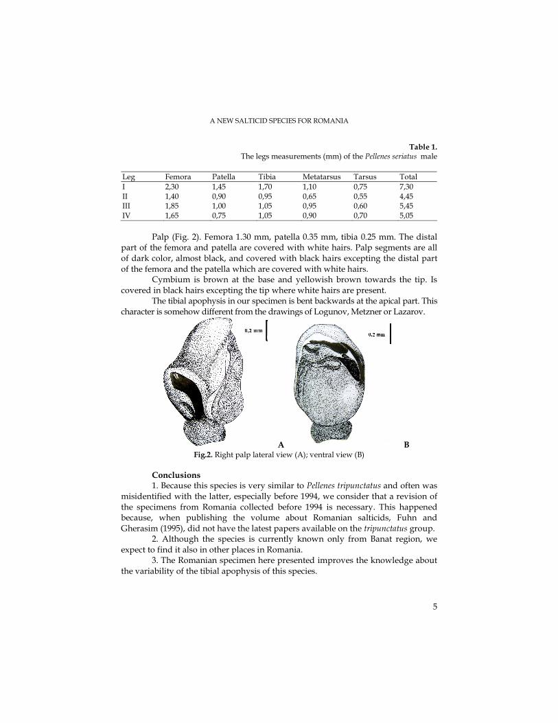
Palp (Fig. 2). Femora 1.30 mm, patella 0.35 mm, tibia 0.25 mm. The distal
part of the femora and patella are covered with white hairs. Palp segments are all of dark color, almost black, and covered with black hairs excepting the distal part of the femora and the patella which are covered with white hairs.
Cymbium is brown at the base and yellowish brown towards the tip. Is covered in black hairs excepting the tip where white hairs are present.
The tibial apophysis in our specimen is bent backwards at the apical part. This character is somehow different from the drawings of Logunov, Metzner or Lazarov.
A B Fig.2. Right palp lateral view (A); ventral view (B)
Conclusions 1. Because this species is very similar to Pellenes tripunctatus and often was
misidentified with the latter, especially before 1994, we consider that a revision of the specimens from Romania collected before 1994 is necessary. This happened because, when publishing the volume about Romanian salticids, Fuhn and Gherasim (1995), did not have the latest papers available on the tripunctatus group.
2. Although the species is currently known only from Banat region, we expect to find it also in other places in Romania.
3. The Romanian specimen here presented improves the knowledge about the variability of the tibial apophysis of this species.
I. DUMA
6
Acknowledgements I want to thank my wife Alina for collecting the material and for all her support.
REFERENCES Fuhn, I. E., Gherasim V., Familia Salticidae. Arachnida. Fauna României., 5 (5), pp. 1-297. Edit.
Academiei Române. 1995 Helsdingen, P., 2007. Fauna Europaea available from: http://www.faunaeur.org/full_results.php?id=182788. Logunov, D. V., Marusik, Y. M., New data on the jumping spiders of the Palearctic fauna (Aranei
Salticidae). ''Arthropoda Selecta'', 3(1-2), 1994, 101-115. Logunov, D. V., Marusik, Y. M., Rakov, S. Y., A review of the genus Pellenes in the fauna of Central
Asia and the Caucasus (Araneae, Salticidae). ''J. nat. Hist.'', 33, 1999, 89-148. Logunov, D.V., Marusik, Y. M., Catalogue of the jumping spiders of northern Asia (Arachnida, Araneae,
Salticidae), pp. 159, map 33, KMK Scientific Press Ltd., Moscow, 2000 Lazarov, S., Deltshev, C. & Blagoev, G., (2001). The spiders (Araneae) of Sashtinska Sredna Gora
Mountain (Bulgaria). Faunistic and zoogeographical analysis. ''Acta zool. bulg.'', 53, 2001, 3-28. Metzner, H., Die Springspinnen (Araneae, Salticidae) Griechenlands, pp. 122-123, 241, table 87a-l,
map 94, Staatliches Museum fur Naturkunde, Karlsruhe, 1999. Platnick, N. I., 2007. The world spider catalog, version 7.5. available from:
http://research.amnh.org/entomology/spiders/catalog/index.html. Proszynski, J., 2003. Salticidae (Aranae) of the World, (revised in part on March 1st, 2006) available
at: http://www.miiz.waw.pl/salticid/diagnost/pellenes/grec-ph.htm. Weiss, I., Urak, I., 2000. Checklist of the Romanian spiders (Arachnida: Araneae) available from:
http://members.aol.com/Arachnologie/Faunenlisten.htm.
STUDIA UNIVERSITATIS BABEŞ – BOLYAI, BIOLOGIA, LII, 1, 2007 (p.7-14)
THE KINETICS OF PLASTOQUINONE RE-OXIDATION IN DARKNESS, IN THE CHLOROPLASTS ISOLATED FROM THE
GREEN ALGA Mougeotia sp., STRAIN AICB 560
VICTOR BERCEA1, BOGDAN DRUGĂ1, CĂTĂLINA VASILESCU1, NICOLAE DRAGOŞ2
SUMMARY. The plastoquinone re-oxidation in darkness in the presence of the methyl viologen and ferricyanure acceptors in the Mougeotia isolated chloroplasts was studied. In order to stop the mitochondrial respiration there were used certain inhibitors which act on the electrons transport in the same way like n-propyl gallate (PG), salicylhydroxamic acid (SHAM) and rotenone in the presence of diurone (DCMU) do. In the presence of the methyl viologen acceptor, DCMU and SHAM have enhanced the amount of re-oxidized plastoquinone, while the quantity of Q-A has decreased. Propyl gallate has generated the stimulation of the plastoquinone re-oxidation, but the amount of Q-A has been decreased. In the presence of ferricyanure, DCMU, propyl gallate, SHAM and rotenone have inhibited the plastoquinone re-oxidation in darkness, and the reduced acceptors quantity has been increased. The plastoquinol re-oxidation in darkness followed by the reduction by light has caused the initial fluorescence change. In the presence of DCMU and artificial electrons acceptors, the supply of quinone acceptors becomes re-oxidized in darkness because of the cyclic electrons chain around PS I where the chlororespiration and the Ndh complex are involved, this influencing directly the photosynthesis. KEYWORDS: chlorophyll fluorescence, chlororespiration, ferricyanure, mitochondrial inhibitors, methyl viologen, quantic yield, photochemical efficiency
Introduction The PS II quinone acceptors become reduced after the light-dark transition
and they display fluorescence binary oscillations due to the QB semiquinone accumulation during the slowed period because of the reduction of PQ in the dark (Groom et al., 1993).
The electrons transfer is created through plastoquinone reduction and oxidation, which are independent of the PSII and PSI, by the NADH-dehydrogenase specific enzymes (Teicher and Scheller, 1998, Joët et al., 2002), and quinol-oxydase specific enzymes (Peltier and Cournac, 2002). The plastoquinone (PQ) reduction is accomplished by NAD(P)H dehydrogenase (Bennoun, 2002), while the reoxidation is accomplished with molecular oxygen consuming in the presence of plastoquinol:oxygen oxidoreductase (Bennoun, 1994) that is n-propyl gallate-sensitive (Cournac et al., 2002), an inhibitor for the alternative mitochondrial oxidases (Josse et al., 2000).
1Institute of Biological Research, 48 Republicii Street, 400015 Cluj-Napoca, Romania E-mail: [email protected] 2 Faculty of Biology and Geology, Babeş-Bolyai University, 5-7 Clinicilor Street, 400006 Cluj-Napoca, Romania
Research supported by CERES Program nr. 4-10/2004
V. BERCEA, B. DRUGĂ, C. VASILESCU, N. DRAGOŞ
8
Into the chloroplasts, the NAD(P)H dehydrogenase complex (Ndh) represents a way of the cyclic electrons transport around PS I and it may produce the plastoquinone non-photochemical reduction in darkness, after illumination. If the type of the electrons transporter that is implied in the plastoquinone reduction is known, then the kind the oxidation transporter remains a disputed subject (Cournac et al, 2000). Under stress it avoids the chloroplasts over-reduction (Munshi et al., 2006).
In this study we analyzed the plastoquinone re-oxidation in darkness in the presence of the methyl viologen and ferricyanure acceptors, in the chloroplasts isolated by the selective action of the mitorespiration inhibitors.
Material and methods The green alga Mougeotia sp. (AICB 560) derives from The Algae Culture
Collection of the Institute of Biological Researches from Cluj-Napoca (AICB) (Dragoş et al., 1997). The alga was grown in Bold nutritive solution (BBM), under continuous air agitation, 630 µmol.m-2.s-1, at 220C. The cultivation period was 23 days.
Chloroplasts isolation. The algal cell suspension was concentrated by nylon filtration and it was grinded in a blender for 30 minutes at medium speed, in 50 mM phosphate buffer, pH=7. The resulted chloroplasts and thylakoidal membranes were caught in a suspension medium consisting in 50 mM phosphate buffer, 3 mM KCl and 330 mM sucrose, pH=7,8 and 200 µM methyl viologen or 5 mM potassium ferricyanure.
The measurement of the PSII electrons acceptors amount by fluorescence. In order to measure the electrons acceptors we used the Bennoun (2001) method that sustain that the area which is delimited by the fluorescence rise and its asymptote is proportional with the amount of electrons acceptors available in PS II. In the presence of DCMU, only a charge separation is possible in the PS II reaction center. Thus, the ratio of the area over the fluorescence rise that was observe in the lack or in the presence of DCMU allows us to estimate the amount of the available electrons acceptors in the PSII active reaction centers (Bennoun, 2001). In our experiments this ratio was 2. The plastoquinone acceptor receives 2 e- from PS II, and, judging on the observed ratio, there should exist about one plastoquinone molecule for every PSII reaction center. All the variants were incubated with 3,27 µM DCMU before using the other specific inhibitors, except the control. The amount of the oxidized primary QA acceptors of the PS II reactions centers was estimated by measuring the fluorescence rise area in the presence of DCMU, while the Q-A reduced acceptor was deduced from 1-Q (Bennoun, 1994).
Analysis of the chlorophyll fluorescence. The chlorophyll fluorescence was measured with PAM-210 fluorometer, as Schreiber et al did (1986). The fluorescence parameters and the quenching analysis were accomplished by the method of saturation pulse. The photochemical energy conversion quantic yield was found by the formula: Yield = ∆F/FM, and the FV/FM (FV/FM = FM-F0/FM) ratio displays the photochemical quantic yield of the closed PS II reaction centers.
KINETICS OF THE PLASTOQUINONE RE-OXIDATION
9
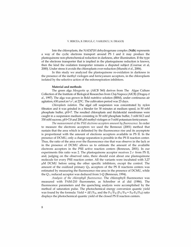
Results and discussion The isolated chloroplasts were exposed to saturable light in the presence of
DCMU in order to achieve the plastoquinone complete oxidation. The in darkness re-oxidation of the plastoquinol amount by using methyl viologen and ferricyanure as electrons acceptors was performed on Mougeotia chloroplasts pre-illuminated for 700 ms and incubated in dark for 30 s, before the second illumination, in order to find the fluorescence together with other mitochondrial inhibitors types. In the methyl viologen control chloroplasts we observed a 35,9 % plastoquinone amount and 64 % Q-A. (Fig. 1).
We considered that the mitochondrial inhibitors caused the ATP decrease, this leading to the chloroplast glycolytic metabolism intensification, thus resulting the enhancement of the reductants able to reduce the plastoquinone (Bennoun, 2001). For the mitochondrial respiration inhibition we used certain inhibitors that act on the electrons transport by inhibiting the quinol:oxygen oxydoreductase which is sensitive to n-propyl gallate (PG), ), salicylhydroxamic acid (SHAM) and rotenone in the presence of diurone (DCMU).
In the chloroplasts that were added with diurone (DCMU) and SHAM the re-oxidized plastoquinol amount has enhance up to 47,9 % comparatively to the control, in darkness (Fig. 1). The reduced acceptors amount was decreased to 52 %. In the presence of propyl gallate there occurred a stimulation up to 71,9 % of plastoquinol re-oxidation in darkness comparatively to the control, but the Q-A quantity decreased down to 28%. The rotenone lead to the inhibition of the plastoquinol amount re-oxidation in darkness down to 29,9 %, this resulting in the Q-A acceptors enhancement up to 70 %.
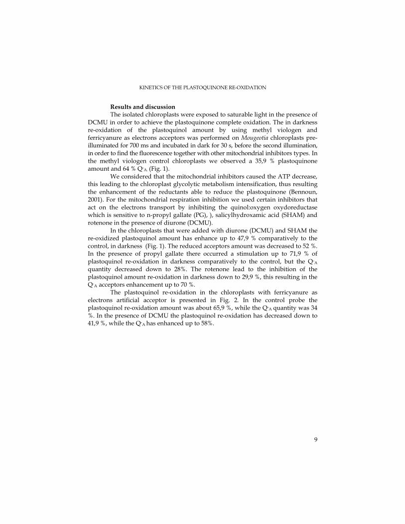
The plastoquinol re-oxidation in the chloroplasts with ferricyanure as electrons artificial acceptor is presented in Fig. 2. In the control probe the plastoquinol re-oxidation amount was about 65,9 %, while the Q-A quantity was 34 %. In the presence of DCMU the plastoquinol re-oxidation has decreased down to 41,9 %, while the Q-A has enhanced up to 58%.
V. BERCEA, B. DRUGĂ, C. VASILESCU, N. DRAGOŞ
10
Fig. 1. Plastoquinol re-oxidation in darkness following reduction by light. M = chloroplasts pre-illuminated for 700 ms and then incubated in dark for 30 seconds; chloroplasts pre-illuminated in the presence of methyl viologen and DCMU (2), PG (3),
SHAM (4) and rotenone (5)
Propyl gallate has inhibited the plastoquinol re-oxidation in darkness with 29,9 %, thus the Q-A amount being increased up to 70 %. SHAM has lead to the decrease of plastoquinol re-oxidation down to 23,9%, while the quantity of reduced electrons acceptors has been enhanced up to 76%. Furthermore, rotenone has inhibited the plastoquinol re-oxidation to 35,9%, while the reduced acceptor amount has enhanced up to 64% (Fig. 2).
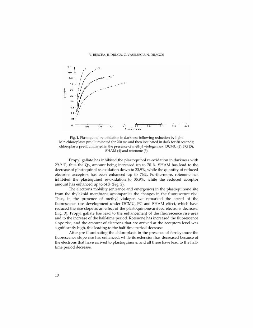
The electrons mobility (entrance and emergence) in the plastoquinone site from the thylakoid membrane accompanies the changes in the fluorescence rise. Thus, in the presence of methyl viologen we remarked the speed of the fluorescence rise development under DCMU, PG and SHAM effect, which have reduced the rise slope as an effect of the plastoquinone-arrived electrons decrease. (Fig. 3). Propyl gallate has lead to the enhancement of the fluorescence rise area and to the increase of the half-time period. Rotenone has increased the fluorescence slope rise, and the amount of electrons that are arrived at the acceptors level was significantly high, this leading to the half-time period decrease.
After pre-illuminating the chloroplasts in the presence of ferricyanure the fluorescence slope rise has enhanced, while its extension has decreased because of the electrons that have arrived to plastoquinone, and all these have lead to the half-time period decrease.
KINETICS OF THE PLASTOQUINONE RE-OXIDATION
11
Fig. 2. Plastoquinol re-oxidation in darkness following reduction by light. M = chloroplasts pre-illuminated for 700 ms and then incubated in dark for 30 seconds; chloroplasts
pre-illuminated in the presence ferricyanure and DCMU (2), PG (3), SHAM (4) and rotenone (5)
MeV
0
0,1
0,2
0,3
0,4
0,5
0,6
0,7
M DCMU DCMU+PG DCMU+SHAM DCMU+Rot
τ τ τ τ s
ec
FeCN
00,050,1
0,150,2
0,250,3
0,350,4
0,450,5
Martor DCMU DCMU + PG DCMU +SHAM
DCMU +Rot
τ τ τ τ s
ec
Fig. 3. The evolution of the half-time period of chlorophyll fluorescence referred to the specific
inhibitors effects after chloroplasts pre-illumination and in the presence of methyl viologen (MeV) and ferricyanure (FeCN)
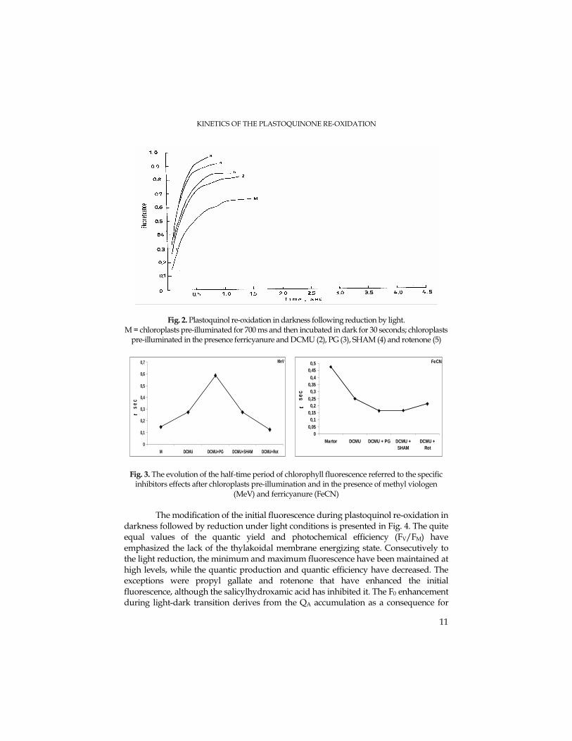
The modification of the initial fluorescence during plastoquinol re-oxidation in
darkness followed by reduction under light conditions is presented in Fig. 4. The quite equal values of the quantic yield and photochemical efficiency (FV/FM) have emphasized the lack of the thylakoidal membrane energizing state. Consecutively to the light reduction, the minimum and maximum fluorescence have been maintained at high levels, while the quantic production and quantic efficiency have decreased. The exceptions were propyl gallate and rotenone that have enhanced the initial fluorescence, although the salicylhydroxamic acid has inhibited it. The F0 enhancement during light-dark transition derives from the QA accumulation as a consequence for
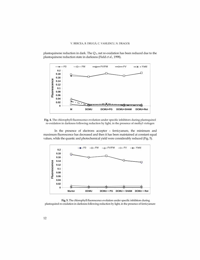
V. BERCEA, B. DRUGĂ, C. VASILESCU, N. DRAGOŞ
12
plastoquinone reduction in dark. The Q-A net re-oxidation has been reduced due to the plastoquinone reduction state in darkness (Field et al., 1998).
00.02
0.040.060.080.1
0.120.140.16
0.180.2
M DCMU DCMU+PG DCMU+SHAM DCMU+Rot
Flu
ore
scen
ce
F0 FM FV/FM FV Yield
Fig. 4. The chlorophyll fluorescence evolution under specific inhibitors during plastoquinol re-oxidation in darkness following reduction by light, in the presence of methyl viologen
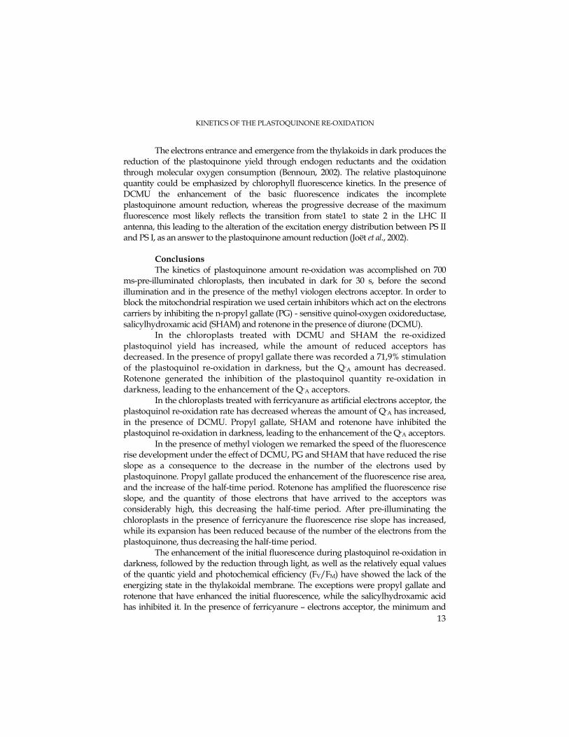
In the presence of electrons acceptor – ferricyanure, the minimum and
maximum fluorescence has decreased and then it has been maintained at constant equal values, while the quantic and photochemical yield were considerably reduced (Fig. 5).
0
0.02
0.04
0.06
0.08
0.1
0.12
0.14
0.16
0.18
0.2
Martor DCMU DCMU + PG DCMU + SHAM DCMU + Rot
Flu
ore
scen
ce
F0 FM FV/FM FV Yield
Fig. 5. The chlorophyll fluorescence evolution under specific inhibitors during plastoquinol re-oxidation in darkness following reduction by light, in the presence of ferricyanure
KINETICS OF THE PLASTOQUINONE RE-OXIDATION
13
The electrons entrance and emergence from the thylakoids in dark produces the reduction of the plastoquinone yield through endogen reductants and the oxidation through molecular oxygen consumption (Bennoun, 2002). The relative plastoquinone quantity could be emphasized by chlorophyll fluorescence kinetics. In the presence of DCMU the enhancement of the basic fluorescence indicates the incomplete plastoquinone amount reduction, whereas the progressive decrease of the maximum fluorescence most likely reflects the transition from state1 to state 2 in the LHC II antenna, this leading to the alteration of the excitation energy distribution between PS II and PS I, as an answer to the plastoquinone amount reduction (Joët et al., 2002).
Conclusions The kinetics of plastoquinone amount re-oxidation was accomplished on 700
ms-pre-illuminated chloroplasts, then incubated in dark for 30 s, before the second illumination and in the presence of the methyl viologen electrons acceptor. In order to block the mitochondrial respiration we used certain inhibitors which act on the electrons carriers by inhibiting the n-propyl gallate (PG) - sensitive quinol-oxygen oxidoreductase, salicylhydroxamic acid (SHAM) and rotenone in the presence of diurone (DCMU).
In the chloroplasts treated with DCMU and SHAM the re-oxidized plastoquinol yield has increased, while the amount of reduced acceptors has decreased. In the presence of propyl gallate there was recorded a 71,9% stimulation of the plastoquinol re-oxidation in darkness, but the Q-A amount has decreased. Rotenone generated the inhibition of the plastoquinol quantity re-oxidation in darkness, leading to the enhancement of the Q-A acceptors.
In the chloroplasts treated with ferricyanure as artificial electrons acceptor, the plastoquinol re-oxidation rate has decreased whereas the amount of Q-A has increased, in the presence of DCMU. Propyl gallate, SHAM and rotenone have inhibited the plastoquinol re-oxidation in darkness, leading to the enhancement of the Q-A acceptors.
In the presence of methyl viologen we remarked the speed of the fluorescence rise development under the effect of DCMU, PG and SHAM that have reduced the rise slope as a consequence to the decrease in the number of the electrons used by plastoquinone. Propyl gallate produced the enhancement of the fluorescence rise area, and the increase of the half-time period. Rotenone has amplified the fluorescence rise slope, and the quantity of those electrons that have arrived to the acceptors was considerably high, this decreasing the half-time period. After pre-illuminating the chloroplasts in the presence of ferricyanure the fluorescence rise slope has increased, while its expansion has been reduced because of the number of the electrons from the plastoquinone, thus decreasing the half-time period.
The enhancement of the initial fluorescence during plastoquinol re-oxidation in darkness, followed by the reduction through light, as well as the relatively equal values of the quantic yield and photochemical efficiency (FV/FM) have showed the lack of the energizing state in the thylakoidal membrane. The exceptions were propyl gallate and rotenone that have enhanced the initial fluorescence, while the salicylhydroxamic acid has inhibited it. In the presence of ferricyanure – electrons acceptor, the minimum and
V. BERCEA, B. DRUGĂ, C. VASILESCU, N. DRAGOŞ
14
maximum fluorescence has decreased and then it has been maintained at constant equal values, while the quantic and photochemical yield were considerably reduced.
In the presence of DCMU and artificial electrons acceptors, the supply of quinone acceptors becomes re-oxidized in darkness because of the cyclic electrons chain around PS I where the chlororespiration and the Ndh complex are involved, this influencing directly the photosynthesis.
REFERENCES
Bennoun, P. (1994). Chlororespiration revisited: mitochondrial-plastid interactions in Chlamydomonas., Biochim. Biophys.Acta, 1186,59-66.
Bennoun, P. (2001). Chlororespiration and the process of carotenoid biosynthesis. Biochim. Biophys.Acta,1506,133-142.
Bennoun, P. (2002). The present model for chlororespiration., Photosznth. Res.,73, 273-277. Cournac, L., Redding, K., Ravenel, J., Rumeau, D., Josse, E.M., Kuntz, M., Peltier, G.(2000). Electron
flow between photosustem II and oxygen in chloroplasts of photosystem I –deficient algae is mediated by a quinol oxidase involved in chlororespiration. J. Biol. Chem., 275, 17256-17262.
Cournac, L., Josse, E.M., Joët, T., Rumeau, D., Redding, K., Kuntz, M., Peltier, G.(2000). Flexibility in photosynthetic electron transport: a newly identified chloroplast oxidase involved in chlororespiration. Phil.Trans.R.Soc.London. B, 355, 1447-1454.
Cournac, L., Latouche, G., Cerovic, Z., Redding, K., Ravenel, J., Peltier, G. (2002). In vivo interactions between photosynthesis mitorespiration and chlororespiration in Chlamydomonas reinhardtii. Plant Physiol., 129, 1921-1928.
Dragoş, N., Péterfi, L. Şt., Momeu, L., Popescu, C. (1997). An introduction to the algae and the culture collection of algae at the Institute of the Biological Research Cluj-Napoca, Cluj University Press.
Feild, T.S., Nedbal, L., Ort, D.R. (1998). Nonphotochemical reduction of the plastochinone pool in sunflower leaves originates from chlororespiration. Plant Physiol., 116, 1209-1218.
Groom, Q., Kramer, D.M., Crofts, A.R., Ort, D.R. (1993). The non-photochemical reduction of plastoquinone in leaves. Photosynth. Res., 36, 205-213.
Joët, T., Cournac, L., Peltier, G., Havaux, M. (2002). Cyclic electron flow around photosystem I in C3 plants. In vivo control by the redox state of chloroplasts and involvement of the NADH-dehydrogenase complex. Plant Pysiol., 128, 760-769.
Josse, E.M., Simkin, A.J., Gaffe, J., Laboure, A.M., Kuntz, M., Carol, P. (2000). A plastid terminal oxidase associated with carotenoid desaturation during chromoplast differetiation. Plant Physiol., 123, 1427-1436.
Munshi, M.K., Kobayashi, Y., Shikanai, T. (2006). Chlororespiratory Reduction 6 is a novel factor required for accumulation of the chloroplast NAD(P)H dehydrogenase complex in Arabidopsis. Plant Physiol., 141, 737-744.
Peltier, G., Cournac, L.(2002). Chlororespiration. Annu.Rev.Plant Physiol., 53, 523-550. Schreiber, U., Schliwa, U., Bilger, W. (1986). Continuous recording of photochemical and non-
photochemical chlorophyll fluorescence quenching with a new type of modulation fluorometer, Photosynth. Res., 10, 51-62.
Teicher, H. B. & Scheller, H. V. (1998) The NAD(P)H-dehydrogenase in barley (Hordeum vulgare L.) thylakoids is photoactivatable and utilizes NADPH as well as NADH. Plant Physiol., 117, 525-532
STUDIA UNIVERSITATIS BABEŞ – BOLYAI, BIOLOGIA, LII, 1, 2007 (p.15-23)
CHLORORESPIRATION STUDY ON ISOLATED CHLOROPLASTS FROM THE GREEN ALGA Mougeotia sp., STRAIN AICB 560
VICTOR BERCEA1, CĂTĂLINA VASILESCU1, BOGDAN DRUGĂ1, NICOLAE DRAGOŞ2
SUMMARY. The kinetics of plastoquinone oxidation in darkness at isolated chloroplasts from Mougeotia in the presence of DCMU and of some mitochondrial respiratory inhibitors was studied. In the presence of the acceptor methyl viologen, DCMU conducted to the increase of the available plastoquinone pool and the fraction Q-A decreased. N-propyl gallate sustained the oxidation of plastoquinone in darkness, and Q-A increased at 64%. Salicylhydroxamic acid (SHAM) sustained the plastoquinone oxidation in darkness, and Q-A decreased. Rotenone inhibited the plastoquinone oxidation in darkness, and Q- decreased at 70%. In the presence of the acceptor ferricyanure, DCMU did not produce changes in plastoquinone pool. PG and SHAM did not change the plastoquinone redox state, rotenone sustained the plastoquinone oxidation in darkness, while Q-A decreased. It was observed that the partial reduction of plastoquinone pool was accompanied by changes in the initial fluorescence. The results showed the presence of respiratory type of electron transfer from NAD(P)H to oxygen in chloroplasts, mediated by plastoquinone pool involved in the photosynthetic electron transfer chain, process driven by chlororespiration. KEYWORDS: chlorophyll fluorescence kinetics, half time, mitochondrial oxidases inhibitors, plastoquinone redox state Introduction Chlororespiration has been defined as the interaction of respiratory electrons
carriers from mitochondria with the electron transport chain from chloroplast thylakoidal membranes (Bennoun, 2001). The electron transfer is generated through the plastoquinone reduction and oxidation, independent of PS II and PS I, by specific enzymes like NADH-dehydrogenase (Teicher and Scheller, 1998) and by quinol oxidase (Peltier and Cournac, 2002). The plastoquinol reoxidation (PQ-H2) in darkness is made on the account of molecular oxygen, in the presence of plastoquinol: oxygen oxidoreductase (Bennoun, 1994; 2002), enzyme sensitive to n-propyl gallate (Cournac et al., 2000), an inhibitor of alternative mitochondrial oxidases (Josse et al., 2000).
At chloroplast level, the NAD(P)H-dehydrogenase complex represent a path of electron cyclic transport. In stress conditions, it protects the chloroplasts from over-reduction (Munshi et al., 2006). The chlororespiration activity and the expression of Ndh complex are being intensified as a response to stress factors (Bukhov et al., 2000). The photooxidative stress increases the nonphotochemical reduction of intersystemic
1 Institute of Biological Research, 48 Republicii Street, 400015 Cluj-Napoca, Romania E-mail: [email protected] 2 Faculty of Biology and Geology, Babeş-Bolyai University, 5-7 Clinicilor Street, 400006 Cluj-Napoca Research supported by CERES Program nr. 4-10/2004
V. BERCEA, C. VASILESCU, B. DRUGĂ, N. DRAGOŞ
16
electron carriers (Havaux, 1996; Casano et al., 1999; 2000; 2001). The chlororespiration may play a photoprotective role in photosynthesis (Endo et al., 1999) through the modulation of the electron cyclic flow around PS I (Deng et al., 2003; Joët et al., 2002).
This paper studied the interaction between the thylakoidal electron transport and the respiratory electron transport, at the level of isolated chloroplast, in the presence of high light and some respiratory inhibitors.
Material and methods. The green alga Mougeotia sp. belongs to the Collection of Algae Cultures of
I.C.B. Cluj-Napoca (AICB) (Dragoş et al., 1997). The strain was grown in Bold nutritive solution (BBM), during continuous air stirring, continuous illumination with 630 µmol.m-2.s-1., at 22oC. The cultivation period was of 23 days.
Chloroplasts isolation. The algal suspension was concentrated by filtration on nylon and was subjected to grinding in a Blender for 30 minutes, at a medium rotation speed, in 50mM phosphate buffer pH=7. The chloroplasts and the thylakoidal membranes were colected in the resuspension medium made up of 50 mM phosphate buffer, 3mM KCl, 330 mM sucrose, and 200 µM methyl viologen or 5 mM potassium ferricyanure, pH=7,8.
The assessment of PS II electron acceptors quantity by fluorescence. The isolated chloroplasts were incubated at dark one hour with DCMU [3-(3,4-dichlorophenyl)-1,1-dimethylurea] before using, enabling thus the complete oxidation of plastoquinone. For electron acceptors measurement was used the Bennoun’s method (2001) which sustains that the area delimitated by the fluorescence rise and its asymptote is proportional with the quantity of electron acceptors available at PSII. In the presence of DCMU, it is possible only one charge separation per PS II. As a consequence, the ratio of the area over the fluorescence rise observed in the absence and in the presence of DCMU enables the estimation of electron acceptor quantity available per PS II active reaction centre (Bennoun, 2001). In our experiments, this ratio was in average about 2. The acceptor plastoquinone gains 2e- from PS II and, on the basis of the observed ratio, would exist in average about one molecule of plastoquinone per PS II reaction centre. Excepting the control, all experimental variants were incubated with 3.27 µM DCMU before adding the other inhibitors.
The quantity of oxidized primary acceptors QA of PSII centers was estimated in the presence of DCMU by measuring the area over the fluorescence rise and the reduced acceptor Q- was calculated as 1-Q (Bennoun, 1994).
Chlorophyll fluorescence analysis. The chlorophyll fluorescence was measured with a PAM-210 fluorometer as was previously described by Schreiber et al. (1986). The fluorescence parameters and the quenching analysis were conducted by the saturation pulse method. The quantum yield of the photochemical energy conversion was determined using the equation Yield = ∆F/FM, and the ratio FV/FM (FV/FM = FM-F0/FM) shows the photochemical quantum yield of the closed PS II reaction centers.
CHLORORESPIRATION STUDY ON ISOLATED CHLOROPLASTS
17
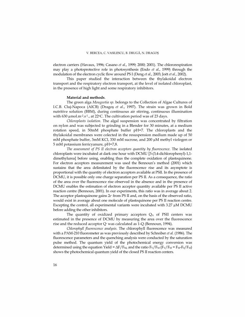
Results and discussion The kinetics of chlorophyll fluorescence in vivo measured by the illumination of
dark adapted chloroplasts, in the presence of methyl viologen electron artificial acceptor and treated with (DCMU) and other inhibitors, is presented in fig. 1. In control chloroplast was determined an available pool of 11.9% QA, and a quantity of 88% Q-A. By treating the chloroplasts with DCMU, which blocks the electron transport between the primary acceptor QA, the secondary acceptor QB and plastoquinone pool, the kinetics mirrors the plastoquinone reduction in darkness by stromal reductants, in the presence of O2 absorption induced by chlororespiration. In the presence of DCMU, the available plastoquinone pool has increased at 53,% comparing to the control. The ratio between areas showed the existence of two molecules of electron acceptors per active PSII reaction centre. The fraction of reduced acceptors (Q-A) has decreased at 46% (Fig. 1). The life time of Q-A in the DCMU presence has increased (Rappaport et al., 1999).
Aiming the determination of the catalytic type involved in plastoquinone oxidation in vivo, was tested the effect of mitochondrial oxidases inhibitors in the DCMU presence. Thus, n-propyl gallate (PG), inhibitor of mitochondrial alternative oxidases, sustained the plastoquinone oxidation in darkness, increasing the available electron acceptors at 35.9% comparing to the control. The molecular ratio of 1.4 showed the presence of 0.7 electron acceptor molecules per PSII active reaction centre. The fraction of reduced acceptors (Q-A) had risen to 64% (Fig. 1).
The plastoquinone redox state in darkness is controlled as following: the reduction is accomplished by stromal reductants which redox state depends on the metabolic and mitochondrial activity, and the oxidation is subjected to the highly sensitive propyl gallate oxidase (Cournac et al., 2002). The salicylhydroxamic acid (SHAM), considered an inhibitor of mitochondrial alternative oxidases, had sustained the plastoquinone oxidation in darkness. The available plastoquinone pool had risen at 59.9% comparing to the control. The molecular ratio had shown the existence of one molecule of electron acceptors per active reaction centre, and the fraction of reduced acceptors (Q-A) was of 40%.
Rotenone is a inhibitor of mitochondrial complex I, acting on the Fe-S reduced centers of ubiquinone. It was observed that rotenone is a week inhibitor of plastoquinone oxidation in darkness, diminishing the available pool of electron acceptors at 29.9% comparing with the control. The ratio between fluorescence areas of 1.2 showed the existence of 0.6 molecules of electron acceptors per reaction centre. The amount of electron acceptors in a reduced state (Q-) had increased at 70% (Fig. 1). The inhibition of plastoquinone oxidation in darkness by rotenone is explained by the inhibitory action conducted upon the NAD(P)H oxidation, confirming that this enzyme activity is of complex I type (Teicher and Scheller, 1998).
V. BERCEA, C. VASILESCU, B. DRUGĂ, N. DRAGOŞ
18
Fig. 1. The chlorophyll fluorescence kinetics in isolated chloroplasts from Mougeotia. Area interval = 4.5 s; M=dark-adapted cells (control). Chloroplasts treated with 200 µM methyl viologen and 3,27 µM DCMU (2), and then inhibited for 4 minutes in darkness with 1 mM PG (n-propyl gallate) (3), 1 mM SHAM (salicylhydroxamic acid) (4), and 20 µM rotenone (5)
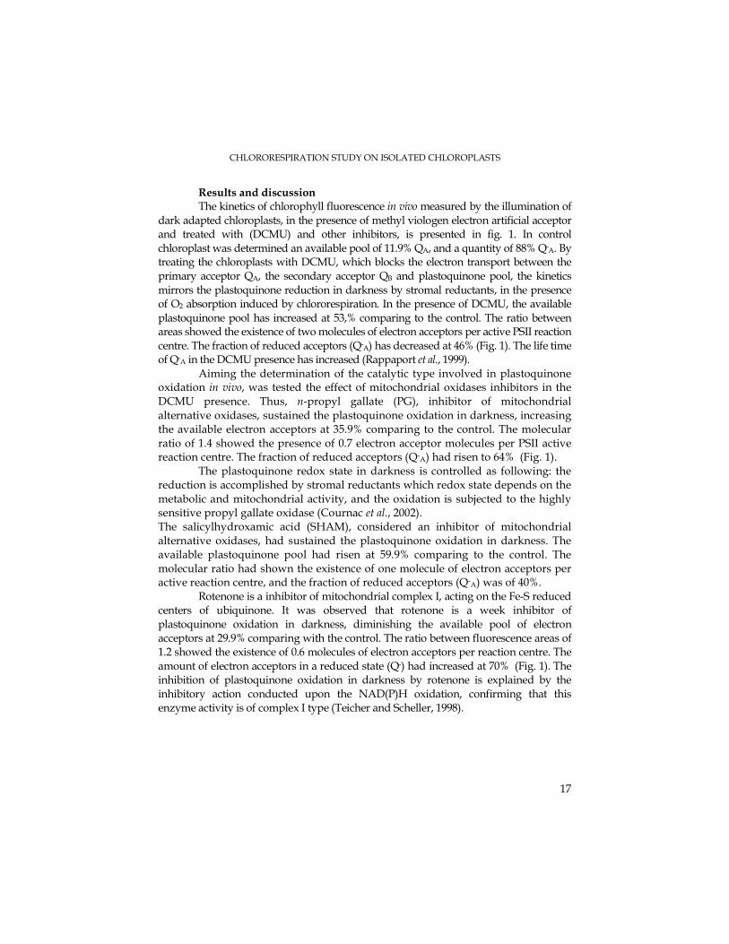
The chlorophyll fluorescence kinetics in vivo, measured by the illumination
of isolated chloroplasts adapted to dark in the presence of an artificial electron acceptor like ferricyanure, is presented in Fig. 2. At the control variant, the plastoquinone pool was of 41.9%, along with a quantity of 58% of Q-A. In the presence of DCMU, the available plastoquinone pool was of 47.9% comparing to the control. The ratio between areas revealed the existence of 0.5 molecules of electron acceptors per active PSII reaction centre. The fraction of reduced acceptors (Q-A) was of 52% (Fig. 2). It was shown that DCMU did not produce changes in the plastoquinone pool. At experimental variants with n-propyl gallate (PG) and SHAM, the plastoquinone redox state remained unchanged comparative to DCMU and control. But it was observed that rotenone sustained the plastoquinone oxidation in darkness, rising up the available electron acceptors pool at 53.9% comparing to the control. The ratio between fluorescence areas of 1.3 showed the existence of 0.6 molecules of electron acceptors per reaction centre. The quantity of electron acceptors in a reduced state (Q-A) decreased at 46% (Fig. 2).

CHLORORESPIRATION STUDY ON ISOLATED CHLOROPLASTS
19
Fig. 2. The chlorophyll fluorescence kinetics in isolated chloroplasts from Mougeotia. Area interval = 4.5 s; M=dark-adapted cells (control). Chloroplasts treated with 5 mM ferricyanure and
3,27 µM DCMU (2), and then inhibited for 4 minutes at dark with 1 mM PG (3), 1 mM SHAM (4), and 20 µM rotenone (5)
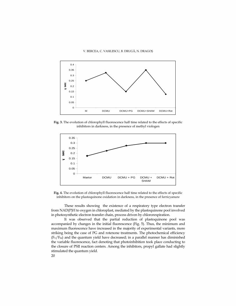
The evolution of the half time in dark conditions (τ) showed two important
phenomena: a- the speed of fluorescence rise recording time was dependent of plastoquinone quantity, and b- the slope of this rise depended on the quantity of electrons arrived at plastoquinone. In the presence of methyl viologen was observed that the available plastoquinone pool was increased in the cases of the variants with DCMU and SHAM, and the arrival of an increased electron number at the plastoquinone system, conducting to the increase of fluorescence rise slope, was observed at PG and rotenone variants where the fluorescence area decreased (Fig. 3).
In the presence ferricyanure as electron acceptor, the slope of fluorescence rise has been slightly decreased by the arrival at the plastoquinone of an relatively constant number of electrons, and the area of the rise remained constant due to the relatively constant values of the half time (Fig. 4).
V. BERCEA, C. VASILESCU, B. DRUGĂ, N. DRAGOŞ
20
0
0.05
0.1
0.15
0.2
0.25
0.3
0.35
0.4
M DCMU DCMU+PG DCMU+SHAM DCMU+Rot
τ τ τ τ se
c
Fig. 3. The evolution of chlorophyll fluorescence half time related to the effects of specific inhibitors in darkness, in the presence of methyl viologen
0
0.05
0.1
0.15
0.2
0.25
0.3
0.35
Martor DCMU DCMU + PG DCMU +SHAM
DCMU + Rot
τ τ τ τ s
ec
Fig. 4. The evolution of chlorophyll fluorescence half time related to the effects of specific inhibitors on the plastoquinone oxidation in darkness, in the presence of ferricyanure
These results showing the existence of a respiratory type electron transfer
from NAD(P)H to oxygen in chloroplast, mediated by the plastoquinone pool involved in photosynthetic electron transfer chain, process driven by chlororespiration.
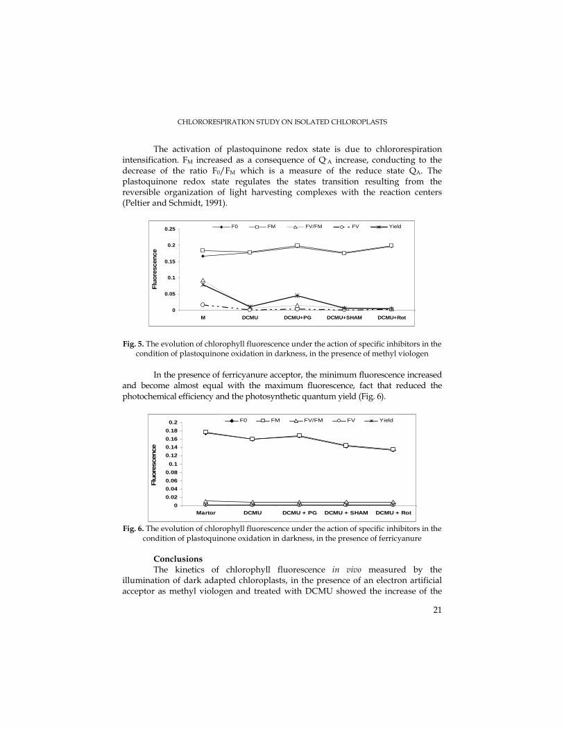
It was observed that the partial reduction of plastoquinone pool was accompanied by changes in the initial fluorescence (Fig. 5). Thus, the minimum and maximum fluorescence have increased in the majority of experimental variants, more striking being the case of PG and rotenone treatments. The photochemical efficiency (FV/FM) and the quantum yield have decreased; in a parallel manner has diminished the variable fluorescence, fact denoting that photoinhibition took place conducting to the closure of PSII reaction centers. Among the inhibitors, propyl gallate had slightly stimulated the quantum yield.
CHLORORESPIRATION STUDY ON ISOLATED CHLOROPLASTS
21
The activation of plastoquinone redox state is due to chlororespiration intensification. FM increased as a consequence of Q-A increase, conducting to the decrease of the ratio F0/FM which is a measure of the reduce state QA. The plastoquinone redox state regulates the states transition resulting from the reversible organization of light harvesting complexes with the reaction centers (Peltier and Schmidt, 1991).
0
0.05
0.1
0.15
0.2
0.25
M DCMU DCMU+PG DCMU+SHAM DCMU+Rot
Flu
ore
scen
ce
F0 FM FV/FM FV Yield
Fig. 5. The evolution of chlorophyll fluorescence under the action of specific inhibitors in the condition of plastoquinone oxidation in darkness, in the presence of methyl viologen
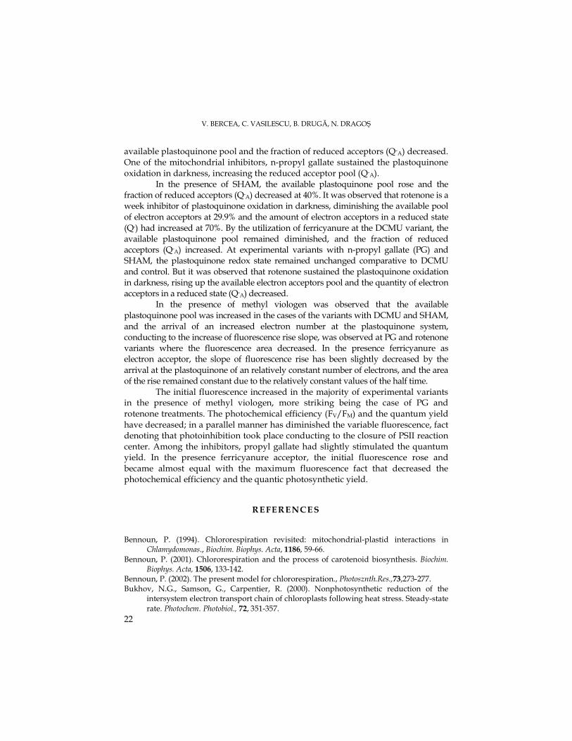
In the presence of ferricyanure acceptor, the minimum fluorescence increased
and become almost equal with the maximum fluorescence, fact that reduced the photochemical efficiency and the photosynthetic quantum yield (Fig. 6).
0
0.02
0.04
0.06
0.08
0.1
0.12
0.14
0.16
0.18
0.2
Martor DCMU DCMU + PG DCMU + SHAM DCMU + Rot
Flu
ore
scen
ce
F0 FM FV/FM FV Yield
Fig. 6. The evolution of chlorophyll fluorescence under the action of specific inhibitors in the
condition of plastoquinone oxidation in darkness, in the presence of ferricyanure Conclusions The kinetics of chlorophyll fluorescence in vivo measured by the
illumination of dark adapted chloroplasts, in the presence of an electron artificial acceptor as methyl viologen and treated with DCMU showed the increase of the
V. BERCEA, C. VASILESCU, B. DRUGĂ, N. DRAGOŞ
22
available plastoquinone pool and the fraction of reduced acceptors (Q-A) decreased. One of the mitochondrial inhibitors, n-propyl gallate sustained the plastoquinone oxidation in darkness, increasing the reduced acceptor pool (Q-A).
In the presence of SHAM, the available plastoquinone pool rose and the fraction of reduced acceptors (Q-A) decreased at 40%. It was observed that rotenone is a week inhibitor of plastoquinone oxidation in darkness, diminishing the available pool of electron acceptors at 29.9% and the amount of electron acceptors in a reduced state (Q-) had increased at 70%. By the utilization of ferricyanure at the DCMU variant, the available plastoquinone pool remained diminished, and the fraction of reduced acceptors (Q-A) increased. At experimental variants with n-propyl gallate (PG) and SHAM, the plastoquinone redox state remained unchanged comparative to DCMU and control. But it was observed that rotenone sustained the plastoquinone oxidation in darkness, rising up the available electron acceptors pool and the quantity of electron acceptors in a reduced state (Q-A) decreased.
In the presence of methyl viologen was observed that the available plastoquinone pool was increased in the cases of the variants with DCMU and SHAM, and the arrival of an increased electron number at the plastoquinone system, conducting to the increase of fluorescence rise slope, was observed at PG and rotenone variants where the fluorescence area decreased. In the presence ferricyanure as electron acceptor, the slope of fluorescence rise has been slightly decreased by the arrival at the plastoquinone of an relatively constant number of electrons, and the area of the rise remained constant due to the relatively constant values of the half time.
The initial fluorescence increased in the majority of experimental variants in the presence of methyl viologen, more striking being the case of PG and rotenone treatments. The photochemical efficiency (FV/FM) and the quantum yield have decreased; in a parallel manner has diminished the variable fluorescence, fact denoting that photoinhibition took place conducting to the closure of PSII reaction center. Among the inhibitors, propyl gallate had slightly stimulated the quantum yield. In the presence ferricyanure acceptor, the initial fluorescence rose and became almost equal with the maximum fluorescence fact that decreased the photochemical efficiency and the quantic photosynthetic yield.
REFERENCES
Bennoun, P. (1994). Chlororespiration revisited: mitochondrial-plastid interactions in Chlamydomonas., Biochim. Biophys. Acta, 1186, 59-66.
Bennoun, P. (2001). Chlororespiration and the process of carotenoid biosynthesis. Biochim. Biophys. Acta, 1506, 133-142.
Bennoun, P. (2002). The present model for chlororespiration., Photosznth.Res.,73,273-277. Bukhov, N.G., Samson, G., Carpentier, R. (2000). Nonphotosynthetic reduction of the
intersystem electron transport chain of chloroplasts following heat stress. Steady-state rate. Photochem. Photobiol., 72, 351-357.
CHLORORESPIRATION STUDY ON ISOLATED CHLOROPLASTS
23
Casano, L.M., Martin, M., Zapata, J.M., Sabater, B.(1999). Leaf age- and paraquat-dependent effects on the levels of enzymes protecting against photooxidative stress. Plant Sci., 149, 13-22.
Casano, L.M., Zapata, J.M., Martin, M., Sabater, B.(2000). Chlororespiration and poising of cyclic electron transport- plastoquinone as electron transporter between thylakoid NADH dehydrohenase and peroxidase. J.Biol.Chem., 275,942-948.
Casano, L.M., Martin, M., Sabater, B.(2001). Hydrogen peroxide mediates the induction of chloroplastic Ndh complex under photooxidative stress in barley. Plant Physiol, 125, 1450-1458.
Cournac, L., Redding, K., Ravenel, J., Rumeau, D., Josse, E.M., Kuntz, M., Peltier, G. (2000). Electron flow between photosustem II and oxygen in chloroplasts of photosystem I –deficient algae is mediated by a quinol oxidase involved in chlororespiration. J. Biol. Chem., 275, 17256-17262.
Cournac, L., Josse, E.M., Joët, T., Rumeau, D., Redding, K., Kuntz, M., Peltier, G. (2000). Flexibility in photosynthetic electron transport: a newly identified chloroplast oxidase involved in chlororespiration. Phil.Trans.R.Soc.London.B,355,1447-1454.
Cournac, L., Latouche, G., Cerovic, Z., Redding, K., Ravenel, J., Peltier, G. (2002). In vivo interactions between photosynthesis mitorespiration and chlororespiration in Chlamydomonas reinhardtii. Plant Physiol.,129,1921-1928.
Deng, Y., Ye, J., Mi, H. (2003). Effects of low CO2 on NAD(P)H dehydrogenase, a mediator of cyclic electron transport around photosystem I in the cyanobacterium Synechocystis PCC 6803. Plant Cell Physiol., 44, 534-540.
Dragoş, N., Péterfi, L. Şt., Momeu, L., Popescu, C. (1997). An introduction to the algae and the culture collection of algae at the Institute of the Biological Research Cluj-Napoca. Cluj University Press.
Endo, T., Shikanai, T., Takabayashi, A., Asada, K., Sato, F.(1999). The rol of chloroplastic NAD(P)H dehydrogenase in photoprotection. FEBS Lett., 457, 5-8.
Havaux, M. (1996). Short-term responses of photosystem I to heat stress. Induction of a PS II-independent electron transport through PS I fed by stromal components. Photosynth. Res., 47, 85-97.
Joët, T., Cournac, L., Peltier, G., Havaux, M. (2002). Cyclic electron flow around photosystem I in C3 plants. In vivo control by the redox state of chloroplasts and involvement of the NADH-dehydrogenase complex. Plant Pysiol., 128, 760-769.
Josse, E.M., Simkin, A.J., Gaffe, J., Laboure, A.M., Kuntz, M., Carol, P. (2000). A plastid terminal oxidase associated with carotenoid desaturation during chromoplast differetiation. Plant Physiol., 123, 1427-1436.
Munshi, M.K., Kobayashi, Y., Shikanai, T. (2006). Chlororespiratory Reduction 6 is a novel factor required for accumulation of the chloroplast NAD(P)H dehydrogenase complex in Arabidopsis. Plant Physiol., 141, 737-744.
Peltier, G., Schmidt, G.W. (1991). Chlororespiration: an adaptation to nitrogen deficiency in Chlamydomonas reinhardtii. Proc.Natl.Acad.Sci.USA, 88, 4791-4795.
Peltier, G., Cournac, L.(2002). Chlororespiration. Annu.Rev.Plant Physiol., 53, 523-550. Rappaport, F., Finazzi, G., Pierre, Y., Bennoun, P. (1999). A new electrochemical gradient
generator in thylakoid membranes of green algae. Biochemistry, 38, 2040-2047. Schreiber, U., Schliwa, U., Bilger, W. (1986). Continuous recording of photochemical and
non-photochemical chlorophyll fluorescence quenching with a new type of modulation fluorometer, Photosynth. Res., 10, 51-62.
Teicher, H. B. & Scheller, H. V. (1998) The NAD(P)H-dehydrogenase in barley (Hordeum vulgare L.) thylakoids is photoactivatable and utilizes NADPH as well as NADH. Plant Physiol., 117, 525-532
STUDIA UNIVERSITATIS BABEŞ – BOLYAI, BIOLOGIA, LII, 1, 2007 (p.25-36)
PRELIMINARY DATA ON ALGAL, MACROINVERTEBRATE AND FISH COMMUNITIES FROM THE ARIEŞ CATCHMENT AREA,
TRANSYLVANIA, ROMANIA
MOMEU, L.1, BATTES, K.W.2, PRICOPE, F.2, AVRAM, A.1, BATTES, K.P.1, CÎMPEAN, M.1, URECHE, D.2, STOICA, I.2
SUMMARY. Water quality assessment studies represent an important part of the monitoring programs. According to the new requirements of the European legislation, these ecological studies should focus on biotic communities. In case of lotic ecosystem monitoring, benthic assemblages, both algae and macroinvertebrates, are very accurate indicators of water quality, due to their particular characteristics. Moreover, nekton organisms, mainly fishes, due to their position on top of the river food webs represent a useful tool in characterizing the ecological status of the ecosystems. These are the reasons why we focused on three main biotic communities for the present paper: algae, macroinvertebrates and fishes. This preliminary report presents only qualitative data on these assemblages, in case of algae and macrozoobenthos, and quantitative preliminary data for ichthyofauna. Subsequent researches will establish the water quality for every sampling site considered, by means of biotic indices. KEYWORDS: algal community, macroinvertebrates, ichthyofauna. Introduction According to the European Union legislation, The Water Frame Directive,
integrate monitoring programs should be based on an ecosystem and biological approach. From this point of view, algae, macroinvertebrates and fishes represent biological components of great importance in running water quality assessment (Wetzel, 2001).
The present study is included in a larger monitoring study of water quality assessment based on biotic indices. This paper presents the preliminary results concerning the algal, macroinvertebrate and fish communities from the Arieş catchment area, not only from the main river course, but also from its main tributaries. The Arieş River drainage basin records a strong heterogeneity, including not only unaffected areas but also severely polluted ones.
The main objective of the present study was to establish the qualitative structure of benthic and nektonic communities from the Arieş catchment area, for subsequent studies regarding the assessment of water quality. Pointing out the main sources of human pressures was another objective of the paper.
1 „Babeş-Bolyai” University, Faculty of Biology and Geology, Department of Taxonomy and Ecology, 400006 Cluj-Napoca, E-mail: [email protected]
2 University of Bacău, Faculty of Sciences, Department of Biology, 600115 Bacău.
L. MOMEU, K.W. BATTES, F. PRICOPE, A. AVRAM, K.P. BATTES, M. CÎMPEAN, D.URECHE, I. STOICA
26
Material and Methods The Arieş River is the largest right tributary of the Mureş River, having a
catchment area of 2970 km2 and a total length of 164 km (Ujvari, 1972). It represents the Southern limit of the Apuseni Natural Park and its source lays East from the main ridge of the Bihor Mountains. The Arieş can be divided in three main areas: the upper Arieş, also called the Arieşul Mare, from the river source to the Câmpeni locality, the middle Arieş, up to the Corneşti locality, and the lower Arieş, that stretches until the junction with the Mureş River. The Arieşul Mare collects its tributaries from the Scărişoara Plateau. They collect the waters coming from the Padiş limestone plateau. The junction of the Arieşul Mare and Arieşul Mic Rivers forms the main Arieş River. In Câmpeni locality, the Abrud River flows into the Arieş. It gathers many watercourses coming from several mining sites (Bucium-Izbita or Roşia Montană). A special attention must be paid to the decantation ponds located in these mining areas. Downstream of Câmpeni, the Arieş River records a strong left asymmetry: its tributaries come from the South-Eastern Bihor and Muntele Mare Mountains (Ujvari, 1972). For example, the Hăşdate River crosses the famous Turzii Gorge. Other smaller tributaries come from the Feleacu Ridge.
The sampling sites considered for this particular study were chosen according to the geographic, geologic and hydrologic characteristics of the river catchment area. They were easy of access and representative for the study area (considering the altitude, substratum nature, shadowing degree, human impacts etc.).
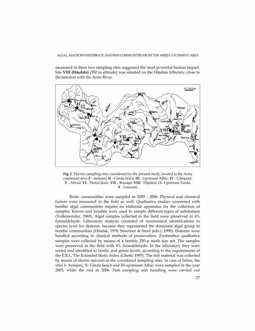
For the preliminary data report, ten representative sampling sites were chosen, as shown in figure 1. Six stations were located on the main river, not only in the upper region but also in the middle and lower reaches. Site I (Arieşeni) was located in Arieşeni locality, at 924 m altitude a.s.l. Site III (upstream Albac) was situated upstream of Albac locality, at 642 m altitude a.s.l. The substratum consisted of boulders and coarse pebble. Site IV (Câmpeni) (534m altitude) was located upstream of Câmpeni locality and it represented the last sampling site from the upper Arieş river. It reflected the human impact characteristic to inhabitted regions – industry, waste pits etc. Site VII (Brăzăşti) was located in the Brăzăşti locality, at 465 m altitude, and the substratum was covered in fine organic-mineral sediment. Site IX (upstream Turda) was situated about 10 km upstream from Turda, at 345 m altitude, at Corneşti, in the lower stretch of the river. A ballast exploitation site located near by represented the main human impact in this area. The last station located on the main river course was site X (Luncani), situated in the homonymous village, at 279 m altitude, downstream of the Turda and Câmpia Turzii localities, thus reflecting their impacts on the river biotic communities. Four stations were chosen on the main river tributaries. Thus, site II (Gârda Seacă) (729 m altitude) was located on the Gârda Seacă brook, in the upper reach of the river, in a clean area. Stations V (Abrud) and VI (Pârâul Şesii) were located on two tributaries that collected waters coming from the mining regions upstream, at 542 and 482 meters altitude, respectively. The aspect of these two tributaries, their orange color, and the physical, chemical and biological parameters
ALGAL, MACROINVERTEBRATE AND FISH COMMUNITIES FROM THE ARIEŞ CATCHMENT AREA
27
measured in these two sampling sites suggested the most powerful human impact. Site VIII (Hăşdate) (350 m altitude) was situated on the Hăşdate tributary, close to the junction with the Aries River.
Fig. 1. The ten sampling sites considered for the present study, located in the Arieş catchment area (I - Arieşeni; II – Gârda Seacă; III - Upstream Albac; IV - Câmpeni; V - Abrud; VI - Pârâul Şesii; VII – Brăzăşti; VIII - Hăşdate; IX- Upstream Turda;
X - Luncani) Biotic communities were sampled in 2005 - 2006. Physical and chemical
factors were measured in the field as well. Qualitative studies concerned with benthic algal communities require no elaborate apparatus for the collection of samples. Knives and brushes were used to sample different types of substratum (Vollenweider, 1969). Algal samples collected in the field were preserved in 4% formaldehyde. Laboratory analysis consisted of taxonomical identifications to species level for diatoms, because they represented the dominant algal group in benthic communities (Hindak, 1978; Stoermer & Smol (eds.), 1999). Diatoms were handled according to classical methods of preservation. Zoobenthos qualitative samples were collected by means of a benthic 250 µ mesh size net. The samples were preserved in the field with 4% formaldehyde. In the laboratory they were sorted and identified to family and genus levels, according to the requirements of the E.B.I., The Extended Biotic Index (Ghetti, 1997). The fish material was collected by means of electro narcosis at the considered sampling sites. In case of fishes, the sites I- Arieşeni, II- Gârda Seacă and III-upstream Albac were sampled in the year 2005, while the rest in 2006. Fish sampling and handling were carried out
L. MOMEU, K.W. BATTES, F. PRICOPE, A. AVRAM, K.P. BATTES, M. CÎMPEAN, D.URECHE, I. STOICA
28
according to methods used in the European Union. Species taxonomy followed the reviewed list of freshwater fish (Nalbant, 2003). Preliminary data in case of ichthyofauna included also quantitative estimations (absolute abundance).
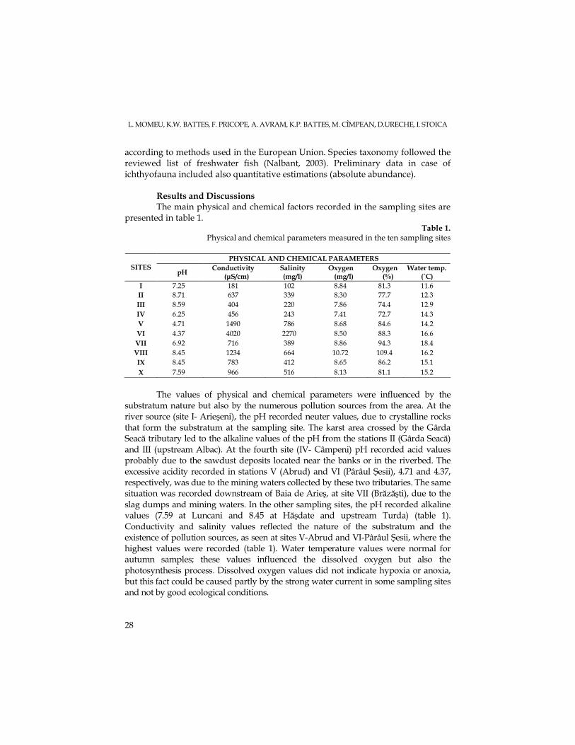
Results and Discussions The main physical and chemical factors recorded in the sampling sites are
presented in table 1. Table 1.
Physical and chemical parameters measured in the ten sampling sites
PHYSICAL AND CHEMICAL PARAMETERS SITES
pH Conductivity
(µS/cm) Salinity (mg/l)
Oxygen (mg/l)
Oxygen (%)
Water temp. (˚C)
I 7.25 181 102 8.84 81.3 11.6 II 8.71 637 339 8.30 77.7 12.3 III 8.59 404 220 7.86 74.4 12.9 IV 6.25 456 243 7.41 72.7 14.3 V 4.71 1490 786 8.68 84.6 14.2 VI 4.37 4020 2270 8.50 88.3 16.6 VII 6.92 716 389 8.86 94.3 18.4 VIII 8.45 1234 664 10.72 109.4 16.2 IX 8.45 783 412 8.65 86.2 15.1 X 7.59 966 516 8.13 81.1 15.2
The values of physical and chemical parameters were influenced by the
substratum nature but also by the numerous pollution sources from the area. At the river source (site I- Arieşeni), the pH recorded neuter values, due to crystalline rocks that form the substratum at the sampling site. The karst area crossed by the Gârda Seacă tributary led to the alkaline values of the pH from the stations II (Gârda Seacă) and III (upstream Albac). At the fourth site (IV- Câmpeni) pH recorded acid values probably due to the sawdust deposits located near the banks or in the riverbed. The excessive acidity recorded in stations V (Abrud) and VI (Pârâul Şesii), 4.71 and 4.37, respectively, was due to the mining waters collected by these two tributaries. The same situation was recorded downstream of Baia de Arieş, at site VII (Brăzăşti), due to the slag dumps and mining waters. In the other sampling sites, the pH recorded alkaline values (7.59 at Luncani and 8.45 at Hăşdate and upstream Turda) (table 1). Conductivity and salinity values reflected the nature of the substratum and the existence of pollution sources, as seen at sites V-Abrud and VI-Pârâul Şesii, where the highest values were recorded (table 1). Water temperature values were normal for autumn samples; these values influenced the dissolved oxygen but also the photosynthesis process. Dissolved oxygen values did not indicate hypoxia or anoxia, but this fact could be caused partly by the strong water current in some sampling sites and not by good ecological conditions.
ALGAL, MACROINVERTEBRATE AND FISH COMMUNITIES FROM THE ARIEŞ CATCHMENT AREA
29
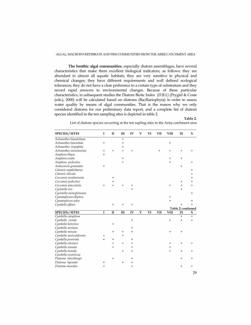
The benthic algal communities, especially diatom assemblages, have several characteristics that make them excellent biological indicators, as follows: they are abundant in almost all aquatic habitats; they are very sensitive to physical and chemical changes; they have different requirements and well defined ecological tolerances; they do not have a clear preference to a certain type of substratum and they record rapid answers to environmental changes. Because of these particular characteristics, in subsequent studies the Diatom Biotic Index (D.B.I.) (Prygiel & Coste (eds.), 2000) will be calculated based on diatoms (Bacillariophyta) in order to assess water quality by means of algal communities. That is the reason why we only considered diatoms for our preliminary data report, and a complete list of diatom species identified in the ten sampling sites is depicted in table 2.
Table 2. List of diatom species occurring at the ten sapling sites in the Arieş catchment area
SPECIES / SITES I II III IV V VI VII VIII IX X
Achnanthes biasoletiana + Achnanthes lanceolata + + + Achnanthes kryophila + Achnanthes minutissima + + + + + + + + Amphora libyca + Amphora ovalis + + + Amphora pediculus + + + Aulacoseira granulata + + Caloneis amphisbaena + Caloneis silicula + Cocconeis neodiminuta + + Cocconeis pediculus + + + Cocconeis placentula + + + + + + + Cyclotella iris + + Cyclotella meneghineana + Cymatopleura elliptica + Cymatopleura solea + + Cymbella affinis + + + + +
Table 2: continued SPECIES / SITES I II III IV V VI VII VIII IX X Cymbella caespitosa + + Cymbella cistula + + + + Cymbella helvetica + Cymbella mesiana + Cymbella minuta + + + + + Cymbella naviculiformis + + Cymbella prostrata + + + Cymbella silesiaca + + + + + + Cymbella sinuata + + + Cymbella tumida Cymbella ventricosa
+ + + + +
Diatoma ehrenbergei + + + + Diatoma hyemale + + + Diatoma mesodon + + + +
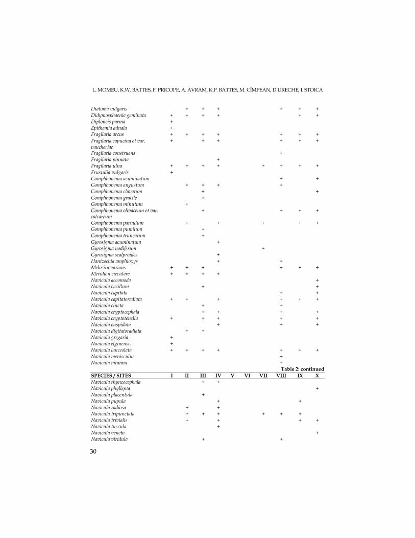
L. MOMEU, K.W. BATTES, F. PRICOPE, A. AVRAM, K.P. BATTES, M. CÎMPEAN, D.URECHE, I. STOICA
30
Diatoma vulgaris + + + + + + Didymosphaenia geminata + + + + + + Diploneis parma + Epithemia adnala + Fragilaria arcus + + + + + + + Fragilaria capucina et var. vaucheriae
+ + + + + +
Fragilaria construens + Fragilaria pinnata + Fragilaria ulna + + + + + + + + Frustulia vulgaris + Gomphhonema acuminatum + + Gomphhonema angustum + + + + Gomphhonema clavatum + + Gomphhonema gracile + Gomphhonema minutum + Gomphhonema olivaceum et var. calcareum
+ + + +
Gomphhonema parvulum + + + + + Gomphhonema pumilum + Gomphhonema truncatum + Gyrosigma acuminatum + Gyrosigma nodiferum + Gyrosigma scalproides + Hantzschia amphioxys + + Melosira varians + + + + + + Meridion circulare + + + + Navicula accomoda + Navicula bacillum + + Navicula capitata + + Navicula capitatoradiata + + + + + + Navicula cincta + + Navicula cryptocephala + + + + Navicula cryptotenella + + + + + Navicula cuspidata + + + Navicula digitatoradiata + + Navicula gregaria + Navicula elginensis + Navicula lanceolata + + + + + + + Navicula menisculus + Navicula minima +
Table 2: continued SPECIES / SITES I II III IV V VI VII VIII IX X Navicula rhyncocephala + + Navicula phyllepta + Navicula placentula + Navicula pupula + + Navicula radiosa + + Navicula tripunctata + + + + + + Navicula trivialis + + + + Navicula tuscula + Navicula veneto + Navicula viridula + +
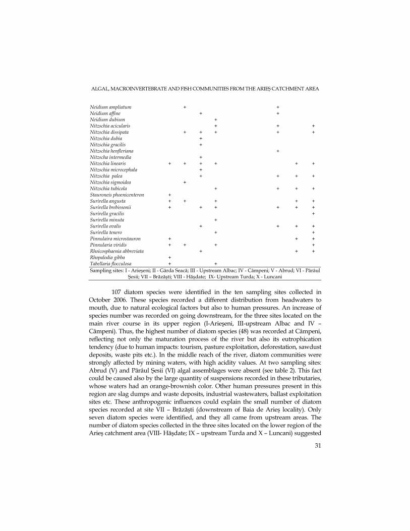
ALGAL, MACROINVERTEBRATE AND FISH COMMUNITIES FROM THE ARIEŞ CATCHMENT AREA
31
Neidium ampliatum + + Neidium affine + + Neidium dubium + Nitzschia acicularis + + + Nitzschia dissipata + + + + + Nitzschia dubia + Nitzschia gracilis + Nitzschia henfleriana + Nitzscha intermedia + Nitzschia linearis + + + + + + Nitzschia microcephala + Nitzschia palea + + + + Nitzschia sigmoidea + Nitzschia tubicola + + + + Stauroneis phoenicenteron + Surirella angusta + + + + + Surirella brebissonii + + + + + + Surirella gracilis + Surirella minuta + Surirella ovalis + + + + Surirella tenero + + Pinnulaira microstauron + + + Pinnularia viridis + + + + Rhoicosphaenia abbreviata + + + Rhopalodia gibba + Tabellaria flocculosa + + Sampling sites: I - Arieşeni; II - Gârda Seacă; III - Upstream Albac; IV - Câmpeni; V - Abrud; VI - Pârâul
Şesii; VII – Brăzăşti; VIII - Hăşdate; IX- Upstream Turda; X - Luncani
107 diatom species were identified in the ten sampling sites collected in
October 2006. These species recorded a different distribution from headwaters to mouth, due to natural ecological factors but also to human pressures. An increase of species number was recorded on going downstream, for the three sites located on the main river course in its upper region (I-Arieşeni, III-upstream Albac and IV – Câmpeni). Thus, the highest number of diatom species (48) was recorded at Câmpeni, reflecting not only the maturation process of the river but also its eutrophication tendency (due to human impacts: tourism, pasture exploitation, deforestation, sawdust deposits, waste pits etc.). In the middle reach of the river, diatom communities were strongly affected by mining waters, with high acidity values. At two sampling sites: Abrud (V) and Pârâul Şesii (VI) algal assemblages were absent (see table 2). This fact could be caused also by the large quantity of suspensions recorded in these tributaries, whose waters had an orange-brownish color. Other human pressures present in this region are slag dumps and waste deposits, industrial wastewaters, ballast exploitation sites etc. These anthropogenic influences could explain the small number of diatom species recorded at site VII – Brăzăşti (downstream of Baia de Arieş locality). Only seven diatom species were identified, and they all came from upstream areas. The number of diatom species collected in the three sites located on the lower region of the Arieş catchment area (VIII- Hăşdate; IX – upstream Turda and X – Luncani) suggested
L. MOMEU, K.W. BATTES, F. PRICOPE, A. AVRAM, K.P. BATTES, M. CÎMPEAN, D.URECHE, I. STOICA
32
the existence of an eutrophication process, caused by industrial and agriculture wastewaters, domestic waste deposits located on the river banks and the ballast exploitation sites located in this area.
As concerns the dominant diatom species identified in the ten sampling sites, species of Navicula dominated at site I –Arieşeni; species of Cymbella and Diatoma vulgaris prevailed in site III, and for the station IV – Câmpeni, Achnanthes minutissima
became most abundant. At station IX – upstream Turda, Fragilaria capucina was dominant, while a few species of Surirella dominated the diatom community from Luncani-site X. As for the main river tributaries, Achnanthes minutissima prevailed at station II-Gârda Seacă. In Hăşdate – site VIII, a green alga (Chlorophyta): Chladophora glomerata developed extensively, next to the diatom Fragilaria ulna.
The dominant algae were cosmopolite, eurybiont elements, except for the species Diatoma vulgaris, which indicated clean, xeno-oligosaprobic waters at site III – Garda Seaca, and species belonging to Surirella genus (identified at site X – Luncani), indicators of beta or alpha mesosaprobic waters.
The benthic macroinvertebrate community represents a good water quality indicator because it includes organisms attached to the substratum; they include numerous populations, with different tolerance levels for limiting factors and having a relatively long life cycle. The Extended Biotic Index (E.B.I.) will be used in subsequent studies to assess the water quality in the Arieş catchment area. Table 3 presents the list of macroinvertebrate taxa, identified to the genus level (in case of Plecoptera, Ephemeroptera, Plathelminthes) or to the family level (in case of Trichoptera, Coleoptera, Diptera, Crustacea, Gastropoda and Oligochaeta), according to the requirements of the Extended Biotic Index.
25 taxa were identified at site I-Arieşeni, most of them belonging to the Orders Plecoptera and Ephemeroptera, known to be indicators of good quality waters. A high number of taxa were collected also at the second and third stations (site II-Gârda Seacă and site III-upstream Albac), 26 and 23 taxa, respectively, indicating a good water quality as well. On going downstream, beginning with site IV-Câmpeni, the number of taxa found in the benthic community decreased drastically. Only the stonefly genus (Leuctra) was identified, the most tolerant Plecoptera genus to ecological factors. At the sampling sites V-Abrud, VI-Pârâul Şesii and VII-Brăzăşti, no zoobenthic organism was collected. These results are similar to the algal community data presented above. As for site VIII - Hăşdate, located on the right tributary of the Arieş River, stoneflies were not found, but all the other groups were well represented, indicating a less drastic human impact compared to the three stations described above.
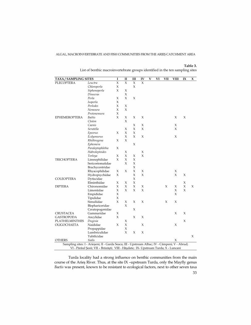
ALGAL, MACROINVERTEBRATE AND FISH COMMUNITIES FROM THE ARIEŞ CATCHMENT AREA
33
Table 3. List of benthic macroinvertebrate groups identified in the ten sampling sites
TAXA / SAMPLING SITES I II III IV V VI VII VIII IX X
Leuctra X X X X Chloroperla X X Siphonoperla X X Dinocras X Perla X X X Isoperla X Perlodes X X Nemoura X X
PLECOPTERA
Protonemura X Baëtis X X X X X X Cloëon X Caenis X X X Seratella X X X X Epeorus X X X Ecdyonurus X X X X Rhithrogena X X Ephemera X Paraleptophlebia X Habroleptoides X X
EPHEMEROPTERA
Torleya X X X X Limnephilidae X X X Sericostomatidae X X Brachycentridae X Rhyacophilidae X X X X X
TRICHOPTERA
Hydropsychidae X X X X X Dytiscidae X COLEOPTERA
Elminthidae X X X X Chironomidae X X X X X X X X Limoniidae X X X X X X Empididae X X Tipulidae X Simuliidae X X X X X X Blephariceridae X
DIPTERA
Ceratopogonidae X CRUSTACEA Gammaridae X X X GASTROPODA Ancylidae X X X PLATHELMINTHES Dugesia X X
Naididae X X X X Propappidae X Lumbriculidae X X X
OLIGOCHAETA
Tubificidae X OTHERS Sialis X
Sampling sites: I - Arieşeni; II - Garda Seaca; III - Upstream Albac; IV - Câmpeni; V - Abrud; VI - Pârâul Şesii; VII – Brăzăşti; VIII - Hăşdate; IX- Upstream Turda; X - Luncani
Turda locality had a strong influence on benthic communities from the main
course of the Arieş River. Thus, at the site IX –upstream Turda, only the Mayfly genus Baetis was present, known to be resistant to ecological factors, next to other seven taxa
L. MOMEU, K.W. BATTES, F. PRICOPE, A. AVRAM, K.P. BATTES, M. CÎMPEAN, D.URECHE, I. STOICA
34
belonging to other groups (see table 3). Downstream from Turda, in Luncani – site X, only chironomids and oligochaetes were identified.
Using fish communities in ecological monitoring programs has several advantages, as follows: fishes are present in all aquatic habitats, including the heavy polluted waters; fish communities are very stable, recording small variations of population characteristics; due to their top position in the food webs, fish communities include the features of most of the lower trophic levels; fishes have long life cycles that last years or dozens of years; fishes are easily to identify in the field. Subsequent studies will assess water quality by means of I.B.I. – the Index of Biological Integrity, introduced and improved by Karr and Dudley (1981), modified by Battes (1999). The index of biological integrity combines twelve parameters of fish communities, divided in three groups: species composition and richness, trophic structure and ichthyofauna abundance and status.
Table 4 depicts the characteristics of considered habitats and fishing areas at the sampling sites located on the Arieş catchment area. At the sites V – Abrud and VI - Pârâul Şesii, no fish community was sampled due to the lack of algal and macroinvertebrate assemblages.
Table 4. Habitat and fishing area characteristics at the sampling sites
Habitat data Collection technical data
Sampling sites Width (m) Depth (m)
Speed (m/s)
Substratum nature
Fishing area (m2)
Tension (V)/ intensity (A)
Fishing depth (m)
I - Arieşeni 5 – 6 0.2 – 0.7 0.4 – 0.6 - boulders >30 300 550 / 1.5 0.3 – 0.5 II - Gârda Seacă 3 – 4 0.1 – 0.5 0.3 - boulders >20 200 540 / 2 0.1 – 0.3 III - upstream Albac 30 0.2 – 0.8 0.3 – 0.6 - boulders <25 1000 550 / 5 0.2 – 0.5 IV - Câmpeni 35 0.8 0.6 - boulders <30 600 550 / 4 0.7 VII - Brăzăşti 60 0.2 – 0.5 0.4 – 0.6 - boulders <10 500 500 / 8 0.4
VIII - Hăşdate 3 0.2 – 0.6 0.3 - boulders. >30 gravel
150 250 / 6 0.2 – 0.8
IX - upstream Turda 40 0.4 0.3 – 0.5 - boulders <10 1010 550 / 8 0.4
X -Luncani 30 0.2 – 0.6 0.3 - boulders. >30 gravel
660 250 / 6 0.4
Thirteen fish species were identified at the considered sampling sites, as
follows: Salmo trutta fario (brown trout); Thymallus thymallus (grayling); Eudontonmyzon danfordi (Danubian lamprey); Cottus gobio (bull head); Phoxinus phoxinus (minnow); Orthrias barbatulus (loach); Alburnoides bipunctatus (schneider); Barbus meridionalis (afterbarbe); Chondrostoma nasus (undermouth); Leuciscus cephalus (chub); Alburnus alburnus (bleak); Gobio gobio (gudgeon) and Carassius auratus (gold fish). At site VII – Brăzăşti, where few algal and macroinvertebrate organisms were collected, no fish individual was sampled. At site X - Luncani, only two fish species were identified, represented by only a small number of individuals, thus reflecting the human impact described above. The absolute abundance of the fish species collected in the sampling sites located on the main river course and on its main tributaries is presented in table 5.
ALGAL, MACROINVERTEBRATE AND FISH COMMUNITIES FROM THE ARIEŞ CATCHMENT AREA
35
Table 5.
The absolute abundance of fish species (number of individuals) collected at the considered sampling sites on the main river course and on its main tributaries
Fish species
Sampling sites 1 2 3 4 5 6 7 8 9 10 11 12 13
Total / site
II 14 - - 16 - - - - - - - - - 30 T VIII - - - - - 2 13 42 - 15 - 1 1 74
I 2 - - 11 - - - - - - - - - 13 III 1 11 - 12 1 1 - 9 - - - - - 35 IV - - - 8 2 1 18 14 12 2 - 1 - 58 VII - - - - - - - - - - - - - - IX - - - - 22 - 1 4 - 57 1 1 1 87
MC
X - - - - - - - - - 4 4 - - 8 Total - tributaries
29 4 - 48 6 2 13 42 - 15 - 1 1 161
Total – the Arieş River
6 14 1 44 25 2 19 28 12 63 5 2 1 232
Total (general)
35 18 1 92 31 4 32 70 12 78 5 3 2 393
Abbreviations: T – tributaries; MC – the main river course Fish species: 1. Salmo trutta fario; 2. Thymallus thymallus; 3. Eudontonmyzon danfordi; 4. Cottus gobio; 5. Phoxinus phoxinus; 6. Orthrias barbatulus; 7. Alburnoides bipunctatus; 8. Barbus meridionalis; 9. Chondrostoma nasus; 10. Leuciscus cephalus; 11. Alburnus alburnus; 12. Gobio gobio; 13. Carassius auratus
Thus, the preliminary study regarding the algal, macroinvertebrate and
fish communities from the Arieş catchment area included data concerning the structure of these biotic assemblages that reflected the main human pressures existing in the area. In the upper reaches of the river, the main human impacts are related to the tourism, deforestations and wood processing industry. In the middle reaches, the main disturbances are caused by the mining waters that flow into the river tributaries, leading to the total absence of biotic assemblages. In the lower catchment area, the Turda and Câmpia Turzii localities cause the main human pressures, by discharges of domestic and industrial wastewaters. Moreover, the waste deposits located in the riverbed or on its banks represent a constant impact on the river and on its main tributaries as well.
Acknowledgements. The authors recognize with gratitude the financial
support provided by CNCSIS (contract no. 1329/2006).
L. MOMEU, K.W. BATTES, F. PRICOPE, A. AVRAM, K.P. BATTES, M. CÎMPEAN, D.URECHE, I. STOICA
36
REFERENCES
Battes, K.W. (1999). Influence of the hydrotechnical development and of BistriŃa rivers pollution on the natural fish population. Studii şi Cercetări; Biologie, 4, 283-294.
Ghetti, P.F. (1997). Manuale di aplicazione - Indice Biotico Esteso (IBE); I macroinvertebrati nel controllo della qualita degli ambienti di acque currenti, Ed. Provincia Autonoma di Trento, Agenzia provinciale per la protezione dell ambiente, Trento.
Hindak, F. (ed.) (1978). Sladkovodne Riasy, Slovenske Pedagogicke Nakladatelstvo, Bratislava. Karr, J.R., Dudley, D.R. (1981). Ecological perspective on Water quality goals, Environment
management, 5-15. Nalbant, T. (2003). Checklist of the fishes of Romania; Part one: freshwater and
brackishwater fish, Studii şi Cercetări; Biologie, 8, 122-128. Prygiel, J., Coste, M. (eds) (2000). Guide Methodologique pour la mise en oeuvre de l’Indice
Biologique Diatomees, Ed. Cemagref, Bordeaux. Stoermer, E.F., Smol, J.P. (eds) (1999). The Diatoms, applications for the Environmental and Earth
Sciences, Cambridge University Press, Cambridge. Ujvari, I. (1972). Geografia apelor României, Editura ŞtiinŃifică, Bucureşti. Vollenweider, R.A. (1969). A Manual on Methods for Measuring Primary Production in Aquatic
Environments, IBP Handbook 12, Blackwell Scientific Publications, Oxford. Wetzel, R.G. (2001). Limnology. Lake and river ecosystems, Third Edition, Academic Press, San Diego.
STUDIA UNIVERSITATIS BABEŞ – BOLYAI, BIOLOGIA, LII, 1, 2007 (p.37-45)
PARTIAL BIOCHEMICAL CHARACTERIZATION OF STORAGE PROTEIN FROM ALEURONE CELLS OF BARLEY
(HORDEUM VULGARE L.)
HORIA BANCIU1, FLORINA OLARU1, VERENA HENGST,2 MANUELA BANCIU1, IOAN PETRESCU1, AURORA MOCANU3,
CORNELIU TARBA1, TRAIANOS YUPSANIS4 and MARIA TOMOAIA-COTISEL3
SUMMARY. The present paper focuses on the characterization of 7S globulin regarding its biochemical and biophysical properties. 7S globulin was extracted by the purification of aleurone cells of barley (Yupsanis et al., 1990). This protein belongs to the storage protein class with important roles in providing the amino acids during germination of plants. 7S globulin from barley aleurone was found to be positively-charged since the measurement of zeta potential revealed a value of +17 mV. Spectrofluorimetric measurements using the anionic probe ANS (anilino-naphtalene sulfonate) showed that the positive amino acids are probably located mainly on the external surface of protein. 7S globulin is chemically stable up to 6 M urea and consists of 4 different subunits with molecular weights of 65, 37, 25 and 20 kDa. Due to its chemical and physical properties this protein may be of interest for nanobiotechnology of surface coatings (glass, mica, silicone etc). KEYWORDS: globulins, chemical and thermal stability, positive charge, surface coatings, storage proteins.
Introduction
With an average of about 10–12% of dry weight, cereal grains contain a relatively low amount of proteins compared to legume seeds,. The embryo and outer aleurone layer of the endosperm contain globulin storage proteins. Globulins are readily soluble in dilute salt solution and have sedimentation coefficients of about 7 (Shewry and Halford, 2002). The storage proteins belonging to the 7S globulin class have limited sequence similarity with, and may be homologous to, the 7S vicilins of legumes and other dicotyledonous plants (Kriz, 1999). Related proteins have been found in embryos and/or aleurone layers of wheat, barley, oats and rice (Burgess and Shewry, 1986; Yupsanis et al., 1990; Heck et al., 1993; Horikoshi and Morita, 1975). The 7S globulins are stored in protein bodies and appear to function solely as storage proteins. Up to now, little is known about their molecular diversity and functions during the plant development. In the present paper we report some biochemical and biophysical features of the 7S globulin previously purified from barley (Hordeum
1 Department of Biology, Faculty of Biology and Geology, Babes-Bolyai University, Cluj-Napoca, Romania 2 Department of Pharmaceutical Technology and Biopharmacy, Philipps-University of Marburg, Ketzerbach 63, 35037 Marburg, Germany
3 Faculty of Chemistry and Chemical Engineering, Babes-Bolyai University, Cluj-Napoca, Romania 3 Department of Biochemistry, School of Chemistry, Aristotle University, 54006 Thessaloniki, Greece
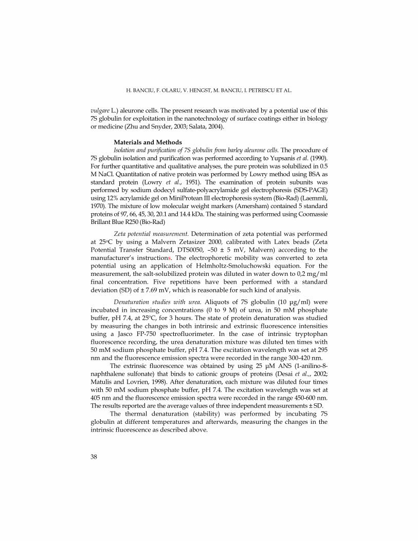
H. BANCIU, F. OLARU, V. HENGST, M. BANCIU, I. PETRESCU ET AL.
38
vulgare L.) aleurone cells. The present research was motivated by a potential use of this 7S globulin for exploitation in the nanotechnology of surface coatings either in biology or medicine (Zhu and Snyder, 2003; Salata, 2004).
Materials and Methods Isolation and purification of 7S globulin from barley aleurone cells. The procedure of
7S globulin isolation and purification was performed according to Yupsanis et al. (1990). For further quantitative and qualitative analyses, the pure protein was solubilized in 0.5 M NaCl. Quantitation of native protein was performed by Lowry method using BSA as standard protein (Lowry et al., 1951). The examination of protein subunits was performed by sodium dodecyl sulfate-polyacrylamide gel electrophoresis (SDS-PAGE) using 12% acrylamide gel on MiniProtean III electrophoresis system (Bio-Rad) (Laemmli, 1970). The mixture of low molecular weight markers (Amersham) contained 5 standard proteins of 97, 66, 45, 30, 20.1 and 14.4 kDa. The staining was performed using Coomassie Brillant Blue R250 (Bio-Rad)
Zeta potential measurement. Determination of zeta potential was performed at 25oC by using a Malvern Zetasizer 2000, calibrated with Latex beads (Zeta Potential Transfer Standard, DTS0050, –50 ± 5 mV, Malvern) according to the manufacturer’s instructions. The electrophoretic mobility was converted to zeta potential using an application of Helmholtz-Smoluchowski equation. For the measurement, the salt-solubilized protein was diluted in water down to 0,2 mg/ml final concentration. Five repetitions have been performed with a standard
deviation (SD) of ± 7.69 mV, which is reasonable for such kind of analysis.
Denaturation studies with urea. Aliquots of 7S globulin (10 µg/ml) were incubated in increasing concentrations (0 to 9 M) of urea, in 50 mM phosphate buffer, pH 7.4, at 25ºC, for 3 hours. The state of protein denaturation was studied by measuring the changes in both intrinsic and extrinsic fluorescence intensities using a Jasco FP-750 spectrofluorimeter. In the case of intrinsic tryptophan fluorescence recording, the urea denaturation mixture was diluted ten times with 50 mM sodium phosphate buffer, pH 7.4. The excitation wavelength was set at 295 nm and the fluorescence emission spectra were recorded in the range 300-420 nm.
The extrinsic fluorescence was obtained by using 25 µM ANS (1-anilino-8-naphthalene sulfonate) that binds to cationic groups of proteins (Desai et al.,, 2002; Matulis and Lovrien, 1998). After denaturation, each mixture was diluted four times with 50 mM sodium phosphate buffer, pH 7.4. The excitation wavelength was set at 405 nm and the fluorescence emission spectra were recorded in the range 450-600 nm. The results reported are the average values of three independent measurements ± SD.
The thermal denaturation (stability) was performed by incubating 7S globulin at different temperatures and afterwards, measuring the changes in the intrinsic fluorescence as described above.
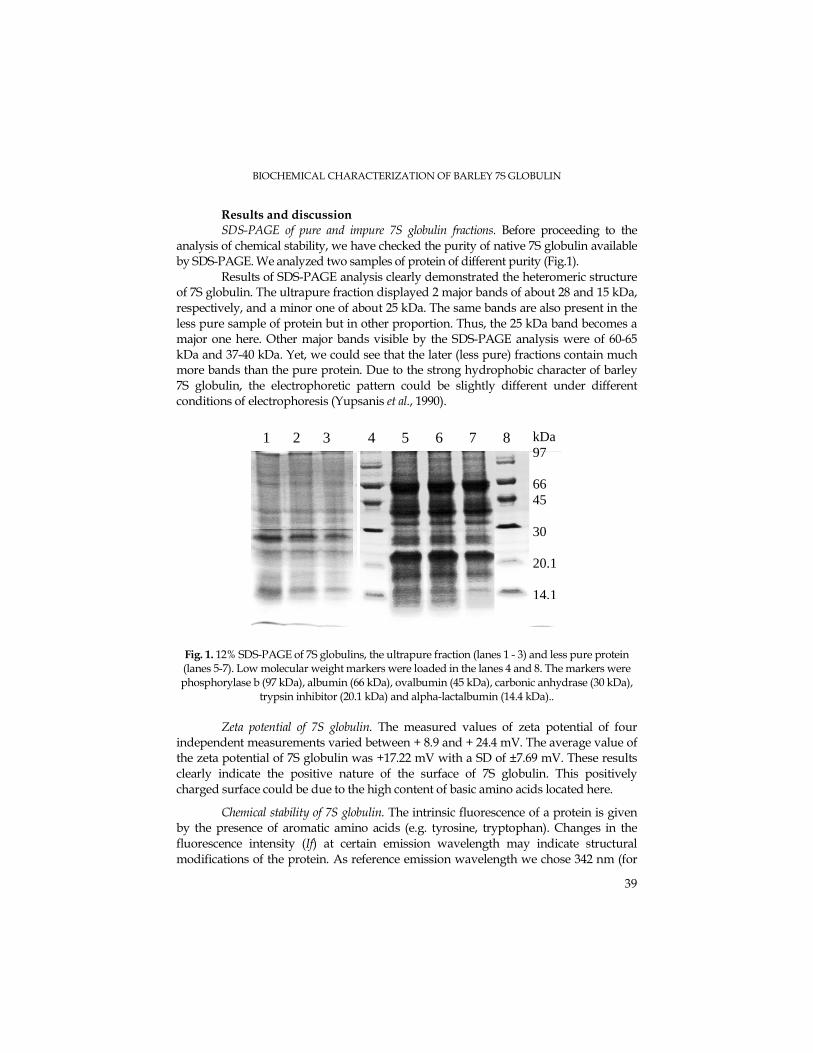
BIOCHEMICAL CHARACTERIZATION OF BARLEY 7S GLOBULIN
39
Results and discussion SDS-PAGE of pure and impure 7S globulin fractions. Before proceeding to the analysis of chemical stability, we have checked the purity of native 7S globulin available by SDS-PAGE. We analyzed two samples of protein of different purity (Fig.1).
Results of SDS-PAGE analysis clearly demonstrated the heteromeric structure of 7S globulin. The ultrapure fraction displayed 2 major bands of about 28 and 15 kDa, respectively, and a minor one of about 25 kDa. The same bands are also present in the less pure sample of protein but in other proportion. Thus, the 25 kDa band becomes a major one here. Other major bands visible by the SDS-PAGE analysis were of 60-65 kDa and 37-40 kDa. Yet, we could see that the later (less pure) fractions contain much more bands than the pure protein. Due to the strong hydrophobic character of barley 7S globulin, the electrophoretic pattern could be slightly different under different conditions of electrophoresis (Yupsanis et al., 1990).
Fig. 1. 12% SDS-PAGE of 7S globulins, the ultrapure fraction (lanes 1 - 3) and less pure protein (lanes 5-7). Low molecular weight markers were loaded in the lanes 4 and 8. The markers were phosphorylase b (97 kDa), albumin (66 kDa), ovalbumin (45 kDa), carbonic anhydrase (30 kDa),
trypsin inhibitor (20.1 kDa) and alpha-lactalbumin (14.4 kDa)..
Zeta potential of 7S globulin. The measured values of zeta potential of four
independent measurements varied between + 8.9 and + 24.4 mV. The average value of the zeta potential of 7S globulin was +17.22 mV with a SD of ±7.69 mV. These results clearly indicate the positive nature of the surface of 7S globulin. This positively charged surface could be due to the high content of basic amino acids located here.
Chemical stability of 7S globulin. The intrinsic fluorescence of a protein is given by the presence of aromatic amino acids (e.g. tyrosine, tryptophan). Changes in the fluorescence intensity (If) at certain emission wavelength may indicate structural modifications of the protein. As reference emission wavelength we chose 342 nm (for
1 2 3 4 5 6 7 8
kDa 97 66 45 30 20.1 14,1
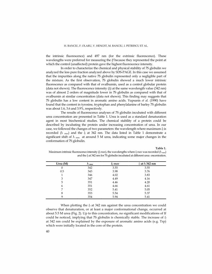
H. BANCIU, F. OLARU, V. HENGST, M. BANCIU, I. PETRESCU ET AL.
40
the intrinsic fluorescence) and 497 nm (for the extrinsic fluorescence). These wavelengths were preferred for measuring the If because they represented the point at which the control (unaffected) protein gave the highest fluorescence intensity. In order to characterize the chemical and physical stability of 7S globulin we analyzed the less pure fraction analyzed above by SDS-PAGE. In this case we assumed that the impurities along the native 7S globulin represented only a negligible part of the mixture. As the first observation, 7S globulin showed a much lower intrinsic fluorescence as compared with that of ovalbumin, used as a control globular protein (data not shown). The fluorescence intensity (If) at the same wavelength value (342 nm) was of almost 2 orders of magnitude lower in 7S globulin as compared with that of ovalbumin at similar concentration (data not shown). This finding may suggests that 7S globulin has a low content in aromatic amino acids. Yupsanis et al. (1990) have found that the content in tyrosine, tryptophan and phenylalanine of barley 7S globulin was about 1.6, 3.6 and 3.9%, respectively. The results of fluorescence analyses of 7S globulin incubated with different urea concentration are presented in Table 1. Urea is used as a standard denaturation agent in most biochemical studies. The chemical stability of a protein could be described by incubating the protein under increasing concentration of urea. In our case, we followed the changes of two parameters: the wavelength where maximum If is recorded (λ max) and the If at 342 nm. The data listed in Table 1 demonstrate a significant shift of λ max at around 5 M urea, indicating some major changes in the conformation of 7S globulin.
Table 1.
Maximum intrinsic fluorescence intensity (If max), the wavelengths where If max was recorded (λ max) and the If at 342 nm for 7S globulin incubated at different urea oncentration.
Urea (M) λ max If max If at λ 342 nm
0 342 3.55 3.55 0.5 343 3.98 3.76 1 346 4.02 3.83 3 347 4.49 4.16 5 351 4.46 4.20 6 351 4.66 4.61 7 352 5.41 5.05 8 353 5.54 5.37 9 354 5.94 5.41
When plotting the If at 342 nm against the urea concentration we could observe that denaturation, or at least a major conformational change, occurred at about 5.5 M urea (Fig. 2). Up to this concentration, no significant modifications of If could be noticed, implying that 7S globulin is chemically stable. The increase of If at 342 nm could be explained by the exposure of aromatic amino acids (e.g. Trp) which were initially located in the core of the protein.
BIOCHEMICAL CHARACTERIZATION OF BARLEY 7S GLOBULIN
41
1
1.5
2
2.5
3
3.5
4
4.5
5
5.5
6
310 330 350 370 390 410
Wavelength (nm)
If 3
42 n
m
a.
2
2.5
3
3.5
4
4.5
5
5.5
6
0 2 4 6 8 10
Urea (M)
I f 3
42 n
m
b.
Fig 2. Changes of intrinsic fluorescence spectra of barley 7S globulin with increasing urea
concentration (a) and denaturation profile of 7S globulin treated with various concentrations
of urea (b). Intrinsic fluorescence of 7S globulin in 50 mM phosphate buffer, pH 7.4, was recorded at 342 nm with excitation wavelength set at 295 nm.
The intrinsic fluorescence studies of chemical denaturation in 7S globulin can be completed by information obtained when following a fluorescent compound that binds to the positively charged amino acid residues. When excited at 405 nm, ANS gave a maximum fluorescence emission at around 495 nm. High values If of ANS
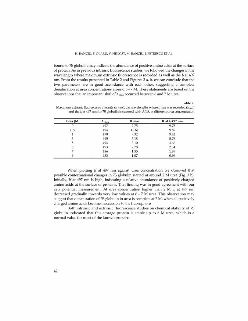
H. BANCIU, F. OLARU, V. HENGST, M. BANCIU, I. PETRESCU ET AL.
42
bound to 7S globulin may indicate the abundance of positive amino acids at the surface of protein. As in previous intrinsic fluorescence studies, we followed the changes in the wavelength where maximum extrinsic fluorescence is recorded as well as the If at 497 nm. From the results presented in Table 2 and Figures 3 a, b, we can conclude that the two parameters are in good accordance with each other, suggesting a complete denaturation at urea concentrations around 6 - 7 M. These statements are based on the observations that an important shift of λ max occurred between 6 and 7 M urea.
Table 2. Maximum extrinsic fluorescence intensity (If max), the wavelengths where If max was recorded (λ max)
and the If at 497 nm for 7S globulin incubated with ANS, at different urea concentration
Urea (M) λ max If max If at λ 497 nm
0 497 9.75 9.75 0.5 494 10.61 9.69 1 498 9.32 9.42
3 495 5.18 5.76 5 494 3.10 3.66 6 493 2.78 2.34 7 486 1.55 1.39 9 483 1.07 0.96
When plotting If at 497 nm against urea concentration we observed that possible conformational changes in 7S globulin started at around 2 M urea (Fig. 3 b). Initially, If at 497 nm is high, indicating a relative abundance of positively charged amino acids at the surface of proteins. That finding was in good agreement with our zeta potential measurement. At urea concentration higher than 2 M, If at 497 nm decreased gradually towards very low values at 6 - 7 M urea. This observation may suggest that denaturation of 7S globulin in urea is complete at 7 M, when all positively charged amino acids become inaccessible to the fluorophore.
Both intrinsic and extrinsic fluorescence studies on chemical stability of 7S globulin indicated that this storage protein is stable up to 6 M urea, which is a normal value for most of the known proteins.
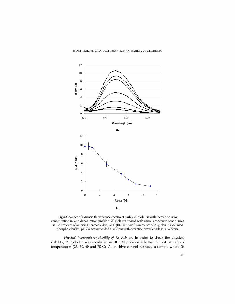
BIOCHEMICAL CHARACTERIZATION OF BARLEY 7S GLOBULIN
43
0
2
4
6
8
10
12
420 470 520 570
Wavelength (nm)
If 4
97 n
m
a.
0
2
4
6
8
10
12
0 2 4 6 8 10
Urea (M)
I f 4
97 n
m
b.
Fig 3. Changes of extrinsic fluorescence spectra of barley 7S globulin with increasing urea
concentration (a) and denaturation profile of 7S globulin treated with various concentrations of urea
in the presence of anionic fluorescent dye, ANS (b). Extrinsic fluorescence of 7S globulin in 50 mM phosphate buffer, pH 7.4, was recorded at 497 nm with excitation wavelength set at 405 nm.
Physical (temperature) stability of 7S globulin. In order to check the physical
stability, 7S globulin was incubated in 50 mM phosphate buffer, pH 7.4, at various temperatures (25, 50, 60 and 70oC). As positive control we used a sample where 7S
H. BANCIU, F. OLARU, V. HENGST, M. BANCIU, I. PETRESCU ET AL.
44
globulin was boiled at 100oC for 30 minutes. The protein concentration in the incubation test tubes was 0.2 mg/ml. Only the parameters for intrinsic fluorescence were monitored during the thermal denaturation of 7S globulin (Fig. 4).
0
2
4
6
8
10
12
14
0 50 100 150 200Time (min)
If 3
42 n
m
25oC
50oC
60oC
70oC
100oC
Fig. 4. Thermal stability of 7S globulin incubated in 50 mM phosphate buffer, pH 7.4, at different temperatures for various time intervals. Intrinsic fluorescence of globulin was
monitored as If at 342 nm, with excitation wavelength set at 295 nm.
The thermal resistance of 7S globulin proved to be high. As well at 50°C and an incubation time of 2.5 h as at 60°C and an incubation time of 2 h, the protein did not show thermal instabilities.. Denaturation at 60oC started after 2 hours of incubation. At higher temperature (e.g. 70oC), 7S globulin was relatively stable for at least 1.5 hours. Interestingly, in contrast to the pattern shown during urea denaturation, the If at 342 nm decreased during thermal denaturation, suggesting a different mechanism for protein unfolding under the temperature stress. When protein incubated for 2.5 hours at 70oC (If 342 nm = 4.5) was stored at 25oC for 12 hours, it showed a partial renaturation (If 342 nm increased to 9.8). However, further studies using alternative methods (NMR, calorimetry) are required for better characterization of 7S globulin chemical and physical stability.
Conclusions
7S globulin is a heteromeric protein, consisting of subunits with various molecular weights. It is arguable which are the main subunits because different bands are visible and highlighted in the SDS-PAGE analysis of pure and less pure fractions, respectively. 7S globulin is chemically stable at up to 6 M urea and showed a thermal stability after 2 hours of incubation at 60oC making it a good candidate for nanotechnology of surfaces coating.
BIOCHEMICAL CHARACTERIZATION OF BARLEY 7S GLOBULIN
45
Acknowledgments. The present research was supported by CEEX grant type P-CD, Nr. 5603/ 07.10.2005 and CNCSIS grant type AT, Nr. 2771/ 23.05.2006.
REFERENCES Burgess, S.R., Shewry, P.R. (1986) Identification of homologous globulins from embryos of
wheat, barley, rye and oats. J. Exp. Botany, 37, 1863–1871. Desai, U.R., Johns, J.L., Lahaye, L., Wright, H.T. (2002) Probing reactive center loop insertion in
serpins: a simple method for ovalbumin. Anal. Biochem. , 302, 81-87 Heck, G.R., Chamberlain, A.K., Ho, D.T.-H. (1993) Barley embryo globulin 1 gene, Beg1:
characterization of cDNA chromosome mapping and regulation of expression. Mol. Gen. Genetics, 239, 209–218.
Horikoshi M., Morita Y. (1975) Localization of c-globulin in rice seed and changes in c-globulin content during seed development and germination. Agric. Biol. Chem., 39, 2309–2314.
Kriz, A.L. (1999) 7S globulins of cereals. In: Shewry P.R., Casey R. (Eds.). Seed proteins. Kluwer Academic Publishers, Dordrecht, 477–498.
Laemmli ,U.K. (1970) Nature (London), 227, 680–685 Lowry, O.H., Rosebrough, N.J., Farr, A.L., Randall, R.J. (1951) Protein measurement with the
Folin phenol reagent. J. Biol. Chem., 193, 265-275. Matulis, D., Lovrien, R. (1998) Anilino-8-naphthalene sulfonate anion-protein binding depends
primarily on ion pair formation. Biophys. J., 74, 422-429 Salata, O.V. (2004) Applications of nanoparticles in biology and medicine. J. Nanobiotehnol., 2, 3-8. Shewry, P.R., Halford, N.G. (2002) Cereal seed storage proteins: structures, properties and
role in grain utilization. J. Exp. Botany, 53, 947-958 Yupsanis, T., Burgess, S.R, Jackson, P.J., Shewry, P.R. (1990) Characterization of the major protein
component from aleurone cells of barley (Hordeum vulgare L). J. Exp. Botany, 41, 385–392. Zhu, H., Snyder, M. (2003) Protein chip technology, Curr. Opinion Chem. Biol., 7, 55-63.
STUDIA UNIVERSITATIS BABEŞ – BOLYAI, BIOLOGIA, LII, 1, 2007 (p.47-61)
ULTRASTRUCTURAL STUDIES CONCERNING THE REACTIVITY OF THE HIPOTHALAMIC-PITUITARY AXIX FOLLOWING
L-MONOSODIUM GLUTAMATE ADMINISTRATION IN JUVENILE RABBITS
CONSTANTIN PUICA1, CONSTANTIN CRACIUN2, MIRCEA RUSU1, IOANA ROMAN1 and MIHAI CRISTESCU3
SUMMARY. Subchronic oral administration for 30 days of L-monosodic-glutamate (GLU) in a daily dose of 0.15 mg/g, body weight in immature prepubertal male rabbits, induced reactive manifestations of mediobasal hypothalamus morphology, characterized by ultrastructural alteration of the majority of neuronal cellular components: nucleus, nucleolus, mitochondria and endoplasmic reticulum. The excitotoxic effect of GLU is expressed also by the blocking in some hypothalamus regions of the axonal and dendrites myelinates processes. At the adenohypophysis level, glutamate has determined drastically reduces of the number of the electron-dense secretory granules in some cellular components associated with mitochondrial balloonization, dilatation of both smooth endoplasmic reticulum and Golgi apparatus, as well as significantly rarefaction of ribosome from cytoplasmatic structure. KEYWORDS: L-monosodium glutamate, rabbits, hypothalamus, pituitary ultrastructure
Introduction. The objective of our study was to determine the modifications induced by L-
monosodium glutamate (MSG) subchronic treatment on the mediobasal hipotalamic (MBH) neurons ultrastructure and on the pituitary cells ultrastructure, in prepubertal, juvenile rabbits.
Glutamate (GLU) is a powerful amino acid neurotransmitter that plays a pivotal role in the formation of synapses and neuronal circuitry, long-term potentiation and depression, and both normal learning and addictive behavior (Meldrum, 2000; Olney, 1995). The neurotoxic properties of GLU have generated significant interest among neuroscientists. In the preceding decades, it was shown that systemic administration of GLU to animals of various species causes acute neurodegeneration of neurons in the retina (Lucas and Newhouse, 1957; Olney, 1969), or several regions of brain thet lack blood brain barriers (Olney, 1969). Moreover, ultrastructural studies (Olney, 1969, 1988), localized the apparent site of toxic action to postsynaptic dendrosomal membranes where GLU excitatory synaptic receptors are believed to be localized. By electron microscopy it has been
1 Institute of Biological Research, 48 Republicii Street, 400015 Cluj-Napoca, Romania. E-mail: [email protected] 2 “Babes-Bolyai” University, Faculty of Biology and Geology, Centre of Electron Microscopy, 1 Mihail Kogălniceanu Street, 400084 Cluj-Napoca, Romania. E-mail: [email protected]
3 University of Agricultural Science, Faculty of Veterinary Medicine, 3-5 Manastur Street, 400372 Cluj-Napoca. E-mail: [email protected]
C. PUICA, C. CRACIUN, M. RUSU, I. ROMAN, M. CRISTESCU
48
shown that the toxic action of GLU impinges selectivelly on dendritic and somal surface of the neuron that posses excitatory receptors trough which the depolarizing effects of GLU putativelly are mediated.
These several observations led to the “excitotoxic” concept that a depolarization mechanism underlies GLU neurotoxicity and the toxic action is mediated through dendrosomal synaptic receptors specialised for glutamatergic transmission.
Excitotoxicity refers to the paradoxical property, shared by GLU and specific excitatory aminoacids (EAA) analogs, of causing acute neuronal degeneration by excesive stimulation of postsynaptic EAA ionotropic receptors-receptors through which GLU functions physiologically as a transmitter.The precise mechanisms by which excessive EAA receptor activation leads to acute neuronal death are not well understood, although increased membrane permeability and abnormal Na+ , Cl- and Ca2+ influx are assumed to play important roles. Studies in tissue culture indicate that GLU receptor–mediated neuronal degeneration can be separed into two distinct forms, acute and delayed, distinguished by the time of course and ionic dependence of neuronal degeneration (Blaylock, 1998; Chandan et al., 1999; Choy, 1993; Coyle and Puttfarcken, 1993; Ho et al., 2003; Rothman, 1989; Caruso et al., 2004; Tursky and Tursky, 1993; Srinivasan et al., 2006; Young and Ajami, 2000). The acute form of neurotoxicity is characterized by neuronal swelling in the presence of agonists, in particularly GLU, (Kubo and Kohira,1993; Lombard, 2002).
Regions of the brain most sensitive to GLU exposure are the median eminence (ME), and the circumventricular organs (CVO) of the hipothalamus. These highly vascularized areas sit outside the blood-brain barrier, which is formed at their inner surfaces by tight junctions between the modified astroglial cells called tanycytes lining their ventricular boundaries, (Matsumoto and Ishii,1992).
The arcuate nucleus-ME region of the early postnatal rodent has often been used for studies of monosodium GLU neurotoxicity because of its heightened responsiveness, consistent cytoarchitecture and conspicuous anatomical landmarks, (Olney 1969). This author noticed that the newborn of GLU exposed mice were grossly obese and short in statue. Further examination also demonstrated hypoplastic organs, including pituitary, thyroid, adrenal as well as reproductive dysfunction. Physiologically, the author demonstrated multiple endocrine deficiencies after GLU administration, including TSH, growth hormone, LH, FSH, and ACTH. When Dr. Olney examined the animal's brain, he discovered discrete lesions of the arcuate nucleus as well as less severe destruction of other hypothalamic nuclei. Later studies indicated that the damage by GLU was much more widespread, including the hippocampus, circumventricular organs, locus cereulus, amygdala-limbic system, subthalamus, and striatum, (Olney, 1995, 2002).
Despite evidence that GLU is a neurotoxin that can destroy central neurons following oral intake by animals of various species, MSG continues to be one of the most widely and heavelly used food aditives in the world (Olney, 1995).
ULTRASTRUCTURAL STUDIES OF HIPOTHALAMIC-PITUITARY AXIX REACTIVITY
49
At present, it is known that endogen and exogen GLU accumulation in the central nervous system (CNS) over a critical level, can produce irreversible damages in rodents, monkeys, etc. (Blaylock, 2002; Gao, 1994; Goldsmith, 2000; Meldrum 2000; Olney, 1995; Puică et al., 1996, 1997, 2004; Puică and Crăciun, 1999; Schwartz, 1988).
Because the arcuate hypothalamic nucleus (ARC), a neuroendocrine regulatory center is one of the circumventricular organs (CVO) brain regions damaged, animals treated with MSG in infancy, manifest multiple neuroendocrine disturbances and have an abnormal body habitus in adulthood. The fully developped syndrome includes obesity, skeletal stunting, reproductive failure and hypoplasia of the adenohypophysis and gonads, together with abnormally low hypothalamic, pituitary or plasma levels of luteinising hormone, growth hormone, gonadotrophs hormone and prolactin. Accumulation of GLU in CNS induces acute perturbations in several neuroendocrine axes.
Such experimental observations suggest that GLU may be involved in neuronal death, leading to neurodegenerative disorders in animals and in humans (Danbolt, 2001; Krajnic et al., 1996; Ferrante et al., 1997; Olney, 1988, 1994, 1995; Puică et al., 2002; Tursky and Tursky, 1993).
Over the past decade, evidence has begun to accumulate for the involvement of EAA, (GLU respectively), and an excitotoxic mechanism in the pathophysiology of a wide variety of neurological disorders, including food poissoning (neuroendocrinopathies, motor neuron disorders and/or dementia), sulfite oxidase deficiency, olivopontocerebellar degeneration, amyotrophic lateral sclerosis, epilepsy, hypoglicemia, hypoxia/ischemia, Huntington’s disease, Alzheimer’s disease and parkinsonism (Blaylock, 1998; Choy, 1993; Olney, 1995; Urushitani et al., 2001; Van Westerlaak et al., 2001; Wolf et al.,1990).
Materials and Methods
Animals and diets. Male 40 days old Supercuni male rabbits with an average weight of 1000 g ± 5 g were divided into two groups of 4 animals. The rabbits were housed in cages, had free access to drinking water and feed with a common rabbits-chow. The animals were maintained in standardized conditions, in a room at 230C, and a 12-h light:dark cycle. Care and treatment of rabbits followed recommended
guidelines (NRC 1985). In the 70th day of life the animals were sacrificed by cervical dislocation, and the hypothalamus and the pituitary was rapidly isolated for ultrastructural investigations. The aims of our study was to evaluate the ultrastructural modifications of medio-bazal hypothalamic neuronal structures and of anterior pituitary, after GLU administration.
Experimental groups. C group - control group trated by oral administration with distilled water. GT group - treated by oral administration for 30 days with L-monosodium glutamate (MSG) (Sigma) in a daily dose of 0,15 mg/g b.w., dissolved in distilled water.
Ultrastructural investigations. For ultrastructural investigations the mediobasal hypothalamus and pituitary were immediately prefixed in a 2,7% glutaraldehyde solution and postfixed in a 2% osmic acid solution. The dehydration of the samples
C. PUICA, C. CRACIUN, M. RUSU, I. ROMAN, M. CRISTESCU
50
was performed in acetone and in dehydred acetone and then embedded in Vestopal W. The ultratin sections were obtained using an LKB-III ultramicrotome and were contrasted with uranyl acetate and lead citrate. Examination of sections were performed in a OLYMPUS electron microscope.
Results Ultrastructural examination of medio-basal hypotalamus (MBH) structures revealed
the normal aspect of all cellular components of the neurons, in control group. The euchromatic nuclei are round or ovalar with one or two reticular nucleols. The neuronal pericarya contains normal conformed and structured cellular organites: numerous polysomes and mitochondries, as well as endoplasmic rough reticulum disposed in narrow profiles. In the some areas of the nervous tissue on can observe apoptotic neuronal cells partial or total destroyed. The axons and the dendritic fibers are normal mielinisated, (Figure 1).
The MSG-treated rabbits (GT group) showed some moderate to intense degenerative changes of neurons. Thus, ultrastructural studies showed that administration of 0,15 mg/g b.w. of GLU for 30 days induced selective degeneration of all subcellular neurons structure of MBH. The nuclei of circumventricular organs of hypothalamus often have irregular outline, condensed chromatine, many of them being picnotic. The cytoplasmic organelles have alterative degenerative modifications characterized by swelling, vacuolisation and loss of cristae of mitochondria, disintegrated mitochondria, and dilatation of the endoplasmic reticulum and Golgi system, in comparison with normal aspects of the neurons in control group.
MSG-treatment induced degeneration of axons (demyelination and interruption of the myelin sheath, vacuolisation at the level of axoplasma, accompanied by losses of axonal substance), in lesioned regions of MBH. Degenerated mossy fibers had shrunk and the terminals had become either electron-dense or were vacuolized and swollen, (Fig. 2).
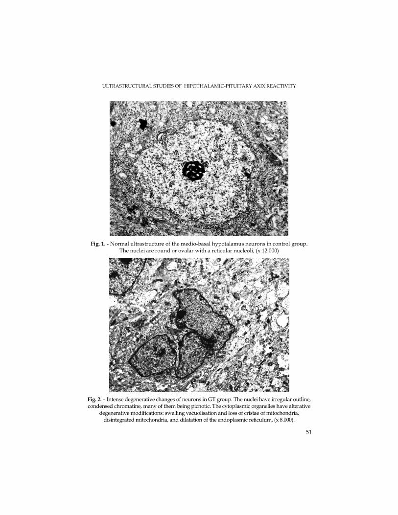
ULTRASTRUCTURAL STUDIES OF HIPOTHALAMIC-PITUITARY AXIX REACTIVITY
51
Fig. 1. - Normal ultrastructure of the medio-basal hypotalamus neurons in control group. The nuclei are round or ovalar with a reticular nucleoli, (x 12.000)
Fig. 2. – Intense degenerative changes of neurons in GT group. The nuclei have irregular outline, condensed chromatine, many of them being picnotic. The cytoplasmic organelles have alterative
degenerative modifications: swelling vacuolisation and loss of cristae of mitochondria, disintegrated mitochondria, and dilatation of the endoplasmic reticulum, (x 8.000).
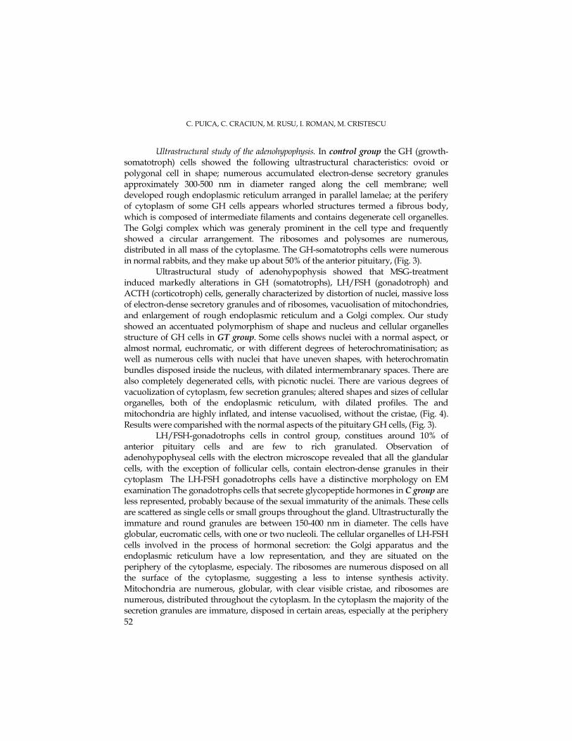
C. PUICA, C. CRACIUN, M. RUSU, I. ROMAN, M. CRISTESCU
52
Ultrastructural study of the adenohypophysis. In control group the GH (growth-somatotroph) cells showed the following ultrastructural characteristics: ovoid or polygonal cell in shape; numerous accumulated electron-dense secretory granules approximately 300-500 nm in diameter ranged along the cell membrane; well developed rough endoplasmic reticulum arranged in parallel lamelae; at the perifery of cytoplasm of some GH cells appears whorled structures termed a fibrous body, which is composed of intermediate filaments and contains degenerate cell organelles. The Golgi complex which was generaly prominent in the cell type and frequently showed a circular arrangement. The ribosomes and polysomes are numerous, distributed in all mass of the cytoplasme. The GH-somatotrophs cells were numerous in normal rabbits, and they make up about 50% of the anterior pituitary, (Fig. 3).
Ultrastructural study of adenohypophysis showed that MSG-treatment induced markedly alterations in GH (somatotrophs), LH/FSH (gonadotroph) and ACTH (corticotroph) cells, generally characterized by distortion of nuclei, massive loss of electron-dense secretory granules and of ribosomes, vacuolisation of mitochondries, and enlargement of rough endoplasmic reticulum and a Golgi complex. Our study showed an accentuated polymorphism of shape and nucleus and cellular organelles structure of GH cells in GT group. Some cells shows nuclei with a normal aspect, or almost normal, euchromatic, or with different degrees of heterochromatinisation; as well as numerous cells with nuclei that have uneven shapes, with heterochromatin bundles disposed inside the nucleus, with dilated intermembranary spaces. There are also completely degenerated cells, with picnotic nuclei. There are various degrees of vacuolization of cytoplasm, few secretion granules; altered shapes and sizes of cellular organelles, both of the endoplasmic reticulum, with dilated profiles. The and mitochondria are highly inflated, and intense vacuolised, without the cristae, (Fig. 4). Results were comparished with the normal aspects of the pituitary GH cells, (Fig. 3).
LH/FSH-gonadotrophs cells in control group, constitues around 10% of anterior pituitary cells and are few to rich granulated. Observation of adenohypophyseal cells with the electron microscope revealed that all the glandular cells, with the exception of follicular cells, contain electron-dense granules in their cytoplasm The LH-FSH gonadotrophs cells have a distinctive morphology on EM examination The gonadotrophs cells that secrete glycopeptide hormones in C group are less represented, probably because of the sexual immaturity of the animals. These cells are scattered as single cells or small groups throughout the gland. Ultrastructurally the immature and round granules are between 150-400 nm in diameter. The cells have globular, eucromatic cells, with one or two nucleoli. The cellular organelles of LH-FSH cells involved in the process of hormonal secretion: the Golgi apparatus and the endoplasmic reticulum have a low representation, and they are situated on the periphery of the cytoplasme, especialy. The ribosomes are numerous disposed on all the surface of the cytoplasme, suggesting a less to intense synthesis activity. Mitochondria are numerous, globular, with clear visible cristae, and ribosomes are numerous, distributed throughout the cytoplasm. In the cytoplasm the majority of the secretion granules are immature, disposed in certain areas, especially at the periphery
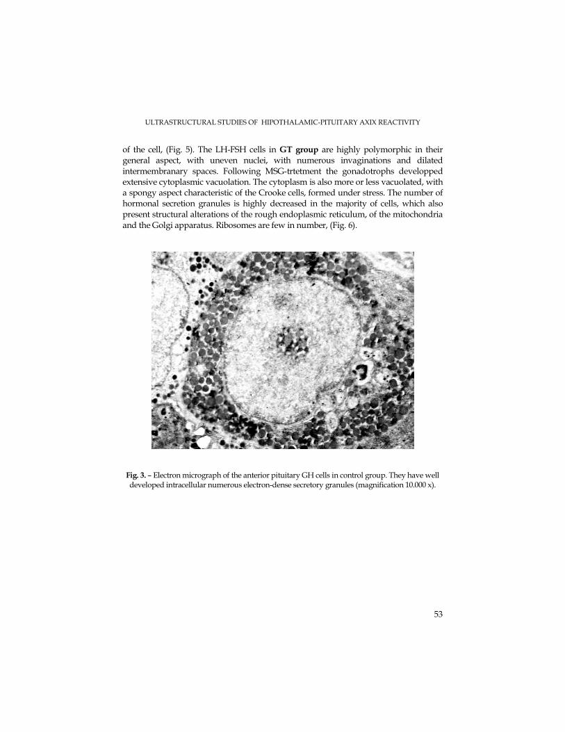
ULTRASTRUCTURAL STUDIES OF HIPOTHALAMIC-PITUITARY AXIX REACTIVITY
53
of the cell, (Fig. 5). The LH-FSH cells in GT group are highly polymorphic in their general aspect, with uneven nuclei, with numerous invaginations and dilated intermembranary spaces. Following MSG-trtetment the gonadotrophs developped extensive cytoplasmic vacuolation. The cytoplasm is also more or less vacuolated, with a spongy aspect characteristic of the Crooke cells, formed under stress. The number of hormonal secretion granules is highly decreased in the majority of cells, which also present structural alterations of the rough endoplasmic reticulum, of the mitochondria and the Golgi apparatus. Ribosomes are few in number, (Fig. 6).
Fig. 3. – Electron micrograph of the anterior pituitary GH cells in control group. They have well developed intracellular numerous electron-dense secretory granules (magnification 10.000 x).
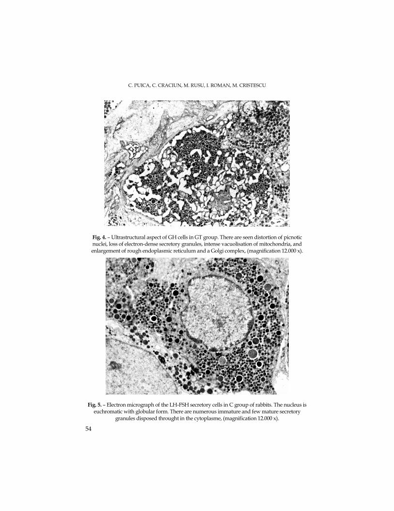
C. PUICA, C. CRACIUN, M. RUSU, I. ROMAN, M. CRISTESCU
54
Fig. 4. – Ultrastructural aspect of GH cells in GT group. There are seen distortion of picnotic nuclei, loss of electron-dense secretory granules, intense vacuolisation of mitochondria, and enlargement of rough endoplasmic reticulum and a Golgi complex, (magnification 12.000 x).
Fig. 5. – Electron micrograph of the LH-FSH secretory cells in C group of rabbits. The nucleus is euchromatic with globular form. There are numerous immature and few mature secretory
granules disposed throught in the cytoplasme, (magnification 12.000 x).
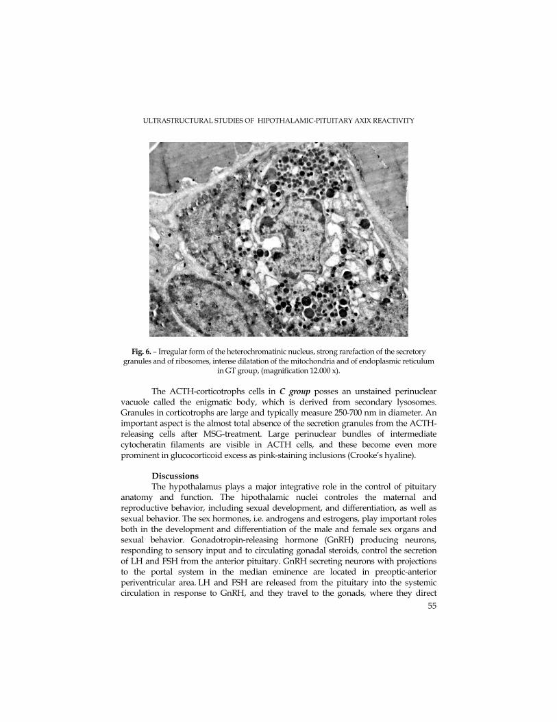
ULTRASTRUCTURAL STUDIES OF HIPOTHALAMIC-PITUITARY AXIX REACTIVITY
55
Fig. 6. – Irregular form of the heterochromatinic nucleus, strong rarefaction of the secretory granules and of ribosomes, intense dilatation of the mitochondria and of endoplasmic reticulum
in GT group, (magnification 12.000 x).
The ACTH-corticotrophs cells in C group posses an unstained perinuclear
vacuole called the enigmatic body, which is derived from secondary lysosomes. Granules in corticotrophs are large and typically measure 250-700 nm in diameter. An important aspect is the almost total absence of the secretion granules from the ACTH-releasing cells after MSG-treatment. Large perinuclear bundles of intermediate cytocheratin filaments are visible in ACTH cells, and these become even more prominent in glucocorticoid excess as pink-staining inclusions (Crooke’s hyaline).
Discussions The hypothalamus plays a major integrative role in the control of pituitary
anatomy and function. The hipothalamic nuclei controles the maternal and reproductive behavior, including sexual development, and differentiation, as well as sexual behavior. The sex hormones, i.e. androgens and estrogens, play important roles both in the development and differentiation of the male and female sex organs and sexual behavior. Gonadotropin-releasing hormone (GnRH) producing neurons, responding to sensory input and to circulating gonadal steroids, control the secretion of LH and FSH from the anterior pituitary. GnRH secreting neurons with projections to the portal system in the median eminence are located in preoptic-anterior periventricular area. LH and FSH are released from the pituitary into the systemic circulation in response to GnRH, and they travel to the gonads, where they direct
C. PUICA, C. CRACIUN, M. RUSU, I. ROMAN, M. CRISTESCU
56
gamete production, as well as gonadal (testosterone in male and estrogen and progesterone in female) hormone production, (Stevens and Lowe, 1993).
On the other hand, the hipothalamic-pituitary axis is a key player in an animal’s response to stressful stimuli. Corticotropin-releasing hormone (CRH or CRF) is a 41 amino-acid peptide expressed in the hypothalamus. In response to CRH stimulation, corticoctropes synthesize and release adrenocorticotropic hormone (ACTH) in anterior pituitary. ACTH through the systemic circulation binds and activates its receptors on the surface of cells of the adrenal cortex. In response to receptor activation, adrenocortical cells synthesize glucocorticoids, (Akil, 2002).
Our results demonstrate that the subchronic administration of MSG as neurotoxicant and stressant factor, for 30 days in prepubertal male rabbits, induce ultrastructural changes in the mediobasal neuronal cells of hypothalamus, characterized by the alterations of all subcellular structures: the heterochromatic nuclei are irregular forms, endoplasmic reticulum and Golgi complex are intense dilatated, the mitochondries are intense dilatated and vacuolised, with very few cristae. Electron microscopy examination of the medio-basal hypothalamus, confirm us, and represents an additional element of the direct neurotoxic effects of MSG on brain structures and functionality.
MSG-treatment in our experiment induced modifications in the forms and number of adenohipophysary cells. Neuronal distrophy and lesions induced by MSG in the specific regions of hypothalamus which controle the activity of pituitary, induced damages (intense vacuolisation, loss of the intracytoplasmatic secretory granules) in all cellular structures of the adenohypophysis.
The presence of Crooke-ACTH cells in the anterior pituitary after MSG administration, represents a tipicaly stress reaction determined by presumably elevated level of plasma concentration of the cortisol. These combinative actions - the excessive glucocorticoid concentration and the decrease of GH level, both contribute to a retardation of the general development of animals, and of body weight, in particularly (data non presented). Ultrastructural modifications of the GH and LH-FSH cells registered in our experimental study, are followed by a presumable decrease secretion of the growth hormone (GH), and of gonadotroph hormone (LH/FSH).
Our experimental data are in agreement to literature data. The recent studies have shown that GLU is the most important neurotransmitter in the hypothalamus. Since this early observation, L-monosodium glutamate and other excitatory substances have become the standard tool in studying the function of the hypothalamus, (Goldsmith, 2000; Ikonomidou et al., 1999; Meldrum, 2000).
Glutamic acid functions in all cells in the body as a metabolic intermediate and protein constituent; in the brain, it has the added role of being an excitatory neurotransmitter (Fonnum, 1984).
A substantial amount of GLU is ingested each day, principally as a component of dietary protein; a small amount is also present in food as free glutamate and monosodium glutamate which occurs naturally in some foods and is added to others as a flavor enhancer). Repeated injection of very large doses of the amino acid into neonatal rodents
ULTRASTRUCTURAL STUDIES OF HIPOTHALAMIC-PITUITARY AXIX REACTIVITY
57
could produce visible brain damage, notably in the circumventricular organs (CVO) of hypothalamus (Fernstrom, 2000).
Elevated plasma glutamate can cause selective loss of the neurons in the selective regions of brains of infant mice. The arcuate nucleus - median eminence regions exhibit the greatest sensitivity to glutamate while it undergoes developmental maturation during early postnatal life. Some related study demonstrated that elevated plasma of glutamate after endogen release or exogen administration of this amino acid in infant rodents, can cause selective loss of neurons in the circumventricular organs of hypothalamus especially, which controls the morphology and the secretory activity of anterior pituitary. The arcuate nucleus is consistently the most sensitive of these nuclei to MSG-toxicity. We know that this nucleus regulates growth hormone secretion, by way of the pituitary, (Blaylock, 2002; Olney, 1995).
It is well recognized that the immature brain is four times more sensitive to the toxic effects of the excitatory amino acids as is the mature brain, (Blaylock, 2000). The experimental data reveals the role of excitotoxins in neurodegenerative process particularly relevant to the developing CNS in early postnatal and in prepubertal period of life, (Meldrum, 2000; Olney, 2002; Puică, 1996).
The data reported by Garattini, (2000) demonstrated that MSG-treated animals showed stunted growth, hypogonadism and moderate brain retardation. There is considerable evidence for the cerebral morphological and functional abnormalities developed by rodents exposed neonatally or in the prepubertal stage of development to GLU. Developmental changes in the expression of ionotropic GLU receptors are known to influence excitotoxic phoenomena and may contribute to the pattern of vulnerability of brain in the neonatal rodent.
The study of Magarinos et al., (1998, 1995) confirmed that neonatal treatment with MSG produced several abnormalities of the CNS-pituitary axis. The rodent exposed neonatally to MSG, showed stunted several neuroendocrine abnormalities, growth, obesity, hypogonadism, sterility and self-mutilation, as adults. The inner layers of the retina and the hypothalamus arcuate nucleus are preferential destroyed by sistemic administration of MSG, but the lessions are not restricted to these structures, as circumventricular organs and other brain nuclei may be damaged to some extent.
It is well established that the paraventricular nucleus release the corticoliberin releasing-factor (CRF), which controls the secretion of glucocorticoids by the corticotroph cells of adenohipophysis. An increase of plasma concentration of the cortisol, determined by the actions of some stress factors, can certains modify the morphology and the functions of adenohipophysary cells in young rabbits. Glucocorticoids also regulate hypothalamic-pituitary-adrenal axis and their own production, via negative feed-back at the level of the hypothalamus and pituitary, (Goldsmith, 2000; Nichols et al., 2001).
The neuronal lesions produced by MSG administration in ventromedial and arcuate nuclei, in paraventricular nuclei of the hypothalamus, in medial and lateral parts of the preoptic area, and in median eminence determined an reduction of glucocorticoid receptors in these brain regions. The damages of these CNS structures, of arcuate nuclei especialy, may be related to the adrenal hyperfunction of GLU-treated rats. Excessive
C. PUICA, C. CRACIUN, M. RUSU, I. ROMAN, M. CRISTESCU
58
glucocorticoids concentrations in vivo inhibit somatic growth in both man and animals, (Ozawa et al., 1999; Nicols et al., 2001).
In agreement to early reports of the recent data of literature (Goldsmith, 2000; Meldrum, 2000; Olney, 1995; Blaylock, 1999, 2002), administration of MSG in our experiment, demonstrate the high vulnerability of brain structures in prepubertal stage of development.
The implications of all autors mentioned above findings, should have been earth-shaking to say the least. Why? Because millions of babies all over the world were eating baby foods containing large amounts of GLU and hydrolyzed vegetable protein (a compound which contains three excitotoxins). In fact, the concentrations of GLU found in baby foods was equal to that used to create brain lesions in experimental animals. And in all of these experiments, immature animals were found to be much more vulnerable to the toxic effects of GLU than were older animals. This was true in all animal species tested. His findings indicated that GLU was not only toxic to the retina, but also to the brain. When they examined the animals' brains they discovered that specialized cells in a critical area of the animals' brain, the hypothalamus, were destroyed, after a single dose of GLU, (Danbolt, 2001; Ikonomidou and Turski,1995; Puică, 1996; Blaylock, 1999).
We specify that the 0,15mg/g b.w. dose of MSG utilised in our experiment, correspond to the concentration of this alimentar aditive which can be found in all human alimentar concentrates. This dose was’nt utilised until to date in experimental procedures, and it represent an novelty in the domaine of GLU neurotoxicity-studies.
Conclusions
• The administration of MSG induced neurodegenerative effects especially in the CVO of hypothalamus, and severe structural and functional alterations in hypothalamo-pituitary axis. These observations emphasizes the important role of CVO in the regulation of some endocrine morphology and functions.
• Ultrastructural aspects of neurons in CVO are very well correlated with morpho-functional modifications of hypothalamo-pituitary axis.
• Subcronical exposure (30 days) of immature rabbits to MSG in this prepubertal critical stage of development entails potential risk, even if brain damage does not occur, that hormonal biorhytms may be disturbed with adverse effects on growth and development.
• Our experimental results are claiming cautions in MSG consumption in prepubertal phase of children development. The degenerative aspects of brain structures observed in MSG-treated rabbits suggests that is reasonable to assume that the same infant-to-adult relationship would be true for the consumption of alimentar concentrates with glutamate, as aditive in earlyes stages of ontogeny. Of particular concern is the toxic effects of these excitotoxic compounds on the developing brain. It is well recognized that the immature brain is four times more sensitive to the neurotoxic effects of the glutamate as is the mature brain. This means that excitotoxic injury is of special concern from the fetal stage to adolescence.
ULTRASTRUCTURAL STUDIES OF HIPOTHALAMIC-PITUITARY AXIX REACTIVITY
59
• Our experimental result that are in accord with previous study performed, about MSG-neurotoxicity, are claiming cautions in MSG consumption in prepubertal phase of children development.
REFERENCES
Akil, T., (2002), Neuroendocrine Systems I: Overview- Thyroid and adrenal axes. In: Fundamental Neuroscience Zigmond et al., (ed), pp 1127-1150.
Blaylock, R.L., (1998), Neurodegeneration and aging of the central nervous system: Prevention and treatment by phytochemicals and metabolic nutrients. Integr. Med., 1, 117-133.
Blaylock, R.L., (1999), Food Additive Excitotoxins and Degenerative Brain Disorders, Medical Sentinel, 4, 212-215.
Blaylock, R., (2002), Aspartame, Monosodium glutamate and other Excitotoxins and the Hypothalamus, From: "Russell M.D.", Borlongan, C.V., Kanning, K., Poulos, T.B., Cahill, D.W., and Sanberg, P.R. Free radical damage and oxidative stress in Huntington's disease, J. Fla. Med. Assoc., 83, 335-341.
Caruso, C., Bottino, M.C., Pampillo, M., Pisera, D., Jaita, G., Duvilanski, B., Seilicovich, A., Lasa M., (2004), Glutamate Induces Apoptosis in Anterior Pituitary Cells through Group II Metabotropic Glutamate Receptor Activation, Endocrinology, 145, 4677-4684.
Chandan, K.S.., Khanna, S., Roy, S., Packer, L., (2000), Molecular Basis of Vitamin E Action. Tocotrienol potently inhibits glutamate-induced pp60c-Src kinase activation and death of HT4 neuronal cells, J. Biol. Chem., 275, 13049-13055.
Choy, D.W., (1993), Glutamate neurotoxicity and diseases of the nervous system, Neuron, 1, 623-634. Coyle, J. T., Puttfarcken, P., (1993), Oxidative stress, glutamate, and neurodegenerative disorders,
Science, 262, 689-695. Danbolt, N.C., (2001), Glutamate uptake, Prog. Neurobiol., 65, 100-105. Ferrante, R.J., Browne, S.E., Shinobu, L.A., Bowling, A.C., Baik, M.J., MacGarvey, U., Kowall,
N.W., Brown, R.H.Jr., Beal, M.F., (1997), Evidence of increased oxidative damage in both sporadic and familial amyotrophic lateral sclerosis. J. Neurochem., 69, 2064-2074.
Fernstrom, J.D., (2000), Pituitary Hormone Secretion in Normal Male Humans: Acute Responses to a Large, Oral Dose of Monosodium Glutamate, J.0 Nutr., 130, 1053S-1057S.
Fonum, F., (1984), Glutamate: a neurotransmitter in mammalian brain, J. Neurochem., 42, 1-11. Gao, J., (1994), Transplacental neurotoxic effects of monosodium glutamate on structures and
function of specific brain areas of filial mice, Acta Phisiol. Sin., 46, 111-118. Garattini, S., (2000), International Symposium on Glutamate: Glutamic Acid, Twenty Years Later,
J. Nutr., 130, 901S–909S. Goldsmith, P.C., (2000), Neuroglial Responses to Elevated Glutamate in the Medial Basal
Hypothalamus of the Infant Mouse, J. Nutr., 130, 1032S-1038S. Ho, P.I., Ortiz, D., Rogers, E., Shea, T., (2003), Multiple aspects of Homocysteine neurotoxicity:
Glutaqmate excitotoxicity, Kinase hyperactivation and DNA damage, J. Neuroscience Res., 70, 694-702.
Ikonomidou, C., Bosch, F., Miksa, M., Bittigau, P., Vöckler, J., Dikranian, K., Tenkova, T., Turski, L., Olney, J.W., (1999), Blockade of NMDA receptors and apoptotic neurodegeneration in the developing brain, Science, 283, 70-74.
C. PUICA, C. CRACIUN, M. RUSU, I. ROMAN, M. CRISTESCU
60
Kay, D., (1967), Techniques for electron microscopy, Second Edition, Blockwell Sci. Pub., Oxford and Edinburgh.
Krajnic, D., Neff, N. H., Hadjiconstantinou, M., (1996), Glutamate, glutamine and glutamine synthetase in the neonatal rat brain following hypoxia, Brain Res., 707, 134-137.
Kubo, T., Kohira, R., (1993), Neonatal glutamate can destroy the hippocampal CA1 structure and impair discrimination learning in rats, Brain Res., 616: 311-314.
Lombard, J., (2002), Antioxidants and neurodegenerative diseases, Brain Behav. Res., 2, 1-19. Lucas, D.R, Newhouse, J.P., (1957), The toxic effect of sodium L-glutamate on the inner layer of
the retina, Arch. Ophthalmol., 58, 193-201. Matsumoto, M., Ishii, T., (1992), A family of metabotropic glutamate receptors, Neuron, 8, 169-179. Magarinos, M.A., Estivariz, F., Morado, M.I., De Nicola, A.F., (1988), Regulation of the central
nervous system-pituitary-adrenal axis in rats after neonatal tratment with monosodium glutamate, Neuroendocrinology, 48, 105-111.
Magariños, M.A., McEwen, B.S., (1995), Stress-induced atrophy of apical dendrites of hippocampal CA3c neurons: comparison of stressors, Neurosci., 69, 83-88.
Meldrum, B.S., (2000), Glutamate as a Neurotransmitter in the Brain: Review of Physiology and Pathology, J. Nutrition, 130, 1007S-1015S.
Nicols, R.N., Zelba, M., Bye, N., (2001), Do glucocorticoids contribute to brain aging ? Brain Res. Reviews, 1, 1-14.
Olney, J.W. (1969) Brain lesions, obesity, and other disturbances in mice treated with monosodium glutamate, Science (Washington, DC) 164, 719–721.
Olney, J.W., (1974), Toxic Effects of Glutamate and Related Amino Acids on the Developing Central Nervous System, Inherited Disorders of Amino Acid Metabolism, , New York, Wiley, W.N. Nylan (ed), pp 133-135..
Olney, J.W., (1988), Excitotoxic food additives: functional teratological aspects, Prog. Brain Res..,18, 283-294.
Olney, J.W., (1995), Excitotoxin in foods, Neurotoxicology, 15, 535-544. Olney, J.W., (2002), Drug-induced Apoptotic Neurodegeneration in the Developing Brain, Brain
Pathol., 12, 488-498. Ozawa, H., Takao, I., Ikno, O., Mitsushiro, K., (1999), Cellular localisation and distribution of
glucocorticoid receptor immunoreactivity and the expression of glucocorticoid receptor messenger RNA in rat pituitary gland: A combined double immunohistochemistry study and in situ hibridisation histochemical analysis, Cell and Tissue Res., 295, 207-214.
Puică, C., (1996), Sodium glutamate-neurotoxic factor, experimental model for the study of neurodegenerative disorders in human, vol. I, Current problems and techniques in Cellular and Molecular Biology, 1, 338-345.
Puică, C., Excitotoxine în alimentaŃie, (1997), Clujul Medical, 1-2, 155-159; 134-137. Puică, C., Crăciun, C., (1999), Aspecte ultrastructurale ale acŃiunii L-glutamatului monosodic la
nivelul hipotalamusului mediobazal la şobolanul alb, 27, BUL. SNBC, 159-161. Puică, C., Crăciun, C., Sandu, D., Cristescu, C., (2002) The protective action of Magnesium ions
and B6 vitamin upon some morpho-functional parametters in hypothalamus and anterior pituitary of white juvenile rats treated with monosodium glutamate, Current Problems and Techniques in Cellular and Molecular Biology, V, 285-291.
Puică, C., Crăciun, C., Ardeleanu, C., Rusu, M., Roman, I. (2004), Protective effects of a bioactive compound treatment on new-born brain morphology and function, following Aspartame ingestion during gestation in white rats, In vol. of The 13th European Microscopy Congress from August 22-27 Aug. ANWERP, BELGIUM, II, 461-462,.
ULTRASTRUCTURAL STUDIES OF HIPOTHALAMIC-PITUITARY AXIX REACTIVITY
61
Rothman, S., (1989), Excitatory amino acid neurotoxicity is produced by passive chloride influx, J. Neurosci., 5, 2221-2229.
Schwartz, G., (1988), In Bad Taste: The MSG Syndrome, Truth in Labeling Campaign, Darien, IL, (ed), USA, pp. 60-67.
Srinivasan, M, Sudheer, A.R., Pillai, K.R., Kumar, P.R., Sudhakaran, P.R., Menon, V.P., (2006), Superoxide Dismutases and Ionic Manganese in Aging”; Biochim Biophys Acta, 23, 112-115,.
Stevens, A., Lowe, J., (1993), HISTOLOGY, Mosby, - Year Book Europe LTD, London, England. Tursky L., Tursky, W. A., (1993), Towards an undersatnding of the role of glutamate in
neurodegenerative disorders: energy metabolism and neuropathology, Experientia, 49, 1064-1070.
Urushitani, M.T., Inoue, R., Sawada, H., Kihara, T., Honda, K., Akaike, A., Shimohama, S., (2001), N-methyl-D-aspartate receptor-mediated mitochondrial Ca(2+) overload in acute excitotoxic motor neuron death: A mechanism distinct from chronic neurotoxicity after Ca(2+) influx, J. Neurosci. Res., 63, 377-387.
Van Westerlaak, M.G, Joosten, E.A, Gribnau, A.A, Cools, A.R, Bar, P.R, (2001), Chronic Mitochondrial Inhibition Induces Glutamate-Mediated Corticomotoneuron Death in an Organotypic Culture Model., Exp. Neurol., 167, 393-400.
Wolf, G., Keilhoff, G., Fisher, S., Hass, P., (1990), Subcutaneosully applied magnesium protects reliably against quinolinate-induced N-methyl-D-aspartate (NMDA)-mediated neurodegeneration and convulsions in rats: are there therapeutical implications? Neurosci. Lett., 117, 207-211.
Young, V.R., Ajami, A. M., (2000), Glutamate: An Amino Acid of Particular Distinction, International Symposium on Glutamate: Keynote Presentation, J. Nutr., 130, 892S–900S.
STUDIA UNIVERSITATIS BABEŞ – BOLYAI, BIOLOGIA, LII, 1, 2007 (p.63-66)
=== SHORT COMMUNICATION ===
1THE EFFECT OF “IN VITRO” CCL4 ACTION AS WELL AS SOME BIOPROTECTIVE SUBSTANCES UPON THE WISTAR RAT LIVER
IOANA ROMAN1, MIRCEA ANTONIU RUSU1, CONSTANTIN PUICĂ1 and MARIA BORŞA AVRAM1
SUMMARY. Pieces of hepatic tissue were put in contact with CCl4 and CCl4 combined with A or E vitamins in the Warburg cup and were incubated during 1 hour at 380C. The in vitro effects upon the biochemical parameters: total proteins and glycogen show a stimulation of those two biochemical parameters catabolism by vitamins, especially at the variant with CCl4 and E vitamin. KEYWORDS: A and E vitamins, CCl4, hepatic tissue, in vitro.
Introduction CCl4 as well as ethanol is one of the most utilized toxic substances with
hepatic tropism being utilized both in vivo and in vitro conditions. Intoxication with CCl4 is often used in experimental hepatology to investigate
pathogenic mechanisms of hepatic fibrosis and cirrhosis (Seifert et al., 1995; Rusu et al., 1996). CCl4 toxicity is due to its bioactivation, especially by cytochrome P450 2E1 to produce the free radical CCl3- (Raucy et al., 1993).This is covalently linked to membrane lipids determining their peroxidation and altering the physico-chemical properties of cell membranes, and activity of enzymes, which are localized in cell organelles.
The free radical CCl3- resulting from CCl4 metabolism can bind to the cell
proteins, too, damaging the normal functionality of the cells. Our previous researches have proved that in the case of an acute
administration of CCl4, one of the most relevant phenomenons is hepatocitolysis – praised by a massive increase of transaminazes level, especially of GPT. Other observations have showed that utilization of some substances of different categories (in our case are vitamins) may partial neutralize the noxious effects of CCl3- free radical (Rusu et al., 2005). Thus, in our researches, administration of A and E vitamins have favorable effects during CCl4 hepatic intoxication. Both A and E vitamins have an antioxidant character which meaning that they are involved in the CCl4-induced oxidative stress and generation of CCl3- free radical. E vitamin had the best effect influencing especially the GPT serum activity (Rusu et al., 2003).
1 Biological Research Institute, 48 Republicii Str., 400015 Cluj-Napoca, Romania. E-mail: [email protected]
I. ROMAN, C. PUICA, M. A. RUSU, M. BORSA AVRAM
64
Majority of liver toxicity studies (chemical intoxication) were made in vivo because they are easier to execute and are closely to the real situation of processes which are produced during the natural contact of the body with different xenobiotics with the keeping of body integrity including the integration and neuroendocrine regulation systems. But, the in vivo studies allow the assessment of direct actions of xenobiotics and free radicals upon functional processes in liver, hepatocites even this situation is breaking up the complex processes which are produced in the organ (Faroon, 1994).
The utilization of both experimental methods, in vivo and in vitro, is a complex and complete way to find out what’s really happens at the hepatocites morph functional level following a contact with xenobiotics, free radicals, as well as antioxidant products.
Thus, the aim of this study was to complete our previous investigations concerning the possible positive effects of A and E vitamins against the alterations induced by the in vivo CCl4 administration by investigation concerning the effects of these vitamins against the CCl4 in vitro intoxication.
Materials and Methods Experiments were performed on healthy adult Wistar rats’ liver. To perform in
vitro studies the liver was cut with a lancet into thin strips from which were taken appreciatively 250 mg tissue sections and were distributed into Warburg cups. Beforehand, in each cup was added 2 ml glucosed and buffered Krebs-Henseilet serum and into central cup was added 0.2 ml KOH 30% and filter paper.
For every experimental variant were utilized 6 cups, as follows: 1) 6 control cups (C). 2) 6 cups in which was added CCl4, 2.5 mM (CCl4)/cup. 3) 6 cups in which was added CCl4, 2.5 mM and A vitamin (ACl4) 66.6 UI/250 mg
tissue/cup. 4) 6 cups in which was added CCl4, 2.5 mM and E vitamin (ECl4) 0.325 mg /250
mg tissue/cup. The cups were incubated in Warburg apparatus at 380C during 1 h. After that
from the incubated tissue were determined the following biochemical parameters: total proteins (TP) (Gornall et al., 1949) and glycogen (G) (Montgomery, 1957). Before the incubation were made initial tests for TP and G.
Results were statistical processed by Student’s “t” test, aberrant values being eliminated after Chauvenet’s criterion. The statistical values were considered semnificative from p ≤ 0.05.
Results and discussion The CCl4 and CCl4 with A or E vitamins, was determined in vitro
conditions, at the liver sections level, modifications of the biochemical parameters. Thus, at the total proteins level we obtained a decrease of their concentration vs.
IN VITRO CCL4 ACTION AS WELL AS SOME BIOPROTECTIVE SUBSTANCES UPON THE RAT LIVER
65
the initial test, which was made to see the metabolic activities that take place during the incubation, synthesis or degradation of some metabolic products.
After incubation it comes out that CCl4 administered in vitro determine an insignificant decreases of total proteins vs. the control group with 5.74% respectively with 9.86% in ACl4 group. In the ECl4 group the decrease is at the significant limit, respectively 23.77% (Fig. 1).
As concerning the glycogen level is decrease vs. the initial test at all experimental groups. Thus, after incubation, the glycogen level increase easier in CCl4 group than the control group with 13.96% and drastic decrease in the ACCl4 group with 98.55% (p<0.01) respectively 91.60% (p<0.001) in ECCl4 group.
-100
-80
-60
-40
-20
0
20
CCl4 ACCl4 ECCl4
TP Glyc.
Fig. 1. Total proteins and glzcogen contents in hepatic tissue exposed to CCl4 and CCl4 +A vitamin or CCl4 + E vitamin
TP = total proteins (mg% tissue); Glyc = glycogen (µg/mg tissue). Results represent the percentage differences versus control.
These modifications, of total proteins and glycogen, suggest an intervention of
vitamins in the hepatocytes metabolism especially those of E vitamin, probably with the aim to counteract the environmental CCl4 hepatotoxic effect (Ayri et al., 1990; Letteron at al., 1990). Otherwise it is well known the important role of E vitamin in metabolism of proteins, glucids, and lipids and in hydroelectrolytic stability. Also, E vitamin protects A vitamin and unsaturated fatty acids of oxidation, thus preventing the atherosclerosis. It has role in A vitamin formation, too, necessary for the body. All these explain the metabolically effects more emphasized in the variant to which was added besides CCl4, E vitamin vs. those in which was added A vitamin. Significantly glycogen consumption in those two experimental variants with CCl4 and vitamins, suggest a stimulation of respiratory processes. By stimulation of enzymes implicated into cellular respiration, this being utilized like energetic material, fact that is confirmed by histochemical examination made in in vivo study. At the protein level we suppose that it tacked place an inhibition of ARNm synthesis by vitamins, parallel with a stimulation of protein catabolism, which
% ± C
I. ROMAN, C. PUICA, M. A. RUSU, M. BORSA AVRAM
66
explain the percentage decreases that is more emphasized to those experimental variants vs. the control and CCl4 variant (Srivastava et al., 1990).
Conclusions In vitro administration of CCl4, CCl4 with A or E vitamins, has determined at
the hepatic tissue level modifications of the proteins and glycogen contents, that suggest a stimulation of those two biochemical parameters catabolism by vitamins especially those of E vitamin, probably with the aim to counteract the environmental CCl4 hepatotoxic effect.
REFERENCE S Ayri, S., Mata, H.P., Gandolfi, A.J., Brendel, K., (1990) CCl4-Induced cytochrome P-450 loss and
lipid peroxidation in rat liver slices. Biological Reactive Intermediates IV, C.M. Witmer et al. (eds.), Plenum Press, New York, pp 669-674.
Faroon, O., (1994) Carbon tetrachloride: healths effects toxicokinetics human exposure and environmental fate. Toxicol. and Health, 10, 40-50.
Gornall A.G., Bardawill G.J., David M.M., 1949. Determination of serum proteins by means of the biuret reaction. J. Biol. Chem., 78, 751-766.
Letteron, P., Labbe, G., Degott, C., Berson, A., Fromenty, B., Delaforge, M., Larrey, D., Pessayre, D., (1990) Mechanism for the protective effects of silymarin against carbon tetrachloride-induced lipid peroxidation and hepatotoxicity in mice. Evidence that silymarin acts both as an inhibitor of metabolic activation and as a chain-breaking antioxidant. Biochem. Pharmacol., 39, 2027-2034.
Montgomery, R., (1957) The determination of glycogen. Arch. Biochem. Biophys., 67, 378-386. Raucy, J.L., Kraner, J.C., Lasker, J., (1993) Bioactivation of halogenated hydrocarbons by
cytochrome P450 2E1. Crit. Rev. Toxicol., 23, 1-20. Rusu, M.A., Bucur, N., Puică, C., (1996) Aspects of hepatic affectation and recovery in CCl4
toxicosis in young and old rats. Ann. Univ. Oradea, 3, 19-25. Rusu, M.A., Roman, I., Puică, C., Borşa, M., Sabadâş, M., (2003) AcŃiunea citoprotectoare a unor
vitamine antioxidante la nivelul ficatului şi rinichiului afectat de radicalul liber –CCl3, Analele SNBC, VIII, Ardelean, A., Crăciun, C. (eds.), Risoprint, Cluj-Napoca, pp 99-103.
Rusu, M.A., Tămaş, M., Puică, C., Roman, I., Sabadâş, M., (2005) The hepatoprotective action of 10 herbal extracts in CCl4 intoxicated liver, Phytother. Res., 19, 744 – 749.
Seifert, W.F., Bosma, A., Hendriks, H.F.J., Leeuwen, R.E.W., (1995) Beta-carotene (provitamin A) decreases the severitz of CCl4-induced hepatic inflammation and fibrosis in rats. Liver, 15, 1-8.
Srivastava, S.P., Chen, N.Q., Holtzman, J.L., (1990) The in vitro NADPH-dependent inhibition by CCl4 of the ATP-dependent calcium uptake of hepatic microsomes from male rats. Studies on the mechanism of the inactivation of the hepatic microsomal calcium pump by the CCl3.radical. J. Biol. Chem., 265, 8392-8399.
STUDIA UNIVERSITATIS BABEŞ – BOLYAI, BIOLOGIA, LII, 1, 2007 (p.67-78)
THE MICROBIAL DISTRIBUTION FROM WATER AND SEDIMENT OF TARNITA DAM RESERVOIR
MANUELA CURTICĂPEAN1 and MIHAIL DRĂGAN-BULARDA2
SUMMARY. The Tarnita dam reservoir takes part from big dam reservoirs disposed in a „water fall” system, in upper basin of the Somesul Mic River. The reservoir has the following functions: mainly – energetic, flood aversion, wave attenuation and, in the future, guarantee with drinking water for Cluj-Napoca city. The study consists in determination of four hygienico-sanitary groups of bacteria from water and sediment of the lake. The samples (fourteen water samples and seven sediment samples) were collected seasonally, from different points and depths. The results of the analyses shown that the number of bacteria from each group had seasonally variations depending on sampling site. Using ANOVA test and correlation coefficient (r) has been established that the next physico-chemical indicators influence the four hygienico-sanitary bacteria groups: temperature, dissolved oxygen, biochemical oxygen demand (BOD) and quantity of organic material determined by KMnO4 method (CCO-Mn). In addition, there have been observed differences between the middle and peripheral zones of the lake, showing the presence of the negative influences in these zones of the lake.
KEYWORDS: hygienico-sanitary bacteria, dam reservoir, physico-chemical indicators
Introduction Taking into consideration the Ward theory from 1976, that each lake has its own
characteristics determining the particulate effects, knowledge of the microorganism communities from these media is necessary, their diversity, abundance, and their role in the biochemical processes (Burian, 2002). From the current concerns of the limnology researches, the microbiological and ecological studies of the dam reservoirs have been imposed like an urgent necessity, through the fundamental limnology interest and through several aspects with applicative character towards water quality supervision and evolution in time of the big dam reservoirs.
Because of many and intense pressures on the water resources, it became essential the creation of the legislative instruments that clearly addresses to the appearing problems and to contribute to the guarantee with water resources for the next generations.
Thus, the dam reservoirs water used to drinking have to fulfill some quality conditions, before entering in the treatment station. Therefore, according with the Water Framework Directive demands (***, 2001), is essential the thoroughly study of these waters, especially from biological and microbiological point of view, in order to
1University of Medicine and Pharmacy, Faculty of Pharmacy, F1, Pharmaceutical Botany and Cell Biology
Department, 38 Gh. Marinescu Street, 540139 Târgu-Mureş, Romania, E-mail: [email protected] 2 „Babeş-Bolyai” University, Faculty of Biology and Geology, Experimental Biology Department, 1 M.
Kogălniceanu Street, Cluj-Napoca, Romania, E-mail: [email protected]
M. CURTICĂPEAN, M. DRĂGAN-BULARDA
68
find the impact ways and to apply the suitable control methods for rehabilitation and maintaining of the water quality at a good and very good potential.
The Tarnita dam reservoir is positioned in the Somesul Mic upper basin that has an area of 3804 km2 and a length of 167 km (***, 1992). In upper basin of the Somesul Mic river have been build a dam reservoirs succession (big dam reservoirs disposed in a „water fall” system), most important ones Fantanele - Tarnita - Somesul Cald - Gilau. The dam of the Tarnita reservoir, positioned upstream the Gilau lake, has been placed into the Somesul Cald valley’s narrowing sector. The reservoir depression is sculptured in crystalline schist covered with quaternary sands and gravels (Serban, 1999).
The Tarnita artificial lake that was opened for use in 1973 represents one of the most severe anthropic effects on the natural course of the Somesul Mic River. The reservoir, with a capacity of 77.4 million m3 water (220 ha surface, 8 km length) (Serban, 1999), has the following functions: mainly - energetics, flood aversion, wave attenuation and, in the future, guarantee with drinking water for Cluj-Napoca city (*** Regulamente de exploatare a lacurilor).
In order to establish the waters quality, the researchers gave a special attention to the hygienico-sanitary state of the waters. In this context, the sanitary quality of water was estimated based on presence or absence of pathogenic microorganisms or that one indicating the possibility of their presence. The main pollution bacteriological indicators from water and sediment are the group of total and fecal coliform bacteria (as a major indicator of fecal water contamination) and fecal enterococcus. Depending on the isolation and association of these bacteria groups and on their seasonal quantity variation, one can appreciate the hygienico-sanitary state of the water and sediment.
In accordance with morphological and biochemical characteristics, the bacteria coliform group belonging to the following species form: Escherichia, Citrobacter, Klebsiella, Erwinia, Enterobacter, Serratia, Yersinia. All these species involve Gram-negative, unsporulated, aerobic and optional anaerobic bacteria (Diudea et al., 1986). Although, the coliform bacteria have not an exclusive fecal origin, there are increased quantities in human and animals with warm blood fecal, fact that allow decelation of this group even at a considerable dilution. Their presence in the natural environments indicates a recent fecal contamination (Kenneth, 2003; Madigan et al., 2000).
The species from the coliform bacteria group are not pathogenic, strictly speaking, but, in some conditions, can often induce infections of the urinary tract. Therefore, they are considered opportunist pathogens. Due to this fact, the coliform germs are considered as indicator organisms of high importance. As higher as is their number in the natural environment this increases the probability of the presence for pathogen microorganisms in the environment.
The sediments constitute a key link in the biogeochemical cycle of the elements in the aquatic systems (Muntean et al., 2001). All the biochemical transformations from sediments depend on the enzymes presence (Gianfreda and Bollag, 1996). The bacteriological indicators have, in sediments, constancy and bigger stability degree, being
MICROBIAL DISTRIBUTION FROM WATER AND SEDIMENT OF TARNITA DAM RESERVOIR
69
less influenced by some changes of the environmental conditions (comparative with waters) and, therefore, reflects in time the evolution of water quality.
Because TarniŃa dam reservoir is an ecosystem with the ecological significance, besides the Gilău dam reservoir, it will be used as a drinking water source and which needs an increased exigency for the water and sediment quality. The results of the microbiological analysis have been correlated with the physico-chemical indicators, in order to understand the role of the studied groups of bacteria in the organic material cycle from these ecosystems.
The bacteriological studies of the water and sediment from TarniŃa dam reservoir have a special significance, because the studied bacteriological indicators can serve as appreciation criteria for the evolution of water quality, especially in some critical situations (the discharge of the accumulation lakes, floods, drought), but also as basis for the decision in case of bringing to normal measures the aquatic ecosystems (Cusa, 1996).
Materials and Methods The bacteriological analysis from water and sediment were performed in April
2003-October 2004 period. The water samples were taken seasonally, from surface but also from different depths in a vertical profile of the lake’s water. In addition, there were taken into account the areas of lakes, namely: dam, middle and tail of the lake. For the TarniŃa dam reservoir, the sampling sites of the water were 14: Dam-0 m (1), Dam-5 m (2), Dam-10 m (3), Dam-15 m (4), Dam-right border 0 m (5), Dam-left border 0 m (6), Middle lake-0 m (7), Middle lake-5 m (8), Middle lake-10 m (9), Middle lake-beach I right border (10), Middle lake-beach I left border (11), Middle lake-beach II right border (12), Tail lake-right border 0 m (13) and Tail lake-left border 0 m (14). The seven sediment samples were taken also seasonally at 0-5 cm depth, in the same time with the water samples: Dam-right border (1), Dam-left border (2), Middle lake-beach I right border (3), Middle lake-beach I left border (4), Middle lake-beach II right border (5), Tail lake-right border (6) and Tail lake-left border (7).
The humidity of each sediment sample was established during their preparation for the analysis, because of different sediment categories that may have variable water content (which can influence the expression of the microbial charge reported to the sediment weight) (Cusa, 1996). From the original samples have been performed a dilution series, between 10-1 and 10-5 for water samples and between 10-1 and 10-7 for sediment samples. The numbers of dilutions have been chosen according with microbial charge degree of the water and sediment.
From the water and sediment of the TarniŃa dam reservoir have been studied four hygienico-sanitary indicators: total number of mesophilic bacteria-TNMB, probable number of total coliform-PNTC, of fecal coliform-PNFC and of fecal enterococcus-PNFE. There were considered necessary the use of the following methods: the cultivation and quantification of the specific bacterial groups through classical microbiological technique, the isolation of some bacterial genus through
M. CURTICĂPEAN, M. DRĂGAN-BULARDA
70
biochemical methods and physico-chemical analysis of the water samples (using culture media and methods according with Cusa (1996), Drăgan-Bularda (2000), Dunca et al. (2004) and STAS 3001 (1991).
In order to establish the nature of lake’s water fecal pollution we used an index, which represent the ratio between the number of fecal coliform bacteria (FC) and fecal enterococcus (FE) (Barbato et al., 1990; Cusa, 1996).
For each set of data, there have been calculated elements of descriptive statistics: extreme values (maximum and minimum); average; standard deviation – S or σ; standard error of average (ES). In order to appreciate the relations that could appear between microbiological and physico-chemical properties there have been performed the signification tests between biotic and abiotic factors and obtained bacterial densities, also between bacterial densities from different sections of water or sediment. For this purpose, have been used one-way ANOVA (monofactorial) test and correlation coefficient test, using GraphPad InStat software version 3.05 (2000). After application of the ANOVA test, there have been selected the data which show that the variation of the resulted characteristic is not influenced by essential factors, data which vary together and are demonstrated by existing p>0.05. For establishing the correlation magnitude between the data, also for showing that these are positive or negative, it has been calculated the correlation coefficient (r) (Marusteri, 2006).
Results and discussions By comparing the obtained results with the maximum values from “The
normative regarding the reference objectives for classification of the surface water quality” (issued in 2002 in the Official Monitor of Romania, Part I, No. 197/27.03.2003) (***, 2002), the water of the TarniŃa dam reservoir have good hygienico-sanitary state because, for total coliform bacteria, the majority of the sections have been framed in the Ist quality category. Based on the values of the fecal coliform bacteria, the water of the lake has been framed in the IInd quality category, because presented increased values in the depth sections of the lake and also in the peripheral sections of the middle of the lake, in the beach zones on the right border (Fig. 1 and 2).
By comparing the obtained values of the bacteriological analysis with the maximum values from “Directive 75/440/EEC – regarding the quality of surface water used as drinking water” (***Directive 75/440/EEC), transposed in the Romanian legislation through NTPA 013 (Quality standards which must have the surface waters used as drinking water) (***NTPA-013), it has been established that the water of the TarniŃa dam reservoir has been framed in A1 (especially for fecal enterococcus) and A2 category, in majority of the sampling sections and in all the sampling periods. Anyway, there were exceptions regarding the numbers of total and fecal coliform, but some increased values framed the water of the lake in A3 category, especially in summer, in the depth sections and in the peripheral zones of the middle of the lake, in the beach sections on the right border.
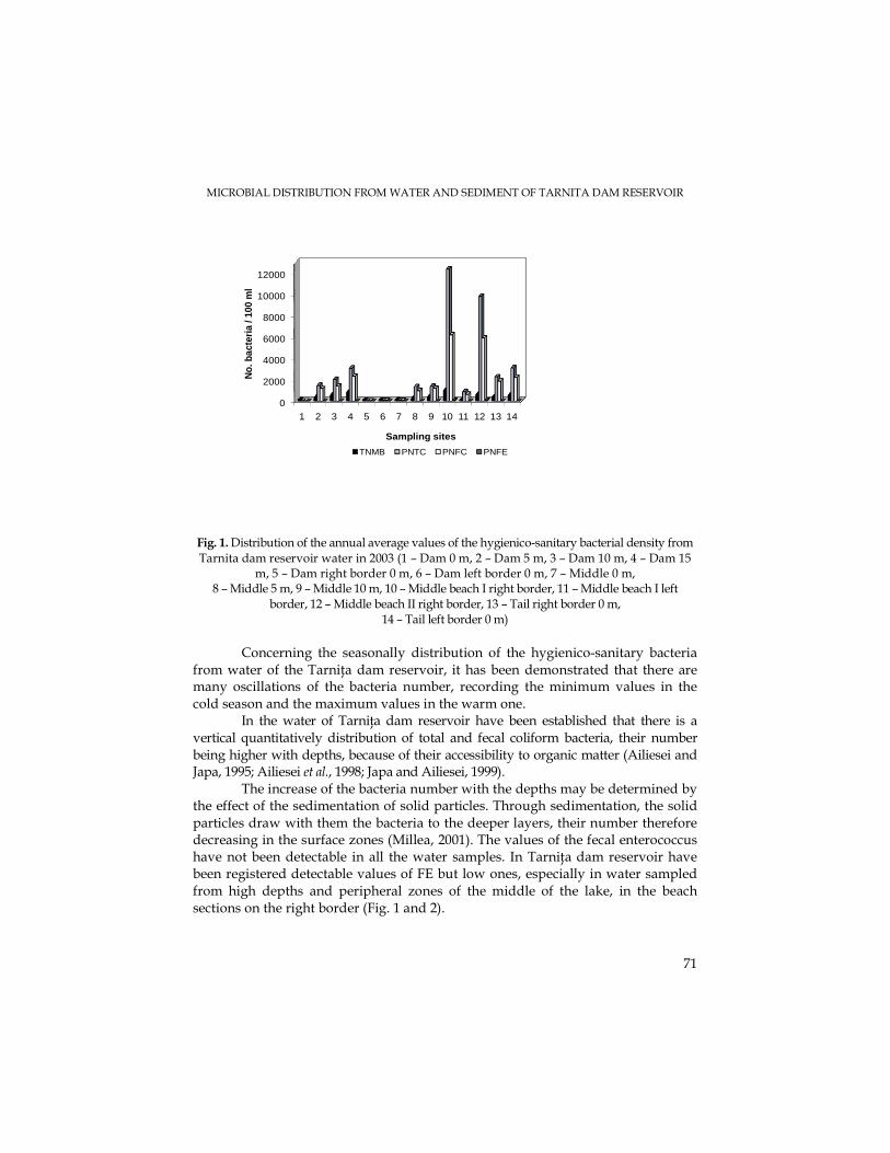
MICROBIAL DISTRIBUTION FROM WATER AND SEDIMENT OF TARNITA DAM RESERVOIR
71
0
2000
4000
6000
8000
10000
12000
1 2 3 4 5 6 7 8 9 10 11 12 13 14
No.
bac
teri
a / 1
00 m
l
Sampling sites
TNMB PNTC PNFC PNFE
Fig. 1. Distribution of the annual average values of the hygienico-sanitary bacterial density from Tarnita dam reservoir water in 2003 (1 – Dam 0 m, 2 – Dam 5 m, 3 – Dam 10 m, 4 – Dam 15
m, 5 – Dam right border 0 m, 6 – Dam left border 0 m, 7 – Middle 0 m, 8 – Middle 5 m, 9 – Middle 10 m, 10 – Middle beach I right border, 11 – Middle beach I left
border, 12 – Middle beach II right border, 13 – Tail right border 0 m, 14 – Tail left border 0 m)
Concerning the seasonally distribution of the hygienico-sanitary bacteria
from water of the TarniŃa dam reservoir, it has been demonstrated that there are many oscillations of the bacteria number, recording the minimum values in the cold season and the maximum values in the warm one.
In the water of TarniŃa dam reservoir have been established that there is a vertical quantitatively distribution of total and fecal coliform bacteria, their number being higher with depths, because of their accessibility to organic matter (Ailiesei and Japa, 1995; Ailiesei et al., 1998; Japa and Ailiesei, 1999).
The increase of the bacteria number with the depths may be determined by the effect of the sedimentation of solid particles. Through sedimentation, the solid particles draw with them the bacteria to the deeper layers, their number therefore decreasing in the surface zones (Millea, 2001). The values of the fecal enterococcus have not been detectable in all the water samples. In TarniŃa dam reservoir have been registered detectable values of FE but low ones, especially in water sampled from high depths and peripheral zones of the middle of the lake, in the beach sections on the right border (Fig. 1 and 2).
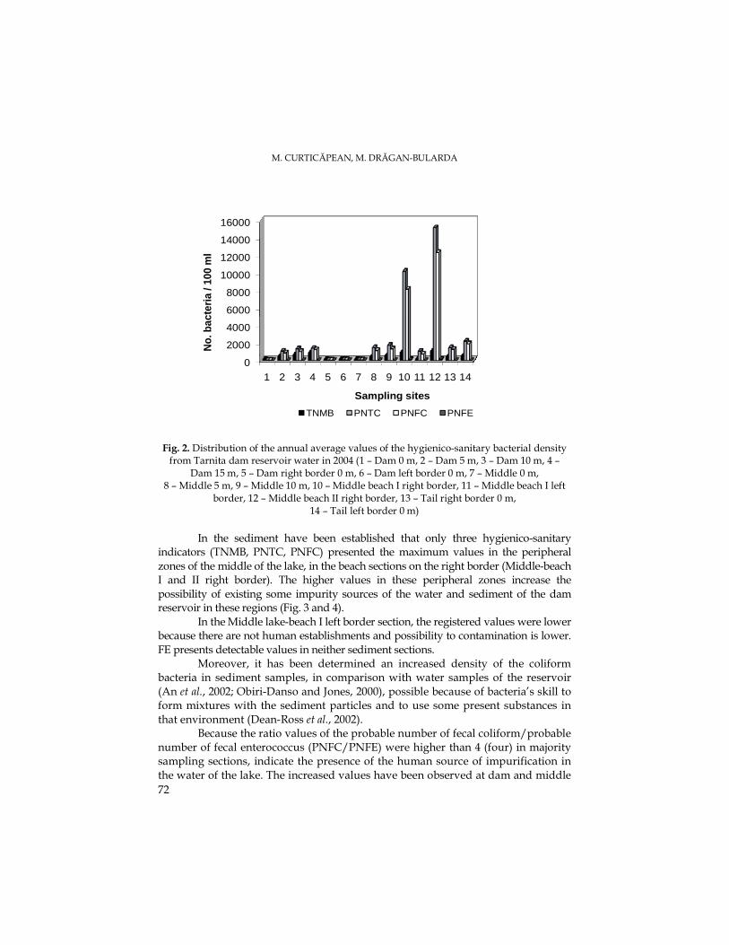
M. CURTICĂPEAN, M. DRĂGAN-BULARDA
72
0
2000
4000
6000
8000
10000
12000
14000
16000
1 2 3 4 5 6 7 8 9 10 11 12 13 14
No.
bac
teri
a / 1
00 m
l
Sampling sites
TNMB PNTC PNFC PNFE
Fig. 2. Distribution of the annual average values of the hygienico-sanitary bacterial density from Tarnita dam reservoir water in 2004 (1 – Dam 0 m, 2 – Dam 5 m, 3 – Dam 10 m, 4 –
Dam 15 m, 5 – Dam right border 0 m, 6 – Dam left border 0 m, 7 – Middle 0 m, 8 – Middle 5 m, 9 – Middle 10 m, 10 – Middle beach I right border, 11 – Middle beach I left
border, 12 – Middle beach II right border, 13 – Tail right border 0 m, 14 – Tail left border 0 m)
In the sediment have been established that only three hygienico-sanitary
indicators (TNMB, PNTC, PNFC) presented the maximum values in the peripheral zones of the middle of the lake, in the beach sections on the right border (Middle-beach I and II right border). The higher values in these peripheral zones increase the possibility of existing some impurity sources of the water and sediment of the dam reservoir in these regions (Fig. 3 and 4).
In the Middle lake-beach I left border section, the registered values were lower because there are not human establishments and possibility to contamination is lower. FE presents detectable values in neither sediment sections.
Moreover, it has been determined an increased density of the coliform bacteria in sediment samples, in comparison with water samples of the reservoir (An et al., 2002; Obiri-Danso and Jones, 2000), possible because of bacteria’s skill to form mixtures with the sediment particles and to use some present substances in that environment (Dean-Ross et al., 2002).
Because the ratio values of the probable number of fecal coliform/probable number of fecal enterococcus (PNFC/PNFE) were higher than 4 (four) in majority sampling sections, indicate the presence of the human source of impurification in the water of the lake. The increased values have been observed at dam and middle
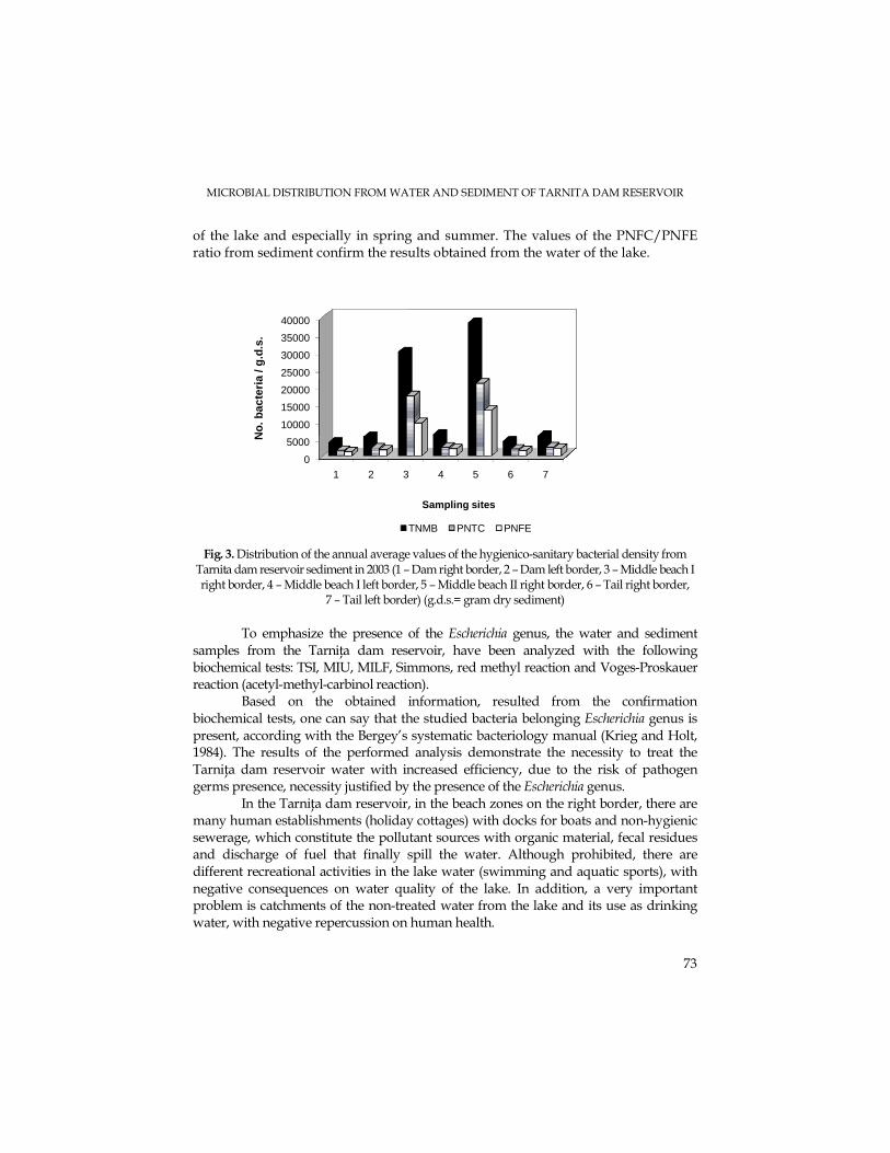
MICROBIAL DISTRIBUTION FROM WATER AND SEDIMENT OF TARNITA DAM RESERVOIR
73
of the lake and especially in spring and summer. The values of the PNFC/PNFE ratio from sediment confirm the results obtained from the water of the lake.
0
5000
10000
15000
20000
25000
30000
35000
40000
1 2 3 4 5 6 7
No
. bac
teri
a / g
.d.s
.
Sampling sites
TNMB PNTC PNFE
Fig. 3. Distribution of the annual average values of the hygienico-sanitary bacterial density from Tarnita dam reservoir sediment in 2003 (1 – Dam right border, 2 – Dam left border, 3 – Middle beach I right border, 4 – Middle beach I left border, 5 – Middle beach II right border, 6 – Tail right border,
7 – Tail left border) (g.d.s.= gram dry sediment) To emphasize the presence of the Escherichia genus, the water and sediment
samples from the TarniŃa dam reservoir, have been analyzed with the following biochemical tests: TSI, MIU, MILF, Simmons, red methyl reaction and Voges-Proskauer reaction (acetyl-methyl-carbinol reaction).
Based on the obtained information, resulted from the confirmation biochemical tests, one can say that the studied bacteria belonging Escherichia genus is present, according with the Bergey’s systematic bacteriology manual (Krieg and Holt, 1984). The results of the performed analysis demonstrate the necessity to treat the TarniŃa dam reservoir water with increased efficiency, due to the risk of pathogen germs presence, necessity justified by the presence of the Escherichia genus.
In the TarniŃa dam reservoir, in the beach zones on the right border, there are many human establishments (holiday cottages) with docks for boats and non-hygienic sewerage, which constitute the pollutant sources with organic material, fecal residues and discharge of fuel that finally spill the water. Although prohibited, there are different recreational activities in the lake water (swimming and aquatic sports), with negative consequences on water quality of the lake. In addition, a very important problem is catchments of the non-treated water from the lake and its use as drinking water, with negative repercussion on human health.
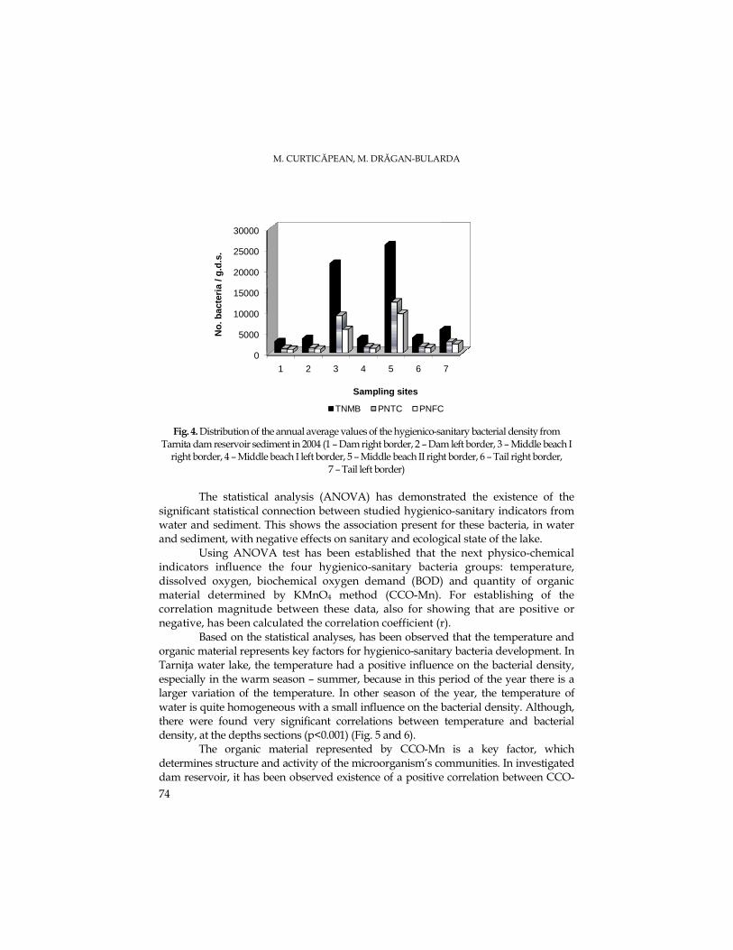
M. CURTICĂPEAN, M. DRĂGAN-BULARDA
74
0
5000
10000
15000
20000
25000
30000
1 2 3 4 5 6 7
No
. bac
teri
a / g
.d.s
.
Sampling sites
TNMB PNTC PNFC
Fig. 4. Distribution of the annual average values of the hygienico-sanitary bacterial density from
Tarnita dam reservoir sediment in 2004 (1 – Dam right border, 2 – Dam left border, 3 – Middle beach I right border, 4 – Middle beach I left border, 5 – Middle beach II right border, 6 – Tail right border,
7 – Tail left border) The statistical analysis (ANOVA) has demonstrated the existence of the
significant statistical connection between studied hygienico-sanitary indicators from water and sediment. This shows the association present for these bacteria, in water and sediment, with negative effects on sanitary and ecological state of the lake.
Using ANOVA test has been established that the next physico-chemical indicators influence the four hygienico-sanitary bacteria groups: temperature, dissolved oxygen, biochemical oxygen demand (BOD) and quantity of organic material determined by KMnO4 method (CCO-Mn). For establishing of the correlation magnitude between these data, also for showing that are positive or negative, has been calculated the correlation coefficient (r).
Based on the statistical analyses, has been observed that the temperature and organic material represents key factors for hygienico-sanitary bacteria development. In TarniŃa water lake, the temperature had a positive influence on the bacterial density, especially in the warm season – summer, because in this period of the year there is a larger variation of the temperature. In other season of the year, the temperature of water is quite homogeneous with a small influence on the bacterial density. Although, there were found very significant correlations between temperature and bacterial density, at the depths sections (p<0.001) (Fig. 5 and 6).
The organic material represented by CCO-Mn is a key factor, which determines structure and activity of the microorganism’s communities. In investigated dam reservoir, it has been observed existence of a positive correlation between CCO-
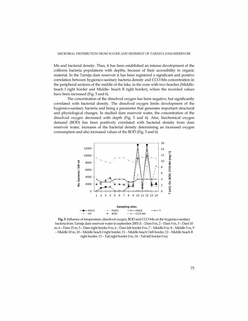
MICROBIAL DISTRIBUTION FROM WATER AND SEDIMENT OF TARNITA DAM RESERVOIR
75
Mn and bacterial density. Thus, it has been established an intense development of the coliform bacteria populations with depths, because of their accessibility to organic material. In the TarniŃa dam reservoir it has been registered a significant and positive correlation between hygienico-sanitary bacteria density and CCO-Mn concentration in the peripheral sections of the middle of the lake, in the zone with two beaches (Middle-beach I right border and Middle- beach II right border), where the recorded values have been increased (Fig. 5 and 6).
The concentration of the dissolved oxygen has been negative, but significantly correlated with bacterial density. The dissolved oxygen limits development of the hygienico-sanitary bacteria and being a parameter that generates important structural and physiological changes. In studied dam reservoir water, the concentration of the dissolved oxygen decreased with depth (Fig. 5 and 6). Also, biochemical oxygen demand (BOD) has been positively correlated with bacterial density from dam reservoir water, increases of the bacterial density determining an increased oxygen consumption and also increased values of the BOD (Fig. 5 and 6).
0
2
4
6
8
10
12
14
16
0
2000
4000
6000
8000
10000
12000
1 2 3 4 5 6 7 8 9 10 11 12 13 14
T (o
C);
O2,
BO
D, C
CO
-Mn
(mg
/ l)
No
. bac
teri
a / 1
00 m
l
Sampling sitesPNTC PNFC PNFE TO2 BOD CCO-Mn
Fig. 5. Influence of temperature, dissolved oxygen, BOD and CCO-Mn on the hygienico-sanitary
bacteria from TarniŃa dam reservoir water in september 2003 (1 – Dam 0 m, 2 – Dam 5 m, 3 – Dam 10 m, 4 – Dam 15 m, 5 – Dam right border 0 m, 6 – Dam left border 0 m, 7 – Middle 0 m, 8 – Middle 5 m, 9 – Middle 10 m, 10 – Middle beach I right border, 11 – Middle beach I left border, 12 – Middle beach II
right border, 13 – Tail right border 0 m, 14 – Tail left border 0 m)

M. CURTICĂPEAN, M. DRĂGAN-BULARDA
76
0
2
4
6
8
10
12
14
16
18
20
0
2000
4000
6000
8000
10000
12000
14000
16000
18000
20000
22000
1 2 3 4 5 6 7 8 9 10 11 12 13 14 T (o
C);
O2,
BO
D, C
CO
-Mn
(mg
/ l)
No
. bac
teri
a / 1
00 m
l
Sampling sitesPNTC PNFC PNFE TO2 BOD CCO-Mn
Fig. 6. Influence of temperature, dissolved oxygen, BOD and CCO-Mn on the hygienico-sanitary bacteria from TarniŃa dam reservoir water in july 2004 (1 – Dam 0 m, 2 – Dam 5 m, 3 – Dam 10 m, 4 – Dam 15 m, 5 – Dam right border 0 m, 6 – Dam left border 0 m, 7 – Middle 0 m, 8 – Middle 5 m, 9 – Middle 10 m, 10 – Middle beach I right border, 11 – Middle beach I left border, 12 – Middle beach II
right border, 13 – Tail right border 0 m, 14 – Tail left border 0 m) The statistical analyses have established that in the Tarnita dam reservoir it has
been obtained the significant positive correlations between all hygienico-sanitary indicators from different samples of water, in all sampling period. The most significant correlations for the surface water sections have been recorded between bacterial density from the sections from the beach zone of the middle of the lake, also between these and those from the tail of the lake. There have been observed differences between the middle and peripheral zones of the lake, showing the presence of the negative influences in these zones of the lake. In the water depths sections it has been established a positive and significant correlation in all the sampling periods (p<0.01). Using the same statistical tests it has been observed that the bacterial density from the sediment sections was positive and significant correlated in all sampling periods (p<0.001).
Conclusions 1. Although generally, the water of the TarniŃa dam reservoir has a good
hygienico-sanitary state, there are the spillage sources, which may confer a danger for health. The framing of waters in A3 category demonstrates the necessity to treat the TarniŃa dam reservoir water with increased efficiency and costs, due to the risk of pathogen germs presence.
2. The presence of the bacteria in a higher number at the peripheral zones is influenced by the dynamics of water and especially by presence of some organic contamination sources.
MICROBIAL DISTRIBUTION FROM WATER AND SEDIMENT OF TARNITA DAM RESERVOIR
77
3. Based on the obtained information resulted from the confirmation biochemical tests, one can say that in TarniŃa dam reservoir, the studied bacteria belonging Escherichia genus is present.
4. The statistical analyses have established that in the Tarnita dam reservoir it has been obtained the significant positive correlations between all hygienico-sanitary indicators from different samples of water, in all sampling period. Using the same statistical tests it has been observed that the bacterial density from the sediment sections was positive and significant correlated in all sampling periods.
Through the study of these dam reservoirs, one can assume that the monitoring of the waters used as drinking water is necessary and very important to maintain a better ecological potential and to establish and remove the possible pollution sources.
Acknowledgements. We would like to thank to Miss Carmen Nap (S.C. Hidroeletrica S.A., Sucursala
Hidrocentrale Cluj), for her support for samples collecting from the Tarnita dam reservoir. Also, to Dr. Marius Marusteri (UMF Tg. Mures, Faculty of Pharmacy) for statistical calculations guidance.
REFERENCE S Ailiesei, O., Jâpa, F. (1995). Microbiological characteristics of the Vaduri dam lake in the
natural conditions and under the influence of salmonid aquaculture, An. Şti. Univ. Al. I. Cuza, Iaşi, 41 (II), 97-104
Ailiesei, O., NimiŃan, E., Jâpa, F., Dunca, S. (1998). Ecology of some bacterial populations in the Şerbăneşti dam lake, An. Şti. Univ. Al. I. Cuza, Iaşi, 44 (II), 163-175
An, Y-J., Kampbell, D.H., Breidenbach, G.P. (2002). Escherichia coli and total coliforms in water and sediments at lake marinas, Environ. Pollution, 120 (3), 771-778
Barbato, G., Grottolo, M., Resola, S. (1990). Indagine sul lago D’Idro-Aspetti chimico-fisici, microgiologici e fitoplanctonici, Monografie di „Natura Bresciana”, Promodis Italia editrice, Brescia, pp. 45-52
Burian, P. (2002). Lacul de acumulare-analiza biologică, Edit. UniversităŃii de Medicină şi Farmacie, University Press, Târgu-Mureş, pp. 13-16, 117
Cuşa, V. (1996). InstrucŃiuni metodologice pentru analiza microbiologică a sedimentelor din ecosistemele acvatice, Inst. Cercet. Ing. Mediului, Bucureşti, 4, 14-20
Dean-Ross, D., Moody, J., Cerniglia, C.E. (2002). Utilization of mixtures of polycyclic aromatic hydrocarbons by bacteria isolated from contaminated sediment, FEMS Microb. Ecol., 41 (1), 1-7
Diudea, M., Tudor, S., Igna, A. (1986). Toxicologie acvatică, Edit. Dacia, Cluj-Napoca, pp. 31-33 Drăgan-Bularda, M. (2000). Lucrări practice de microbiologie generală, Univ. “Babeş-Bolyai”, Cluj-
Napoca, pp. 175-180 Dunca, S., Ailiesei, O., NimiŃan, E., Ştefan, M. (2004). Microbiologie aplicată, Edit. Tehnopress,
Iaşi, pp. 45, 147-148, 158 Gianfreda, L., Bollag, J-M. (1996). Influence of natural and anthropogenic factors on enzyme activity in
soil In: Soil Biochemistry, Stotzky, G. and Bollag, J-M. (Ed.), Vol.9, Marcel Dekker, Inc., New York, pp. 123-193
M. CURTICĂPEAN, M. DRĂGAN-BULARDA
78
Jâpa, F., Ailiesei, O. (1999). The quantitative distribution of the main ecophysiological groups of bacteria involved in the biogenic elements cycle from the Siriu lake, An. Şti. Univ. Al. I. Cuza, Iaşi, 45 (II), 161-166
Kenneth, T. (2003). Procaryotes in the environment and Major groups of procaryotes, In: Bacteriology 303 Main Page, University of Wisconsin-Madison (Ed.), Department of Bacteriology.
Krieg, N.R., Holt, J.G. (1984). Bergey’s Manual of Systematic Bacteriology, Williams and Wilkins Ed., Vol. I, Baltimore, pp. 410-466
Madigan, M.T., Martinko, J.M., Parker, J. (2000). Brock Biology of Microorganisms, 9th ed. Prentice-Hall, Inc., Englewood Cliffs, New Jersey.
Marusteri, M. (2006). Notiuni fundamentale de biostatistica (www.umftgm.ro/info/carte_statistica.pdf)
Millea, L.C. (2001). Preocupări actuale legate de poluarea apelor, Stud. Cercet. Biol., BistriŃa, 6, 9-34 Muntean, V., Crişan, R., Paşca, D. (2001). Cercetări enzimologice asupra nămolului din balta
sărată Alunei-Târnăveni, Stud. Cercet. Biol., BistriŃa, 6, 7-12 Obiri-Danso, K., Jones, K. (2000). Intertidal sediments as reservoirs for hippurate negative
Campylobacters, Salmonellae and faecal indicators in three EU recognised bathing waters in North West England, Wat. Res., 34 (2), 519-527
Şerban, G. (1999). Lacurile de acumulare din bazinul superior al Someşului Mic (partea I), Stud. Univ. Babeş-Bolyai, Geographia, Cluj-Napoca, 43 (2), 10-19
*** STAS 3001, Apa – analiza bacteriologică (1991). *** Atlasul Cadastrului Apelor din România (1992). Ministerul Mediului, Bucureşti. *** Water Framework Directive (2001). *** Normativ privind obiectivele de referinŃă pentru clasificarea calităŃii apelor de suprafaŃă,
(2002), apărut în Monitorul Oficial al României, Partea I, Nr. 197/27.03.2003. *** NTPA-013, Norme de calitate pe care trebuie să le îndeplinească apele de suprafaŃă utilizate
pentru potabilizare, apărut în Monitorul Oficial al României, Partea I, Nr. 130/19.02.2002. *** Directive 75/440/EEC – regarding the quality of surface water used as drinking water. *** Regulamente de exploatare a lacurilor, DirecŃia Apelor Someş-Tisa, Cluj-Napoca.
STUDIA UNIVERSITATIS BABEŞ – BOLYAI, BIOLOGIA, LII, 1, 2007 (p.79-85)
THE QUALITATIVE ENZYMATIC ACTIVITY OF GILAU AND TARNITA DAM RESERVOIR SEDIMENT
MANUELA CURTICĂPEAN1 and MIHAIL DRĂGAN-BULARDA2
SUMMARY. The enzymological studies on the sediment of the accumulation lake that has the main purpose of supplying drinking water to the city of Cluj-Napoca, were aimed at the comprehensive understanding of the complex processes that happen in these habitats of special significance. In the sediment samples, the following enzymatic activities have been qualitatively determined: maltase, saccharase, lactase, cellobiase, amylase, dextranase, levanase, cellulase and inulinase. Seven sediment samples were collected seasonally, from different sites. The qualitative enzymatic activities have a uniform behavior without big differences from one season to another, and from one year to another. One can observe that not all activities have detectable values for the analyzed sediment samples. There were used the statistical chi-square test (χ2) to emphasize the presence of some correlations between the enzymatic activities from sediment sections of the Gilau and Tarnita dam reservoirs. KEYWORDS: enzymatic activity, dam reservoir, sediment
Introduction The ecological succession in lakes, respectively the evolution of these aquatic
ecosystems in time, is the result of the complex interactions between biocenotic communities, respectively between these and abiotic characteristics of the life media, being in a constant modification (Burian, 2002).
The sediments consist of three major components: the detritic material derived from the erosion, the biogenic material formed from the biological productivity and the autogenic material formed in situ (Wetzel, 1991). The sediments are very heterogeneous systems where the different physical phases (solid, liquid and gases) and numerous biotic (microorganisms, the small organisms, enzymes) and abiotic (minerals, humus materials, organo-mineral aggregate) components are involved in physical, chemical and biological processes. All the biochemical transformations from sediments depend on the enzymes presence (Gianfreda and Bollag, 1996).
The sediments constitute a key link in the biogeochemical cycle of the elements in the aquatic systems. Here are finalized the mineralization processes of the organic substances that were not degraded in the water column (Muntean et al., 2001).
The action of the microorganisms on the environmental substrates takes the enzymatic way through oxidoreduction and hydrolysis, respectively through the action of some final products of the microbial metabolism (Muntean et al., 2004).
1 University of Medicine and Pharmacy, Faculty of Pharmacy, F1, Pharmaceutical Botany and Cell Biology Department, 38 Gh. Marinescu Street, 540139 Târgu-Mureş, Romania, E-mail: [email protected]
2 „Babeş-Bolyai” University, Faculty of Biology and Geology, Experimental Biology Department, 1 M. Kogălniceanu Street, Cluj-Napoca, Romania, E-mail: [email protected]
M. CURTICĂPEAN, M. DRĂGAN-BULARDA
80
The stage decomposition of the organic material have as result the accumulation of soluble monomers, oligomers and macromolecules, based on the type of the substrate and the involved enzymes. The bacteria and microplankton include some of these compounds, while others suffer further enzymatic decomposition (Chróst, 1991; Meyer-Reil, 1987, 1991). The sediment medium is a complex function of very different factors, like the major mineral matrix, texture, quantity of organic carbon, geographical localization (Malcolm and Stanley, 1982). Thus, the enzymatic activity determination offers, in a shorter time than microbiological analysis, the suggestive data regarding the processes that are taking place in sediments or in other natural habitats. Their determination from sediments of the aquatic ecosystems consist a research method in evaluation the functional diversity of the microbiota involved in the biogeochemical cycles.
The Gilău and TarniŃa dam reservoirs are positioned in the Someşul Mic upper basin that has an area of 3804 km2 and a length of 167 km (***, 1992). The Gilău artificial lake, positioned downstream the TarniŃa lake, is the first accumulation opened in 1972. The Gilău reservoir has 4.1 million m3 water capacity. It serves several functions, but mainly to guarantee the drinking and industrial water for Cluj-Napoca city and the nearby villages (Gherla city and Aghireş area). Other functions are energetic, flood aversion, wave attenuation and providing water to the Gilău trout nursery (Serban, 1999). The TarniŃa artificial lake that was opened for use in 1973 represents one of the most severe anthropic effects on the natural course of the Someşul Mic River. The reservoir, with a capacity of 77.4 million m3 water has the following functions: mainly – energetic, flood aversion, wave attenuation and, in the future, guarantee with drinking water for Cluj-Napoca city (*** Regulamente de exploatare a lacurilor).
Thus, the dam reservoirs water used to drinking have to fulfill some quality conditions, before entering in the treatment station. Therefore, according with the Water Framework Directive demands (***, 2001), the thoroughly study of these waters is essential, especially from microbiological and enzymological point of view, in order to find the impact ways and to apply the suitable control methods for rehabilitation and maintaining of the water quality at a good and very good potential.
The present paper analyze for the first time the evolution of the enzymatic activities from these dam reservoirs, which is necessary and has a special importance in the case of water ecosystems with ecological value. The water ecosystems used as drinking water require these studies, for increased exactingness of the water and sediment quality.
Materials and Methods The enzymological analysis from sediment were performed in April 2002-
October 2003 period for Gilău dam reservoir and in April 2003-October 2004 period for TarniŃa dam reservoir. The seven sediment samples were taken seasonally at 0-5 cm depth (with Ponar dredger - USA Wildlife Supply Company, 48602), in the same time with the water samples. The humidity of each sediment sample was established during their preparation for the analysis, because of different sediment categories that may have variable water content (which can influence the expression of the
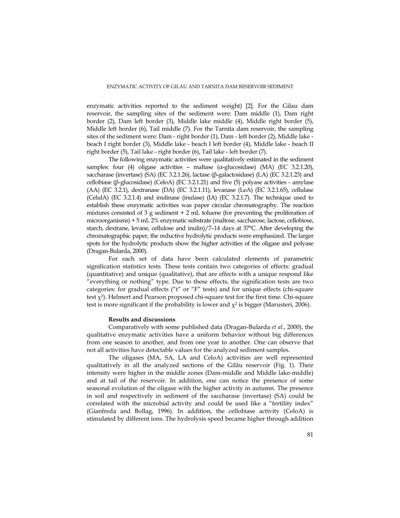
ENZYMATIC ACTIVITY OF GILAU AND TARNITA DAM RESERVOIR SEDIMENT
81
enzymatic activities reported to the sediment weight) [2]. For the Gilau dam reservoir, the sampling sites of the sediment were: Dam middle (1), Dam right border (2), Dam left border (3), Middle lake middle (4), Middle right border (5), Middle left border (6), Tail middle (7). For the Tarnita dam reservoir, the sampling sites of the sediment were: Dam - right border (1), Dam - left border (2), Middle lake - beach I right border (3), Middle lake - beach I left border (4), Middle lake - beach II right border (5), Tail lake - right border (6), Tail lake - left border (7).
The following enzymatic activities were qualitatively estimated in the sediment samples: four (4) oligase activities – maltase (α-glucosidase) (MA) (EC 3.2.1.20), saccharase (invertase) (SA) (EC 3.2.1.26), lactase (β-galactosidase) (LA) (EC 3.2.1.23) and cellobiase (β-glucosidase) (CeloA) (EC 3.2.1.21) and five (5) polyase activities - amylase (AA) (EC 3.2.1), dextranase (DA) (EC 3.2.1.11), levanase (LeA) (EC 3.2.1.65), cellulase (CelulA) (EC 3.2.1.4) and inulinase (inulase) (IA) (EC 3.2.1.7). The technique used to establish these enzymatic activities was paper circular chromatography. The reaction mixtures consisted of 3 g sediment + 2 mL toluene (for preventing the proliferation of microorganisms) + 5 mL 2% enzymatic substrate (maltose, saccharose, lactose, cellobiose, starch, dextrane, levane, cellulose and inulin)/7–14 days at 37°C. After developing the chromatographic paper, the reductive hydrolytic products were emphasized. The larger spots for the hydrolytic products show the higher activities of the oligase and polyase (Dragan-Bularda, 2000).
For each set of data have been calculated elements of parametric signification statistics tests. These tests contain two categories of effects: gradual (quantitative) and unique (qualitative), that are effects with a unique respond like “everything or nothing” type. Due to these effects, the signification tests are two categories: for gradual effects (“t” or “F” tests) and for unique effects (chi-square test χ2). Helmert and Pearson proposed chi-square test for the first time. Chi-square test is more significant if the probability is lower and χ2 is bigger (Marusteri, 2006).
Results and discussions Comparatively with some published data (Dragan-Bularda et al., 2000), the
qualitative enzymatic activities have a uniform behavior without big differences from one season to another, and from one year to another. One can observe that not all activities have detectable values for the analyzed sediment samples.
The oligases (MA, SA, LA and CeloA) activities are well represented qualitatively in all the analyzed sections of the Gilău reservoir (Fig. 1). Their intensity were higher in the middle zones (Dam-middle and Middle lake-middle) and at tail of the reservoir. In addition, one can notice the presence of some seasonal evolution of the oligase with the higher activity in autumn. The presence in soil and respectively in sediment of the saccharase (invertase) (SA) could be correlated with the microbial activity and could be used like a “fertility index” (Gianfreda and Bollag, 1996). In addition, the cellobiase activity (CeloA) is stimulated by different ions. The hydrolysis speed became higher through addition
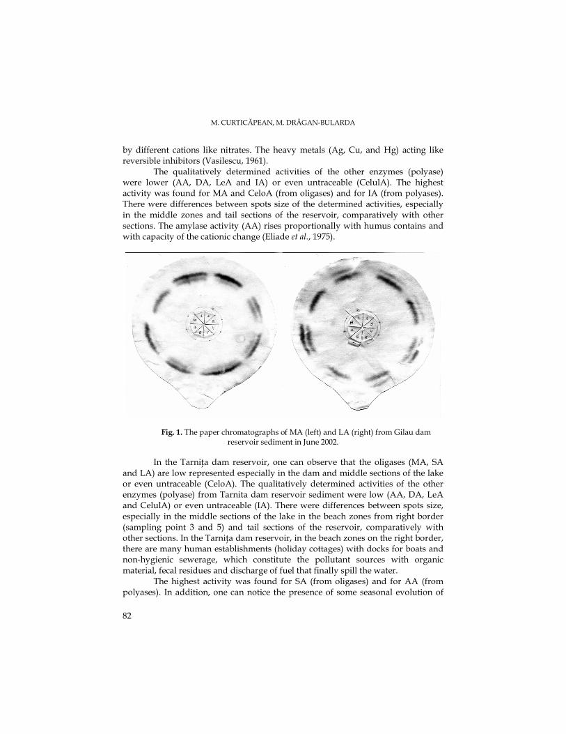
M. CURTICĂPEAN, M. DRĂGAN-BULARDA
82
by different cations like nitrates. The heavy metals (Ag, Cu, and Hg) acting like reversible inhibitors (Vasilescu, 1961).
The qualitatively determined activities of the other enzymes (polyase) were lower (AA, DA, LeA and IA) or even untraceable (CelulA). The highest activity was found for MA and CeloA (from oligases) and for IA (from polyases). There were differences between spots size of the determined activities, especially in the middle zones and tail sections of the reservoir, comparatively with other sections. The amylase activity (AA) rises proportionally with humus contains and with capacity of the cationic change (Eliade et al., 1975).
Fig. 1. The paper chromatographs of MA (left) and LA (right) from Gilau dam reservoir sediment in June 2002.
In the TarniŃa dam reservoir, one can observe that the oligases (MA, SA
and LA) are low represented especially in the dam and middle sections of the lake or even untraceable (CeloA). The qualitatively determined activities of the other enzymes (polyase) from Tarnita dam reservoir sediment were low (AA, DA, LeA and CelulA) or even untraceable (IA). There were differences between spots size, especially in the middle sections of the lake in the beach zones from right border (sampling point 3 and 5) and tail sections of the reservoir, comparatively with other sections. In the TarniŃa dam reservoir, in the beach zones on the right border, there are many human establishments (holiday cottages) with docks for boats and non-hygienic sewerage, which constitute the pollutant sources with organic material, fecal residues and discharge of fuel that finally spill the water.
The highest activity was found for SA (from oligases) and for AA (from polyases). In addition, one can notice the presence of some seasonal evolution of
ENZYMATIC ACTIVITY OF GILAU AND TARNITA DAM RESERVOIR SEDIMENT
83
the oligase and polyase activity with higher activity in summer, due to the difference in the size of the spots.
Table 1. The contingency table with enzimatic activity from Gilau dam reservoir sediment in 2002-2003 period Oligase Polyase
Enzymatic activity
S1 S2 S3 Enzymatic activity
S1 S2 S3
- ; +/- + ; ++ +++ ; > +++
11 31 14
0 25 30
2 23 31
- +/- + ; ++
50 5 14
32 13 25
41 14 15
S1 – spring; S2 – summer; S3 – autumn; - nedectable enzymatic activity; +/- and + lower enzymatic activity; ++ medium enzymatic activity; +++ ; > +++ higher enzymatic activity
Table 2. The contingency table with enzimatic activity from Tarnita dam reservoir sediment in 2003-2004 period
Oligase Polyase
Enzymatic activity
S1 S2 S3 Enzymatic activity
S1 S2 S3
- ; +/- + ; ++ +++ ; > +++
38 8 10
38 4 14
38 10 8
- +/- + ; ++
56 9 5
48 14 8
58 5 6
S1 – spring; S2 – summer; S3 – autumn; - nedectable enzymatic activity; +/- and + lower enzymatic activity; ++ medium enzymatic activity; +++ ; > +++ higher enzymatic activity
The hydrolases (polysaccharidases and proteinases) are extracelular
esential enzymes that transform macromolecular substrates in smaller compounds. In the aquatic sediment, very important in the biological cycle of carbon and azot, was proteines depolymerisation and mineralisation of their monomeric products. Also, the hydrolitic enzymatic activities could be used for pesticides and other xenobiotics transformation (Gianfreda and Bollag, 1996).
The chi-square test (χ2) was used to emphasize the presence of some correlations between the enzymatic activities from sediment sections of the Gilau and Tarnita dam reservoirs. For statistical correlation, the results of the enzymatic activities were grouped in contingency tables (Table 1 and 2) and using GraphPad InStat software version 3.05 (2000) was calculated chi-square test.
In the Gilau dam reservoir sediment, the oligases activities are significantly associated (the row and column variables) with χ2 = 24.383 and p < 0.0001. In addition, the polyases activities are significantly associated with χ2 = 12.640 and p < 0.0132. In the Tarnita dam reservoir sediment, the oligase and polyase activities are not significantly associated: χ2 = 4.295 and p = 0.3675 (oligase); χ2 = 6.126 and p = 0.1900 (polyase). Chi-square test could be more significant if the probability is lower and χ2 is bigger.
M. CURTICĂPEAN, M. DRĂGAN-BULARDA
84
Based on the statistical test it has been established that in the Gilău dam reservoir sediment there are significant statistical connection between the studied enzymatic activities. These facts demonstrate that the sediment of the lake is a very heterogeneous system (with a mud consistence), where the enzymatic activities interact for determining the complex biochemical changes from sediment and form a connection between sediment particles and enzymes.
The nonsignificant statistical connection between the studied enzymatic activities from Tarnita dam reservoir sediment could appear probably due to the sediment consistence (sand and gravel, nonsignificant muddy).
The qualitative analysis of the oligase and polyase increases the complexity of evaluation of the total enzymatic potential of the lake sediment.
Conclusions The above-mentioned enzymatic activities were chosen because their
determination in the aquatic sediments constitutes a research tool to evaluate the functional diversity of the microbiota involved in the biogeochemical cycles.
1. The qualitative enzymatic activities have a uniform behavior without big differences from one season to another and from one year to another.
2. One can observe that not all activities have detectable values for the analyzed sediment samples.
3. Based on the statistical test it has been established that in the Gilău dam reservoir sediment there are significant statistical connection between the studied enzymatic activities.
4. The nonsignificant statistical connection between the studied enzymatic activities from Tarnita dam reservoir sediment could appear probably due to the sediment consistence.
Acknowledgements. We would like to thank our colleagues from S.C. Hidroelectrica S.A., Sucursala
Hidrocentrale Cluj and from “Somes-Tisa” Water Division, Cluj-Napoca, for their support with sample collection from the Gilau and Tarnita dam reservoirs. Also, to dr. Marius Marusteri (UMF Tg. Mures, Faculty of Pharmacy) for statistical calculations guidance.
REFERENCES Burian, P. (2002). Lacul de acumulare-analiza biologică, Edit. UniversităŃii de Medicină şi Farmacie,
University Press, Târgu-Mureş, pp. 13-16, 117 Chróst, J., R. (1991). Environmental control of the synthesis and activity of aquatic microbial ectoenzymes
In: Microbial enzymes in aquatic environments, Ryszard J. Chróst (Ed.), Springer-Verlag Ed., New York, pp. 29-31, 51-54
ENZYMATIC ACTIVITY OF GILAU AND TARNITA DAM RESERVOIR SEDIMENT
85
Drăgan-Bularda, M. (2000). Lucrări practice de microbiologie generală, Univ. “Babeş-Bolyai”, Cluj-Napoca, pp. 175-180
Drăgan-Bularda, M., Poptelecan, I-C., Muntean, V., Crişan, R., Paşca, D. (2000). Cercetări enzimologice asupra nămolurilor din lacurile saline de la Bazna şi Blaj, Stud. Univ. Babeş-Bolyai, Biol., Cluj-Napoca, 45 (1), 121-128
Eliade, G., Ghinea, L., Ştefania, G. (1975). Microbiologia solului, Edit. „Ceres”, Bucureşti, pp. 139-144 Gianfreda, L., Bollag, J-M., (1996). Influence of natural and anthropogenic factors on enzyme activity in
soil, In: Soil Biochemistry, Stotzky, G. and Bollag, J-M. (Ed.), Vol.9, Marcel Dekker, Inc., New York, pp. 123-129
Malcolm S.J., Stanley, S.D. (1982). The sediment environment, In: Sediment Microbiology, Nedwell D.B. (Ed.), Acad. Press, London, pp. 1-14
Marusteri, M. (2006). Notiuni fundamentale de biostatistica (www.umftgm.ro/info/carte_statistica.pdf)
Meyer-Reil, L-A., (1987). Seasonal and spatial distribution of extracellular enzymatic activities and microbial incorporation of dissolved organic substrates in marine sediments, Appl. Environ. Microbiol., 53, 1748-1755
Meyer-Reil, L-A. (1991). Ecological aspects of enzymatic activity in marine sediment, In: Microbial Enzymes in Aquatic Environment, R.J. Chrόst (Ed.), Springer-Verlag Ed., Berlin, pp. 84-94
Muntean, V., Crişan, R., Paşca, D. (2001). Cercetări enzimologice asupra nămolului din balta sărată Alunei-Târnăveni, Stud. Cercet. Biol., BistriŃa, 6, 7-12
Muntean, V., Ştef, L.C., Drăgan-Bularda, M. (2004). Cercetări enzimologice asupra unor sedimente din râul Mureş, Romanian Biological Sciences, I (3-4), 107-114
Şerban, G. (1999). Lacurile de acumulare din bazinul superior al Someşului Mic (partea I), Stud. Univ. Babeş-Bolyai, Geographia, Cluj-Napoca, 43 (2), 10-19
Vasilescu, I. (1961). Enzimele, Edit. Academiei RPR, pp. 159-178 Wetzel, R.G. (1991). Extracellular enzymatic interactions: storage, redistribution and interspecific
communication, In: Microbial enzymes in aquatic environments, Ryszard J. Chróst (Ed.), Spriger-Verlag Ed., New York, pp. 19-25
*** Atlasul Cadastrului Apelor din România (1992). Ministerul Mediului, Bucureşti. *** Water Framework Directive (2001). *** Regulamente de exploatare a lacurilor, DirecŃia Apelor Someş-Tisa, Cluj-Napoca.
STUDIA UNIVERSITATIS BABEŞ – BOLYAI, BIOLOGIA, LII, 1, 2007 (p.87-92)
BACTERIAL AND ENZYMATIC INDICATORS OF WATER AND SEDIMENT POLLUTION IN THE ARIEŞ RIVER
VASILE MUNTEAN1
SUMMARY. Eleven sediment and water samples from the Arieş River were studied microbiologically, enzymologically and physico-chemically. The following four ecophysiological bacterial groups have been studied: aerobic mesophilic heterotrophs, ammonifiers, denitrifiers and iron-reducers. The following four enzymatic activities have been measured: phosphatase, catalase, actual and potential dehydrogenase. Some physico-chemical parameters of water were also analyzed: pH, Eh, conductivity and O2 concentration. The presence of all four ecophysiological bacterial groups was registered in all the studied sediments. The descending ranking of their abundance was: aerobic mesophilic heterotrophs > ammonifiers > denitrifiers > iron-reducers. Based on the bacteria number of each ecophysiological group, the bacterial indicators of sediment (BISQ) and water (BIWQ) quality were calculated. The four enzymatic activities were noticed in all the studied samples. On the base of the analytical data, the enzymatic indicators of sediment quality (EISQ) were calculated. As in the case of the microbiological analyses, the enzymatic potential of sediments increases downstream the river. The lowest values of both bacterial and enzymatic indicators were registered in the P11 site, where the river Abrud flows into Arieş, indicating a strong local pollution, probably caused by the mining enterprise in Roşia Montană. The physico-chemical parameters were also very different as compared to the other sampling sites. A statistically significant strong positive correlation has been established between the bacterial and enzymatic indicators. KEYWORDS: enzymes, bacteria, sediments, water, pollution, indicators of water and sediment quality
Introduction. The water pollution is a pressing matter of our times worldwide, but
insufficiently studied in our country. The importance of microbial and enzymatic activity as an indicator of water and sediment pollution was frequently underlined (Papp et. al., 2002, Luna et al., 2002, Noble et. al., 2003, Ştef et. al., 2004). Decomposition and mineralization of organic matter are processes of great importance for the releasing of biogenic elements in the aquatic environments. A part of the organic matter which originate in phytoplankton and in zooplankton, enter into the dissolved organic phase of the water. The particulate phase is partly incorporated by the secondary consumers. The rest is converted in detritus and subsequently submitted to decomposition. Some of these compounds are incorporated by bacteria or micro plankton, while others undergo further enzymatic decomposition. The compounds of low molecular weight resulting from the exoenzyme activities are rapidly metabolized by the heterotrophic bacteria. One can consider that the rate of the organic matter degradation is, probably, controlled by the exoenzymatic hydrolysis
1 Babeş-Bolyai University, 1 Kogălniceanu Street, 400084/Cluj-Napoca, Romania, E-mail: [email protected]
V. MUNTEAN
88
(Meyer-Reyl, 1987). This emphasizes the importance of the enzymological analyses for detection the effect of the pollutants on the normal occurence of the biological cycles in different habitats.
The present paper aimed to evaluate the bacterial and enzymatic potential in water and sediments of the Arieş River, in order to detect the effect of some putative pollutants. We choose to determine the enzymatic activity because it offers relevant data, regarding the processes that take place in sediments in shorter time than the classic microbiological analysis.
Material and Methods. The microbiological, enzymological and physico-chemical analyses were
carried out on water and sediments samples collected from eleven sites, as follows (odd number – upstream; even number – downstream): P1,2 – Câmpeni; P3,4 – Baia de Arieş; P5,6 – Sălciua; P7,8 – Turda; P9,10 – Câmpia Turzii; P11 – site where the Abrud river flows into the Arieş.
We studied the ecophysiological bacterial groups implied in the biogeochemical cycles of carbon and nitrogen, as well as iron-reducers, taking into account the existence of iron mines along the river Arieş. The following four ecophysiological bacterial groups have been studied: aerobic mesophilic heterotrophs, ammonifiers, denitrifiers, and iron-reducers. The following methods and media were used: aerobic mesophilic heterotrophs (agar plates), ammonifiers (peptone medium) (Atlas, 2004), denitrifiers (De Barjac culture medium) (Pochon, 1954) and iron-reducers (Ottow medium) (Pârvu et al., 1978). Except for the aerobic mesophilic heterotrophs (where the method of successive dilutions, was used), the most probable number of bacteria was calculated according to the statistical table of Alexander (1965).
The following four enzymatic activities in sediments were tested: phosphatase (Krámer and Erdei, 1959), catalase (Kappen, 1913), actual and potential dehydrogenase (Casida et al., 1964).
Some physico-chemical characteristics of water were also analyzed: pH, Eh (redox potential), conductivity and O2 concentration. The physico-chemical parameters were accomplished using a portable multiparameter.
Results and Discussions.
Results of the physico-chemical analyses of water are presented in Table 1. One can notice the difference between the sample collected at P11, the site where the Abrud River flows into the Arieş, and the other sampling sites. In all sites excepting P11, the pH was alkaline (>7.5), and the redox potential (Eh) was negative. In the P11 site, pH was 4.5, and the Eh was positive, less reducing.
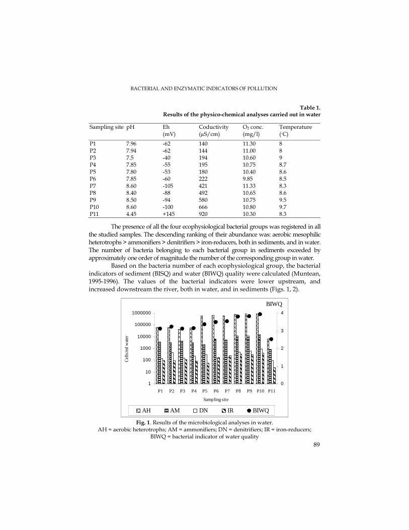
BACTERIAL AND ENZYMATIC INDICATORS OF POLLUTION
89
Table 1. Results of the physico-chemical analyses carried out in water
Sampling site pH Eh (mV)
Coductivity (µS/cm)
O2 conc. (mg/l)
Temperature (◦C)
P1 7.96 -62 140 11.30 8 P2 7.94 -62 144 11.00 8 P3 7.5 -40 194 10.60 9 P4 7.85 -55 195 10.75 8.7
P5 7.80 -53 180 10.40 8.6 P6 7.85 -60 222 9.85 8.5 P7 8.60 -105 421 11.33 8.3 P8 8.40 -88 492 10.65 8.6 P9 8.50 -94 580 10.75 9.5 P10 8.60 -100 666 10.80 9.7 P11 4.45 +145 920 10.30 8.3
The presence of all the four ecophysiological bacterial groups was registered in all the studied samples. The descending ranking of their abundance was: aerobic mesophilic heterotrophs > ammonifiers > denitrifiers > iron-reducers, both in sediments, and in water. The number of bacteria belonging to each bacterial group in sediments exceeded by approximately one order of magnitude the number of the corresponding group in water.
Based on the bacteria number of each ecophysiological group, the bacterial indicators of sediment (BISQ) and water (BIWQ) quality were calculated (Muntean, 1995-1996). The values of the bacterial indicators were lower upstream, and increased downstream the river, both in water, and in sediments (Figs. 1, 2).
1
10
100
1000
10000
100000
1000000
P1 P2 P3 P4 P5 P6 P7 P8 P9 P10 P11
Sampling site
Cel
ls/m
l wat
er
0
1
2
3
4
AH AM DN IR BIWQ
BIWQ
Fig. 1. Results of the microbiological analyses in water.
AH = aerobic heterotrophs; AM = ammonifiers; DN = denitrifiers; IR = iron-reducers; BIWQ = bacterial indicator of water quality
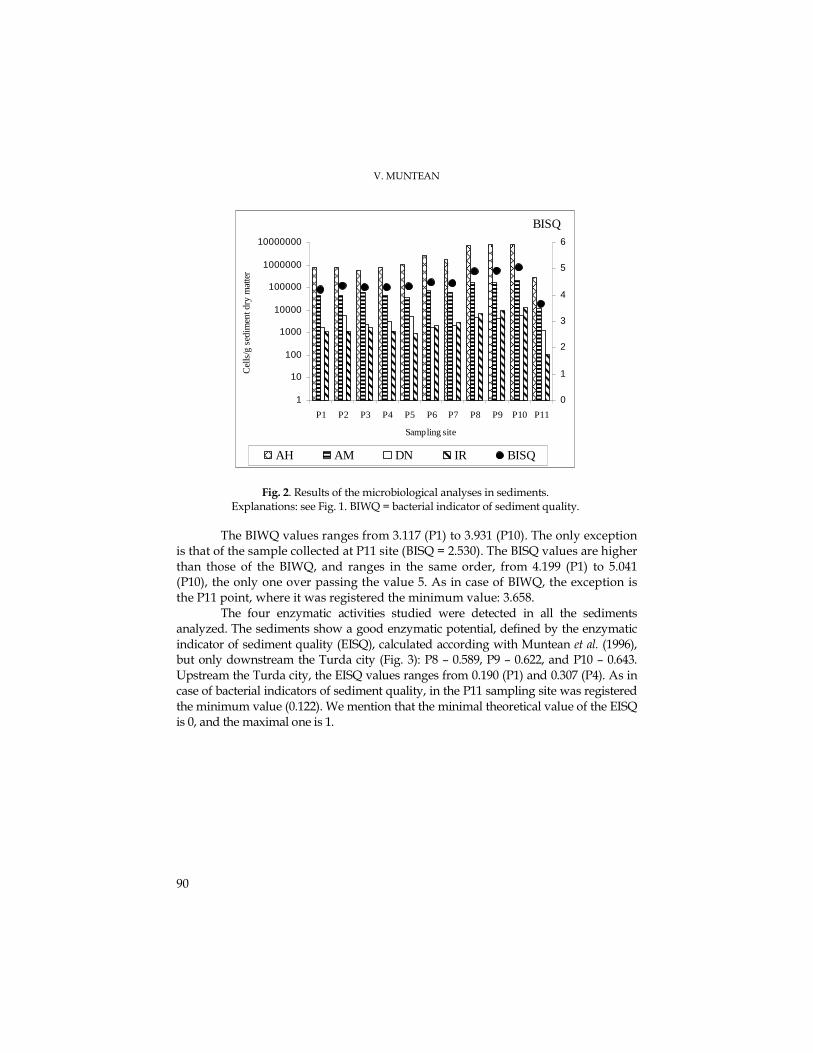
V. MUNTEAN
90
1
10
100
1000
10000
100000
1000000
10000000
P1 P2 P3 P4 P5 P6 P7 P8 P9 P10 P11
Sampling site
Cel
ls/g
sed
imen
t dry
mat
ter
0
1
2
3
4
5
6
AH AM DN IR BISQ
BISQ
Fig. 2. Results of the microbiological analyses in sediments. Explanations: see Fig. 1. BIWQ = bacterial indicator of sediment quality.
The BIWQ values ranges from 3.117 (P1) to 3.931 (P10). The only exception is that of the sample collected at P11 site (BISQ = 2.530). The BISQ values are higher than those of the BIWQ, and ranges in the same order, from 4.199 (P1) to 5.041 (P10), the only one over passing the value 5. As in case of BIWQ, the exception is the P11 point, where it was registered the minimum value: 3.658.
The four enzymatic activities studied were detected in all the sediments analyzed. The sediments show a good enzymatic potential, defined by the enzymatic indicator of sediment quality (EISQ), calculated according with Muntean et al. (1996), but only downstream the Turda city (Fig. 3): P8 – 0.589, P9 – 0.622, and P10 – 0.643. Upstream the Turda city, the EISQ values ranges from 0.190 (P1) and 0.307 (P4). As in case of bacterial indicators of sediment quality, in the P11 sampling site was registered the minimum value (0.122). We mention that the minimal theoretical value of the EISQ is 0, and the maximal one is 1.
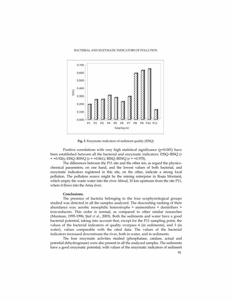
BACTERIAL AND ENZYMATIC INDICATORS OF POLLUTION
91
0.000
0.100
0.200
0.300
0.400
0.500
0.600
0.700
EIS
Q
P1 P2 P3 P4 P5 P6 P7 P8 P9 P10 P11
Sampling site
Fig. 3. Enzymatic indicators of sediment quality (EISQ)
Positive correlations with very high statistical significance (p<0.001) have
been established between all the bacterial and enzymatic indicators: EISQ–BISQ (r = +0.926); EISQ–BIWQ (r = +0.861); BISQ–BIWQ (r = +0.978).
The differences between the P11 site and the other ten, as regard the physico-chemical parameters, on one hand, and the lowest values of both bacterial, and enzymatic indicators registered in this site, on the other, indicate a strong local pollution. The pollution source might be the mining enterprise in Roşia Montană, which empty the waste water into the river Abrud, 10 km upstream from the site P11, where it flows into the Arieş river.
Conclusions. The presence of bacteria belonging to the four ecophysiological groups
studied was detected in all the samples analyzed. The descending ranking of their abundance was: aerobic mesophilic heterotrophs > ammonifiers > denitrifiers > iron-reducers. This order is normal, as compared to other similar researches (Muntean, 1995-1996; Ştef et al., 2003). Both the sediments and water have a good bacterial potential, taking into account that, except for the P11 sampling point, the values of the bacterial indicators of quality overpass 4 (in sediments), and 3 (in water), values comparable with the cited data. The values of the bacterial indicators increased downstream the river, both in water, and in sediments.
The four enzymatic activities studied (phosphatase, catalase, actual and potential dehydrogenase) were also present in all the analyzed samples. The sediments have a good enzymatic potential, with values of the enzymatic indicators of sediment
V. MUNTEAN
92
quality higher than 0.5, only downstream the Turda city. We mention that, according to the formula used for calculation, the enzymatic indicator can theoretically have values between 0 and 1.
Strong positive correlations with very high statistical significance (p<0.001) have been established between the bacterial and enzymatic indicators: EISQ–BISQ (r = +0.926); EISQ–BIWQ (r = +0.861); BISQ–BIWQ (r = +0.978).
The bacterial and enzymatic potential of sediments was seriously affected in the sampling site P11, where the Abrud River flows into the Arieş. The lowest values of both bacterial, and enzymatic indicators registered in this site, were probably caused by the waste water spilt into the river Abrud from the mining enterprise in Roşia Montană, 10 km upstream from the site P11.
REFERENCES
Alexander, M. (1965). Most probable-number method for microbial populations. In Black, C.A., Evans, D.D., White, J.L., Ensminger, L.E., Clark, F.E. (eds.), “Methods of Soil Analysis”, Ed. Am. Soc. Agron., Madison.
Atlas, R.M. (2004). Handbook of Microbiological Media. 3rd edition, CRC Press, New York. Casida, L.E.Jr., Klein, D.A., Santoro, T. (1964). Soil dehydrogenase activity. Soil Sci., 98, 371-376. Kappen, H. (1913). Die katalytische Kraft des Ackerbodens. Fühlings Landw. Ztg., 62, 377-392. Krámer, M., Erdei, G. (1959). Primenenie metoda opredeleniya aktivnosti fosfatazy v
agrokhimicheskikh issledovaniyakh. Pochvovedenie, 9, 99-102. Luna, G.M., Manini, E., Danovaro, R. (2002). Large fraction of dead and inactive bacteria in
coastal marine sediments: comparison of protocols for determination and ecological significance. Appl. Environ. Microbiol., 68, 3509-3513.
Meyer-Reil, L.A. (1987). Seasonal and spatial distribution of extracellular enzymatic activities and microbial incorporation of dissolved organic substrates in marine sediments. Appl. Environ. Microbiol., 53, 1748-1755.
Muntean, V. (1995-1996). Bacterial indicator of mud quality. Contrib. Bot., 73-76. Muntean, V., Crişan, R., Paşca, D., Kiss, S., Drăgan-Bularda, M. (1996). Enzymological
classification of salt lakes in Romania. Int. J. Salt Lake Res., 5 (1), 35-44. Noble, R.T., Weisberg, S.B., Leecaster, M.K., McGee, C.D., Ritter, K. Walker, K.O., Vainik, P.
(2003). Comparison of beach bacterial water quality indicator measurement methods. Environ. Monit. Assess., 81, 301–312.
Papp, J., Fodorpataki, L. (2002). Evaluation of the organic pollution of the Mureş river on the study of the indicator microflora. Contrib. Bot., 231-237.
Pârvu, R., Stanciu, E., Lorinczi, F., Kiss, S., Drăgan-Bularda, M., Rădulescu, D. (1977). Iron-reducing capacity of soil micromycetes. In Fourth Symp. Soil Biol. (Cluj-Napoca, 1977), Ed. Ceres, Bucureşti, 149-154.
Pochon, J. (1954). Manuel technique d'analyse microbiologique du sol. Ed. Masson, Paris.
Ştef, L.C., Muntean, V., Drăgan-Bularda, M. (2004). Microbiological research on the Mureş sediments from Ocna Mureş affected by pollution. Contrib. Bot., 39, 227-232.
STUDIA UNIVERSITATIS BABEŞ – BOLYAI, BIOLOGIA, LII, 1, 2007 (p.93-101)
THE QUANTITATIVE VARIATIONS OF SOME ECOPHYSIOLOGICAL GROUPS OF BACTERIA FROM THE SEDIMENTS
OF THE CRIŞUL ALB RIVER MARIOARA NICOLETA FILIMON1 and MIHAIL DRĂGAN-BULARDA2
SUMMARY. Sediment samples from the Crişul Alb River was taken and submitted to microbiological analyses during 2005. We have determined five ecophysiological groups of bacteria: ammonifying, nitrifying, denitrifying, iron-reducing bacteria, sulfate-reducing bacteria. In all sediment samples taken, bacteria from all of the 5 ecophysiological groups were found. The order of their abundance in the water was: ammonifying > denitrifying > nitrifying > iron-reducing > anaerobic sulfate-reducing bacteria. Based on the values of the bacteria from each ecophysiological group we determined the bacterial indicator of the sediment quality (BISQ) and we observed its seasonal variations. The seasonal values of the BISQ increase from the winter to the autumn. Regarding the sampling points we observed that the lower values of BISQ in all seasons of 2005 are registered downstream Buteni, due to the chemical pollution of the river in this locality. KEYWORDS: ecophysiological groups, sediments, Crişul Alb River, Romania Introduction The aquatic microorganisms have an essential role in the conversion of
organic matter produced by plants and animals into inorganic material. This is realized through complex processes that decompose the organic molecules into simple inorganic ones and so renew the essential nutrients like N and P, essential for the activity of photosynthesizing organisms.
So on a global scale the microorganisms are very important in the cycle of Carbon, Phosphorus, Nitrogen and other elements (S, P, Mn, Fe) in the nature.
Besides this role, some bacteria have the possibility to transform the xenobiotic chemical substances (Colwell, 1980), and so reduce their concentrations and negative effects on the environment.
The bacterial fauna of the running streams may prove to be of great importance when it comes to ecological characterization of aquatic habitats, to understand the fundamental processes that ensure the circulation of matter and energy in that ecosystem and also to understand the basic principles that interfere with the ecosystem productivity (Zarnea, 1994).
In all ecological periods of the 2005 (winter, spring, summer, autumn) we took samples of water from the Crişul Alb River in five points: Brad, GurahonŃ,
1 West University of Timişoara, Faculty of Chemistry-Biology-Geography, Dept. of Biology, 16 Pestalozzi Street, Timişoara, Romania; E-mail:[email protected]
2 Faculty o f Biology and Geology, Babeş-Bolyai University,1 Kogalniceanu Street, 400008 Cluj-Napoca, Romania, E-mail: [email protected]
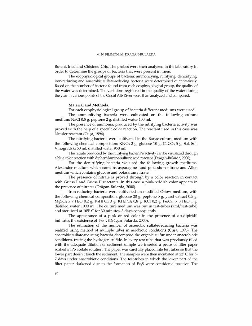
M. N. FILIMON, M. DRĂGAN-BULARDA
94
Buteni, Ineu and Chişineu-Criş. The probes were then analyzed in the laboratory in order to determine the groups of bacteria that were present in them.
The ecophysiological groups of bacteria: ammonifying, nitrifying, denitrifying, iron-reducing and anaerobic sulfate-reducing bacteria were determined quantitatively. Based on the number of bacteria found from each ecophysiological group, the quality of the water was determined. The variations registered in the quality of the water during the year in various points of the Crişul Alb River were than analyzed and compared.
Material and Methods. For each ecophysiological group of bacteria different mediums were used.
The ammonifying bacteria were cultivated on the following culture medium: NaCl 0.5 g, peptone 2 g, distilled water 100 ml.
The presence of ammonia, produced by the nitrifying bacteria activity was proved with the help of a specific color reaction. The reactant used in this case was Nessler reactant (Cuşa, 1996).
The nitrifying bacteria were cultivated in the Barjac culture medium with the following chemical composition: KNO3 2 g, glucose 10 g, CaCO3 5 g, Sal. Sol. Vinogradski 50 ml, distilled water 950 ml.
The nitrate produced by the nitrifying bacteria’s activity can be visualized through a blue color reaction with diphenylamine-sulfuric acid reactant (Drăgan-Bularda, 2000).
For the denitrifying bacteria we used the following growth mediums: Alexander medium which contains asparagines and potassium nitrate and Allen medium which contains glucose and potassium nitrate.
The presence of nitrate is proved through by a color reaction in contact with Griess I and Griess II reactants. In this case a pink-reddish color appears in the presence of nitrates (Drăgan-Bularda, 2000).
Iron-reducing bacteria were cultivated on modified Ottow medium, with the following chemical composition: glucose 20 g, peptone 5 g, yeast extract 0,5 g, MgSO4 x 7 H2O 0,2 g, K2HPO4 3 g, KH2PO4 0,8 g, KCl 0,2 g, Fe2O3 x 3 H2O 1 g, distilled water 1000 ml. The culture medium was put in test-tubes (7ml/test-tube) and sterilized at 105º C for 30 minutes, 3 days consequently.
The appearance of a pink or red color in the presence of αα-dipiridil indicates the existence of Fe2+. (Drăgan-Bularda, 2000). The estimation of the number of anaerobic sulfate-reducing bacteria was realized using method of multiple tubes in aerobiotic conditions (Cuşa, 1996). The anaerobic sulfate-reducing bacteria decompose the organic sulfur under anaerobiotic conditions, freeing the hydrogen sulfide. In every test-tube that was previously filled with the adequate dilution of sediment sample we inserted a peace of filter paper soaked in Pb acetate solution. The paper was carefully placed into test tubes so that the lower part doesn’t touch the sediment. The samples were then incubated at 22˚ C for 5-7 days under anaerobiotic conditions. The test-tubes in which the lower part of the filter paper darkened due to the formation of Fe2S were considered positive. The
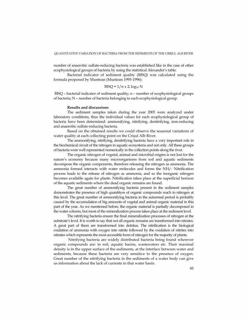
QUANTITATIVE VARIATION OF BACTERIA FROM THE SEDIMENTS OF THE CRIŞUL ALB RIVER
95
number of anaerobic sulfate-reducing bacteria was established like in the case of other ecophysiological groups of bacteria by using the statistical Alexander’s table. Bacterial indicator of sediment quality (BISQ) was calculated using the formula proposed by Muntean (Muntean 1995-1996):
BISQ = 1/n x Σ log10 N
BISQ – bacterial indicator of sediment quality; n – number of ecophysiological groups of bacteria; N – number of bacteria belonging to each ecophysiological group.
Results and discussions The sediment samples taken during the year 2005 were analyzed under
laboratory conditions, thus the individual values for each ecophysiological group of bacteria have been determined: ammonifying, nitrifying, denitrifying, iron-reducing and anaerobic sulfate-reducing bacteria.
Based on the obtained results we could observe the seasonal variations of water quality at each collecting point on the Crişul Alb River.
The ammonifying, nitrifying, denitrifying bacteria have a very important role in the biochemical circuit of the nitrogen in aquatic ecosystems and not only. All these groups of bacteria were well represented numerically in the collection points along the river.
The organic nitrogen of vegetal, animal and microbial origins is not lost for the nature’s economy because many microorganisms from soil and aquatic sediments decompose the organic components, therefore releasing the nitrogen as ammonia. The ammonia formed interacts with water molecules and forms the NH4+. Nitrification process leads to the release of nitrogen as ammonia, and so the inorganic nitrogen becomes available again for plants. Nitrification takes place at the superficial horizon of the aquatic sediments where the dead organic remains are found.
The great number of ammonifying bacteria present in the sediment samples demonstrates the presence of high quantityes of organic compounds reach in nitrogen at this level. The great number of ammonifying bacteria in the autumnal period is probably caused by the accumulation of big amounts of vegetal and animal organic material in this part of the year. As we mentioned before, the organic material is partially decomposed in the water column, but most of the mineralization process takes place at the sediment level.
The nitrifying bacteria ensure the final mineralization processes of nitrogen at the substrate’s level. It is worth to say that not all organic remains are transformed into nitrates. A great part of them are transformed into detritus. The nitrification is the biological oxidation of ammonia with oxygen into nitrite followed by the oxidation of nitrites into nitrates which represents the most accessible form of nitrogen for the majority of plants. Nitrifying bacteria are widely distributed bacteria being found wherever organic compounds are: in soil, aquatic basins, wastewaters etc. Their maximal density is in the upper surface of the sediments, at the interface between water and sediments, because these bacteria are very sensitive to the presence of oxygen. Great number of the nitrifying bacteria in the sediments of a water body can give us information about the lack of currents in that water basin.
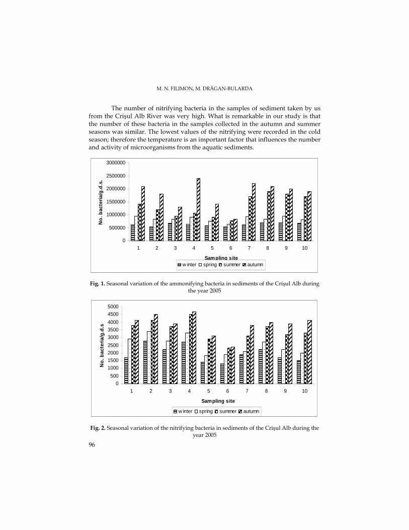
M. N. FILIMON, M. DRĂGAN-BULARDA
96
The number of nitrifying bacteria in the samples of sediment taken by us from the Crişul Alb River was very high. What is remarkable in our study is that the number of these bacteria in the samples collected in the autumn and summer seasons was similar. The lowest values of the nitrifying were recorded in the cold season; therefore the temperature is an important factor that influences the number and activity of microorganisms from the aquatic sediments.
0
500000
1000000
1500000
2000000
2500000
3000000
1 2 3 4 5 6 7 8 9 10
Sampling site
No
. bac
teri
a/g
.d.s
.
w inter spring summer autumn
Fig. 1. Seasonal variation of the ammonifying bacteria in sediments of the Crişul Alb during the year 2005
0
500
1000
15002000
2500
3000
3500
4000
4500
5000
1 2 3 4 5 6 7 8 9 10
Sampling site
No
. bac
teri
a/g
.d.s
w inter spring summer autumn
Fig. 2. Seasonal variation of the nitrifying bacteria in sediments of the Crişul Alb during the year 2005
QUANTITATIVE VARIATION OF BACTERIA FROM THE SEDIMENTS OF THE CRIŞUL ALB RIVER
97
Denitrification represents an important process that in the end will release nitrogen back into the atmosphere. Also it is one of the most efficient mechanisms when it comes to lowering the nitrogen content of wastewaters rich in nitrates.
Denitrification is taking place in waters and sediments with a low oxygen content and in waters and also in sediments with high oxygen levels, because the denitrification bacteria are facultative anaerobic. In the waters and sediments poorly aerated great quantities of organic substances accumulate, especially nitrites and so the denitrification is stimulated. This stimulation may also take place if chemical fertilizers, especially the ones that lead to a notable increase of nitrate concentration, get accidentally in the water. Regarding the number of bacteria involved in the nitrogen’s cycle we can say that in our samples denitrifying bacteria were in the highest number. In the following order were ammonifying, nitrifying and denitrifying bacteria.
Just like in the case of other two groups of bacteria, the lowest values were noticed in the cold season, winter and spring. The maximal values were registered in the warm season, in the summer and autumn. The ratio between the numbers of bacteria in the warm and cold seasons differs with the collection points of the samples. At the downriver points of sampling the water quality is lower, as expected, and so the number of bacteria from this group is higher.
0
10000
20000
30000
40000
50000
60000
1 2 3 4 5 6 7 8 9 10
Sampling site
No
.bac
teri
a/g
.d.s
w inter spring summer autumn
Fig. 3. Seasonal variation of the denitrifying bacteria in sediments of the Crişul Alb during the year 2005
The microorganisms have an important role in the iron’s cycle in the biosphere. The main stages of this circuit are the iron’s biological oxidation and the reduction of iron compounds. The biologic reduction of iron may be direct or indirect. The direct reduction is strongly connected with the metabolism of the microorganisms and plays an
M. N. FILIMON, M. DRĂGAN-BULARDA
98
important role in the soil. On the other hand the indirect reduction of iron is made by the organic substances produced by the microorganisms or derived from their metabolism (glucose). In anaerobiotic conditions the bacteria reduces the iron while oxidizing organic substances, Fe3+ having the role as terminal electron acceptor.
Iron-reducing bacteria have a major role in the iron’s mineral compounds transformation processes, especially in the water and less in the soil, while the unspecific heterotrophic microflora (putrefaction flora) intervenes in these transformations in the water as well as in the soil (Topală, 1978). The number of iron-reducing bacteria is under hundreds or lower. As expected, the number of this bacteria group has minimal values during the cold season, in winter, and maximal values in the summer and autumn seasons. An exception from this was found at the Buteni collection point where the number of these bacteria was lower all year long. The relatively low number of iron-reducing bacteria in the collection point Buteni during may have been caused by the existence in the area of a pollution source from the leather manufacturing factories.
The totality of transformations that the organic and mineral sulfur compounds undergo in the biosphere represents the sulfur’s circuit. The main stages of the sulfur’s circuit are: the incorporation of mineral sulfur into organic substances; mineralization of organic sulfur; oxidation of mineral compounds of sulfur (sulfur oxidation); reduction of oxidized mineral compounds of sulfur (sulfate oxidation), this stage taking place under anaerobiotic conditions in the soil, mud, marine sediments, marine waters, residual waters and consists in the reduction of sulfates to H2S (Ailiesei et al., 1980).
The anaerobic sulfate-reducing bacteria are largely spread trough out the aquatic and terrestrial anaerobic environments that serve as a microbial decomposition place for the vegetal and animal organic substances. Their capacity of producing large quantities of H2S leads to a variety of economical, industrial and ecological effects. Their ecological role is notable when the waters are polluted, when their number increases, the waters gaining an unpleasant smell because of the H2S accumulation and also the darkening of waters and sediments takes place due to the iron sulfide formed.
The anaerobic sulfate-reducing are present in the Crişul Alb River’s sediments by order of tenths to hundreds, their values being relatively low. The minimal values for this group of bacteria are met in the cold season (winter) and the maximal in the autumn period. Also downstream of the localities mentioned we noticed high values, which could be due to the pollution sources with industrial waters.
QUANTITATIVE VARIATION OF BACTERIA FROM THE SEDIMENTS OF THE CRIŞUL ALB RIVER
99
0
200
400
600
800
1000
1 2 3 4 5 6 7 8 9 10
Sampling site
No
. bac
teri
a/g
.d.s
w inter spring summer autumn
Fig. 4. Seasonal variation of the iron-reducing bacteria sediments of the Crişul Alb River
during the year 2005
0
50
100
150
200
250
300
350
400
450
1 2 3 4 5 6 7 8 9 10
Sampling site
No
. bac
teri
a/g
.d.s
.
w inter spring summer autumn
Fig. 5. Seasonal variation of the anaerobic sulfate-reducing in sediments of the Crişul Alb
River during the year 2005 Based on the number of the ecophysiological groups of bacteria analyzed and
using the calculation formula proposed by Muntean (1995-1996) we calculated the sediment’s quality bacterial indicator. Following the seasonal variations of this indicator during the year 2005 was observed that the minimal values, as expected, was
M. N. FILIMON, M. DRĂGAN-BULARDA
100
registered in the colder months and the maximal values in the warm months. The lowest value of BISQ during the 2005 was recorded at the Buteni collection point due to high levels of pollution in the area. Downstream on the river the values for the SCBI are relatively similar, thus indicating the lack of strong perturbational sources on the populations of microorganisms. The higher values recorded here rather than at other collection points were probably due to the running speed of the river that decreases, thus favoring the sedimentation process and so the intense microbiological activity.
1,0500
1,1000
1,1500
1,2000
1,2500
1,3000
1 2 3 4 5 6 7 8 9 10
Sampling site
BIS
Q
w inter spring summer autumn
Fig. 6. Seasonal variation of the bacterial indicator of sediment quality from the Crişul Alb
River during the year 2005 Conclusions The ammonifying, nitrifying, denitrifying, iron-reducing, anaerobic sulfate-
reducing bacteria were determined numerically in all the sediment samples. The bacterial indicator of sediment quality (BISQ) presents seasonal variations
and also variations according to the with the collection point of the samples. The lowest values were recorded in the winter season and the highest in the autumn.
The temperature and organic substances dissolved in the water had a major role in the development of the microorganism populations, influencing the numerical values registered during the four seasons.
The polluting substances that accidentally or not enter into the river have a negative effect on some bacterial populations. The BISQ minimal values were recorded in the samples gathered from Buteni, a chemically polluted area.
QUANTITATIVE VARIATION OF BACTERIA FROM THE SEDIMENTS OF THE CRIŞUL ALB RIVER
101
The relatively close values of BISQ registered at the downstream collection points were caused by the decrease of the velocity of water flow, this favoring sedimentation process and so influencing the increase of microorganism density.
REFERENCES Ailiesei, O., NimiŃan, E., Comănescu, Şt., 1980-Lucrări practice de microbiologie generală, Iaşi. Burlage,D., Atlas,R., Stahl,D., Geesey,G.,Sayler,G., 1998, Techniques in Microbial Ecology, Oxford
University Press, New York, 31-78. Colwell R., 1980-Aquatic Microbial Ecology- Introduction, ed. Schlessinger D., “Microbiology”
Am. Soc. Microbiol., Washington D. C., 307-308. Cuşa V.,-InstrucŃiuni metodologice pentru analiza microbiologică a sedimentelor din
ecosistemele acvatice, Inst.Cercet. Ing.Mediului, Bucuresti, 4: 14-20, 1996. Drăgan-Bularda M.,-Microbiologie generală, Lucrări practice, Cluj-Napoca, 2000. Muntean V., 1995-1996-Bacterial indicator of mud quality, Inst. de Cercet Biologice,ContribuŃii
Botanice, 73-76. Topală, N.D., 1978, Microbiologie generală, Vol II, Univ. Al. I. Cuza, Iaşi. Zarnea G., 1994-Tratat de Microbiologie generală, Vol. V, Ed. Academiei, Bucureşti.
STUDIA UNIVERSITATIS BABEŞ – BOLYAI, BIOLOGIA, LII, 1, 2007 (p.103-110)
SEASONAL POLLUTION CAUSED BY SEWAGE WASTE WATERS IN THE CRIŞUL ALB RIVER DURING 2005
MARIOARA NICOLETA FILIMON1 and MIHAIL DRĂGAN-BULARDA2
SUMMARY. The purpose of this paper was to establish the presence and degree of pollution caused by sewage wastes in the waters of the Crişul Alb River. The seasonal variation of the bacteria associated with the sewage wastes is also reported. The quantitative determinations of coliform bacteria, faecal coliform bacteria and enterococci were established in 10 sampling points along the Crisul Alb River. Based on the results obtained, we classified the waters of Crisul Alb River into two quality classes, I and II (according to the existing regulations). Water of first quality class was registered upstream the river, and of second quality, downstream. The ratio between the coliform bacteria and the enterococci, which is an indicator of the nature of pollution, showed that the water of the Crişul Alb River is in most of cases loaded with bacteria from the human sewage. However, in some sampling points, the pollution was of animal origin. This was the case downstream of GurahonŃ. The water from the Crişul Alb River has a moderate rate of pollution caused by the sewage wastes. KEYWORDS: total coliform bacteria, faecal coliform bacteria, faecal streptococci, Crişul Alb River Introduction The running streams of water are an environment that can be easily polluted
by the human activities. Any change in quality or quantity of the organic compounds in the water has a direct effect on the bacterial populations and also on all living things in the ecosystem. Many diseases transmitted thru water are caused by microorganisms eliminated from the digestive tract of different animals and humans (Mănescu, 1989).
The aquatic environments like rivers, lakes and marshes contain normally a high and varied number of bacteria due to the organic matter of animal or plant origin found in them. Input of organic matter is a normal feature of aquatic ecosystems. The detritivores of streams and the sediment communities of slow flowing rivers depend upon it for most of their energy. The organic matter is rapidly converted to inorganic substances or left in a refractory state. The main difference between natural organic input and pollution by organic matter is that the former tends to be in large packets, like lives with a low surface to volume ratio, while the latter is usually soluble or finely divided and very labile. In the past centuries humans added more and more organic substances in water streams and in ground water polluting so the aquatic environment and affecting the ecosystems (Herlea et al., 1995).
1 West University of Timişoara, Chemistry-Biology-Geography, Faculty Biology Department,Pestalozzi, 16, Timişoara, Romania, E-mail:[email protected]
2 Faculty o f Biology and Geology, Babeş-Bolyai University,1, Kogalniceanu Street, 400006/ Cluj-Napoca, Romania, E-mail: [email protected]
M.N. FILIMON, M. DRĂGAN-BULARDA
104
In the case of surface waters the pollution sources are numerous and complex, but in general, certain industrial and agricultural branches are incriminated. The most pollutant human activities are mainly in the chemical industry and in agriculture because the water used here (in majority of cases) is flushed back into the rivers, lakes or swamps usually without being treated. Despite the fact that within these natural receptors some physical and chemical phenomena are taking place and in some degree pollutants are decomposed, many of them remain for longer periods in the environment. The nature itself can manage to absorb some quantities of pollutants and neutralize them by so called self-purification but also nature’s capabilities are limited and at some point the equilibrium will fail with negative consequences.
Humans or certain animals are sources of pathogenic agents that can be transmitted through contaminated water. Many species of bacteria have pathogenic and nonpathogenic strains, or they opportunistic, causing diseases to people with a suppressed immune system. An individual eliminates daily thru the feces billions of coliform bacilli, fundamentally nonpathogenic. Most species of bacteria have a degree of specificity for some species, but some do not show such pattern and can cause diseases to more than one species of animal.
In practice the presence of a pathogen agent in water is not determined, due to the difficulties involved, but another method is used: analyses are made to determine the presence of the faecal contamination. A large amount of bacteria associated with human or animal feces is a certain sign that pathogens exist. The indicators of sewage pollution (total coliform bacteria, faecal coliform bacteria, faecal streptococci) are not adequate enough for estimating the risks of infection through contact with the contaminated water. The treatment of the used waters and also the monitorisation of bacteria species are needed. The self-purification mechanisms of the rivers or other water bodies reduce the bacterial contamination, but their efficiency is low in case of low temperatures or in case of a high level of pollution.
This paper deals with hygienically and sanitary state of the Crişul Alb River. The data were obtained after analyzing water samples taken from various points along the river’s course along the year 2005. Based on the hygienic indicators analyzed: total coliform bacteria, faecal coliform bacteria and faecal streptococci, we have made a map of water quality of Crişul Alb River. Our study showed that the on a great length, Crisul Alb River has clean waters that and can be categorized in the first class quality. Only when the river riches the plain region its waters get polluted (due to the human activities) and are categorized into IInd quality class.
We conclude that the water of the Crişul Alb River is unpolluted in the mountain parts and has a moderate rate of pollution downstream.
Material and Methods The samples were taken from stations along the Crisul Alb River. We have
covered an area of 238 km of river’s course from the spring till the place it leaves the country at Vărşand. The stations were placed near the major localities crossed
FAECAL POLLUTION OF THE CRIŞUL ALB RIVER DURING 2005
105
by the river: Brad, GurahonŃ, Buteni, Ineu, Chişineu-Criş in order to follow the effects of the human pressure on the microorganism from the water.
In order to determine the hygienicall and bacteriological parameters, water samples were submitted to analyzation, the following culture mediums were used: nutritive gelose (heterotrophic mesophile bacteria), sodium laurilsulphat (simple or double concentrated) and Geam-Levine (total coliform bacteria), MacConkey medium (faecal coliform), simple or double concentrated medium of sodium azide and azide with purple cresol medium (faecal enterococci).
The presence of mesophile bacteria was proved by putting a small amount of probe on a special solid medium and incubated at 37° C for 48 hours. After this, follows the counting of colonies that have grown. The result is expressed in CFU – colony-forming unit per 1 milliliter.
The presence of coliform bacteria (total coliform) can be revealed also through the presumption test (presumptive test). First of all we put samples of water in several test tubes with medium and after that the confirmation test is done on a solid medium at a temperature of 37° C and a period of incubation of 24 hours. Starting from the number of tubes with positive reaction we calculate the probable number of coliform bacteria (total coliform). The coliform bacteria (total coliform) are confirmed if characteristic colonies have developed: flat colonies of dark blue-violet with metallic gloss or convex, opaque, mucous with central metallic gloss or pink colored with the centre blue-violet (STAS 3001/1991).
Based on the positive results of the presumption test for the coliform bacteria further on we can establish the presence of the coliform thermotolerant bacteria (faecal coliform). This is done with the help of liquid selective mediums at an incubation temperature of 44° C. After this time depending on the number of positive test tubes the probable number of thermotolerant coliform bacteria (faecal coliform) can be calculated. In order to confirm the thermotolerant coliform bacteria from the same test-tubes considered positive in the presumption test a few droplets are taken with a with Pasteur pipette and passed in test-tubes which contain either Bromocresol purple Lactose Bouillon or Brilliant Green Bile Lactose Bouillon.
The samples are incubated at 44° C for 24 hours. If the color of the medium turns into yellow and simultaneously a gas is released due to the fermentation processes of the lactose is clear that the thermotolerant coliform bacteria are present (faecal coliform).
The streptococci are evidenced also through the presumption test. In this case are used also test tubes with an enriched liquid medium. This medium with the bacteria are incubated at 37° C. The positive reaction is revealed through confirmation test on selective liquid medium incubated for 48 hours at 44° C. Starting from the number of confirmed positive tubes the probable number of streptococci is calculated. After the confirmation of streptococci in liquid medium, one or two drops from each test-tube considered positive in the presumption test are passed with a Pasteur pipette into a test-tube with Bromocresol purple Sodium azide Bouillon. We incubate this at 44.5º C for 24 hours. The turning of the color in yellow
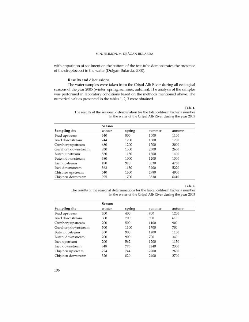
M.N. FILIMON, M. DRĂGAN-BULARDA
106
with apparition of sediment on the bottom of the test-tube demonstrates the presence of the streptococci in the water (Drăgan-Bularda, 2000).
Results and discussions The water samples were taken from the Crişul Alb River during all ecological
seasons of the year 2005 (winter, spring, summer, autumn). The analysis of the samples was performed in laboratory conditions based on the methods mentioned above. The numerical values presented in the tables 1, 2, 3 were obtained.
Tab. 1. The results of the seasonal determination for the total coliform bacteria number
in the water of the Crişul Alb River during the year 2005
Season Sampling site winter spring summer autumn Brad upstream 640 800 1000 1100 Brad downstream 744 1200 1600 1700 GurahonŃ upstream 680 1200 1700 2000 GurahonŃ downstream 830 1300 2300 2600 Buteni upstream 560 1150 1300 1400 Buteni downstream 380 1000 1200 1300 Ineu upstream 490 910 3830 4760 Ineu downstream 562 1150 3900 5220 Chişineu upstream 540 1300 2980 4900 Chişineu dowstream 925 1700 3830 6410
Tab. 2. The results of the seasonal determinations for the faecal coliform bacteria number
in the water of the Crişul Alb River during the year 2005
Season Sampling site winter spring summer autumn Brad upstream 200 400 900 1200 Brad downstream 300 700 900 610 GurahonŃ upstream 200 500 1100 900 GurahonŃ downstream 500 1100 1700 700 Buteni upstream 350 900 1200 1100 Buteni downstream 200 900 700 340 Ineu upstream 200 562 1200 1150 Ineu downstream 348 775 2240 2300 Chişineu upstream 224 744 2200 2600 Chişineu dowstream 326 820 2400 2700
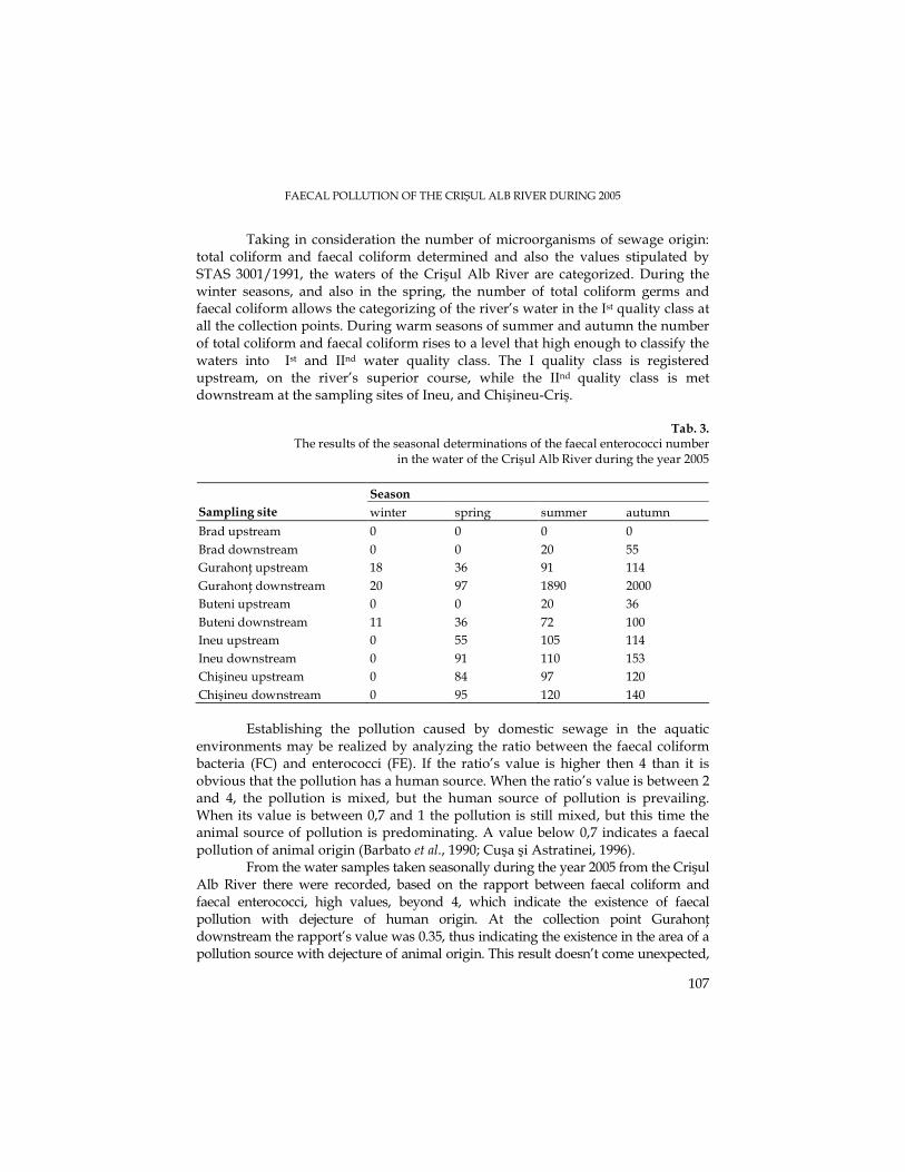
FAECAL POLLUTION OF THE CRIŞUL ALB RIVER DURING 2005
107
Taking in consideration the number of microorganisms of sewage origin: total coliform and faecal coliform determined and also the values stipulated by STAS 3001/1991, the waters of the Crişul Alb River are categorized. During the winter seasons, and also in the spring, the number of total coliform germs and faecal coliform allows the categorizing of the river’s water in the Ist quality class at all the collection points. During warm seasons of summer and autumn the number of total coliform and faecal coliform rises to a level that high enough to classify the waters into Ist and IInd water quality class. The I quality class is registered upstream, on the river’s superior course, while the IInd quality class is met downstream at the sampling sites of Ineu, and Chişineu-Criş.
Tab. 3.
The results of the seasonal determinations of the faecal enterococci number in the water of the Crişul Alb River during the year 2005
Season Sampling site winter spring summer autumn Brad upstream 0 0 0 0 Brad downstream 0 0 20 55 GurahonŃ upstream 18 36 91 114 GurahonŃ downstream 20 97 1890 2000 Buteni upstream 0 0 20 36 Buteni downstream 11 36 72 100 Ineu upstream 0 55 105 114 Ineu downstream 0 91 110 153 Chişineu upstream 0 84 97 120 Chişineu downstream 0 95 120 140
Establishing the pollution caused by domestic sewage in the aquatic environments may be realized by analyzing the ratio between the faecal coliform bacteria (FC) and enterococci (FE). If the ratio’s value is higher then 4 than it is obvious that the pollution has a human source. When the ratio’s value is between 2 and 4, the pollution is mixed, but the human source of pollution is prevailing. When its value is between 0,7 and 1 the pollution is still mixed, but this time the animal source of pollution is predominating. A value below 0,7 indicates a faecal pollution of animal origin (Barbato et al., 1990; Cuşa şi Astratinei, 1996).
From the water samples taken seasonally during the year 2005 from the Crişul Alb River there were recorded, based on the rapport between faecal coliform and faecal enterococci, high values, beyond 4, which indicate the existence of faecal pollution with dejecture of human origin. At the collection point GurahonŃ downstream the rapport’s value was 0.35, thus indicating the existence in the area of a pollution source with dejecture of animal origin. This result doesn’t come unexpected,
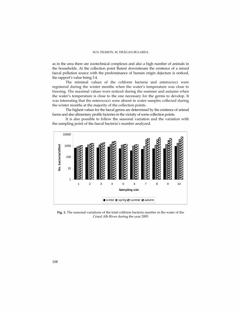
M.N. FILIMON, M. DRĂGAN-BULARDA
108
as in the area there are zootechnical complexes and also a high number of animals in the households. At the collection point Buteni downstream the existence of a mixed faecal pollution source with the predominance of human origin dejecture is noticed, the rapport’s value being 3.4.
The minimal values of the coliform bacteria and enterococci were registered during the winter months when the water’s temperature was close to freezing. The maximal values were noticed during the summer and autumn when the water’s temperature is close to the one necessary for the germs to develop. It was interesting that the enterococci were absent in water samples collected during the winter months at the majority of the collection points.
The highest values for the faecal germs are determined by the existence of animal farms and also alimentary profile factories in the vicinity of some collection points.
It is also possible to follow the seasonal variation and the variation with the sampling point of the faecal bacteria’s number analyzed.
1
10
100
1000
10000
1 2 3 4 5 6 7 8 9 10
Sampling site
No
. bac
teri
a/10
0ml
w inter spring summer autumn
Fig. 1. The seasonal variations of the total coliform bacteria number in the water of the Crişul Alb River during the year 2005
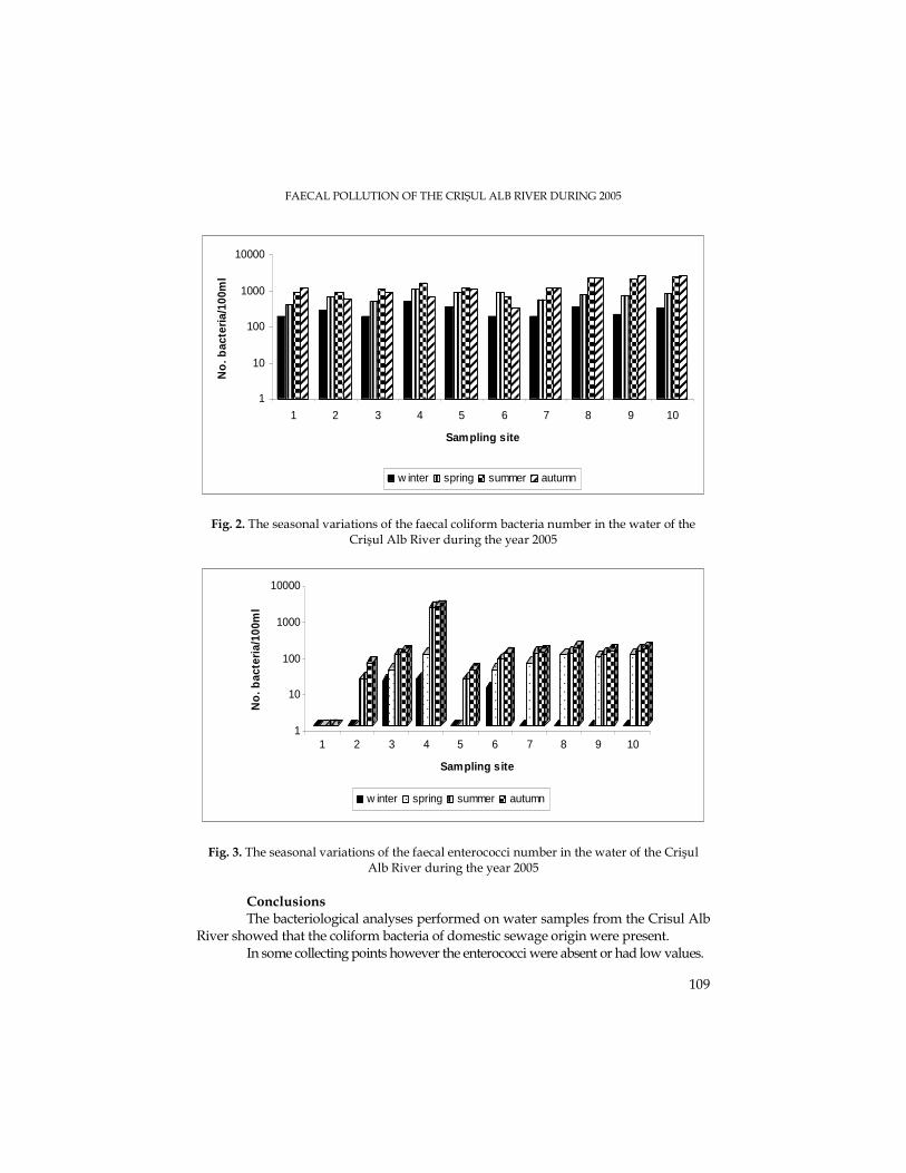
FAECAL POLLUTION OF THE CRIŞUL ALB RIVER DURING 2005
109
1
10
100
1000
10000
1 2 3 4 5 6 7 8 9 10
Sampling site
No
. bac
teri
a/10
0ml
w inter spring summer autumn
Fig. 2. The seasonal variations of the faecal coliform bacteria number in the water of the Crişul Alb River during the year 2005
1
10
100
1000
10000
No
. bac
teri
a/10
0ml
1 2 3 4 5 6 7 8 9 10
Sampling site
w inter spring summer autumn
Fig. 3. The seasonal variations of the faecal enterococci number in the water of the Crişul Alb River during the year 2005
Conclusions The bacteriological analyses performed on water samples from the Crisul Alb
River showed that the coliform bacteria of domestic sewage origin were present. In some collecting points however the enterococci were absent or had low values.
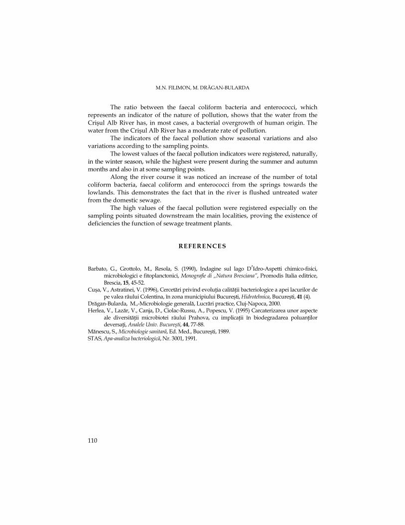
M.N. FILIMON, M. DRĂGAN-BULARDA
110
The ratio between the faecal coliform bacteria and enterococci, which represents an indicator of the nature of pollution, shows that the water from the Crişul Alb River has, in most cases, a bacterial overgrowth of human origin. The water from the Crişul Alb River has a moderate rate of pollution.
The indicators of the faecal pollution show seasonal variations and also variations according to the sampling points.
The lowest values of the faecal pollution indicators were registered, naturally, in the winter season, while the highest were present during the summer and autumn months and also in at some sampling points.
Along the river course it was noticed an increase of the number of total coliform bacteria, faecal coliform and enterococci from the springs towards the lowlands. This demonstrates the fact that in the river is flushed untreated water from the domestic sewage.
The high values of the faecal pollution were registered especially on the sampling points situated downstream the main localities, proving the existence of deficiencies the function of sewage treatment plants.
REFERENCES Barbato, G., Grottolo, M., Resola, S. (1990), Indagine sul lago D �́��Idro-Aspetti chimico-fisici,
microbiologici e fitoplanctonici, Monografie di „Natura Bresciana”, Promodis Italia editrice, Brescia, 15, 45-52.
Cuşa, V., Astratinei, V. (1996), Cercetări privind evoluŃia calităŃii bacteriologice a apei lacurilor de pe valea râului Colentina, în zona municipiului Bucureşti, Hidrotehnica, Bucureşti, 41 (4).
Drăgan-Bularda, M.,-Microbiologie generală, Lucrări practice, Cluj-Napoca, 2000. Herlea, V., Lazăr, V., Canja, D., Ciolac-Russu, A., Popescu, V. (1995) Carcaterizarea unor aspecte
ale diversităŃii microbiotei râului Prahova, cu implicaŃii în biodegradarea poluanŃilor deversaŃi, Analele Univ. Bucureşti, 44, 77-88.
Mănescu, S., Microbiologie sanitară, Ed. Med., Bucureşti, 1989. STAS, Apa-analiza bacteriologică, Nr. 3001, 1991.