Embed Size (px)
DESCRIPTION
aa
Citation preview
Structure–activity relationship of indomethacin analogues forMRP-1, COX-1 and COX-2 inhibition: identification ofnovel chemotherapeutic drug resistance modulators
S. Touheya,1, R. O’Connora,*,1, S. Plunkettb, A. Maguireb, M. Clynesa
aThe National Cell and Tissue Culture Centre, Dublin City University, Glasneuin, Dublin 9, IrelandbDepartment of Chemistry, University College Cork, Ireland
Received 5 December 2001; received in revised form 28 February 2002; accepted 9 April 2002
Abstract
We report the screening of analogues of indomethacin to investigate the structure–activity relationship (SAR) of indomethacin-
mediated multidrug resistance associated protein-1 (MRP-1) inhibition. By examining the activities of compounds with minorvariations of the parent structure, we were able to separate MRP-1, glutathione-S-transferase (GST), cyclooxygenase (COX)-1 andCOX-2 inhibitory activities. Combination cytotoxicity assays were utilised to identify agents which possess synergistic potential in
MRP-1-expressing cell lines. MRP-1 Inside Out Vesicles (IOVs) were utilised to demonstrate the ability of the indomethacin ana-logues to inhibit the pump directly. Most of the indomethacin analogues active as MRP-1 inhibitors were poor GST inhibitorswhen compared with the GST-inhibitory activity of indomethacin. Two of the MRP-1 inhibitory analogues were found to have no
COX-1 inhibitory activity and low COX-2 inhibitory activity, suggesting potentially reduced clinical toxicity. One MRP-1 inhibi-tory indomethacin analogue was also found to have low COX-1 inhibitory activity, but significant COX-2 inhibitory activity,making this analogue again interesting in terms of low potential toxicity, but with the possibility of direct inhibitory effects ontumour growth. # 2002 Elsevier Science Ltd. All rights reserved.
Keywords: Cancer; MRP; NSAID; Resistance circumvention; Indomethacin; GST; Cyclooxygenase; Structure–activity relationship; COX-1; COX-2
1. Introduction
The development of drug resistance is one of the mostsignificant obstacles to effective treatment of cancer.Elucidation of the mechanisms determining inherent orchemotherapy-induced resistance in human tumours istherefore an important challenge [1].Multidrug resistance in model systems has been
shown to be conferred by different integral membraneproteins including the 170 kDa P-glycoprotein (Pgp) [2]and the 190 kDa multidrug resistance associated protein(MRP) [3]. These proteins belong to the adenosine tri-phosphate (ATP) binding cassette (ABC) family oftransport proteins. Since the discovery of MRP (now
termed MRP-1), an additional six homologues of MRP(MRP2–MRP7) have been described [4,5].MRP-1 can confer cellular resistance to natural product
drugs, including anthracyclines, some vinca alkaloids, andepipodophyllotoxins [6,7]. This 190 kDa multidrug resis-tance-associated protein mediates the ATP driven uni-directional transport of a broad range of neutral, as wellas anionic, compounds across cellular membranes [8].MRP-1 can transport drugs conjugated to glutathione(GSH) out of the cell [9] and can actively co-transportGSH and unmodified vincristine, possibly via an interac-tion with leukotriene C4 (LTC4) binding site(s) [4,8].The anti-inflammatory action of non-steriodal anti-
inflammatory drugs (NSAIDs) is due to reduced syn-thesis of prostaglandins through inhibition of theenzyme prostaglandin endoperoxide synthase or cyclo-oxygenase (COX) which exists in two isoforms andwhich transforms arachidonic acid, liberated by phos-pholipase A2 (PLA2) to prostaglandins [10]. COX-1 isconstitutively produced and is involved in regulating
0959-8049/02/$ - see front matter # 2002 Elsevier Science Ltd. All rights reserved.
PI I : S0959-8049(02 )00128-4
European Journal of Cancer 38 (2002) 1661–1670
www.ejconline.com
* Corresponding author. Tel.: +353-1-700-5691; fax: +353-1-700-
5484.
E-mail address: [email protected] (R. O’Connor).1 The first two authors contributed equally to this study.
normal cellular processes, such as gastrointestinal (GI)cytoprotection, vascular homeostasis and renal function[11]. In contrast, COX-2, identified as an inducible syn-thase [12], is generally undetectable in most normal tis-sues. However, its expression can be increaseddramatically after exposure of fibroblasts, vascularsmooth muscle or endothelial cells to growth factors,hypoxia, phorbol esters, cytokines and by lipopoly-saccharides (LPS) in monocytes/macrophages [13]. Evi-dence suggests that the gastrointestinal toxicityassociated with NSAID use is primarily the result ofinhibition of COX-1 and anti-inflammatory effects arelargely due to the inhibition of COX-2 [14]. The expres-sion of COX-2 in a number of cancer types, includingFamilial Adenomatous Polyposis (FAP) has been asso-ciated with increased tumorigenesis [15]. COX-2 is alsoassociated with angiogenesis and may therefore repre-sent a specific target for anti-tumour therapy [16].A number of NSAIDs (indomethacin, sulindac, tol-
metin, acemetacin, zomepirac and mefenamic acid) atnon-toxic levels, have been demonstrated by our group[17] to have the ability to significantly enhance thecytotoxicity of a number of anti-cancer drugs (includingdoxorubicin, daunorubicin, epirubicin, teniposide,VP-16 and vincristine) in vitro when co-administered incell lines which express MRP-1. These results are con-sistent with other published data [18,19]. In addition,two sulindac metabolites, sulindac sulphide and sulin-dac sulphone, were found to be active in the combination
toxicity assay. As previously described, sulindac sul-phone is, by definition, not a NSAID due to the factthat it is not a COX inhibitor [20].Similarities in the chemical structures of the NSAIDs
which act as chemosensitising agents suggested thatthere might be specific structural requirements for thesynergistic combination effect. Hence, a number ofcompounds, structurally related to indomethacin, weresynthesised and screened to further investigate thestructure–activity relationship (SAR) of indomethacin-mediated MRP inhibition (Table 1) [21].
2. Materials and methods
2.1. Cell lines
DLKP (a human lung squamous carcinoma), A549 (ahuman lung adenocarcinoma), COR L23P (a humanlarge cell lung cancer), COR L23R (an anthracycline-resistant variant of COR L23), HL60 (a human promy-elocytic leukaemia) and HL60/ADR (an anthracycline-resistant variant of HL60) cells were cultured as previouslydescribed in Ref. [17].
2.2. Chemicals
Doxorubicin (Pharmacia, UK), cisplatin, and 5-fluoro-uracil (David Bull Labs, UK), etoposide (VP-16), and
Table 1
List of the analogues of indomethacin investigated in this study. Both analogue number and chemical nomenclature are given for each compound
Compound Mol. Wt. Chemical description
Indo. 358 1-(4-Chlorobenzoyl)-5-methoxy-2-methylindole-3-acetic acid
4 344 1-(4-Chlorobenzyl)-5-methoxy-2-methylindole-3-acetic acid
5 391 1-(4-Methylthiobenzyl)-5-methoxy-2-methylindole-3-acetic acid
7 388 1-(4-Bromobenzyl)-5-methoxy-2-methylindole-3-acetic acid
8 261 1-Benzylindole-3-acetic acid
9 296 1-(4-Chlorobenzyl)indole-3-acetic acid
10 341 1-(4-Bromobenzyl)indole-3-acetic acid
11 292 1-(4-Methoxybenzyl)indole-3-acetic acid
13 308 1-Benzyl-5-methoxy-2-methylindole-3-acetic acid
14 327 1-(4-Fluorobenzyl)-5-methoxy-2-methylindole-3-acetic acid
16 344 1-(3-Chlorobenzyl)-5-methoxy-2-methylindole-3-acetic acid
17 344 1-(2-Chlorobenzyl)-5-methoxy-2-methylindole-3-acetic acid
18 327 1-(3-Fluorobenzyl)-5-methoxy-2-methylindole-3-acetic acid
19 329 1-(4-Chlorobenzyl)-5-methoxyindole-3-acetic acid
20 314 1-(4-Chlorobenzyl)-2-methylindole-3-acetic acid
21 322 1-Benzoyl-5-methoxy-2-methylindole-3-acetic acid
22 402 1-(4-Bromobenzoyl)-5-methoxy-2-methylindole-3-acetic acid
23 372 Methyl 1-(4-chlorobenzoyl)-5-methoxy-2-methylindole-3-acetate
24 447 N-Tolyl 1-(4-chlorobenzoyl)-5-methoxy-2-methylindole-3-acetamide
25 328 1-(4-Chlorobenzoyl)-2-methylindole-3-acetic acid
26 370 4-[3(Amidomethyl)-2-methyl-1-(phenylmethyl)- indol-5-yl]oxy]butanoic acid
27 384 4-[3(Amidomethyl)-2-ethyl-1-(phenylmethyl)- indol-5-yl]oxy]butanoic acid
28 406 [3-[3(Amidomethyl)-2-methyl-1-(phenylmethyl)-indol-5 yl]oxy]propyl]phosphonic acid
29 420 [3-[3(Amidomethyl)-2-ethyl-1-(phenylmethyl)-indol-5-yl]oxy]propyl]phosphonic acid
Mol. Wt., molecular weight.
1662 S. Touhey et al. / European Journal of Cancer 38 (2002) 1661–1670
vincristine (Bristol-Myers Squibb Pharmaceuticals Ltd.,UK) were all supplied as solutions and were diluted foruse in culture medium. Indomethacin was purchasedfrom Sigma (UK) and was dissolved at 5 mg/ml indimethyl sulphoxide (DMSO) before being diluted to itsworking concentration with culture medium. The indo-methacin analogues were synthesised in our laboratories[21] and were dissolved in the same manner as indo-methacin. All compounds synthesised were fully char-acterised spectroscopically and analytically.
2.3. In vitro toxicity testing
Cytotoxicity testing of drugs and drug combinationswas measured by colormetric assays as previouslydescribed [17,22,23]. Briefly, on day 1, cells were seededat 1�103 cells/well in a 96-well plate and left to attachovernight in a 5% CO2 incubator at 37
�C. The appro-priate concentrations of drug and/or compound wereadded to the plate on day 2, and the assay was termi-nated on day 7. All assays were performed in triplicate.
2.4. Preparation of inside-out vesicles from plasmamembrane of HL60/ADR cells
Inside out plasma membrane vesicles were preparedfrom HL60/ADR cells as previously described in Refs.[17,24]. Cells (1–2�109 cells) were harvested by cen-trifugation and washed once with ice-cold phosphatebuffered saline (PBS). The cell pellet was diluted 40-foldin hypotonic buffer (0.5 mM sodium phosphate, pH 7.0,0.1 mM ethylene glycol-aminoethyl-tetra-acetic acid(EGTA) and 0.1 mM phenyl methyl sulphonyl fluoride(PMSF) and stirred gently at 4 �C for 1.5 h. The result-ing cell lysate was centrifuged at 100 000g for 30 min at4 �C. The subsequent pellet was resuspended in hypo-tonic buffer (10 ml) and homogenised. The homogenatewas then diluted with 10-ml incubation buffer (250 mMsucrose/10 mM Tris–HCl, pH 7.4). The diluted homo-genate was layered over 38% sucrose/10 mM Tris–HCl,pH 7.4 and centrifuged at 100 000g for 30 min at 4 �C.Following centrifugation, the interface was collected,diluted with 20 ml incubation buffer, and centrifuged at100 000g for 30 min at 4 �C. The pellets were resus-pended in 0.3 ml incubation buffer and vesicles wereformed by passing the resuspended pellets through a 27G needle 20 times, using a 1-ml syringe. Aliquots (50 ml)of the vesicle mixture (5 mg/ml) were stored at �80 �C.
2.5. Vesicle transport assay using LTC4
ATP-dependent transport of [3H] LTC4 into themembrane vesicles was measured by a rapid filtrationmethod, using a Millipore sampling manifold as pre-viously described in Ref. [17]. The membrane vesicleswere thawed at 37 �C before use and kept on ice. The
reaction components consisted of 0.25 mM sucrose/10mM Tris–HCl pH 7.4/1 mM ATP/10 mM MgCl2/10mM creatine phosphate/100 mg/ml creatine kinase and10 nM [3H]LTC4 (Dupont-NEN, USA), with 50 mg ofthe inside-out vesicle preparation, in a final volume of110 ml. This suspension was incubated at 37 �C, withgentle mixing and aliquots were taken up to 3 min.Those aliquots were diluted in ice-cold incubation bufferbefore being applied to 0.22 mm Millipore GSWPnitrocellulose filters, presoaked in ice-cold incubationbuffer, under vacuum. The filters were subsequentlywashed with ice-cold incubation buffer and absorbedradioactivity was measured using a scintillation counter(Beckman). In control experiments, ATP was replacedwith adenosine monophosphate (AMP) to determine theATP-dependent transport.
2.6. Assay for glutathione-S-transferase activity
Total cellular glutathione-S-transferase (GST) activitywas assayed, using 1-chloro-2,4-dinitrobenzene as asubstrate, according to the method of Habig andJakoby [25]. Because of spectrophotometric interferenceat 340 nm, caused by indomethacin and analogues, theassays were performed at 360 nm, without any apparenteffect on the measurement of the rate of the reaction.
2.7. Doxorubicin/indomethacin/indomethacin analogueefflux studies
DLKP cells were seeded into 75-cm2 flasks at 0.5�106
cells/10 ml American Type Culture Collection (ATCC)medium. The cells were incubated for 2 days, the ATCCmedium was removed and fresh medium containingdoxorubicin (10 mM), indomethacin/indomethacin ana-logue (28 mM) or a combination of both compound anddrug were added. Following a 2-h incubation, thismedium was removed. The flasks were washed twicewith PBS. 10 ml ATCC medium or ATCC mediumcontaining the test NSAID was added and the cells wereincubated for a further 5 h. The cells were trypsinised,counted and the pellet was then frozen at �20 �C.
2.8. Quantification of doxorubicin in DLKP cells
The level of doxorubicin in DLKP cells was quanti-fied using liquid–liquid extraction and reverse-phasehigh performance liquid chromatography (HPLC) ana-lysis as previously described in Ref. [17].
2.9. Quantification of indomethacin in DLKP cells
The frozen pellets were thawed and resuspended inglass tubes in 1000 ml ultra high purity (UHP)/100 mlmefenamic acid (10 mg/ml)/500 ml 1 M citrate buffer (pH3.0)/7 ml dichloromethane. The tubes were mixed for 10
S. Touhey et al. / European Journal of Cancer 38 (2002) 1661–1670 1663
min and were then centrifuged at 4000 rotations perminute (r.p.m.) for 15 min. 1.1 ml was removed fromthe dichloromethane phase (bottom phase) and addedto the HPLC autosampler vials. The vial contents wereevaporated under nitrogen and resuspended in 50 ml ofHPLC mobile phase. Measurement of the test samplesby HPLC was carried out as previously described inRefs. [17,26].
2.10. Measurement of inhibition of COX-1 byindomethacin and analogues
The spectrophotometric assay, for measuring COX-1activity, has been previously described in Refs. [27,28].In brief, COX-1 (Cayman Chemical, Ann Arbor, MI,USA) was incubated with 100 mM arachidonic acid(Cayman Chemical, AnnArbor,MI, USA) and co-factors(0.5 mM glutathione, 0.5 mM hydroquinone, 0.625 mMhaemoglobin and 1.25 mM CaCl2 in 100 mM Tris–HCl,pH 7.4) at 37 �C for 20 min in the presence of variousNSAIDs or their solvent (1% DMSO final concen-tration). 12 mg/ml of indomethacin and analogues wereadded to the reaction mixture to assess the ability ofthese compounds to inhibit COX-1 enzyme activity(preliminary experiments indicated that 12 mg/ml inhib-ited approximately 50% COX-1 activity under the con-ditions used in this assay). The reaction was terminatedby the addition of trichloroacetic acid. Enzyme activitywas measured by the thiobarbituric colour reaction ofmalonaldehyde formed in the reaction and determinedby a spectrophotometer at 530 nm.
2.11. Measurement of inhibition of COX-2 byindomethacin and analogues
A prostaglandin E2 (PGE2) specific enzyme-linkedimmunosorbent assay (ELISA) (Cayman Chemicals,Ann Arbor, MI, USA) was utilised to determine theCOX-2 inhibitory activity of indomethacin and ana-logues. Cells were seeded at high density (2.5�105 cellsper well) in 6 well plates. (The cell line, A549, a humanlung adenocarcinoma, was chosen for the experiment asit was demonstrated by Asano and colleagues [29], thatCOX-2 is the constitutive and dominant isoform inunstimulated and stimulated cultured human lung epi-thelial cells. A549 cells express COX-2 mRNA and/orprotein when they are stimulated with epidermal growthfactor or pro-inflammatory cytokines such as inter-leukin-1b (IL-1b). The plates were incubated overnightin serum-containing media. The media was removedafter this incubation and washed twice with fresh media.The compounds of interest (such as NSAID (10 nM)and IL-1b (Sigma, UK) (10 ng/ml)) were then added tothe cells. Control wells were treated with media only.After 24 h, the media was removed from the wells,placed into appropriately labelled eppendorfs and
stored at �80 �C. Samples were then analysed using thePGE2 enzyme immunoassay kit. Concentrations ofPGE2 present in the samples were determined from astandard curve of absorbance at 405 nm versus PGE2
concentration.
2.12. Statistical analysis of combination toxicity assays
Results obtained from the analysis of data using thefractional method were confirmed using a computerpackage for multiple drug effect analysis, ‘Dose–EffectAnalysis with Microcomputers’ (Biosoft, UK) [30]. Theprogram provides combination index (CI) values whichare a quantitative measure of drug interaction in termsof an additive (CI=1), synergistic (CI<1) or antago-nistic (CI>1) effect for a given endpoint of the assayused, adapted from Chou and Talalay [31]. CI valuespresented in this paper were calculated using a widerange of concentrations and related effects for both anti-cancer drug and indomethacin/indomethacin analogues.
3. Results
3.1. Combination toxicity assays
The combination toxicity assay was the preliminarybiological assay carried out on the compounds to assessif the analogues of indomethacin (at non-toxic con-centrations) were capable of potentiating the toxicity ofdoxorubicin in MRP-expressing cell lines. DLKP cellswere used in the combination toxicity assay as this cellline had previously been shown to express MRP-1 [17].Of the 23 indomethacin analogues analysed, 9 haddoxorubicin toxicity-enhancing ability in DLKP cells(Table 2).The first of the compounds to be produced, 8, 9, 10,
11, were very dissimilar to indomethacin and wereinactive in the screening assay. A number of N-benzylanalogues of indomethacin were then produced. In thecombination toxicity assays, the potentiation activitywas maintained after removing the carbonyl groupalone from the indomethacin structure (4). However,removal of the methyl (19) and/or methoxy (20) and/orhalogen groups (13) from the structure rendered thebenzyl compounds inactive. When chlorine was replacedwith bromine (7) or fluorine (14), the analogues werestill active in the combination toxicity assay. But whenthis chlorine was replaced with a methylthio substituent(5) the compound was rendered inactive. Movingchlorine from the para- to the ortho- (17) or meta- (16)position in the benzyl series resulted in inactive ana-logues. Analogue 18 proved to be the only exception tothese findings in that when fluorine was changed fromthe para- to the meta- position the compound was stillactive in the combination toxicity assay.
1664 S. Touhey et al. / European Journal of Cancer 38 (2002) 1661–1670
In the N-benzoyl series, two compounds were deve-loped in which the acetic acid side chains were manipu-lated to form indomethacin methyl ester (23) andindomethacin tolyl amide (24). Both these compoundswere more difficult to dissolve in DMSO than indo-methacin due to removal of the carboxylic acid, andwere inactive in the combination toxicity assay. Ana-logues 21 and 22, in which the chlorine was removedcompletely (21) or was replaced with bromine (22) werefound to be active in the combination toxicity assay.Analogue 25 in which the methoxy substituent wasremoved was also found to be active.Only six of the positive indomethacin analogues could
be used in the combination toxicity assay at concen-trations comparable to indomethacin and the results ofthese combinations are summarised in Table 3.As indomethacin is a known phospholipase A2 (PLA2)
inhibitor [32], an indomethacin analogue (27) was gen-erated, the structure of which was similar to indo-methacin and based on the structures of known PLA2
inhibitors [21]. This compound was positive in thecombination toxicity assay, but at concentrationsapproximately 10 times higher than those required forindomethacin to enhance toxicity. Replacing the ethylsubstituent in 27 with a methyl group (26) resulted in a
compound that was also positive in the combinationtoxicity assay, but only at 8 times the concentrationused for indomethacin. A phosphonic acid analogue of26, (28), and an ethyl derivative of 28, (29), were inac-tive in this assay.A number of the analogues, active in the combination
toxicity assay in DLKP cells, were analysed in combi-nation toxicity assays in the COR L23 parental (low,but detectable MRP-1 levels) and resistant cell lines(high MRP-1 levels) (Table 4). Indomethacin, 27, 21and 22, in combination with doxorubicin, vincristine orVP-16 (where determined) resulted in an increase in thetoxicity of these chemotherapeutic drugs in the CORL23R cells. There was also evidence of some increase inthe toxicity of the same drugs in the COR L23 parentalcell line.
3.1.1. GST assaysOnly three of the indomethacin analogues, 21, 22 and
25, which were active in the combination toxicity assay,were also positive in the GST assay (Table 5). Interest-ingly, these analogues are all benzoyl derivatives ofindomethacin. The results indicate that 22 was a stron-ger inhibitor of GST than indomethacin. The remainderof the indomethacin analogues that were positive in thecombination toxicity assay were not as good GST inhi-bitors as indomethacin.
3.1.2. Inside out vesicle assays (IOVs)The influence of indomethacin and indomethacin
analogues on multidrug resistance associated protein-1(MRP-1) activity in HL60/ADR IOVs was investigatedby measuring the ability of the compounds to inhibit thetransport of [3H]-LTC4.The results of the IOV assay indicate that all of the
compounds positive in the combination toxicity assay
Table 2
CI values for indomethacin and indomethacin analogues with doxo-
rubicin in DLKP cellsa
Compound
(mg/ml)NSAID+doxorubicin
CIb value
Indo (2.5) 0.557
4 (5) 0.483
5 (10) 1.000
7 (10) 0.437
13 (10) 1.020
14 (10) 0.563
16 (10) 1.035
17 (5) 1.000
18 (15) 0.340
19 (5) 1.074
20 (5) 1.044
21 (5) 0.609
22 (5) 0.566
23 (5) 1.024
24 (2.5) 1.286
25 (5) 0.550
26 (20) 0.551
27 (50) 0.397
28 (25) 1.385
29 (50) 1.050
NSAID, non-steroidal anti-inflammatory drug.a The highest non-toxic concentrations of indomethacin and analo-
gues are shown in brackets. Data are expressed as combination index
(CI) values for the combination as described in the Materials and
Methods. All combination data were obtained from triplicate deter-
minations.b CI, Combination Index: (CI=1) additive, (CI<1) synergistic or
(CI>1) antagonistic.
Table 3
Combination of indomethacin or indomethacin analogues at 2.5 mg/ml(concentrations are also given in mM for direct molar comparison),
with doxorubicin in DLKP cellsa
Molar concentration
in assay (mM)
NSAID+doxorubicin
CIb value
Indomethacin 0.0070 0.557
4 0.0075 0.619
7 0.0064 0.571
14 0.0076 0.754
21 0.0080 0.619
22 0.0062 0.566
25 0.0075 0.544
NSAID, non-steroidal anti-inflammatory drug.a Data are expressed as combination index (CI) values for the
combination as described in the Materials and Methods. All combi-
nation data were obtained from triplicate determinations.b CI=Combination Index: (CI=1) additive, (CI<1) synergistic or
(CI>1) antagonistic.
S. Touhey et al. / European Journal of Cancer 38 (2002) 1661–1670 1665
were inhibitors of MRP (Table 6). The most activeMRP inhibitors were indomethacin analogues 22, 14, 7and 27, followed closely by 21. One of the analogues,18, which was very active in the combination toxicityassay was not a very strong inhibitor of [3H]-LTC4
transport in the IOV assay, although it was still a morepotent inhibitor than analogue 28, which was used asthe negative agent in this assay, as it was inactive in thecombination toxicity assay.
3.1.3. Inhibition of COX-1 and COX-2 by indomethacinand indomethacin analoguesIndomethacin is considered to be a non-selective
inhibitor of COX-1 and COX-2 [33]. The indomethacinanalogues were analysed to assess if they displayed anyCOX selectivity in vitro. The results of the COX-1 assay(Table 7) indicate that, of the compounds positive in thecombination toxicity assay, 21 and 25 had similar COX-1inhibitory ability to indomethacin. Analogue 22 provedto be a better COX-1 inhibitor than indomethacin,while analogues 4, 7, 14 and 26 were weak inhibitors ofCOX-1. However, two of the indomethacin analogues18 and 27 had almost no COX-1 inhibitory activity. Theresults for 18 and the para-form of this compound, 14,suggests that moving the halogen from the para- to themeta-position resulted in a loss of COX-1 inhibitoryactivity.The results obtained from the COX-2 assays indicated
that indomethacin and analogues 4, 21, 22 and 25 werethe most capable of inhibiting COX-2-mediated PGE2
production, with analogue 22 being the most potentinhibitor (Table 8). Analogues 7 and 14 were weakerinhibitors of PGE2 production and analogues 27, 18 and26 were the least active inhibitors of COX-2.
3.1.4. Doxorubicin efflux studies using HPLC analysisReduced drug accumulation and enhanced drug efflux
is usually observed in drug-selected cells that over-express MRP-1 [34]. To investigate the possibility thatindomethacin and the active analogues may also be
Table 4
Synergistic combination of indomethacin or indomethacin analogues, at their highest non-toxic concentrations (bracketed values), with doxorubicin,
vincristine, VP-16 and 5-FU in COR L23R cells (a) and COR L23S parental cells (b)a
COR L23R No anticancer
agent
Doxorubicin
(250 ng/ml)
Vincristine
(12 ng/ml)
VP-16
(2.5 mg/ml)5-FU
(2.0 mg/ml)
No NSAID 100�0.0 46.7�3.1 57.5�15.1 34.5�5.3 15. 5�1.2
Indo (2.5 mg/ml) 101.0�4.3 16.1�5.7 16.9�3.0 9.9�3.4 14.0�5.0
21 (5mg/ml) 103.0�0.9 5.8�1.0 15.2�1.5 9.8�1.1 14.9�1.4
22 (5 mg/ml) 102.0�2.0 9.7�0.2 11.5�3.7 10.1�5.1 15.7�3.5
27 (50 mg/ml) 97.5�5.8 13.0�1.4 ND ND ND
28 (2.5 mg/ml) 103.2�6.4 47.0�1.4 ND ND ND
COR L23S No anticancer
agent
Doxorubicin
(20 ng/ml)
Vincristine
(1ng/ml)
VP-16
(0.1mg/ml)5-FU
(0.4mg/ml)
No NSAID 100�0.0 45.8�10.1 57.5�15.1 23.4�2.8 29.0�1.1
Indo (2.5 mg/ml) 101.6�5.6 31.9�6.4 11.3�3.2 15.5�6.7 27.7�7.0
21 (5 mg/ml) 103.0�5.6 17.2�1.3 12.9�1.2 16.2�1.3 28.4�3.1
22 (5 mg/ml) 102.0�11.7 24.6�8.6 19.0�2.36 16.8�4.4 28.2�9.3
27 (50 mg/ml) 97.5�5.9 34.8�5.0 ND ND ND
28 (2.5 mg/ml) 98.1�6.0 36.3�4.2 ND ND ND
Indo, indomethacin; 5-FU, 5-fluorouracil; NSAID, non-steroidal anti-inflammatory drug; ND, not determined.a Data are expressed as % cell survival � standard deviation for a minimum of three determinations.
Table 5
Glutathione-S-transferase (GST) assay results showing the % inhibi-
tion of production of glutathione conjugates by indomethacin and
indomethacin analogues (�standard deviation (S.D.))a
Compound Molarity of compound
in test solution (mM)
Average%
inhibition
Indomethacin 0.9 93.1�3.5
4 1.0 7.2�1.0
5 0.9 0.0�0.0
7 1.0 15.1�2.7
13 1.1 17.6�2.3
14 1.0 21.2�2.6
16 1.0 2.0�0.2
17 1.0 46.7�0.7
18 1.0 8.2�0.8
19 1.0 0.1�0.1
20 1.1 4.0�0.3
21 1.1 70.9�4.7
22 0.9 94.3�2.6
23 0.9 0.1�0.1
24 0.8 0.1�0.1
25 1.0 74.0�3.8
26 0.8 12.1�0.9
27 0.8 13.4�1.2
28 0.8 9.7�1.4
29 0.8 11.7�2.1
a All results are the average of a minimum of two readings for each
compound from a minimum of three assay repeats. Data is expressed
as % inhibition relative to an untreated control (negative control). %
Inhibition values are expressed as a percentage of untreated control,
taken as 100%.
1666 S. Touhey et al. / European Journal of Cancer 38 (2002) 1661–1670
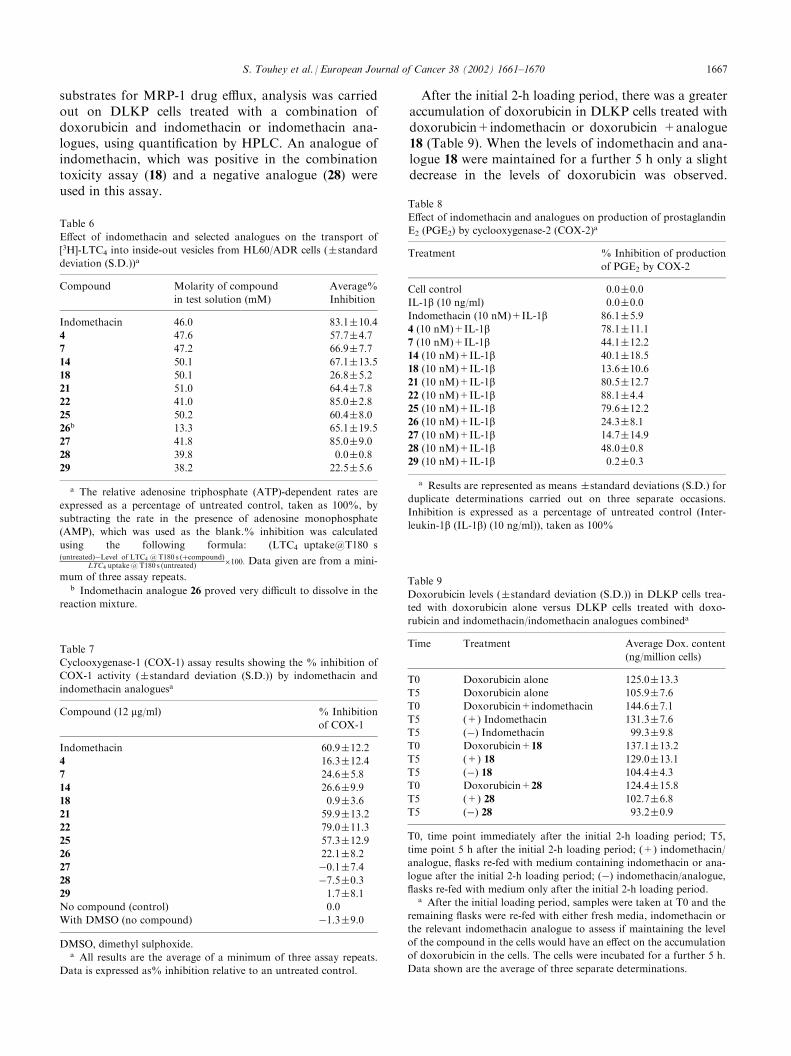
substrates for MRP-1 drug efflux, analysis was carriedout on DLKP cells treated with a combination ofdoxorubicin and indomethacin or indomethacin ana-logues, using quantification by HPLC. An analogue ofindomethacin, which was positive in the combinationtoxicity assay (18) and a negative analogue (28) wereused in this assay.
After the initial 2-h loading period, there was a greateraccumulation of doxorubicin in DLKP cells treated withdoxorubicin+indomethacin or doxorubicin +analogue18 (Table 9). When the levels of indomethacin and ana-logue 18 were maintained for a further 5 h only a slightdecrease in the levels of doxorubicin was observed.
Table 6
Effect of indomethacin and selected analogues on the transport of
[3H]-LTC4 into inside-out vesicles from HL60/ADR cells (�standard
deviation (S.D.))a
Compound Molarity of compound
in test solution (mM)
Average%
Inhibition
Indomethacin 46.0 83.1�10.4
4 47.6 57.7�4.7
7 47.2 66.9�7.7
14 50.1 67.1�13.5
18 50.1 26.8�5.2
21 51.0 64.4�7.8
22 41.0 85.0�2.8
25 50.2 60.4�8.0
26b 13.3 65.1�19.5
27 41.8 85.0�9.0
28 39.8 0.0�0.8
29 38.2 22.5�5.6
a The relative adenosine triphosphate (ATP)-dependent rates are
expressed as a percentage of untreated control, taken as 100%, by
subtracting the rate in the presence of adenosine monophosphate
(AMP), which was used as the blank.% inhibition was calculated
using the following formula: (LTC4 uptake@T180 suntreatedð Þ�Level of LTC4 @T180 s þcompoundð Þ
LTC4 uptake@T180 s untreatedð Þ�100. Data given are from a mini-
mum of three assay repeats.b Indomethacin analogue 26 proved very difficult to dissolve in the
reaction mixture.
Table 8
Effect of indomethacin and analogues on production of prostaglandin
E2 (PGE2) by cyclooxygenase-2 (COX-2)a
Treatment % Inhibition of production
of PGE2 by COX-2
Cell control 0.0�0.0
IL-1b (10 ng/ml) 0.0�0.0
Indomethacin (10 nM)+IL-1b 86.1�5.9
4 (10 nM)+IL-1b 78.1�11.1
7 (10 nM)+IL-1b 44.1�12.2
14 (10 nM)+IL-1b 40.1�18.5
18 (10 nM)+IL-1b 13.6�10.6
21 (10 nM)+IL-1b 80.5�12.7
22 (10 nM)+IL-1b 88.1�4.4
25 (10 nM)+IL-1b 79.6�12.2
26 (10 nM)+IL-1b 24.3�8.1
27 (10 nM)+IL-1b 14.7�14.9
28 (10 nM)+IL-1b 48.0�0.8
29 (10 nM)+IL-1b 0.2�0.3
a Results are represented as means �standard deviations (S.D.) for
duplicate determinations carried out on three separate occasions.
Inhibition is expressed as a percentage of untreated control (Inter-
leukin-1b (IL-1b) (10 ng/ml)), taken as 100%
Table 7
Cyclooxygenase-1 (COX-1) assay results showing the % inhibition of
COX-1 activity (�standard deviation (S.D.)) by indomethacin and
indomethacin analoguesa
Compound (12 mg/ml) % Inhibition
of COX-1
Indomethacin 60.9�12.2
4 16.3�12.4
7 24.6�5.8
14 26.6�9.9
18 0.9�3.6
21 59.9�13.2
22 79.0�11.3
25 57.3�12.9
26 22.1�8.2
27 �0.1�7.4
28 �7.5�0.3
29 1.7�8.1
No compound (control) 0.0
With DMSO (no compound) �1.3�9.0
DMSO, dimethyl sulphoxide.a All results are the average of a minimum of three assay repeats.
Data is expressed as% inhibition relative to an untreated control.
Table 9
Doxorubicin levels (�standard deviation (S.D.)) in DLKP cells trea-
ted with doxorubicin alone versus DLKP cells treated with doxo-
rubicin and indomethacin/indomethacin analogues combineda
Time Treatment Average Dox. content
(ng/million cells)
T0 Doxorubicin alone 125.0�13.3
T5 Doxorubicin alone 105.9�7.6
T0 Doxorubicin+indomethacin 144.6�7.1
T5 (+) Indomethacin 131.3�7.6
T5 (�) Indomethacin 99.3�9.8
T0 Doxorubicin+18 137.1�13.2
T5 (+) 18 129.0�13.1
T5 (�) 18 104.4�4.3
T0 Doxorubicin+28 124.4�15.8
T5 (+) 28 102.7�6.8
T5 (�) 28 93.2�0.9
T0, time point immediately after the initial 2-h loading period; T5,
time point 5 h after the initial 2-h loading period; (+) indomethacin/
analogue, flasks re-fed with medium containing indomethacin or ana-
logue after the initial 2-h loading period; (�) indomethacin/analogue,
flasks re-fed with medium only after the initial 2-h loading period.a After the initial loading period, samples were taken at T0 and the
remaining flasks were re-fed with either fresh media, indomethacin or
the relevant indomethacin analogue to assess if maintaining the level
of the compound in the cells would have an effect on the accumulation
of doxorubicin in the cells. The cells were incubated for a further 5 h.
Data shown are the average of three separate determinations.
S. Touhey et al. / European Journal of Cancer 38 (2002) 1661–1670 1667
However, there was a decreased amount of doxorubicinafter 5 h in those cells treated with doxorubicin alone orwith the analogue 28. The results demonstrate that theexport of doxorubicin from MRP-1-expressing cells canbe inhibited by indomethacin and its active analogues.
3.1.5. Indomethacin efflux studiesIndomethacin efflux was also analysed, using HPLC
quantification, in cells treated with indomethacin aloneor doxorubicin and indomethacin combined. After the
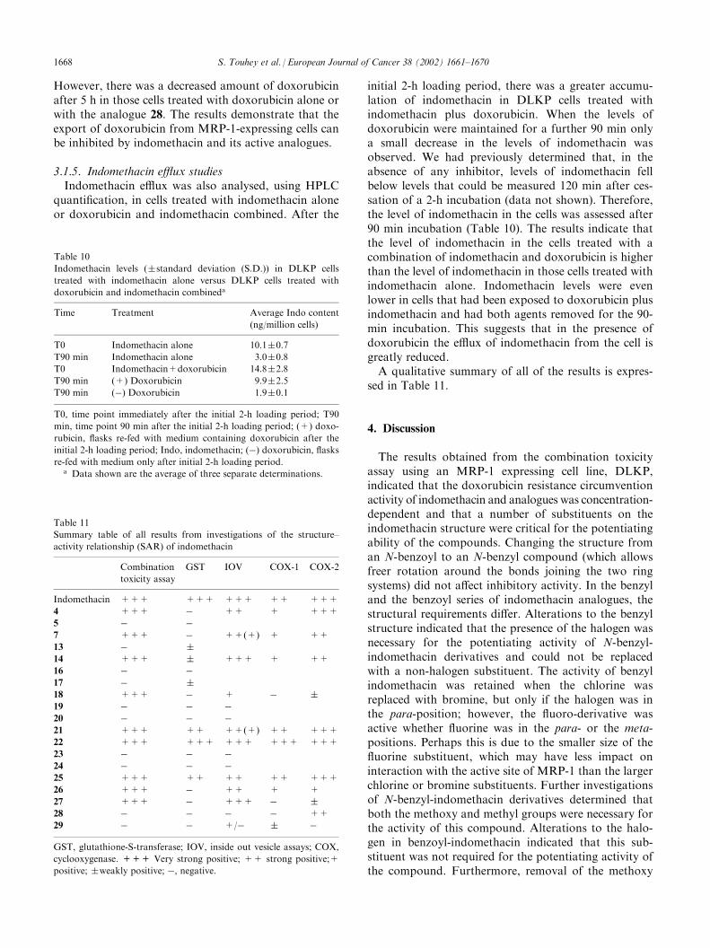
initial 2-h loading period, there was a greater accumu-lation of indomethacin in DLKP cells treated withindomethacin plus doxorubicin. When the levels ofdoxorubicin were maintained for a further 90 min onlya small decrease in the levels of indomethacin wasobserved. We had previously determined that, in theabsence of any inhibitor, levels of indomethacin fellbelow levels that could be measured 120 min after ces-sation of a 2-h incubation (data not shown). Therefore,the level of indomethacin in the cells was assessed after90 min incubation (Table 10). The results indicate thatthe level of indomethacin in the cells treated with acombination of indomethacin and doxorubicin is higherthan the level of indomethacin in those cells treated withindomethacin alone. Indomethacin levels were evenlower in cells that had been exposed to doxorubicin plusindomethacin and had both agents removed for the 90-min incubation. This suggests that in the presence ofdoxorubicin the efflux of indomethacin from the cell isgreatly reduced.A qualitative summary of all of the results is expres-
sed in Table 11.
4. Discussion
The results obtained from the combination toxicityassay using an MRP-1 expressing cell line, DLKP,indicated that the doxorubicin resistance circumventionactivity of indomethacin and analogues was concentration-dependent and that a number of substituents on theindomethacin structure were critical for the potentiatingability of the compounds. Changing the structure froman N-benzoyl to an N-benzyl compound (which allowsfreer rotation around the bonds joining the two ringsystems) did not affect inhibitory activity. In the benzyland the benzoyl series of indomethacin analogues, thestructural requirements differ. Alterations to the benzylstructure indicated that the presence of the halogen wasnecessary for the potentiating activity of N-benzyl-indomethacin derivatives and could not be replacedwith a non-halogen substituent. The activity of benzylindomethacin was retained when the chlorine wasreplaced with bromine, but only if the halogen was inthe para-position; however, the fluoro-derivative wasactive whether fluorine was in the para- or the meta-positions. Perhaps this is due to the smaller size of thefluorine substituent, which may have less impact oninteraction with the active site of MRP-1 than the largerchlorine or bromine substituents. Further investigationsof N-benzyl-indomethacin derivatives determined thatboth the methoxy and methyl groups were necessary forthe activity of this compound. Alterations to the halo-gen in benzoyl-indomethacin indicated that this sub-stituent was not required for the potentiating activity ofthe compound. Furthermore, removal of the methoxy
Table 11
Summary table of all results from investigations of the structure–
activity relationship (SAR) of indomethacin
Combination
toxicity assay
GST IOV COX-1 COX-2
Indomethacin +++ +++ +++ ++ +++
4 +++ � ++ + +++
5 � �
7 +++ � ++(+) + ++
13 � �
14 +++ � +++ + ++
16 � �
17 � �
18 +++ � + � �
19 � � �
20 � � �
21 +++ ++ ++(+) ++ +++
22 +++ +++ +++ +++ +++
23 � � �
24 � � �
25 +++ ++ ++ ++ +++
26 +++ � ++ + +
27 +++ � +++ � �
28 � � � � ++
29 � � +/� � �
GST, glutathione-S-transferase; IOV, inside out vesicle assays; COX,
cyclooxygenase. +++ Very strong positive; ++ strong positive;+
positive; �weakly positive; �, negative.
Table 10
Indomethacin levels (�standard deviation (S.D.)) in DLKP cells
treated with indomethacin alone versus DLKP cells treated with
doxorubicin and indomethacin combineda
Time Treatment Average Indo content
(ng/million cells)
T0 Indomethacin alone 10.1�0.7
T90 min Indomethacin alone 3.0�0.8
T0 Indomethacin+doxorubicin 14.8�2.8
T90 min (+) Doxorubicin 9.9�2.5
T90 min (�) Doxorubicin 1.9�0.1
T0, time point immediately after the initial 2-h loading period; T90
min, time point 90 min after the initial 2-h loading period; (+) doxo-
rubicin, flasks re-fed with medium containing doxorubicin after the
initial 2-h loading period; Indo, indomethacin; (�) doxorubicin, flasks
re-fed with medium only after initial 2-h loading period.a Data shown are the average of three separate determinations.
1668 S. Touhey et al. / European Journal of Cancer 38 (2002) 1661–1670

substituent from the benzoyl indomethacin structure didnot render the compound inactive.The active analogues also showed synergy in other cell
lines possessing MRP-1 indicating that the effect is notcell line-specific.Compounds known to be PLA2 inhibitors were tes-
ted, but only two of these were capable of potentiatingthe toxicity of doxorubicin. These were less toxic to thecells, but also had to be used at higher concentrations toproduce the same potentiation effect. These resultsindicated that the potentiating ability of the NSAIDswas unlikely to involve PLA2 inhibition.Indomethacin or indomethacin analogues might exert
their toxicity-enhancing effect on the chemotherapeuticdrug through inhibition of the activity of GST. Gluta-thione conjugates are transported very effectively byMRP-1 and these glutathione conjugates are formed bythe GST enzyme. Indomethacin is also a classic inhi-bitor of GST. Results demonstrated varying abilityamong the indomethacin analogues to inhibit GST. Ofthe indomethacin analogues shown to have the ability topotentiate the toxicity of doxorubicin, only the benzoylanalogues were good GST inhibitors. The remainingindomethacin analogues demonstrated insignificant orno GST inhibitory ability which indicates that thepotentiating activity of the analogues on the chemo-therapeutic drugs is not through inhibition of GST.Using inside-out vesicles prepared from the plasma
membrane of HL60/ADR cells, which overexpressMRP-1, we demonstrated that the active indomethacinanalogues had the ability to inhibit the uptake of theMRP-1 substrate [3H] LTC4 into the vesicles indicatinga direct interaction between the analogues and MRP-1.The efflux of doxorubicin from DLKP cells treated
with indomethacin or active indomethacin analogueswas significantly retarded relative to untreated cells orcells treated with an inactive indomethacin analogue.Conversely, doxorubicin also inhibited the efflux ofindomethacin in these cells suggesting that this effect isdue to direct competitive inhibition of the export pump.A number of the active indomethacin analogues were
found to potentiate the toxicity of MRP-1 substrates,doxorubicin, VP-16 and vincristine in an MRP-1-over-expressing cell line, COR L23R. There was a lowerpotentiation of the same drugs in the parental COR L23cell line when combined with the active analogues.MRP-1 is expressed in both the COR L23 variants [5],although at relatively low levels in the COR L23 par-ental cells. MRP-1 is also expressed at a low level inDLKP cells (i.e. it was only visible on Western blotswhen a concentrated plasma membrane preparation wasused [17]), but it is clearly functional as indicated by ourdata. We also demonstrated that indomethacin and theactive analogues were unable to potentiate the toxicityof 5-FU in the COR L23 parental and resistant cells.5-FU is not a substrate forMRP-1, and this result further
supports the idea that the action of the active analoguesis through an interaction with MRP-1.The variation between the magnitude of toxicity syner-
gism seen between the different inhibitors and differentchemotherapy drugs (and in different cell lines) suggeststhat there may be quantitative differences in the activity ofinhibitors with different MRP substrates. For example,there is little difference between the synergistic activity ofany of the agents combined with etoposide, whereas 21may be a better potentiator of doxorubicin activity in CORL23R and COR L23S cells, while 25 is more active inDLKP cells. This may reflect the interactions of the differ-ent physiochemical environments in each cell causingminor modifications in substrate and inhibitor specificity,and/or inhibition of other efflux pumps, the expression ofwhich varies from one cell line to another.We had previously reported that the enhancement
effect is not due to the COX inhibitory activity ofNSAIDs [17]. However, we analysed the indomethacinanalogues for COX-1 and COX-2 inhibitory activity toassess if any of the active analogues were potentiallycapable of enhancing chemotherapeutic drug toxicitywithout the potential for damage that is associated withNSAIDs, due to COX-1 inhibition. The N-benzoyl-indomethacin analogues were effective at inhibitingCOX-1 and COX-2, while the N-benzyl indomethacinanalogues were weak COX-1 inhibitors. This suggeststhat the carbonyl group in indomethacin may beimportant for COX-1 inhibition. Of these benzyl analo-gues, only 4 was equi-potent to the benzoyl analogues atinhibiting COX-2. It seems that removing the chlorinefrom the benzene ring, changing its position from para-,or replacing it with another halogen, affected the abilityof the compound to inhibit COX-2.Of particular interest are those active indomethacin
analogues with reduced COX-1 inhibitory ability, as theymight provide an approach by which the toxicity of theanticancer drugs could be enhanced without the gastro-intestinal toxic side-effects associated with indomethacin.In particular, analogues 18 and 27, N-benzyl indometha-cin analogues, active in the combination toxicity assay,were less toxic than indomethacin and demonstrated noCOX-1 and low COX-2 inhibitory activity. Indometha-cin analogue 4 also demonstrated very low COX-1inhibitory ability but, in contrast to 18 and 27, was alsoa very potent COX-2 inhibitor. This suggests that 4 isworthy of clinical evaluation due to strong MRP-1inhibitory activity combined with possible lower toxicside-effects than indomethacin with additional ability tosuppress tumour growth due to inhibition of COX-2 [35].Taken together, the results also show that it is possi-
ble to distinguish molecular structures which give betterMRP inhibition or better COX inhibition and althoughsome of the structural determinants are similar, suffi-cient differences exist to permit the design of specificinhibitors of individual proteins.
S. Touhey et al. / European Journal of Cancer 38 (2002) 1661–1670 1669
Overall, these results have demonstrated that indo-methacin and a number of indomethacin analogueshave the ability to potentiate (via MRP-1 inhibition) thetoxicity of a number of clinically important chemo-therapeutic drugs at non-toxic concentrations. For cer-tain cancers, where drug resistance is a result of MRP-1-overexpression, these active analogues are promising aspotentiators of the toxicity of chemotherapeutic drugspotentially enhancing existing treatments for cancerpatients.
Acknowledgements
Bioresearch Ireland and DCU Educational Trust aregratefully acknowledged for their financial support ofthis work.
References
1. Clynes M. Multiple Drug Resistance in Cancer 2— Molecular,
Cellular and Clinical Aspects. Cytotechnology. London, Kluwer
Academic Publishers, 1998.
2. Clynes M. Cellular models for multiple drug resistance in cancer.
In Vitro Cell Dev Biol 1993, 29A, 171–179.
3. Cole SPC, Bhardwaj G, Gerlech JH, et al. Overexpression of a
transporter gene in a multidrug-resistant human lung cancer cell
line. Science 1992, 258, 1650–1654.
4. Borst P, Evers R, Kool M, Wijnholds J. A family of drug trans-
porters: the multidrug resistance-associated proteins. J Natl Can-
cer Inst 2000, 92, 1295–1302.
5. Kool M, de Haas M, Scheffer G, et al. Analysis of expression of
cMOAT (MRP2), MRP3, MRP4 and MRP5, homologues of the
multidrug resistance associated protein (MRP1), in human cancer
cell lines. Cancer Res 1997, 57, 3537–3547.
6. Cole SPC, Deeley R. Multidrug resistance mediated by the ATP
binding cassette transporter protein MRP. Bioessays 1998, 20,
931–940.
7. Oude Elferink RP, Meijer DK, Kuipers F, Jansen PL, Groen
AK, Groothuis GM. Hepatobiliary secretion of organic com-
pounds; molecular mechanisms of membrane transport. Biochim
Biophys Acta 1995, 1241, 215–268.
8. Loe DW, Deeley R, Cole SPC. Characterisation of vincristine
transport by the multidrug resistant 190,000 multidrug resistance
protein (MRP): evidence for co-transport with reduced gluta-
thione. Cancer Res 1998, 58, 5130–5136.
9. Jedlitschky G, Leier I, Buchholz U, Centre M, Keppler D. ATP-
dependent transport of glutathione S-conjugates by the multi-
drug resistance-associated protein. Cancer Res 1994, 54, 4833–
4836.
10. Vane JR, Botting RM. Mechanisms of action of anti-inflamma-
tory drugs. Int J Tissue React 1998, 20, 3–15.
11. DeWitt D. Prostaglandin endoperoxide synthase: regulation of
enzyme expression. Biochim Biophys Acta 1991, 1083, 121–134.
12. Fu JY, Masferrer JL, Seibert K, Raz A, Needleman P. The
induction and suppression of prostaglandin H2 synthase (cyclo-
oxygenase) in human monocytes. J Biol Chem 1990, 265, 16737–
16740.
13. Bolten WW. Scientific rational for specific inhibition of Cox-2.
J Rheumatol Suppl 1998, 51, 2–7.
14. Fosslien E. Adverse effects of nonsteroidal anti-inflammatory
drugs on the gastro-intestinal tract. Ann Clin Lab Sci 1998, 28,
67–81.
15. Khan KNM, Masferrer JL, Woerner BM, Soslow R, Koki AT.
Enhanced cyclooxygenase-2 expression in sporadic and familial
adenomatous polyposis of the human colon. Scan J Gastroenterol
2001, 36, 865–869.
16. Dannhardt G, Kiefer W. Cyclooxygenase inhibitors— current
status and future prospects. Eur J Med Chem 2001, 36, 109–126.
17. Duffy C, Elliot C, O’Connor R, et al. Enhancement of chemo-
therapeutic drug toxicity to human tumour cells in vitro by a
subset of non-steroidal anti-inflammatory drugs (NSAIDs). Eur J
Cancer 1998, 34, 1250–1259.
18. Draper M, Martell R, Levy S. Indomethacin-mediated reversal of
multidrug resistance and drug efflux in human and murine cell
lines overexpressing MRP, but not P-glycoprotein. Br J Cancer
1997, 75, 810–815.
19. Roller A, Bahr O, Streffer J, et al. Selective potentiation of drug
cytotoxicity by NSAID in human glioma cells: the role of COX-1
and MRP. Biochem Biophys Res Commun 1999, 259, 600–605.
20. Piazza GA, Alberts DS, Hixson LJ, et al. Sulindac sulfone inhi-
bits Azoxymethane-induced colon carcinogenesis in rats without
reducing prostaglandin levels. Cancer Res 1997, 57, 2909–2915.
21. Maguire AR, Plunkett SJ, Papot S, Clynes M, O’Connor R,
Touhey S. Synthesis of indomethacin analogues for evaluation as
moderators of MRP activity. Bioorganic and Medicinal Chemistry
2001, 9, 745–762.
22. Martin A, Clynes M. Acid phosphatase: endpoint for in vitro
toxicity tests. In Vitro Cell Dev Biol 1991, 27A, 183–184.
23. Martin A, Clynes M. Comparison of five microplate colorimetric
assays for in vitro cytotoxicity testing and cell proliferation
assays. Cytotechnology 1993, 11, 49–58.
24. Ishikawa T, Wright CD, Ishizuka H. GS-X pump is functionally
overexpressed in cis-diamminedichloroplatinum (II)-resistant
human leukemia HL-60 cells and down regulated by cell differ-
entiation. J Biol Chem 1994, 269, 29085–29093.
25. Habig WH, Jacoby WB. Glutathione S-tranferases (rat and
human). Methods Enzymol 1981, 77, 218–231.
26. Johnson AG, Ray JE. Improved high-performance liquid chro-
matographic method for the determination of indomethacin
inplasma. Ther Drug Monit 1992, 14, 61–65.
27. Boopathy R, BalasubramanianA. Purification and characterisation
of sheep platelet cyclooxygenase. Biochem J 1986, 239, 371–377.
28. Piazza GA, Rahm AK, Finn TS, et al. Apoptosis primarily
accounts for the growth inhibitory properties of sulindac meta-
bolites and involves a mechanism that is independent of cyclo-
oxygenase inhibition, cell cycle arrest and p53 induction. Cancer
Res 1997, 57, 2452–2459.
29. Asano K, Lilly C, Deazen J. Prostaglandin G/H synthase-2 is the
constitutive and dominant isoform in cultured human lung epi-
thelial cells. Am J Physiol 1996, 271, L126–L131.
30. Chou J, Chou T. Dose–Effect Analysis with Microcomputers.
Cambridge, UK, Biosoft, 1987.
31. Chou T, Talalay P. Analysis of combined drug effects: a new look
at a very old problem. Trends Pharmacol Sci 1983, 4, 450–454.
32. Kaplan L, Weiss J, Elsbach P. Low concentrations of indo-
methacin inhibit phospholipase A2 of rabbit polymorphonuclear
leukocytes. Proc Natl Acad Sci 1978, 75, 2955–2958.
33. Riendeau D, Charleson S, Cromlish W, Mancini JA, Wong E,
Guay J. Comparison of the cyclooxygenase-1 inhibitory proper-
ties of nonsteroidal anti-inflammatory drugs (NSAIDs) and
selective Cox-2 inhibitors, using sensitive microsomal and platelet
assays. Can J Physiol Pharmacol 1997, 75, 1088–1095.
34. Paul S, Breuninger L, Tew K, Shen H, Kruh G. ATP-dependent
uptake of natural product cytotoxic drugs by membrane vesicles
establishes MRP as a broad specificity transporter. Proc Natl
Acad Sci USA 1996, 93, 6929–6934.
35. Watson AJM. Chemopreventative effects of NSAIDs against
colorectal cancer: regulation of apoptosis and mitosis by Cox-1
and Cox-2. Histol Histopathol 1998, 13, 591–597.
1670 S. Touhey et al. / European Journal of Cancer 38 (2002) 1661–1670

![Indomethacin Capsules, USP 25 mgdiuretics, ACE inhibitors, or angiotensin receptor blockers (ARBs)] [see Precautions; Drug Interactions]. Avoid the use of indomethacin capsules in](https://img.pdfslide.us/doc/110x75/5f723d43e581ab752c2e347b/indomethacin-capsules-usp-25-mg-diuretics-ace-inhibitors-or-angiotensin-receptor.jpg)