Embed Size (px)
Citation preview
electronic reprintActa Crystallographica Section F
Structural Biologyand CrystallizationCommunications
ISSN 1744-3091
Editors: H. M. Einspahr and M. S.Weiss
Structure of Pisum sativum Rubisco with bound ribulose1,5-bisphosphate
Peter C. Loewen, Allison L. Didychuk, Jacek Switala, Rosa Perez-Luque,Ignacio Fita and Michele C. Loewen
Acta Cryst. (2013). F69, 10–14
Copyright c© International Union of Crystallography
Author(s) of this paper may load this reprint on their own web site or institutional repository provided thatthis cover page is retained. Republication of this article or its storage in electronic databases other than asspecified above is not permitted without prior permission in writing from the IUCr.
For further information see http://journals.iucr.org/services/authorrights.html
Acta Crystallographica Section F
Structural Biologyand CrystallizationCommunicationsEditors: H. M. Einspahr and M. S. Weiss
journals.iucr.org
International Union of CrystallographyWiley-Blackwell
ISSN 1744-3091
Volume 69
Part 1
January 2013Acta Crystallographica Section F: Structural Biology and Crystallization Communicationsis a rapid all-electronic journal, which provides a home for short communications onthe crystallization and structure of biological macromolecules. Structures determinedthrough structural genomics initiatives or from iterative studies such as those used in thepharmaceutical industry are particularly welcomed. Articles are available online whenready, making publication as fast as possible, and include unlimited free colour illus-trations, movies and other enhancements. The editorial process is completely electronicwith respect to deposition, submission, refereeing and publication.
Crystallography Journals Online is available from journals.iucr.org
Acta Cryst. (2013). F69, 10–14 Loewen et al. · Rubisco
structural communications
10 doi:10.1107/S1744309112047549 Acta Cryst. (2013). F69, 10–14
Acta Crystallographica Section F
Structural Biologyand CrystallizationCommunications
ISSN 1744-3091
Structure of Pisum sativum Rubisco with boundribulose 1,5-bisphosphate
Peter C. Loewen,a Allison L.
Didychuk,b Jacek Switala,a Rosa
Perez-Luque,c Ignacio Fitac and
Michele C. Loewenb,d*
aDepartment of Microbiology, University of
Manitoba, 418 Buller Building, Winnipeg,
MB R3T 2N2, Canada, bNational Research
Council of Canada, 110 Gymnasium Place,
Saskatoon, SK S7N 0W9, Canada, cInstitut de
Biologia Molecular de Barcelona (IBMB–CSIC),
Parc Cientıfic, Baldiri Reixac 10,
08028 Barcelona, Spain, and dDepartment of
Biochemistry, University of Saskatchewan,
107 Wiggins Road, Saskatoon, SK S7N 5E5,
Canada
Correspondence e-mail:
Received 19 October 2012
Accepted 19 November 2012
PDB Reference: Rubisco, 4hhh
The first structure of a ribulose-1,5-bisphosphate carboxylase/oxygenase
(Rubisco) from a pulse crop is reported. Rubisco was purified from Pisum
sativum (garden pea) and diffraction-quality crystals were obtained by hanging-
drop vapour diffusion in the presence of the substrate ribulose 1,5-bisphosphate.
X-ray diffraction data were recorded to 2.20 A resolution from a single crystal at
the Canadian Light Source. The overall quaternary structure of non-activated
P. sativum Rubisco highlights the conservation of the form I Rubisco
hexadecameric complex. The electron density places the substrate in the active
site at the interface of the large-subunit dimers. Lys201 in the active site is not
carbamylated as expected for this non-activated structure. Some heterogeneity
in the small-subunit sequence is noted, as well as possible variations in the
conformation and contacts of ribulose 1,5-bisphosphate in the large-subunit
active sites. Overall, the active-site conformation most closely correlates with
the ‘closed’ conformation observed in other substrate/inhibitor-bound Rubisco
structures.
1. Introduction
The photosynthetic assimilation of atmospheric CO2 into carbo-
hydrates in plants occurs via the Calvin cycle and utilizes ribulose-
1,5-bisphosphate carboxylase/oxygenase (Rubisco) for the primary
CO2-fixation step (Ellis, 1979). A molecule of CO2 is first added to the
five-carbon substrate ribulose 1,5-bisphosphate (RuBP), and the
resulting six-carbon intermediate is cleaved to two molecules of
3-phosphoglycerate (3-PGA). The effectiveness of Rubisco in CO2
fixation is compromised by its competing catalysis of RuBP oxyge-
nation, which yields one molecule of 3-PGA and one molecule of
phosphoglycolate. The first Rubisco structures revealed complexes
comprised of large or of large and small subunits (Knight et al., 1989;
Andersson et al., 1989; Chapman et al., 1987; Suh et al., 1987;
Schneider et al., 1986). In the case of higher plants, the complex was
found to consistently have a hexadecameric structure with an L8S8 (L,
large subunit, 475 residues; S, small subunit, 123 residues) arrange-
ment referred to as form I Rubisco (Andersson, 2008). Catalysis
begins with an initial activation step involving the conversion of an
active-site lysine (Lys201 in the spinach enzyme) in the L subunit to
its carbamylated derivative by the addition of CO2, which also
facilitates the binding of a magnesium ion required to complete the
active site. RuBP can bind to both activated and non-activated
Rubisco, but the latter forms a nonproductive complex that leads to a
gradual loss of enzymatic activity in vitro (Zhu & Jensen, 1991). A
comparison of structures reveals the substrate-bound or inhibitor-
bound forms to have a more constrained or isolated RuBP-binding
site, which is a result of localized changes in flexible loop regions
(Taylor & Andersson, 1997b). Despite decades of study and the
availability of Rubisco structures from spinach, tobacco, rice, algae,
cyanobacteria and phototropic bacteria, many aspects of the regu-
lation of Rubisco activity, such as the partitioning between carbox-
ylation and oxygenation reactions, remain enigmatic (Andersson &
Backlund, 2008). Recently, in the context of climate change and food
security, Rubisco has become a major target of international crop-
improvement consortia (Parry et al., 2011). The focus of these efforts
is on the modulation of Rubisco to increase its expression level,# 2013 International Union of Crystallography
All rights reserved
electronic reprint
catalytic rate, thermal stability and availability of substrates, with the
aim of developing higher yielding crops that sequester larger amounts
of carbon dioxide from the environment. In this light, a recent
structure of rice Rubisco has been published that yielded novel
insights into mechanistic aspects of its regulation by specific effector
molecules (Matsumura et al., 2012). Here, we report the structure of
non-activated Pisum sativum (garden pea) Rubisco (PsRubisco) with
bound RuBP, which is the first representative structure from a pulse
crop.
2. Materials and methods
2.1. Protein purification and crystallization
Rubisco was purified from P. sativum plants. Plant homogenates in
50 mM Tris–HCl pH 7.2 were strained through gauze, centrifuged at
15 000g for 20 min and subjected to 2.5% streptomycin and 30–50%
ammonium sulfate (AS) precipitations. The 40% AS fraction was
resuspended in 50 mM Tris–HCl pH 7.2 and dialyzed overnight into
the same buffer. This simple protocol yielded a final dialyzed Rubisco
concentration of 25–33 mg ml�1 in repeated preparations, with >95%
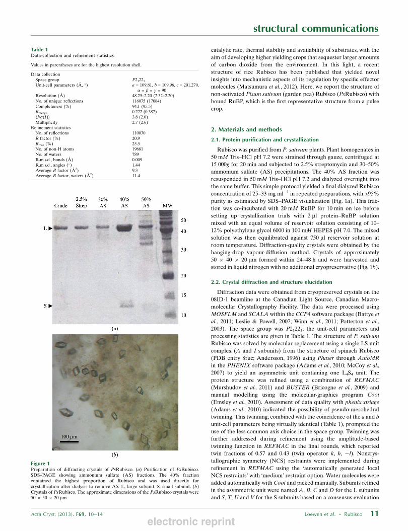
purity as estimated by SDS–PAGE visualization (Fig. 1a). This frac-
tion was co-incubated with 20 mM RuBP for 10 min on ice before
setting up crystallization trials with 2 ml protein–RuBP solution
mixed with an equal volume of reservoir solution consisting of 10–
12% polyethylene glycol 6000 in 100 mM HEPES pH 7.0. The mixed
solution was then equilibrated against 750 ml reservoir solution at
room temperature. Diffraction-quality crystals were obtained by the
hanging-drop vapour-diffusion method. Crystals of approximately
50 � 40 � 20 mm formed within 24–48 h and were harvested and
stored in liquid nitrogen with no additional cryopreservative (Fig. 1b).
2.2. Crystal diffraction and structure elucidation
Diffraction data were obtained from cryopreserved crystals on the
08ID-1 beamline at the Canadian Light Source, Canadian Macro-
molecular Crystallography Facility. The data were processed using
MOSFLM and SCALA within the CCP4 software package (Battye et
al., 2011; Leslie & Powell, 2007; Winn et al., 2011; Potterton et al.,
2003). The space group was P21221; the unit-cell parameters and
processing statistics are given in Table 1. The structure of P. sativum
Rubisco was solved by molecular replacement using a single LS unit
complex (A and I subunits) from the structure of spinach Rubisco
(PDB entry 8ruc; Andersson, 1996) using Phaser through AutoMR
in the PHENIX software package (Adams et al., 2010; McCoy et al.,
2007) to yield an asymmetric unit containing one L4S4 unit. The
protein structure was refined using a combination of REFMAC
(Murshudov et al., 2011) and BUSTER (Bricogne et al., 2009) and
manual modelling using the molecular-graphics program Coot
(Emsley et al., 2010). Assessment of data quality with phenix.xtriage
(Adams et al., 2010) indicated the possibility of pseudo-merohedral
twinning. This twinning, combined with the coincidence of the a and b
unit-cell parameters being virtually identical (Table 1), prompted the
use of the less common axis choice in the space group. Twinning was
further addressed during refinement using the amplitude-based
twinning function in REFMAC in the final rounds, which reported
twin fractions of 0.57 and 0.43 (twin operator k, h, �l). Noncrys-
tallographic symmetry (NCS) restraints were implemented during
refinement in REFMAC using the ‘automatically generated local
NCS restraints’ with ‘medium’ restraint option. Water molecules were
added automatically with Coot and picked manually. Subunits refined
in the asymmetric unit were named A, B, C and D for the L subunits
and S, T, U and V for the S subunits based on a consensus evaluation
structural communications
Acta Cryst. (2013). F69, 10–14 Loewen et al. � Rubisco 11
Figure 1Preparation of diffracting crystals of PsRubisco. (a) Purification of PsRubisco.SDS–PAGE showing ammonium sulfate (AS) fractions. The 40% fractioncontained the highest proportion of Rubisco and was used directly forcrystallization after dialysis to remove AS. L, large subunit; S, small subunit. (b)Crystals of PsRubisco. The approximate dimensions of the PsRubisco crystals were50 � 50 � 20 mm.
Table 1Data-collection and refinement statistics.
Values in parentheses are for the highest resolution shell.
Data collectionSpace group P21221
Unit-cell parameters (A, �) a = 109.81, b = 109.96, c = 201.270,� = � = � = 90
Resolution (A) 48.25–2.20 (2.32–2.20)No. of unique reflections 116075 (17084)Completeness (%) 94.1 (95.5)Rmerge 0.222 (0.387)hI/�(I)i 3.8 (2.0)Multiplicity 2.7 (2.6)
Refinement statisticsNo. of reflections 110030R factor (%) 20.9Rfree (%) 25.5No. of non-H atoms 19681No. of waters 789R.m.s.d., bonds (A) 0.009R.m.s.d., angles (�) 1.44Average B factor (A2) 9.3Average B factor, waters (A2) 11.4
electronic reprint
of available Rubisco structure nomenclature (Supplementary Table
S1). Refinement statistics are shown in Table 1. Figures were gener-
ated using PyMOL (Schrodinger LLC). The atomic coordinates and
structure factors have been deposited in the PDB as entry 4hhh.
3. Results and discussion
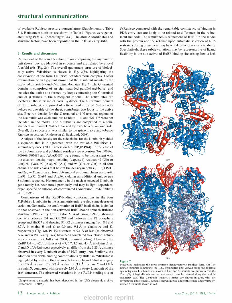
Refinement of the four LS subunit pairs comprising the asymmetric
unit shows they are identical in structure and are related by a local
fourfold axis (Fig. 2a). The overall quaternary structure of biologi-
cally active PsRubisco is shown in Fig. 2(b), highlighting the
conservation of the form I Rubisco hexadecameric complex. Closer
examination of an L2S2 unit shows that the L subunit maintains the
expected discrete N- and C-terminal domains (Fig. 3). The C-terminal
domain is comprised of an eight-stranded parallel �/�-barrel and
includes the active site formed by loops connecting the C-terminal
end of �-strands to the subsequent �-helix. The active sites are
located at the interface of each L2 dimer. The N-terminal domain
of the L subunit, comprised of a five-stranded mixed �-sheet with
helices on one side of the sheet, contributes two loops to the active
site. Electron density for the C-terminal and N-terminal regions of
the L subunits was weak and thus residues 1–11 and 470–475 were not
included in the model. The S subunits are comprised of a four-
stranded antiparallel �-sheet flanked by two helices on one side.
Overall, the structure is very similar to the spinach, rice and tobacco
Rubisco structures (Andersson & Backlund, 2008).
Analysis of the density for the side chains for the L subunit yielded
a sequence that is in agreement with the available PsRubisco L-
subunit sequence (NCBI accession No. NP_054944). In the case of
the S subunits, several published residues (see accession Nos. P00868,
P00869, P07689 and AAA33686) were found to be inconsistent with
the electron-density maps, including (expected) residues 47 (Glu or
Leu), 91 (Val), 92 (Ala), 93 (Ala) and 96 (Glu or Gln) in all four
chains. The side chains that best fit the density in both Fo � Fc OMIT
and 2Fo � Fc maps in all four determined S-subunit chains are Lys47,
Lys91, Lys92, Glu93 and Arg96, yielding an additional unique pea
S-subunit sequence. Heterogeneity in the nuclear-encoded S-subunit
gene family has been noted previously and may be light-dependent,
organ-specific or chloroplast-coordinated (Andersson, 1996; Shibata
et al., 1996).
Comparisons of the RuBP-binding conformations in the four
PsRubisco L subunits in the asymmetric unit revealed some degree of
variation. Generally, the conformation of RuBP in all chains is similar
to that observed in the non-activated RuBP-bound spinach Rubisco
structure (PDB entry 1rcx; Taylor & Andersson, 1997b), showing
contacts between O4 and Glu204 and between the P2 phosphate
group and His327 and showing P1–P2 distances ranging from 8.6 and
8.7 A in chains B and C to 9.0 and 9.1 A in chains A and D,
respectively (Fig. 4a). P1–P2 distances of 9.1 A or less (as observed
here and in PDB entry 1rcx) have been correlated to a ‘closed’ active-
site conformation (Duff et al., 2000; discussed below). However, the
RuBP O3—Lys201 distances of 4.7, 5.7, 3.7 and 4.4 A in chains A, B,
C and D of PsRubisco, respectively, all differ from the 3.23 A distance
observed in every L-subunit chain of PDB entry 1rcx. Similarly, the
adoption of variable binding conformations by RuBP in PsRubisco is
highlighted by shifts in the distance between O4 and Glu204 ranging
from 2.8 A in chain B to 3.2 A in chain A, 3.4 A in chain C and 4.8 A
in chain D, compared with precisely 2.96 A in every L subunit of the
1rcx structure. The observed variations in the RuBP-binding site of
PsRubisco compared with the remarkable consistency of binding in
PDB entry 1rcx are likely to be related to differences in the refine-
ment methods. The simultaneous refinement of RuBP in the model
with the protein and the reliance upon automatic selection of NCS
restraints during refinement may have led to the observed variability.
Speculatively, these subtle variations may be representative of ligand
flexibility in the non-activated RuBP-binding site arising from a lack
structural communications
12 Loewen et al. � Rubisco Acta Cryst. (2013). F69, 10–14
Figure 2PsRubisco maintains the most common hexadecameric Rubisco form. (a) Therefined subunits comprising the L4S4 asymmetric unit viewed along the fourfoldsymmetry axis. L subunits are shown in blue and S subunits are shown in red. (b)The L8S8 biologically relevant hexadecameric complex viewed along the twofoldsymmetry axis. The L-subunit symmetry mates are shown in grey, with theasymmetric unit refined L subunits shown in blue and both refined and symmetry-related S subunits shown in red.
1 Supplementary material has been deposited in the IUCr electronic archive(Reference: TT5035).
electronic reprint
of constraint in the absence of a carbamylated Lys201 and the Mg2+.
However, the possibility that the variations might arise from crystal-
packing defects or errors in estimation of the twin fraction cannot be
strictly eliminated.
As alluded to above, spinach Rubisco has been shown to adopt two
different active-site conformational states. In the unliganded form or
in the presence of the product 3-PGA (e.g. PDB entries 1aus and
1aa1; Taylor & Andersson, 1996, 1997a) the active site is in an ‘open’
conformation. In these structures, the C-terminal region (residues
466–475) as well as loop 6 (residues 333–338) are disordered and are
not actually represented in the available models. In the presence of
ligand, be it RuBP in a non-activated enzyme (PDB entry 1rcx; Taylor
& Andersson, 1997b) or inhibitor in the activated form (PDB entry
8ruc; Andersson, 1996), the C-terminal strand is stabilized against
loop 6, causing loop 6 to fold over the ligand-access channel yielding a
stable ‘closed’ conformation. A third, possibly intermediate, confor-
mation was isolated upon activation of spinach Rubisco with Ca2+
instead of Mg2+, yielding an inactive activated enzyme which could be
cocrystallized with RuBP (PDB entry 1rxo; Taylor et al., 1996). The
structural communications
Acta Cryst. (2013). F69, 10–14 Loewen et al. � Rubisco 13
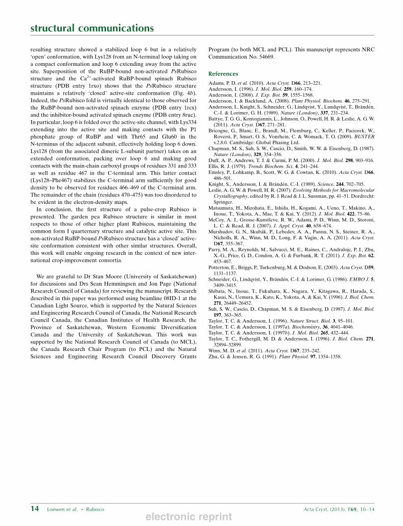
Figure 4Details of the catalytic site. (a) A representation of RuBP in the active site of non-activated PsRubisco. (b) Comparison of non-activated RuBP-bound PsRubiscowith Ca2+-activated inactive RuBP-bound spinach Rubisco. The substrate-bindingchannel in the PsRubisco structure is shown as a grey surface. Loop 6 of thePsRubisco L subunit (green loop) is folded over and pinned down by theoverlapping Lys128 residue (grey surface), yielding a ‘closed’ active-siteconformation. This is in contrast to the ‘open’ conformation observed for spinachRubisco when activated in the presence of Ca2+ with RuBP bound (PDB entry1rxo), which shows loop 6 (yellow loop) folded back and away from the channel. Inboth panels, discrete atoms are shown with pink phosphate, red O, yellow C andblue N atoms.
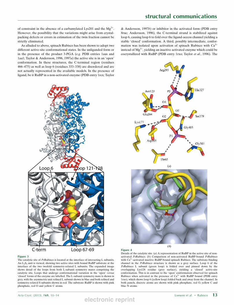
Figure 3The catalytic site of PsRubisco is located at the interface of interacting L subunits.An L2S2 unit is viewed, showing two active sites with bound RuBP substrate at theinterface of the two twofold symmetry-related L subunits. The expanded imageshows detail of the loops from both L-subunit symmetry mates comprising thecatalytic site. Loops that undergo conformational variation in the ‘open’ versus‘closed’ forms of the enzyme are labelled. The L-subunit symmetry mate is shown ingrey, with the asymmetric unit refined L subunit shown in blue and both refined andsymmetry-related S subunits shown in red. The substrate RuBP is shown with pinkphosphate, red O and yellow C atoms.
electronic reprint
resulting structure showed a stabilized loop 6 but in a relatively
‘open’ conformation, with Lys128 from an N-terminal loop taking on
a compact conformation and loop 6 extending away from the active
site. Superposition of the RuBP-bound non-activated PsRubisco
structure and the Ca2+-activated RuBP-bound spinach Rubisco
structure (PDB entry 1rxo) shows that the PsRubisco structure
maintains a relatively ‘closed’ active-site conformation (Fig. 4b).
Indeed, the PsRubisco fold is virtually identical to those observed for
the RuBP-bound non-activated spinach enzyme (PDB entry 1rcx)
and the inhibitor-bound activated spinach enzyme (PDB entry 8ruc).
In particular, loop 6 is folded over the active-site channel, with Lys334
extending into the active site and making contacts with the P1
phosphate group of RuBP and with Thr65 and Glu60 in the
N-terminus of the adjacent subunit, effectively holding loop 6 down.
Lys128 (from the associated dimeric L-subunit partner) takes on an
extended conformation, packing over loop 6 and making good
contacts with the main-chain carboxyl groups of residues 331 and 333
as well as residue 467 in the C-terminal arm. This latter contact
(Lys128–Phe467) stabilizes the C-terminal arm sufficiently for good
density to be observed for residues 466–469 of the C-terminal arm.
The remainder of the chain (residues 470–475) was too disordered to
be evident in the electron-density maps.
In conclusion, the first structure of a pulse-crop Rubisco is
presented. The garden pea Rubisco structure is similar in most
respects to those of other higher plant Rubiscos, maintaining the
common form I quarternary structure and catalytic active site. This
non-activated RuBP-bound PsRubisco structure has a ‘closed’ active-
site conformation consistent with other similar structures. Overall,
this work will enable ongoing research in the context of new inter-
national crop-improvement consortia.
We are grateful to Dr Stan Moore (University of Saskatchewan)
for discussions and Drs Sean Hemmingsen and Jon Page (National
Research Council of Canada) for reviewing the manuscript. Research
described in this paper was performed using beamline 08ID-1 at the
Canadian Light Source, which is supported by the Natural Sciences
and Engineering Research Council of Canada, the National Research
Council Canada, the Canadian Institutes of Health Research, the
Province of Saskatchewan, Western Economic Diversification
Canada and the University of Saskatchewan. This work was
supported by the National Research Council of Canada (to MCL),
the Canada Research Chair Program (to PCL) and the Natural
Sciences and Engineering Research Council Discovery Grants
Program (to both MCL and PCL). This manuscript represents NRC
Communication No. 54669.
References
Adams, P. D. et al. (2010). Acta Cryst. D66, 213–221.Andersson, I. (1996). J. Mol. Biol. 259, 160–174.Andersson, I. (2008). J. Exp. Bot. 59, 1555–1568.Andersson, I. & Backlund, A. (2008). Plant Physiol. Biochem. 46, 275–291.Andersson, I., Knight, S., Schneider, G., Lindqvist, Y., Lundqvist, T., Branden,
C.-I. & Lorimer, G. H. (1989). Nature (London), 337, 231–234.Battye, T. G. G., Kontogiannis, L., Johnson, O., Powell, H. R. & Leslie, A. G. W.
(2011). Acta Cryst. D67, 271–281.Bricogne, G., Blanc, E., Brandl, M., Flensburg, C., Keller, P., Paciorek, W.,
Roversi, P., Smart, O. S., Vonrhein, C. & Womack, T. O. (2009). BUSTERv.2.8.0. Cambridge: Global Phasing Ltd.
Chapman, M. S., Suh, S. W., Cascio, D., Smith, W. W. & Eisenberg, D. (1987).Nature (London), 329, 354–356.
Duff, A. P., Andrews, T. J. & Curmi, P. M. (2000). J. Mol. Biol. 298, 903–916.Ellis, R. J. (1979). Trends Biochem. Sci. 4, 241–244.Emsley, P., Lohkamp, B., Scott, W. G. & Cowtan, K. (2010). Acta Cryst. D66,
486–501.Knight, S., Andersson, I. & Branden, C.-I. (1989). Science, 244, 702–705.Leslie, A. G. W. & Powell, H. R. (2007). Evolving Methods for Macromolecular
Crystallography, edited by R. J. Read & J. L. Sussman, pp. 41–51. Dordrecht:Springer.
Matsumura, H., Mizohata, E., Ishida, H., Kogami, A., Ueno, T., Makino, A.,Inoue, T., Yokota, A., Mae, T. & Kai, Y. (2012). J. Mol. Biol. 422, 75–86.
McCoy, A. J., Grosse-Kunstleve, R. W., Adams, P. D., Winn, M. D., Storoni,L. C. & Read, R. J. (2007). J. Appl. Cryst. 40, 658–674.
Murshudov, G. N., Skubak, P., Lebedev, A. A., Pannu, N. S., Steiner, R. A.,Nicholls, R. A., Winn, M. D., Long, F. & Vagin, A. A. (2011). Acta Cryst.D67, 355–367.
Parry, M. A., Reynolds, M., Salvucci, M. E., Raines, C., Andralojc, P. J., Zhu,X.-G., Price, G. D., Condon, A. G. & Furbank, R. T. (2011). J. Exp. Bot. 62,453–467.
Potterton, E., Briggs, P., Turkenburg, M. & Dodson, E. (2003). Acta Cryst. D59,1131–1137.
Schneider, G., Lindqvist, Y., Branden, C.-I. & Lorimer, G. (1986). EMBO J. 5,3409–3415.
Shibata, N., Inoue, T., Fukuhara, K., Nagara, Y., Kitagawa, R., Harada, S.,Kasai, N., Uemura, K., Kato, K., Yokota, A. & Kai, Y. (1996). J. Biol. Chem.271, 26449–26452.
Suh, S. W., Cascio, D., Chapman, M. S. & Eisenberg, D. (1987). J. Mol. Biol.197, 363–365.
Taylor, T. C. & Andersson, I. (1996). Nature Struct. Biol. 3, 95–101.Taylor, T. C. & Andersson, I. (1997a). Biochemistry, 36, 4041–4046.Taylor, T. C. & Andersson, I. (1997b). J. Mol. Biol. 265, 432–444.Taylor, T. C., Fothergill, M. D. & Andersson, I. (1996). J. Biol. Chem. 271,
32894–32899.Winn, M. D. et al. (2011). Acta Cryst. D67, 235–242.Zhu, G. & Jensen, R. G. (1991). Plant Physiol. 97, 1354–1358.
structural communications
14 Loewen et al. � Rubisco Acta Cryst. (2013). F69, 10–14
electronic reprint