Embed Size (px)
Citation preview

Structure-Function Study of the N-terminal Domain ofExocyst Subunit Sec3*□S
Received for publication, December 21, 2009, and in revised form, January 28, 2010 Published, JBC Papers in Press, February 5, 2010, DOI 10.1074/jbc.M109.096966
Kyuwon Baek‡1, Andreas Knodler§1,2, Sung Haeng Lee‡3, Xiaoyu Zhang§4, Kelly Orlando§, Jian Zhang§,Trevor J. Foskett‡, Wei Guo§5, and Roberto Dominguez‡6
From the ‡Department of Physiology, School of Medicine, and §Department of Biology, University of Pennsylvania,Philadelphia, Pennsylvania 19104
The exocyst is an evolutionarily conservedoctameric complexinvolved in polarized exocytosis fromyeast to humans. The Sec3subunit of the exocyst acts as a spatial landmark for exocytosisthrough its ability to bind phospholipids and small GTPases.The structure of the N-terminal domain of Sec3 (Sec3N) wasdetermined ab initio and defines a new subclass of pleckstrinhomology (PH) domains along with a new family of proteinscarrying this domain. Respectively, N- andC-terminal to the PHdomain Sec3Npresents an additional�-helix and two�-strandsthat mediate dimerization through domain swapping. Thestructure identifies residues responsible for phospholipid bind-ing, which whenmutated in cells impair the localization of exo-cyst components at the plasmamembrane and lead to defects inexocytosis. Through its ability to bind the small GTPase Cdc42and phospholipids, the PH domain of Sec3 functions as a coin-cidence detector at the plasma membrane.
The exocyst is an evolutionarily conserved octameric proteincomplex composed of subunits Sec3, Sec5, Sec6, Sec8, Sec10,Sec15, Exo70, and Exo84. This complex was first identified bygenetic and biochemical methods in the budding yeast Saccha-romyces cerevisiae (1, 2). A homologous complex was subse-quently discovered in mammalian cells (3). The exocyst medi-ates initial tethering of post-Golgi secretory vesicles to the
plasma membrane, a step that precedes SNARE7-driven mem-brane fusion (4, 5). The exocyst is regulated by numerous cel-lular factors, and in particular small GTPases, which are pri-marily responsible for the spatiotemporal control of exocytosis(6).Recent studies have provided insights into the molecular
architecture and function of tethering proteins. Crystal struc-tures of nearly full-length Exo70 (7–9) and large fragments ofSec6 (10), Sec15 (11), and Exo84 (7) have been determined.Despite the lack of sequence similarity, these structures allreveal a similar fold, consisting of elongated tandem repeats ofhelical bundles, which are predicted to pack against oneanother during assembly of the exocyst complex (4). Therecently determined structure of the yeast Dsl1p compleximplicated in Golgi-to-endoplasmic reticulum transport pro-vided the first glance into an assembled tethering complex con-sisting of helical bundles similar to those of exocyst subunits(12). The structure suggested a similar architecture, and possi-bly a common origin, among multisubunit tethering com-plexes. Structures have also been determined of the RalA-bind-ing domains of the mammalian exocyst subunits Sec5 (13) andExo84 (14), which display immunoglobulin-like and pleckstrinhomology (PH) folds, respectively. However, these two do-mains are missing in the yeast complex and are not consideredpart of the conserved core of the exocyst (4).Studies in yeast suggest that subunit Sec3 plays a pivotal role
in exocyst function and vesicle tethering. Sec3 localizes,together with Exo70, to the growing end of the daughter cell(known as the “bud tip”). Although the localization of otherexocyst components relies on the actin cables that serve astracks for motor-driven vesicle transport to the daughter cell,the polarized localization of Sec3 is independent of actin assem-bly (15–17). Genetic analyses and live cell imaging have shownthat theN-terminal 320-amino acid region of Sec3 is importantfor localization of the exocyst to the plasma membrane andexocytosis (18). This region has also been implicated in thebinding of small Rho-family GTPases and phosphatidylinositol4,5-bisphosphate (19–22).As an important step toward understanding exocyst-medi-
ated vesicle tethering, we identified the precise domain of Sec3involved in plasma membrane and Cdc42 binding, determined
* This work was supported, in whole or in part, by National Institutes of HealthGrants R01-GM073791 (to R. D.) and R01-GM64690 (to W. G.). Use of theIndustrial Macromolecular Crystallography Association-CollaborativeAccess Team (IMCA-CAT) beamline 17-BM was supported by the IndustrialMacromolecular Crystallography Association through a contract withHauptman-Woodward Medical Research Institute. The Advanced PhotonSource was supported by Department of Energy Contract W-31-109-Eng-38.
The atomic coordinates and structure factors (code 3HIE) have been deposited inthe Protein Data Bank, Research Collaboratory for Structural Bioinformatics,Rutgers University, New Brunswick, NJ (http://www.rcsb.org/).
□S The on-line version of this article (available at http://www.jbc.org) containssupplemental Figs. S1–S4 and Movie S1.
1 Both authors contributed equally to this work.2 Supported by a fellowship from the Deutsche Forschungsgemeinschaft.3 Present address: Chosun University School of Medicine, 375 Seosuk-dong,
Dong-gu, Gwangju, Korea 501-759.4 Supported by a Scientist Development Grant from the American Heart
Association.5 To whom correspondence may be addressed: Dept. of Biology, University of
Pennsylvania, Philadelphia, PA 19104. E-mail: [email protected] To whom correspondence may be addressed: Dept. of Physiology, School of
Medicine, University of Pennsylvania, A507 Richard Building, 3700 Hamil-ton Walk, Philadelphia, PA 19104. Tel.: 215-573-4559; Fax: 215-573-5851;E-mail: [email protected].
7 The abbreviations used are: SNARE, soluble NSF attachment protein recep-tors; PH, pleckstrin homology; Bicine, N,N-bis(2-hydroxyethyl)glycine; GFP,green fluorescent protein; Bgl2, endo-�1,3-glucanase; 5-FOA, 5-fluoroo-rotic acid.
THE JOURNAL OF BIOLOGICAL CHEMISTRY VOL. 285, NO. 14, pp. 10424 –10433, April 2, 2010© 2010 by The American Society for Biochemistry and Molecular Biology, Inc. Printed in the U.S.A.
10424 JOURNAL OF BIOLOGICAL CHEMISTRY VOLUME 285 • NUMBER 14 • APRIL 2, 2010
by guest on May 17, 2018
http://ww
w.jbc.org/
Dow
nloaded from
its crystal structure, and studied its function in cells. We showthat the core of this domain, consisting of residues 71–241 andreferred to as Sec3N, defines a new subclass of PH domains.Respectively, N- and C-terminal to the PH domain, Sec3N con-tains an�-helix and two�-strands thatmediate dimerization ofthe PH domain through domain swapping. A phosphate ionand the C terminus of a neighboring molecule in the crystalbind in the predicted phospholipid-binding pockets at the dis-tal ends of the dimer, mimicking protein-phospholipid interac-tions. Mutations of positively charged residues identified bythese interactions impair the polarized localization of exocystcomponents at the daughter cell membrane and lead to growthand secretion defects. Based on this structure, a new family ofproteins was identified containing Sec3-like PH domains,including amisyn, a protein implicated in the regulation ofSNARE complex assembly.
EXPERIMENTAL PROCEDURES
Protein Preparation—The cDNA encoding for yeast Sec3N(residues 71–241) was amplified by PCR and cloned into theBamHI site of vector pGEX4T-1 (AmershamBiosciences). Thisvector comprises a glutathione S-transferase affinity purifica-tion tag and a thrombin-cleavage site. BL21(DE3) competentcells (Invitrogen) were transformed with this construct andgrown in LB medium at 37 °C to A600 � 0.6. Expression wasinduced by addition of 1mM isopropyl-�-D-thiogalactopyrano-side and carried out overnight at 20 °C. Cells were harvested bycentrifugation, resuspended in phosphate-buffered saline (140mM NaCl, 2.7 mM KCl, 10 mM Na2HPO4, 1.8 mM KH2PO4 (pH7.4)), and lysed using aMicrofluidizer apparatus (MicroFluidicsCorp.). Affinity purification on a glutathione-Sepharose col-umn (Amersham Biosciences) was done according to the man-ufacturer’s protocol. Sec3N-glutathione S-transferase waseluted with 50 mM Tris-HCl (pH 7.5), 10 mM glutathione anddialyzed against 20 mM Tris-HCl (pH 7.5), 20 mM NaCl, 1 mM
dithiothreitol. Cleavage of the glutathione S-transferase tagwasdone during dialysis with addition of thrombin at amolar rationof 1:1000 (Hematologic Technologies Inc.). Further purifica-tion was carried out on aMono S ion-exchange column (Amer-sham Biosciences) in 20 mM Tris-HCl, (pH 7.5), 1 mM dithio-threitol, and 1 M NaCl gradient. Selenomethionine-substitutedSec3N was obtained by a similar procedure, growing cells inSelenoMet medium (Athena Enzyme Systems), supplementedwith 70 mg/ml selenomethionine (Acros Organics).Crystallization—Sec3Nwas dialyzed against 20mMTris-HCl
(pH 7.5), 50 mMNaCl, 5 mM dithiothreitol and concentrated to10mg/ml using a Vivaspin centrifugal device (Sartorius StedimBiotech). Crystals were obtained using the hanging drop,vapor-diffusion method. A typical setup consisted of a 1:1(v/v) mixture of protein solution and a well solution contain-ing 100 mM Bicine (pH 9.5), 14% polyethylene glycolmonomethyl ether 5000, 1 mM dithiothreitol at 20 °C. Crys-tal quality was improved with addition of 10 mM of CdCl2 orBaCl2. Crystals were flash-frozen in liquid nitrogen usingParatone-N (Hampton Research) as cryoprotectant.Data Collection, Structure Determination, and Analysis—X-
ray datasets were collected from selenomethionine-substitutedcrystals (Table 1) using beamline 17-BM at the Industrial Mac-
romolecularCrystallographyAssociation-CollaborativeAccessTeam facility of the Advance Photon Source (Argonne, IL).Data indexation and scaling were carried out with the programHKL2000 (HKL Research Inc.). The structure was determinedusing the multiwavelength anomalous dispersion method at2.8-Å resolution. The positions of four selenium atoms (one perSec3Nmonomer in the asymmetric unit) were found using theprogram SnB (23). The positions of the selenium atoms wererefined, and phases were calculated with the program Phenix(24). The electron densitymapwas improved by 4-fold symme-try averaging and density modification with the CCP4 programDM (25). Most of the structure was built into the averagedmapusing the graphics programCoot (26). Phases were extended to2.0-Å resolution, and subsequent model building and refine-ment iterations were performed with the programs Coot andPhenix. Illustrations were prepared with the program PyMOL(DeLano Scientific LLC.). The solvent-accessible area buried atthe dimer interface, defined as the locus of the center of a sol-vent probe of radius 1.4 Å as it rolls over the van der Waalssurface of the protein, was calculated with the CCP4 programAREAIMOL.Plasmids and Yeast Strains—Sec3mutations were generated
with the QuikChange site-directed mutagenesis kit (AgilentTechnologies) using a CEN-LEU2 plasmid (pG1273) contain-ing the full-length Sec3 gene as a template. To generate exo70sec3 double mutants, yeast cells bearing the sec3�N mutation,exo70::HIS3 deletion, and carrying an EXO70 balancer plasmid(CEN-URA3) and plasmids with exo70–45 or exo70-47 weretransformed with various sec3 mutant variants. The transfor-mants were tested for growth upon losing the EXO70 balanceron 5-fluoroorotic acid (5-FOA) plates as described previously(18).Bgl2 Secretion Assay—Wild-type, exo70-47, and exo70-47
sec3-303 cells were grown to early log phase at 25 °C. sec10-2mutant cells were grown at 25 °C and moved for 1 h to 37 °C.NaF and NaN3 were added to a concentration of 10 mM. Anamount of cells equivalent to 10 A600/ml was collected andwashed three times with washing buffer (20 mM Tris-HCl, pH7.5, 10 mM NaN3, and 10 mM NaF). Cells were resuspended in300 �l of spheroplast solution (50 mM Tris-HCl, pH 7.5, 1.4 M
sorbitol, 10 mM NaF, 10 mM NaN3, 30 mM 2-mercaptoethanol,and 0.2 mg/ml zymolyase), and incubated for 30 min at 37 °C.The resulting spheroplasts were pelleted at 2000 rpm, andsupernatants (external pools) were collected. After washing,spheroplasts were dissolved in lysis buffer (20mMTris-HCl, pH7.5, 100 mM NaCl, 2 mM MgCl2, 0.5% Triton X-100, and prote-ase inhibitor) on ice for 10 min. The samples were centrifugedat 500� g for 5min, and the supernatants (internal pools) werecollected. Fractions from both pools were analyzed byWesternblot using a rabbit anti-Bgl2 polyclonal antibody.Microscopy—Chromosomal GFP tagging of exocyst compo-
nents was performed as described (15). Cells were grown toearly log phase (A600 0.6) in synthetic completemedia and fixedinmethanol/acetone beforemicroscopy. Images were collectedusing a Leica DM IRB microscope equipped with a 100� oilimmersion objective and a high resolution charge-coupleddevice camera (ORCA-ER, Hamamatsu Photonics). The fluo-rescence distribution in cell images was analyzed with the pro-
A New Subclass of PH Domain Revealed by Sec3 Structure
APRIL 2, 2010 • VOLUME 285 • NUMBER 14 JOURNAL OF BIOLOGICAL CHEMISTRY 10425
by guest on May 17, 2018
http://ww
w.jbc.org/
Dow
nloaded from
gram ImageJ (rsb.info.nih.gov/ij). A straight line was drawnacross the bud and into themother cell. Plot Profile ImageJ wasused to determine the gray value of each point along the line, ameasure of the fluorescence intensity.
RESULTS
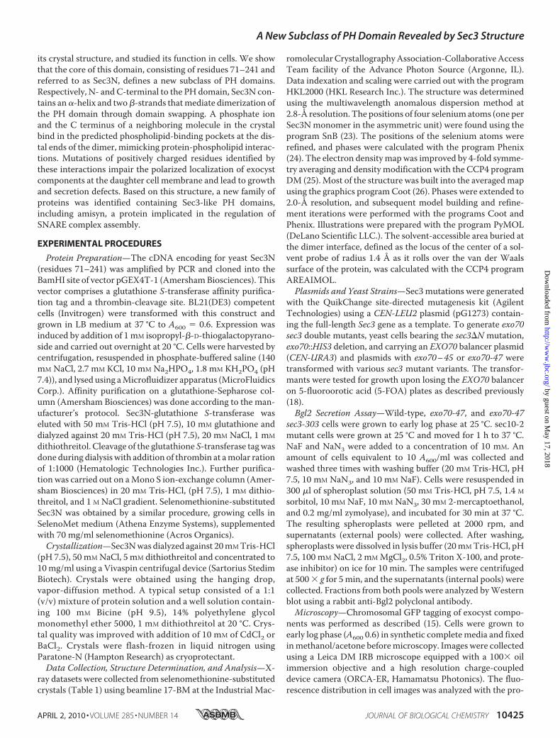
Sec3N Folds into a PH Domain—We had previously mappedthe GTPase and membrane-binding activities of Sec3 to anN-terminal fragment comprising residues 1–320 (18, 19).Sequence analysis suggested that Sec3-(1–320) contained N-and C-terminal disordered, non-conserved regions. The frag-ment 71–250 appeared to form an independent globulardomain, conserved across species and lacking the predicted dis-ordered regions. Our genetic analysis also indicated that dele-tion of the first 70 amino acids of Sec3 did not affect its functionin cells (supplemental Fig. S1). Attempts to crystallize Sec3-(71–250) yielded needle-like crystals of poor quality that couldnot be improved. Analysis of protein samples from old crystal-lization setups showed partial protein degradation, a conditionknown to affect crystallization. A stable fragment of slightlysmaller mass (residues 71–241, referred to here as Sec3N) wasidentified using a combination of mass spectrometry andN-terminal sequencing. This fragment was subcloned andyielded excellent quality crystals, diffracting the x-rays to�2.0-Å resolution (Table 1 and “Experimental Procedures”).Importantly, Sec3N binds Cdc42 (supplemental Fig. S2) and, asshown below, accounts for the plasma membrane-bindingactivity of Sec3.Sec3Nhad no detectable similarity to any known structure in
the Protein Data Bank. Therefore, the structure was deter-mined ab initio bymultiple anomalous dispersion from crystalsof selenomethionine-substituted protein. Sec3N has a singlemethionine residue in 171 amino acids, resulting in a weakanomalous signal. However, the crystal contained four Sec3Nmolecules in the asymmetric unit, and the implementation of4-fold non-crystallographic symmetry averaging improved sig-nificantly the quality of the electron densitymap. The structurewas built and refined to 2.0-Å resolution (Table 1).During model building, the core domain of the Sec3N mon-
omer was recognized as a PH domain. The PH domain belongs
to a structural superfamily that includes the phosphotyrosine-binding, Ena/VASP homology, and Ran-binding domains (27).These domains lack sequence similarity, but all share a corestructure of 100–120 amino acids consisting of a seven-stranded, semi-open, antiparallel �-barrel (strands �1 to �7),capped at one end by a C-terminal �-helix (�1). At the otherend of the barrel three inter-strand variable loops (VL1, VL2,andVL3) form the canonical phosphoinositide-binding pocket.Sec3N shares this core structure, although it lacks statisticallysignificant sequence identity with classic PH domains. Forinstance, the PH domain of Sec3 and that of the prototypicalPH-domain-containing protein phospholipase C� (28) (PDBcode: 1mai) superimpose with r.m.s.d. of 2.15 Å for 87 equiva-lent C� positions, whereas the two proteins share only 7%sequence identity (Fig. 1A). The PH fold appears to be con-served among Sec3 sequences from different species(supplemental Fig. S3), including human Sec3, which shares16.4% sequence identity (37.4% sequence similarity) with yeastSec3 in this region (Fig. 1B). Importantly, amino acids predictedto be involved in phospholipid binding are well conserved (dis-cussed below).In addition to Sec3-related sequences from different species,
a BLAST search identified proteins previously unsuspected tocontain PH domains (examples are given in Fig. 1 andsupplemental Fig. S3). Most of these proteins are still unchar-acterized. However, an interesting example is the protein ami-syn (also known as syntaxin-binding protein 6 or STXBP6), a210-amino acid polypeptide implicated in the regulation ofSNARE complex assembly and exocytosis through its C-termi-nal coiled-coil, a vesicle-SNARE-homology domain (29, 30).Although its characterization is still limited, amisyn has beenproposed to form part of a vesicle-docking complex and isknown to partially co-sediment withmembranes (30). Based onits relationship with Sec3N (Fig. 1B), we now predict that theassociation of amisyn with cellular membranes results from thepresence of an N-terminal PH domain (residues 1–153).Dimerization of Sec3N—The Sec3N fragment crystallized
here is 50–70 amino acids longer than the canonical PHdomain, and the structure includes an additional �-helix at the
TABLE 1Crystallographic data, phasing, and refinement statisticsValues in parentheses correspond to the highest resolution shell.
Native Selenium peak Selenium inflection Selenium remote
Wavelength, Å 1.0 0.9796 0.9798 0.9740Space group P1 P1Unit cell a/b/c, Å 40.7/68.5/68.0 40.6/67.2/67.6Unit cell �/�/�, ° 89.9/82.2/71.8 85.5/82.0/73.5Resolution, Å 2.00-50 (2.00-2.07) 2.85-50 (2.85-2.95)Completeness, % 93.6 (62.7) 96.3 (77.9) 95.8 (73.5) 95.9 (74.5)Multiplicity 10.1 (3.8) 2.0 (1.9) 2.0 (1.9) 2.0 (1.9)Rmerge,a % 6.9 (52.0) 3.4 (11.0) 3.4 (11.7) 3.5 (14.0)I/� 24.6 (1.5) 24.0 (6.2) 23.8 (6.0) 22.5 (4.9)Rfactor,b % 20.2Rfree,c % 24.8Root mean square bonds, Å 0.025Root mean square angles, ° 1.251B-factor protein, Å2 60.25B-factor solvent, Å2 58.46PDB code 3HIE
aRmerge � �hkl(I � �I�)/�I, where I and �I� are the observed and mean intensities of all observations of reflection hkl, including its symmetry-related equivalents.bRfactor � �hkl�Fobs� � �Fcalc�/��Fobs�, where Fobs and Fcalc are the observed and calculated structure factors of reflection hkl.c Rfree and Rfactor were calculated for a randomly selected subset of the reflections (5%) that were omitted during refinement.
A New Subclass of PH Domain Revealed by Sec3 Structure
10426 JOURNAL OF BIOLOGICAL CHEMISTRY VOLUME 285 • NUMBER 14 • APRIL 2, 2010
by guest on May 17, 2018
http://ww
w.jbc.org/
Dow
nloaded from
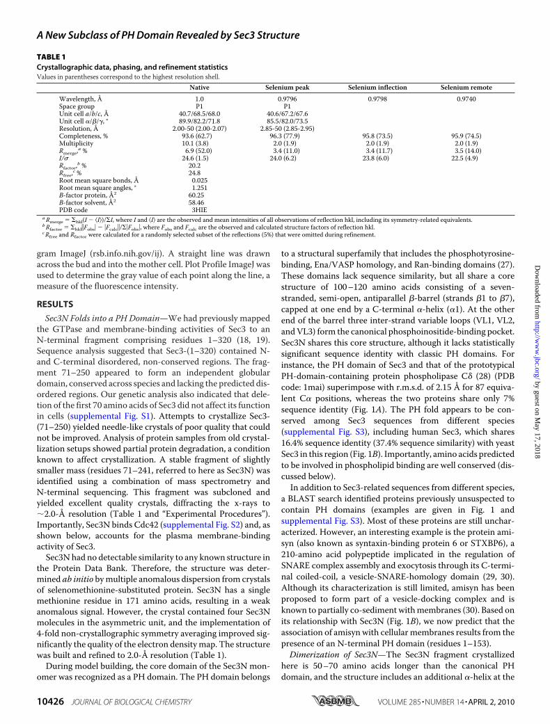
N terminus (�0) and two �-strands at the C terminus (�8 and�9). To our surprise, the four Sec3N molecules in the asym-metric unit of the crystal are organized into two independentantiparallel dimers (Fig. 2). Dimerization involves domainswapping of strands �8 and �9 of each monomer, whichoverlap in “crossed arms” arrangement (supplementalmovie S1). The dimer interface is extensive, burying 4074 Å2
(2037 Å2 per monomer) of solvent-accessible surface area(“Experimental Procedures”). This value is much larger thanthe average crystal packing contact (570 Å2) or protein-pro-tein complex interface (1910 Å2), and even greater than theaverage homodimer interface (3880 Å2) (31–33). The dimer-ization interface is hydrophobic in character and involves
residues from strands �8 and �9 and helix �1 (Tyr-217, Ile-218, Phe-221, Val-229, Trp-231, Phe-236, and Leu-238).Additional stabilization of the dimer interface results fromincorporation of strands �8 and �9 from two differentmonomers into extended antiparallel �-sheets with strands�5, �6, and �7 of each of the PH domains (Fig. 2).The dimerization of several PH domains has been observed
in crystal structures, but never confirmed in solution (34–38).Similarly, analysis by three differentmethods, including analyt-ical size-exclusion chromatography, analytical ultracentrifuga-tion, and multiangle light scattering (supplemental Fig. S4),suggests that Sec3N is amonomer in solution, at lower concen-tration. However, we cannot totally exclude the possibility that
FIGURE 1. Sec3N folds into a PH domain. A, two perpendicular views of a superimposition of the structure of the Sec3N monomer with that of the PH domainof phospholipase C� (28) (PDB code: 1MAI). Phospholipase C� (PLCD1) is colored blue and Sec3N is colored green (helices), yellow (�-strands in front), red(�-strands in back), and cyan (variable loops 1–3). Compared with PLCD1, Sec3N contains an additional �-helix at the N terminus (�0) and two �-strands at theC terminus (magenta). The two structures superimpose with an r.m.s.d. of 2.15 Å for 87 equivalent C� positions. B, sequence alignment of yeast Sec3N with theequivalent region of human Sec3, and the Sec3N-related proteins amisyn and maize roothairless (or rth1). PLCD1 was also aligned based on a structuresuperimposition. The percentage identity and similarity are indicated in the bottom right corner. Secondary structure assignment is shown above the alignmentand colored according to A. Residues predicted to be important for phosphoinositide binding are highlighted (red contour). Uniprot accession codes:SEC3_YEAST (P33332), SEC3_HUMAN (Q9NV70), AMISYN_HUMAN (Q8NFX7), rth1_MAIZE (Q5YLM3), and PLCD1_RAT (P10688).
A New Subclass of PH Domain Revealed by Sec3 Structure
APRIL 2, 2010 • VOLUME 285 • NUMBER 14 JOURNAL OF BIOLOGICAL CHEMISTRY 10427
by guest on May 17, 2018
http://ww
w.jbc.org/
Dow
nloaded from
the region C-terminal to the PH domain contributes to dimer-ization. Indeed, the fact that there are twonearly identical (crys-tallographically independent) dimers in the asymmetric unit(r.m.s.d. of 0.85 Å between equivalent C� atoms) suggests thatdimerization is not a serendipitous crystallization artifact.Moreover, C-terminal to the PH domain Sec3 presents a regionof predicted coiled-coil structure, which is also conserved in thehuman sequence (39). Finally, recruitment at the plasmamem-brane may increase the local concentration of Sec3 and facili-tate dimerization, a possibility that has been proposed for otherPH domains (34–36).Lipid-binding Pocket—By analogy with other PH domains,
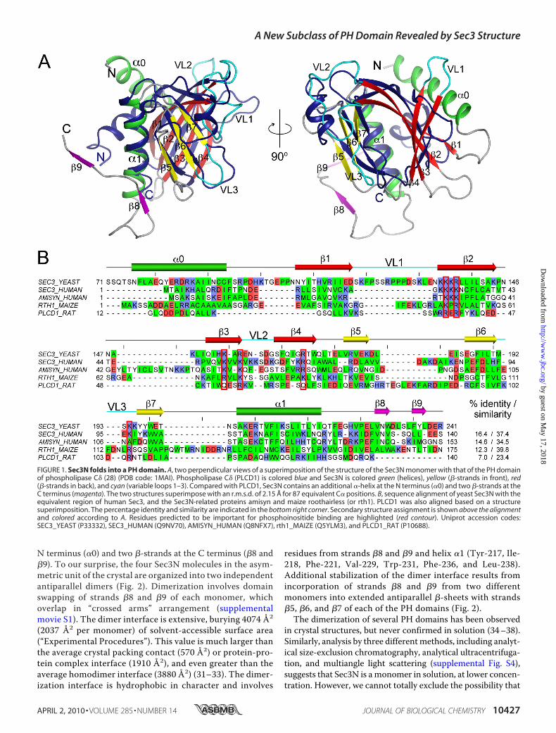
the predicted phospholipid-binding site in Sec3 consists of alarge, positively charged pocket, surrounded by VL1–3 (Fig.3A). Two such pockets are symmetrically disposed at the distalends of the Sec3N dimer (Fig. 2 and supplemental movie S1).
Despite multiple attempts, Sec3Ncould not be co-crystallized withbound phosphoinositides. Further-more, crystals of the unligandedprotein soaked with various phos-phoinositides dissolved immedi-ately, suggesting that phosphoinosi-tides bound Sec3N in the crystals,but their binding was incompatiblewith crystal packing contacts.It has been previously shown that
the locations of inorganic sulfateand phosphate ions in phosphoino-sitide-free structures of PH do-mains, such as those of Grp1 andDapp1, coincide with the locationsof the phosphate groups of phos-phoinositides in the structures ofthe complexes (40). In this regard, itis very informative that one of theSec3N dimers contains a phosphateion bound in each of the phospho-lipid-binding pockets at the distalends of the dimer (Fig. 2). The phos-phates, which are distinguishablefrom solvent atoms at 2.0-Å resolu-tion, present B-factors of �40 Å2,comparable to that of the neighbor-ing residues, and were probablyincorporated during purification, asphosphate was present in the purifi-cation buffer. What is more, the Cterminus and the carboxylic groupof Glu-240 from a neighboringmolecule in the crystal also bind inthe phospholipid-binding pocketnear the phosphate ion (Fig. 3B).Although only one of the Sec3Ndimers shows this arrangement,nearly identical interactions occurat the two distal ends of this dimer.Because the molecules of the
dimer are crystallographically independent, this corre-sponds to two independent observations of the same set ofinteractions. The presence of these crystal contacts probablyexplains why the crystals could not withstand phosphoino-sitide soaking. More importantly, these contacts probablymimic protein-phosphoinositide interactions. We thusattempted to model inositol 1,4,5-trisphosphate in thephospholipid-binding pocket, so as to position the threephosphate groups of the phosphoinositide on top of thephosphate ion, the C terminus and the carboxylic group ofGlu-240 from the neighboring molecule (Fig. 3C). Althoughthe resulting model is unlikely to reflect the actual bindingorientation or phosphoinositide specificity of the Sec3Npocket, it points to the residues most likely to be involved inprotein-phosphoinositide interactions (Arg-137, Lys-155,Arg-157, Arg-168, Lys-194, and possibly Lys-135).
FIGURE 2. Sec3N forms an antiparallel dimer of PH domains. The figure shows a ribbon diagram and surfacerepresentations of the Sec3 dimer. One of the molecules of the dimer is labeled and colored according to Fig. 1,while the other is colored blue. The phosphate ions that bind in the predicted phosphoinositide-bindingpockets at the distal ends of the dimer are also shown. Note that dimerization results from domain swapping ofstrands �8 and �9. An enlarged view shows some of the hydrophobic amino acids involved in interactions at thedimer interface.
FIGURE 3. Phosphoinositide-binding pocket. A, electrostatic surface representation of Sec3N near the pre-dicted phosphoinositide-binding pocket surrounded by VL1–3. Note that the pocket is positively charged (bluecolor) due to the presence of various basic amino acids. B, electron density map (2Fo � Fc, contoured at 1.5�) inthe area corresponding to the yellow square in A. The C terminus of a symmetry-related molecule binds in thephosphoinositide-binding pocket (green backbone and red electron density map). A phosphate ion, the Cterminus, and the carboxylic group of Glu-240 from the symmetry-related molecule are likely to mimic protein-phosphoinositide interactions. C, using these three negative charges as reference, inositol 1,4,5-trisphosphatecan be modeled in the pocket. Although it is unlikely that phosphoinositides bind precisely in this manner, thismodel identifies some of the amino acids most likely to participate in protein-phosphoinositide interactions.
A New Subclass of PH Domain Revealed by Sec3 Structure
10428 JOURNAL OF BIOLOGICAL CHEMISTRY VOLUME 285 • NUMBER 14 • APRIL 2, 2010
by guest on May 17, 2018
http://ww
w.jbc.org/
Dow
nloaded from
Structural studies have identified the determinants of PHdomain-phosphoinositide interaction and specificity (28, 35,40–43). Classic PH domains present a basic signature motif,KXm(R/K)XRXn(Y/N),with the first lysine residue locatednearthe C terminus of strand �1, the (R/K)XR sequence near the Nterminus of strand �2, and a tyrosine (or asparagine) residue instrand�3. The interactions of thismotif are thought tomediateindiscriminate phosphoinositide binding but not specificity.Specificity appears to depend on additional interactions withnon-conserved residues in the VLs, which for this reason arealso known as the specificity determining regions.Sec3 shares some of these features with classic PH domains
but also displays important differences, defining a new subclassof PH domains. Thus, the position corresponding to the lysineresidue in strand�1 is occupied by glutamic acid in Sec3, and inall themembers of its PH domain subclass, except human Sec3,amisyn, and RTH1 (Fig. 1 and supplemental Fig. S3). This sub-stitution of a deeply buried residue of the signature sequence islikely to affect the orientation of the bound phosphoinositide.Note that the overall orientation of bound phosphoinositidesvaries significantly among PH domains (40). A compensatorymutation occurs in Sec3, and all the members of its PH domainsubclass, which present a lysine residue in strand�3 (Lys-155 inSec3), corresponding to the location of the canonical tyrosine(or asparagine) residue of classic PHdomains. The amino groupof lysine 155 in Sec3 (Fig. 3) occupies a position very similar tothat of the amino group of the conserved lysine residue instrand �1 of classic PH domains. Despite these important dif-ferences Sec3 shares with classic PH domains the (R/K)XRsequence in strand �2 (Lys-135, Lys-136, and Arg-137 in Sec3).This sequence corresponds to the basic patch (Lys-134 to Arg-137) previously identified by us as a critical determinant of Sec3membrane binding and localization (18). In addition, otheramino acids are likely to participate in lipid binding in Sec3 andcould play a role in determining phosphoinositide specificity,including Arg-157, Arg-168, and Lys-194. These positions arehighly conserved within the Sec3 PH domain subclass, but notamong classic PH domains. It thus appears that the Sec3 sub-class of PH domains has highly conserved basic amino acids,some ofwhich are also present in classic PHdomains, which aresuitably positioned in the structure to allow for the specificbinding of phosphoinositides. A more detailed knowledge ofthe specific lipid interactions will require the determination ofphosphoinositide-bound structures of this subclass of PHdomains. However, the information obtained here was invalu-able in designing mutagenesis experiments to address the roleof lipid binding in vivo.Synthetic Growth Defects of sec3 Mutants with exo70-47—
The exocyst subunits Exo70 and Sec3 display some functionalredundancy, asmutants of either protein are individually viable,but lethal when combined (18, 44, 45). For this reason, mutantsec3�N (deletion of residues 1–320) lacks any detectable exo-cytosis defect but leads to severe exocytosis and growth defects,and even lethality, when combined with exo70 mutations.Therefore, to understand the functions of positively chargedresidues identified by the crystal structure to form part of thephosphoinositide-binding pocket, we studied sec3 mutants inan exo70mutant background. Among the exo70mutations pre-
viously studied by us, the point mutation Arg-595 3 Ala inallele exo70-47 disrupts phosphoinositide binding of Exo70 andcauses a synthetic growth defect with sec3�N (44). Therefore,we generated various combinations of mutants of Sec3 target-ing residues Lys-134, Lys-135, Lys-136, Arg-137, Lys-155, Arg-157, and Arg-168 in an exo70-47mutant background (Table 2).
Comparedwith the singlemutant exo70-47, the doublemutantexo70-47 sec3-303, containing Sec3 mutations Lys-135 3Glu and Arg-137 3 Glu, showed reduced growth on 5-FOAplates in which the wild-type exo70 balancer was removed (Fig.4A). Moreover, single mutations of Sec3 residues Lys-135 orArg-137 (alleles sec3-301 and sec3-300, respectively) also led togrowth defects in the exo70-47 mutant background. In con-trast, the double mutant exo70-47 sec3-302, containing Sec3mutations Lys-1343 Glu and Arg-1363 Glu displayed nor-mal growth. This is all consistent with the crystal structure inwhich the side chains of residues Lys-135 and Arg-137 are ori-ented toward the phosphoinositide-binding pocket (Fig. 3),whereas those of Lys-134 andArg-136 are oriented in the oppo-site direction. Alleles sec3-306 (carrying mutations Lys-1353Glu, Arg-1373Glu, Lys-1553Glu, and Arg-1573Glu) andsec3-307 (mutations Lys-1353Glu, Arg-1373Glu, and Arg-168 3 Glu) both resulted in lethality in the exo70-47 back-ground, which is also consistentwith the structure that predictsan important role for these residues in lipid binding (Fig. 3).Taken together, the results suggest that residues Lys-135,
Arg-137, Lys-155, Arg-157, and Arg-168, which are solvent-exposed and clustered together in a basic pocket in the crystalstructure (Fig. 3), play an important role in Sec3 function inyeast cells, most likely through their involvement in phospho-lipid binding.The Double Mutant exo70-47 sec3-303 Is Defective in Bgl2
Secretion—We next asked whether the double mutantexo70-47 sec3-303 displays exocytosis defects. This mutantwas selected, because it showed growth defects, but was stillviable (Fig. 4A), thus allowing us to perform secretion assaysand fluorescence microscopy analyses. We examined thesecretion of the cell wall modification enzyme endo-�1,3-glucanase (Bgl2), a widely used marker of polarized secretionat the daughter cell membrane. Wild-type cells did not accu-mulate Bgl2 (Fig. 4B), whereas cells expressing the singlemutant allele exo70-47 showed moderate accumulation of
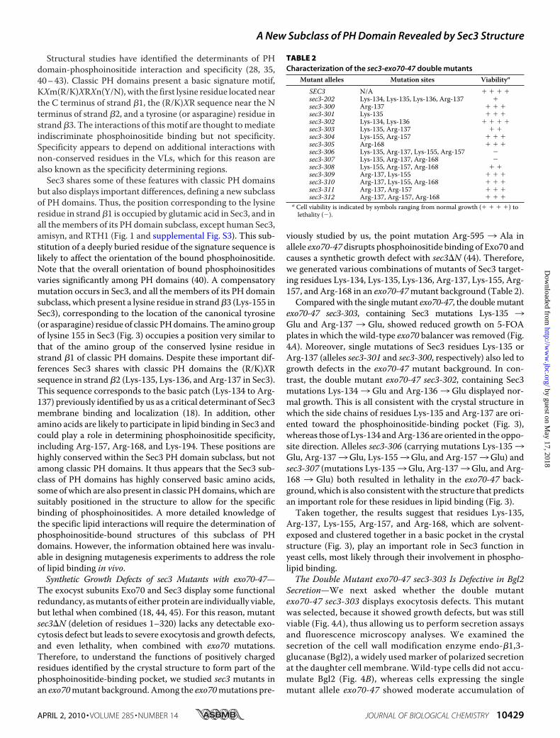
TABLE 2Characterization of the sec3-exo70-47 double mutants
Mutant alleles Mutation sites Viabilitya
SEC3 N/A sec3-202 Lys-134, Lys-135, Lys-136, Arg-137 sec3-300 Arg-137 sec3-301 Lys-135 sec3-302 Lys-134, Lys-136 sec3-303 Lys-135, Arg-137 sec3-304 Lys-155, Arg-157 sec3-305 Arg-168 sec3-306 Lys-135, Arg-137, Lys-155, Arg-157 �sec3-307 Lys-135, Arg-137, Arg-168 �sec3-308 Lys-155, Arg-157, Arg-168 sec3-309 Arg-137, Lys-155 sec3-310 Arg-137, Lys-155, Arg-168 sec3-311 Arg-137, Arg-157 sec3-312 Arg-137, Arg-157, Arg-168
a Cell viability is indicated by symbols ranging from normal growth ( ) tolethality (�).
A New Subclass of PH Domain Revealed by Sec3 Structure
APRIL 2, 2010 • VOLUME 285 • NUMBER 14 JOURNAL OF BIOLOGICAL CHEMISTRY 10429
by guest on May 17, 2018
http://ww
w.jbc.org/
Dow
nloaded from
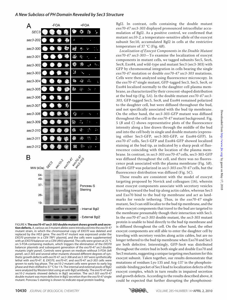
Bgl2. In contrast, cells containing the double mutantexo70-47 sec3-303 displayed pronounced intracellular accu-mulation of Bgl2. As a positive control, we confirmed thatmutant sec10-2, a temperature-sensitive allele of the exocystsubunit Sec10, accumulated Bgl2 in cells at the restrictivetemperature of 37 °C (Fig. 4B).Localization of Exocyst Components in the Double Mutant
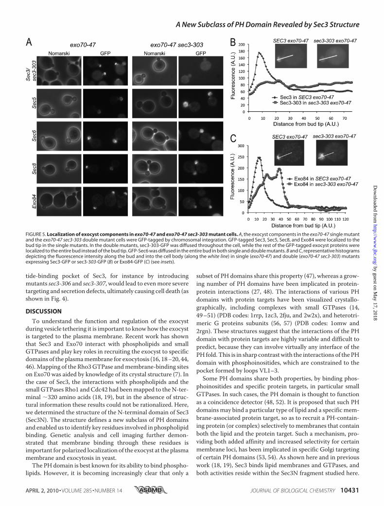
exo70-47 sec3-303—To examine the localization of exocystcomponents in mutant cells, we tagged subunits Sec5, Sec6,Sec8, Exo84, and wild-type and mutant Sec3 (sec3-303) withGFP by chromosomal integration in cells bearing the singleexo70-47 mutation or double exo70-47 sec3-303 mutations.Cells were then analyzed using fluorescence microscopy. Inthe exo70-47 single mutant, GFP-tagged Sec3, Sec5, Sec8, orExo84 localized normally to the daughter cell plasma mem-brane, as characterized by their crescent-shaped distributionat the bud tip (Fig. 5A). In the double mutant exo70-47 sec3-303, GFP-tagged Sec5, Sec8, and Exo84 remained polarizedto the daughter cell, but were diffused throughout the bud,and not specifically associated with the bud tip membrane.On the other hand, the sec3-303-GFP mutant was diffusedthroughout the cell in the exo70-47mutant background. Fig.5 (B and C) shows representative plots of the fluorescenceintensity along a line drawn through the middle of the budand into the cell body in single and double mutants (express-ing either Sec3-GFP, sec3-303-GFP, or Exo84-GFP). Inexo70-47 cells, Sec3-GFP and Exo84-GFP showed localizedstaining at the bud tip, as indicated by a sharp peak of fluo-rescence coinciding with the location of the plasma mem-brane. In contrast, in sec3-303 exo70-47 cells, sec3-303-GFPwas diffused throughout the cell, and there was no fluores-cence peak associated with the plasma membrane (Fig. 5B).Exo84-GFP was polarized in sec3-303 exo70-47 cells, but thefluorescence distribution was diffused (Fig. 5C).These results are consistent with the model of exocyst
targeting proposed by Novick and colleagues (16), whereinmost exocyst components associate with secretory vesiclestraveling toward the bud tip along actin cables, whereas Sec3and Exo70 bind to the bud tip membrane and act as land-marks for vesicle tethering. Thus, in the exo70-47 singlemutant, Sec3 can still localize to the bud tipmembrane, and theother exocyst components that travel to the bud are targeted tothe membrane presumably though their interaction with Sec3.In the exo70-47 sec3-303 double mutant, the sec3-303 mutantprotein is unable to bind directly to the bud tip membrane andis diffused throughout the cell. On the other hand, the otherexocyst components are still able to enter the daughter cell bytraveling with secretory vesicles along actin cables, but are nolonger tethered to the bud tipmembrane when Exo70 and Sec3are both defective. Interestingly, GFP-Sec6 was distributedthroughout the entire bud in both single and double Exo70 andSec3mutants, suggesting a unique targetingmechanism for thisexocyst subunit. Taken together, our results demonstrate thatmutations of residues Lys-135 and Arg-137 in the phosphoin-ositide-binding pocket of Sec3 lead to localization defects of theexocyst complex, which in turn results in impaired secretionand growth defects. According to the results described above, itcould be expected that further disrupting the phosphoinosi-
FIGURE 4. The exo70-47 sec3-303 double mutant shows growth and secre-tion defects. A, various sec3 mutant alleles were introduced into the exo70-47mutant strain, in which the chromosomal copy of EXO70 was deleted andreplaced by the HIS3 gene. The exo70-47 mutant was expressed under theEXO70 promoter in a CEN TRP1 plasmid, and the cells were supplementedwith an EXO70 balancer on a CEN URA3 plasmid. The cells were grown at 25 °Con 5-FOA-containing medium, which triggers the elimination of the EXO70balancer plasmids and subsequent generation of the exo70-47 sec3 doublemutants (right panel). Controls were grown on medium without 5-FOA (leftpanel). sec3-303 and several other mutants showed different degrees of syn-thetic growth defects with exo70-47. sec3-306 and sec3-307 were syntheticallylethal with exo70-47. B, EXO70, exo70-47, and exo70-47 sec3-303 cells weregrown to early log phase. The sec10-2 mutant cells were grown to early logphase and then shifted to 37 °C for 1 h. The internal and external pools of Bgl2were analyzed by Western blot using an anti-Bgl2 antibody. The exo70-47 andsec10-2 mutants showed defects in Bgl2 secretion. The sec3-303 exo70-47double mutant was more defective in Bgl2 secretion than the exo70-47 singlemutant. Ponceau S staining is shown to indicate equal protein loading.
A New Subclass of PH Domain Revealed by Sec3 Structure
10430 JOURNAL OF BIOLOGICAL CHEMISTRY VOLUME 285 • NUMBER 14 • APRIL 2, 2010
by guest on May 17, 2018
http://ww
w.jbc.org/
Dow
nloaded from
tide-binding pocket of Sec3, for instance by introducingmutants sec3-306 and sec3-307, would lead to evenmore severetargeting and secretion defects, ultimately causing cell death (asshown in Fig. 4).
DISCUSSION
To understand the function and regulation of the exocystduring vesicle tethering it is important to knowhow the exocystis targeted to the plasma membrane. Recent work has shownthat Sec3 and Exo70 interact with phospholipids and smallGTPases and play key roles in recruiting the exocyst to specificdomains of the plasmamembrane for exocytosis (16, 18–20, 44,46).Mapping of the Rho3GTPase andmembrane-binding siteson Exo70 was aided by knowledge of its crystal structure (7). Inthe case of Sec3, the interactions with phospholipids and thesmall GTPases Rho1 andCdc42 had beenmapped to theN-ter-minal �320 amino acids (18, 19), but in the absence of struc-tural information these results could not be rationalized. Here,we determined the structure of the N-terminal domain of Sec3(Sec3N). The structure defines a new subclass of PH domainsand enabled us to identify key residues involved in phospholipidbinding. Genetic analysis and cell imaging further demon-strated that membrane binding through these residues isimportant for polarized localization of the exocyst at the plasmamembrane and exocytosis in yeast.The PHdomain is best known for its ability to bind phospho-
lipids. However, it is becoming increasingly clear that only a
subset of PH domains share this property (47), whereas a grow-ing number of PH domains have been implicated in protein-protein interactions (27, 48). The interactions of various PHdomains with protein targets have been visualized crystallo-graphically, including complexes with small GTPases (14,49–51) (PDB codes: 1rrp, 1zc3, 2fju, and 2w2x), and heterotri-meric G proteins subunits (56, 57) (PDB codes: 1omw and2rgn). These structures suggest that the interactions of the PHdomain with protein targets are highly variable and difficult topredict, because they can involve virtually any interface of thePH fold. This is in sharp contrastwith the interactions of the PHdomain with phosphoinositides, which are constrained to thepocket formed by loops VL1–3.Some PH domains share both properties, by binding phos-
phoinositides and specific protein targets, in particular smallGTPases. In such cases, the PH domain is thought to functionas a coincidence detector (48, 52). It is proposed that such PHdomainsmay bind a particular type of lipid and a specificmem-brane-associated protein target, so as to recruit a PH-contain-ing protein (or complex) selectively to membranes that containboth the lipid and the protein target. Such a mechanism, pro-viding both added affinity and increased selectivity for certainmembrane loci, has been implicated in specific Golgi targetingof certain PH domains (53, 54). As shown here and in previouswork (18, 19), Sec3 binds lipid membranes and GTPases, andboth activities reside within the Sec3N fragment studied here.
FIGURE 5. Localization of exocyst components in exo70-47 and exo70-47 sec3-303 mutant cells. A, the exocyst components in the exo70-47 single mutantand the exo70-47 sec3-303 double mutant cells were GFP-tagged by chromosomal integration. GFP-tagged Sec3, Sec5, Sec8, and Exo84 were localized to thebud tip in the single mutants. In the double mutants, sec3-303-GFP was diffused throughout the cell, while the rest of the GFP-tagged exocyst proteins werelocalized to the entire bud instead of the bud tip. GFP-Sec6 was diffused in the entire bud in both single and double mutants. B and C, representative histogramsdepicting the fluorescence intensity along the bud and into the cell body (along the white line) in single (exo70-47) and double (exo70-47 sec3-303) mutantsexpressing Sec3-GFP or sec3-303-GFP (B) or Exo84-GFP (C) (see insets).
A New Subclass of PH Domain Revealed by Sec3 Structure
APRIL 2, 2010 • VOLUME 285 • NUMBER 14 JOURNAL OF BIOLOGICAL CHEMISTRY 10431
by guest on May 17, 2018
http://ww
w.jbc.org/
Dow
nloaded from

We have now shown that a PH domain mediates both theseinteractions. Thus, Sec3, and probably other members of itsstructural subclass, join the growing number of PH domain-containing proteins that function as coincidence detectors atthe membrane.Yeast Sec3 is a large protein of 1336 amino acids. The region
C-terminal to the PH domain is predicted to be predominantlyhelical in structure. Residues 320–470 within this region arepredicted with high probability to form a coiled-coil structure.Fold recognition programs further suggest that the 727-aminoacid region starting from residue 610 to the C terminus of theprotein consists of a series of long helices separated by shortloops. This is the signature pattern of helical bundles, charac-teristic of other subunits of the exocyst (4, 10, 39). Indeed, thecrystal structures of four other subunits of the exocyst show arelated fold consisting of consecutively stacked helical bundles(7–11). Each bundle typically contains three helices. The thirdhelix of each bundle is usually longer and contributes its C-ter-minal half to the next bundle, helping to stabilize bundle-bun-dle interactions, a fold somewhat related to that of the spectrinrepeat (55). The human Sec3 sequence is relatively shorter (894amino acids) and displays low overall sequence identity (�12%)with yeast Sec3 but appears to share a similar domain organi-zation, including an N-terminal PH domain (residues 1–140),followed by a predicted coiled-coil domain (residues 155–255)and a series of C-terminal helical bundles (residues 258–894).As we have shown here, residues implicated in phospholipidbinding in yeast Sec3N are also well conserved in the humansequence, as well as in other eukaryotes. This fact was previ-ously underappreciated, as the N terminus of Sec3 shows rela-tively low sequence similarity across species. Our preliminaryresults indicate that the N terminus of human Sec3 also bindsphospholipids8; further cell biological studies are being carriedout to investigate the function of this domain in mammaliancells.The structure of Sec3N also identifies a number of proteins
that carries this new subclass of PH domains. One of theseproteins is amisyn, implicated in binding to the t-SNARE pro-tein syntaxin in neuronal cells and known to partially co-sedi-ment with membranes (30). Future studies will examinewhether the PH domain of amisyn binds phospholipids andwhether this interaction is implicated in the recruitment ofamisyn to the plasma membrane and regulation of SNAREfunction during vesicle docking and fusion.
REFERENCES1. Novick, P., Field, C., and Schekman, R. (1980) Cell 21, 205–2152. TerBush, D. R., and Novick, P. (1995) J. Cell Biol. 130, 299–3123. Hsu, S. C., Ting, A. E., Hazuka, C. D., Davanger, S., Kenny, J. W., Kee, Y.,
and Scheller, R. H. (1996) Neuron 17, 1209–12194. Munson, M., and Novick, P. (2006) Nat. Struct. Mol. Biol. 13, 577–5815. He, B., and Guo, W. (2009) Curr. Opin. Cell Biol. 21, 537–5426. Hsu, S. C., TerBush, D., Abraham, M., and Guo, W. (2004) Int. Rev. Cytol.
233, 243–2657. Dong, G., Hutagalung, A. H., Fu, C., Novick, P., and Reinisch, K.M. (2005)
Nat. Struct. Mol. Biol. 12, 1094–11008. Hamburger, Z. A., Hamburger, A. E., West, A. P., Jr., and Weis, W. I.
(2006) J. Mol. Biol. 356, 9–219. Moore, B. A., Robinson, H. H., and Xu, Z. (2007) J. Mol. Biol. 371,
410–42110. Sivaram, M. V., Furgason, M. L., Brewer, D. N., and Munson, M. (2006)
Nat. Struct. Mol. Biol. 13, 555–55611. Wu, S., Mehta, S. Q., Pichaud, F., Bellen, H. J., and Quiocho, F. A. (2005)
Nat. Struct. Mol. Biol. 12, 879–88512. Tripathi, A., Ren, Y., Jeffrey, P. D., and Hughson, F. M. (2009)Nat. Struct.
Mol. Biol. 16, 114–12313. Fukai, S., Matern, H. T., Jagath, J. R., Scheller, R. H., and Brunger, A. T.
(2003) EMBO J. 22, 3267–327814. Jin, R., Junutula, J. R., Matern, H. T., Ervin, K. E., Scheller, R. H., and
Brunger, A. T. (2005) EMBO J. 24, 2064–207415. Finger, F. P., Hughes, T. E., and Novick, P. (1998) Cell 92, 559–57116. Boyd, C., Hughes, T., Pypaert, M., and Novick, P. (2004) J. Cell Biol. 167,
889–90117. Zhang, X., Zajac, A., Zhang, J., Wang, P., Li, M., Murray, J., TerBush, D.,
and Guo, W. (2005) J. Biol. Chem. 280, 20356–2036418. Zhang, X., Orlando, K., He, B., Xi, F., Zhang, J., Zajac, A., and Guo, W.
(2008) J. Cell Biol. 180, 145–15819. Guo, W., Tamanoi, F., and Novick, P. (2001) Nat. Cell Biol. 3, 353–36020. Zhang, X., Bi, E., Novick, P., Du, L., Kozminski, K. G., Lipschutz, J. H., and
Guo, W. (2001) J. Biol. Chem. 276, 46745–4675021. Adamo, J. E., Rossi, G., and Brennwald, P. (1999) Mol. Biol. Cell 10,
4121–413322. Robinson, N. G., Guo, L., Imai, J., Toh-E., A., Matsui, Y., and Tamanoi, F.
(1999)Mol. Cell. Biol. 19, 3580–358723. Weeks, C. M., and Miller, R. (1999) J. Appl. Crystallogr. 32, 120–12424. Zwart, P.H., Afonine, P. V., Grosse-Kunstleve, R.W.,Hung, L.W., Ioerger,
T. R., McCoy, A. J., McKee, E., Moriarty, N. W., Read, R. J., Sacchettini,J. C., Sauter, N. K., Storoni, L. C., Terwilliger, T. C., and Adams, P. D.(2008)Methods Mol. Biol. 426, 419–435
25. Cowtan, K., and Main, P. (1998) Acta Crystallogr. D Biol. Crystallogr. 54,487–493
26. Emsley, P., and Cowtan, K. (2004)Acta Crystallogr. D Biol. Crystallogr. 60,2126–2132
27. Lemmon, M. A. (2004) Biochem. Soc. Trans. 32, 707–71128. Ferguson, K. M., Lemmon, M. A., Schlessinger, J., and Sigler, P. B. (1995)
Cell 83, 1037–104629. Constable, J. R., Graham, M. E., Morgan, A., and Burgoyne, R. D. (2005)
J. Biol. Chem. 280, 31615–3162330. Scales, S. J., Hesser, B. A., Masuda, E. S., and Scheller, R. H. (2002) J. Biol.
Chem. 277, 28271–2827931. Janin, J., and Rodier, F. (1995) Proteins 23, 580–58732. Lo Conte, L., Chothia, C., and Janin, J. (1999) J. Mol. Biol. 285, 2177–219833. Bahadur, R. P., Chakrabarti, P., Rodier, F., and Janin, J. (2004) J. Mol. Biol.
336, 943–95534. Depetris, R. S.,Wu, J., and Hubbard, S. R. (2009)Nat. Struct. Mol. Biol. 16,
833–83935. Baraldi, E., Djinovic Carugo, K., Hyvonen, M., Surdo, P. L., Riley, A. M.,
Potter, B. V., O’Brien, R., Ladbury, J. E., and Saraste,M. (1999) Structure 7,449–460
36. Murayama, K., Kato-Murayama, M., Mishima, C., Akasaka, R., Shirouzu,M., Fukui, Y., and Yokoyama, S. (2008) Biochem. Biophys. Res. Commun.377, 23–28
37. Jackson, S. G., Zhang, Y., Bao, X., Zhang, K., Summerfield, R., Haslam, R. J.,and Junop, M. S. (2006) Acta Crystallogr. D Biol. Crystallogr. 62, 324–330
38. Ferguson, K. M., Lemmon, M. A., Schlessinger, J., and Sigler, P. B. (1994)Cell 79, 199–209
39. Croteau,N. J., Furgason,M. L., Devos, D., andMunson,M. (2009) PloS one4, e4443
40. Cronin, T. C., DiNitto, J. P., Czech, M. P., and Lambright, D. G. (2004)EMBO J. 23, 3711–3720
41. Lietzke, S. E., Bose, S., Cronin, T., Klarlund, J., Chawla, A., Czech, M. P.,and Lambright, D. G. (2000)Mol. Cell 6, 385–394
42. Ferguson, K. M., Kavran, J. M., Sankaran, V. G., Fournier, E., Isakoff, S. J.,Skolnik, E. Y., and Lemmon, M. A. (2000)Mol. Cell 6, 373–384
43. DiNitto, J. P., Cronin, T. C., and Lambright, D. G. (2003) Sci. STKE 2003,8 K. Baek, A. Knodler, S. H. Lee, X. Zhang, K. Orlando, J. Zhang, T. J. Foskett, W.
Guo, and R. Dominguez, unpublished observation.
A New Subclass of PH Domain Revealed by Sec3 Structure
10432 JOURNAL OF BIOLOGICAL CHEMISTRY VOLUME 285 • NUMBER 14 • APRIL 2, 2010
by guest on May 17, 2018
http://ww
w.jbc.org/
Dow
nloaded from

re1644. He, B., Xi, F., Zhang, X., Zhang, J., and Guo, W. (2007) EMBO J. 26,
4053–406545. Hutagalung, A. H., Coleman, J., Pypaert, M., and Novick, P. J. (2009)Mol.
Biol. Cell 20, 153–16346. Liu, J., Zuo, X., Yue, P., and Guo,W. (2007)Mol. Biol. Cell 18, 4483–449247. Yu, J. W., Mendrola, J. M., Audhya, A., Singh, S., Keleti, D., DeWald, D. B.,
Murray, D., Emr, S. D., and Lemmon, M. A. (2004)Mol. Cell 13, 677–68848. Lemmon, M. A. (2007) Biochem. Soc. Symp. 81–9349. Vetter, I. R., Nowak, C., Nishimoto, T., Kuhlmann, J., andWittinghofer, A.
(1999) Nature 398, 39–4650. Jezyk, M. R., Snyder, J. T., Gershberg, S., Worthylake, D. K., Harden, T. K.,
and Sondek, J. (2006) Nat. Struct. Mol. Biol 13, 1135–114051. Bunney, T. D., Opaleye, O., Roe, S. M., Vatter, P., Baxendale, R. W., Wal-
liser, C., Everett, K. L., Josephs, M. B., Christow, C., Rodrigues-Lima, F.,Gierschik, P., Pearl, L. H., and Katan, M. (2009)Mol. Cell 34, 223–233
52. Lemmon, M. A. (2008) Nat. Rev. Mol. Cell Biol. 9, 99–11153. Godi, A., Di Campli, A., Konstantakopoulos, A., Di Tullio, G., Alessi, D. R.,
Kular, G. S., Daniele, T., Marra, P., Lucocq, J. M., and De Matteis, M. A.(2004) Nat. Cell Biol. 6, 393–404
54. Levine, T. P., and Munro, S. (2002) Curr. Biol. 12, 695–70455. Djinovic-Carugo, K., Gautel, M., Ylanne, J., and Young, P. (2002) FEBS
Lett. 513, 119–12356. Lodowski, D. T., Pitcher, J. A., Capel, W. D., Lefkowitz, R. J., and Tesmer,
J. J. (2003) Science 300, 1256–126257. Lutz, S., Shankaranarayanan, A., Coco, C., Ridilla,M.,Nance,M. R., Vettel,
C., Baltus, D., Evelyn, C. R., Neubig, R. R., Wieland, T., and Tesmer, J. J.(2007) Science 318, 1923–1927
A New Subclass of PH Domain Revealed by Sec3 Structure
APRIL 2, 2010 • VOLUME 285 • NUMBER 14 JOURNAL OF BIOLOGICAL CHEMISTRY 10433
by guest on May 17, 2018
http://ww
w.jbc.org/
Dow
nloaded from
Zhang, Trevor J. Foskett, Wei Guo and Roberto DominguezKyuwon Baek, Andreas Knödler, Sung Haeng Lee, Xiaoyu Zhang, Kelly Orlando, Jian
Structure-Function Study of the N-terminal Domain of Exocyst Subunit Sec3
doi: 10.1074/jbc.M109.096966 originally published online February 5, 20102010, 285:10424-10433.J. Biol. Chem.
10.1074/jbc.M109.096966Access the most updated version of this article at doi:
Alerts:
When a correction for this article is posted•
When this article is cited•
to choose from all of JBC's e-mail alertsClick here
Supplemental material:
http://www.jbc.org/content/suppl/2010/02/05/M109.096966.DC1
http://www.jbc.org/content/285/14/10424.full.html#ref-list-1
This article cites 56 references, 16 of which can be accessed free at
by guest on May 17, 2018
http://ww
w.jbc.org/
Dow
nloaded from