Embed Size (px)
Citation preview
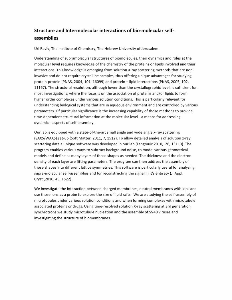
StructureandIntermolecularinteractionsofbio‐molecularself‐assemblies
UriRaviv,TheInstituteofChemistry,TheHebrewUniversityofJerusalem.
Understandingofsupramolecularstructuresofbiomolecules,theirdynamicsandrolesatthe
molecularlevelrequiresknowledgeofthechemistryoftheproteinsorlipidsinvolvedandtheirinteractions.ThisknowledgeisemergingfromsolutionX‐rayscatteringmethodsthatarenon‐invasiveanddonotrequirecrystallinesamples,thusofferinguniqueadvantagesforstudying
protein‐protein(PNAS,2004,101,16099)andprotein–lipidinteractions(PNAS,2005,102,11167).Thestructuralresolution,althoughlowerthanthecrystallographiclevel,issufficientfor
mostinvestigations,wherethefocusisontheassociationofproteinsand/orlipidstoformhigherordercomplexesundervarioussolutionconditions.Thisisparticularlyrelevantforunderstandingbiologicalsystemsthatareinaqueousenvironmentandarecontrolledbyvarious
parameters.Ofparticularsignificanceistheincreasingcapabilityofthosemethodstoprovidetime‐dependentstructuralinformationatthemolecularlevel‐ameansforaddressingdynamicalaspectsofself‐assembly.
Ourlabisequippedwithastate‐of‐the‐artsmallangleandwideanglex‐rayscattering
(SAXS/WAXS)set‐up(SoftMatter,2011,7,1512).Toallowdetailedanalysisofsolutionx‐rayscatteringdataauniquesoftwarewasdevelopedinourlab(Langmuir,2010,26,13110).Theprogramenablesvariouswaystosubtractbackgroundnoise,tomodelvariousgeometrical
modelsanddefineasmanylayersofthoseshapesasneeded.Thethicknessandtheelectrondensityofeachlayerarefittingparameters.Theprogramcanthenaddresstheassemblyofthoseshapesintodifferentlatticesymmetries.Thissoftwareisparticularlyusefulforanalyzing
supra‐molecularself‐assembliesandforreconstructingthesignalinit'sentirety(J.Appl.Cryst.,2010,43,1522).
Weinvestigatetheinteractionbetweenchargedmembranes,neutralmembraneswithionsandusethoseionsasaprobetoexplorethesizeoflipidrafts.Wearestudyingtheself‐assemblyof
microtubulesundervarioussolutionconditionsandwhenformingcomplexeswithmicrotubuleassociatedproteinsordrugs.Usingtime‐resolvedsolutionX‐rayscatteringat3rdgenerationsynchrotronswestudymicrotubulenucleationandtheassemblyofSV40virusesand
investigatingthestructureofbiomembranes.
Structure and Intermolecular interactions of biomolecular
self-assemblies
Uri Raviv
The Institute of Chemistry
May 6 2011
General Aims
• How biological molecules self-assemble and interact with one another in solutions?
• Focus on: Charged and Dipolar Systems

General Approach
• Similar to liquid crystals
• Focus on important length scales and study their variation
• X-ray scattering in solutions (combined with microscopy and other biophysical methods) – Non-invasive – Good statistics – Structures – Domain size – Fluctuations – Intermolecular interactions – Elastic and mechanical properties
Analysis
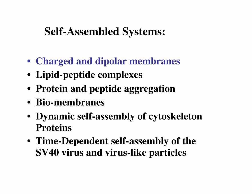
• Charged and dipolar membranes • Lipid-peptide complexes • Protein and peptide aggregation • Bio-membranes • Dynamic self-assembly of cytoskeleton
Proteins • Time-Dependent self-assembly of the
SV40 virus and virus-like particles
Self-Assembled Systems:
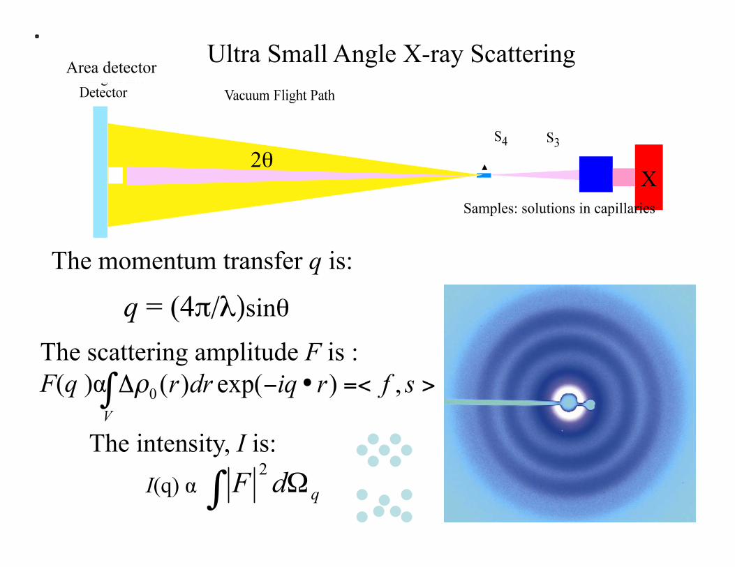
X 2θ
q = (4π/λ)sinθ
Ultra Small Angle X-ray Scattering
Samples: solutions in capillaries
The scattering amplitude F is : F(q )α
I(q) α
The intensity, I is:
The momentum transfer q is:
Area detector
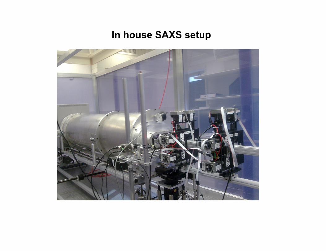
In house SAXS setup
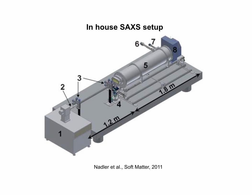
Nadler et al., Soft Matter, 2011
In house SAXS setup
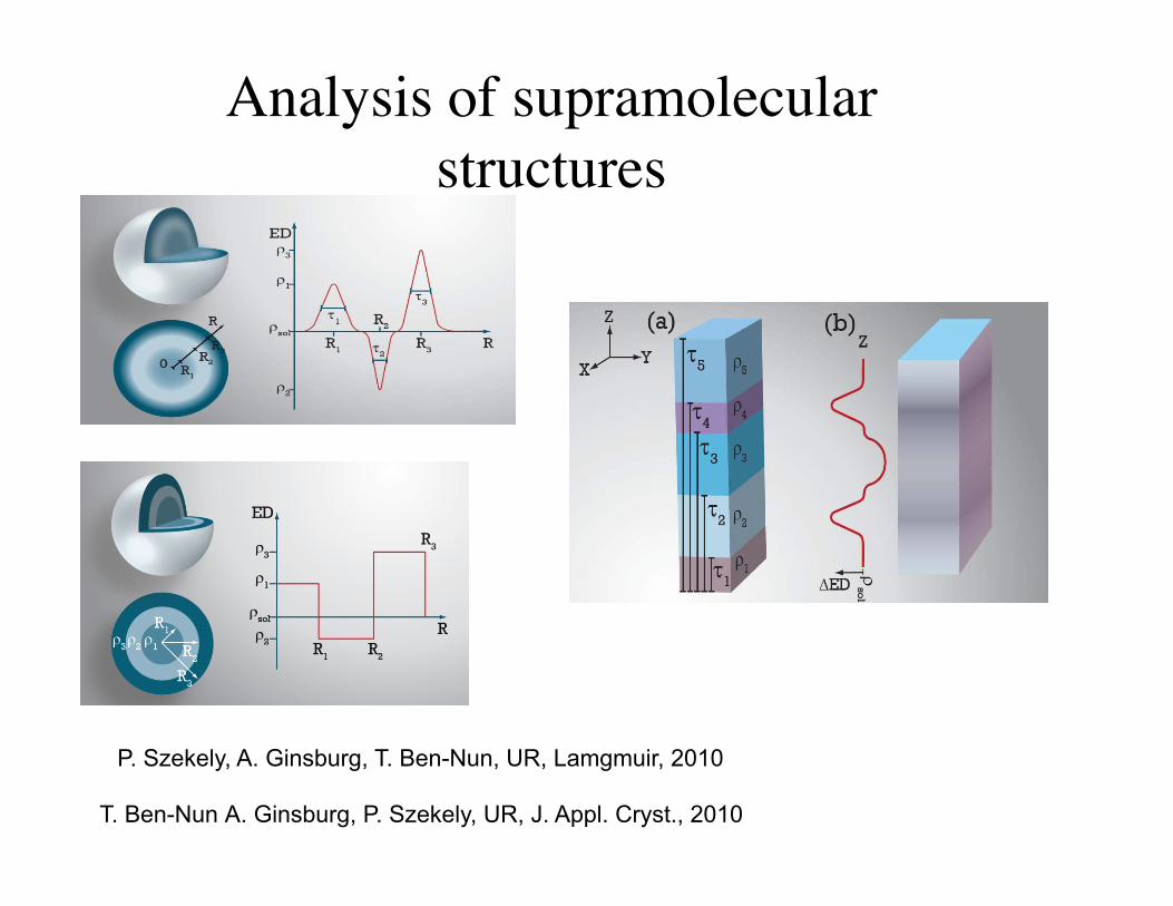
Analysis of supramolecular structures
P. Szekely, A. Ginsburg, T. Ben-Nun, UR, Lamgmuir, 2010
T. Ben-Nun A. Ginsburg, P. Szekely, UR, J. Appl. Cryst., 2010
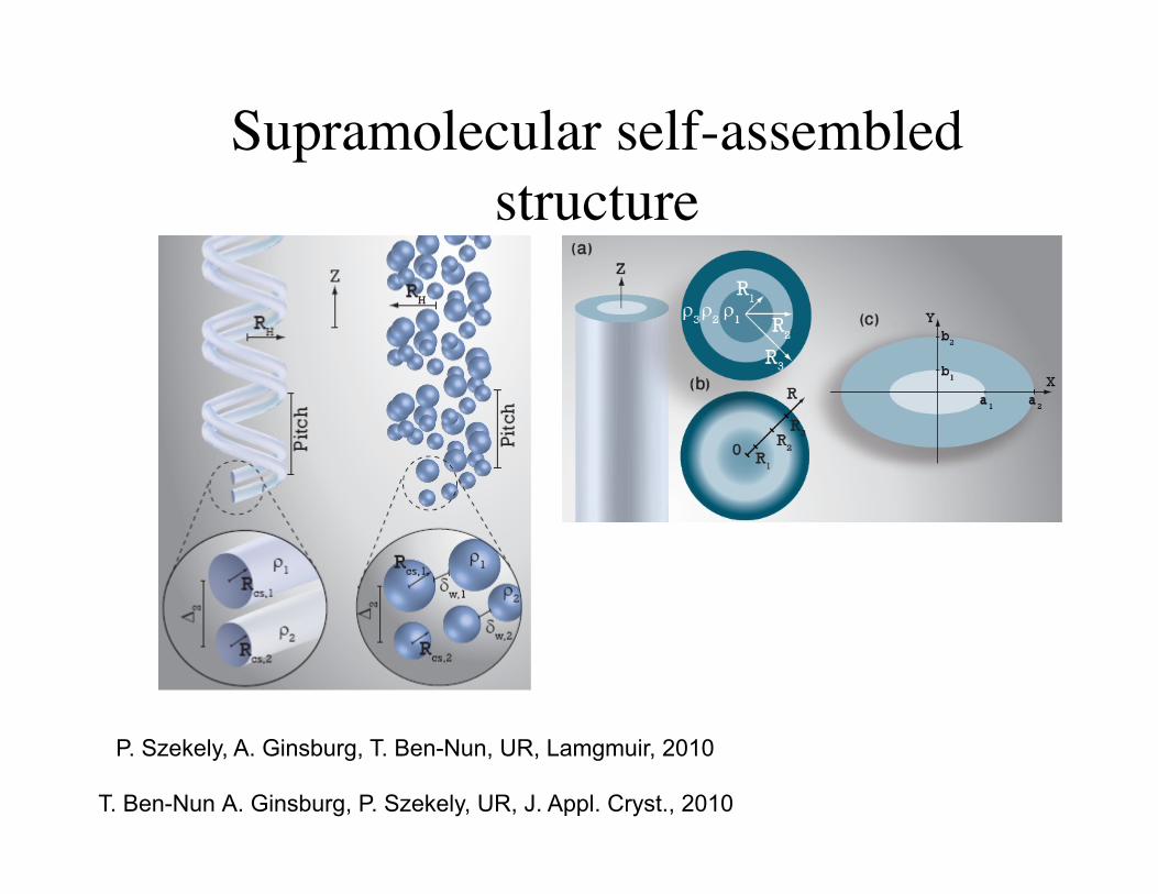
Supramolecular self-assembled structure
P. Szekely, A. Ginsburg, T. Ben-Nun, UR, Lamgmuir, 2010
T. Ben-Nun A. Ginsburg, P. Szekely, UR, J. Appl. Cryst., 2010
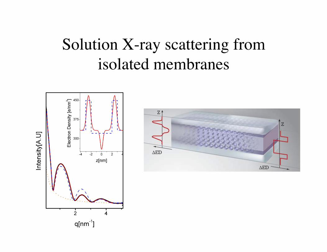
Solution X-ray scattering from isolated membranes
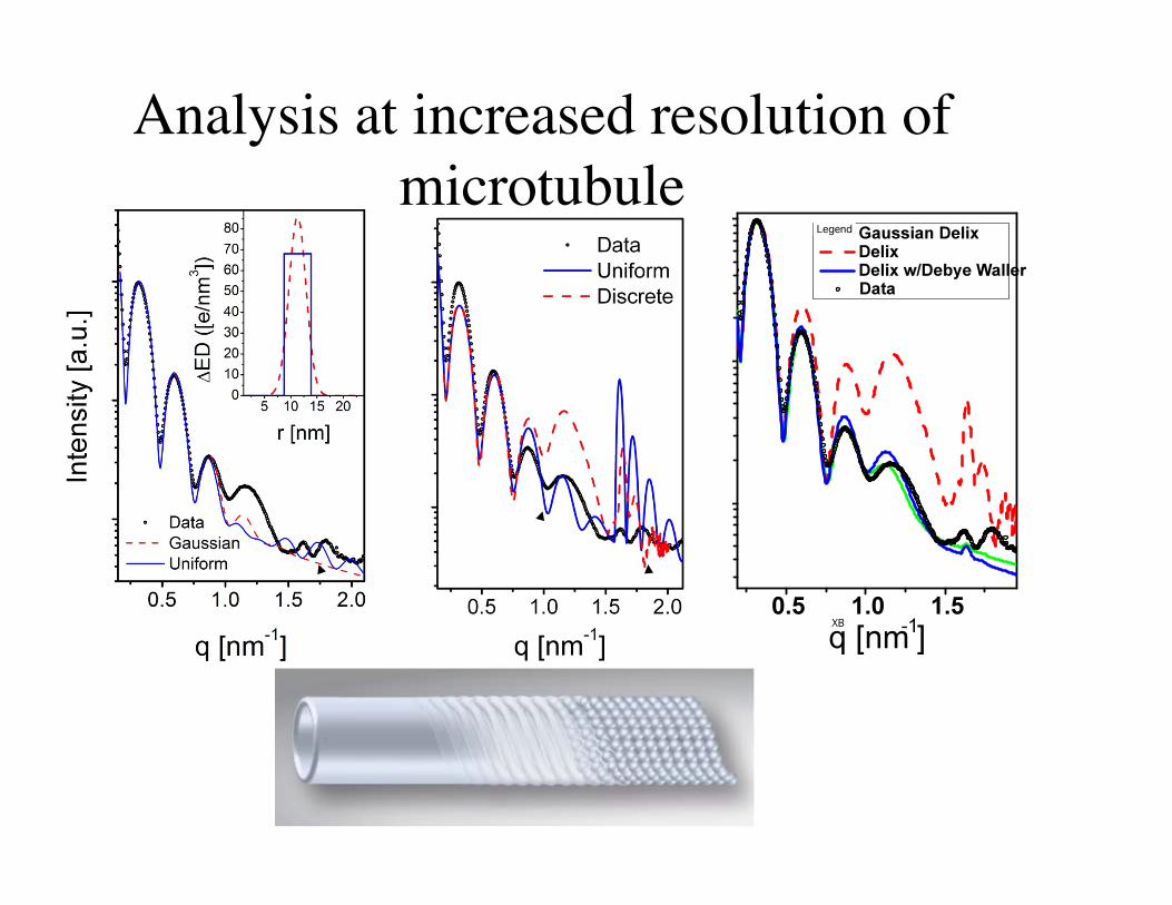
Analysis at increased resolution of microtubule
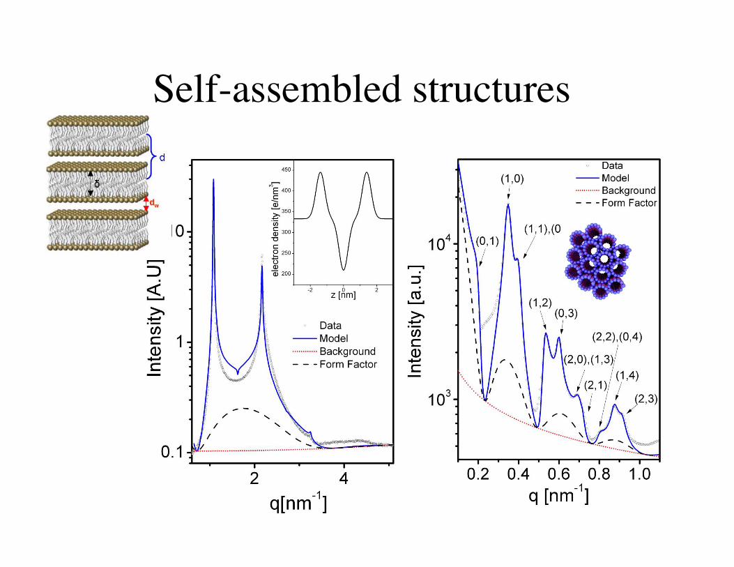
Self-assembled structures
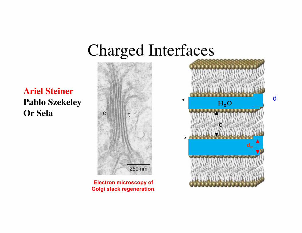
Charged Interfaces
Ariel Steiner Pablo Szekeley Or Sela
dw
δ
d
Electron microscopy of Golgi stack regeneration.
Charged interfaces
• Solid like-charged interfaces, such as clay minerals, swell indefinitely when diluted in water
• Very soft charged interfaces can swell only up to a maximum distance. Further dilution leads to their continuous unbinding, driven by thermal fluctuations
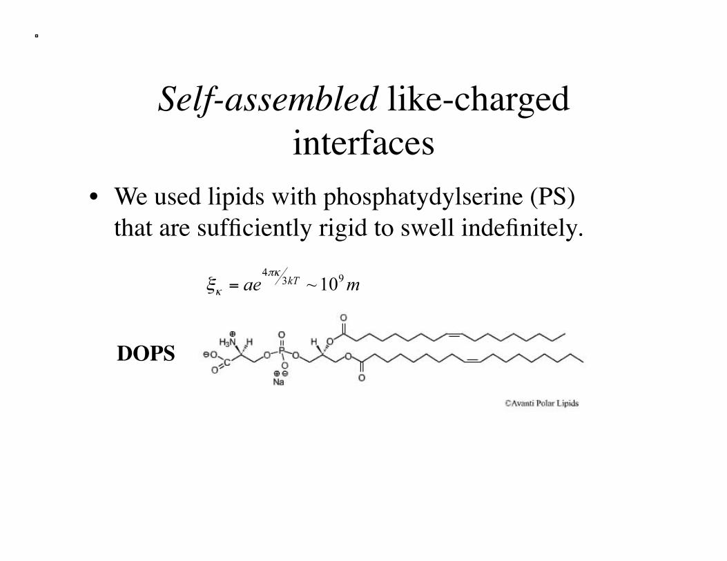
Self-assembled like-charged interfaces
• We used lipids with phosphatydylserine (PS) that are sufficiently rigid to swell indefinitely.
DOPS
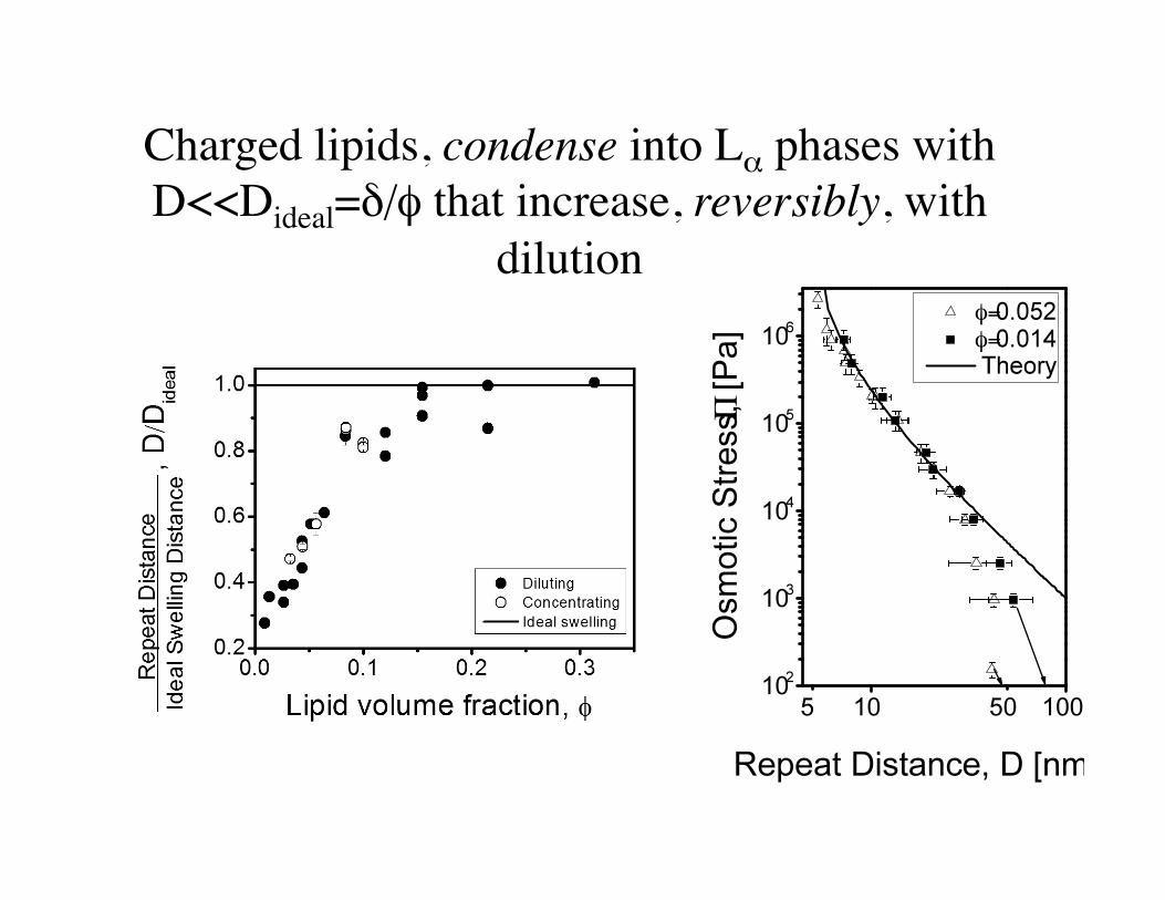
Charged lipids, condense into Lα phases with D<<Dideal=δ/φ that increase, reversibly, with
dilution
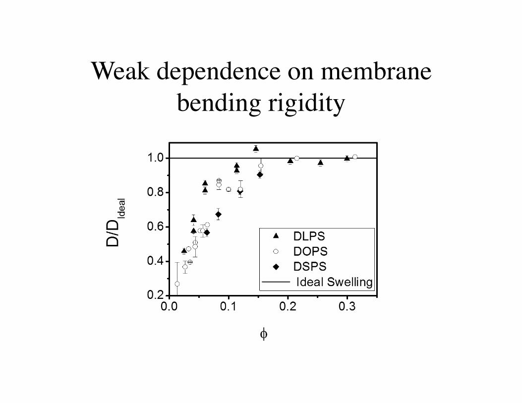
Weak dependence on membrane bending rigidity
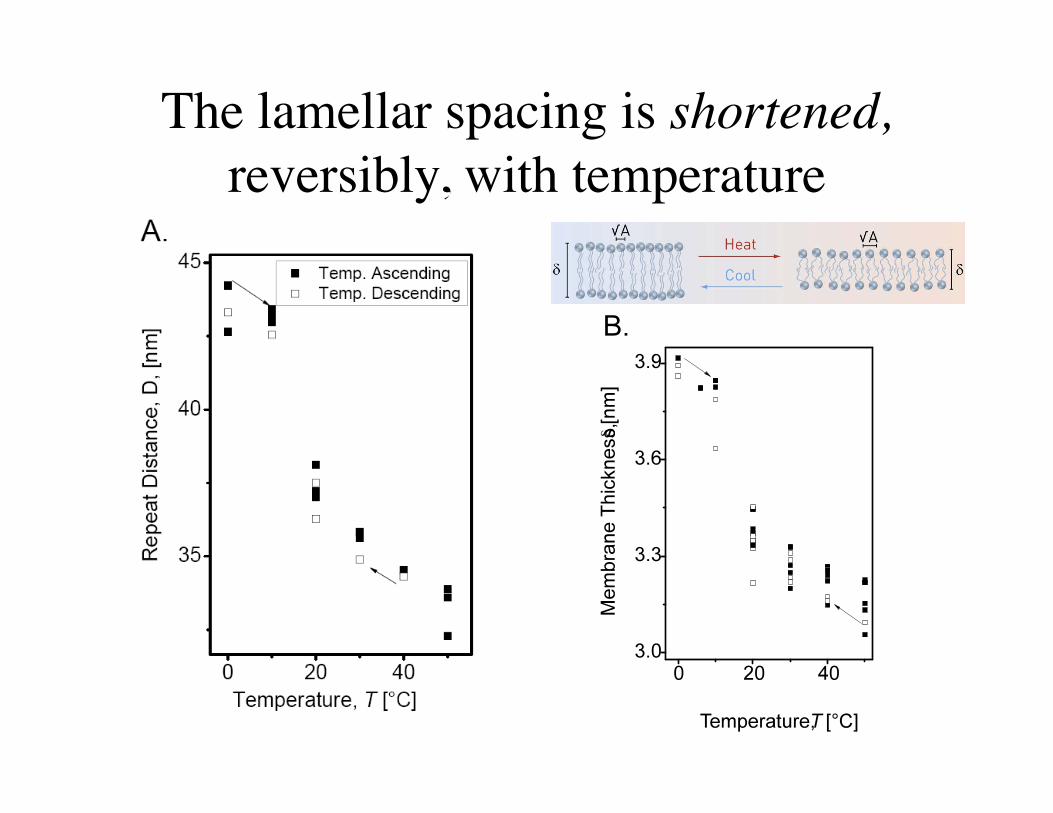
The lamellar spacing is shortened, reversibly, with temperature
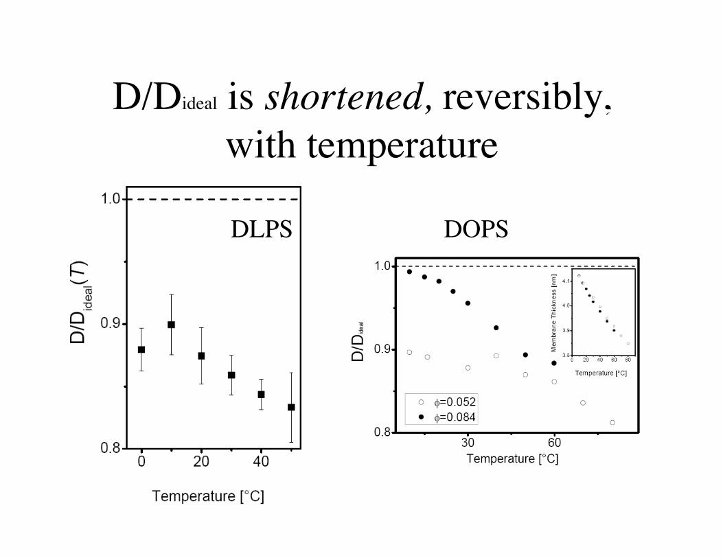
D/Dideal is shortened, reversibly, with temperature
DLPS DOPS
scale bar= 50 μm
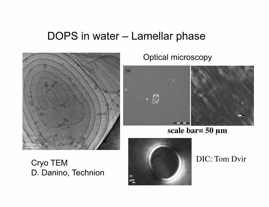
DOPS in water – Lamellar phase
Cryo TEM D. Danino, Technion
Optical microscopy
10 µm
DIC: Tom Dvir
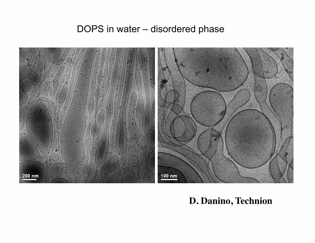
DOPS in water – disordered phase
D. Danino, Technion
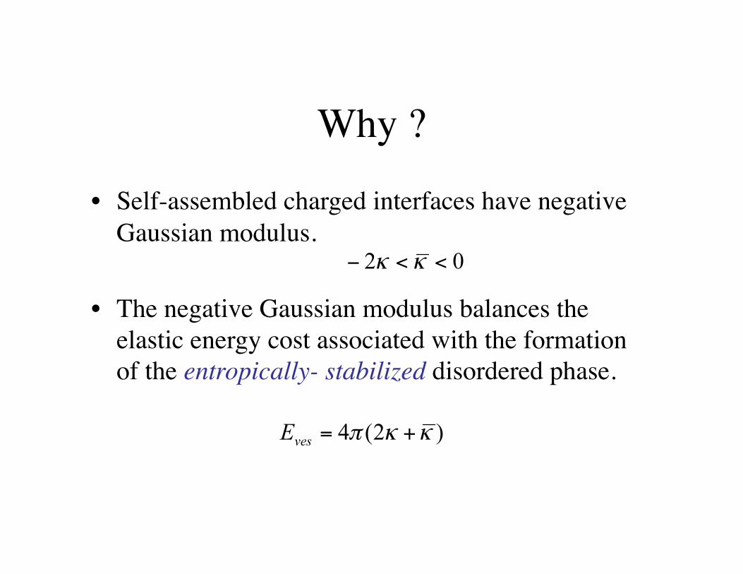
Why ?
• Self-assembled charged interfaces have negative Gaussian modulus.
• The negative Gaussian modulus balances the elastic energy cost associated with the formation of the entropically- stabilized disordered phase.
• The disordered phase is depleted from the lamellar phase and applies an osmotic stress on it.
• The lamellar spacing is set by equating the water chemical potential and the pressures of the two phases.
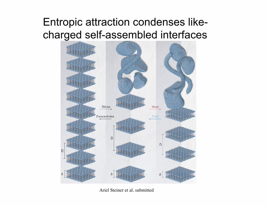
Entropic attraction condenses like-charged self-assembled interfaces
Ariel Steiner et al, submitted
Ions at dipolar interfaces: Regulating the size of lipid rafts
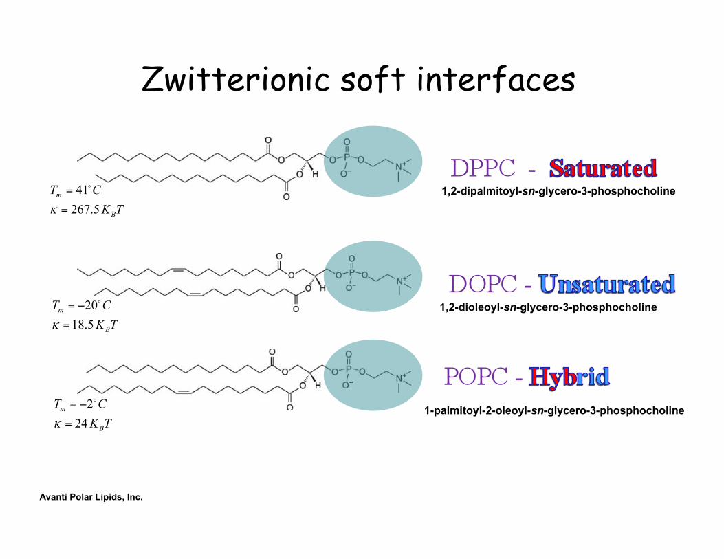
Zwitterionic soft interfaces
Avanti Polar Lipids, Inc.
1,2-dipalmitoyl-sn-glycero-3-phosphocholine
1,2-dioleoyl-sn-glycero-3-phosphocholine
1-palmitoyl-2-oleoyl-sn-glycero-3-phosphocholine
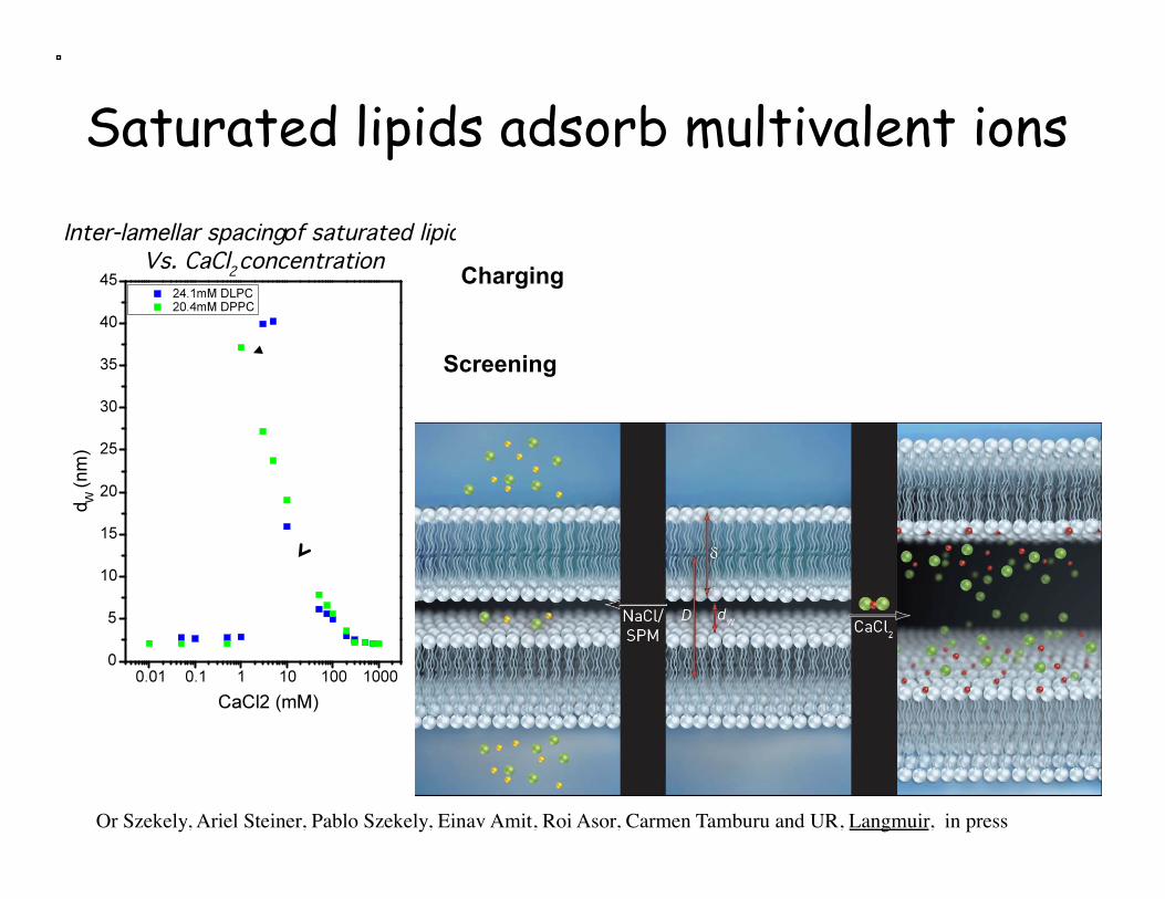
Saturated lipids adsorb multivalent ions
Or Szekely, Ariel Steiner, Pablo Szekely, Einav Amit, Roi Asor, Carmen Tamburu and UR, Langmuir, in press
Charging
Screening
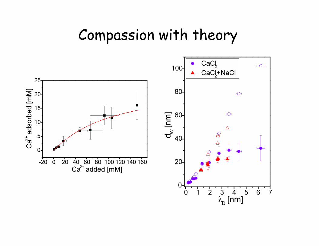
Compassion with theory
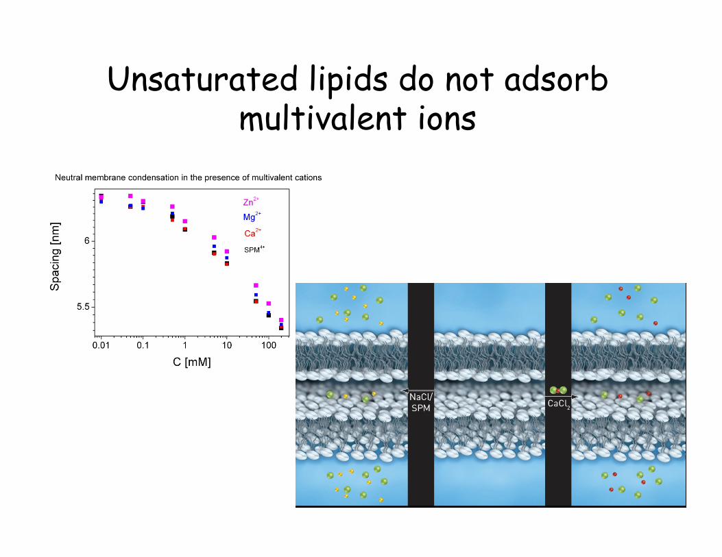
Unsaturated lipids do not adsorb multivalent ions
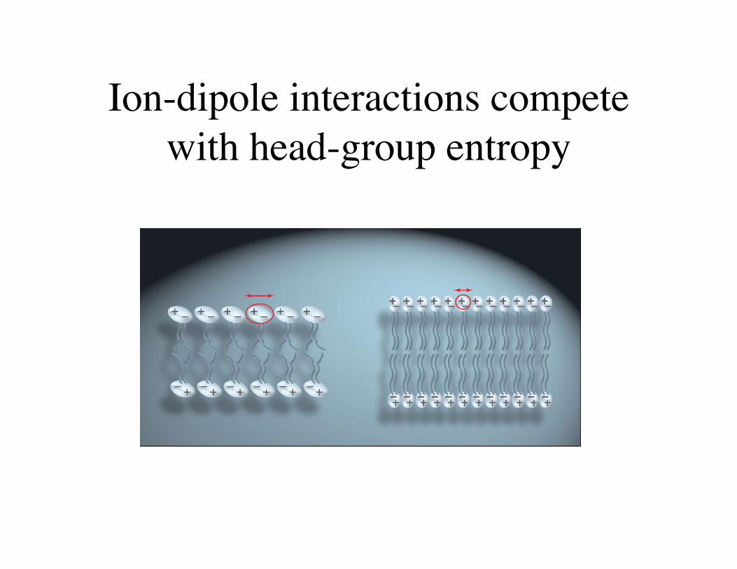
Ion-dipole interactions compete with head-group entropy
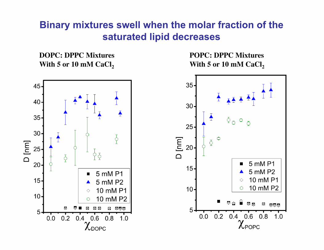
Binary mixtures swell when the molar fraction of the saturated lipid decreases
DOPC: DPPC Mixtures With 5 or 10 mM CaCl2
POPC: DPPC Mixtures With 5 or 10 mM CaCl2
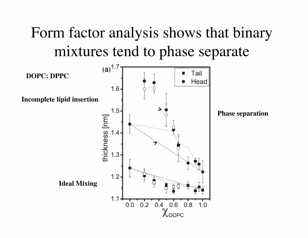
Form factor analysis shows that binary mixtures tend to phase separate
DOPC: DPPC
Incomplete lipid insertion
Phase separation
Ideal Mixing
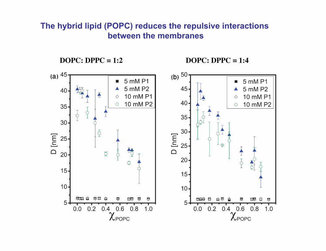
The hybrid lipid (POPC) reduces the repulsive interactions between the membranes
DOPC: DPPC = 1:2 DOPC: DPPC = 1:4
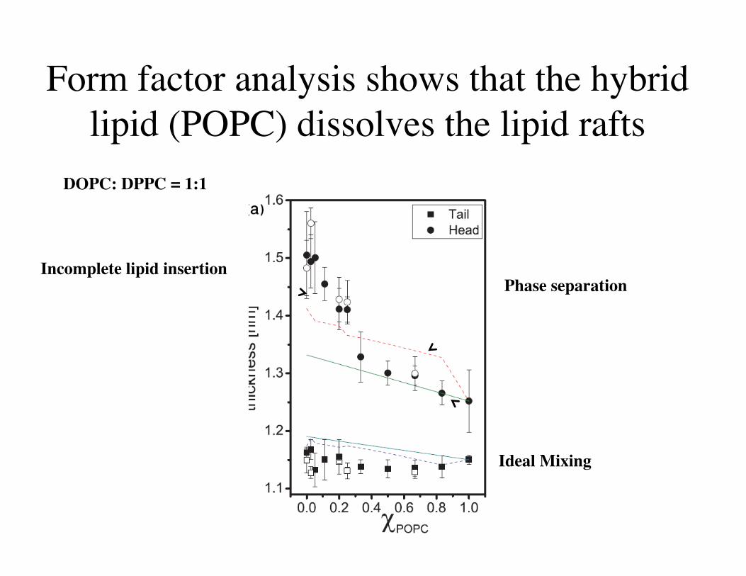
Form factor analysis shows that the hybrid lipid (POPC) dissolves the lipid rafts
DOPC: DPPC = 1:1
Incomplete lipid insertion Phase separation
Ideal Mixing

The Hybrid lipid reduces the line-tension and regulates the size of lipid rafts
Or Szekely and UR, submitted
Conclusions
• Entropic attraction condenses like-charged self-assembled interfaces
• Ion- dipole interaction depends strongly on lipid tails and ion-structure.
• Hybrid lipid regulates the size of lipid rafts.
Acknowledgements
Synchrotrons: ESRF, EMBL, SOLEIL, Elettra
Funding: HFSP, ISF, BSF, JF
Grad Students: Ariel Steiner, Or Szekely, Pablo Szekely, Avi Ginsburg, Tal Ben-Nun, Roi Asor Under-grad students: Tom Dvir. Einav Amir, Roy Resh Research Staff: Carmen Tamburu, Yaelle

The total isotropic phase work decrease with lipid concentration
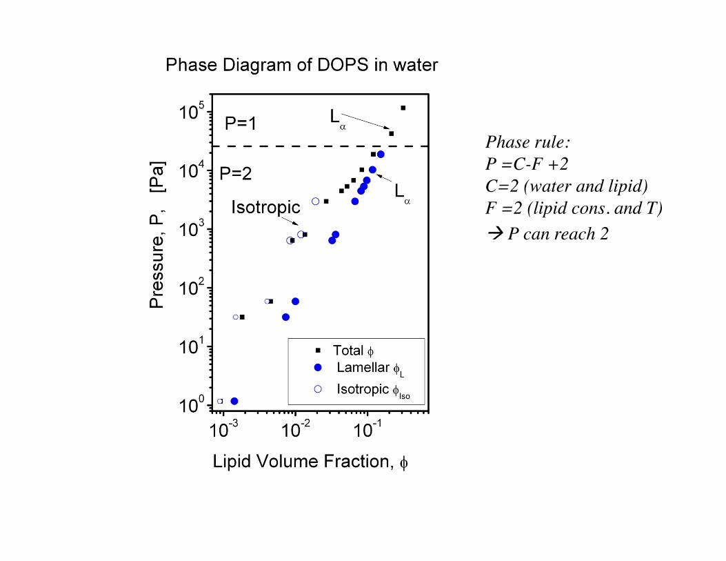
Phase rule: P =C-F +2 C=2 (water and lipid) F =2 (lipid cons. and T) P can reach 2
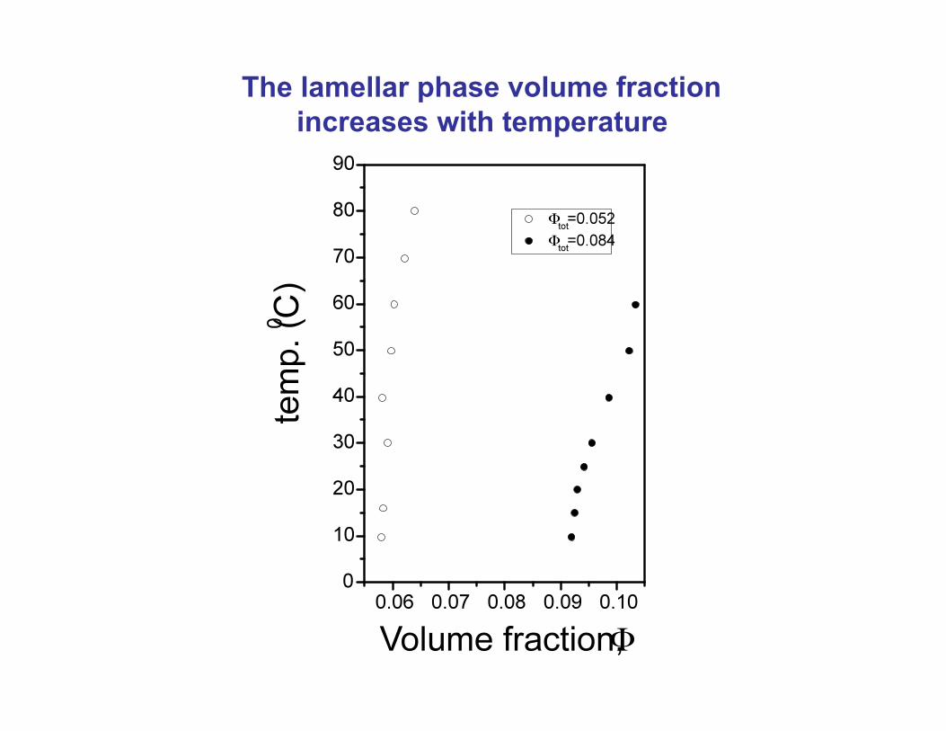
The lamellar phase volume fraction increases with temperature
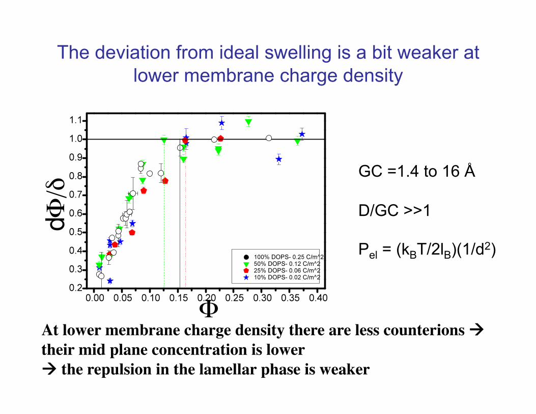
The deviation from ideal swelling is a bit weaker at lower membrane charge density
GC =1.4 to 16 Å
D/GC >>1
Pel = (kBT/2lB)(1/d2)
At lower membrane charge density there are less counterions their mid plane concentration is lower the repulsion in the lamellar phase is weaker
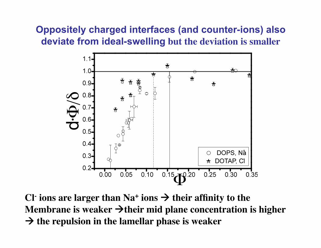
Oppositely charged interfaces (and counter-ions) also deviate from ideal-swelling but the deviation is smaller
Cl- ions are larger than Na+ ions their affinity to the Membrane is weaker their mid plane concentration is higher the repulsion in the lamellar phase is weaker
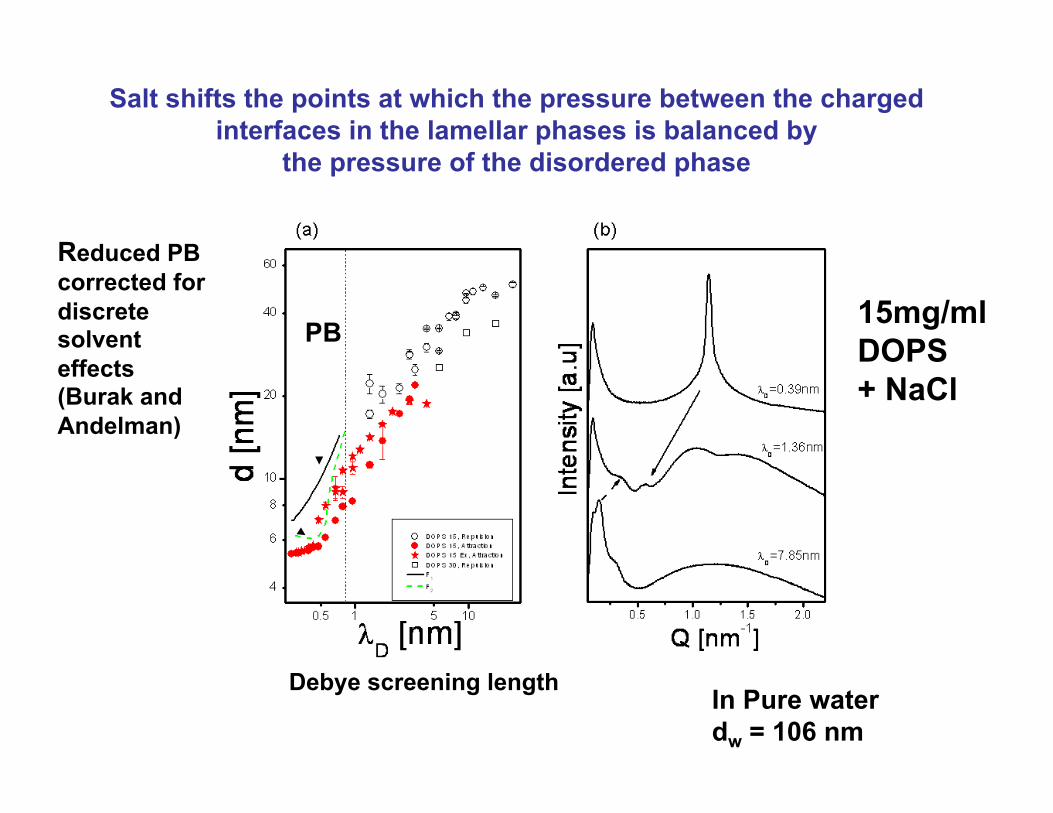
In Pure water dw = 106 nm
Salt shifts the points at which the pressure between the charged interfaces in the lamellar phases is balanced by
the pressure of the disordered phase
PB
Reduced PB corrected for discrete solvent effects (Burak and Andelman)
15mg/ml DOPS + NaCl
Debye screening length