Embed Size (px)
Citation preview
Structure and Function of Nucleoside Hydrolases fromPhyscomitrella patens and Maize Catalyzing theHydrolysis of Purine, Pyrimidine, andCytokinin Ribosides1[W]
Martina Kope�cná, Hanna Blaschke, David Kope�cný*, Armelle Vigouroux, Radka Kon�citíková,Ond�rej Novák, Ond�rej Kotland, Miroslav Strnad, Solange Moréra*, and Klaus von Schwartzenberg*
Department of Protein Biochemistry and Proteomics, Center of the Region Haná for Biotechnological andAgricultural Research (M.K., D.K., R.K.), Department of Biochemistry (R.K., O.K.), Faculty of Science, PalackýUniversity, CZ–783 71 Olomouc, Czech Republic; Laboratory of Growth Regulators, Palacký University andInstitute of Experimental Botany, Academy of Sciences of the Czech Republic, CZ–783 71 Olomouc, CzechRepublic (O.N., O.K., M.S.); Biozentrum Klein Flottbek und Botanischer Garten, Universität Hamburg,D–22609 Hamburg, Germany (H.B., K.v.S.); and Laboratoire d’Enzymologie et Biochimie Structurales, CNRS,F–91198 Gif-sur-Yvette cedex, France (A.V., S.M.)
We present a comprehensive characterization of the nucleosideN-ribohydrolase (NRH) family in twomodel plants, Physcomitrella patens(PpNRH) and maize (Zea mays; ZmNRH), using in vitro and in planta approaches. We identified two NRH subclasses in the plantkingdom; one preferentially targets the purine ribosides inosine and xanthosine, while the other is more active toward uridine andxanthosine. Both subclasses can hydrolyze plant hormones such as cytokinin ribosides. We also solved the crystal structures of twopurine NRHs, PpNRH1 and ZmNRH3. Structural analyses, site-directed mutagenesis experiments, and phylogenetic studies wereconducted to identify the residues responsible for the observed differences in substrate specificity between the NRH isoforms. Thepresence of a tyrosine at position 249 (PpNRH1 numbering) confers high hydrolase activity for purine ribosides, while an aspartateresidue in this position confers high activity for uridine. Bud formation is delayed by knocking out single NRH genes in P. patens, andunder conditions of nitrogen shortage, PpNRH1-deficient plants cannot salvage adenosine-bound nitrogen. All PpNRH knockout plantsdisplay elevated levels of certain purine and pyrimidine ribosides and cytokinins that reflect the substrate preferences of the knocked outenzymes. NRH enzymes thus have functions in cytokinin conversion and activation as well as in purine and pyrimidine metabolism.
Nucleoside hydrolases or nucleoside N-ribohydrolases(NRHs; EC 3.2.2.-) are glycosidases that catalyze the
cleavage of the N-glycosidic bond in nucleosides to en-able the recycling of the nucleobases and Rib (Fig. 1A).The process by which nucleosides and nucleobases arerecycled is also known as salvaging and is a way ofconserving energy, which would otherwise be neededfor the de novo synthesis of purine- and pyrimidine-containing compounds. During the salvage, bases andnucleosides can be converted into nucleoside mono-phosphates by the action of phosphoribosyltransfer-ases and nucleoside kinases, respectively, and furtherphosphorylated into nucleoside diphosphates and tri-phosphates (Moffatt et al., 2002; Zrenner et al., 2006;Fig. 1B). Uridine kinase and uracil phosphoribosyltransferase are key enzymes in the pyrimidine-salvagingpathway in plants (Mainguet et al., 2009; Chen andThelen, 2011). Adenine phosphoribosyltransferase andadenosine kinase (ADK) are important in purine sal-vaging (Moffatt and Somerville, 1988; Moffatt et al.,2002), and their mutants cause reductions in fertility orsterility, changes in transmethylation, and the formationof abnormal cell walls. In addition, both enzymes werealso reported to play roles in cytokinin metabolism(Moffatt et al., 1991, 2000; von Schwartzenberg et al.,1998; Schoor et al., 2011). Cytokinins (N6-substitutedadenine derivatives) are plant hormones that regulate
1 This work was supported by the Czech Science Foundation(grant no. P501/11/1591), the Ministry of Education, Youth, andSports of the Czech Republic (grant no. MSM 6198959215), PalackýUniversity (grant no. PrF_2013_037), Operational Programme Re-search and Development for Innovation (grant no. ED0007/01/01),the Centre National de la Recherche Scientifique (to S.M. and A.V.),the Deutsch Forschungsgemeinschaft (grant no. SCHW687/6 toK.v.S. and H.B.), and the Appuhn Foundation (Hamburg). M.K.was a Federation of European Microbiological Societies fellow.
* Address correspondence to [email protected], [email protected], and [email protected].
The author responsible for distribution of materials integral to thefindings presented in this article in accordance with the policy de-scribed in the Instructions for Authors (www.plantphysiol.org) is:David Kope�cný ([email protected]).
D.K., S.M., and K.v.S. designed the research; M.T., H.B., D.K., andR.K. analyzed enzyme kinetics; A.V. and S.M. performed the crystal-lographic study and contributed material and tools for crystallogra-phy; S.M. and D.K. analyzed the crystal structures; H.B. and K.v.S.generated and analyzed P. patens mutants, and O.N., O.K., and M.S.analyzed themetabolite levels. D.K., S.M., and K.v.S. wrote the paper.
[W] The online version of this article contains Web-only data.www.plantphysiol.org/cgi/doi/10.1104/pp.113.228775
1568 Plant Physiology�, December 2013, Vol. 163, pp. 1568–1583, www.plantphysiol.org � 2013 American Society of Plant Biologists. All Rights Reserved. www.plantphysiol.orgon February 23, 2020 - Published by Downloaded from
Copyright © 2013 American Society of Plant Biologists. All rights reserved.
cell division and numerous developmental events (Mokand Mok, 2001; Sakakibara, 2006). Cytokinin ribosidesare considered to be transport forms and have little orno activity.
NRHs are metalloproteins first identified and char-acterized in parasitic protozoa such as Trypanosoma,Crithidia, and Leishmania species that rely on the importand salvage of nucleotide derivatives. They have sincebeen characterized in other organisms such as bacteria,yeast, and insects (Versées and Steyaert, 2003) butnever in mammals (Parkin et al., 1991). They have beendivided into four classes based on their substrate spec-ificity: nonspecific NRHs, which hydrolyze inosine anduridine (IU-NRHs; Parkin et al., 1991; Shi et al., 1999);purine-specific inosine/adenosine/guanosine NRHs(Parkin, 1996); the 6-oxopurine-specific guanosine/inosine NRHs (Estupiñán and Schramm, 1994); andthe pyrimidine nucleoside-specific cytidine/uridineNRHs (CU-NRHs; Giabbai and Degano, 2004). AllNRHs exhibit a stringent specificity for the Rib moietyand differ in their preferences regarding the natureof the nucleobase. Crystal structures are available forempty NRH or in complex with inhibitors from Crithidiafasciculata (CfNRH; Degano et al., 1998), Leishmaniamajor (LmNRH; Shi et al., 1999), and Trypanosoma vivax(TvNRH; Versées et al., 2001, 2002). The structures oftwo CU-NRHs from Escherichia coli, namely YeiK (Iovaneet al., 2008) and YbeK (rihA; Muzzolini et al., 2006; Garauet al., 2010), are also available. NRHs are believed tocatalyze N-glycosidic bond cleavage by a direct dis-placement mechanism. An Asp from a conserved motifacts as a general base and abstracts a proton from acatalytic water molecule, which then attacks the C19atom of the Rib moiety of the nucleoside. Kinetic isotope-effect studies on CfNRH (Horenstein et al., 1991) showedthat the substrate’s hydrolysis proceeds via an oxo-carbenium ion-like transition state and is preceded byprotonation at the N7 atom of the purine ring, whichlowers the electron density on the purine ring and de-stabilizes the N-glycosidic bond. A conserved active-siteHis is a likely candidate for this role in IU-NRHs andCU-NRHs. In the transition state, the C19-N9 glyco-sidic bond is almost 2 Å long, with the C19 atom beingsp2 hybridized while the C39 atom adopts an exo-conformation, and the whole ribosyl moiety carries asubstantial positive charge (Horenstein et al., 1991).
Several NRH enzymes have been identified in plants,including a uridine-specific NRH from mung bean(Phaseolus radiatus; Achar and Vaidyanathan, 1967), aninosine-specific NRH (EC 3.2.2.2) and a guanosine-inosine-specific NRH, both from yellow lupine (Lupinusluteus; Guranowski, 1982; Szuwart et al., 2006), andan adenosine-specific NRH (EC 3.2.2.7) from coffee
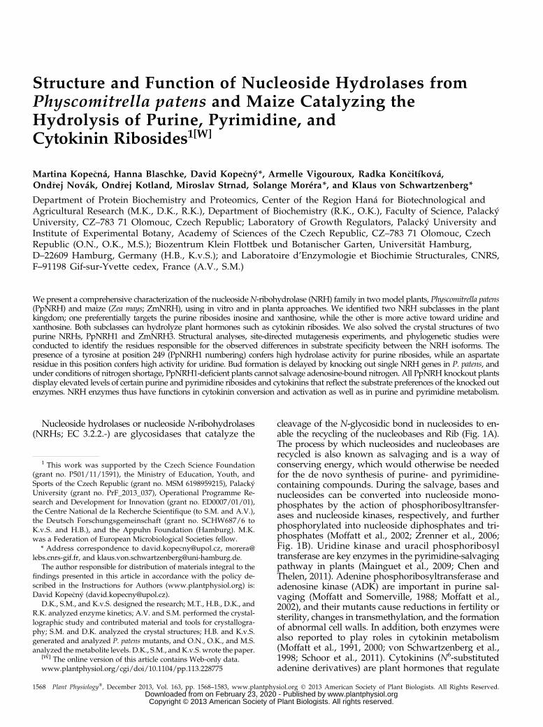
Figure 1. A, Scheme of the reactions catalyzed by plant NRHs whenusing purine (inosine), pyrimidine (uridine), and cytokinin (iPR) ribo-sides as the substrates. B, Simplified schematic overview of cytokinin,purine, and pyrimidine metabolism in plants. The diagram is adaptedfrom the work of Stasolla et al. (2003) and Zrenner et al. (2006) withmodifications. Themetabolic components shown are as follows: 1, cytokininnucleotide phosphoribohydrolase; 2, adenine phosphoribosyltransfer-ase; 3, adenosine kinase; 4, 59-nucleotidase; 5, adenosine phospho-rylase; 6, purine/pyrimidine nucleoside ribohydrolase; 7, cytokininoxidase/dehydrogenase; 8, AMP deaminase; 9, hypoxanthine phos-phoribosyltransferase; 10, inosine kinase; 11, inosine-guanosinephosphorylase; 12, IMP dehydrogenase; 13, xanthine dehydrogenase; 14,
59-nucleotidase; 15, GMP synthase; 16, hypoxanthine-guanine phos-phoribosyltransferase; 17, guanosine deaminase; 18, guanine deaminase;19, guanosine kinase; 20, uracil phosphoribosyltransferase; 21, uridinecytidine kinase; 22, pyrimidine 59-nucleotidase; 23, cytidine deaminase;24, adenosine/adenine deaminase. CK, Cytokinin; CKR, cytokinin ribo-side; CKRMP, cytokinin riboside monophosphate.
Plant Physiol. Vol. 163, 2013 1569
Nucleoside Hydrolases from Moss and Maize
www.plantphysiol.orgon February 23, 2020 - Published by Downloaded from Copyright © 2013 American Society of Plant Biologists. All rights reserved.
(Coffea arabica), barley (Hordeum vulgare), and wheat(Triticum aestivum; Guranowski and Schneider, 1977;Chen and Kristopeit, 1981; Campos et al., 2005). How-ever, their amino acid sequences have not been reportedso far. A detailed study of the NRH gene family fromArabidopsis (Arabidopsis thaliana) has recently beenreported (Jung et al., 2009, 2011). The AtNRH1 enzymeexhibits highest hydrolase activity toward uridine andxanthosine. It can also hydrolyze the cytokinin ribosideN6-(2-isopentenyl)adenosine (iPR), which suggests thatit may also play a role in cytokinin homeostasis. However,Riegler et al. (2011) analyzed the phenotypes of homo-zygous nrh1 and nrh2 single mutants along with the ho-mozygous double mutants and concluded that AtNRHsare probably unimportant in cytokinin metabolism.
Here, we identify and characterize plant IU-NRHsfrom two different model organisms, Physcomitrella patensand maize (Zea mays), combining structural, enzymatic,and in planta functional approaches. The moss P. patenswas chosen to represent the bryophytes, which can beregarded as being evolutionarily basal terrestrial plants,and is suitable for use in developmental and metabolicstudies (Cove et al., 2006; von Schwartzenberg, 2009),while maize is an important model system for cerealcrops. We report the crystal structures of NRH enzymesfrom the two plant species, PpNRH1 and ZmNRH3.Based on these structures, we performed site-directedmutagenesis experiments and kinetic analyses of pointmutants of PpNRH1 in order to identify key residuesinvolved in nucleobase interactions and catalysis. Toanalyze the physiological role of the PpNRHs, singleknockout mutants were generated. NRH deficiencycaused significant changes in the levels of purine, py-rimidine, and cytokinin metabolites relative to thoseseen in the wild type, illustrating the importance ofthese enzymes in nucleoside and cytokinin metabolism.
RESULTS AND DISCUSSION
Gene Models of NRHs from P. patens and Maize
Ongoing genomic analyses suggest that most plantgenomes contain at least two genes coding for NRHs.
Several plant genomes, such as those of moss (P. patens),maize, Arabidopsis, rice (Oryza sativa), tomato (Solanumlycopersicum), and wheat, appear to contain severalNRHs. We focused on two model plant organisms, themoss P. patens and maize, due to the availability ofdetailed information on cytokinin metabolism in bothspecies (Massonneau et al., 2004; von Schwartzenberg,2009; Vyroubalová et al., 2009). The genome databasesfor the moss P. patens (www.phytozome.net, version 9.1)and maize (www.maizesequence.org, version 5b.60) in-dicate that these species contain three and five NRHgenes, respectively. In order to identify the correct genemodels in each case and obtain the corresponding re-combinant proteins, we cloned the complementaryDNAs (cDNAs) of these eight NRHs using gene-specificprimers and deposited their sequences to GenBank (see“Materials and Methods”).
The cDNA sequence obtained for PpNRH1 corre-sponds to the predicted gene model (Pp1s357_22V6.1),those obtained for PpNRH2 and PpNRH3 do not matchthe predicted gene models in the genome annotationversion 6.1 (Pp1s140_172V6.1 and Pp1s5_276V6.1; see“Materials and Methods”; Supplemental Fig. S1). Thetwo paralogous genes ZmNRH1a and ZmNRH1b arelocalized on chromosomes 8 and 3 (GRMZM2G029845and GRMZM2G134149), respectively. The two otherparalogs, ZmNRH2a and ZmNRH2b, lie on chromosomes4 and 1 (GRMZM2G085960 and GRMZM2G015344),respectively. The last ZmNRH3 gene (GRMZM2G104999)is localized on chromosome 2. These genes encode pro-teins of between 315 and 341 residues. All plant NRHsexhibit a conserved sequence motif, DTDPGIDD, at theN terminus (Fig. 2), which is involved in the binding ofa calcium ion and the Rib moiety of the substrate (Verséesand Steyaert, 2003). Another group of extracellular NRHswith two domains was recently identified in Arabidopsis(At5g18860; Jung et al., 2011). The authors suggested thatthese NRHs could correspond with the extracellularadenosine hydrolase activity found in potato (Solanumtuberosum) tubers (Riewe et al., 2008). Although the firstdomain carries a DTDVDTDD motif, the second con-tains an unconserved DMDMSXGD motif. Analogoussequences are also present in the genomes of other
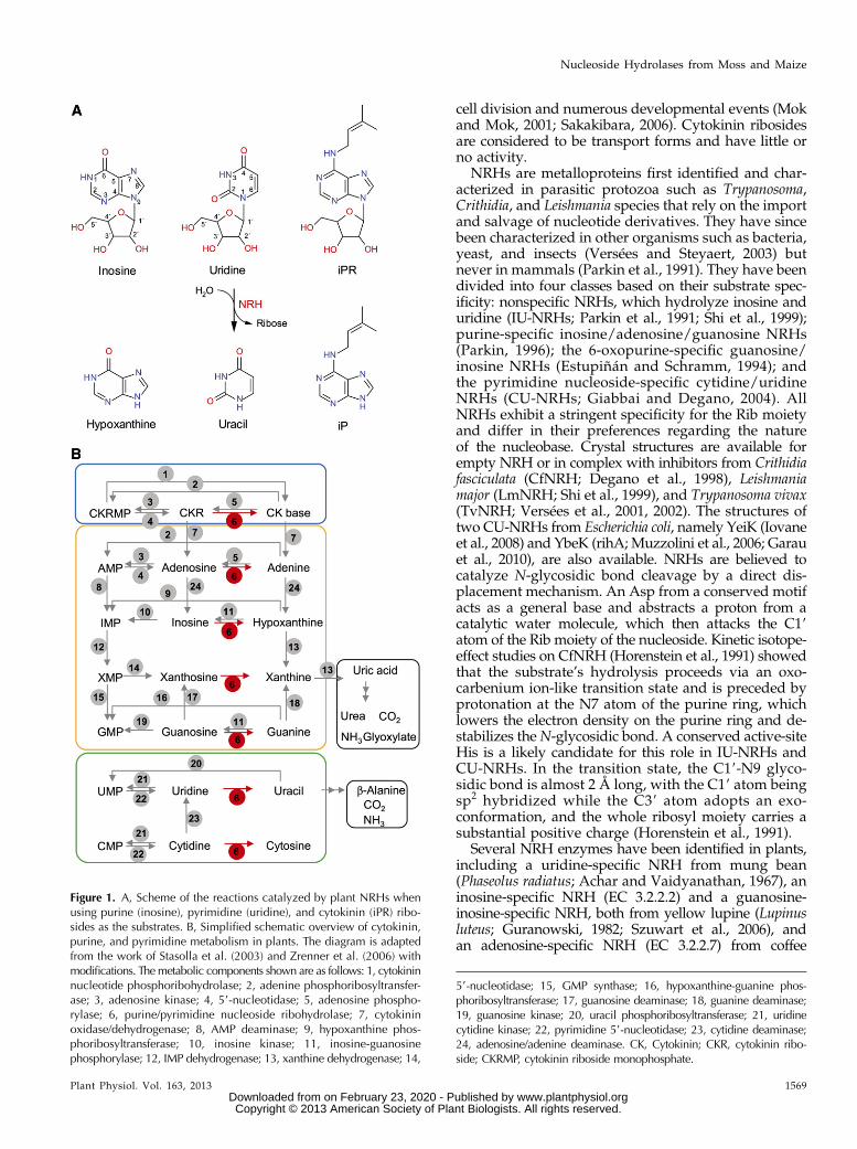
Figure 2. Multiple alignment of the amino acidsequences of NRHs from plants, yeast, bacteria,and protozoa, focusing on the DXDXXXDD motifand the a3 and a11 helix regions. Asp residuesinvolved in calcium ion coordination are high-lighted in blue. The Asp-binding 2-OH group ofRib is highlighted in green. Crucial residues in-volved in substrate binding in plant NRHs arehighlighted in red. Residues are numberedaccording to the PpNRH1 sequence. Accessionnumbers for the sequences shown are listed in“Materials and Methods.”
1570 Plant Physiol. Vol. 163, 2013
Kope�cná et al.
www.plantphysiol.orgon February 23, 2020 - Published by Downloaded from Copyright © 2013 American Society of Plant Biologists. All rights reserved.
plants such as maize (GRMZM2G386229) and rice(Os05g33644 and Os05g33630).
Substrate Specificity of Plant NRHs
The pH effect on the specific activity of PpNRH1(Supplemental Fig. S2) was analyzed, and high activitywas found between pH 7.0 and 9.0. All subsequentkinetic analyses, therefore, were performed at pH 7.5.PpNRH1, ZmNRH2a, ZmNRH2b, and ZmNRH3 wereobtained active and in high yield in soluble form. Incontrast, the production of PpNRH2, ZmNRH1a, andZmNRH1b primarily resulted in the formation of in-clusion bodies, and refolding attempts did not lead torestoration of the enzymatic activity. Only very smallquantities of these enzymes could be obtained in sol-uble form. We thus were only able to briefly screen thethree NRHs with possible natural substrates, includingpurine, pyrimidine, and cytokinin ribosides, at 200 mM
concentration (Table I). It was possible to analyze thesubstrate preferences for PpNRH2, whereas ZmNRH1aand ZmNRH1b show only negligible activity. How-ever, cytidine and adenosine could be substrates of thetwo maize enzymes, but we cannot rule out that un-tested ribosides can be more suitable substrates. So far,the production of recombinant protein was not ob-served for any of the four splicing variants of PpNRH3,in contrast to the studied NRHs.PpNRH1 is most active toward the two purine ribo-
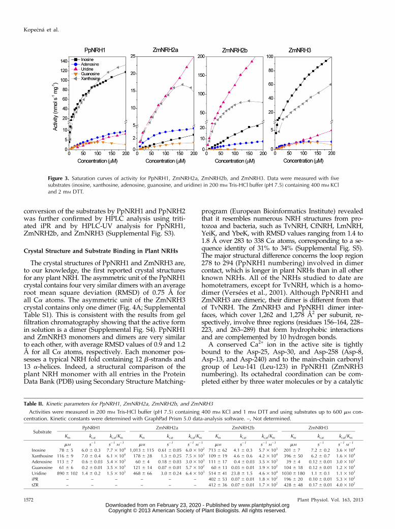
sides xanthosine and inosine and exhibits weaker activitywith adenosine, uridine, and guanosine. In contrast,PpNRH2 prefers the pyrimidine riboside uridine andis less active toward xanthosine and inosine. ZmNRH2aand ZmNRH2b are also most active toward uridine.The activity of ZmNRH2b is 10 and 200 times higherthan that of ZmNRH2a and PpNRH2, respectively. Thecomparatively low activity of PpNRH2 is likely due tobeing poorly expressed in E. coli and its low stability.ZmNRH3 preferentially hydrolyzes inosine and xan-thosine, while the remaining nucleosides are weakersubstrates. PpNRH1 and ZmNRH3 exhibit similar sub-strate preferences. However, subtle differences betweenthese two enzymes are shown in Figure 3.
PpNRH1, ZmNRH2a, ZmNRH2b, and ZmNRH3 werefurther analyzed to determine their Km and catalyticconstant (kcat) values (Table II) and confirmed the resultsdiscussed above. Both PpNRH1 and ZmNRH3 showthe highest catalytic efficiency with inosine and xan-thosine, while ZmNRH2a and ZmNRH2b display thehighest catalytic efficiency for uridine and xanthosine.Although ZmNRH2a and ZmNRH2b have relativelyhigh Km values for uridine, it is also the substrate forwhich they have the highest kcat values. Based on thesekinetic values and the current system of classification,the investigated plant NRHs belong to the nonspecificIU-NRH class (Parkin et al., 1991; Shi et al., 1999).However, both PpNRH1 and ZmNRH3 (which preferinosine and xanthosine) are apparently kinetically dif-ferent from ZmNRH2a, ZmNRH2b, and PpNRH2 (all ofwhich prefer uridine and xanthosine) and the AtNRH1from Arabidopsis (Jung et al., 2009, 2011). Therefore, itseems that there are at least two subclasses of IU-NRHsin plants. Details of the sequences of these two sub-classes are discussed below.
Inosine, xanthosine, and guanosine are all interme-diates in the purine catabolic pathway (Fig. 1B) thatstarts with AMP (Zrenner et al., 2006). The xanthineand hypoxanthine nucleobases are formed by the actionof nucleoside hydrolases and are further processed byxanthine dehydrogenase to give uric acid. Adenosineand adenine are by-products of the cytokinin degradationreactions catalyzed by cytokinin oxidase/dehydrogenase(EC 1.5.99.12; Houba-Hérin et al., 1999). In contrast to thetwo known bacterial CU-NRHs, the five plant IU-NRHsexhibit very weak activity toward cytidine (Table I). Thisis consistent with the fact that plants preferentiallyconvert cytidine to uridine via cytidine deaminase(EC 3.5.4.5; Stasolla et al., 2003). To analyze the con-version of cytokinin ribosides, we first determined theextinction coefficients of iPR and trans-zeatin riboside(tZR) by spectrometric measurement according to Parkin(1996). All of the studied plant IU-NRHs have onlyweak activity toward iPR and tZR, between 1% and0.1% of that toward their best substrates (Table I).ZmNRH2b and ZmNRH3 exhibit catalytic efficienciesbetween 1.7 and 5.3 3 102 M
21 s21 (Table II). The
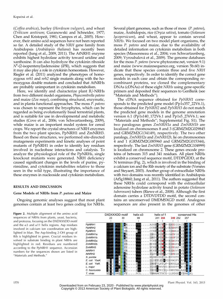
Table I. Substrate specificity of NRHs from P. patens and maize
Relative reaction rates (%) were measured at 200 mM substrate concentration. Activities were measured in 200 mM Tris-HCl buffer (pH 7.5)containing 400 mM KCl and 1 mM DTT. The specific activities for PpNRH1 and ZmNRH3 with 200 mM xanthosine were 135 and 61 nkat mg21,respectively. The specific activities for PpNRH2, ZmNRH2a, and ZmNRH2b with 200 mM uridine were 1.3, 26, and 226 nkat mg21, respectively.+ indicates very low activity.
SubstrateRelative Rate
PpNRH1 PpNRH2 ZmNRH2a ZmNRH2b ZmNRH3
Inosine 87 22 9.3 9.5 100Xanthosine 100 61 53 34 70Adenosine 6.1 1.6 15 3.5 2Guanosine 1.7 0 0.1 0.5 1.5Uridine 5.7 100 100 100 5Cytidine 0.1 3.9 5.0 0.9 0.1iPR 0.1 + 0.03 0.3 1.4tZR 0.1 + 0.03 0.3 1.5
Plant Physiol. Vol. 163, 2013 1571
Nucleoside Hydrolases from Moss and Maize
www.plantphysiol.orgon February 23, 2020 - Published by Downloaded from Copyright © 2013 American Society of Plant Biologists. All rights reserved.
conversion of the substrates by PpNRH1 and PpNRH2was further confirmed by HPLC analysis using triti-ated iPR and by HPLC-UV analysis for PpNRH1,ZmNRH2b, and ZmNRH3 (Supplemental Fig. S3).
Crystal Structure and Substrate Binding in Plant NRHs
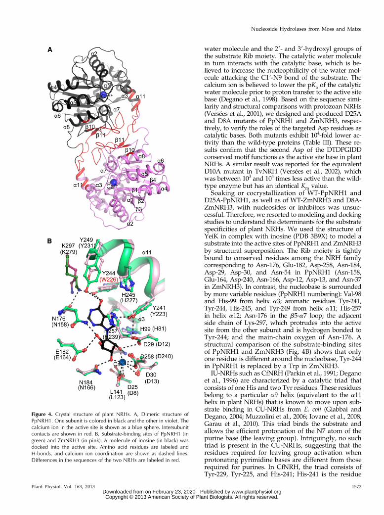
The crystal structures of PpNRH1 and ZmNRH3 are,to our knowledge, the first reported crystal structuresfor any plant NRH. The asymmetric unit of the PpNRH1crystal contains four very similar dimers with an averageroot mean square deviation (RMSD) of 0.75 Å forall Ca atoms. The asymmetric unit of the ZmNRH3crystal contains only one dimer (Fig. 4A; SupplementalTable S1). This is consistent with the results from gelfiltration chromatography showing that the active formin solution is a dimer (Supplemental Fig. S4). PpNRH1and ZmNRH3 monomers and dimers are very similarto each other, with average RMSD values of 0.9 and 1.2Å for all Ca atoms, respectively. Each monomer pos-sesses a typical NRH fold containing 12 b-strands and13 a-helices. Indeed, a structural comparison of theplant NRH monomer with all entries in the ProteinData Bank (PDB) using Secondary Structure Matching-
program (European Bioinformatics Institute) revealedthat it resembles numerous NRH structures from pro-tozoa and bacteria, such as TvNRH, CfNRH, LmNRH,YeiK, and YbeK, with RMSD values ranging from 1.4 to1.8 Å over 283 to 338 Ca atoms, corresponding to a se-quence identity of 31% to 34% (Supplemental Fig. S5).The major structural difference concerns the loop region278 to 294 (PpNRH1 numbering) involved in dimercontact, which is longer in plant NRHs than in all otherknown NRHs. All of the NRHs studied to date arehomotetramers, except for TvNRH, which is a homo-dimer (Versées et al., 2001). Although PpNRH1 andZmNRH3 are dimeric, their dimer is different from thatof TvNRH. The ZmNRH3 and PpNRH1 dimer inter-faces, which cover 1,262 and 1,278 Å2 per subunit, re-spectively, involve three regions (residues 156–164, 228–223, and 263–289) that form hydrophobic interactionsand are complemented by 10 hydrogen bonds.
A conserved Ca2+ ion in the active site is tightlybound to the Asp-25, Asp-30, and Asp-258 (Asp-8,Asp-13, and Asp-240) and to the main-chain carbonylgroup of Leu-141 (Leu-123) in PpNRH1 (ZmNRH3numbering). Its octahedral coordination can be com-pleted either by three water molecules or by a catalytic
Figure 3. Saturation curves of activity for PpNRH1, ZmNRH2a, ZmNRH2b, and ZmNRH3. Data were measured with fivesubstrates (inosine, xanthosine, adenosine, guanosine, and uridine) in 200 mM Tris-HCl buffer (pH 7.5) containing 400 mM KCland 2 mM DTT.
Table II. Kinetic parameters for PpNRH1, ZmNRH2a, ZmNRH2b, and ZmNRH3
Activities were measured in 200 mM Tris-HCl buffer (pH 7.5) containing 400 mM KCl and 1 mM DTT and using substrates up to 600 mM con-centration. Kinetic constants were determined with GraphPad Prism 5.0 data-analysis software. –, Not determined.
SubstratePpNRH1 ZmNRH2a ZmNRH2b ZmNRH3
Km kcat kcat/Km Km kcat kcat/Km Km kcat kcat/Km Km kcat kcat/Km
mM s21 s21M21 mM s21 s21
M21 mM s21 s21
M21 mM s21 s21
M21
Inosine 78 6 5 6.0 6 0.3 7.7 3 104 1,013 6 115 0.61 6 0.05 6.0 3 102 713 6 62 4.1 6 0.3 5.7 3 103 201 6 7 7.2 6 0.2 3.6 3 104
Xanthosine 116 6 9 7.0 6 0.4 6.1 3 104 178 6 28 1.3 6 0.25 7.5 3 103 109 6 19 4.6 6 0.6 4.2 3 104 396 6 50 6.2 6 0.7 1.6 3 104
Adenosine 113 6 7 0.6 6 0.03 5.4 3 103 60 6 4 0.18 6 0.03 3.0 3 103 111 6 17 0.4 6 0.03 3.5 3 103 39 6 4 0.12 6 0.01 3.0 3 103
Guanosine 61 6 6 0.2 6 0.01 3.5 3 103 121 6 14 0.07 6 0.01 5.7 3 102 60 6 13 0.03 6 0.01 3.9 3 102 104 6 18 0.12 6 0.01 1.2 3 103
Uridine 890 6 102 1.4 6 0.2 1.5 3 103 468 6 66 3.0 6 0.24 6.4 3 103 514 6 41 23.8 6 1.5 4.6 3 104 1030 6 180 1.1 6 0.1 1.1 3 103
iPR – – – – – – 402 6 53 0.07 6 0.01 1.8 3 102 196 6 20 0.10 6 0.01 5.3 3 102
tZR – – – – – – 412 6 36 0.07 6 0.01 1.7 3 102 428 6 48 0.17 6 0.01 4.0 3 102
1572 Plant Physiol. Vol. 163, 2013
Kope�cná et al.
www.plantphysiol.orgon February 23, 2020 - Published by Downloaded from Copyright © 2013 American Society of Plant Biologists. All rights reserved.
water molecule and the 29- and 39-hydroxyl groups ofthe substrate Rib moiety. The catalytic water moleculein turn interacts with the catalytic base, which is be-lieved to increase the nucleophilicity of the water mol-ecule attacking the C19-N9 bond of the substrate. Thecalcium ion is believed to lower the pKa of the catalyticwater molecule prior to proton transfer to the active sitebase (Degano et al., 1998). Based on the sequence simi-larity and structural comparisons with protozoan NRHs(Versées et al., 2001), we designed and produced D25Aand D8A mutants of PpNRH1 and ZmNRH3, respec-tively, to verify the roles of the targeted Asp residues ascatalytic bases. Both mutants exhibit 104-fold lower ac-tivity than the wild-type proteins (Table III). These re-sults confirm that the second Asp of the DTDPGIDDconserved motif functions as the active site base in plantNRHs. A similar result was reported for the equivalentD10A mutant in TvNRH (Versées et al., 2002), whichwas between 103 and 104 times less active than the wild-type enzyme but has an identical Km value.
Soaking or cocrystallization of WT-PpNRH1 andD25A-PpNRH1, as well as of WT-ZmNRH3 and D8A-ZmNRH3, with nucleosides or inhibitors was unsuc-cessful. Therefore, we resorted to modeling and dockingstudies to understand the determinants for the substratespecificities of plant NRHs. We used the structure ofYeiK in complex with inosine (PDB 3B9X) to model asubstrate into the active sites of PpNRH1 and ZmNRH3by structural superposition. The Rib moiety is tightlybound to conserved residues among the NRH familycorresponding to Asn-176, Glu-182, Asp-258, Asn-184,Asp-29, Asp-30, and Asn-54 in PpNRH1 (Asn-158,Glu-164, Asp-240, Asn-166, Asp-12, Asp-13, and Asn-37in ZmNRH3). In contrast, the nucleobase is surroundedby more variable residues (PpNRH1 numbering): Val-98and His-99 from helix a3; aromatic residues Tyr-241,Tyr-244, His-245, and Tyr-249 from helix a11; His-257in helix a12; Asn-176 in the b5-a7 loop; the adjacentside chain of Lys-297, which protrudes into the activesite from the other subunit and is hydrogen bonded toTyr-244; and the main-chain oxygen of Asn-176. Astructural comparison of the substrate-binding sitesof PpNRH1 and ZmNRH3 (Fig. 4B) shows that onlyone residue is different around the nucleobase, Tyr-244in PpNRH1 is replaced by a Trp in ZmNRH3.
IU-NRHs such as CfNRH (Parkin et al., 1991; Deganoet al., 1996) are characterized by a catalytic triad thatconsists of one His and two Tyr residues. These residuesbelong to a particular a9 helix (equivalent to the a11helix in plant NRHs) that is known to move upon sub-strate binding in CU-NRHs from E. coli (Giabbai andDegano, 2004; Muzzolini et al., 2006; Iovane et al., 2008;Garau et al., 2010). This triad binds the substrate andallows the efficient protonation of the N7 atom of thepurine base (the leaving group). Intriguingly, no suchtriad is present in the CU-NRHs, suggesting that theresidues required for leaving group activation whenprotonating pyrimidine bases are different from thoserequired for purines. In CfNRH, the triad consists ofTyr-229, Tyr-225, and His-241; His-241 is the residue
Figure 4. Crystal structure of plant NRHs. A, Dimeric structure ofPpNRH1. One subunit is colored in black and the other in violet. Thecalcium ion in the active site is shown as a blue sphere. Intersubunitcontacts are shown in red. B, Substrate-binding sites of PpNRH1 (ingreen) and ZmNRH3 (in pink). A molecule of inosine (in black) wasdocked into the active site. Amino acid residues are labeled andH-bonds, and calcium ion coordination are shown as dashed lines.Differences in the sequences of the two NRHs are labeled in red.
Plant Physiol. Vol. 163, 2013 1573
Nucleoside Hydrolases from Moss and Maize
www.plantphysiol.orgon February 23, 2020 - Published by Downloaded from Copyright © 2013 American Society of Plant Biologists. All rights reserved.
that protonates the N7 atom (Fig. 5A; Gopaul et al.,1996). PpNRH1, which is an IU-NRH, possesses anequivalent triad consisting of His-245, Tyr-241, andHis-257, in which His-257 is expected to protonate theleaving group. However, in this work, we mutatedHis-99 into Ala in PpNRH1 and found this mutantinactive with any riboside tested as a substrate. His-99is conserved in all NRHs except for TvNRH. In ourdocking experiments (see below), His-99 is only 3.2 to3.5 Å away from the N9 atom of the purine ring as wellas the N1 and O2 atoms of a docked pyrimidine ring,indicating that it is another potential proton donor.Interestingly, Giabbai and Degano (2004) concluded fromtheir mutagenesis analysis on two potential catalytic acidsin bacterial YeiK (His-82 and His-239, equivalent toHis-99 and His-257 in PpNRH1) that other active-siteresidues can function as alternative proton donorsthat could contribute to the N-glycosidic bond cleav-age in this CU-NRH. The PpNRH1 substrate-binding siteinvolves two additional residues, Tyr-244 and Tyr-249,which point toward the nucleobase compared with theirequivalent residues Ile-228 and Tyr-234 in CfNRH. InCfNRH, the substrate-binding site is completed by theArg-233 residue, which is located in the loop followingthe mobile a9 helix. Interestingly, the side chain of Lys-297(from the neighboring subunit in PpNRH1) occupies aposition similar to that of Arg-233 (Fig. 5A), suggestingthat the dimer formation has an important role in en-zymatic function of plant NRHs.
Docking Analysis and Site-Directed Mutagenesisof PpNRH1
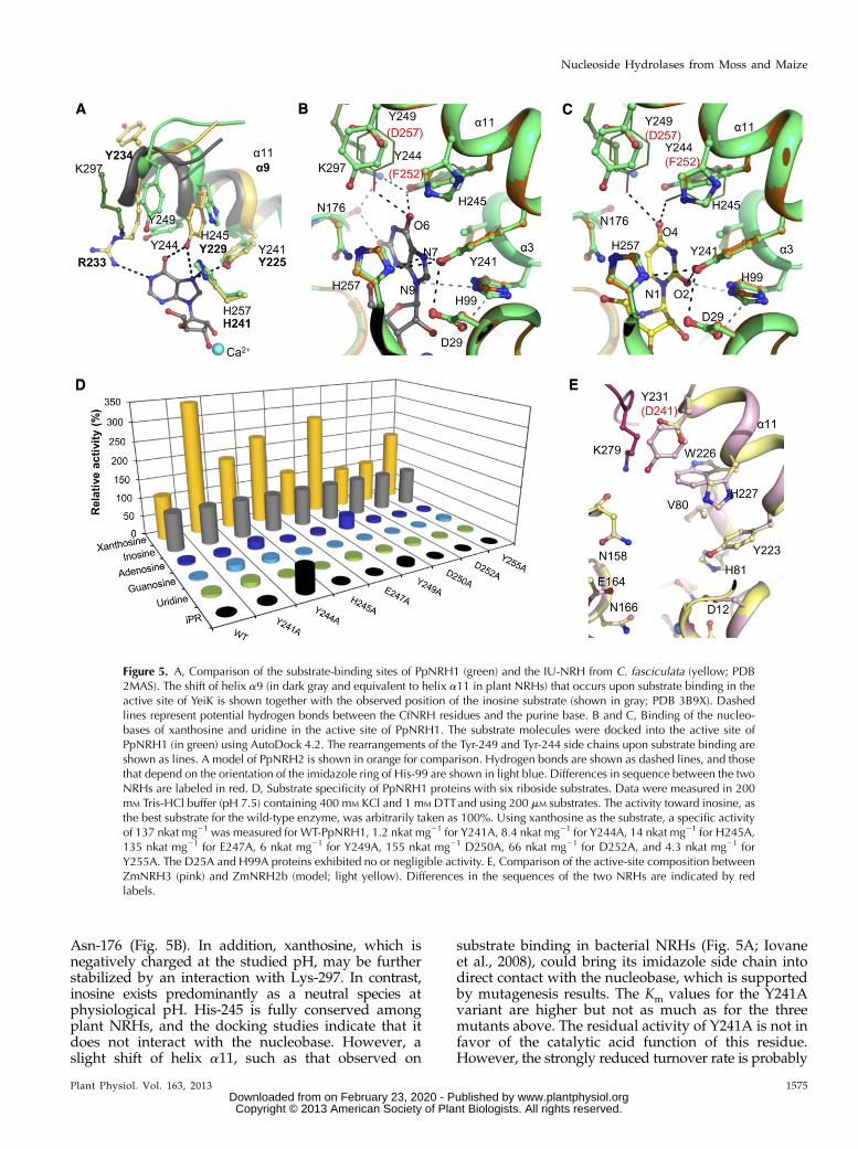
Xanthosine and inosine show similar binding modeswhen docked into the PpNRH1 active site (Fig. 5B),with conserved interactions between the 6-oxo groupand the side chains of Tyr-244 and Tyr-249 (and pos-sibly the side chain of Lys-297) as well as between the
N7 atom (protonated during catalysis) and the sidechain of Tyr-241 (3.5-Å distance). Both the 2-oxo groupand the N1 atom of xanthosine can form additionalhydrogen bonds to the main chain carbonyl group ofAsn-176. Docking experiments with uridine revealedthat the 4-oxo group can interact with the hydroxylgroups of Tyr-244 and Tyr-249. The 2-oxo group pointstoward a cluster formed by four residues: the twoimidazole rings of His-99 and His-257, the hydroxyl ofTyr-241, and the carboxyl group of Asp-29 (Fig. 5C).
To verify the results of our docking studies onsubstrate binding based on the PpNRH1 structure, weperformed site-directed mutagenesis of Tyr-241, Tyr-244,His-245, and Tyr-249 from the mobile helix a11 andreplaced them with Ala. We also mutated another resi-due from the a11 helix, Glu-247, whose side chain doesnot project into the substrate-binding site. Three otherresidues, Asp-250 and Asp-252, both positioned in theloop between helix a11 and strand b5, and Tyr-255, werealso targeted in order to determine if more mobileregions are involved in substrate binding, as shownpreviously for TvNRH (Versées et al., 2002). Circulardichroism spectroscopy measurements indicate that thefolding in solution of each mutant variant resembles thatof WT-PpNRH1 (Supplemental Fig. S6).
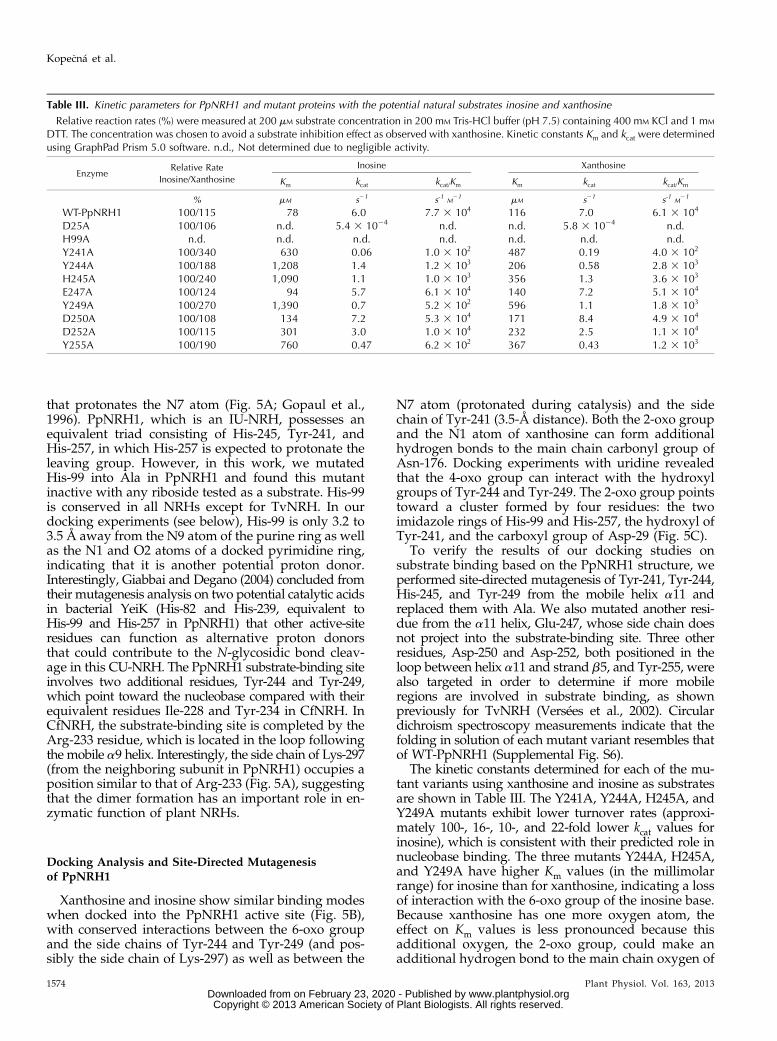
The kinetic constants determined for each of the mu-tant variants using xanthosine and inosine as substratesare shown in Table III. The Y241A, Y244A, H245A, andY249A mutants exhibit lower turnover rates (approxi-mately 100-, 16-, 10-, and 22-fold lower kcat values forinosine), which is consistent with their predicted role innucleobase binding. The three mutants Y244A, H245A,and Y249A have higher Km values (in the millimolarrange) for inosine than for xanthosine, indicating a lossof interaction with the 6-oxo group of the inosine base.Because xanthosine has one more oxygen atom, theeffect on Km values is less pronounced because thisadditional oxygen, the 2-oxo group, could make anadditional hydrogen bond to the main chain oxygen of
Table III. Kinetic parameters for PpNRH1 and mutant proteins with the potential natural substrates inosine and xanthosine
Relative reaction rates (%) were measured at 200 mM substrate concentration in 200 mM Tris-HCl buffer (pH 7.5) containing 400 mM KCl and 1 mM
DTT. The concentration was chosen to avoid a substrate inhibition effect as observed with xanthosine. Kinetic constants Km and kcat were determinedusing GraphPad Prism 5.0 software. n.d., Not determined due to negligible activity.
EnzymeRelative Rate
Inosine/Xanthosine
Inosine Xanthosine
Km kcat kcat/Km Km kcat kcat/Km
% mM s21 s-1 M21 mM s21 s-1 M
21
WT-PpNRH1 100/115 78 6.0 7.7 3 104 116 7.0 6.1 3 104
D25A 100/106 n.d. 5.4 3 1024 n.d. n.d. 5.8 3 1024 n.d.H99A n.d. n.d. n.d. n.d. n.d. n.d. n.d.Y241A 100/340 630 0.06 1.0 3 102 487 0.19 4.0 3 102
Y244A 100/188 1,208 1.4 1.2 3 103 206 0.58 2.8 3 103
H245A 100/240 1,090 1.1 1.0 3 103 356 1.3 3.6 3 103
E247A 100/124 94 5.7 6.1 3 104 140 7.2 5.1 3 104
Y249A 100/270 1,390 0.7 5.2 3 102 596 1.1 1.8 3 103
D250A 100/108 134 7.2 5.3 3 104 171 8.4 4.9 3 104
D252A 100/115 301 3.0 1.0 3 104 232 2.5 1.1 3 104
Y255A 100/190 760 0.47 6.2 3 102 367 0.43 1.2 3 103
1574 Plant Physiol. Vol. 163, 2013
Kope�cná et al.
www.plantphysiol.orgon February 23, 2020 - Published by Downloaded from Copyright © 2013 American Society of Plant Biologists. All rights reserved.
Asn-176 (Fig. 5B). In addition, xanthosine, which isnegatively charged at the studied pH, may be furtherstabilized by an interaction with Lys-297. In contrast,inosine exists predominantly as a neutral species atphysiological pH. His-245 is fully conserved amongplant NRHs, and the docking studies indicate that itdoes not interact with the nucleobase. However, aslight shift of helix a11, such as that observed on
substrate binding in bacterial NRHs (Fig. 5A; Iovaneet al., 2008), could bring its imidazole side chain intodirect contact with the nucleobase, which is supportedby mutagenesis results. The Km values for the Y241Avariant are higher but not as much as for the threemutants above. The residual activity of Y241A is not infavor of the catalytic acid function of this residue.However, the strongly reduced turnover rate is probably
Figure 5. A, Comparison of the substrate-binding sites of PpNRH1 (green) and the IU-NRH from C. fasciculata (yellow; PDB2MAS). The shift of helix a9 (in dark gray and equivalent to helix a11 in plant NRHs) that occurs upon substrate binding in theactive site of YeiK is shown together with the observed position of the inosine substrate (shown in gray; PDB 3B9X). Dashedlines represent potential hydrogen bonds between the CfNRH residues and the purine base. B and C, Binding of the nucleo-bases of xanthosine and uridine in the active site of PpNRH1. The substrate molecules were docked into the active site ofPpNRH1 (in green) using AutoDock 4.2. The rearrangements of the Tyr-249 and Tyr-244 side chains upon substrate binding areshown as lines. A model of PpNRH2 is shown in orange for comparison. Hydrogen bonds are shown as dashed lines, and thosethat depend on the orientation of the imidazole ring of His-99 are shown in light blue. Differences in sequence between the twoNRHs are labeled in red. D, Substrate specificity of PpNRH1 proteins with six riboside substrates. Data were measured in 200mM Tris-HCl buffer (pH 7.5) containing 400 mM KCl and 1 mM DTTand using 200 mM substrates. The activity toward inosine, asthe best substrate for the wild-type enzyme, was arbitrarily taken as 100%. Using xanthosine as the substrate, a specific activityof 137 nkat mg21 was measured for WT-PpNRH1, 1.2 nkat mg21 for Y241A, 8.4 nkat mg21 for Y244A, 14 nkat mg21 for H245A,135 nkat mg21 for E247A, 6 nkat mg21 for Y249A, 155 nkat mg21 D250A, 66 nkat mg21 for D252A, and 4.3 nkat mg21 forY255A. The D25A and H99A proteins exhibited no or negligible activity. E, Comparison of the active-site composition betweenZmNRH3 (pink) and ZmNRH2b (model; light yellow). Differences in the sequences of the two NRHs are indicated by redlabels.
Plant Physiol. Vol. 163, 2013 1575
Nucleoside Hydrolases from Moss and Maize
www.plantphysiol.orgon February 23, 2020 - Published by Downloaded from Copyright © 2013 American Society of Plant Biologists. All rights reserved.
a consequence of ineffective stabilization of the nega-tive charge in the leaving group, leading to an in-creased energy barrier to reach the transition state.Because Tyr-241 is fully conserved among plant NRHs,it is most likely involved in this process and in linewith similar findings on the equivalent residue in YeiKfrom E. coli and CfNRH (Iovane et al., 2008).
As expected from the structure, the E247A variantresembles the wild-type enzyme regarding activity.Both D250A and D252A also behave similarly to thewild-type enzyme, meaning that no more conforma-tional change occurs upon substrate binding, and theseresidues are not in interaction with the substrate. Apuzzling result was obtained with Y255A, which ex-hibits a 30-fold decrease in kcat and a 10-fold increase inKm values for inosine (Table III). This residue is not indirect contact with the substrate, based on dockingmodels, but it lies in the vicinity of His-245 and His-257. Thus, it seems that the mutation has an indirecteffect on catalysis by influencing the function of atleast one of the two His residues.
Substrate specificity differences measured for allPpNRH1 variants with six various substrates are shownin Figure 5D. Two significant changes appear in thesubstrate specificity of the Y244A and Y249A proteins.First, both of the corresponding mutants have substan-tially higher reaction rates with adenosine (20% and 38%,respectively) compared with inosine (Fig. 5D), whileWT-PpNRH1 hydrolyzes adenosine at only an 8% rate.Second, they also show higher relative rates with thecytokinin riboside iPR compared with inosine (62%and 10%, respectively), while the wild type shows only0.1% activity. Therefore, it appears that Tyr-244 andTyr-249 are both essential for the enzyme’s activity butalso have negative effects on the binding of the 6-aminogroup of the purine ring (in the case of adenosine) andthat of the isoprenoid side chain (in the case of cytokininribosides).
All kinetic data correlate well with the docking ex-periments and highlight the essential roles of tworesidues, Tyr-244 and Tyr-249, in the binding of purineribosides in PpNRH1. Notably, even though ZmNRH3contains a Trp (Trp-226) at a position equivalent toTyr-244 in PpNRH1, both PpNRH1 and ZmNRH3 arekinetically very similar. This suggests that the conservedTyr-249 (Tyr-231) may be very important in determiningtheir substrate specificity. PpNRH2, ZmNRH2a, andZmNRH2b, which are all highly active toward uridineand xanthosine, carry an Asp residue at position 249(Fig. 2), while all the other active-site residues remainidentical (Fig. 5, B, C, and E). The Tyr replacement byan Asp is accompanied by three to five times higher Kmvalues for inosine and about one-half lower Km valuesfor uridine (Table II). This may imply that the uncharac-terized PpNRH3, which also possesses an Asp, will pref-erentially catalyze the hydrolysis of uridine. ZmNRH1aand ZmNRH1b should behave similarly to ZmNRH3and PpNRH1. Interestingly, AtNRH1, which exhibitshigher activity toward uridine (Jung et al., 2009), alsohas an Asp residue at this position. We believe that
the other Arabidopsis enzyme, AtNRH2, so far notkinetically characterized, should behave as ZmNRH3and PpNRH1.
Phenotypes and Growth of P. patens NRH KnockoutMutants in Medium with Nucleosides as the SoleNitrogen Source
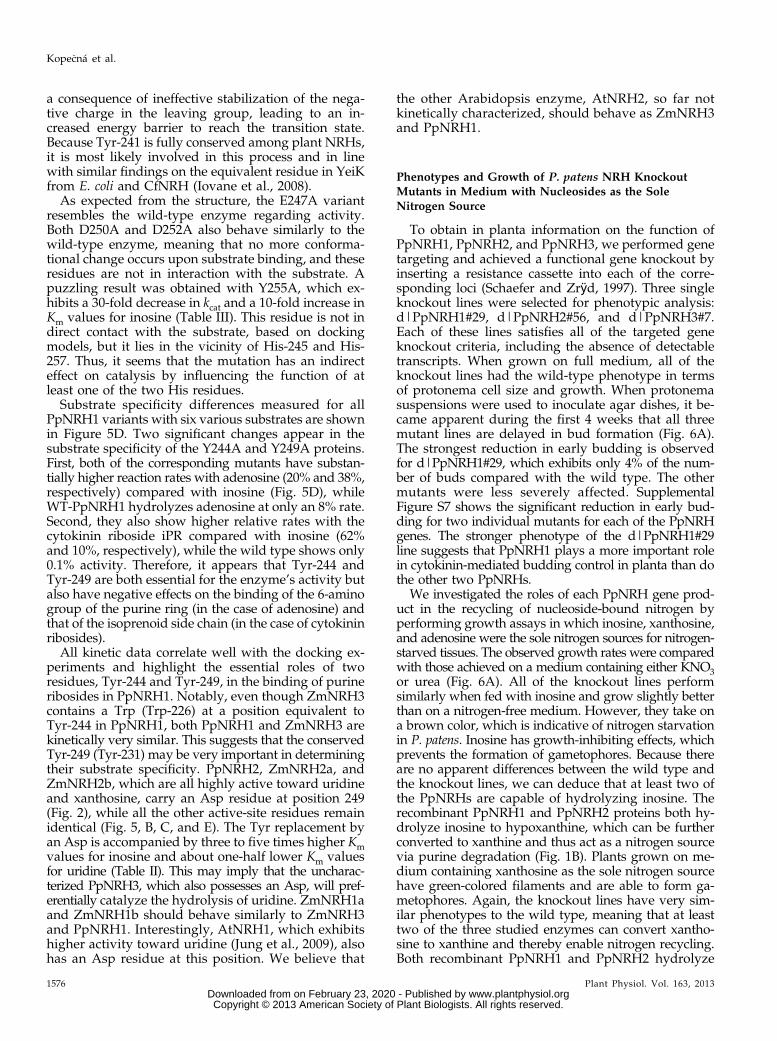
To obtain in planta information on the function ofPpNRH1, PpNRH2, and PpNRH3, we performed genetargeting and achieved a functional gene knockout byinserting a resistance cassette into each of the corre-sponding loci (Schaefer and Zrÿd, 1997). Three singleknockout lines were selected for phenotypic analysis:d|PpNRH1#29, d|PpNRH2#56, and d|PpNRH3#7.Each of these lines satisfies all of the targeted geneknockout criteria, including the absence of detectabletranscripts. When grown on full medium, all of theknockout lines had the wild-type phenotype in termsof protonema cell size and growth. When protonemasuspensions were used to inoculate agar dishes, it be-came apparent during the first 4 weeks that all threemutant lines are delayed in bud formation (Fig. 6A).The strongest reduction in early budding is observedfor d|PpNRH1#29, which exhibits only 4% of the num-ber of buds compared with the wild type. The othermutants were less severely affected. SupplementalFigure S7 shows the significant reduction in early bud-ding for two individual mutants for each of the PpNRHgenes. The stronger phenotype of the d|PpNRH1#29line suggests that PpNRH1 plays a more important rolein cytokinin-mediated budding control in planta than dothe other two PpNRHs.
We investigated the roles of each PpNRH gene prod-uct in the recycling of nucleoside-bound nitrogen byperforming growth assays in which inosine, xanthosine,and adenosine were the sole nitrogen sources for nitrogen-starved tissues. The observed growth rates were comparedwith those achieved on a medium containing either KNO3or urea (Fig. 6A). All of the knockout lines performsimilarly when fed with inosine and grow slightly betterthan on a nitrogen-free medium. However, they take ona brown color, which is indicative of nitrogen starvationin P. patens. Inosine has growth-inhibiting effects, whichprevents the formation of gametophores. Because thereare no apparent differences between the wild type andthe knockout lines, we can deduce that at least two ofthe PpNRHs are capable of hydrolyzing inosine. Therecombinant PpNRH1 and PpNRH2 proteins both hy-drolyze inosine to hypoxanthine, which can be furtherconverted to xanthine and thus act as a nitrogen sourcevia purine degradation (Fig. 1B). Plants grown on me-dium containing xanthosine as the sole nitrogen sourcehave green-colored filaments and are able to form ga-metophores. Again, the knockout lines have very sim-ilar phenotypes to the wild type, meaning that at leasttwo of the three studied enzymes can convert xantho-sine to xanthine and thereby enable nitrogen recycling.Both recombinant PpNRH1 and PpNRH2 hydrolyze
1576 Plant Physiol. Vol. 163, 2013
Kope�cná et al.
www.plantphysiol.orgon February 23, 2020 - Published by Downloaded from Copyright © 2013 American Society of Plant Biologists. All rights reserved.
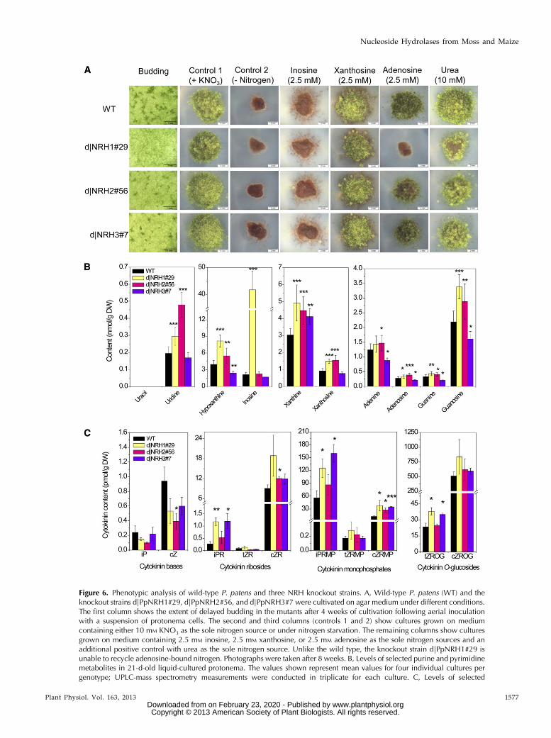
Figure 6. Phenotypic analysis of wild-type P. patens and three NRH knockout strains. A, Wild-type P. patens (WT) and theknockout strains d|PpNRH1#29, d|PpNRH2#56, and d|PpNRH3#7 were cultivated on agar medium under different conditions.The first column shows the extent of delayed budding in the mutants after 4 weeks of cultivation following aerial inoculationwith a suspension of protonema cells. The second and third columns (controls 1 and 2) show cultures grown on mediumcontaining either 10 mM KNO3 as the sole nitrogen source or under nitrogen starvation. The remaining columns show culturesgrown on medium containing 2.5 mM inosine, 2.5 mM xanthosine, or 2.5 mM adenosine as the sole nitrogen sources and anadditional positive control with urea as the sole nitrogen source. Unlike the wild type, the knockout strain d|PpNRH1#29 isunable to recycle adenosine-bound nitrogen. Photographs were taken after 8 weeks. B, Levels of selected purine and pyrimidinemetabolites in 21-d-old liquid-cultured protonema. The values shown represent mean values for four individual cultures pergenotype; UPLC-mass spectrometry measurements were conducted in triplicate for each culture. C, Levels of selected
Plant Physiol. Vol. 163, 2013 1577
Nucleoside Hydrolases from Moss and Maize
www.plantphysiol.orgon February 23, 2020 - Published by Downloaded from Copyright © 2013 American Society of Plant Biologists. All rights reserved.
xanthosine (Table I). A remarkable result was obtainedwith medium containing adenosine as the sole nitrogensource: while the d|PpNRH2#56 and d|PpNRH3#7knockout lines exhibited similar levels of growth to thewild type, no growth was observed for the d|PpNRH1#29knockout line. Although recombinant PpNRH2 is weaklyactive toward adenosine (Table I), this finding stronglysuggests that PpNRH1 is the only P. patens NRH that iscapable of effectively recycling nitrogen from adenosinein vivo. This result clearly demonstrates that, togetherwith xanthine dehydrogenase, NRHs play a central rolein purine degradation and the recycling of nucleoside-bound nitrogen.
Changes in the Levels of Purine, Pyrimidine, andCytokinin Metabolites in d|PpNRH Knockout Mutants
As expected from the kinetic data (Tables I and II),we observe accumulations of inosine (20-fold), uridine(1.4-fold), and xanthosine (1.5-fold) in the d|PpNRH1#29mutant line relative to the wild type (Fig. 6B). The levelsof uracil and cytidine in this mutant line are below thelimit of detection. Similar accumulation is observedfor uridine (2.5-fold) and xanthosine (1.6-fold) ind|PpNRH2#56 plants. Surprisingly, the level of hy-poxanthine (which is produced by the hydrolysis ofinosine) in the d|PpNRH1 plants is twice that in the wildtype, indicating that purine metabolism is modified inthis knockout. Because inosine has been found to causegrowth inhibition in P. patens (Fig. 6A), we may assumethat a harmful excess of inosine in the d|PpNRH1 mu-tant is eliminated by (unspecified) alternative pathways.It should be noted that Riegler et al. (2011) did not ob-serve an accumulation of inosine over the detection limitin any of their nrh1 and nrh2 single and double mutantsin Arabidopsis. In contrast, the AtNRH1 knockout mutantaccumulates high levels of uridine in roots, which isconsistent with the preferential activity of this NRHtoward this substrate.
Although the xanthosine levels in the d|PpNRH1and d|PpNRH2 plants are only slightly higher thanthose in the wild type, the difference is statistically sig-nificant (P, 0.001) and is consistent with the observationthat both of the corresponding recombinant enzymes canhydrolyze this riboside. Overall, these data indicate thatP. patens produces at least two NRHs hydrolyzing xan-thosine. The levels of xanthine (a product of xanthosinehydrolysis) are increased significantly in all three knock-out lines, which again suggests that they have abnormalpurine metabolism due to deficiencies in NRH activity.
Interestingly, the far less pronounced accumulation ofxanthosine in d|PpNRH1#29 and d|PpNRH2#56 com-pared with that seen in the nrh1 (urh1) Arabidopsismutant (Riegler et al., 2011) suggests that bryophytesand seed plants differ in terms of xanthosine homeo-stasis. A comparable result (i.e. a relatively low butstatistically significant increase in d|PpNRH1#29 andd|PpNRH2#56) is also found for the other purine nu-cleosides adenosine and guanosine. Conversely, thelevels of these metabolites in the d|PpNRH3#7 plantsare slightly lower than in the wild type. The finding thatthe levels of both the bases and the ribosides are alteredindicates that their endogenous levels are regulated byboth the NRHs and by enzymes that are active in othermetabolic pathways, which will presumably respond tochanges in the abundance of the various purine de-rivatives. The absence of an accumulation of uridine,inosine, and xanthosine in the d|PpNRH3#7 plantsindicates that PpNRH3 probably plays a minor role intheir in vivo hydrolysis, either due to low expression ofthe functional PpNRH3 or because PpNRH3 expressionis specific to some developmental stage other thanthose examined in this work.
The first report on the functionality of AtNRH1 (Junget al., 2009) demonstrated that recombinant enzyme iscapable of hydrolyzing iPR. To date, no in planta ex-periments have been published showing the relevanceof NRHs in cytokinin activation. The major pathway forcytokinin activation is that involving phosphoribohy-drolase (LONELY GUY; Kurakawa et al., 2007), whichreleases cytokinin bases from the corresponding nucle-otides. In mosses, N6-(2-isopentenyl)adenine (iP) is animportant cytokinin base that has significant effects ondevelopment because it induces bud formation in pro-tonema (von Schwartzenberg et al., 2007). Our enzy-matic study reveals that PpNRH1 and PpNRH2 arecapable of releasing iP from the corresponding iPR, albeitat low rates (Table I). The kinetic data for ZmNRH2b andZmNRH3 indicate that their Km values for iPR and tZRare similar to those for other purine substrates, but theirturnover rates for these substrates are much lower.
Cytokinin analyses demonstrate that the levelsof endogenous iP and cis-zeatin in all three of thed|PpNRH mutant lines are slightly lower than in thewild type (the trans-zeatin levels are below the limit ofdetection). This is consistent with the observed hy-drolysis of these cytokinin ribosides in vitro by thevarious PpNRH enzymes. The lower iP levels seen inour mutant lines are also consistent with their re-duced levels of early bud formation. The ribosides iPRand cis-zeatin riboside as well as the ribonucleotides
Figure 6. (Continued.)cytokinins in 21-d-old liquid-cultured protonema as determined by UPLC-mass spectrometry (three cultures per genotype).Asterisks indicate significant differences between the mutant lines and the wild type at P value thresholds of 0.05 (*), 0.01 (**),and 0.001 (***), according to Student’s t test. Error bars indicate SD. cZ, cis-Zeatin; DW, dry weight; cZRMP, cis-zeatin riboside59-monophosphate; cZROG, cis-zeatin riboside-O-glucoside; iPRMP, isopentenyladenosine 59-monophosphate; tZROG, trans-zeatin riboside-O-glucoside.
1578 Plant Physiol. Vol. 163, 2013
Kope�cná et al.
www.plantphysiol.orgon February 23, 2020 - Published by Downloaded from Copyright © 2013 American Society of Plant Biologists. All rights reserved.
isopentenyladenosine 59-monophosphate and cis-zeatinriboside 59-monophosphate accumulate in all three mu-tant genotypes (Fig. 6C), whereas the accumulation oftrans-zeatin derivatives is less pronounced. A significantaccumulation of trans-zeatin riboside-O-glucoside is ob-served in d|PpNRH1#29 and d|PpNRH3#7 knockoutplants. However, the levels of the cis-zeatin riboside-O-glucoside are only slightly elevated in d|PpNRH1#29.These observations indicate that an excess of zeatin-typeribosides is glucosylated by zeatin-O-glucosyltrans-ferases. Cytokinin O-glucosides are generally assumedto be storage products that can be activated throughhydrolysis by b-glucosidases. In summary, we can statethat all three PpNRHs have an impact on cytokininhomeostasis, although we cannot rule out that some ofthe effects might be due to indirect or unspecific re-sponses of the whole cytokinin homeostatic system in-volving other pathways in addition to NRHs.Taken together, changes in the metabolite profiles
are generally compatible with the substrate preferencesdetermined for the recombinant PpNRHs. Unexpect-edly, the increased levels of hypoxanthine, xanthine, oradenine can be possibly attributed to the activity of near
pathways, including up-regulation of cytokinin oxidase/dehydrogenase, adenine deaminase, or xanthine oxidase,which might compensate the levels of these metabolites.Moreover, Jung et al. (2011) reported that the accumu-lation of nucleosides in the nrh1Arabidopsis mutant wasstrongly increased under conditions of prolonged dark-ness, when salvage pathways are more important thanthe biosynthetic routes. The metabolite profiling for theP. patens single knockout mutants was undertaken understandard conditions. In the case of ADK, another purine/cytokinin-interconverting enzyme, the expected decreaseof AMP was not observed in ADK-silenced Arabidopsisplants, which was also explained by the activity of relatedmetabolic pathways leading to AMP production (Schooret al., 2011). NRH, ADK, and adenine phosphoribosyl-transferase are similar in that the cytokinins are not theirmajor substrates in vitro (Moffatt et al., 2000; Allen et al.,2002). Nevertheless, it was shown for the ArabidopsisADK knockdown plants (Schoor et al., 2011) as well asfor adenine phosphoribosyltransferase1-1 mutants (Zhanget al., 2013) that the levels of endogenous cytokininsare significantly changed, revealing an impact of theseenzymes on the cytokinin interconversion.
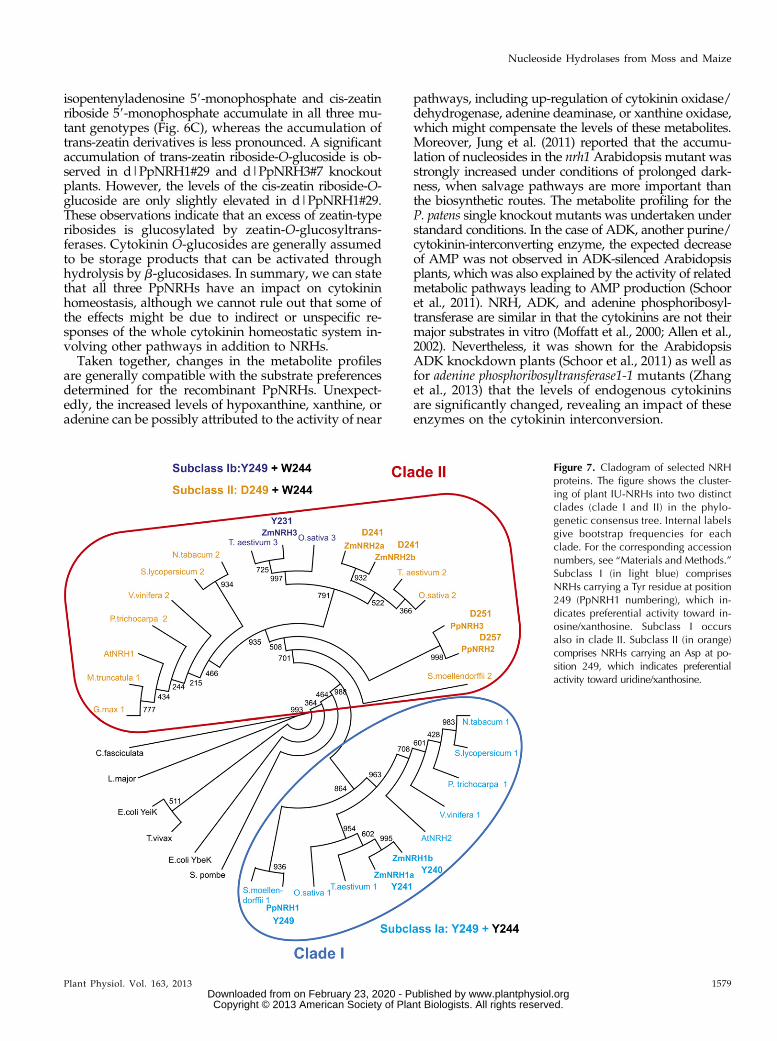
Figure 7. Cladogram of selected NRHproteins. The figure shows the cluster-ing of plant IU-NRHs into two distinctclades (clade I and II) in the phylo-genetic consensus tree. Internal labelsgive bootstrap frequencies for eachclade. For the corresponding accessionnumbers, see “Materials and Methods.”Subclass I (in light blue) comprisesNRHs carrying a Tyr residue at position249 (PpNRH1 numbering), which in-dicates preferential activity toward in-osine/xanthosine. Subclass I occursalso in clade II. Subclass II (in orange)comprises NRHs carrying an Asp at po-sition 249, which indicates preferentialactivity toward uridine/xanthosine.
Plant Physiol. Vol. 163, 2013 1579
Nucleoside Hydrolases from Moss and Maize
www.plantphysiol.orgon February 23, 2020 - Published by Downloaded from Copyright © 2013 American Society of Plant Biologists. All rights reserved.
Phylogeny of Plant IU-NRHs
Phylogenetic analysis (Fig. 7) shows clustering of plantIU-NRHs (Supplemental Fig. S8) into two clades. Theoutgroup comprising NRHs from L. major, C. fasciculata,T. vivax, Schizosaccharomyces pombe, and E. coli are, asexpected, distant to the plant IU-NRHs. PpNRHs andZmNRHs share 49% to 75% sequence identity withother plant NRHs (Supplemental Table S2). PpNRH1 isin the same branch (clade I) as ZmNRH1a and ZmNRH1b,while ZmNRH3 is in the same branch as ZmNRH2a,ZmNRH2b, PpNRH2, and PpNRH3 (clade II). It seemsthat early divergent land plants such as P. patens andSelaginella moellendorffii already had two NRH isoforms,which in most cases were preserved during the evolu-tion of the seed plant line leading to two NRH isoforms,with one in each branch, in higher plants. The apparentduplication in P. patens resulted in PpNRH2 and PpNRH3(clade II), probably due to a whole-genome duplication(Rensing et al., 2007). The Poaceae species maize, rice,and wheat display apparent diversification, leading to atleast two isoforms in clade II. In the case of maize, afurther diversification led to ZmNRH2a and ZmNRH2b.
There is a clear correlation between the clusteringof the NRH genes and their enzymatic properties.Clade I comprises subclass 1a NRHs carrying Tyr-249and Tyr-244 (using the PpNRH1 numbering), indica-tive of inosine and xanthosine preference. Clade IIcomprises mainly subclass II NRHs carrying an Asp atthe same position (together with Trp-244), indicative ofuridine/xanthosine preference. Interestingly, enzymesin clade II from the Poaceae family are also func-tionally diversified (Fig. 7). Although ZmNRH3 andZmNRH2a/ZmNRH2b share 74.2% and 75.8% sequenceidentity (Supplemental Table S2), ZmNRH3 functionallybelongs to subclass Ib (with Tyr-249 and Trp-244) andprefers the substrates inosine and xanthosine.
In the current classification, plant NRHs analyzed inthis work (excluding two-domain NRHs) belong tononspecific IU-NRHs. Our data show that assigningnames of the plant IU-NRHs based on their in vitrosubstrate preferences can be misleading. For example,PpNRH1, which has a xanthosine/inosine preference,shows uridine as a weak substrate in vitro. However,in planta, the d|PpNRH1 line accumulates significantquantities of uridine, demonstrating that PpNRH1 isimportant for uridine conversion. This was the case forAtNRH1, which was initially named as uridine ribo-hydrolase (AtURH1; Jung et al., 2009). Plant IU-NRHsare obviously able to act on a wide range of ribosides,including cytokinin ribosides, and cannot be classifiedas having an exclusive preference for either purines orpyrimidines.
CONCLUSION
This work provides a comprehensive analysis ofIU-NRHs from two plant species, maize and P. patens.It reveals the presence of several NRH genes per plantspecies, leading to the existence of at least two enzyme
groups differing in substrate specificity, either preferringxanthosine/inosine (subclass I) or uridine/xanthosine(subclass II). Structural analysis combined with site-directed mutagenesis identified several residues re-sponsible for nucleoside binding and catalysis. Thesingle knockout mutants in P. patens show changes inthe levels of purine, pyrimidine, and cytokinin metab-olites and point out the importance of NRHs for nu-cleoside and cytokinin metabolism. Here, we prove theparticipation of plant IU-NRHs from both subclasses incytokinin activation in vivo.
MATERIALS AND METHODS
Plant Material and Culture Conditions
The wild-type Physcomitrella patens (Funariaceae) strain used in this workwas derived from the ‘Gransden 2004’ strain. Photoautotrophic growth wasinduced by keeping cultures under axenic conditions in growth chambers(RUMED 1602) at 25°C, illuminated with white light under a 16/8-h light/dark regime, with a flux of 50 mmol m–2 s–1. For metabolite content analysis,tissue was cultivated in liquid medium (Wang et al., 1980) containing0.359 mM Ca(NO3)2, 0.035 mM FeSO4, 1.01 mM MgSO4, 1.84 mM KH2PO4, and10 mM KNO3, to which 1 mL of Hoagland trace element solution was added(Ashton and Cove, 1977).
Cloning, Expression, and Gene Models of NRHs fromP. patens and Maize
Total RNA for reverse transcription was isolated from P. patens (at theprotonema stage) and from 5-d-old maize (Zea mays var saccharata) seedlingsusing the Plant RNA Isolation Aid solutions from Ambion. The RNA wastreated twice with the TURBO DNase-free kit (Ambion). The cDNA was thensynthesized using the SuperScript II reverse transcriptase (Invitrogen) and theRevertAid H Minus reverse transcriptase (Fermentas). Sequences coding forthe PpNRH1 (999 bp), PpNRH2 (1,026 bp), and PpNRH3 (1,017 bp) genes wereamplified using gene-specific primers and the Accuprime Pfx polymerase(Invitrogen; Supplemental Table S3) and then cloned into a pCDFDuet His-tagvector (Novagen). In the case of PpNRH3, four splicing variants were obtained.In addition, five NRH coding sequences from maize were cloned (the primersused are shown in Supplemental Table S3) and submitted to GenBank. Thesesequences were as follows: ZmNRH1a (981 bp), ZmNRH1b (978 bp), ZmNRH2a(978 bp), ZmNRH2b (978 bp), and ZmNRH3 open reading frame (ORF; 948 bp).The plasmids were transformed into T7 express cells (New England Biolabs).Protein expression was induced with 0.5 mM isopropyl-b-thiogalactopyrano-side, after which the cultures were incubated at 20°C overnight.
Except for splicing variants of PpNRH3_v3 and PpNRH3_v4, all of theanalyzed NRH sequences consist of nine exons (Supplemental Fig. S1), withexons 2, 3, 6, and 7 all having the same length. Four variants of PpNRH3 wereidentified due to alternative splicing at the 39 ends of the fifth and eighthexons, none of which matches the current PpNRH3 model. The eighth exonleads to a protein either with a FIAT C-terminal sequence (variants 3 and 4) oran SRLK C terminus (variants 1 and 2), in case the exon is spliced into two,meaning nine exons in total like all other NRH genes. The fifth exon, longer by36 bp, introduces the VSLKRQKSHSRN peptide into the final protein (variants2 and 4). Based on an alignment of plant NRH sequences, variant 1 most likelycorresponds to the active form of PpNRH3. All five of the genes identified inmaize also contain nine exons (Supplemental Fig. S1).
Site-Directed Mutagenesis of PpNRH1
Site-directed mutagenesis was performed on PpNRH1 ORF in a pCDFDuetvector. The mutant H245A was prepared using two complementary primerscontaining the desired mutation (Supplemental Table S3). All of the othermutants were cloned using tail-to-tail-oriented phosphorylated primers, withthe mutation being located at the 59 end of one of the primers. PCR wasperformed using Accuprime Pfx polymerase (Invitrogen) in 30 cycles. Theproducts were treated with DpnI, gel purified, and ligated using the T4 DNA
1580 Plant Physiol. Vol. 163, 2013
Kope�cná et al.
www.plantphysiol.orgon February 23, 2020 - Published by Downloaded from Copyright © 2013 American Society of Plant Biologists. All rights reserved.
ligase (Promega). The sequenced clones were transformed into T7 expresscompetent cells (New England Biolabs). Mutant proteins were screened forthe expression of the His-tagged protein by SDS-PAGE and using activitymeasurements.
Circular Dichroism Spectroscopy
The far-UV circular dichroism spectra of WT-PpNRH1 and its mutantvariants were recorded on a J-815 spectropolarimeter (JASCO) at a concen-tration of 0.5 mg mL21 in 20 mM Tris-HCl (pH 9.0) using a 0.1-cm quartz cell.
Phylogenetic Analysis
Amino acid alignments were performed using MUSCLE version 3.8 (Edgar,2004). A maximum likelihood phylogeny with bootstrap analysis was performedwith PhyML version 3.0 (Guindon et al., 2010) using the LG amino acid re-placement matrix. NRH sequences from the following species were obtainedfrom the National Center for Biotechnology Information, Phytozome, TheGene Index Project (http://compbio.dfci.harvard.edu/tgi/plant.html; Tenta-tive Consensus accessions), or The Institute for Genomic Research (http://blast.jcvi.org/euk-blast/plantta_blast.cgi; Transcript Assembly accessions) data-bases: Schizosaccharomyces pombe (CAB91168), Escherichia coli YbeK (AAN79208),E. coli YeiK (AAA60514), Leishmania major (AY533501), Crithidia fasciculata (CFU43371),and Trypanosoma vivax (AF311701). Further complete plant NRH sequenceswere used:P. patens PpNRH1 (JQ649322), PpNRH2 (JX861385), and PpNRH3 (JX861386);maize ZmNRH1a (HQ825159), ZmNRH1b (HQ825160), ZmNRH2a (HQ825161),ZmNRH2b (JQ594984), and ZmNRH3 (HQ825162); Arabidopsis AtNRH1(At2g36310) and AtNRH2 (At1g05620); soybean (Glycine max 1; BT097166),Medicago truncatula 1 (XM_003625740), tobacco (Nicotiana tabacum 1/2; TC91311 andTC81540), rice (Oryza sativa 1/2/3; Os03g31170.1, Os08g44370.1, and Os09g39440.1),Populus trichocarpa 1/2 (XM_002310348 and XM_002309011), Selaginella moellendorffii1/2 (XM_002974764 and XM_002984237), tomato (Solanum lycopersicum 1/2;AK325443 and AK322170), wheat (Triticum aestivum 1/2/3; TA56209_4565,TA76228_4565, and TA76014_4565), and grape (Vitis vinifera 1/2; XM_002280235and XM_002283117).
Activity Measurement
Purine and pyrimidine ribosides were purchased from Sigma-Aldrich. TheNRH activity was measured spectrophotometrically at 30°C according to amethod described by Parkin (1996). The reaction in 200 mM Tris-HCl buffer,pH 7.5, 400 mM KCl, 1 mM dithiothreitol (DTT), and a riboside substrate wasinitiated by adding an appropriate amount of the enzyme (up to 50 mg forWT-PpNRH1 and 100–500 mg for the mutants). Kinetic constants were de-termined using the GraphPad Prism 5.0 software (GraphPad Software) bymonitoring the absorption decrease of adenosine (D«276 = 21.4 mM
21 cm21),inosine (D«280 = 20.92 mM
21 cm21), uridine (D«280 = 21.8 mM21 cm21), cytidine
(D«280 = 23.42 mM21 cm21), and thymidine (D«265 = 21.7 mM
21 cm21; Parkin,1996). The differential extinction coefficients for xanthosine and guanosinewere determined to be D«248 = 23.7 and 24.1 mM
21 cm21, respectively. Sim-ilarly, D«289 values of 21.37 and 21.48 mM
21 cm21 were determined for iPRand tZR, respectively.
Purification, Crystallization, and Structure Determination
All NRHs were purified on Co-Sepharose columns, and both PpNRH1 andZmNRH3 were further purified by gel filtration chromatography on a HiLoad26/60 Superdex 200 column using 50 mM Tris-HCl buffer (pH 8.0) and 150 mM
NaCl. The purified PpNRH1 and ZmNRH3 fractions were concentrated to 30to 35 mg mL21. Crystallization conditions for both NRHs were screened usingQiagen kits (Valencia) with a Cartesian nanodrop robot (Genomic Solutions).Crystals were obtained in hanging drops by mixing equal volumes of proteinsolution and a precipitant solution containing 0.1 M HEPES (pH 7.5), 100 mM
sodium acetate, 10% (w/v) polyethylene glycol (PEG) 4000, and 10% ethyleneglycol for PpNRH1 and containing 50 mM Tris-HCl (pH 8.0), 150 mM NaCl,and 20% (w/v) PEG 2000 monomethyl ether for ZmNRH3. Crystals weretransferred to a cryoprotectant solution (the mother liquor supplemented with25% PEG 400) and flash frozen in liquid nitrogen. Diffraction data for thePpNRH1 and ZmNRH3 crystals were collected at 100 K on the Proxima 1beamline at the SOLEIL synchrotron at resolutions of 3.35 and 2.49 Å,
respectively. Intensities were integrated using the XDS program (Kabsch,2010; Supplemental Table S1).
The crystal structures of ZmNRH3 and PpNRH1 were determined byperforming molecular replacement with Phaser (Storoni et al., 2004), using themonomer of YbeK (PDB 1YOE) and the dimer of ZmNRH3 as search models,respectively. Both models were refined with strong non-crystallographicsymmetry restraints using Buster 2.10 (Bricogne et al., 2011). One translation/libration/screw-motion group was assigned for the dimer in the 2.49-Å struc-ture and the four dimers in the lower resolution structure. Electron densitymaps were evaluated using COOT (Emsley and Cowtan, 2004). Refinementstatistics are presented in Supplemental Table S1. No electron density wasobserved for residues 230 to 234 in subunit B of the ZmNRH3 structure. Insubunit A, the electron density map was poorly defined for the side chains inthe region comprising residues 228 to 337. Only two dimers (AB and CD) ofthe four were well defined in the electron density maps of the PpNRH1 structure.The dimer GH and mostly the molecule H present many disordered side chainsand poor electron density maps in a few regions. Molecular graphics images weregenerated using PYMOL (www.pymol.org).
Substrate Docking into the Active Sites of PpNRH1and ZmNRH3
The AutoDock suite 4.2.5.1 (Morris et al., 2009) was used for docking ex-periments. Both target active sites were kept rigid, while Tyr-241, Tyr-244, andTyr-249 in PpNRH1 (Tyr-223, Trp-226, and Tyr-231 in ZmNRH3) were keptflexible. Hydrogen atoms were added and Gasteiger partial charges werecomputed. Calcium atom charges were added manually. Coordinates for inosinewere taken from the structure of a YeiK complex (PDB 3B9X), while xanthosineand uridine were built in Avogadro 1.0.0 (http://avogadro.openmolecules.net/).The Rib moiety was constrained to maintain the C49-endo puckered confor-mation, as commonly found in ribosides bound to NRHs. Docking calcula-tions were performed using a Lamarckian genetic algorithm and a maximumof 100 conformers.
Generation of P. patens NRH Knockout Mutants
Functional gene knockouts of PpNRH1, PpNRH2, and PpNRH3 wereprepared using three gene-replacement vectors. The vectors were alldesigned using a resistance cassette flanked by 800- to 1,000-bp-long ge-nomic fragments from the 59 and 39 regions of the corresponding genomicNRH locus. Details of the construction of the replacement vectors are pro-vided in Supplemental Methods S1, and primer sequences are given inSupplemental Table S4. The transformation of P. patens protoplasts wascarried out according to Schaefer et al. (1991) with minor modifications.Each transformation assay was performed using 25 mg of vector DNA. Be-tween 15 and 20 stable moss lines were obtained after three rounds of se-lection using the appropriate antibiotic for each knockout construct. Thehaploid status of the transformants was verified, and eight to 10 transgeniclines for each mutant were arbitrarily chosen. These were then analyzed byPCR for recombination events at the corresponding loci and by reversetranscription-PCR for the absence of the transcript. Three knockout lines,d|PpNRH1#29, d|PpNRH2#56, and d|PpNRH3#7, were chosen for biochemicaland phenotypic characterization.
Extraction and Determination of Purine, Pyrimidine, andCytokinin Metabolites
P. patens wild-type and mutant lines were collected, freeze dried, andpurified in triplicate (10 mg dry weight per sample). For quantification of thepurine/pyrimidine bases and ribosides, the samples were homogenized, extrac-ted in cold water with 25% ammonia (ratio, 4:1), and purified by solid-phaseextraction with the addition of the stable-labeled internal standards. All sampleswere further purified on mixed-mode anion-exchange sorbent Oasis MAXcartridges (Waters) and analyzed using an Acquity ultra-performance liquidchromatography (UPLC) system connected to a triple quadrupole massspectrometer (Xevo TQ MS; Waters MS Technologies). Further details aregiven in Supplemental Methods S1. Ultra-performance liquid chromatography-tandemmass spectrometry analysis was used to determine the cytokinin contentof each sample (von Schwartzenberg et al., 2007) using a modified procedure ofNovák et al. (2008).
Plant Physiol. Vol. 163, 2013 1581
Nucleoside Hydrolases from Moss and Maize
www.plantphysiol.orgon February 23, 2020 - Published by Downloaded from Copyright © 2013 American Society of Plant Biologists. All rights reserved.
Sequence data can be found in the GenBank/EMBL data libraries underaccession numbers JQ649322 (PpNRH1), JX861385 (PpNRH2), JX861386 to JX861389(the four splicing variants of PpNRH3), HQ825159 (ZmNRH1a), HQ825160(ZmNRH1b), HQ825161 (ZmNRH2a), JQ594984 (ZmNRH2b), and HQ825162(the ZmNRH3 ORF). The atomic coordinates and structure factors have beendeposited in the PDB under accession codes 4KPN (PpNRH1) and 4KPO(ZmNRH3).
Supplemental Data
The following materials are available in the online version of this article.
Supplemental Figure S1. NRH gene models for P. patens, maize, and Ara-bidopsis.
Supplemental Figure S2. Influence of pH and temperature on catalyticactivity of recombinant PpNRH1.
Supplemental Figure S3. Confirmation of cytokinin riboside conversion byZmNRH3, ZmNRH2b, and PpNRH1.
Supplemental Figure S4. Gel permeation chromatography of PpNRH1and ZmNRH3.
Supplemental Figure S5. Structural comparison of plant NRH (ZmNRH3,this article) with NRH from E. coli (YeiK, PDB 3B9X).
Supplemental Figure S6. Production of PpNRH1 protein variants.
Supplemental Figure S7. Delayed bud development within 4 weeks afteraerial inoculation with protonema suspension.
Supplemental Figure S8. Alignment of plant NHR sequences.
Supplemental Table S1. Data collection and refinement statistics of plantNRHs.
Supplemental Table S2. Identities of ZmNRH3 and PpNRH1 with otherNRHs from maize and P. patens and from other species.
Supplemental Table S3. Primer pairs used for the cloning of NRHs and forthe site-directed mutagenesis of PpNRH1.
Supplemental Table S4. Primers used for generation of gene replacementvectors and genetic analysis of P. patens knockout mutants.
Supplemental Methods S1. Generation of P. patens knockout mutants andquantification of ribosides.
ACKNOWLEDGMENTS
The pBZRF vector was provided by the courtesy of Fabien Nogué (InstitutNational de la Recherche Agronomique, Versailles). We thank Barbara Moffattand Katja Engel (University of Waterloo) for contributions to enzyme assays inan early phase of the project, Jeanette Klein (Charité Berlin) for initial purineand pyrimidine measurements, Dr. Petr Tarkowski (University of Olomouc)for HPLC measurement, Vera Schwekendiek and Susanne Bringe (Universityof Hamburg) for excellent technical support, Beatriz Guimaraes for help indata collection on Proxima 1 at the SOLEIL synchrotron, Pierre Legrand (Syn-chrotron SOLEIL) and Gérard Bricogne (Global Phasing Ltd) for help in dataprocessing and refinement strategy, and Andrew Thompson for help withmanuscript preparation. This work benefited from the IMAGIF platform fa-cilities (www.imagif.cnrs.fr) at the Centre de Recherche de Gif-sur-Yvette.
Received September 18, 2013; accepted October 28, 2013; published October29, 2013.
LITERATURE CITED
Achar BS, Vaidyanathan CS (1967) Purification and properties of uridinehydrolase from mung-bean (Phaseolus radiatus) seedlings. Arch BiochemBiophys 119: 356–362
Allen M, Qin W, Moreau F, Moffatt B (2002) Adenine phosphoribosyl-transferase isoforms of Arabidopsis and their potential contributions toadenine and cytokinin metabolism. Physiol Plant 115: 56–68
Ashton NW, Cove DJ (1977) The isolation and preliminary characterisationof auxotrophic and analogue resistant mutants of the moss, Physcomi-trella patens. Mol Gen Genet 154: 87–95
Bricogne G, Blanc E, Brandl M, Flensburg C, Keller P, Paciorek W, Roversi P,Sharff A, Smart OS, Vonrhein C, et al (2011) BUSTER Version 2.1.0. GlobalPhasing, Cambridge, UK
Campos A, Rijo-Johansen MJ, Carneiro MF, Fevereiro P (2005) Purifica-tion and characterisation of adenosine nucleosidase from Coffea arabicayoung leaves. Phytochemistry 66: 147–151
Chen CM, Kristopeit SM (1981) Metabolism of cytokinin: deribosylation ofcytokinin ribonucleoside by adenosine nucleosidase from wheat germcells. Plant Physiol 68: 1020–1023
Chen M, Thelen JJ (2011) Plastid uridine salvage activity is required forphotoassimilate allocation and partitioning in Arabidopsis. Plant Cell 23:2991–3006
Cove D, Bezanilla M, Harries P, Quatrano R (2006) Mosses as modelsystems for the study of metabolism and development. Annu Rev PlantBiol 57: 497–520
Degano M, Almo SC, Sacchettini JC, Schramm VL (1998) Trypanosomalnucleoside hydrolase: a novel mechanism from the structure with atransition-state inhibitor. Biochemistry 37: 6277–6285
Degano M, Gopaul DN, Scapin G, Schramm VL, Sacchettini JC (1996)Three-dimensional structure of the inosine-uridine nucleoside N-ribohydrolasefrom Crithidia fasciculata. Biochemistry 35: 5971–5981
Edgar RC (2004) MUSCLE: multiple sequence alignment with high accur-acy and high throughput. Nucleic Acids Res 32: 1792–1797
Emsley P, Cowtan K (2004) Coot: model-building tools for moleculargraphics. Acta Crystallogr D Biol Crystallogr 60: 2126–2132
Estupiñán B, Schramm VL (1994) Guanosine-inosine-preferring nucleosideN-glycohydrolase from Crithidia fasciculata. J Biol Chem 269: 23068–23073
Garau G, Muzzolini L, Tornaghi P, Degano M (2010) Active site plasticityrevealed from the structure of the enterobacterial N-ribohydrolase RihAbound to a competitive inhibitor. BMC Struct Biol 10: 14
Giabbai B, Degano M (2004) Crystal structure to 1.7 a of the Escherichiacoli pyrimidine nucleoside hydrolase YeiK, a novel candidate for cancergene therapy. Structure 12: 739–749
Gopaul DN, Meyer SL, Degano M, Sacchettini JC, Schramm VL (1996)Inosine-uridine nucleoside hydrolase from Crithidia fasciculata: geneticcharacterization, crystallization, and identification of histidine 241 as acatalytic site residue. Biochemistry 35: 5963–5970
Guindon S, Dufayard JF, Lefort V, Anisimova M, Hordijk W, Gascuel O(2010) New algorithms and methods to estimate maximum-likelihood phy-logenies: assessing the performance of PhyML 3.0. Syst Biol 59: 307–321
Guranowski A (1982) Purine catabolism in plants: purification and someproperties of inosine nucleosidase from yellow lupin (Lupinus luteus L.)seeds. Plant Physiol 70: 344–349
Guranowski A, Schneider Z (1977) Purification and characterization ofadenosine nucleosidase from barley leaves. Biochim Biophys Acta 482:145–158
Horenstein BA, Parkin DW, Estupiñán B, Schramm VL (1991) Transition-state analysis of nucleoside hydrolase from Crithidia fasciculata. Bio-chemistry 30: 10788–10795
Houba-Hérin N, Pethe C, d’Alayer J, Laloue M (1999) Cytokinin oxidasefrom Zea mays: purification, cDNA cloning and expression in mossprotoplasts. Plant J 17: 615–626
Iovane E, Giabbai B, Muzzolini L, Matafora V, Fornili A, Minici C,Giannese F, Degano M (2008) Structural basis for substrate specificity ingroup I nucleoside hydrolases. Biochemistry 47: 4418–4426
Jung B, Flörchinger M, Kunz HH, Traub M, Wartenberg R, Jeblick W,Neuhaus HE, Möhlmann T (2009) Uridine-ribohydrolase is a key reg-ulator in the uridine degradation pathway of Arabidopsis. Plant Cell 21:876–891
Jung B, Hoffmann C, Möhlmann T (2011) Arabidopsis nucleoside hy-drolases involved in intracellular and extracellular degradation of pu-rines. Plant J 65: 703–711
Kabsch W (2010) XDS. Acta Crystallogr D Biol Crystallogr 66: 125–132Kurakawa T, Ueda N, Maekawa M, Kobayashi K, Kojima M, Nagato Y,
Sakakibara H, Kyozuka J (2007) Direct control of shoot meristem ac-tivity by a cytokinin-activating enzyme. Nature 445: 652–655
Mainguet SE, Gakière B, Majira A, Pelletier S, Bringel F, Guérard F,Caboche M, Berthomé R, Renou JP (2009) Uracil salvage is necessaryfor early Arabidopsis development. Plant J 60: 280–291
1582 Plant Physiol. Vol. 163, 2013
Kope�cná et al.
www.plantphysiol.orgon February 23, 2020 - Published by Downloaded from Copyright © 2013 American Society of Plant Biologists. All rights reserved.
Massonneau A, Houba-Hérin N, Pethe C, Madzak C, Falque M, Mercy M,Kopecny D, Majira A, Rogowsky P, Laloue M (2004) Maize cytokininoxidase genes: differential expression and cloning of two new cDNAs.J Exp Bot 55: 2549–2557
Moffatt B, Pethe C, Laloue M (1991) Metabolism of benzyladenine is im-paired in a mutant of Arabidopsis thaliana lacking adenine phosphori-bosyltransferase activity. Plant Physiol 95: 900–908
Moffatt B, Somerville C (1988) Positive selection for male-sterile mutantsof Arabidopsis lacking adenine phosphoribosyl transferase activity.Plant Physiol 86: 1150–1154
Moffatt BA, Stevens YY, Allen MS, Snider JD, Pereira LA, Todorova MI,Summers PS, Weretilnyk EA, Martin-McCaffrey L, Wagner C (2002)Adenosine kinase deficiency is associated with developmental abnor-malities and reduced transmethylation. Plant Physiol 128: 812–821
Moffatt BA, Wang L, Allen MS, Stevens YY, Qin W, Snider J, vonSchwartzenberg K (2000) Adenosine kinase of Arabidopsis: kineticproperties and gene expression. Plant Physiol 124: 1775–1785
Mok DW, Mok MC (2001) Cytokinin metabolism and action. Annu RevPlant Physiol Plant Mol Biol 52: 89–118
Morris GM, Huey R, Lindstrom W, Sanner MF, Belew RK, Goodsell DS,Olson AJ (2009) AutoDock4 and AutoDockTools4: automated dockingwith selective receptor flexibility. J Comput Chem 30: 2785–2791
Muzzolini L, Versées W, Tornaghi P, Van Holsbeke E, Steyaert J, DeganoM (2006) New insights into the mechanism of nucleoside hydrolasesfrom the crystal structure of the Escherichia coli YbeK protein bound tothe reaction product. Biochemistry 45: 773–782
Novák O, Hauserová E, Amakorová P, Dole�zal K, Strnad M (2008) Cytokininprofiling in plant tissues using ultra-performance liquid chromatography-electrospray tandem mass spectrometry. Phytochemistry 69: 2214–2224
Parkin DW (1996) Purine-specific nucleoside N-ribohydrolase from Try-panosoma brucei brucei: purification, specificity, and kinetic mechanism.J Biol Chem 271: 21713–21719
Parkin DW, Horenstein BA, Abdulah DR, Estupiñán B, Schramm VL (1991)Nucleoside hydrolase from Crithidia fasciculata: metabolic role, purification,specificity, and kinetic mechanism. J Biol Chem 266: 20658–20665
Rensing SA, Ick J, Fawcett JA, Lang D, Zimmer A, Van de Peer Y, Reski R(2007) An ancient genome duplication contributed to the abundance ofmetabolic genes in the moss Physcomitrella patens. BMC Evol Biol 7: 130
Riegler H, Geserick C, Zrenner R (2011) Arabidopsis thaliana nucleosidasemutants provide new insights into nucleoside degradation. New Phytol191: 349–359
Riewe D, Grosman L, Fernie AR, Zauber H, Wucke C, Geigenberger P (2008)A cell wall-bound adenosine nucleosidase is involved in the salvage of ex-tracellular ATP in Solanum tuberosum. Plant Cell Physiol 49: 1572–1579
Sakakibara H (2006) Cytokinins: activity, biosynthesis, and translocation.Annu Rev Plant Biol 57: 431–449
Schaefer D, Zryd JP, Knight CD, Cove DJ (1991) Stable transformation ofthe moss Physcomitrella patens. Mol Gen Genet 226: 418–424
Schaefer DG, Zrÿd JP (1997) Efficient gene targeting in the moss Phys-comitrella patens. Plant J 11: 1195–1206
Schoor S, Farrow S, Blaschke H, Lee S, Perry G, von Schwartzenberg K,Emery N, Moffatt B (2011) Adenosine kinase contributes to cytokinininterconversion in Arabidopsis. Plant Physiol 157: 659–672
Shi W, Schramm VL, Almo SC (1999) Nucleoside hydrolase from Leish-mania major: cloning, expression, catalytic properties, transition stateinhibitors, and the 2.5-Å crystal structure. J Biol Chem 274: 21114–21120
Stasolla C, Katahira R, Thorpe TA, Ashihara H (2003) Purine and py-rimidine nucleotide metabolism in higher plants. J Plant Physiol 160:1271–1295
Storoni LC, McCoy AJ, Read RJ (2004) Likelihood-enhanced fast rotationfunctions. Acta Crystallogr D Biol Crystallogr 60: 432–438
Szuwart M, Starzynska E, Pietrowska-Borek M, Guranowski A (2006)Calcium-stimulated guanosine-inosine nucleosidase from yellow lupin(Lupinus luteus). Phytochemistry 67: 1476–1485
Versées W, Decanniere K, Pellé R, Depoorter J, Brosens E, Parkin DW,Steyaert J (2001) Structure and function of a novel purine specific nu-cleoside hydrolase from Trypanosoma vivax. J Mol Biol 307: 1363–1379
Versées W, Decanniere K, Van Holsbeke E, Devroede N, Steyaert J (2002)Enzyme-substrate interactions in the purine-specific nucleoside hydro-lase from Trypanosoma vivax. J Biol Chem 277: 15938–15946
Versées W, Steyaert J (2003) Catalysis by nucleoside hydrolases. Curr OpinStruct Biol 13: 731–738
von Schwartzenberg K (2009) Hormonal regulation of development byauxin and cytokinin in moss. Annu Plant Rev 36: 246-281
von Schwartzenberg K, Kruse S, Reski R, Moffatt B, Laloue M (1998)Cloning and characterization of an adenosine kinase from Physcomi-trella involved in cytokinin metabolism. Plant J 13: 249–257
von Schwartzenberg K, Núñez MF, Blaschke H, Dobrev PI, Novák O,Motyka V, Strnad M (2007) Cytokinins in the bryophyte Physcomitrellapatens: analyses of activity, distribution, and cytokinin oxidase/dehydrogenaseoverexpression reveal the role of extracellular cytokinins. Plant Physiol 145:786–800
Vyroubalová S, Václavíková K, Turecková V, Novák O, Smehilová M,Hluska T, Ohnoutková L, Frébort I, Galuszka P (2009) Characterizationof new maize genes putatively involved in cytokinin metabolism andtheir expression during osmotic stress in relation to cytokinin levels.Plant Physiol 151: 433–447
Wang TL, Cove DJ, Beutelmann P, Hartmann E (1980) Isopentenyladeninefrom mutants of the moss, Physcomitrella patens. Phytochemistry 19:1103–1105
Zhang X, Chen Y, Lin X, Hong X, Zhu Y, Li W, He W, An F, Guo H (2013)Adenine phosphoribosyl transferase 1 is a key enzyme catalyzing cy-tokinin conversion from nucleobases to nucleotides in Arabidopsis. MolPlant 6: 1661–1672
Zrenner R, Stitt M, Sonnewald U, Boldt R (2006) Pyrimidine and purinebiosynthesis and degradation in plants. Annu Rev Plant Biol 57: 805–836
Plant Physiol. Vol. 163, 2013 1583
Nucleoside Hydrolases from Moss and Maize
www.plantphysiol.orgon February 23, 2020 - Published by Downloaded from Copyright © 2013 American Society of Plant Biologists. All rights reserved.


![The Moss Physcomitrium (Physcomitrella) patens: …REVIEW ARTICLE The Moss Physcomitrium (Physcomitrella) patens: A Model Organism for Non-Seed Plants[OPEN] Stefan A. Rensing,a,1 Bernard](https://img.pdfslide.us/doc/110x75/5f1a2556a4775c2a2e7d8505/the-moss-physcomitrium-physcomitrella-patens-review-article-the-moss-physcomitrium.jpg)