Embed Size (px)
Citation preview
Biosynthesis of Lysosomal Hydrolases : Their Synthesis
in Bound Polysomes and the Role of
Co- and Post-translational Processing
in Determining Their Subcellular Distribution
M. G. ROSENFELD, G . KREIBICH, D. POPOV, K. KATO, and D. D . SABATINIDepartment of Cell Biology, New York University School of Medicine, New York 10016; Institute of CellBiology and Pathology, Bucuresti, Romania; and Department of Biochemistry, Faculty of PharmaceuticalSciences, Kyushu University, Fukuoka 812, Japan
ABSTRACT By in vitro translation of mRNA's isolated from free and membrane-bound poly-somes, direct evidence was obtained for the synthesis of two lysosomal hydrolases, ß-glucu-ronidase of the rat preputial gland and cathepsin D of mouse spleen, on polysomes bound torough endoplasmic reticulum (ER) membranes. When the mRNA's for these two proteins weretranslated in the presence of microsomal membranes, the in vitro synthesized polypeptideswere cotranslationally glycosylated and transferred into the microsomal lumen . Polypeptidessynthesized in the absence of microsomal membranes were 2,000 daltons larger than therespective unglycosylated microsomal polypeptides found after short times of labeling incultured rat liver cells treated with tunicamycin. This strongly suggests that nascent chains ofthe lysosomal enzymes bear transient amino terminal signals which determine synthesis onbound polysomes and are removed during the cotranslational insertion of the polypeptidesinto the ER membranes .
In the line of cultured rat liver cells used for this work, newly synthesized lysosomalhydrolases showed a dual destination; -60% of the microsomal polypeptides detected aftershort times of labeling were subsequently processed proteolytically to lower molecular weightforms characteristic of the mature enzymes. The remainder was secreted from the cells withoutfurther proteolytic processing . As previously observed by other investigations in culturedfibroblasts (A . Gonzalez-Noriega, J . H . Grubbs, V. Talkad, and W. S. Sly, 1980, J. Cell Biol. 85 :839-852; A. Hasilik and E. F. Neufeld, 1980, /. Biol. Chem., 255:4937-4945 .) the lysosomotropicamine chloroquine prevented the proteolytic maturation of newly synthesized hydrolases andenhanced their secretion . In addition, unglycosylated hydrolases synthesized in cells treatedwith tunicamycin were exclusively exported from the cells without undergoing proteolyticprocessing . These results support the notions that modified sugar residues serve as sorting outsignals which address the hydrolases to their lysosomal destination and that final proteolyticcleavage of hydrolase precursors takes place within the lysosome itself .
Structural differences in the carbohydrate chains of intracellular and secreted precursors ofcathepsin D were detected from their differential sensitivity to digestion with endoglycosidasesH and D . These observations suggest that the hydrolases exported into the medium follow thenormal secretory route and that some of their oligosaccharides are subject to modificationsknown to affect many secretory glycoproteins during their passage through the Golgi apparatus.
THE JOURNAL OF CELL BIOLOGY " VOLUME 93 APRIL 1982 135-143©The Rockefeller University Press - 0021-9525/82/04/0135/09 $1 .00 135
on January 12, 2019jcb.rupress.org Downloaded from http://doi.org/10.1083/jcb.93.1.135Published Online: 1 April, 1982 | Supp Info:

The study oflysosomal biogenesis promises to cast light on thediscriminating mechanisms which direct the subcellular trafficof proteins. In several respects, lysosomal hydrolases resemblesecretory proteins and indeed in many cell lines a substantialproportion ofthese proteins are exported into the extracellularmedium (23). It may be inferred from this fact that lysosomalenzymes, like genuine secretory proteins, are synthesized inmembrane bound ribosomes and at least initially follow thesecretory route (9, 52) .
This notion was originally based on cytochemical observa-tions (3, 38) demonstrating the presence of hydrolases in thelumina ofthe endoplasmic reticulum (ER), Golgi complex, andGERL membrane systems. It is also supported by the glyco-protein character common to lysosomal enzymes and manysecretory proteins, as well as by the functional analogy whichcan be made between the secretory discharge which takes placeon the surface of the cell and the release of lysosomal contentinto phagocytic vacuoles. Yet, a direct demonstration of thesynthesis of lysosomal enzymes on membrane-bound poly-somes has not been provided, although in cell-free systemsprogrammed with total mRNA preparations the cotranslationalinsertion of cathepsin D into microsomal vesicles has beendemonstrated (11) .Here we show that mRNA's for the lysosomal enzymes
cathepsin D and,ß-glucuronidase are segregated intracellularlyin membrane-bound polysomes and provide evidence indicat-ing that the nascent lysosomal polypeptides contain amino-terminal insertion signals which may be functionally analogousto those determining the cotranslational insertion of secretorypolypeptides into the ER membranes .
Considerable evidence suggests (19, 20, 23, 25, 48, 54) thatmannosyl phosphate groups on the oligosaccharides of newlysynthesized lysosomal hydrolases serve as sorting-out signalsthat determine that these proteins are sequestered within lyso-somes . We also present experiments which strongly support thehypothesis that carbohydrate moieties play a role in divertinglysosomal hydrolases from the secretory pathway to their func-tional destination within lysosomes. Using tunicamycin to pre-vent the glycosylation of nascent polypeptides synthesized incultured hepatocytes, we found that unglycosylated lysosomalenzymes are completely secreted from cultured cells withoutundergoing proteolytic cleavages that normally seem to accom-pany their incorporation into the organelles.
MATERIALS AND METHODS
Most reagents were purchasedfrom Sigma Chemical Co. (St . Louis, MO) or fromFisher Scientific Co. (Pittsburgh, PA). "S-Methionine (specific activity 1,000 Ci/mmol) was purchased from New England Nuclear (Boston, MA), Trasylol fromMobay Chemical Corp. (New York, NY), ohgo(dT)-cellulose from CollaborativeResearch Inc. (Waltham, MA). RPMI 1640 medium and horse serum werepurchased from Gibco Laboratories (Grand Island Biological Co., Grand Island,NY). Wheat germ was a gift of the General Mills Corporation (Minneapolis,MI).
AnimalsSprague-Dawley female rats (150 g) and adult New Zealand white rabbits
were obtained from Taconic Farms (Germantown, NY) and Swiss female micefrom the Jackson Laboratory (Bar Harbor, ME).
Preparation of Antiserumfl-Glucuronidase was purified from rat preputial glands (24) and cathepsin D
from ratspleen (6 l) . Theproteins (1-2 mg each) emulsified in complete Freund'sadjuvant were injected intradermally at 10-15 sites on the back of rabbits . Abooster injection(0.5 mg) was given 3 wk later and after anotherweek the rabbits
136
THE JOURNAL OF CELL BIOLOGY " VOLUME 93, 1982
were bled from the marginal ear vein. Antiserum was assayed by Ouchterlonydouble-diffusion analysis (40), and specific antibodies were isolated by affinitychromatography (33).
Cell CultureArat hepatocyte line (clone 9) was kindly provided by Dr . M. Kaighn of the
Pasadena Foundation for Medical Research . Cells were cultured at 37°C in anair 5% COz atmosphere in RPMI 1640 medium supplemented with 10% horseserum and antibiotics.
Labeling and Preparation of Cell ExtractsHepatocyte monolayers were grown in 35-mm dishes to a density of-5 x 10`
cells/dish . Before labeling, the cultures were rinsed with RPMIfree of methionineand incubatedin this medium for I h to allow for the depletion of the intracellularmethionine pool . Cells were pulse-labeled for 1 h in methionine-free mediumsupplemented with 10% dialyzed horse serum and ""S-methionine (1001ac'i/ml).For pulse-chase experiments, the labeling was terminated by adding to t timesthe normal concentration of L-methionine in the RPMI medium.
In experiments employing tunicamycin (2 Wg/ml) or chloroquine (25 AM),confluent cultures were first incubated for 1 h in complete medium with therespective drug and then placed in methionine-free media for 1 h before thepulse-chase . The drugs were present throughout the incubation and pulse-chaseperiods .
At different time-points medium was collected and centrifuged for 2 min inan Eppendorf Model 5412 table-top centrifuge (Brinkmann Instruments, Inc.,Westbury, NY) (15,000 g) . After adding 0.8% SDS and 1% Triton X-100 thesample was boiled for 2 min and then diluted with an equal volume of 50 mMTris-HC17.4, 190mM NaCl, 6 mM EDTA, 2.5% Triton X-100, 0.02% NaN3, and100 U/ml Trasylol (solution A) .
Monolayers were washed twice with Moscona's phosphate-buffered saline(36), and cells were scraped off the dish into an SDS solution (20 mM Tris-HCI,7.4, 2% SDS) andsonicated for 10-30 s in a sonifier with a microtip (Heat SystemUltrasonics, Plainview, NY). The suspension was then adjusted to 1% Triton X-100, boiled for 2 min and centrifuged (2 min, 15,000 g) in an Eppendorf 5412 toremove unsolubilized material. The supernatant was diluted fourfold with solu-tion A.
RNA ExtractionMouse spleens, hepatocytes, or rat preputial glands which were frozen in
liquid Nz and pulverized in a mortar were homogenized in a solution containing6 M guanidine-HCI (GuHCI), 10 mM dithiothreitol, and 25 mM NaAcetate atpH 5.0 . RNAwas separated by centrifugation through 0.2 volume of5.7 M CSC12as described by Liu et al . (32).
Free and membrane-bound polysomes were prepared from mouse spleen orrat preputial gland by the procedure ofRamsey and Steele (43) . Polysome pelletswere rinsed with sterile water and the RNA was extracted with 6 M guanidine-HCl as described above. Poly A(+) RNA was prepared by chromatography onoligo(dT)-cellulose (2) .
Cell Free Protein SynthesisWheat germ extracts (34) containing 12.5 yCi of 35S-methionine and 0.03 Also
U ofmRNA in 0.025 ml were incubated at 25°C for 90 min.We added dog pancreas microsomal membranes (49) at a final concentration
of2 Also U/ml . To probe for the localization of polypeptides synthesized in thepresence of membranes, samples were incubated with a mixture of trypsin andchymotrypsin (80 )Ag/ml of each) for 3 h at O°C. Proteolysis was terminated bythe addition of 30 U of Trasylol.
ImmunoprecipitationSamples from translation mixtures (0.2 ml)were boiled in 2% SDS, centrifuged
(2 min at 15,000 g), and diluted fourfold with solution A (16) . Supernatants wereincubated with affinity-purified immunoglobulins (1001Lg/ml) at room temper-ature for 1 h and at 4°C for 14 h before addition of protein-A-sepharose beads .After incubation for 3 h atroom temperature, the beads weresedimented, washedthree times with solution A containing 0.2% SDS, and boiled for 2 min in asolution of 10% SDS, 1 MDTT, 2 mM EDTA.
Treatment with Endoglycosidase H andEndoglycosidase D N-Acetyl Clucosaminidases
Hepatocyte monolayers (5 x IOs cells) in 35-mm dishes were labeled for 30min with "S-methionine . Media were collected and cellular debris were removed
by centrifugation . Monolayers were washed and cell extracts were prepared bysonication in 2% SDS as described above. After removing insoluble material bycentrifugation, supernatants of cell extracts and media were diluted with 1 Mcitrate-phosphate buffer (pH 5.0 or pH 6.4) to a final concentration of 50 mMand incubated with endoglycosidase (endo) H (8 X 10- U/ml) or a crudepreparation of endo D (obtained from Dr. P. Atkinson, Albert Einstein Collegeof Medicine) containing f3-galactosidase, ,B-N-acetylglucosaminidase and traceamounts of neuraminadase (37), for 15 h at 37°C at pH 5 or 6.4, respectively.Additional neuraminadase (10 U/ml) was added to endo D-treated and controlsamples . Samples were then adjusted to 0.8% SDS-1% Triton X-100, boiled for2 min, diluted with an equal volume of solution A, and processed for immuno-precipitation to be followed by gel electrophoresis and tluorography .
RESULTS
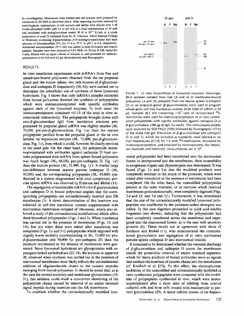
In vitro translation experiments with mRNA's from free andmembrane-bound polysomes obtained from the rat preputialgland and the mouse spleen, two rich sources of ß-glucuroni-dase and cathepsin D respectively (30, 61), were carried out todetermine the subcellular site of synthesis of these lysosomalhydrolases . Fig. 1 shows that only mRNA's samples extractedfrom bound polysomes directed the synthesis of polypeptideswhich were immunoprecipitated with specific antibodiesagainst each of the lysosomal enzymes. In both cases theimmunoprecipitates contained <0.1% of the total in vitro in-corporated radioactivity. The polypeptide brought down withanti-ß-glucuronidase IgG from translation mixtures pro-grammed by preputial gland mRNA was slightly smaller (Mr,70,000; pre-pro-ß-glucuronidase, Fig. 1 a) than the maturepolypeptide purified from the preputial gland or the in vivolabeled rat hepatocyte enzyme (Mr, 72,000; pro-/3-glucuroni-dase, Fig. 1 c), from which it could, however, be clearly resolvedin the same gels. On the other hand, the polypeptide immu-noprecipitated with antibodies against cathepsin D from sys-tems programmed with mRNA from spleen bound polysomeswas much larger (Mr, 48,000 ; pre-pro-cathepsin D, Fig. 1 d)than the mature protein (Mr, 31,000, Fig. If). A similar largesize-difference between mature porcine cathepsin D (Mr,30,000) and the corresponding polypeptide (Mr, 43,000) syn-thesized in a system programmed with total cytoplasmic por-cine spleen mRNA was observed by Erickson and Blobel (11) .The segregation oftranslatable mRNA's for,ß-glucuronidase
and cathepsin D in bound polysomes implies that the corre-sponding polypeptides are cotranslationally inserted into ERmembranes (1) . A direct demonstration of this insertion wasachieved in cell-free translation systems supplemented withmicrosomal membranes stripped of ribosomes, which also al-lowed a study of the cotranslational modifications which affectthese lysosomal polypeptides (Figs. 2 and 3) . When translationwas carried out in the presence of membranes (Figs. 2 b and3 b), but not when these were added after translation wascompleted (Figs. 2 c and 3 c), polypeptides which migrated withslightly lower mobility (corresponding to Mr, 72,000 for pro-ß-glucuronidase and 50,000 for pro-cathepsin D) than theproducts synthesized in the absence of membranes were gen-erated. Since lysosomal hydrolases are glycoproteins with as-paragine-linked carbohydrate (23, 54), the increase in apparentMr observed when synthesis was carried out in the presence ofmicrosomal membranes most likely reflects the cotranslationaladdition of oligosaccharide chains to the nascent peptidesemerging from bound polysomes. It should be noted that, as isthe case for several secretory and membrane glycoproteins (10,31), this addition could mask a concurrent shortening of thepolypeptide chains caused by removal of an amino terminalsignal peptide during insertion into the ER membranes.To establish whether the cotranslationally modified lyso-
FIGURE 1
In vitro biosynthesis of lysosomal enzymes. MessengerRNA samples isolated from free (b and e) or membrane-boundpolysomes (a and d) prepared from the mouse spleen (cathepsinD) or rat preputial gland (ß-glucuronidase) were used to programwheat germ cell-free translation systems (0.06 A260 of mRNA in 50pl) . Samples (0 .2 ml) containing -10' cpm of incorporated 35S-
methionine were used for immunoprecipitation of in vitro synthe-sized polypeptides with specific antibodies against cathepsin D orß-glucuronidase (100 Wg of IgG for each) . The immunoprecipitateswere analyzed by SDS PAGE (10%) followed by fluorography (72 h)of the dried slab gel . Precursors of B-glucuronidase and cathepsinD (c and f), which are included as standards, were labeled in ratliver hepatocytes (C1-9) for 1 h with 355-methionine, recovered byimmunoprecipitation, and analyzed by electrophoresis (for details,see Materials and Methods) . Measurements are X 103.
somal polypeptides had been transferred into the microsomallumen or incorporated into the membranes, their accessibilityto exogenous trypsin and chymotrypsin was determined. It wasfound (Figs. 2 e and 3 e) that the modified products werecompletely resistant to the attack of the proteases, which wereadded after translation in the presence of membraneshad beencompleted. On the other hand, the unmodified polypeptidespresent in the same mixtures, or in mixtures which receivedmembranes posttranslationally, were completely digested (Figs .2 d and 2f, and 3d and 3f). Furthermore, it was determinedthat the size of the cotranslationally modified lysosomal poly-peptides was unaffected by the proteases unless detergent wasadded. In this case digestion proceeded to yield acid-solublefragments (not shown), indicating that the polypeptides hadbeen completely transferred across the membrane and segre-gated into the microsomal lumen, as is the case with secretoryproteins (6) . These results are in agreement with those ofErickson and Blobel (11), who demonstrated the cotransla-tional glycosylation and segregation of in vitro synthesizedporcine spleen cathepsin D into microsomal vesicles .
It remained to be determined whether the vectorial dischargeof /3-glucuronidase and cathepsin D across the membranesentails the proteolytic removal of amino terminal segments,which for many products of bound polysomes serve as signalsthat mediate the insertion ofnascent chains into the membranes(cf. Kreibich et al. 1271) . To this effect, the electrophoreticmobilities of the unmodified and cotranslationally modified invitro synthesized polypeptides were compared with the mobil-ities of polypeptides synthesized in vivo, which were immu-noprecipitated after a short time of labeling from controlcultured cells and from cells treated with tunicamycin to pre-vent glycosylation (29). A tissue culture system of rat hepato-
ROSENFELD IT At .
Biosynthesis of Lysosomal Hydrolases
137
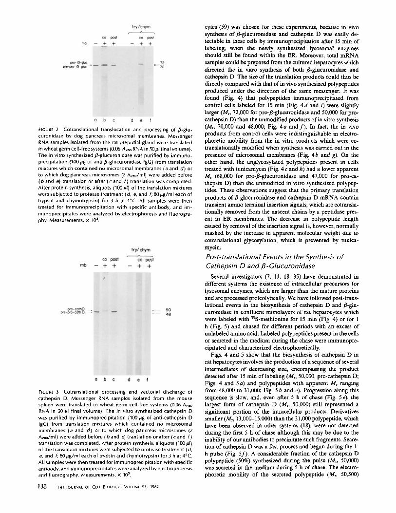
FIGURE 2 Cotranslational translocation and processing of ,B-glu-curonidase by dog pancreas microsomal membranes . MessengerRNA samples isolated from the rat preputial gland were translatedin wheat germ cell-free systems (0 .06 Also RNA in 50ul final volume) .The in vitro synthesized ,ß-glucuronidase was purified by immuno-precipitation (100 pg of anti-,8-glucuronidase IgG) from translationmixtures which contained no microsomal membranes (a and d) orto which dog pancreas microsomes (2 A260/ml) were added before(b and e) translation or after (c and f) translation was completed.After protein synthesis, aliquots (100 WI) of the translation mixtureswere subjected to protease treatment (d, e, and f; 80gg/ml each oftrypsin and chymotrypsin) for 3 h at 4°C . All samples were thentreated for immunoprecipitation with specific antibody, and im-munoprecipitates were analyzed by electrophoresis and fluorogra-phy . Measurements, x 103 .
FIGURE 3 Cotranslational processing and vectorial discharge ofcathepsin D . Messenger RNA samples isolated from the mousespleen were translated in wheat germ cell-free systems (0.06 Azs0RNA in 50 ,al final volume) . The in vitro synthesized cathepsin Dwas purified by immunoprecipitation (1001Ag of anti-cathepsin DIgG) from translation mixtures which contained no microsomalmembranes (a and d) or to which dog pancreas microsomes (2Also/ml) were added before (b and e) translation or after (c and f )translation was completed . After protein synthesis, aliquots (100ILI)of the translation mixtures were subjected to protease treatment (d,e, and f; 80 tLg/ml each of trypsin and chymotrypsin) for 3 h at 4°C .All samples were then treated for immunoprecipitation with specificantibody, and immunoprecipitates were analyzed by electrophoresisand fluorography . Measurements, x 103.
138
THE JOURNAL OF CELL BIOLOGY " VOLUME 93, 1982
cytes (59) was chosen for these experiments, because in vivosynthesis of 8-glucuronidase and cathepsin D was easily de-tectable in these cells by immunoprecipitation after 15 min oflabeling, when the newly synthesized lysosomal enzymesshould still be found within the ER. Moreover, total mRNAsamples could be prepared from the cultured hepatocytes whichdirected the in vitro synthesis of both 8-glucuronidase andcathepsin D . The size of the translation products could thus bedirectly compared with that ofin vivo synthesized polypeptidesproduced under the direction of the same messenger. It wasfound (Fig . 4) that polypeptides immunoprecipitated fromcontrol cells labeled for 15 min (Fig. 4d and i) were slightlylarger (Mr, 72,000 for pro-,8-glucuronidase and 50,000 for pro-cathepsin D) than the unmodified products ofin vitro synthesis(Mr, 70,000 and 48,000 ; Fig. 4 a and f) . In fact, the in vivoproducts from control cells were indistinguishable in electro-phoretic mobility from the in vitro products which were co-translationally modified when synthesis was carried out in thepresence of microsomal membranes (Fig. 46 and g) . On theother hand, the unglycosylated polypeptides present in cellstreated with tunicamycin (Fig. 4c and h) had a lower apparentMr (68,000 for pro-,8-glucuronidase and 47,000 for pro-ca-thepsin D) than the unmodified in vitro synthesized polypep-tides . These observations suggest that the primary translationproducts of ß-glucuronidase and cathepsin D mRNA containtransient amino terminal insertion signals, which are cotransla-tionally removed from the nascent chains by a peptidase pres-ent in ER membranes. The decrease in polypeptide lengthcaused by removal of the insertion signal is, however, normallymasked by the increase in apparent molecular weight due tocotranslational glycosylation, which is prevented by tunica-mycin.Post-translational Events in the Synthesis ofCathepsin D and,ß-GlucuronidaseSeveral investigators (7, 11, 18, 35) have demonstrated in
different systems the existence of intracellular precursors forlysosomal enzymes, which are larger than the mature proteinsand are processed proteolytically. We have followed post-trans-lational events in the biosynthesis of cathepsin D and ,8-glu-curonidase in confluent monolayers of rat hepatocytes whichwere labeled with 35S-methionine for 15 min (Fig. 4) or for 1h (Fig. 5) and chased for different periods with an excess ofunlabeled amino acid. Labeled polypeptides present in the cellsor secreted in the medium during the chase were immunopre-cipitated and characterized electrophoretically .
Figs. 4 and 5 show that the biosynthesis of cathepsin D inrat hepatocytes involves the production ofa sequence ofseveralintermediates of decreasing size, encompassing the productdetected after 15 min of labeling (Mr, 50,000, pro-cathepsin D ;Figs . 4 and 5a) and polypeptides with apparent Mr rangingfrom 48,000 to 31,000 ; Fig. 5 b and e) . Progression along thissequence is slow, and, even after 5 h of chase (Fig. 5 e), thelargest form of cathepsin D (Mr, 50,000) still represented asignificant portion of the intracellular products . Derivativessmaller (Mr , 13,000-15,000) than the 31,000 polypeptide, whichhave been observed in other systems (18), were not detectedduring the first 5 h of chase although this may be due to theinability of our antibodies to precipitate such fragments . Secre-tion ofcathepsin D was a fast process and began during the 1-h pulse (Fig. 5j) . A considerable fraction of the cathepsin Dpolypeptide (50%) synthesized during the pulse (Mr, 50,000)was secreted in the medium during 5 h of chase . The electro-phoretic mobility of the secreted polypeptide (Mr, 50,500)

FIGURE 4 Effect of cotranslational cleavage and/or glycosylationon the apparent molecular weight of ß-glucuronidase and cathepsinD . Messenger RNA, isolated from rat hepatocyte cultures (C1-9),was translated in a wheat germ cell-free system (0 .06 A26o RNA/50Id) in the presence (b and g) or absence (a and f) of dog pancreasmicrosomal membranes (2 A260/Ml) . The in vitro products are com-pared with polypeptides labeled in vivo in rat hepatocyte cultureswhich were incubated for 15 min with ssS-methionine in the pres-ence (c and h) or absence (d, e, i, and j) of tunicamycin (2 Wg/ml)and were prepared for immunoprecipitation immediately after thepulse (c, h, d, and i) or after a 5-h chase (e and j) . All samples wereimmunoprecipitated and analyzed by polyacrylamide gel (10%)electrophoresis followed by fluorography. Measurements, x 10 3.
appeared, however, slightly though detectably slower than thatof the corresponding intracellular form (Fig . 5f-j) . It shouldbe noted that only the largest detected cathepsin D polypeptidewas exported from the hepatocytes. A similar secretion of thecathepsin D precursor has been observed with human fibroblast(18) and endothelial cells (21) although in one of these casessmall amounts of the mature enzyme were detected in themedium (18) .
In contrast to the relatively rapid intracellular processing ofcathepsin D, labeled immunoprecipitable ß-glucuronidasepolypeptides found at the end of the pulse did not show anapparent change in electrophoretic mobility until the chase wasextended beyond 5 h (Fig. 5 k-o) . After 8 h of incubation incold medium a slightly smaller (Mr , 71,000) form of iß-glucu-ronidase was detected, which accumulated slowly during thenext 12 h. These observations confirm the reports of Swankand his associates (7), who first detected a small change in sizeduring the maturation of,ß-glucuronidase in mouse kidney andfibroblasts. The change involved in the maturation of iß-glu-curonidase (1,000 daltons) in rat hepatocytes is, however, evensmaller than the one reported with other systems (1,500-3,000daltons) . It should be noted (Fig . 5p-t) that, as was the casewith cathepsin D, secretion of iß-glucuronidase was much fasterthan intracellular maturation by proteolytic processing . A frac-tion of the newly synthesized enzyme detected at the end ofthe pulse began to be secreted into the medium without de-tectable processing as soon as 3 h after the pulse . The matureform of,ß-glucuronidase (71,000) which is seen intracellularlyafter 8 h of chase was not secreted even when incubation wasprolonged to 20 h when up to 40% of the pulse-labeled productwas found in the medium.
Enhancement of the Secretion of Cathepsin Dand iß-Glucuronidase in Cells Treatedwith Chloroquine
Previous studies have shown that lysosomotropic aminessuch as chloroquine enhance the secretion of lysosomal en-
zymes by cultured fibroblasts (17, 18) and impair the intracel-lular processing of newly synthesized hydrolases (18) . Figs . 6and 7 demonstrate that chloroquine had a dramatic effect onthe secretion of cathepsin D and ,ß-glucuronidase by rat hepa-tocytes . When these cells were incubated with the drugthroughout the pulse and chase periods, conversion of thelabeled precursors (Mr, 50,000 and 72,000 for cathepsin D and,ß-glucuronidase, respectively) to lower molecular weight formswas abolished (Figs . 6 and 7 i-1) and secretion ofthe precursorswas accelerated (Figs. 6 and 7u-x) . While control cells, evenduring prolonged chases (up to 20 h), exported to the mediumno >50% of the newly synthesized enzymes, chloroquine-treated cells were almost totally devoid of labeled hydrolasesafter 5 h of chase. The complete secretion of unprocessedprecursors caused by chloroquine reinforces the suggestion thatfinal proteolytic processing of lysosomal enzymes takes placewithin the lysosomes (18) .
FIGURE 5
Post-translational events in the synthesis of cathepsin Dand ,ß-glucuronidase. Confluent monolayers of rat hepatocytes werelabeled with 35S-methionine for 1 h and chased for different timeswith an excess of the unlabeled amino acid . Labeled polypeptidespresent in the cells or secreted in the medium during the chasewere immunoprecipitated (100 hg/ml IgG) and characterized elec-trophoretically. (A) Intracellular cathepsin D immunoprecipitatedimmediately following a 1-h pulse (a) or after a chase of 1, 3, 5, or8 h (b, c, d, and e) . Conversion of the pro-cathepsin D (a, 50,000daltons) to the 31,000-dalton cathepsin D occurs through a numberof intermediates (b, c, d, and e, 46,000 and 36,000 daltons) during" 5 h . (B) Immunoprecipitated cathepsin D secreted during a 1-hpulse ( f) or during a chase of 1, 3, 5 or 8 h ( g, h, i, and j) . Thesecreted polypeptide (50,500dalton) has an electrophoretic mobilityslightly slower than that of the corresponding intracellular form(50,000 daltons) . (C) Intracellular ,ß-glucuronidase immunoprecipi-tated immediately following a 1-h pulse (k) or after a chase of 1, 3,5, or 8 h (l, m, n, and o) . In contrast to cathepsin D, an apparentchange in electrophoretic mobility did not appear until the chasewas extended beyond 5 h (lane o, 71,000 dalton) . (D) Immunopre-cipitated,ß-glucuronidase secreted during a 1-h pulse ( p) or duringa chase of 1, 3, 5, or 8 h (q, r, s, and t) . A fraction of the newlysynthesized polypeptide detected at the end of the pulse began tobe secreted into the medium as soon as 3 h after the chase (lanes r,s, and t, 72,000 daltons) . The mature form of /3-glucuronidase wasnot secreted .
ROSENEELD ET AL .
Biosynthesis of Lysosomal Hydrolases
139

FIGURE 6
Effect of tunicamycin and chloroquine on the kinetics ofintracellular processing and secretion of cathepsin D. Confluentrrtonolayers of rat hepatocytes were labeled for 1 h with 35 S-methi-onine and chased for different periods with an excess of the unla-beled amino acid . Cultures were incubated with tunicamycin (2 Wg/ml) or chloroquine (25 pM) for 1 h before the pulse, and the drugswere present throughout the pulse-chase periods. Labeled polypep-tides present in the cells (A, C, and E) or secreted in the medium(8, D, and F) during the chase were immunoprecipitated (100 Wg/ml anti-cathepsin D IgG) and characterized electrophoretically .Intracellular (A) and secreted (8) cathepsin D immunoprecipitatedfollowing a 1-h pulse (a and m) and chased for 1, 3, or 5 h (b, c, d;n, o, p) . Conversion of the pro-cathepsin D (a, 50,000 dalton) to the31,000-dalton cathepsin D occurs through a number of intermedi-ates (b, c, and d; 46,000 and 36,000 daltons) during ^-5 h. Only theunprocessed precursor is secreted (m, n, o, and p) . Intracellular ( C)and secreted (D) cathepsin D immunoprecipitated following a 1-hpulse (e and q) and chased for 1, 3, or 5 h ( f, g, h; r, s, t) in thepresence of tunicamycin. The unglycosylated form of pro-cathepsinD (e; 47,000 dalton) was not processed to any lower molecularweight derivatives ( f, g, and h), and the intracellular concentrationcontinuously decreased throughout the chase in parallel with anaccumulation in the medium (r, s, t) . Intracellular (E) and secreted(F) cathepsin D immunoprecipitated following a 1-h pulse (i and u)and chased for 1, 3, or 5 h ( j, k, 1; v, w, x) in the presence ofchloroquine. Conversion of the pro-cathepsin Dprecursor (i, 50,000daltons) to lower molecular weight forms was abolished, and theintracellular concentration of pro-cathepsin D decreased through-out the chase period with a concomitant increase in the concentra-tion of pro-cathepsin Dsecreted in the medium (v, w, and x, 50,500daltons) .
Sensitivity of Intracellular and SecretedPrecursors of Lysosomal Hydrolases to Digestionby Endo H and DAn examination of the sensitivity to digestion by endo H and
D provided a means to detect structural differences in thecarbohydrate side chains of intracellular and secreted precur-sors of cathepsin D . The fact that the intracellular precursorsof lysosomal hydrolases have high-mannose oligosaccharidechains (54) was corroborated by the complete sensitivity of thecathepsin D precursor to digestion with endo H . This enzymereduced the apparent molecular weight of pro-cathepsin Dfrom 50,000 to 48,500 (Fig. 8b). Furthermore, incubation withendo D (an enzyme which cleaves off oligosaccharides withfive or less mannose residues from which terminal sugars havebeen removed) did not affect the size of the intracellular
140
THE IOURNAL OF CELL ETIOLOGY " VOLUME 93, 1982
cathepsin D precursor (Fig. 8f). On the other hand, digestionof the secreted precursor (50,500 dalton) with endo H showedthat this material represents a mixture of polypeptides whichdiffered in the extent and type of oligosaccharide processing .While some of the secreted cathepsin D chains behaved likethe intracellular precursors in their sensitivity to endo H,decreasing in size to 48,500 dalton, others decreased only to50,000 dalton (Fig . 8 d) . A likely explanation for these findingsis that each secreted polypeptide molecule may contain morethan one type of oligosaccharide chain and that, while in somepolypeptides all chains are susceptible to endo H, in otherssome oligosaccharides are resistant to this glycosidase, whichtherefore only causes a smaller reduction in molecular size . Inthis case it may be expected that at least some of the oligosac-charides resistant to endo H may be sensitive to digestion withendo D. Indeed, incubation with this glycosidase showed that,in contrast to the polypeptides found intracellularly, which
FIGURE 7
Effect of tunicamycin and chloroquine on the kinetics ofintracellular processing and secretion ofß-glucuronidase. Confluentmonolayers of rat hepatocytes were labeled for 1 h with 35 S-methi-onine and chased for different periods with an excess of the unla-beled amino acid . Cultures were incubated with tunicamycin (2 hg/ml) or chloroquine (25 AM) for 1 h before the chase, and the drugswere present throughout the pulse-chase periods. Labeled polypep-tides present in the cells (A, C, and E) or secreted in the medium(8, D, and F) during the chase were immunoprecipitated (100 tLg/ml anti-ß-glucuronidase) and characterized electrophoretically. In-tracellular (A) and secreted (8) fl-glucuroniclase immunoprecipi-tated after a 1-h pulse (a and m) and chased for 1, 3, or 5 h (b, c, d;n, o, p) . No change in electrophoretic mobility was observedthrough 5 h of chase (72,000 daltons) . Only when the chase was
extended beyond 5 h (Fig, 5, lane o, 71,000 daltons) was theconversion to the mature form seen . In contrast to cathepsin D (Fig.
6 m, n, o,and p),ß-glucuronidase secretion was slower and appearedafter 3 h of chase (o) . Only the unprocessed form offl-glucuroniclasewas secreted (72,000 daltons) . Intracellular (C) and secreted (D) fl-glucuroniclase immunoprecipitated after a 1-h pulse (e and q) andchased for 1, 3, or 5 h ( f, g, h; r, s, t) in the presence of tunicamycin.
The unglycosylated form of pro-p-glucuronidase (e, 68,000 daltons)decreased throughout the chase and began to accumulate in themedium (r, s, and t, 68,000 daltons) . Intracellular (E) and secreted( F) fl-glucuroniclase immunoprecipitated after a 1-h pulse (i and u)
and chased for 1, 3, or 5 h (j, k, l; v, w, x) in the presence ofchloroquine. The intracellular concentration of pro-ß-glucuronidasedecreased throughout the chase period with a concomitant increasein the concentration of pro-p-glucuronidase secreted in the medium(v, w, and x, 72,000 daltons) .

FIGURE 8
Sensitivity of cathepsin D to endoglycosidase H or en-doglycosidase D. Confluent monolayers of rat hepatocytes werelabeled during 30 min with 35S-methionine, and the cell extracts (a,b, e, and f) and medium (c, d, g, and h) were kept as controls (a,c, e, and g) or treated with endoglycosidase H (8 x 10-° U/ml ; 15h at 37°C); b, d) or a crude preparation of endoglycosidase D (15 h37°C ; f and h) before immunoprecipitation with anti-cathepsin DIgG (100ttg/ml) as described in Materials and Methods. Intracellularpro-cathepsin D incubated with endoglycosidase H (b) showed anincrease in electrophoretic mobility (48,500 daltons) . Incubation ofsecreted pro-cathepsin D (50,500 daltons) with endoglycosidase Hgave rise to two polypeptides of Mr , 48,500 and to 50,000 daltons(d) .
were all resistant to endo D, all those in the extracellularmedium contained oligosaccharides susceptible to this glyco-sidase (Fig . 8 h) .
Inhibition of Post- translational Processing andSecretion of Unglycosylated LysosomalHydrolases in Cells Treated with TunicamycinConsiderable evidence has accumulated (19, 20, 23, 25, 48,
54) suggesting that phosphomannosyl groups in oligosaccha-ride chains of newly synthesized lysosomal enzymes serve asrecognition markers which mediate the transfer of the enzymesto lysosomes . It was therefore of interest to determine the fateofthe unglycosylated lysosomal polypeptides, which were iden-tified after short times of labeling in cells treated with tunica-mycin . As seen in Figs. 6 and 7 the behavior of the unglyco-sylated forms of pro-cathepsin D (Mr, 47,000) and pro-,8-glu-curonidase (Mr, 68,000) after a labeling pulse differed fromthat of the normal polypeptides synthesized in control cells intwo respects : (a) the unglycosylated forms were not processedto any lower molecular weight derivatives (Figs . 6 and 7f-h) .(b) their intracellular concentration continuously decreasedthroughout the chase, in parallel with their accumulation inthe extracellular compartment, so that eventually complete(pro-cathepsin D), or almost complete (pro-,8-glucuronidase)secretion of the unglycosylated lysosomal polypeptides oc-curred (Figs . 6 and 7q-t) . These observations are consistentwith the operation of a mechanism in which carbohydratechains bearing phosphomannosyl groups are necessary for thesorting-out of lysosomal hydrolases from other proteins con-tained in the ER (13, 18) . The enhancement of secretion causedby tunicamycin stresses the considerable overlap which mustexist between the early stages of the secretory pathway and thepathway leading to lysosomes .
DISCUSSION
The above results demonstrate directly that mRNA's for thelysosomal enzymes cathepsin D and,8-glucuronidase are trans-lated on polysomes bound to ER membranes . For cathepsin Dthis had already been inferred by Erickson and Blobel (11)from their finding that addition of microsomal vesicles to cell-free systems programmed with total porcine spleen mRNA
leads to the cotranslational modification ofthe in vitro synthe-sized polypeptide and to its sequestration within the micro-somal lumen. In agreement with these observations we foundthat lysosomal polypeptides transferred into the microsomallumen in vitro were electrophoretically indistinguishable fromprecursors of the mature enzymes which were labeled duringshort radioactive pulses in vivo and can therefore be presumedto be contained within the ER lumen . As would be expected ifthe lysosomal polypeptides undergo glycosylation during theirtransfer across the microsomal membrane, the putative micro-somal forms of,8-glucuronidase and cathepsin D immunopre-cipitated from cultured hepatocytes were of larger apparentmolecular weight than the primary translation products syn-thesized in vitro in the absence of membranes . On the otherhand, the corresponding unglycosylated polypeptides immu-noprecipitated from cells treated with tunicamycin were smallerthan the unmodified products of in vitro translation . Thisstrongly suggests that cotranslational processing of the lyso-somal hydrolases includes, in addition to core glycosylation, ashortening of the nascent polypeptide, which can only occurby removal ofan amino terminal segment . Similar observationsusing tunicamycin first revealed the presence oftransient aminoterminal insertion signals in membrane glycoproteins (10), forwhich the decrease in polypeptide length caused by signalremoval is masked by an overall gain in size caused by coreglycosylation . It is worth noting that removal ofamino terminalsegments which may serve as insertion signals is not an oblig-atory step during the synthesis of all polypeptides made onbound ribosomes. Indeed, original amino terminal segmentshave been found to be retained in secretory (42), ER (4), andplasma membrane proteins (8), and the possibility cannot bediscarded that such segments may be conserved in some lyso-somal proteins.Taken together, however, the observations just discussed
suggest that insertion of lysosomal hydrolases into the ERmembrane is a process completely analogous to the vectorialdischarge of secretory proteins into the microsomal lumen . Ofcourse, amino acid sequence determinations are necessary toestablish whether amino terminal signals in prelysosomal andpresecretory proteins are structurally equivalent. This seemslikely, since signals in secretory and membrane proteins showstriking similarities and both classes of polypeptides appear touse the same microsomal translocation apparatus (14, 26, 27) .
Several investigators (18, 35, 50) have reported that lyso-somal enzymes are synthesized as larger molecular weightprecursors which only after post-translational proteolytic proc-essing yield the mature forms found in lysosomes . As reportedfor human placenta (5) and porcine spleen (11, 12), the biosyn-thesis of cathepsin D (31,000 daltons) in rat hepatocytes wasfollowed by the production of a series of intermediates derivedby successive cleavages from a large microsomal precursor(50,000 daltons) . On the other hand, only a single post-trans-lational proteolytic cleavage product was detected after thebiosynthesis of 8-glucuronidase . This appeared >5 h after thepolypeptide was synthesized and represented an even smallerchange in molecular weight than those reported for ,8-glucu-ronidase in mouse kidney and macrophages (7, 51) .The functional significance of the proteolytic processing
steps which follow the biosynthesis of lysosomal enzymes is yetunclear . Removal of peptide segments does not appear to berequired for the activation of many hydrolases which aresecreted as enzymatically active precursors (18), although proc-essing may be required for the activation of cathepsin D .
RosENFELD Er AE .
Biosynthesis of Lysosomaf Hydrofases
141

Furthermore, trimming of precursors is a slow process whichtakes place several hours after synthesis ; thus, even thoughsegments removed by proteolysis maybe important in guidingthe newly synthesized hydrolases to their destination, it appearsunlikely that their removal takes place along the pathwaywhich leads to the lysosomes. Indeed, the fact that chloroquine,a drug which impairs intralysosomal digestion by raising thelysosomal pH (39, 60), completely prevented processing of thenewly synthesized hydrolases supports the suggestion, made byHasilik and Neufeld (18) on the basis of similar observationswith human fibroblasts, that trimming of the precursors takesplace only after their arrival in the lysosomes.The fact that lysosomal hydrolases can be secreted has long
been recognized (23, 48) and serves to emphasize the commonorigin of secretory and lysosomal polypeptides in the ER. Incultured hepatocytes, secretion of cathepsin Dand ß-glucuron-idase was a quantitatively important process which involved-30-40% of the newly synthesized enzymes and began wellbefore any post-translational proteolytic cleavage was detect-able . As was first noted for the enzymes of human fibroblasts(18), only uncleaved precursors of cathepsin D and iB-glucu-ronidase were exported from the cultured hepatocytes . Thedramatic effect ofchloroquine in promoting amassive secretionofthe unprocessed hydrolases is in accordance with the notion(17) that this drug eventually interferes with the transfer ofnewly synthesized hydrolases to lysosomes by impairing therecycling or re-using of membrane receptors which mediatethis process.
It is currently thought (13, 19, 25) that phosphomannosylgroups which have been detected on the oligosaccharide chainsof newly synthesized hydrolases (20, 54) serve as recognitionmarkers which are necessary to ensure the correct destinationof the polypeptides to lysosomes. In addition, membrane-bound receptors capable of binding lysosomal polypeptidescarrying these markers have been identified on the cell surface(13, 21, 45, 56) and in intracellular membranes (13) . Thehypothesis that phosphomannosyl groups serve as sorting-outsignals for lysosomal enzymes implies that unglycosylated poly-peptides of hydrolases synthesized in the presence of tunica-mycin should be unable to reach their lysosomal destination .It is notable that although it has been reported (57) thattunicamycin changes the distribution ofhydrolytic activities inthe intracellular and extracellular compartments, the newlysynthesized unglycosylated polypeptides present in tunicamy-cin-treated cells have not been characterized and their fate hasnot been established. We found that unglycosylated cathepsinD and /3-glucuronidase synthesized in tunicamycin-treatedhepatocytes were quantitatively secreted into the medium asunprocessed precursors. These observations support the notionthat modified sugar residues are necessary for addressing thepolypeptides to lysosomes and suggest that in the absence ofthe phosphomannosyl marker the polypeptides are secreted .As is the case with other secretory products (53), the absenceof sugars does not impair secretion of lysosomal hydrolases .Indeed, secretion became the fate for all newly synthesizedunglycosylated hydrolases . Furthermore, in the presence oftunicamycin, proteolytic processing was completely abolished.These observations provide independent support for the notionthat the cleavage steps which follow the biosynthesis of lyso-somal hydrolases occur only after the polypeptides arrive inlysosomes.The alternative possibility that unglycosylated polypeptides
cannot serve as substrates for processing proteases is rendered
142
THE JOURNAL OF CELL BIOLOGY " VOLUME 93, 1982
unlikely by the finding that proteolytic processing of viralenvelope glycoproteins, which reach the plasma membrane(15), proceeds unimpaired on unglycosylated polypeptides syn-thesized in tunicamycin-treated cells .
Recent studies (22, 44) suggest that phosphorylation ofnewlysynthesized hydrolases is a relatively early event which involvestransfer of an G1cNAc phosphate residue from UDP-G1cNAc.The "covered" phosphate groups generated by this transfer donot appear, however, to be recognized by the specific mem-brane-bound receptors until the G1cNAc cover is removed bya phosphodiesterase which may be localized in the Golgiapparatus or GERL membrane system (58, but cf. reference55). The differences in the sensitivity to endoglycosidases whichwe have detected between the intracellular and secreted formsof hydrolase precursors suggest that the secreted polypeptidesindeed follow the normal secretory route which includes theGolgi apparatus and provides for trimming of oligosaccharideresidues and possibly terminal glycosylation. It still remains tobe determined at which point the secretory and lysosomalpathways diverge, in particular, whether enzymes which reachthe lysosomes depart intracellularly fromthe secretory pathway(cf. references 28, 41, 47) or whether they first reach the cellsurface but are later interiorized by endocytosis (21, 56).The lack of sensitivity to endo D of the intracellular form of
lysosomal enzymes may reflect the inability of Golgi complexenzymes to modify precursors bearing "uncovered" phosphateresidues or more unlikely a bypass of these activities by lyso-somal precursors .
We are grateful to Ms . H. Snitkin and S. Malamet for their help withthe tissue culture experiments, Ms. H. Plesken and Mr . B. Zeitlow fortheir illustrations, Dr. P. Atkinson forthe gift of endoglucosaminidases,and Ms . M. Chung, S. Martinez, and T. Singfield for typing themanuscript .
This work was supported by grants GM 20277, GM 21971 and AG01461. M. G. Rosenfeld is a recipient of United States Public HealthService Postdoctoral Fellowship GM-07177 .A preliminary account of this work was presented at the American
Society for Cell Biology, Cincinnati, Ohio, November, 1980 (46) .
Receivedfor publication 13August 1981, andin revisedform 2 November1981 .
REFERENCES
I . Adelman, M . R., D . D. Sabatini, and G. Blobel. 1973. Ribosome-membrane interaction.Nondestructive disassembly of rat liver rough microsomes into ribosomal and numerouscomponents . J. Cell Mot. 56:206-229.
2 . Aviv, H., and P . Leder. 1972. Purification of biologically active globin messenger RNA bychromatography on oligo thymidylic acid-cellulose . Proc. NaIL Acad. Sci. U. S. A . 69:1408-1412 .
3 . Bainton, D. F., and M. G. Farquhar. 1970. Segregatio n and packaging of granule enzymesin eosmophilic leucocytes. J. Cell Biol. 45:54-73 .
4 . Bar-Nun, S., G . Kreibich, M . Adesnik, L . Alterman, M . Negishi, and D. D . Sabatini .1980 . Synthesis and insertion ofcytochrome P-450 into endoplasmic reticulum membranes.Proc. Natl. Acad. Sci. U. S. A . 77 :965-969 .
5 . Barrett, A. J . 1970 . Cathepsin D . Purification of isoenzymes human and chicken liver.Biochem. J. 117 :601-607 .
6 . Blobel, G ., and B. Dobberstein . 1975 . Transfer of proteins across membranes. 1. Presenceof proteolytically processed and unprocessed nascent immunoglobulin light chains onmembrane-bound ribosomes of murine myeloma . J. Cell Biol. 67 :835-851 .
7 . Brown, 1 . A ., G . P . Jahveis, and R . T. Swank. 1981 . The synthesis and processing of ß-glucuronidase in normal and egasyn deficient mouse kidney . Biochem. Biophys. Res.Comet. 99 :691-699 .
8 . Chyn, T . L., A . N . Martonosi, T . Morimoto, and D . D . Sabatini. 1979 . In vitro synthesisof the Ca" transport ATPase by ribosomes bound to sarcoplasmic reticulum membranes.Proc. Narl. Acad. Sci. U. S. A . 76 :1241-1245 .
9 . DeDuve, C ., and R . Wattiaux. 1966. Functions of lysosomes . Ann. Rev. Physiol. 28:435-492 .
10. Dobberstein, B ., H. Garoff, G. Warren, and P. J . Robinson . 1979 . Cell-free synthesis andmembrane insertion of mouse H-2D^ histocompatibility antigen and B,-microglobulin.Cell. 17 :759-769.
11 . Erickson, A . H ., and G . Blobel . 1979. Early events in the biosynthesis of the lymsomalenzyme cathepsin D . J. Biol. Chem 154:11771-11774.
12 . Erickson, A . H., G. E. Conner, and G . Blobel. 1981 . Cathepsin D, a lysosomal proteasesynthesized with two transient amino terminal sequence extensions . Fed. Proc. 40:1860(Abstr.) .
13 . Fisher, H. D ., A. Gonzalez-Noriega, and W. S . Sly. 1980. ß-glucuronidase binding tohuman fibroblast membrane receptors . J BioL Chem. 255 :5069-5074.
14 . Ghosh, H . P. 1980 . Synthesis and maturation of glycoproteins of enveloped animal viruses .Rev . Inject. Dis. 2 :26-39.
15 . Gibson, R., S . Schlesinger, and S. Kornfeld. 1979 . The nonglycosylated glycoprotein ofvesicular stomatitis virus is temperature-sensitive and undergoes intracellular aggregationat elevated temperatures. J. Biol. Chem. 254 :3600-3607.
16 . Goldman, B . M., and G. Blobel. 1978 . Biogenesis of peroxisomes: intracellular site ofsynthesis of catalane and uricase . Proc. Nod. A cad. Sci. U. S. A . 75:5066-5070 .
17 . Gonzalez-Noriega, A ., J . H . Brugg, V. Talkad, and W . S. Sly . 1980 . Chloroquine inhibitslysosomal enzyme pinocytosis and enhances lysosomal enzyme secretion by impairingreceptor recycling. J. Cell BioL 85 :839-852.
18 . Hasilik, A., and E . F. Neufeld. 1980 . Biosynthesis of lysosomal enzymes in fibroblasts-synthesis as precursors of higher molecular weight. J. Biol. Chem. 255 :4937-4945.
19 . Hasilik, A., and E . F. Neufeld. 1980 . Biosynthesis of lysosomal enzymes in fibroblasts-phosphorylation of marmose residues. J. Biol Chem . 255 :4946-4950.
20 . Hasilik, A., U . Klein, A . Waheed, G. Strecker, and K . Von Figure . 1980 . Phosphorylatedoliogosaccharides in lysosomal enzymes : identification of a-N-acetyl-glucosemine(1)phospho(6)mannose diester groups. Proc. NaIL Acad. Sci. U. S. A . 77 :7074-7078.
21 . Hasilik, A ., B. Voss, and K. Von Figure. 1981 . Transport and processing of lysosomalenzymes by smooth muscle cells and endothelial cells. Exp. Cell Res. 133:23-30 .
22 . Hasilik, A ., A. Waheed, and K . Von Figure. 1981 . Enzymatic phosphorylation of lysosomalenzymes in the presence of UDP-N-acetyl glucosemine. Absence of the activity in I-cellfibroblasts. Biochem. Biophys . Res. Commun. 98:761-767 .
23. Hickman, S., L. J. Shapiro, and E. F . Neufeld . 1974. A recognition marker for uptake ofa lysosomal enzyme by cultured fibroblasts. Biochem. Biophys. Res. Commun . 57:55-61 .
24 . Himeno, M ., H . Ohhara, Y . Akakaway, and K . Kato . 1975. ß-Glucuronidase of ratpreputial gland. J. Biochem. (Tokyo) . 77:427-438.
25. Kaplan,A.,D. T. Achord, andW . S . Sly . 1977. Phosphohexosy l components of a lysosomalenzyme are recognized by pinocytosis receptors on human fibroblasts . Proc. Nall. Acad.Sci. U. S. A . 74 :2026-2030.
26 . Katz, F .N ., l . E . Rothman,V . R. Lingappa, G. Blobel, andH. F . Lodish . 1977 . Membraneassembly in vitro : synthesis, glycosylation and asymmetric insertion of a transmembraneprotein . Proc. Nail. Acad. Sci. U. S. A . 74:3278-3282.
27. Kreibich, G ., M. Czako-Graham, R. C . Grebenau, and D . D . Sabatini . 1980 . Functionaland structural characteristics of endoplasmic reticulum proteins associated with ribosomebinding sites . Ann . N. Y Acad. Sci. 343 :17-33.
28 . Kreibich, G ., M . Czako-Graham, W. Mok, E, Nack, Y . Okada, M . G . Rosenfeld, and D .D. Sabatini . 1980. The role of free and membrane-bound polysomes in organelle biogen-esis. In Biological Chemistry of Organelle Formation. T . H . Bucher, W . Sebald, and H.Weirs, editors . Springer-Verlag, New York. 147-164.
29. KOO, S. C., and J. P . Lampen. 1974 . Tunicamycin-an inhibitor of yeast glycoproteinsynthesis. Biochem . Biophys. Res. Commun. 58:287-295 .
30. Levvy, G . A ., A. McAllan, and G. A. Marsh. 1958. Purification of ß-glucuronidase fromthe preputial gland of the female rat. Biochem. J. 69 :22-27.
31 . Lingappa, V . R., D . Shields, S. L. Woo, and G . Blobel . 1979 . Nascent chicken ovalbumincontains the functional equivalent of a signal sequence . J. Cell Biol. 79:567-572.
32. Liu, C . P ., D . L. Slate, R. Gravel, and F. H. Ruddle. 1979. Biological detection of specificmRNA molecules by microinjection. Proc . Nail. Aced. Sci. U. S. A. 76:4503-4506.
33 . Mayer, R . J ., and 1 . H, Walker. 1980- Immunochemical Methods in the BiologicalSciences : Enzymes and Proteins . Academic Press, Inc ., New York.
34. McMullen, M ., P. Shaw, and P . E. Martin . 1979. Characterization of poly(A') RNA infree messenger ribonucleoprotein and polysomes of mouse taper ascites cells . J. Mol. Biol.132:679-694.
35 . Meyerowitz, R � and E. F . Neufeld . 1981 . Maturation of a-L-iduronidase in culturedhuman fibroblasts . J. Biol. Chem. 256:3044-3048 .
36 . Moscona, A . 1961 . Rotation-mediated histogenetic aggregation of dissociated cells. Exp.Cell Res. 22:455-475 .
37 . Muramatsu, T, P . H . Atkinson, S . G . Nathenson, and C . Ceccarini . 1973. Cell-surface
glycoproteins: growth-dependent changes in the carbohydrate-peptide linkage region . J.Mot. Biol. 80:781-799 .
38. Novikoff, A . B . 1976 . The endoplasmic reticulum: a cytochemists view (a review) . Proc.Nod. Aced. Sci. U. S. A . 73 :2781-2787.
39. Ohkuma, S ., and B . Poole. 1978. Fluorescence probe measurement of the intralysosomalpH in living cells and the perturbation ofpH by various agents . Proc. Natl. Acad. Sci. U.S. A . 75 :3327-3331 .
40 . Ouchterlony, O . 1964 . Gel diffusion techniques. In Immunological Methods-a Sympo-sium, J. F . Ackroyd, editor. Blackwell, Oxford . 55-78 .
41 . Palade, G . E. 1975 . Intracellular aspects of the process of protein synthesis . Science (Wash.D. C). 189:347-358 .
42 . Palmiter, R. D ., J . Gagnon, andK. A . Walsh . 1978 . Ovalbumin: a secreted protein withouta transient hydrophobic leder sequence . Proc. Nall. Acad. Sci. U, S. A . 75 :94-98.
43 . Ramsey, J. C., and W . J . Steele. 1976. A procedure for the quantitative recovery ofhomogeneous populations of undegrated free and bound polysomes from rat liver.Biochemistry. 15 :1704-1711 .
44. Reitman, M. D � and S. Kornfeld. 1981 . UDP-N-Acetyl-glucosamine :glycoprotein N-acetylglucosemine-l-phosphotramferase . J Biol. Chem . 256:4275-4281 .
45 . Rome, L . H ., B. Weissman, and E. H . Neufeld. 1979 . Direct demonstration of binding ofa lysosomal enzyme, a-L-iduronidase, to receptors on cultured fibroblasts. Proc. Nad.Acad. Sci. U. S. A . 76:2331-2334.
46 . Rosenfeld, M ., D. D. Sabatini, E . Sabban, K, Kato, and G . Kreibich . 1980 . Synthesis andmaturation of lysosomal enzymes. J. Cell Biol. 87(2, Pt . 2) :30ßa (Abstr.).
47 . Sabatini, D . D ., and G. Kreibich. 1976. Functional specialization of membrane boundribosomes in eukaryotic cells . In The Enzymes of Biological Membranes . A . Martonisi,editor . Plenum Publishing Co ., N. Y . 531-579 .
48 . Sando, G . N., and E . F. Neufeld . 1977 . Recognition and receptor-mediated uptake of alysosomal enzyme, a-L-idoronidase, by cultured human fibroblasts . Cell. 12:619-627 .
49 . Shields, D., and G. Blobel . 1978. Efficient cleavage and segregation of nascent presecretoryproteins in a reticulocyte lysate supplemented with microsomal membranes . J. Blot. Chem.253 :3753-3756 .
50 . Skudlarek, M. D., and R . T . Swank. 1979, Biosynthesis of two lysosomal enzymes inmacrophages. J. Biol. Chem . 254 :9939-9942.
51 . Skudlarek, M. D ., and R. T . Swank. 1980. Turnover of two lysosomal enzymes in mouseperitoneal macrophages. J. Cell Mot. 87(2, Pt . 2) :305a (Abstr.).
52 . Strawser, L. D ., and O . Touster. 1980 . The cellular processing of lysosomal enzymes andrelated proteins. Rev. Physiol. Biochem. Pharmacol. 87 :169-210.
53 . Struck, D . K ., P. B. Suite, M . D. Lane, and W . J. Lane, 1978 . Effect of tunicamycin on thesecretion of serum proteins by primary cultures of rat and chick hepatocytes. J. Biol.Chem. 253 :5332-5337 .
54. Tabas, I ., and S. Komfeld. 1980 . Biosynthetic intermediates of ß-glucuronidase containhigh mannose oligosaccharides with blocked phosphate residues. J Biol. Chem . 255 :6633-6639 .
55 . Varki, A., and S . Kornfeld . 1980. Identification of a rat liver a-N-acetyl glucosaminylphosphodiesterase capable of removing "blocking" a-N-acetyl glucosemine residues fromphosphorylated high mannose oligosaccharides of lysosomal enzymes. J. Biol. Chem. 255 :8398-8401 .
56 . Von Figure, K., and E. Weber. 1978 . An alternative hypothesis of cellular transport oflysosomal enzymes in fibroblasts. Biochem. J. 176 :943-950 .
57 . Von Figure, K ., M . Rey, R . Prinz, B. Voss, and K. Ullrich. 1979. Effect of tunicamycin ontransport of lysosomal enzymes in cultured skin fibroblasts . Eur. J. Biochem. 101 :103-109 .
58 . Waheed, A., R . Pohlmann, A . Hasilik, and K. Von Figure. 1981 . Subcellular localizationof two enzymes involved in the synthesis of phosphorylated recognition markers inlysosomal enzymes . J BioL Chem. 256:41504152.
59 . Weinstein, B ., N . Yamaguchi, J . M . Orenstein, R. Gebert, and M . E. Kaighn . 1974 .Mechanisms of chemical carcinogenesis analyzed in rat liver and hepatoma cell cultures .In Gene Expression and Carcinogenesis in Cultured Liver . L . Kruse and E. Patterson,editors. Academic Press, Inc., New York . 441-458 .
60 . Wibo, M ., and B . Poole . 1974 . Protein degradation in cultured cells . J. Cell Biol. 63 :430-440 .
61 . Yamamoto, K., N . Katsuda, M . Himeno, and K . Kato. 1979 . Cathepsin D of rat spleen .Eur. J Biochem. 95:459-467 .
ROSENFELD ET AL .
Biosynthesis of Lysosomal Hydrolases
143