Embed Size (px)
Citation preview

Structure and Biosynthesis of FungalSecondary Metabolites
Studies of the Root Rot Pathogen Heterobasidion annosum s.l. and the Biocontrol
Fungus Phlebiopsis gigantea
David HanssonFaculty of Natural Resources and Agricultural Sciences
Department of ChemistryUppsala
Doctoral ThesisSwedish University of Agricultural Sciences
Uppsala 2013

Acta Universitatis agriculturae Sueciae2013:63
ISSN 1652-6880ISBN (print version) 978-91-576-7864-5ISNB (electronic version) 978-91-576-7865-2© 2013 David Hansson, UppsalaPrint: SLU Repro, Uppsala 2013

Structure and Biosynthesis of Fungal Secondary Metabolites.Studies of the Root Rot Pathogen Heterobasidion annosum s.l.and the Biocontrol Fungus Phlebiopsis gigantea.
AbstractThe root rot pathogen Heterobasidion annosum s.l., i.e. H. abietinum, H. parviporum,H. annosum s.s., H. irregulare and H. occidentale, and the biocontrol fungus Phlebiopsis gigantea were investigated regarding their secondary metabolites.
Thirty-three compounds, in total, were identified from H. annosum s.l. by HRMS and NMR, including six new fomannosin related sesquiterpenes (illudolone A and B, illudolactone A and B and deoxyfomannosin A and B), one new fomajorin-type compound and seven previously unreported natural products with fomannoxin related structures. The new fomannosin related compounds were proposed to be intermediates in the biosynthesis of the known phytoxin fomannosin.
Fomannoxin is a benzohydrofuran that previously has been suggested to be involved in the pathogenicity of H. annosum s.l. The biosynthesis of fomannoxin was investigated through an isotopic enrichment study utilizing [1-13C]glucose as metabolic tracer. The results showed that fomannoxin is produced by a combination of the MVA pathway and the shikimic acid pathway.
The secondary metabolite production of the species within H. annosum s.l. was analyzed by LC-HRMS. Subsequent principal component analysis showed that the samples from the five species grouped in accord with the respective species, preferred host and previously established phylogeny.
The biocontrol fungus P. gigantea is used to prevent root rot caused by H. annosums.l. Its mode of action was studied by investigating the production of secondary metabolites. Five secondary metabolites were isolated and identified by HRMS and NMR, out of which three were new compounds (phlebiopsin A-C), one was a new natural product (methyl-terfestatin A) and one was a known compound (o-orsellinaldehyde). Only o-orsellinaldehyde showed growth inhibiting activity against H. occidentale.
Keywords: Heterobasidion annosum s.l., Phlebiopsis gigantea, secondary metabolites, biosynthesis, root rot, biocontrol, MS, NMR, HPLC.
Author’s address: David Hansson, SLU, Department of Chemistry,P.O. Box 7015, 750 07 Uppsala, Sweden E-mail: [email protected]

DedicationTill Therese
Det är bättre att fråga en gång för mycket än att vara dum hela livet.Jan-Ove Hansson
ContentsList of Publications 7
Abbreviations 10
1 Introduction 13 1.1 Fungal secondary metabolites 13 1.2 Root rot and Heterobasidion annosum s.l. 14 1.3 Biocontrol and Phlebiopsis gigantea 15 1.4 Aims and objectives 16
2 Classes of secondary metabolites and their biosynthesis 17 2.1 The MVA and MEP pathway: Terpenoids 17 2.2 The shikimic acid pathway: Aromatic amino acids and
phenylpropanoids 23 2.3 The acetate pathway: Polyketides 26 2.4 Alkaloids, carbohydrates, fatty acids, peptides and proteins 28
2.4.1 Alkaloids 28 2.4.2 Carbohydrates 29 2.4.3 Fatty acids 30 2.4.4 Peptides and proteins 30
3 Experimental 33 3.1 Sample preparation 33
3.1.1 Solvent extraction 33 3.1.2 Solid phase extraction 34
3.2 Separation and isolation 35 3.2.1 High performance liquid chromatography 35
3.3 Structure determination and characterization 36 3.3.1 Mass spectrometry 36 3.3.2 Nuclear magnetic resonance 40 3.3.3 The process of structure elucidation 42
3.4 Stable isotope labeling 44
4 Results & Discussion 45 4.1 Secondary metabolites of H. annosum s.l. (Papers I-III) 45
4.1.1 Sesquiterpenes – Fomannosin-type 45 4.1.2 Sesquiterpenes – Fomajorin-type 47

4.1.3 Sesquiterpenes – Drimane-type 48 4.1.4 Fomannoxin and fomannoxin related compounds 49 4.1.5 Indole-type 50
4.2 Secondary metabolites comparison between the H. annosum s.l. species (Paper III) 50
4.3 Biosynthesis of secondary metabolites produced by H. annosum s.l. (Papers I-III) 52 4.3.1 Biosynthesis of sesquiterpene compounds 52 4.3.2 Biosynthesis of fomannoxin and fomannoxin related
compounds 55 4.3.3 Biosynthesis of indole-type compounds 58
4.4 Secondary metabolites of P. gigantea (paper IV) 59 4.5 Biosynthesis of secondary metabolites produced by P.gigantea 60
5 Conclusions and suggestions for further research 63
References 65
Acknowledgements 71

7
List of PublicationsThis thesis is based on the work contained in the following papers, referred to by Roman numerals in the text:
I Hansson, D., Menkis, A., Himmelstrand, K., Thelander, M., Olson, Å., Stenlid, J. and Broberg, A. (2012). Sesquiterpenes from the conifer root rot pathogen Heterobasidion occidentale. Phytochemistry 82, 158-165.
II Hansson, D., Menkis, A., Olson, Å., Stenlid, J., Broberg, A. and Karlsson, M. (2012). Biosynthesis of fomannoxin in the root rotting pathogen Heterobasidion occidentale. Phytochemistry 84, 31-39.
III Hansson, D., Wubshet, S. G., Olson, Å., Karlsson, M., Staerk, D. and Broberg, A. Secondary metabolite comparison of the species within the Heterobasidion annosum s.l. complex (manuscript).
IV Hansson, D., Menkis, A. and Broberg, A. Secondary metabolites from the root rot biocontrol fungus Phlebiopsis gigantea (submitted toPhytochemistry).
Papers I-II are reproduced with the permission of the publishers.
Paper not included in the thesis:
Olson, Å., Aerts, A., Asiegbu, F., Belbahri, L., Bouzid, O., Broberg, A., Canbäck, B., Coutinho, P., Cullen, D., Dalman, K., Deflorio, G., van Diepen, L., Dunand, C., Duplessis, S., Durling, M., Gonthier, P., Grimwood, J., Fossdal, C.-G., Hansson, D., Henrissat, B., Hietala, A., Himmelstrand, K., Hoffmeister, D., Högberg, N., James, T., Karlsson, M.,

8
Kohler, A., Kües, U., Lee, Y.-H., Lin, Y.-C., Lind, M., Lindquist, E., Lombard, V., Lucas, S., Lundén, K., Morin, E., Murat, C., Park, J., Raffaello, T., Rouzé, P., Salamov, A., Schmutz, J., Solheim, H., Ståhlberg, J., Vélëz, H., de Vries, R., Wiebenga, A., Woodward, S., Yakovlev, I., Garbelotto, M., Martin, F., Grigoriev, I. and Stenlid, J. (2012). Insight into trade-off between wood decay and parasitism from the genome of a fungal forest pathogen. New Phytologist 194, 1001-1013.

9
The contribution of David Hansson to the papers included in this thesis was as follows:
I Planning the work together with the co-authors. All chemical work (except the HRMS analysis) and writing the majority of the chemical related part.Interpretation of chemical data was made together with Anders Broberg. Overall conclusions were made together with all co-authors.
II Planning the work together with the co-authors. All chemical work (except the HRMS analysis) and writing the majority of the chemical related part. Interpretation of chemical data was made together with Anders Broberg. Overall conclusions were made together with all co-authors.
III Planning the work together with the co-authors. Did the LC-HRMS analysisbut the principal component analysis and interpretation of these data wasdone together with Anders Broberg. Isolation and NMR analysis as well as interpretation of these data were done together with Sileshi G. Wubshet and Anders Broberg. Writing much of the chemical related part together with Anders Broberg and Sileshi G. Wubshet. Overall conclusions were madetogether with Anders Broberg, Magnus Karlsson and Åke Olson.
IV Planning the work together with the co-authors. Majority of the chemical work (except GC-MS analysis), the interpretation of data and the writing.
10
AbbreviationsACP acyl carrier proteinAPCI atmospheric-pressure chemical ionizationCDCl3 deuterated chloroformCoA coenzyme ACOSY correlation spectroscopyDMAPP dimethylallyl diphosphateE4P erythrose-4-phosphateEI electron ionizationEnz enzymeESI electrospray ionizationFAB fast atom bombardmentFID free induction delayFPP farnesyl diphosphateGC gas chromatographyGFPP geranylfarnesyl diphosphateGGPP geranylgeranyl diphosphateGPP geranyl diphosphateHMBC heteronuclear multiple bond correlationHMG 3-hydroxy-3-methylglutarylHPLC high performance liquid chromatographyHR high resolutionHSQC heteronuclear single quantum coherenceIPP isopentenyl diphosphateKS ketoacyl synthaseLLE liquid-liquid extractionm/z mass-to-charge ratioMALDI matrix-assisted laser desorptionMeCN acetonitrileMEP methylerythritol phosphate
11
MS mass spectrometryMVA mevalonic acidNMR nuclear magnetic resonanceNOE nuclear Overhauser effectNOESY nuclear Overhauser effect spectroscopyNRPS non-ribosomal peptide synthaseP phosphatePCA principal component analysisPEP phosphoenolpyruvatePKS polyketide synthasePP diphosphateppm parts per millionQ quadrupoleQqQ triple quadrupoleROESY rotating-frame nuclear Overhauser effect spectroscopy RP reversed phases.l. sensu latos.s. sensu strictoSPE solid phase extractionTMS tetramethylsilaneTOF time-of-flightUHPLC ultra high performance liquid chromatographytR retention time
chemical shift

12
13
1 Introduction
1.1 Fungal secondary metabolites
The science of natural product chemistry covers the study of all different compounds produced by living organisms, including polymeric macromolecules such as nucleic acids, proteins, and carbohydrates, as well as,low molecular weight compounds. The latter includes compounds called secondary metabolites and is what the present thesis focuses on. Secondary metabolites are loosely defined as organic compounds that are not directly involved in primary metabolic processes such as cell growth, cell division, cell respiration or photosynthesis. Furthermore, secondary metabolites are derived from a few common biosynthetic pathways which branch off the primary metabolic pathways and are often produced as families of related compounds,often specific for a group of organisms (Dewick, 2009; Hartmann, 2007; Hanson, 2003).
Many organisms produce large numbers of secondary metabolites and the complexity and diversity is sometimes astonishing. Secondary metaboliteshave had great impact on society for centuries in traditional medicine and in modern times, as pharmaceuticals, fragrances in cosmetics, flavouring in foods and drinks, agrochemicals, etc. But what is the biological reason for producing these compounds? Why do organisms put huge amounts of energy on producing them?
Secondary metabolites perform many different functions for their producer including functions as (1) volatile alarm pheromones, (2) sex attractants in many insects, (3) defense against predators and (4) weapons for establishment in their respective ecological niche, just to name a few. However, many of the biological functions are still to be determined.
Fungi are a rich source of secondary metabolites and have been of interest for humans for thousands of years. However, it was not until when Alexander
14
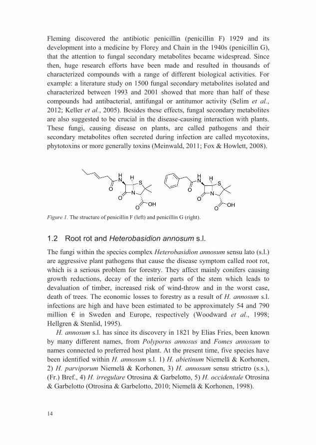
Fleming discovered the antibiotic penicillin (penicillin F) 1929 and its development into a medicine by Florey and Chain in the 1940s (penicillin G),that the attention to fungal secondary metabolites became widespread. Since then, huge research efforts have been made and resulted in thousands of characterized compounds with a range of different biological activities. Forexample: a literature study on 1500 fungal secondary metabolites isolated and characterized between 1993 and 2001 showed that more than half of these compounds had antibacterial, antifungal or antitumor activity (Selim et al.,2012; Keller et al., 2005). Besides these effects, fungal secondary metabolites are also suggested to be crucial in the disease-causing interaction with plants. These fungi, causing disease on plants, are called pathogens and their secondary metabolites often secreted during infection are called mycotoxins,phytotoxins or more generally toxins (Meinwald, 2011; Fox & Howlett, 2008).
Figure 1. The structure of penicillin F (left) and penicillin G (right).
1.2 Root rot and Heterobasidion annosum s.l.
The fungi within the species complex Heterobasidion annosum sensu lato (s.l.)are aggressive plant pathogens that cause the disease symptom called root rot, which is a serious problem for forestry. They affect mainly conifers causinggrowth reductions, decay of the interior parts of the stem which leads to devaluation of timber, increased risk of wind-throw and in the worst case, death of trees. The economic losses to forestry as a result of H. annosum s.l.infections are high and have been estimated to be approximately 54 and 790 million € in Sweden and Europe, respectively (Woodward et al., 1998; Hellgren & Stenlid, 1995).
H. annosum s.l. has since its discovery in 1821 by Elias Fries, been known by many different names, from Polyporus annosus and Fomes annosum to names connected to preferred host plant. At the present time, five species have been identified within H. annosum s.l. 1) H. abietinum Niemelä & Korhonen, 2) H. parviporum Niemelä & Korhonen, 3) H. annosum sensu strictro (s.s.), (Fr.) Bref., 4) H. irregulare Otrosina & Garbelotto, 5) H. occidentale Otrosina & Garbelotto (Otrosina & Garbelotto, 2010; Niemelä & Korhonen, 1998).
15
The species within the species complex have different distributions and host preferences. H. irregulare and H. occidentale are North American species whereas the other three are appearing in Europe. Generally, H. parviporum, H. abietinum and H. occidentale infect spruce and H. annosum s.s. and H. irregulare pine. However, overlap does occur and other tree species can be affected as well (Otrosina & Garbelotto, 2010).
H. annosum s.l. infections are initially spread by airborne basidiospores tofreshly cut stumps or wounds on the roots or stem. The fresh surface is subsequently colonized and the infection spreads rapidly down the stem to the root system. Once the infection has reached the root system it can, by root to root contacts infect nearby healthy trees. The infection grows subsequently up through the trunk and can reach several meters high (Stenlid & Redfern, 1998).
1.3 Biocontrol and Phlebiopsis gigantea
Major efforts have been made to prevent H. annosum s.l. root rot resulting in many different control methods including biological, chemical and silvicultural methods. The biological method includes the use of other microorganisms to suppress disease and is referred to as biocontrol. The fungus Phlebiopsis gigantea is such a biocontrol agent used to prevent H. annosum s.l. infections.
P. gigantea is a highly competitive fungus and is, like H. annosum s.l., an early colonizer of freshly cut conifer wood. The ability to outcompete H.annosum s.l. with P. gigantea in pine stumps and roots was observed already in the fifties of the last century by Rishbeth (1951), but it was not until a decade later that it was used as a biocontrol in practical use. A arhroconidia-based powder formulation of P. gigantea, based on a Finnish strain isolated by Korhonen et al. (1994), was later developed into a commercially available product, Rotstop®. Rotstop® is now the most widely applied stump treatment against H.annosum s.l. infections in Europe, used on more than 200 000 ha annually (Menkis et al., 2012; Holdenrieder & Greig, 1998).
Stump treatment with aqueous solution of urea or disodium octaborate tetrahydrate are two chemical methods that are in use, especially in Great Britain and in North America, but less frequently in Sweden. Urea acts by increasing the pH of the treated surface which can favor other competitive fungi but it is also proposed to be poisonous for H. annosum s.l. (Johansson et al., 2002). Boron is poisonous for fungi in general but also for other organism, for example insects. It acts by blocking the growth of cells in H. annosum s.l. (Sturesson et al., 2011).
Silvicultural methods that have been developed to reduce H. annosum s.l. infections are, among others, stump removal, choice of tree species to less
16
susceptible species, mixed stands (mix of differently susceptible species) and thinning in the winter when temperatures are below 5 °C and germination of basidiospores is poor (Cleary et al., 2013; Korhonen et al., 1998).
1.4 Aims and objectives
Sweden is one of the world´s largest exporters of sawn timber, pulp and paper and the forest is a cornerstone in Swedish industry. A severe threat against healthy tree stands are root rot caused primarily by the root rooting fungi H.annosum s.l. The overall aim of the present thesis was to further our understanding of interactions between H. annosum s.l., their host and the biocontrol fungus P. gigantea. As part of this work, it is important to gain knowledge about involved secondary metabolites, their structure and biosynthesis. The specific objectives of the individual projects were:
H. annosum s.l. produces several bioactive secondary metabolites including the benzofuran fomannoxin and the sesquiterpene fomannosin.The objective of this study was to further investigate bioactive secondary metabolites produced by H. occidentale. (paper I)
Fomannoxin is a biologically active benzohydrofuran produced by H. annosum s.l. and has been proposed to be involved in the fungus pathogenicity. The objective of this study was, by incorporation of 13C, to investigate the biosynthesis of fomannoxin. (paper II)
The objective of this study was to investigate if there are differenceswithin the H. annosum s.l. species complex regarding the production of secondary metabolites and if this could be related to host preferences.(paper III)
Little is known about how the biocontrol fungus P. gigantea act as a biocontrol against H. annosum s.l. The objective of this project was to characterize P. gigantea regarding the production of secondary metabolites and investigate whether these compounds could contribute to its biocontrol effect against H. annosum s.l. (paper IV)

17
2 Classes of secondary metabolites andtheir biosynthesis
Secondary metabolites are biosynthesized of building blocks that are put together in various metabolic pathways. The pathways are usually named after enzymes or intermediates involved and are commonly also used to classify secondary metabolites. The diversity and complexity of the structures that these relatively few building blocks can provide is both surprising and fascinating.
This section aims to briefly explain the different pathways with focus on the pathways relevant for the compounds within this thesis. For further reading see comprehensive textbooks on the biosynthesis subject e.g. Dewick (2009) or Mann et al. (1994).
2.1 The MVA and MEP pathway: Terpenoids
Terpenoids are found in essentially all forms of life and they form a large group of secondary metabolites with more than 40 000 structures (Bohlmann & Keeling, 2008). They are built up from five-carbon segments, so called “isoprene units”, which can combine to form different classes of terpenoids: hemi- (C5), mono- (C10), sesqui- (C15), di- (C20), sester- (C25), tri- (C30) and tetraterpenes (C40). The name “isoprene units” might be misleading since isoprene itself is a hemiterpene. The actual fundamental building block to all terpenes and terpenoids is isopentenyl diphosphate (IPP) and its isomer dimethylallyl diphosphate (DMAPP). Depending on the producing organism, IPP and DMAPP are biosynthesized either via the mevalonic acid (MVA) pathway or the methylerythritol phosphate (MEP) pathway. Fungi and animals have only access to the MVA pathway whereas algae and most bacteria use the MEP pathway. Plants and some bacteria have the possibility to use both pathways (Grawert et al., 2011; Walter et al., 2000; Rohmer, 1999).
18
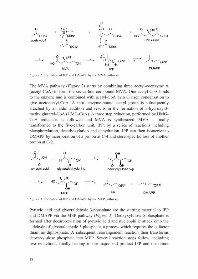
Figure 2. Formation of IPP and DMAPP by the MVA pathway.
The MVA pathway (Figure 2) starts by combining three acetyl-coenzyme A (acetyl-CoA) to form the six-carbon compound MVA. One acetyl-CoA bindsto the enzyme and is combined with acetyl-CoA by a Claisen condensation to give acetoacetyl-CoA. A third enzyme-bound acetyl group is subsequently attached by an aldol addition and results in the formation of 3-hydroxy-3-methylglutaryl-CoA (HMG-CoA). A three step reduction, performed by HMG-CoA reductase, is followed and MVA is synthesized. MVA is finally transformed to the five-carbon unit, IPP, by a series of reactions including phosphorylation, decarboxylation and dehydration. IPP can then isomerize to DMAPP by incorporation of a proton at C-4 and stereospecific loss of another proton at C-2.
Figure 3. Formation of IPP and DMAPP by the MEP pathway.
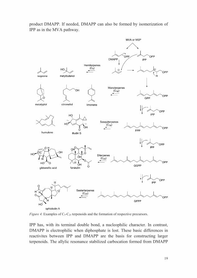
Pyruvic acid and glyceraldehyde 3-phosphate are the starting material to IPP and DMAPP via the MEP pathway (Figure 3). Deoxyxylulose 5-phosphate is formed after decarboxylation of pyruvic acid and nucleophilic attack onto the aldehyde of glyceraldehyde 3-phosphate, a process which requires the cofactor thiamine diphosphate. A subsequent rearrangement reaction then transforms deoxyxylulose phosphate into MEP. Several reaction steps follow, including two reductions, finally leading to the major end product IPP and the minor
19
product DMAPP. If needed, DMAPP can also be formed by isomerization of IPP as in the MVA pathway.
Figure 4. Examples of C5-C25 terpenoids and the formation of respective precursors.
IPP has, with its terminal double bond, a nucleophilic character. In contrast,DMAPP is electrophilic when diphosphate is lost. These basic differences in reactivites between IPP and DMAPP are the basis for constructing larger terpenoids. The allylic resonance stabilized carbocation formed from DMAPP
20
can go on to form different hemiterpenes, exemplified in Figure 4 by isoprene and metylbutenol, or it can react with IPP and form, after loss of a proton, geranyl diphosphate (GPP). GPP, in turn, is the precursor of monoterpenes but can also be extended by addition of one further IPP to form farnesyl diphosphate (FPP), the precursor of sesquiterpenes. Precursors of diterpenes and sesterterpenes, geranylgeranyl diphosphate (GGPP) and geranylfarnesyl diphosphate (GFPP), respectively, are formed in analogous additions reactions of IPP to FPP and GGPP, respectively, as illustrated in Figure 4. Electrophiles as DMAPP, GPP, FPP etc. can also act as alkylating agents in the construction of meroterpenoids, i.e. other secondary metabolites that contain terpenoid moieties. Alkylation by the C5 carbon segement from DMAPP is, for example, a very common reaction to extend the diversity of aromatics in bacteria, fungi and plants (Saleh et al., 2009; Yazaki et al., 2009).
Several examples of C5-C25 terpenoids are given in Figure 4 and by looking at the structure of these molecules one can see some of all the different modifications that can be done to respective precursor, contributing to the diversity of terpenoids. From different carbon skeletons; linear, mono-, bi and multicyclic to various functional groups e.g. carboxylic acids, alcohols, aldehydes, ketones, ester etc.
Terpenoids possess many biological properties and they are widely used asflavors, fragrances, pharmaceuticals, food additives etc. For example, the monoterpene limonene has a strong smell of oranges and is apparent in considerable amounts in many citrus fruits. Citronellol, found in roseoil, is like limonene used as fragrance in many perfumes but also as insecticide (Forster-Fromme & Jendrossek, 2010; Dewick, 2009). Another monoterpene, eucalyptol, the main constituent of the Eucalyptus plant, is used as an antibacterial, anti-inflammatory, antihypertensive agent etc. (Liapi et al.,2007). The sesquiterpene illudin S, produced by the mushroom Omphalotus illudens, is an extremely toxic compound. The toxicity is proposed to be due to the presence of the strained cyclopropyl group which makes it reactive against nucleophiles and has been shown to alkylate DNA (Schobert et al., 2011).Another sesquiterpene, humulene, found in hops is an important fragrance of beer, but it is also a key intermediate in the formation of e.g. fomannosin (paper I) and protoilludanes (Abraham, 2001). Forskolin, isolated from roots of Coleus forskohlii, an Indian plant, has been used in traditional medicine and has been shown to lower blood pressure and has cardioprotective properties(Asada et al., 2012). A well-known plant hormone, gibberellic acid, first isolated from the fungus Gibberella fujikuroi, as a phytotoxin, is produced commercially and used, for example, in the production of seedless grapes (Hanson, 2003). The last example, Ophiobolin A, a sesterterpene found in the
21
plant pathogen Helminthosporium maydis, has a broad spectrum of biological activity against bacteria, fungi and nematodes (Dewick, 2009).
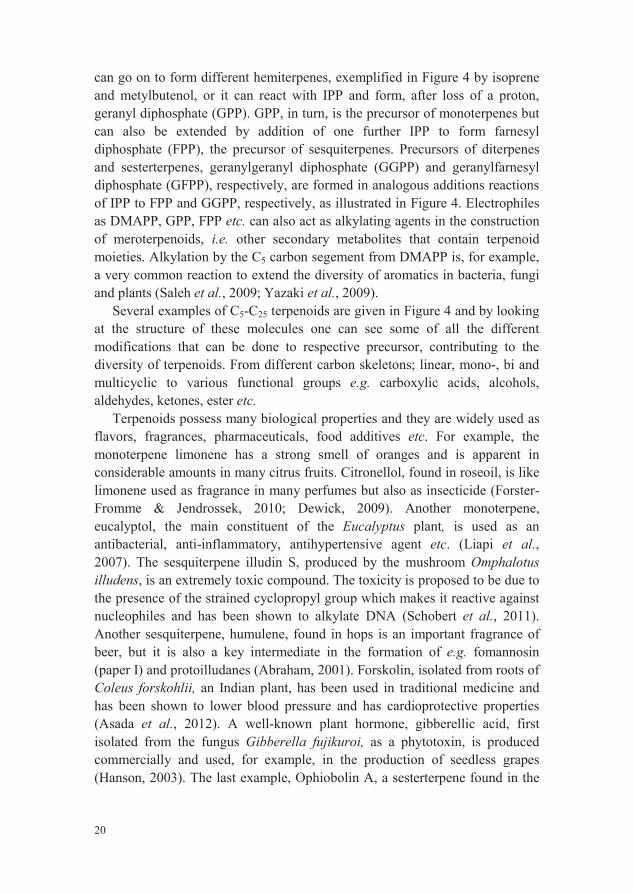
All different structures and properties of terpenoids are the results of modifications accomplished via various enzymatic reactions such as changes to the oxidation state of a molecule by oxidation and reduction reactions, alkylations, decarboxylations, glycosylations, rearrangements and cyclization reactions etc. Many of them, as for example, rearrangement reactions and cyclization reactions are often carbocation driven. The cyclization of E,E-FPP to humulene can serve as an example and is illustrated in Figure 5. The cyclization to the 11-membered ring system of the humulyl cation is the result of a SN1 reaction, involving a carbocation intermediate formed upon loss of diphosphate from FPP. Subsequent loss of a proton leads to humulene. Humulene can then be further transformed to structurally complex bicyclic and tricyclic sesquiterpenes, with the most important pathway generating theprotoilludane skeleton (Abraham, 2001) present in for example illudolone A and B, (Paper I).
Figure 5. Cyclization of humulene.
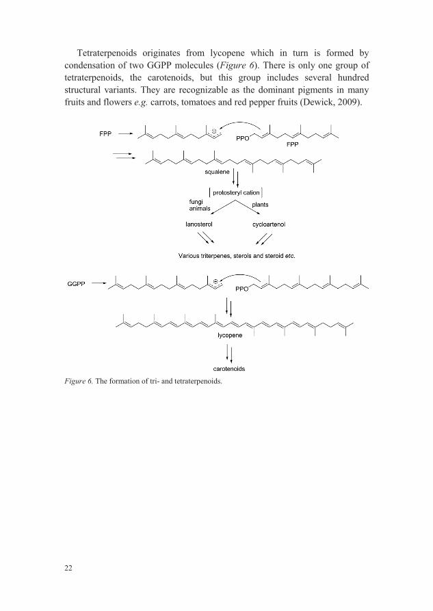
Triterpenoids and tetraterpenoids are not formed by adding further IPP building blocks to GFPP, instead pairwise combination of FPP and GGPP, respectively, give rise to these two classes of terpenoids (Figure 6).
Squalene, first isolated from shark liver oil, is the precursor of triterpenesand is formed, after a sequence of reactions catalyzed by squalene synthase, from two FPP molecules. Cyclization of squalene then leads, via the intermediate protosteryl cation, to lanosterol or cycloartenol. Lanosterol, formed in fungi and animals, is in turn the precursor for many triterpenoids and modified triterpenoids e.g. steroids and sterols. Cycloartenol has the corresponding function as lanosterol, but is formed exclusively in plants(Dewick, 2009).
22
Tetraterpenoids originates from lycopene which in turn is formed by condensation of two GGPP molecules (Figure 6). There is only one group of tetraterpenoids, the carotenoids, but this group includes several hundred structural variants. They are recognizable as the dominant pigments in many fruits and flowers e.g. carrots, tomatoes and red pepper fruits (Dewick, 2009).
Figure 6. The formation of tri- and tetraterpenoids.
23
2.2 The shikimic acid pathway: Aromatic amino acids andphenylpropanoids
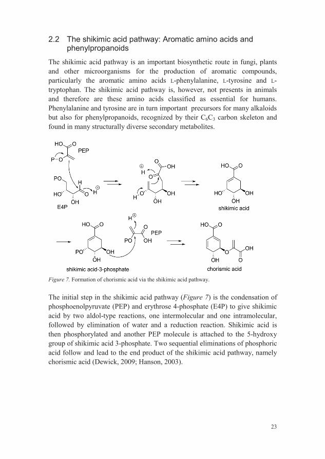
The shikimic acid pathway is an important biosynthetic route in fungi, plants and other microorganisms for the production of aromatic compounds, particularly the aromatic amino acids L-phenylalanine, L-tyrosine and L-tryptophan. The shikimic acid pathway is, however, not presents in animals and therefore are these amino acids classified as essential for humans. Phenylalanine and tyrosine are in turn important precursors for many alkaloids but also for phenylpropanoids, recognized by their C6C3 carbon skeleton andfound in many structurally diverse secondary metabolites.
Figure 7. Formation of chorismic acid via the shikimic acid pathway.
The initial step in the shikimic acid pathway (Figure 7) is the condensation of phosphoenolpyruvate (PEP) and erythrose 4-phosphate (E4P) to give shikimic acid by two aldol-type reactions, one intermolecular and one intramolecular, followed by elimination of water and a reduction reaction. Shikimic acid is then phosphorylated and another PEP molecule is attached to the 5-hydroxy group of shikimic acid 3-phosphate. Two sequential eliminations of phosphoric acid follow and lead to the end product of the shikimic acid pathway, namely chorismic acid (Dewick, 2009; Hanson, 2003).
24
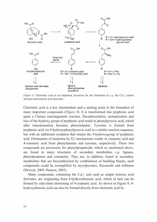
Figure 8. Chorismic acid as an important precursor for the formation of e.g. the C6C3 carbon skeleton and benzoic acid derivates.
Chorismic acid is a key intermediate and a starting point in the formation of many important compounds (Figure 8). It is transformed into prephenic acid upon a Claisen rearrangement reaction. Decarboxylation, aromatization and loss of the hydroxy group of prephenic acid result in phenylpyruvic acid, which after transamination becomes phenylalanine. Tyrosine is formed from prephenic acid via 4-hydroxyphenylpyruvic acid in a similar reaction sequence, but with an additional oxidation that retains the 4-hydroxygroup of prephenic acid. Elimination of ammonia by E2 mechanisms results in cinnamic acid and 4-coumaric acid from phenylalanine and tyrosine, respectively. These twocompounds are precursors for phenylpropanoids, which as mentioned above, are found in many structures of secondary metabolites e.g. lignans,phenylpropenes and coumarins. They are, in addition, found in secondary metabolites that are biosynthesized by combination of building blocks, such compounds could be exemplified by styrylpyrones, flavanoids and stilbenes(Dewick, 2009; Hanson, 2003).
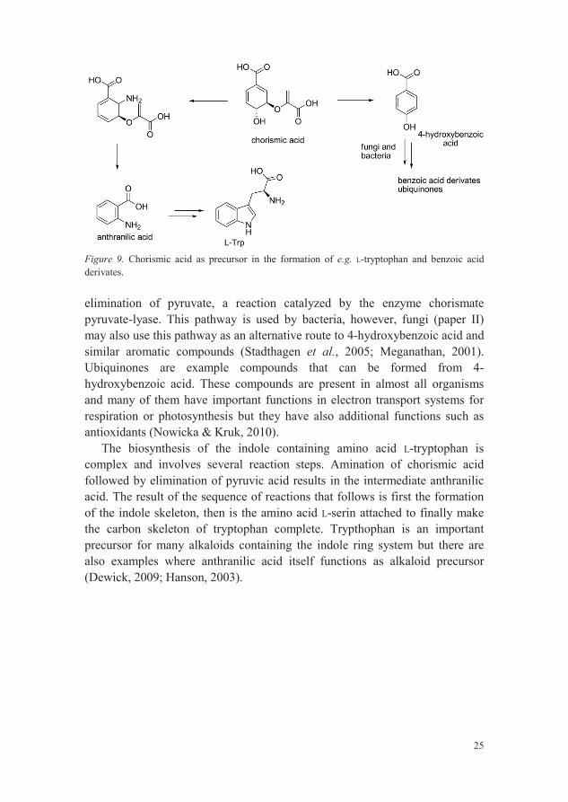
Many compounds, containing the C6C1 unit such as simple benzoic acid derivates, are originating from 4-hydroxybenzoic acid, which in turn can be formed by side-chain shortening of 4-coumaric acid. As shown in Figure 9, 4-hydroxybenzoic acid can also be formed directly from chorismic acid by
25
Figure 9. Chorismic acid as precursor in the formation of e.g. L-tryptophan and benzoic acid derivates.
elimination of pyruvate, a reaction catalyzed by the enzyme chorismate pyruvate-lyase. This pathway is used by bacteria, however, fungi (paper II) may also use this pathway as an alternative route to 4-hydroxybenzoic acid and similar aromatic compounds (Stadthagen et al., 2005; Meganathan, 2001).Ubiquinones are example compounds that can be formed from 4-hydroxybenzoic acid. These compounds are present in almost all organisms and many of them have important functions in electron transport systems for respiration or photosynthesis but they have also additional functions such as antioxidants (Nowicka & Kruk, 2010).
The biosynthesis of the indole containing amino acid L-tryptophan is complex and involves several reaction steps. Amination of chorismic acid followed by elimination of pyruvic acid results in the intermediate anthranilic acid. The result of the sequence of reactions that follows is first the formation of the indole skeleton, then is the amino acid L-serin attached to finally make the carbon skeleton of tryptophan complete. Trypthophan is an important precursor for many alkaloids containing the indole ring system but there are also examples where anthranilic acid itself functions as alkaloid precursor (Dewick, 2009; Hanson, 2003).

26
2.3 The acetate pathway: Polyketides
The polyketides is a large family of secondary metabolites found in bacteria, fungi and plants. They are particularly important for fungi as the most abundant fungal secondary metabolites and many of them have important biological activities. The cholesterol lowering compound lovastatin, produced by the fungi Monascus ruber and Aspergillus terreus, serves as a good example. It works as an inhibitor of the enzyme HMG-CoA reductase which is active in the formation of MVA and was the first statin to be marketed (Dewick, 2009; Keller et al., 2005).
Polyketides are derived from poly- -keto chains, formed by stepwise condensation of mainly acetyl-CoA (starter unit) and malonyl-CoA (extender unit). The reactions involved in the construction of the poly- -keto chain arecatalyzed by polyketide synthase (PKS), a family of multidomain enzymes or enzyme complexes. There are several types of PKSs found in different organisms but the principal of constructing the poly- -keto chain are the same in all PKSs (Figure 10). The initial step is the loading, as thioesters, of both the starter unit and the extender unit to the respective site on the PKS. Acetyl-CoA is bound to a ketoacyl-CoA synthase (KS) domain whereas the extender unit is bound to an acyl carrier (ACP) domain. Condensation then occurs between malonyl-ACP and acetyl-KS by a Claisen-type reaction and simultaneous decarboxylation of the ACP-bound extender unit. -ketothioester bound to the ACP domain can then be transferred to a KS domain and extended by another malonyl-ACP. This cycle repeats -keto chain has reached the desired length. Once the chain is completed it can be folded and activated at desired positions to allow for intramolecular reactions.This is exemplified in Figure 10 by the biosynthesis of orsellinic acid and phloracetophenone. Other more complicated aromatic compounds like the anthraquinones can be syn -keto chain as precursor (Dewick, 2009; Hanson, 2003).

27
Figure 10. Polyketide synthesis.
The carbon skeleton of macrolides, another type of polyketides characterized by large lactone rings, is formed by the same general process but with some modifications. The modifications include reductions, dehydrations, etc., of the
-carbonyl group and may take place after each condensation step and before the next chain extension (Figure 11). The carbon chain of the polyketide leading to macrolides is thus partially reduced. The biosynthesis of macrolides is exemplified in Figure 11 by zearalenone, a mycotoxin produced by e.g.several Fusarium species (Dewick, 2009; Elsharkawy & Abulhajj, 1987).
In addition to the variability that can be achieved through the use of the above mentioned capabilities, (number of extender units, different folding, reduction reactions etc.), the use of other starter and/or extender units than acetyl-CoA and malonyl-CoA further extend the structural diversity of polyketides. Flavanoids and stilbenes are, for example, biosynthesized from a cinnamoyl-CoA starter unit (derivied from the shikimic acid pathway) with chain extension using three malonyl-CoA units.

28
Figure 11. Formation of macrolides.
2.4 Alkaloids, carbohydrates, fatty acids, peptides and proteins
2.4.1 Alkaloids
Alkaloids are cyclic organic compounds that contain one or more nitrogen atoms and are often basic. They have usually pronounced effects on the nervous system of humans and other animals. A few well-known examples ofalkaloids are presented in Figure 12.
.
Figure 12. Structure of various familiar alkaloids.

29
Alkaloids are commonly produced from amino acids such as ornithine, lysine, tryptophan and tyrosine but other building blocks, e.g. terpenes or acetate pathway derived moieties, are also often incorporated into structures of alkaloids.
Ergolines are a group of alkaloids containing the indole ring system and have been found in several fungal genera e.g. Claviceps. These fungi are responsible for a fungal disease called ergot that affect cultivated grass such as wheat and rye and can be poisonous for humans and animals upon feeding. D-(+)-lysergic acid (Figure 13) is one of more than 50 alkaloids characterized from these fungi and are formed from tryptophan and DMAPP, originating from the shikimic acid pathway and the MVA/MEP pathway, respectively(Dewick, 2009; Schiff, 2006).
Figure 13. Formation of lysergic acid from tryptophan.
2.4.2 Carbohydrates
Carbohydrate biosynthesis and degradation are essential for all organisms and are typical components of the primary metabolism. However, some of these carbohydrates e.g. glucose, are attached to secondary metabolites in glycosides. The non-carbohydrate moiety of such a compound is known as the aglycone and may originate from one or more biosynthetic pathway e.g. the shikimic acid pathway and the acetate pathway. Fusicoccin A (Figure 14), a fungal secondary metabolite from Fusicoccum amygdale, is an example of a glucopyranoside of a diterpene that has, in a recent study, been shown to have growth inhibiting effects on the deadly brain tumor, glioblastoma multiforme (Bury et al., 2013).

30
Figure 14. Chemical structure of fusicoccin A.
2.4.3 Fatty acids
Common fatty acids are mostly regarded as primary metabolites as they are important components of some vital functions in organisms. They are usually combined as esters with glycerol, forming glycerides. Triglycerides as an example, form fats and oils, depending of the individual fatty acids, and are an
-oxidation.Fatty acids are biosynthesized via the acetate pathway in a very similar way
as the polyketides, although different enzymes are working. Fatty acids may also, as carbohydrates, be attached to other metabolites. One example is the 16-palmityl ester of cucurbitacin B (Figure 15), found in the fungus Leucopaxillus gentianeus (Clericuzio et al., 2004).
Figure 15. Structure of the 16-palmityl ester of cucurbitacin B.
2.4.4 Peptides and proteins
Peptides and proteins consist of amino acids that are linked to form chains with different lengths, the length determine whether the product will be referred to as peptide (up to 40-50 amino acids) or protein (generally more than 40-50amino acids). Peptides and proteins, as a group, are difficult to classify as primary or secondary metabolites since many of them are large in size and found in materials that are widely distributed and occur in many different organisms while other are small and restricted in occurrence.

31
Peptides and proteins are produced by either ribosomal (produced on the ribosome) or non-ribosomal biosynthesis. Non-ribosomal peptides are synthesized by multidomain, multimodular enzymes called non-ribosomal peptide synthases (NRPSs) and are, in contrast to ribosomal synthesis, not dependent of mRNA. The sequential extension in non-ribosomal peptide synthesis can be compared to and is similar to polyketide synthesis. Each module in an NRPS contains several domains and each domain has specific function e.g. recognition of amino acid, bond formation between amino acids,or release of synthesized peptide (Dewick, 2009; Keller et al., 2005).
-(L- -aminoadipyl)-L-cysteinyl-D-valine synthetase, the first identified fungal NRPS, catalyses the assembly of the L-aminoacids and the epimerization of L-valine to D-valine in the formation of the tripeptide that, after cyclization by isopenicillin N synthase, become isopenicillin N (Figure 16). Isopenici -lactam antibiotics e.g.penicillin G (Keller et al., 2005; Smith et al., 1990).
Figure 16. Biosynthesis of isopenicillin N and subsequent modification to penicillin G.

32
33
3 ExperimentalThis chapter aims to briefly explain the principal of techniques and methodsused during this thesis work. Examining the structure and biosynthesis of secondary metabolites involves (1) sample preparation, (2) separation and isolation, (3) structure determination and characterization.
There is also, at the end, a brief description of the isotopic labeling methodused for studying the biosynthesis of fomannoxin.
3.1 Sample preparation
Sample preparation isolates analytes of interest and simultaneous reduces impurities, from a matrix that is sometimes both large and complex. The aim of sample preparation is a purified and concentrated sample ready for further analysis.
3.1.1 Solvent extraction
Solvent extraction is one of the oldest and most common extraction methods. It was used for extracting semi-solid samples in this work but is generally applicable for both solid and liquid samples. If a liquid is to be extracted, the extraction method is commonly referred to as liquid-liquid extraction (LLE).Before extraction of solids, the samples are often homogenized by chopping or pulverizing. The homogenized solid or liquid sample is then repeatedly extracted with a suitable organic solvent. Following extraction, the extract iscommonly filtrated or centrifuged and concentrated by rotary evaporation(Zhang et al., 2012; Novakova & Vlckova, 2009).
Drawbacks of solvent extraction is that this method is relative time-consuming and large amounts of environmentally harmful organic solvents are used. However, alternative extraction methods for liquids or dissolved solids have been developed e.g. solid phase extraction.
34
3.1.2 Solid phase extraction
Solid phase extraction (SPE) is nowadays the most popular sample preparation method and has been used extensively in this thesis for e.g. sampleconcentration and clean up. The general SPE procedure is simple: a sample commonly containing lipophilic analytes in an aqueous solution, are subjected to a short column which contains the solid phase. The column has previously been activated and equilibrated by e.g. methanol and water, respectively. Activation solvates the dry solid phase and makes it available for the analytes whereas the equilibration aims to prevent losses upon sample loading. The lipophilic analytes are adsorbed to the solid phase whereas the impurities and solvent are passed through. The column is then washed, with water, in order to remove impurities and the analytes are then finally desorbed and eluted by adding a small amount of an organic solvent e.g. methanol or acetonitrile (Zhang et al., 2012).
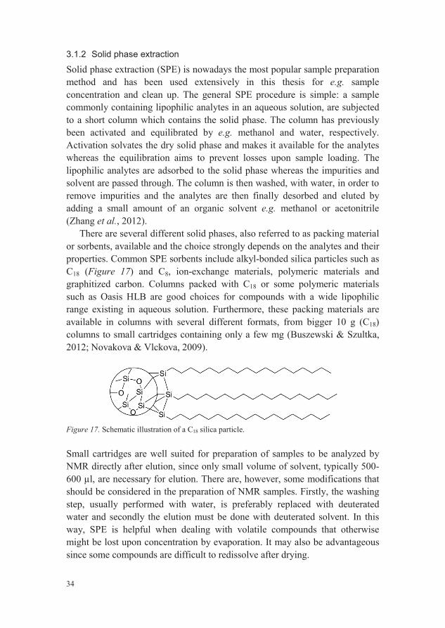
There are several different solid phases, also referred to as packing material or sorbents, available and the choice strongly depends on the analytes and their properties. Common SPE sorbents include alkyl-bonded silica particles such as C18 (Figure 17) and C8, ion-exchange materials, polymeric materials and graphitized carbon. Columns packed with C18 or some polymeric materials such as Oasis HLB are good choices for compounds with a wide lipophilic range existing in aqueous solution. Furthermore, these packing materials are available in columns with several different formats, from bigger 10 g (C18)columns to small cartridges containing only a few mg (Buszewski & Szultka, 2012; Novakova & Vlckova, 2009).
Figure 17. Schematic illustration of a C18 silica particle.
Small cartridges are well suited for preparation of samples to be analyzed by NMR directly after elution, since only small volume of solvent, typically 500-600 l, are necessary for elution. There are, however, some modifications that should be considered in the preparation of NMR samples. Firstly, the washing step, usually performed with water, is preferably replaced with deuterated water and secondly the elution must be done with deuterated solvent. In this way, SPE is helpful when dealing with volatile compounds that otherwise might be lost upon concentration by evaporation. It may also be advantageous since some compounds are difficult to redissolve after drying.
35
3.2 Separation and isolation
A fungus may produce tens or perhaps hundreds of secondary metabolites in different amounts and with various physical and chemical properties. To facilitate structure elucidation and characterization of individual secondary metabolites, the compounds need to be isolated. Chromatography has become an excellent tool for such a task and several chromatographic methods havebeen developed since it first was described about hundred years ago by the Russian botanist Michail Tswett (Ettre, 2003). High performance liquid chromatography (HPLC) and gas chromatography (GC) are the two most important chromatographic techniques for low molecular weight compounds.GC uses an inert gas e.g. helium as mobile phase whereas HPLC uses a liquid.Chromatography can be performed either for analytical purposes or for preparative purposes. Analytic chromatography aims to separate, identify and/or quantify components of a sample and can be performed by both GC and HPLC. Preparative chromatography, on the other hand, aims to not only separate but also physically isolate components by collecting them as they are eluted from the column and cannot be performed by GC. In addition, samples to be analyzed by GC must be volatile, a characteristic lacking in many components of a fungal extract. Therefore, HPLC has been the major chromatographic technique used in this thesis.
3.2.1 High performance liquid chromatography
Among the various HPLC techniques; normal phase HPLC, reversed phase HPLC (RP-HPLC), ion exchange and size exclusion HPLC, RP-HPLC is by far the most common. RP-HPLC is also the techniques that has been used during this thesis work and is from now on referred to as simply HPLC.
A typical HPLC system consist of a high-pressure pump system, an automated sample injector, a separation column, a detector and a computer for displaying the results and controlling the system. Additional common components in a modern HPLC system include solvent degasser, pre-column and column oven. An injected sample is transported by the mobile phase to the column where the actual separation begins. The column is packed with a non-polar stationary phase through which a moderately polar mobile phase is pumped. The compounds are separated because they interact with the stationary phase and are retained differently. Compounds, poorly retained, travel faster through the column and are said to have shorter retention time (tR)than compounds that are strongly retained by the stationary phase.
There are several factors that influence the retention and thus the separation e.g. selected stationary phase, mobile phase modifier, mobile phase
36
composition, temperature, and flow rate. The most commonly used stationary phases are based on microporous silica particles (typically 3-5 m in diameter) to which alkyl chains e.g. C18, C8 or substituted alkyl chains e.g. (CH2)3NH2,(CH2)CN are attached. Water miscible organic solvents such as methanol and acetonitrile are typical mobile phase modifiers that are used in different composition with water. The composition of the mobile phase can either be kept constant as in isocratic elution or it can be continuous changed during the chromatography to increase the eluent strength. The latter method, called gradient elution, is often a good starting point for complex samples and a typical gradient profile starts at 5% organic modifier and progress to 95% within 10-40 minutes. An optimized isocratic elution method, on the other hand, is sometimes a good alternative for compounds with similar tR and difficult to separate with a gradient method. For further reading of HPLC, seefor e.g. Miller (2009).
Fast analysis and high efficiency are desired in HPLC and have driven the development of, mainly, columns and pump systems. Smaller columns with particle size less than 2 m are nowadays available that permit separation of rather complex mixtures in just a few minutes. However, these columns need pump systems able to perform at higher pressure and these new systems, called ultra high performance LC (UHPLC), are rather expensive compared to regular HPLC systems. Superficially porous silica particle columns also referred to as fused core columns are a new type of columns with a solid inner core (typically 1.6-1.9 m) covered by a porous silica shell (typically 0.5 m). Columns packed with fused core material create a much lower back-pressure and allow high flow rates to be maintained resulting in shorter analysis time withoutlosing chromatographic quality. Furthermore, these columns can be used by regular HPLC systems and could be an alternative to UHPLC (Ali et al., 2012; Novakova & Vlckova, 2009).
3.3 Structure determination and characterization
3.3.1 Mass spectrometry
Mass spectrometry (MS) can be divided into three basic steps: (1) ionization,(2) mass analysis and (3) detection.
Ionization takes place in the ion source and aims to convert analytes into gas-phas ions. There are a number of different methods and the choice depends on the sample phase e.g. liquid or solid and the nature of the analytes. Common ionization methods includes electrospray ionization (ESI), electron ionization (EI), fast atom bombardment (FAB), matrix-assisted laser desorption ionization (MALDI), atmospheric-pressure chemical ionization (APCI) among
37
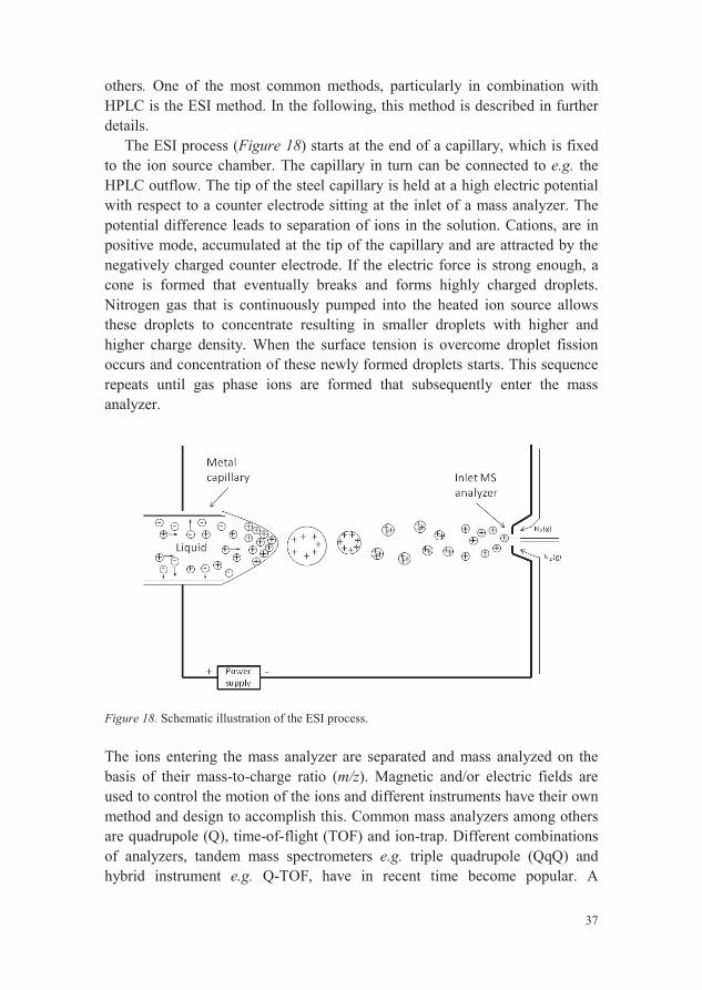
others. One of the most common methods, particularly in combination with HPLC is the ESI method. In the following, this method is described in further details.
The ESI process (Figure 18) starts at the end of a capillary, which is fixedto the ion source chamber. The capillary in turn can be connected to e.g. the HPLC outflow. The tip of the steel capillary is held at a high electric potential with respect to a counter electrode sitting at the inlet of a mass analyzer. The potential difference leads to separation of ions in the solution. Cations, are in positive mode, accumulated at the tip of the capillary and are attracted by the negatively charged counter electrode. If the electric force is strong enough, acone is formed that eventually breaks and forms highly charged droplets.Nitrogen gas that is continuously pumped into the heated ion source allowsthese droplets to concentrate resulting in smaller droplets with higher and higher charge density. When the surface tension is overcome droplet fission occurs and concentration of these newly formed droplets starts. This sequence repeats until gas phase ions are formed that subsequently enter the mass analyzer.
Figure 18. Schematic illustration of the ESI process.
The ions entering the mass analyzer are separated and mass analyzed on the basis of their mass-to-charge ratio (m/z). Magnetic and/or electric fields are used to control the motion of the ions and different instruments have their own method and design to accomplish this. Common mass analyzers among others are quadrupole (Q), time-of-flight (TOF) and ion-trap. Different combinations of analyzers, tandem mass spectrometers e.g. triple quadrupole (QqQ) and hybrid instrument e.g. Q-TOF, have in recent time become popular. A
38
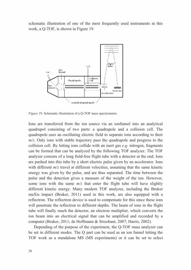
schematic illustration of one of the most frequently used instruments in this work, a Q-TOF, is shown in Figure 19.
Figure 19. Schematic illustration of a Q-TOF mass spectrometer.
Ions are transferred from the ion source via an ionfunnel into an analytical quadrupol consisting of two parts: a quadrupole and a collision cell. The quadrupole uses an oscillating electric field to separate ions according to their m/z. Only ions with stable trajectory pass the quadrupole and progress to the collision cell. By letting ions collide with an inert gas e.g. nitrogen, fragments can be formed that can be analyzed by the following TOF analyzer. The TOF analyzer consists of a long field-free flight tube with a detector at the end. Ions are pushed into this tube by a short electric pulse given by an accelerator. Ions with different m/z travel at different velocities, assuming that the same kinetic energy was given by the pulse, and are thus separated. The time between the pulse and the detection gives a measure of the weight of the ion. However, some ions with the same m/z that enter the flight tube will have slightly different kinetic energy. Many modern TOF analyzer, including the Bruker maXis impact (Bruker, 2011) used in this work, are also equipped with a reflectron. The reflectron device is used to compensate for this since these ions will penetrate the reflectron to different depths. The beam of ions in the flight tube will finally reach the detector, an electron multiplier, which converts the ion beam into an electrical signal that can be amplified and recorded by a computer (Bruker, 2011; de Hoffmann & Stroobant, 2007; Harris, 2002).
Depending of the purpose of the experiment, the Q-TOF mass analyzer can be set in different modes. The Q part can be used as an ion funnel letting the TOF work as a standalone MS (MS experiments) or it can be set to select

39
specific ions for fragmentation and let the TOF analyze the fragments inMS/MS experiment mode. Since the TOF has high resolution and mass accuracy, determination of elemental composition of both fragment ions and precursor ions are feasible. This information is highly valuable for structure elucidation of unknown compounds. It is, however, important to calibrate the system when accurate mass determination is the aim of the analysis. The Q-TOF system used was calibrated by injecting a sodium formate solution.Sodium formate forms a series of cluster over a rather large mass range and is suitable for analysis of low molecular weight compounds with ESI.
The result of a MS analysis is presented in a mass spectrum and when the MS is connected to a HPLC, also a chromatogram. The chromatogram can be based on different types of data e.g. UV data, total ion current etc. As an example of a typical HPLC-HRMS (high resolution MS) analysis, the chromatogram and the mass spectra of phlebiopsin A and B from a 20%acetonitrile SPE eluate of P. gigantea are presented in Figure 20.

40
Figure 20. Base-peak chromatogram (top) from a HPLC-HRMS analysis and mass spectra of phlebiopsin A (middle; tR 21.7 min) and B (bottom; tR 25.8 min). Sample: A 20% MeCN SPE eluate of P. gigantea.
3.3.2 Nuclear magnetic resonance
Nuclear magnetic resonance (NMR) is a powerful method for determination of molecular structures. It is a technique that takes advantage of the fact that nuclei of different atoms in molecular structures absorb and re-emit electromagnetic radiation differently. Provided, however, that the molecule under study is placed in a strong magnetic field. The NMR data recorded can
41
then be used to elucidate the surrounding of each atom and ultimately the molecular structure.
Briefly, many atomic nuclei have what is called nuclear spin, i.e. the nuclei spin around their own axis and generate a small magnetic field and a magnetic moment that is oriented along the axis of spin. The magnetic moment is a vector and has both a direction and a magnitude. The direction of the magnetic moment in the atoms studied during this thesis work, 1H and 13C, can have two directions in the present of a strong magnetic field. That is, either with or against the external magnetic field. Nuclei whose magnetic moment are alignedwith the external field have slightly lower energy than the nuclei alignedagainst, resulting in a net magnetization that is aligned with the external magnetic field (+z). The system is in this situation in equilibrium. Furthermore, the force from the external magnetic field causes the magnetic moments to precess or circulate around the z-axis with a specific frequency, the Larmor frequency. If a pulse of electromagnetic radiation with a frequency that match the Larmor frequency now is applied along the x-axis, the net magnetization will be rotated around the x-axis in the yz-plane. The extent of rotation depends upon the given pulse. A 90 degree pulse, for example, will rotate the net magnetization so that it is along the y-axis. Once the pulse has beenterminated, the nuclei start to relax and return to equilibrium. It is during this process that a signal is recorded. The magnetic moment starts, immediately after the pulse, to precess around the z-axis with the Larmor frequency but now with magnetic moment in the xy-plane. This motion creates a signal that is detected by a receiver coil commonly located along the x-axis. As the relaxation proceeds the magnetization in the xy-plane steadily decreaseresulting in a signal that look like a decaying sinus wave. The recorded signal is called free induction decay (FID) and is transformed by Fourier transformation to a NMR spectrum (Lambert & Mazzola, 2003; Hore, 1995; Breitmaier, 1993).
The reason NMR is useful for structure elucidation is that many nuclei in a molecule have slightly different magnetic environments, depending of the electron density around them. This will result in signals at different frequencies which are, in the NMR spectrum, expressed as chemical shifts ( ) in parts per million (ppm) compared to the signal of tetramethylsilane (TMS). Nuclei do also affect each other and are said to couple to different extent. The major factor on the size of a coupling is the number of bonds between the interacting nuclei and the dihedral angle. The coupling results in a splitting of an NMR signal. For example, a coupling between two aromatic ortho protons will result in two doublets, with a mutual coupling constant in the order of 6-9 Hz.
42
3.3.3 The process of structure elucidation
This section aims to generally describe the process of how data from MS and various NMR experiments can be used to elucidate the chemical structure of an unknown compound. Structure elucidation normally involves (1) calculation of molecular formula, (2) determination of connectivity between atoms. This step can be divided into (i) determination of small fragments (ii) combining small fragments, (3) determination of relative configuration, (4) determination of absolute configuration.
As mentioned earlier, the data from high resolution MS can be used to calculate the molecular formula. This is commonly the first step in a structure elucidation analysis and can give a hint of the compound under investigation. For example, a molecular formula of C15H22O3, may suggest that it is a sesquiterpene, particularly if the organism is known to produce this type of compounds. More importantly, the molecular formula can be used to calculate the unsaturation index (five in C15H22O3) which is the sum of the number of rings and multiple bonds in the molecule.
There are a large amount of NMR experiments available that give different information. However, for routine structural elucidations a basic set of relatively simple, sensitive and fast experiments is most often sufficient. A basic set of experiments includes, for example, one dimensional 1H and 13C,1H-1H correlation spectroscopy (COSY), 1H-13C heteronuclear single quantum coherence (HSQC), 1H-13C heteronuclear multiple bond correlation (HMBC),1H-1H nuclear Overhauser effect spectroscopy (NOESY) or rotating frame nuclear Overhauser effect spectroscopy (ROESY). A good quality 1D 13C spectrum is, however, not always feasible to obtain as it requires more sample material compared to e.g. an HMBC experiment.
Except considering the molecular formula, a sample elucidation starts by determining the connectivity between the atoms in the molecule and the first analysis is often of the 1D 1H spectrum. The 1H spectrum might give an idea of the types of protons present in the molecule e.g. aldehyde, aromatic or methyl. In addition, each signal can be integrated to give the number of protons related to a certain signal. The number of protons should of course also be compared to the molecular formula. Missing signals could be a valuable source of information as exchangeable protons are deuterated in a protic deuterated solvent such as methanol-d4.
The next step is often to determine small fragments in the molecule by using a combination of data from COSY, HSQC and if recorded also 1D 13C. HSQC is a phase-sensitive two dimensional experiment which is used to identify the carbons and their chemical shifts to which certain protons are attached. This is possible since each peak in the HSQC spectrum represent the

43
chemical shift of every proton and its directly attached carbon i.e. one bond 1H-13C. Another advantage of the HSQC experiment is that the phase of each peak shows whether its corresponding carbon atom is bonded to an even or odd number of protons. The remaining carbons not bonded to any proton could now be identified from the 1D 13C data. Obtained 1H and 13C chemical shifts can be compared to tabulated chemical shifts available in for example many books (Lambert & Mazzola, 2003; Pavia et al., 2000). These can give a good picture of functional groups present in the molecule. COSY is a proton-proton correlated experiments where each peak represent a through bond correlation between certain protons. These correlations could be two (geminal), three (vicinal) or four bonds long (long-range) and sometimes even longer. Geminal couplings can be excluded from the information obtained from HSQC data and long-range couplings are often weaker and could therefore also be temporary excluded. Vicinal couplings are therefore left and small fragments can be determined.
HMBC data shows the connection between a certain proton and the carbons two and three bonds away, sometimes longer depending on the molecular structure. This is a very useful experiment to combine small fragments and finally determine the overall connectivity of the molecule. In addition, since 1D 13C data sometimes can be difficult to obtain, HMBC data can be sufficientto extract all the carbon chemical shifts in the molecule. The number of rings and multiple bonds as calculated from the molecular formula is, at this point, an excellent source of information to support in the puzzle of putting pieces together.
Once the connectivity is determined, the next step is to determine the relative configuration of the stereogenic carbons. NOESY and ROESY experiments are often used for this purpose. These two dimensional experiments show cross-peaks for protons close to each other in space.
Finally, when both the connectivity and the relation between groups on stereogenic carbons, i.e. relative configuration, have been established only absolute configuration remains to be determined. This is often one of the most difficult parts in the elucidation process but is needed for full structure determination.
Several methods exists e.g. X-ray crystallography, circular dichroism and different NMR based methods. The method used in this work was based on biosynthetic considerations. For example, a precursor involved in the biosynthesis of a certain secondary metabolite is known to be produced with a certain configuration. Logical reasoning could then be used to propose the absolute configuration of at least one stereogenic carbon of the investigated
44
compound and then, by considering the relative configuration, the absolute configuration of all stereogenic carbons.
3.4 Stable isotope labeling
Feeding living systems with isotopically labeled substrates is a common method to elucidate which metabolic pathways that have been used in the construction of a certain secondary metabolites. The method is based on that an organism is grown in the presence of a labeled substrate which is incorporated into the molecular structure of the metabolite under study. The metabolite is then analyzed to detect the presence of the labeling. It is thus essential that the organism produce the metabolite during the feeding period.
Radioactive isotopes such as 14C and 3H were frequently used in the middle of the 20th century but biosynthetic elucidations using these isotopes were dependent on careful laborious chemical degradation of the product to locate the labeling. 14C and 3H were later displaced with substrates labeled with stable isotopes e.g. 13C. 13C is an excellent tracer since it can by studied by NMR.13C-labeled positions are identified by their enhanced signals compared to unlabeled postions. Assignment of NMR signals will, in addition, give information of which carbons that have been labeled without any chemical degradation (Bacher et al., 1998).
All biosynthetic pathways leading to secondary metabolites use starting materials from the central metabolic pathways where glucose is a key compound. 13C-labeled glucose e.g. [1-13C]glucose, used in the biosynthetic study of fomannoxin (paper II), is therefore a qualified and common tracer to elucidate biosynthetic pathways. Depending on which metabolic pathwaysorganisms use to incorporate glucose derived carbons, the 13C-labeling of glucose end up at characteristic positions in the metabolites. The observed labeling pattern of a metabolite under study can thus reveal which biosynthetic pathways that have been used for the formation.
45
4 Results & Discussion
4.1 Secondary metabolites of H. annosum s.l. (Papers I-III)
One of the objectives of this thesis work was to further investigate the secondary metabolites produced by H. annosum s.l. This subsection aims to conclude all known secondary metabolites with focus on the compounds that have been identified during this work and are described in papers I-III. In addition, the differences and/or similarities, regarding the production of secondary metabolites, between the species within the H. annosum s.l. that were investigated in paper III are concluded and discussed.
4.1.1 Sesquiterpenes – Fomannosin-type
Fomannosin is a sesquiterpene secondary metabolite that has previously been isolated from H. annosum s.l. and was first described 1967 by Kepler et al.(1967). The phytotoxic properties of fomannosin presented in paper I, was in accord with previously published data where fomannosin was shown to have activity against the pine Pinus taeda (Bassett et al., 1967). Because of its phytotoxicity, fomannosin has been claimed to be involved in the plant pathogenic activity of H. annosum s.l.
With the interest to further investigate H. annosum s.l. regarding bioactive secondary metabolites, a small scale experiment were set up and all five species within the H. annosum s.l. species complex were cultivated in different medium (Hagem or Melin Norkrans medium) and with or without stirring. The liquid cultures were subsequently analyzed with LC-MS/UV to determine which species, strain and cultivating parameters that produce most secondary metabolites. H. occidentale strain TC122-12, cultivated in Hagem medium at stationary conditions was shown to be a promising candidate.
Hence, H. occidentale strain TC122-12 were cultivated in liquid Hagem media, filtered and subjected to SPE. The 95% MeCN eluate was subjected to
46
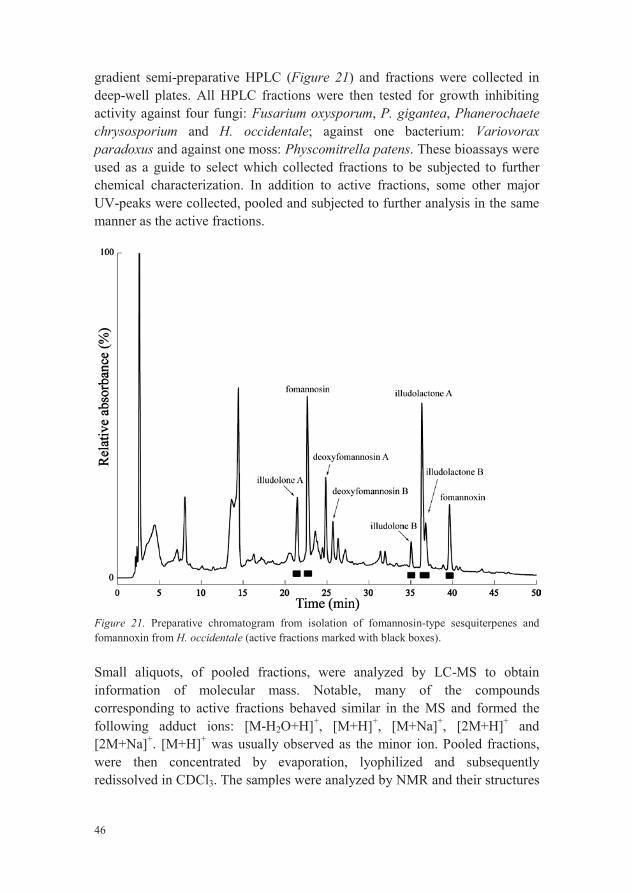
gradient semi-preparative HPLC (Figure 21) and fractions were collected in deep-well plates. All HPLC fractions were then tested for growth inhibiting activity against four fungi: Fusarium oxysporum, P. gigantea, Phanerochaete chrysosporium and H. occidentale; against one bacterium: Variovorax paradoxus and against one moss: Physcomitrella patens. These bioassays were used as a guide to select which collected fractions to be subjected to furtherchemical characterization. In addition to active fractions, some other majorUV-peaks were collected, pooled and subjected to further analysis in the same manner as the active fractions.
Figure 21. Preparative chromatogram from isolation of fomannosin-type sesquiterpenes and fomannoxin from H. occidentale (active fractions marked with black boxes).
Small aliquots, of pooled fractions, were analyzed by LC-MS to obtain information of molecular mass. Notable, many of the compounds corresponding to active fractions behaved similar in the MS and formed the following adduct ions: [M-H2O+H]+, [M+H]+, [M+Na]+, [2M+H]+ and [2M+Na]+. [M+H]+ was usually observed as the minor ion. Pooled fractions, were then concentrated by evaporation, lyophilized and subsequently redissolved in CDCl3. The samples were analyzed by NMR and their structures
47
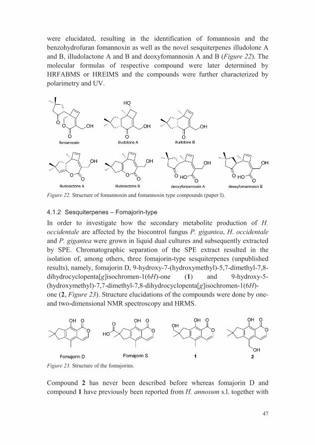
were elucidated, resulting in the identification of fomannosin and the benzohydrofuran fomannoxin as well as the novel sesquiterpenes illudolone A and B, illudolactone A and B and deoxyfomannosin A and B (Figure 22). The molecular formulas of respective compound were later determined by HRFABMS or HREIMS and the compounds were further characterized by polarimetry and UV.
Figure 22. Structure of fomannosin and fomannosin type compounds (paper I).
4.1.2 Sesquiterpenes – Fomajorin-type
In order to investigate how the secondary metabolite production of H. occidentale are affected by the biocontrol fungus P. gigantea, H. occidentaleand P. gigantea were grown in liquid dual cultures and subsequently extracted by SPE. Chromatographic separation of the SPE extract resulted in the isolation of, among others, three fomajorin-type sesquiterpenes (unpublished results), namely, fomajorin D, 9-hydroxy-7-(hydroxymethyl)-5,7-dimethyl-7,8-dihydrocyclopenta[g]isochromen-1(6H)-one (1) and 9-hydroxy-5-(hydroxymethyl)-7,7-dimethyl-7,8-dihydrocyclopenta[g]isochromen-1(6H)-one (2, Figure 23). Structure elucidations of the compounds were done by one-and two-dimensional NMR spectroscopy and HRMS.
Figure 23. Structure of the fomajorins.
Compound 2 has never been described before whereas fomajorin D and compound 1 have previously been reported from H. annosum s.l. together with

48
fomajorin S (Sonnenbichler et al., 1989; Donnelly et al., 1982). Furthermore, fomajorin D was later also identified in paper III.
The 1H NMR spectrum of compound 2 (Figure 24) contained signals at almost identical chemical shifts as fomajorin D, except that the signal from the
H 2.24 ppm (CDCl3, 30 °C) was H 4.73 that integrated for two protons. HMBC
experiments further demonstrated that this methylene group was attached to the aromatic ring, which further strengthen the proposed structure of compound 2.
Figure 24. 1H NMR spectrum of compound 2.
4.1.3 Sesquiterpenes – Drimane-type
A third type of sesquiterpenes, the drimane-type, was also identified to be produced by H. annosum s.l. (paper III). The identification of 7 ,8 ,11-trihydroxydrimane (Figure 25) was done by NMR and HRMS -Trihydroxydrimane has previously been described in H. annosum s.l. and the recorded NMR data were in agreement with literature data (Liu et al., 2008; Donnelly et al., 1980). Epoxydrimenol (Figure 25) was proposed to be a true secondary metabolite of H. annsoum s.l. although no NMR data was recorded for this compound. It was indirectly identified as the epoxide of -trihydroxydrimane (paper III). The molecular formula of epoxydrimenol,
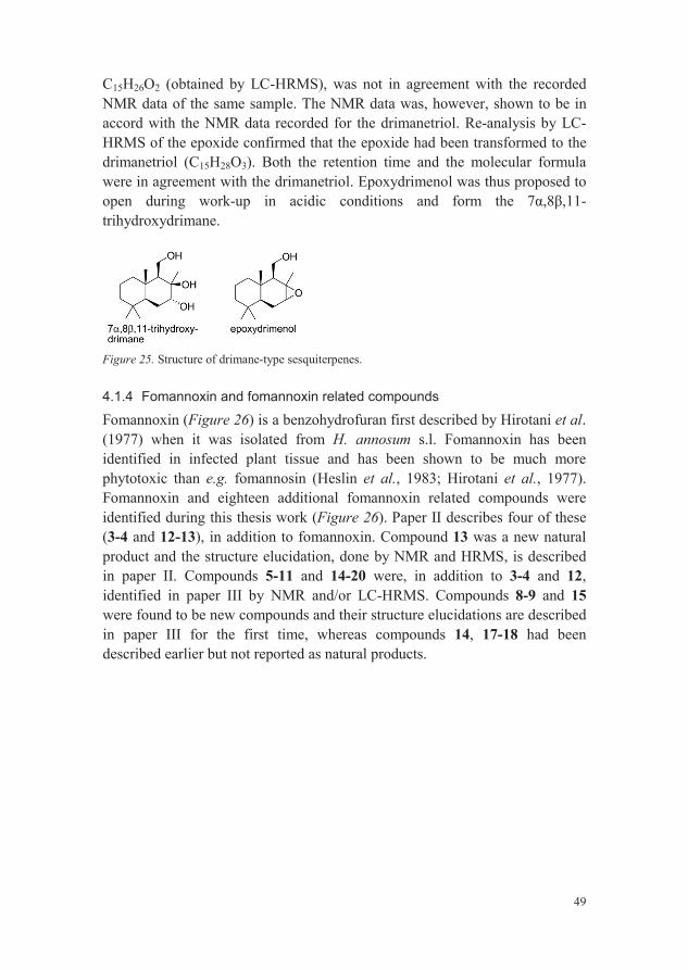
49
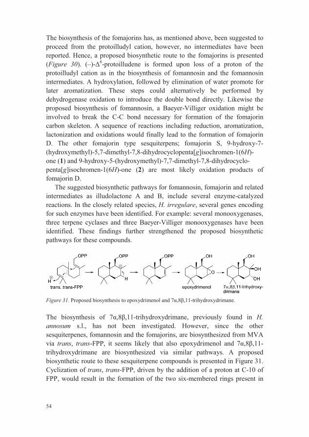
C15H26O2 (obtained by LC-HRMS), was not in agreement with the recorded NMR data of the same sample. The NMR data was, however, shown to be in accord with the NMR data recorded for the drimanetriol. Re-analysis by LC-HRMS of the epoxide confirmed that the epoxide had been transformed to the drimanetriol (C15H28O3). Both the retention time and the molecular formula were in agreement with the drimanetriol. Epoxydrimenol was thus proposed to open during work-up in acidic conditions and form the 11-trihydroxydrimane.
Figure 25. Structure of drimane-type sesquiterpenes.
4.1.4 Fomannoxin and fomannoxin related compounds
Fomannoxin (Figure 26) is a benzohydrofuran first described by Hirotani et al.(1977) when it was isolated from H. annosum s.l. Fomannoxin has been identified in infected plant tissue and has been shown to be much more phytotoxic than e.g. fomannosin (Heslin et al., 1983; Hirotani et al., 1977).Fomannoxin and eighteen additional fomannoxin related compounds were identified during this thesis work (Figure 26). Paper II describes four of these (3-4 and 12-13), in addition to fomannoxin. Compound 13 was a new natural product and the structure elucidation, done by NMR and HRMS, is described in paper II. Compounds 5-11 and 14-20 were, in addition to 3-4 and 12,identified in paper III by NMR and/or LC-HRMS. Compounds 8-9 and 15were found to be new compounds and their structure elucidations are described in paper III for the first time, whereas compounds 14, 17-18 had been described earlier but not reported as natural products.

50
Figure 26. Structure of fomannoxin, fomannoxin-type and fomannoxin related compounds.
4.1.5 Indole-type
Tryptophan and 2,3,4,9-tetrahydro-1H- -carboline-3-carboxylic acid (21) were identified as two compounds that differ in either relative concentration or occurrence between the species within the H. annsoum s.l. complex (paper III). Both compounds were identified by HRMS and NMR. The structure of tryptophan was confirmed by comparing 1H NMR data with reference L-tryptophan whereas NMR data of 2,3,4,9-tetrahydro-1H- -carboline-3-carboxylic acid was compared to literature data (Kotanen et al., 2003).
Figure 27. Structures of identified indole-type compounds.
4.2 Secondary metabolites comparison between the H.annosum s.l. species (Paper III)
With the aim to investigate if the species within the H. annosum s.l. complex could be distinguished on their secondary metabolite profiles, all five species
51
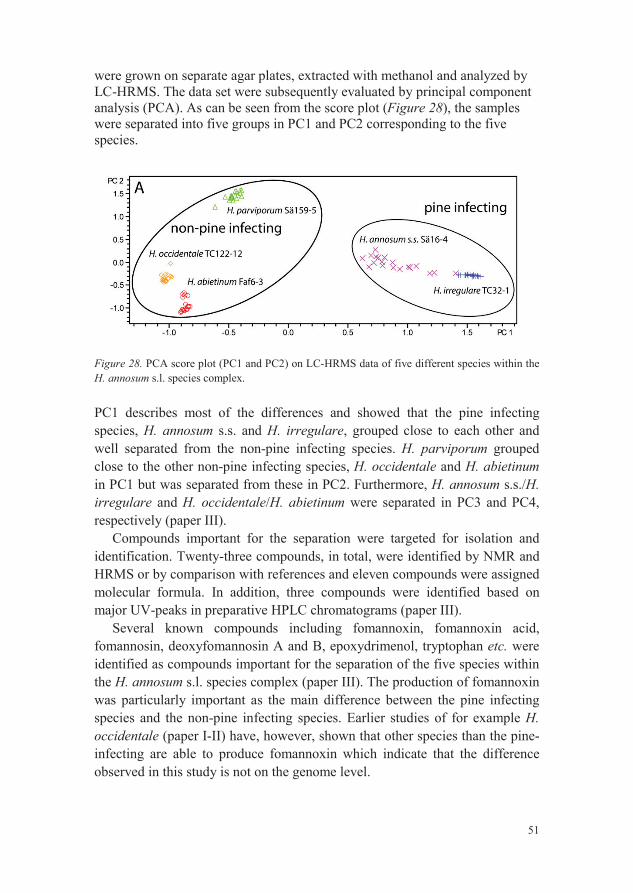
were grown on separate agar plates, extracted with methanol and analyzed by LC-HRMS. The data set were subsequently evaluated by principal component analysis (PCA). As can be seen from the score plot (Figure 28), the samples were separated into five groups in PC1 and PC2 corresponding to the five species.
Figure 28. PCA score plot (PC1 and PC2) on LC-HRMS data of five different species within theH. annosum s.l. species complex.
PC1 describes most of the differences and showed that the pine infecting species, H. annosum s.s. and H. irregulare, grouped close to each other andwell separated from the non-pine infecting species. H. parviporum grouped close to the other non-pine infecting species, H. occidentale and H. abietinumin PC1 but was separated from these in PC2. Furthermore, H. annosum s.s./H. irregulare and H. occidentale/H. abietinum were separated in PC3 and PC4, respectively (paper III).
Compounds important for the separation were targeted for isolation and identification. Twenty-three compounds, in total, were identified by NMR and HRMS or by comparison with references and eleven compounds were assigned molecular formula. In addition, three compounds were identified based on major UV-peaks in preparative HPLC chromatograms (paper III).
Several known compounds including fomannoxin, fomannoxin acid, fomannosin, deoxyfomannosin A and B, epoxydrimenol, tryptophan etc. were identified as compounds important for the separation of the five species within the H. annosum s.l. species complex (paper III). The production of fomannoxin was particularly important as the main difference between the pine infecting species and the non-pine infecting species. Earlier studies of for example H. occidentale (paper I-II) have, however, shown that other species than the pine-infecting are able to produce fomannoxin which indicate that the difference observed in this study is not on the genome level.
52
Nevertheless, the study revealed that it is feasible to differentiate the species by their respective metabolite pattern, but whether these differences remain when the fungi are grown on other medium as e.g. their respective host is uncertain. Furthermore, the samples were separated in accord with the previously identified phylogeny (Dalman et al., 2010) and host preferences.
4.3 Biosynthesis of secondary metabolites produced by H. annosum s.l. (Papers I-III)
4.3.1 Biosynthesis of sesquiterpene compounds
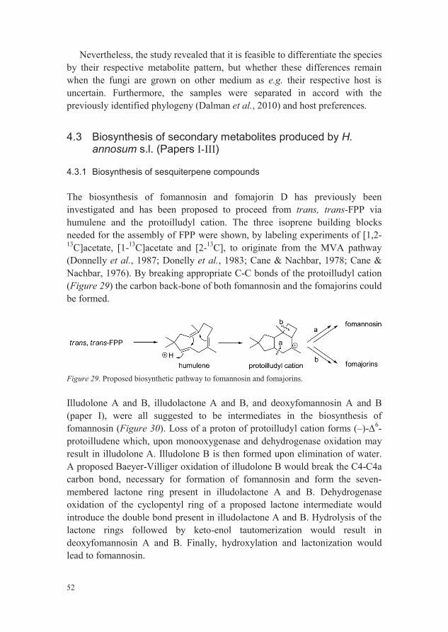
The biosynthesis of fomannosin and fomajorin D has previously been investigated and has been proposed to proceed from trans, trans-FPP via humulene and the protoilludyl cation. The three isoprene building blocks needed for the assembly of FPP were shown, by labeling experiments of [1,2-13C]acetate, [1-13C]acetate and [2-13C], to originate from the MVA pathway (Donnelly et al., 1987; Donelly et al., 1983; Cane & Nachbar, 1978; Cane & Nachbar, 1976). By breaking appropriate C-C bonds of the protoilludyl cation (Figure 29) the carbon back-bone of both fomannosin and the fomajorins could be formed.
Figure 29. Proposed biosynthetic pathway to fomannosin and fomajorins.
Illudolone A and B, illudolactone A and B, and deoxyfomannosin A and B(paper I), were all suggested to be intermediates in the biosynthesis of fomannosin (Figure 30). Loss of a proton of protoilludyl cation forms (–)- 6-protoilludene which, upon monooxygenase and dehydrogenase oxidation may result in illudolone A. Illudolone B is then formed upon elimination of water.A proposed Baeyer-Villiger oxidation of illudolone B would break the C4-C4acarbon bond, necessary for formation of fomannosin and form the seven-membered lactone ring present in illudolactone A and B. Dehydrogenase oxidation of the cyclopentyl ring of a proposed lactone intermediate would introduce the double bond present in illudolactone A and B. Hydrolysis of the lactone rings followed by keto-enol tautomerization would result in deoxyfomannosin A and B. Finally, hydroxylation and lactonization would lead to fomannosin.

53
The absolute configuration assigned for illudolone A and B, illudolactone A and B, and deoxyfomannosin A and B were based on the above described biosynthetic pathway and that they all share the protoilludyl cation as a common precursor. Furthermore, it was assumed that the configuration of the stereogenic carbon in the lactone ring of fomannosin is set during the cyclization of humulene.
Figure 30. Proposed biosynthetic pathway for fomannosin (paper I) and the fomajorins from the common precursor, (–)- 6-protoilludene.
54
The biosynthesis of the fomajorins has, as mentioned above, been suggested to proceed from the protoilludyl cation, however, no intermediates have been reported. Hence, a proposed biosynthetic route to the fomajorins is presented(Figure 30). (–)- 6-protoilludene is formed upon loss of a proton of the protoilludyl cation as in the biosynthesis of fomannosin and the fomannosin intermediates. A hydroxylation, followed by elimination of water promote for later aromatization. These steps could alternatively be performed by dehydrogenase oxidation to introduce the double bond directly. Likewise the proposed biosynthesis of fomannosin, a Baeyer-Villiger oxidation might be involved to break the C-C bond necessary for formation of the fomajorin carbon skeleton. A sequence of reactions including reduction, aromatization, lactonization and oxidations would finally lead to the formation of fomajorin D. The other fomajorin type sesquiterpens; fomajorin S, 9-hydroxy-7-(hydroxymethyl)-5,7-dimethyl-7,8-dihydrocyclopenta[g]isochromen-1(6H)-one (1) and 9-hydroxy-5-(hydroxymethyl)-7,7-dimethyl-7,8-dihydrocyclo-penta[g]isochromen-1(6H)-one (2) are most likely oxidation products of fomajorin D.
The suggested biosynthetic pathways for fomannosin, fomajorin and related intermediates as illudolactone A and B, include several enzyme-catalyzed reactions. In the closely related species, H. irregulare, several genes encoding for such enzymes have been identified. For example: several monooxygenases, three terpene cyclases and three Baeyer-Villiger monooxygenases have been identified. These findings further strengthened the proposed biosynthetic pathways for these compounds.
Figure 31. -trihydroxydrimane.
The -trihydroxydrimane, previously found in H. annosum s.l., has not been investigated. However, since the other sesquiterpenes, fomannosin and the fomajorins, are biosynthesized from MVA via trans, trans-FPP, it seems likely that also epoxydrimenol and -trihydroxydrimane are biosynthesized via similar pathways. A proposed biosynthetic route to these sesquiterpene compounds is presented in Figure 31. Cyclization of trans, trans-FPP, driven by the addition of a proton at C-10 of FPP, would result in the formation of the two six-membered rings present in

55
the carbon backbone of drimanes. Loss of a proton to neutralize the positive charge forms a double bond, which upon oxidation and hydrolysis of diphosphate leads to epoxydrimenol. 7 -Trihydroxydrimane couldfinally be formed after opening of the epoxide.
4.3.2 Biosynthesis of fomannoxin and fomannoxin related compounds
Fomannoxin is a known phytotoxin of H. annosum s.l. and is one of the major secondary metabolites produced by several of the species within the species complex. One of the objectives of this thesis work was to investigate the biosynthesis of this compound (for details see paper II).
Liquid cultures of H. occidentale were grown in Hagem media and small aliquots were regularly taken and analyzed by LC-MS. When the presence of fomannoxin was observed (after ca. three weeks of growth), by the detection of an ion at m/z 189 corresponding to the [M+H]+ ion of fomannoxin, [1-13C]glucose was added. The labeled cultures were then grown for another two weeks before they were harvested by filtration and extracted by SPE. Unlabeled cultures were cultivated in parallel. The SPE extracts were subsequently fractionated by preparative HPLC and fractions corresponding to fomannoxin and other major UV-peaks were pooled and prepared for NMR analysis. 13C NMR spectra of fomannoxin, cultivated in both labeled and unlabeled media, are presented in Figure 32.
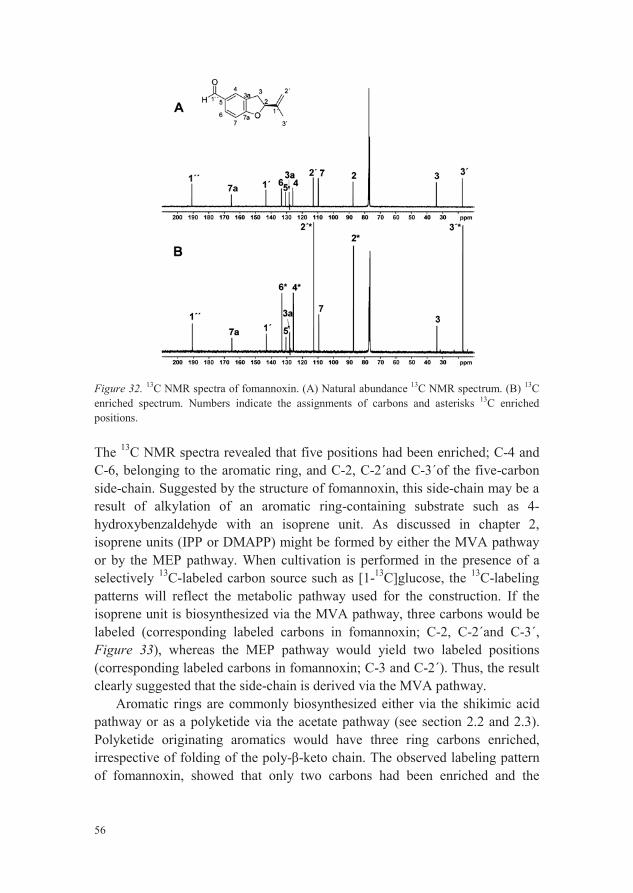
56
Figure 32. 13C NMR spectra of fomannoxin. (A) Natural abundance 13C NMR spectrum. (B) 13Cenriched spectrum. Numbers indicate the assignments of carbons and asterisks 13C enriched positions.
The 13C NMR spectra revealed that five positions had been enriched; C-4 and C-6, belonging to the aromatic ring, and C-2, C-2´and C-3´of the five-carbon side-chain. Suggested by the structure of fomannoxin, this side-chain may be a result of alkylation of an aromatic ring-containing substrate such as 4-hydroxybenzaldehyde with an isoprene unit. As discussed in chapter 2, isoprene units (IPP or DMAPP) might be formed by either the MVA pathway or by the MEP pathway. When cultivation is performed in the presence of aselectively 13C-labeled carbon source such as [1-13C]glucose, the 13C-labeling patterns will reflect the metabolic pathway used for the construction. If the isoprene unit is biosynthesized via the MVA pathway, three carbons would be labeled (corresponding labeled carbons in fomannoxin; C-2, C-2´and C-3´,Figure 33), whereas the MEP pathway would yield two labeled positions(corresponding labeled carbons in fomannoxin; C-3 and C-2´). Thus, the result clearly suggested that the side-chain is derived via the MVA pathway.
Aromatic rings are commonly biosynthesized either via the shikimic acid pathway or as a polyketide via the acetate pathway (see section 2.2 and 2.3).Polyketide originating aromatics would have three ring carbons enriched, irrespective of folding of the poly- -keto chain. The observed labeling pattern of fomannoxin, showed that only two carbons had been enriched and the
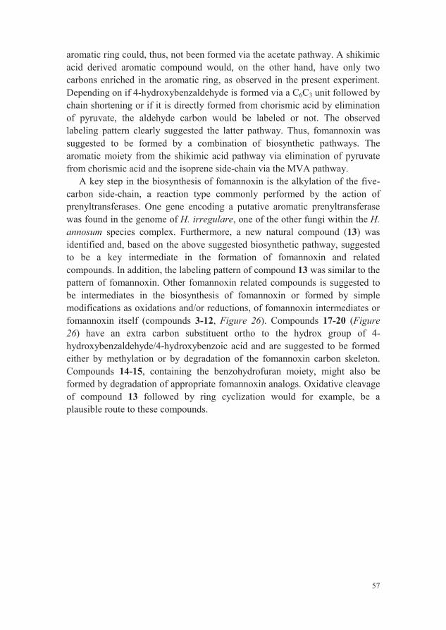
57
aromatic ring could, thus, not been formed via the acetate pathway. A shikimic acid derived aromatic compound would, on the other hand, have only two carbons enriched in the aromatic ring, as observed in the present experiment.Depending on if 4-hydroxybenzaldehyde is formed via a C6C3 unit followed bychain shortening or if it is directly formed from chorismic acid by elimination of pyruvate, the aldehyde carbon would be labeled or not. The observed labeling pattern clearly suggested the latter pathway. Thus, fomannoxin was suggested to be formed by a combination of biosynthetic pathways. The aromatic moiety from the shikimic acid pathway via elimination of pyruvate from chorismic acid and the isoprene side-chain via the MVA pathway.
A key step in the biosynthesis of fomannoxin is the alkylation of the five-carbon side-chain, a reaction type commonly performed by the action of prenyltransferases. One gene encoding a putative aromatic prenyltransferase was found in the genome of H. irregulare, one of the other fungi within the H. annosum species complex. Furthermore, a new natural compound (13) was identified and, based on the above suggested biosynthetic pathway, suggested to be a key intermediate in the formation of fomannoxin and related compounds. In addition, the labeling pattern of compound 13 was similar to the pattern of fomannoxin. Other fomannoxin related compounds is suggested to be intermediates in the biosynthesis of fomannoxin or formed by simple modifications as oxidations and/or reductions, of fomannoxin intermediates or fomannoxin itself (compounds 3-12, Figure 26). Compounds 17-20 (Figure 26) have an extra carbon substituent ortho to the hydrox group of 4-hydroxybenzaldehyde/4-hydroxybenzoic acid and are suggested to be formed either by methylation or by degradation of the fomannoxin carbon skeleton. Compounds 14-15, containing the benzohydrofuran moiety, might also be formed by degradation of appropriate fomannoxin analogs. Oxidative cleavage of compound 13 followed by ring cyclization would for example, be a plausible route to these compounds.
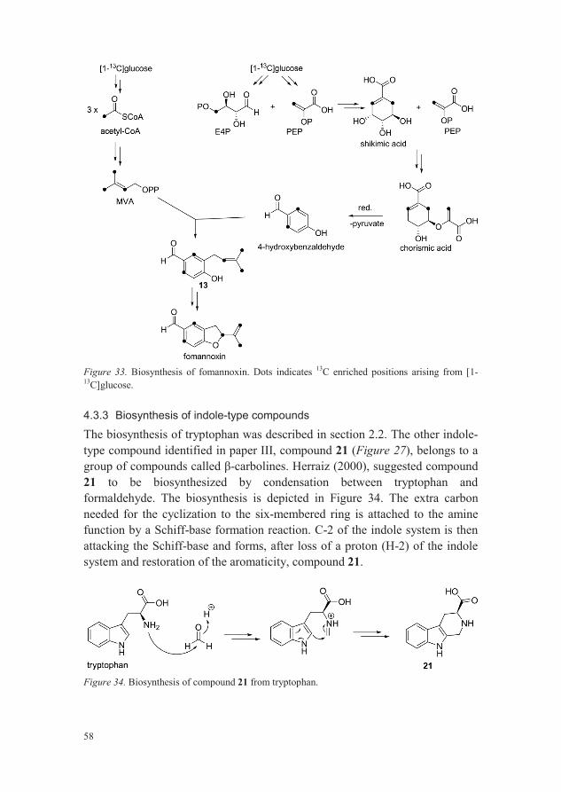
58
Figure 33. Biosynthesis of fomannoxin. Dots indicates 13C enriched positions arising from [1-13C]glucose.
4.3.3 Biosynthesis of indole-type compounds
The biosynthesis of tryptophan was described in section 2.2. The other indole-type compound identified in paper III, compound 21 (Figure 27), belongs to a
-carbolines. Herraiz (2000), suggested compound 21 to be biosynthesized by condensation between tryptophan and formaldehyde. The biosynthesis is depicted in Figure 34. The extra carbon needed for the cyclization to the six-membered ring is attached to the amine function by a Schiff-base formation reaction. C-2 of the indole system is then attacking the Schiff-base and forms, after loss of a proton (H-2) of the indole system and restoration of the aromaticity, compound 21.
Figure 34. Biosynthesis of compound 21 from tryptophan.

59
4.4 Secondary metabolites of P. gigantea (paper IV)
P. gigantea is used as biocontrol fungus against H. annosum s.l. but the mechanism of the biocontrol action is not fully understood. The objective of this study was to investigate P. gigantea with regard to its production of secondary metabolites and to test whether these compounds have an effect on H. occidentale and thus, could be involved in the in vivo biocontrol action between P. gigantea and the H. annosum s.l.
P. gigantea was cultivated in Melin Norkrans media, harvested by filtration and extracted by SPE. The SPE extracts were then fractionated by preparative HPLC and major UV-peaks were collected and analyzed by one- and two-dimensional NMR spectroscopy and HRMS. Five compounds were identified, phlebiopsin A-C, methyl-terfestatin A and o-orsellinaldehyde (Figure 34).
Figure 35. Structure of secondary metabolites identified in P. gigantea.
Phlebiopsin A-C are structurally similar to a group of compounds that are rarely found in nature called diarylcyclopentenones. These compounds have been described as responsible for the bluing reactions observed when the fruiting bodies of certain mushrooms of the order Boletales are bruised. Their activities against three fungi including H. occidentale were tested. However, no activities against either of the fungi were observed at the tested concentrations.
The aglycone of the identified compound methyl-terfestatin A, is very similar to 2´,3´,5´-trimethoxy-p-terphenyl, a compound that was not found in this study but has been reported from P. gigantea previously (Briggs et al.,1975). Methyl-terfestatin A is also very similar to terfestatin A, the only difference being the methoxy group at C-3´. Terfestatin A has previously been isolated from Streptomyces bacteria and has shown to inhibit auxin signaling in
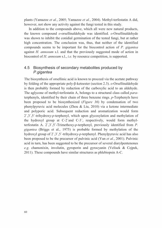
60
plants (Yamazoe et al., 2005; Yamazoe et al., 2004). Methyl-terfestatin A did, however, not show any activity against the fungi tested in this study.
In addition to the compounds above, which all were new natural products, the known compound o-orsellinaldehyde was identified. o-Orsellinaldehyde was shown to inhibit the conidial germination of the tested fungi, but at rather high concentration. The conclusion was, thus, that neither of the identified compounds seems to be important for the biocontrol action of P. giganteaagainst H. annosum s.l. and that the previously suggested mode of action in biocontrol of H. annosum s.l., i.e. by resource competition, is supported.
4.5 Biosynthesis of secondary metabolites produced byP.gigantea
The biosynthesis of orsellinic acid is known to proceed via the acetate pathway by folding of the appropriate poly- -ketoester (section 2.3). o-Orsellinaldehydeis then probably formed by reduction of the carboxylic acid to an aldehyde. The aglycone of methyl-terfestatin A, belongs to a structural class called para-terphenyls, identified by their chain of three benzene rings. p-Terphenyls have been proposed to be biosynthesized (Figure 36) by condensation of two phenylpyruvic acid molecules (Zhou & Liu, 2010) via a ketone intermediate and polyporic acid. Subsequent reduction and aromatization would form 2´,3´,5´-trihydroxy-p-terphenyl, which upon glycosylation and methylation of the hydroxyl group at C-2´and C-3´, respectively, would form methyl-terfestatin A. 2´,3´,5´-Trimethoxy-p-terphenyl, previously identified from P. gigantea (Briggs et al., 1975) is probable formed by methylation of the hydroxyl group of 2´,3´,5´-trihydroxy-p-terphenyl. Phenylpyruvic acid has also been proposed to be the precursor of pulvinic acid (Yun et al., 2001). Pulvinic acid in turn, has been suggested to be the precursor of several diarylpentenones e.g. chamonixin, involutin, gyroporin and gyrocyanin (Velisek & Cejpek, 2011). These compounds have similar structures as phlebiopsin A-C.
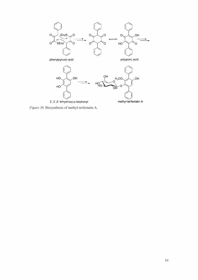
61
Figure 36. Biosynthesis of methyl-terfestatin A.

62
63
5 Conclusions and suggestions for further research
The objectives of the present thesis work included investigating H. annosums.l. and P. gigantea regarding their production of secondary metabolites, how they are biosynthesized and their biological activity.
In total, 33 compounds were identified during the investigation of H. annosum s.l. Six new sesquiterpenes (illudolone A and B, illudolactone A and B and deoxyfomannosin A and B) belonging to the fomannosin class of compounds were identified and described. Furthermore, three sesquiterpenesbelonging to the fomajorins and two drimanes were identified out of which one was a new compound (2). The benzohydrofuran fomannoxin and several (19)fomannoxin related compounds were identified. Three of these were shown to be previously unknown compounds and four had not been described from any natural sources. In addition, tryptophan and another indole-containing
-carboline, were identified. Fomannoxin has been suggested to be important for the pathogenicity of H.
annosum s.l. Its biosynthesis was investigated by an isotopic enrichment study utilizing [1-13C]glucose as metabolic tracer. The labeling pattern, analyzed by 13C-NMR analysis, showed that fomannoxin is biosynthesized by a combination of the MVA pathway and the shikimic acid pathway.
The species within H. annosum s.l. were compared regarding their production of secondary metabolites. The results showed that the five species could be separated by their respective metabolite profile and that the separation was in accord with both host preference and previously established phylogeny.
The biocontrol fungus P. gigantea, was investigated regarding its production of secondary metabolites. The study resulted in five identified compounds out of which three were new compounds (phlebiopsin A-C), one was a new natural product (methyl-terfestatin A) and one (o-orsellinaldehyde) was a known compound. o-Orsellinaldehyde showed weak activity against H.

64
occidentale wheras none of the new natural products showed any activity at the tested concentrations.
H. annosum s.l. and P. gigantea produce, for sure, a lot more secondary metabolites than were identified during this work. For example, recent studies of H. annosum s.l. have shown that there are several NRPSs, PKSs and halogenases present in the genome (Olson et al., 2012). However, neither NRPS, PKS derived nor halogenated secondary metabolites were identified to be produced in H. annosum s.l. during this work. Possibly, other “secondary metabolite genes” are expressed in the presence of natural competitors or during infection of host in vivo. An emerging technology within the natural product chemistry field is epigenetics. Epigenetics manipulate which genes that are expressed. The result could be that earlier “silent genes” became active and that other secondary metabolites are formed. Regardless of how the previously silent genes are made active, it would be interesting to investigate the resulting metabolic profiles.
A natural continuation on the study of P. gigantea would be to investigate the biosynthesis of the diarylcyclopentenones that were identified. Previously suggested pathways have not been established by labeled substrates, but only by discussions based on structure similarities.
65
ReferencesAbraham, W.R. (2001). Bioactive sesquiterpenes produced by fungi: are they
useful for humans as well? Current Medicinal Chemistry 8(6), 583-606.Ali, I., AL-Othman, Z.A., Nagae, N., Gaitonde, V.D. & Dutta, K.K. (2012). Recent
trends in ultra-fast HPLC: new generation superficially porous silica columns. Journal of Separation Science 35(23), 3235-3249.
Asada, Y., Li, W., Terada, T., Kuang, X.Z., Li, Q., Yoshikawa, T., Hamaguchi, S., Namekata, I., Tanaka, H. & Koike, K. (2012). Labdane-type diterpenoids from hairy root cultures of Coleus forskohlii, possible intermediates in the biosynthesis of forskolin. Phytochemistry 79, 141-146.
Bacher, A., Rieder, C., Eichinger, D., Arigoni, D., Fuchs, G. & Eisenreich, W. (1998). Elucidation of novel biosynthetic pathways and metabolite flux patterns by retrobiosynthetic NMR analysis. Fems Microbiology Reviews22(5), 567-598.
Bassett, C., Sherwood, R.T., Kepler, J.A. & Hamilton, P.B. (1967). Production and biological activity of fomannosin a toxic sesquiterpene metabolite of fomes annosus. Phytopathology 57(10), 1046-&.
Bohlmann, J. & Keeling, C.I. (2008). Terpenoid biomaterials. Plant Journal 54(4), 656-669.
Breitmaier, E. (1993). Structure elucidation by NMR in organic chemistry.Chichester, UK: John Wiley & Sons Ltd. ISBN 0471933813.
Briggs, L.H., Cambie, R.C., Dean, I.C., Dromgoole, S.H., Fergus, B.J., Ingram, W.B., Lewis, K.G., Small, C.W., Thomas, R. & Walker, D.A. (1975). Chemistry of fungi. 10. Metabolites of some fungal species. New Zealand Journal of Science 18(4), 565-576.
Bruker (2011). Bruker maXis impact user manual. Bremen, DE: Bruker Daltonik GmbH.
Bury, M., Andolfi, A., Rogister, B., Cimmino, A., Megalizzi, V., Mathieu, V., Feron, O., Evidente, A. & Kiss, R. (2013). Fusicoccin A, a phytotoxic carbotricyclic diterpene glucoside of fungal origin, reduces proliferation and invasion of glioblastoma cells by targeting multiple tyrosine kinases. Translational Oncology 6(2), 112-123.
66
Buszewski, B. & Szultka, M. (2012). Past, present, and future of solid phase extraction: A review. Critical Reviews in Analytical Chemistry 42(3), 198-213.
Cane, D.E. & Nachbar, R.B. (1976). Biosynthesis of fomannosin from 1,2-13C2-acetate. Tetrahedron Letters (25), 2097-2100.
Cane, D.E. & Nachbar, R.B. (1978). Stereochemical studies of isoprenoid biosynthesis - biosynthesis of fomannosin from[1,2-13C2]acetate. Journal of the American Chemical Society 100(10), 3208-3212.
Cleary, M.R., Arhipova, N., Morrison, D.J., Thomsen, I.M., Sturrock, R.N., Vasaitis, R., Gaitnieks, T. & Stenlid, J. (2013). Stump removal to control root disease in Canada and Scandinavia: a synthesis of results from long-term trials. Forest Ecology and Management 290, 5-14.
Clericuzio, M., Mella, M., Vita-Finzi, P., Zema, M. & Vidari, G. (2004). Culcurbitane triterpenoids from Leucopaxillus gentianeus. Journal of Natural Products 67(11), 1823-1828.
Dalman, K., Olson, A. & Stenlid, J. (2010). Evolutionary history of the conifer root rot fungus Heterobasidion annosum sensu lato. Molecular Ecology19(22), 4979-4993.
de Hoffmann, E. & Stroobant, V. (2007). Mass spectrometry: principles and applications. Chichester, UK: John Wiley & Sons, Ltd. ISBN 987047033111.
Dewick, P.M. (2009). Medicinal natural products: a biosynthetic approach. 3rd. ed. Chichester, UK: John Wiley & Sons, Ltd. ISBN 9780470741689.
Donelly, D.M.X., Oreilly, J., Polonsky, J. & Sheidan, M.H. (1983). Biosynthesis of fomajorin D: incorporations of [1-13C]-, [2-13C]-, and [1,2-13C2]-acetates. Journal of the Chemical Society-Chemical Communications (11), 615-616.
Donnelly, D.M.X., Oreilly, J., Chiaroni, A. & Polonsky, J. (1980). Crystal structure and absolute configuration of a new sesquiterpenoid metabolite of fomes annosus, 7 8 11-trihydroxydrimane. Journal of the Chemical Society-Perkin Transactions 1 (10), 2196-2199.
Donnelly, D.M.X., Oreilly, J., Polonsky, J. & Sheridan, M.H. (1987). In vitroproduction and biosynthesis of fomajorin D and fomajorin S by fomes annosus (fr) Cooke. Journal of the Chemical Society-Perkin Transactions 1 (8), 1869-1872.
Donnelly, D.M.X., Oreilly, J., Polonsky, J. & Vaneijk, G.W. (1982). Fomajorin S and fomajorin D from Fomes annosus (Fr) Cooke. Tetrahedron Letters23(51), 5451-5452.
Elsharkawy, S. & Abulhajj, Y. (1987). Microbial transformation of zearalenone. 1. Formation of zearalenone-4-o- -glucoside. Journal of Natural Products50(3), 520-521.
Ettre, L.S. (2003). M.S. Tswett and the invention of chromatography. Lc Gc Europe 16(9), 632-.

67
Forster-Fromme, K. & Jendrossek, D. (2010). Catabolism of citronellol and related acyclic terpenoids in pseudomonads. Applied Microbiology and Biotechnology 87(3), 859-869.
Fox, E.M. & Howlett, B.J. (2008). Secondary metabolism: regulation and role in fungal biology. Current Opinion in Microbiology 11(6), 481-487.
Grawert, T., Groll, M., Rohdich, F., Bacher, A. & Eisenreich, W. (2011). Biochemistry of the non-mevalonate isoprenoid pathway. Cellular and Molecular Life Sciences 68(23), 3797-3814.
Hanson, J.R. (2003). Natural products: the secondary metabolites. Cambridge, UK: Royal society of chemistry. ISBN 0854044906.
Harris, D.C. (2002). Quantitative chemical analysis. 6th. ed. New York, USA: W. H. Freeman and Company. ISBN 9780716744641.
Hartmann, T. (2007). From waste products to ecochemicals: fifty years research of plant secondary metabolism. Phytochemistry 68(22-24), 2831-2846.
Hellgren, M.B. & Stenlid, J. (1995). Long-term reduction in the diameter growth of butt rot affected Norway spruce, Picea abies. Forest Ecology and Management 74(1-3), 239-243.
Herraiz, T. (2000). Tetrahydro- -carboline-3-carboxylic acid compounds in fish and meat: possible precursors of co-mutagenic -carbolines norharman and harman in cooked foods. Food Additives and Contaminants 17(10), 859-866.
Heslin, M.C., Stuart, M.R., Murchu, P.O. & Donnelly, D.M.X. (1983).Fomannoxin, a phytotoxic metabolite of Fomes annosus - in vitroproduction, host toxicity and isolation from naturally infected sitka spruce heartwood. European Journal of Forest Pathology 13(1), 11-23.
Hirotani, M., Oreilly, J., Donnelly, D.M.X. & Polonsky, J. (1977). Fomannoxin -toxic metabolite of Fomes annosus. Tetrahedron Letters (7), 651-652.
Holdenrieder, O. & Greig, B.J.W. (1998). Biological methods of control. Heterobasidion annosum: Biology, ecology, impact and control (Woodward, S., Stenlid, J., Karjaleinen, R. & Hüttermann, A.) Wallingford, UK: CAB International. ISBN 0851992757.
Hore, P.J. (1995). Nuclear magnetic resonance. Oxford, UK: Oxford Science Publications. ISBN 9780198556824.
Johansson, S.M., Pratt, J.E. & Asiegbu, F.O. (2002). Treatment of Norway spruce and Scots pine stumps with urea against the root and butt rot fungus Heterobasidion annosum - possible modes of action. Forest Ecology and Management 157(1-3), 87-100.
Keller, N.P., Turner, G. & Bennett, J.W. (2005). Fungal secondary metabolism -from biochemistry to genomics. Nature Reviews Microbiology 3(12), 937-947.
Kepler, J.A., Wall, M.E., Mason, J.E., Basset, C., McPhail, A.T. & Sim, G.A. (1967). Structure of fomannosin a novel sesquiterpene metabolite of fungus Fomes annosus. Journal of the American Chemical Society 89(5), 1260-&.
68
Korhonen, K., Delatour, C., Greig, B.J.W. & Schönhar, S. (1998). Silvicultural control. Heterobasidion annosum: Biology, ecology, impact and control (Woodward, S., Stenlid, J., Karjaleinen, R. & Hüttermann, A.).Wallingford, UK: CAB International. ISBN 0851992757.
Korhonen, K., Lipponen, K., Bendz, M., Johansson, M., Ryen, I., Venn, K., Seiskari, P. & Niemi, M. (1994). Control of Heterobasidion annosum by stump treatment with "Rotstop", a new commercial formulation of Phlebiopsis gigantea. In M. Johansson, & J. Stenlid, Proceedings of the eighth international conference on root and butt rots. Sweden and Finland, 9-16 August 1993.
Kotanen, S., Huybrechts, J., Cerstiaens, A., Zoltan, K., Daloze, D., Baggerman, G., Forgo, P., De Loof, A. & Schoofs, L. (2003). Identification of tryptophan and -carboline as paralysins in larvae of the yellow mealworm, Tenebrio molitor. Biochemical and Biophysical Research Communications 310(1), 64-71.
Lambert, J.B. & Mazzola, E.P. (2003). Nuclear magnetic resonance spectroscopy: an introduction to principles, applications, and experimental methods.New Jersey, USA: Pearson Education, Inc. ISBN 9780130890665.
Liapi, C., Anifantis, G., Chinou, I., Kourounakis, A.P., Theodosopoulos, S. & Galanopoulou, P. (2007). Antinociceptive properties of 1,8-cineole and -pinene, from the essential oil of eucalyptus camaldulensis leaves, in Rodents. Planta Medica 73(12), 1247-1254.
Liu, Y.J., Liu, Y. & Zhang, K.Q. (2008). Xanthothone, a new nematicidal N-compound from Coprinus xanthothrix. Chemistry of Natural Compounds44(2), 203-205.
Mann, J., Davidson, R.S., Hobbs, J.B., Banthorpe, D.V. & Harborne, J.B. (1994).Natural products: their chemistry and biological significance. Harlow, UK: Longman group. ISBN 0582060095.
Meganathan, R. (2001). Ubiquinone biosynthesis in microorganisms. Fems Microbiology Letters 203(2), 131-139.
Meinwald, J. (2011). Natural products as molecular messengers. Journal of Natural Products 74(3), 305-309.
Menkis, A., Burokiene, D., Gaitnieks, T., Uotila, A., Johannesson, H., Rosling, A., Finlay, R.D., Stenlid, J. & Vasaitis, R. (2012). Occurrence and impact of the root-rot biocontrol agent Phlebiopsis gigantea on soil fungal communities in Picea abies forests of northern Europe. Fems Microbiology Ecology 81(2), 438-445.
Miller, J.M. (2009). Chromatography: concepts and contrasts. 2nd. ed. New Jersey, USA: John Wiley & Sons, Inc. ISBN 9780470530252.
Niemelä, T. & Korhonen, K. (1998). Taxonomy of the genus Heterobasidion. Heterobasidion annosum: Biology, ecology, impact and control (Woodward, S., Stenlid, J., Karjaleinen, R. & Hüttermann, A.) Wallingford, UK: CAB international. ISBN 0851992757.
69
Novakova, L. & Vlckova, H. (2009). A review of current trends and advances in modern bio-analytical methods: chromatography and sample preparation. Analytica Chimica Acta 656(1-2), 8-35.
Nowicka, B. & Kruk, J. (2010). Occurrence, biosynthesis and function of isoprenoid quinones. Biochimica Et Biophysica Acta-Bioenergetics1797(8), 1587-1605.
Olson, A., Aerts, A., Asiegbu, F., Belbahri, L., Bouzid, O., Broberg, A., Canback, B., Coutinho, P.M., Cullen, D., Dalman, K., Deflorio, G., van Diepen, L.T.A., Dunand, C., Duplessis, S., Durling, M., Gonthier, P., Grimwood, J., Fossdal, C.G., Hansson, D., Henrissat, B., Hietala, A., Himmelstrand, K., Hoffmeister, D., Hogberg, N., James, T.Y., Karlsson, M., Kohler, A., Kues, U., Lee, Y.H., Lin, Y.C., Lind, M., Lindquist, E., Lombard, V., Lucas, S., Lunden, K., Morin, E., Murat, C., Park, J., Raffaello, T., Rouze, P., Salamov, A., Schmutz, J., Solheim, H., Stahlberg, J., Velez, H., de Vries, R.P., Wiebenga, A., Woodward, S., Yakovlev, I., Garbelotto, M., Martin, F., Grigoriev, I.V. & Stenlid, J. (2012). Insight into trade-off between wood decay and parasitism from the genome of a fungal forest pathogen. New Phytologist 194(4), 1001-1013.
Otrosina, W.J. & Garbelotto, M. (2010). Heterobasidion occidentale sp nov and Heterobasidion irregulare nom. nov.: a disposition of North American Heterobasidion biological species. Fungal Biology 114(1), 16-25.
Pavia, D.L., Lampman, G.M. & Kriz, G.S. (2000). Introduction to spectroscopy.3rd. ed: Thomson Brooks/Cole. ISBN 9780030319617.
Rishbeth, J. (1951). Observations on the biology of fomes annosus, with particular reference to east anglian pine plantations. 2. Spore production, stump infection, and saprophytic activity in stumps. Annals of Botany, New series 15, 1-21.
Rohmer, M. (1999). The mevalonate-independent methylerythritol 4-phosphate (MEP) pathway for isoprenoid biosynthesis, including carotenoids. Pure and Applied Chemistry 71(12), 2279-2284.
Saleh, O., Haagen, Y., Seeger, K. & Heide, L. (2009). Prenyl transfer to aromatic substrates in the biosynthesis of aminocoumarins, meroterpenoids and phenazines: the ABBA prenyltransferase family. Phytochemistry 70(15-16), 1728-1738.
Schiff, P.L. (2006). Ergot and its alkaloids. American Journal of Pharmaceutical Education 70(5), 10.
Schobert, R., Knauer, S., Seibt, S. & Biersack, B. (2011). Anticancer active illudins: recent developments of a potent alkylating compound class. Current Medicinal Chemistry 18(6), 790-807.
Selim, K.A., El-Beih, A.A., Abdel-Rahman, T.M. & El-Diwany, A.I. (2012). Biology of endophytic fungi. Current research in enviromental & applied mycology 2(1), 31-82.
Smith, D.J., Earl, A.J. & Turner, G. (1990). The multifunctional peptide synthetase performing the 1st step of penicillin biosynthesis in Penicillium
70
chrysogenum is a 421 073 Dalton protein similar to Bacillus brevispeptide antibiotic synthetases. Embo Journal 9(9), 2743-2750.
Sonnenbichler, J., Bliestle, I.M., Peipp, H. & Holdenrieder, O. (1989). Secondary fungal metabolites and their biological activities. 1. Isolation of antibiotic coompounds from cultures of Heterobasidion annosum synthesized in the presence of antagonistic fungi or host plant cells. Biological Chemistry Hoppe-Seyler 370(12), 1295-1303.
Stadthagen, G., Kordulakova, J., Griffin, R., Constant, P., Bottova, I., Barilone, N., Gicquel, B., Daffe, M. & Jackson, M. (2005). p-Hydroxybenzoic acid synthesis in Mycobacterium tuberculosis. Journal of Biological Chemistry280(49), 40699-40706.
Stenlid, J. & Redfern, D.B. (1998). Spread within the tree and stand. Heterobasidion annosum: Biology, ecology, impact and control (Woodward, S., Stenlid, J., Karjaleinen, R. & Hüttermann, A.).Wallingford, UK: CAB International. ISBN 0851992757.
Sturesson, C., Rönnberg, J., Berglund, M. & Norman, J. (2011). Rotröta: -om rotröta i allmänhet och rotticka på gran i synnerhet: Studentlitteratur AB, (In Swedish). ISBN 9789144071961.
Walter, M.H., Fester, T. & Strack, D. (2000). Arbuscular mycorrhizal fungi induce the non-mevalonate methylerythritol phosphate pathway of isoprenoid biosynthesis correlated with accumulation of the 'yellow pigment' and other apocarotenoids. Plant Journal 21(6), 571-578.
Velisek, J. & Cejpek, K. (2011). Pigments of higher fungi: a review. Czech Journal of Food Sciences 29(2), 87-102.
Woodward, S., Stenlid, J., Karjaleinen, R. & Hüttermann, A. (1998).Heterobasidion annosum: Biology, ecology, impact and control.Wallingford, UK: CAB International. ISBN 0851992757.
Yamazoe, A., Hayashi, K., Kepinski, S., Leyser, O. & Nozaki, H. (2005). Characterization of terfestatin A, a new specific inhibitor for auxin signaling. Plant Physiology 139(2), 779-789.
Yamazoe, A., Hayashi, K., Kuboki, A., Ohira, S. & Nozaki, H. (2004). The isolation, structural determination, and total synthesis of terfestatin A, a novel auxin signaling inhibitor from Streptomyces sp. Tetrahedron Letters45(45), 8359-8362.
Yazaki, K., Sasaki, K. & Tsurumaru, Y. (2009). Prenylation of aromatic compounds, a key diversification of plant secondary metabolites. Phytochemistry 70(15-16), 1739-1745.
Yun, B.S., Kang, H.C., Koshino, H., Yu, S.H. & Yoo, I.D. (2001). Suillusin, a unique benzofuran from the mushroom Suillus granulatus. Journal of Natural Products 64(9), 1230-1231.
Zhang, L.J., Liu, S.W., Cui, X.Y., Pan, C.P., Zhang, A.L. & Chen, F. (2012). A review of sample preparation methods for the pesticide residue analysis in foods. Central European Journal of Chemistry 10(3), 900-925.
Zhou, Z.Y. & Liu, J.K. (2010). Pigments of fungi (macromycetes). Natural Product Reports 27(11), 1531-1570.

71
AcknowledgementsJag hade inte kommit hit till de sista meningarna om det inte vore för alla fantastiska människor jag har haft runt omkring mig.
Tack till:
Anders Broberg, min huvudhandledare, för allt stöd. Du är en duktig kemist och du delar gärna med dig av din kunskap. Tack för det och tack för att du alltid tar dig tid, det har varit ovärderligt under de här åren. Tack också för att du tog mig till Uppsala och antog mig som doktorand, det har varit ett nöje att vara i ditt lag (även på innebandyplanen).
Audrius Menkis, min biträdande handledare, för dina många idéer och för att du alltid tagit dig tid att göra ytterligare odlingar. Tack också för dina snabba korrekturer.
Corine Sandström, min biträdande handledare, för att du visat intresse i mitt arbete och för att du förklarat ett och annat om NMR.
framlidne Lennart Kenne, min tidigare biträdande handledare, för din humor, din kunskap och för att du lyssnade.
Åke, Magnus, Kajsa och Janne på skoglig mykologi och patologi för gott samarbete under de här åren.
Suresh, för hjälp med MS körningar och för att du är en mycket trevlig rumskamrat.
Janne, för att du lyssnar, kommer med goda råd och för att du gärna delarmed dig om dina kunskaper om masspektrometri.
Anders S och Bernt, för att ni gör allt ni kan för studenterna och för att niunderlättar för oss som undervisar på labb.
Sonja och Lena, för hjälp med administrativa ärenden.

72
Jag vill också tacka alla övriga kollegor på kemi: Ali, Anke, Daniel, Gulaim, Gunnar, Ingmar, Mattias, Peter, Vadim, m.fl. Tack för att ni gör kemiinstitutionen till en trevlig och lärorik arbetsplats. Särskilt tack till Anke för språkgranskningen.
Tack också till före detta doktorander och blivande doktorer på kemiinstitutionen för utebliven söndagsångest, det är alltid roligt att träffa er.Johan, Josephina, Lars, Martin, Natalia, Olesya, Shahin, Tobias, m.fl. Ett särskilt tack till: Pierre, vi har jobbat tätt ihop under de här åren och du har blivit en god vän. Tack för att du många gånger varit en hjälpande hand på labboch ett bollplank i kemirelaterade funderingar. Tack också för oförglömliga fisketur-, pubkväll-, Thailand-, Leiden-, och poker- minnen. Johan B, du har varit en trevlig rumskompis genom åren. Tack också för roliga fisketurer. Kai, för trevliga pokerkvällar och gymtips. Organ-doktoranderna: Gustav, Lena, Eric, Christina och Frida, ni är ett gott gäng. Gustav, du är en sorts förebild, god lyssnare, äter långsamt och får saker gjorda alla dagar. Lena, du är alltid på gott humör och piggar upp stämningen. Stort lycka till med det kommande nytillskottet. Eric, du är en annan förebild, du gnäller aldrig och kan bita ihop som få. Tack också för trevliga pubkvällar och snygga passningar på innebandyplanen. Christina, det är ett tag kvar tills du ska skriva ihop din lilla bok, men jag vill ändå passa på att önska dig lycka till (hoppas hästen kommer att ha överseende med tiden det kommer att ta). Frida, lycka till med arbetet du har framför dig och till kommande nytillskott.
Tack också till mina vänner utanför kemi. Mads, det har blivit många både bra (och mindre bra) golfrundor, pubrundor och pokerkvällar som har fått mig att tänka på annat än jobb. Martin, löprundorna, boxningen och annat skoj har varit nödvändiga och uppskattade. De ”gamla” vännerna: Andreas, Daniel, Fredrik, Johan, Jonas, Leif och Mats, ni är jätte viktiga för mig och det är alltid kul att träffa er.
Tack till Anki, Chrille och Mattias för att ni alltid ställer upp. Ni har varit räddarna i nöden vid flertalet tillfällen när det har varit svårt att befinna sig på två ställen samtidigt. Tack också till Tommy och Inger som ställt upp med barnpassning när tiden inte räckt till.

73
Ett stort tack till mamma och pappa, ni har verkligen stöttat mig i allt jag har gjort genom åren. Bättre föräldrar kan man inte ha! Tack också till mina systrar: Sarah, Anne-Linn och Felicia. Sarah, du inspirerade mig till universitetsstudier och har peppat mig många gånger. Anne-Linn och Felicia, tack för att ni visat intresse och så många gånger frågat ”Vad gör du egentligen?”…det är nyttigt att förklara även om jag inte alltid lyckats. Tack också till farmor, farfar, Marianne, Enar, Brane och Johan för att ni är dem ni är.
Och grabbarna där hemma Rasmus, Neo och Alexander, ni tar mycket energi men ni ger desto mer. Rasmus, nu när detta är klart ska vi laga den radiostyrda bilen. Neo, nästa sommar ska vi spela mycket golf. Alexander, min lilla ögonsten, nu kommer jag att ha massor med tid för bus.
Therese, min kärlek, tack för att du tålmodigt orkat med alla krumelurer, för att du lyssnat och för att du stöttat mig på alla möjliga vis. Men framför allt tack för att du alltid gör mig på gott humör och till världens lyckligaste.


![Terpenoids as potential chemopreventive and therapeutic ... · PDF fileTriterpenoids are the metabolites of iso- ... ing enzyme in cholesterol biosynthesis in mammals[68]. Diterpenes:](https://img.pdfslide.us/doc/110x75/5a77abaa7f8b9ad22a8e5b3c/terpenoids-as-potential-chemopreventive-and-therapeutic-a-triterpenoids.jpg)