Embed Size (px)
Citation preview
De novo GTP Biosynthesis Is Critical for Virulence of theFungal Pathogen Cryptococcus neoformansCarl A. Morrow1,2, Eugene Valkov2,3,4, Anna Stamp2, Eve W. L. Chow1,2, I. Russel Lee1,2, Ania Wronski2,
Simon J. Williams1,2, Justine M. Hill2,5, Julianne T. Djordjevic6, Ulrike Kappler1,2, Bostjan Kobe1,2,4,
James A. Fraser1,2*
1 Australian Infectious Diseases Research Centre, University of Queensland, Brisbane, Queensland, Australia, 2 School of Chemistry and Molecular Biosciences, University of
Queensland, Brisbane, Queensland, Australia, 3 MRC Laboratory of Molecular Biology, Cambridge, United Kingdom, 4 Division of Chemistry and Structural Biology,
Institute for Molecular Bioscience, University of Queensland, Brisbane, Queensland, Australia, 5 Centre for Advanced Imaging, University of Queensland, Brisbane,
Queensland, Australia, 6 Centre for Infectious Diseases and Microbiology, Westmead Millennium Institute, University of Sydney at Westmead Hospital, Sydney, New South
Wales, Australia
Abstract
We have investigated the potential of the GTP synthesis pathways as chemotherapeutic targets in the human pathogenCryptococcus neoformans, a common cause of fatal fungal meningoencephalitis. We find that de novo GTP biosynthesis, butnot the alternate salvage pathway, is critical to cryptococcal dissemination and survival in vivo. Loss of inosinemonophosphate dehydrogenase (IMPDH) in the de novo pathway results in slow growth and virulence factor defects, whileloss of the cognate phosphoribosyltransferase in the salvage pathway yielded no phenotypes. Further, the Cryptococcusspecies complex displays variable sensitivity to the IMPDH inhibitor mycophenolic acid, and we uncover a rare drug-resistant subtype of C. gattii that suggests an adaptive response to microbial IMPDH inhibitors in its environmental niche.We report the structural and functional characterization of IMPDH from Cryptococcus, revealing insights into the basis fordrug resistance and suggesting strategies for the development of fungal-specific inhibitors. The crystal structure reveals theposition of the IMPDH moveable flap and catalytic arginine in the open conformation for the first time, plus unique,exploitable differences in the highly conserved active site. Treatment with mycophenolic acid led to significantly increasedsurvival times in a nematode model, validating de novo GTP biosynthesis as an antifungal target in Cryptococcus.
Citation: Morrow CA, Valkov E, Stamp A, Chow EWL, Lee IR, et al. (2012) De novo GTP Biosynthesis Is Critical for Virulence of the Fungal Pathogen Cryptococcusneoformans. PLoS Pathog 8(10): e1002957. doi:10.1371/journal.ppat.1002957
Editor: Leah E. Cowen, University of Toronto, Canada
Received June 3, 2012; Accepted August 26, 2012; Published October 11, 2012
Copyright: � 2012 Morrow et al. This is an open-access article distributed under the terms of the Creative Commons Attribution License, which permitsunrestricted use, distribution, and reproduction in any medium, provided the original author and source are credited.
Funding: This work was supported by NHMRC Project Grant 455980 and NHMRC CDA 569673 to JAF. CAM was supported by an ANZ Trustees PhD Scholarship inMedical Research. BK is a National Health and Medical Research Council Research Fellow. JMH was supported by NHMRC RD Wright Fellowship 401748. Thefunders had no role in study design, data collection and analysis, decision to publish, or preparation of the manuscript.
Competing Interests: The authors have declared that no competing interests exist.
* E-mail: [email protected]
Introduction
Fungal infections of humans are highly refractive to pharma-
cological intervention due to the similarities in eukaryotic cell
physiology. The limited array of fungal cell-specific features has
therefore been the focus of antifungal drug research for many
years, with the fungal cell wall and cell membrane being primary
targets. Recent studies exploring potential drug targets in fungal
genomes have found a surprisingly small number of essential
targets with little identity to a human homologue [1–4]. An
alternate approach to targeting fungal-specific components is
therefore to instead target shared proteins that are well charac-
terized in both the host and pathogen, and exploit more subtle
differences between the two. This approach is exemplified by the
novel antifungal sordarin and its derivatives [5,6].
One of the leading life-threatening fungal infections worldwide
is cryptococcal meningitis caused by Cryptococcus neoformans, a
pathogen that infects primarily immunocompromised individuals,
and its sister species Cryptococcus gattii, which generally infects
immunocompetent individuals [7]. The estimated annual global
incidence of cryptococcal meningitis is estimated to be 1.1 million
cases annually, causing ,624,000 deaths per year, mostly in areas
with high HIV rates, such as sub-Saharan Africa. These are
alarming numbers for a pathogen whose treatment regimen has
not altered significantly in over a decade [8–10]. Treatment of
systemic fungal infections predominantly relies on a small group of
antifungals comprising azoles, polyenes, echinocandins and the
antimetabolite flucytosine. Numerous problems exist with these
treatments however, including their notoriously variable efficacy
across the limited spectrum of human fungal pathogens, high cost
and toxicity, a frequent requirement for hospitalization, and
emerging drug resistance [10,11]. The design of new classes of
effective, readily available and affordable antifungals is therefore a
matter of urgency.
Rational drug design was pioneered in the purine metabolic
pathway, a conserved series of processes responsible for providing
the cell with a ready supply of ATP and GTP as both an energy
source and for critical cellular processes including replication,
transcription, translation and signal transduction. This pathway
has continued to serve as a fertile source of therapeutic agent
development for over fifty years [12], and growing evidence
supports it as a potential source of effective antifungal targets.
PLOS Pathogens | www.plospathogens.org 1 October 2012 | Volume 8 | Issue 10 | e1002957
Disruption of de novo ATP or GTP biosynthesis genes in Candida
albicans and Aspergillus fumigatus leads to complete avirulence in
mammalian models [13–15]. In Cryptococcus, mutations that
globally affect ATP and GTP biosynthesis lead to attenuated or
complete loss of virulence in vivo, as well as general growth defects
and impaired virulence factor expression [16,17].
Two key enzymes supplying guanine nucleotides to a cell are
inosine monophosphate (IMP) dehydrogenase (IMPDH), the rate-
limiting catalyst and first committed step of de novo GTP
biosynthesis, and hypoxanthine-xanthine-guanine phosphoribosyl-
transferase (HXGPRT), responsible for recycling purine nucleo-
bases into nucleoside monophosphates in the GTP and ATP
salvage pathways. As a key metabolic enzyme, IMPDH is highly
expressed in proliferating cells and has become a major target of
immunosuppressive and antiviral chemotherapy, and has attracted
great interest as an anticancer, antiprotozoal, antibacterial and
antifungal target [18–21]. Four IMPDH inhibitors are currently
approved for treatments: the immunosuppressants mycophenolic
acid (MPA) and mizoribine, the anticancer agent tiazofurin, and
the antiviral ribavirin. There are significant structural and
functional differences between microbial and human IMPDHs,
suggesting that species-specific inhibitors of key metabolic
pathways hold considerable potential as novel therapeutics
[19,21–23]. In this study we have investigated the potential of
the GTP biosynthesis pathway and the enzymes IMPDH and
HXGPRT as candidate antifungal targets using genetic, structural
and functional approaches to validate purine metabolism as a
viable chemotherapeutic target in C. neoformans.
Results
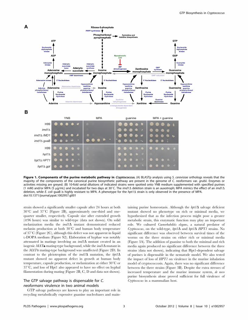
Purine metabolism in Cryptococcus lacks severalcanonical pathway elements
Unlike the purine-rich pigeon guano natural environment of C.
neoformans, the human central nervous system inhabited during systemic
infection is purine-poor [24], suggesting that during infection de novo
purine synthesis could be important for cell survival. A bioinformatic
survey of the available C. neoformans and C. gattii genomes to identify
components of the purine biosynthetic pathway identified Cryptococcus
homologs of most genes of the canonical purine pathway (Figure 1A),
with the exception of adenosine deaminase, adenine deaminase, and
GMP reductase. As previously reported [25], xanthine dehydrogenase
is also absent but a potential equivalent, an a-ketoglutarate-dependent
dioxygenase, is present. Each gene identified is present as a single copy,
including those encoding two key components of the GTP biosynthetic
pathway: IMPDH required for de novo GTP synthesis (IMD1) and a
phosphoribosyltransferase (PRTase) required for the GTP salvage
pathway (HPT1).
IMPDH is essential for de novo GTP biosynthesisIMPDH performs the rate-limiting, first step in de novo GTP
biosynthesis, the NAD+-dependent conversion of inosine mono-
phosphate (IMP) to xanthosine monophosphate (XMP) via a two-
step oxidation and hydrolysis reaction. The reaction mechanism is
complex and involves a large conformational change mid-reaction,
which a number of inhibitors exploit [26–30]. To determine if the
C. neoformans IMD1 gene encodes a bona fide IMPDH, we deleted it
in the well-characterized C. neoformans var. grubii strain H99. The
imd1D strain was a guanine auxotroph and could not grow on
minimal medium (Figure 1B). Supplementation of the medium
with exogenous guanine restored growth, which we later showed
was via a salvage pathway. Introduction of the E. coli IMPDH guaB
into the deletion mutant fully restored growth on minimal
medium, confirming that the IMD1-encoded enzyme performs
the conversion of IMP to XMP (Figure 1B). Finally, the imd1Dphenotype could be mimicked by the addition of the IMPDH
inhibitor MPA to the growth medium; both the wild-type and
IMD1 complemented strains were equally sensitive to MPA at low
concentrations (5 mg/mL). This phenotype is abolished when the
media is supplemented with guanine, as the salvage pathway is
able to bypass the block at IMPDH.
imd1D rescue via purine salvage is mediated byphosphoribosyltransferase Hpt1
Purines present in both the bird guano ecological niche of
Cryptococcus and within the human host are potential substrates of a
GTP salvage pathway. Purine salvage is mediated by PRTase
enzymes, which catalyze the transfer of a ribose 5-phosphate-
derived phosphoribosylpyrophosphate group to guanine or other
purine nucleobases [31]. Upon deletion of the identified HPT1 gene
in strain H99, the mutant exhibited wild-type growth on rich and
minimal media (Figure 1B). MPA-mediated growth inhibition was
abolished by the presence of guanine in all strains but the hpt1Dmutant, demonstrating that Hpt1 mediates salvage of guanine
(Figure 1B). Heterologous expression of the E. coli PRTase gpt in the
hpt1D mutant restored wild-type growth on guanine, confirming the
role of the C. neoformans gene as a PRTase. Further testing revealed
that Hpt1 could accept hypoxanthine, xanthine and guanine as
substrates, defining the enzyme as a hypoxanthine-xanthine-
guanine phosphoribosyltransferase (HXGPRT) (Figure S1).
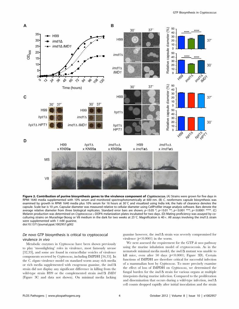
Loss of IMPDH delays synthesis of the cryptococcalpolysaccharide capsule and melanin
As GTP biosynthesis is crucial to many cellular processes, we
sought to determine if guanine auxotrophy affected pathogenicity
via effects on virulence factor production. Despite addition of
guanine, the wild-type phenotype was not completely restored in
the imd1D mutant, which exhibited slow growth at 30uC and was
unable to grow on rich media. Accordingly, in a growth curve
assay the imd1D mutant had an extended lag phase of almost
48 hours compared to wild-type and reached a much lower final
cell density (Figure 2A). In capsule-inducing conditions the imd1D
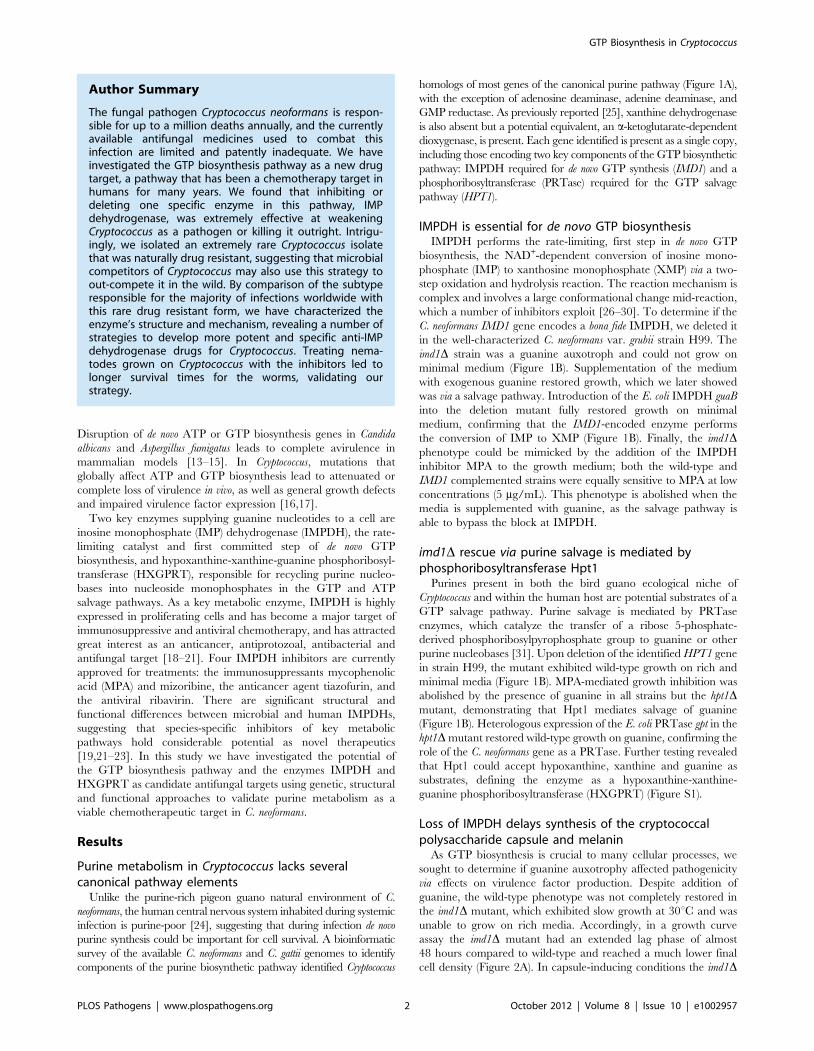
Author Summary
The fungal pathogen Cryptococcus neoformans is respon-sible for up to a million deaths annually, and the currentlyavailable antifungal medicines used to combat thisinfection are limited and patently inadequate. We haveinvestigated the GTP biosynthesis pathway as a new drugtarget, a pathway that has been a chemotherapy target inhumans for many years. We found that inhibiting ordeleting one specific enzyme in this pathway, IMPdehydrogenase, was extremely effective at weakeningCryptococcus as a pathogen or killing it outright. Intrigu-ingly, we isolated an extremely rare Cryptococcus isolatethat was naturally drug resistant, suggesting that microbialcompetitors of Cryptococcus may also use this strategy toout-compete it in the wild. By comparison of the subtyperesponsible for the majority of infections worldwide withthis rare drug resistant form, we have characterized theenzyme’s structure and mechanism, revealing a number ofstrategies to develop more potent and specific anti-IMPdehydrogenase drugs for Cryptococcus. Treating nema-todes grown on Cryptococcus with the inhibitors led tolonger survival times for the worms, validating ourstrategy.
GTP Biosynthesis in Cryptococcus
PLOS Pathogens | www.plospathogens.org 2 October 2012 | Volume 8 | Issue 10 | e1002957
strain showed a significantly smaller capsule after 24 hours at both
30uC and 37uC (Figure 2B), approximately one-third and one-
quarter smaller, respectively. Capsule size after extended growth
($96 hours) was similar to wild-type (data not shown). On solid
melanization media the imd1D mutant demonstrated reduced
melanin production at both 30uC and human body temperature
of 37uC (Figure 2C), although this defect was not apparent in liquid
L-DOPA medium (Figure S2). Elaboration of hyphae was notably
attenuated in matings involving an imd1D mutant created in an
isogenic MATa mating-type background, while the imd1D mutant in
the MATa mating-type background was unaffected (Figure 2D). In
contrast to the pleiotropism of the imd1D mutation, the hpt1Dmutant showed no apparent defect in growth at human body
temperature, capsule production, or melanization at either 30uC or
37uC, and loss of Hpt1 also appeared to have no effect on hyphal
filamentation during mating (Figure 2B, C, D and data not shown).
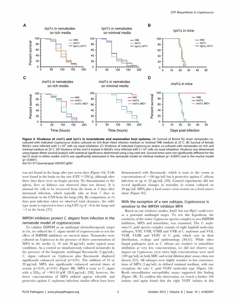
The GTP salvage pathway is dispensable for C.neoformans virulence in two animal models
GTP salvage pathways are known to play an important role in
recycling metabolically expensive guanine nucleobases and main-
taining purine homeostasis. Although the hpt1D salvage deficient
mutant showed no phenotype on rich or minimal media, we
hypothesized that as the infection process might pose a greater
metabolic strain, this enzymatic function may play an important
role. We cultured Caenorhabditis elegans, a natural predator of
Cryptococcus, on the wild-type, hpt1D and hpt1D HPT1 strains. No
significant difference was observed between survival times of the
worms on the three strains on either rich or minimal media
(Figure 3A). The addition of guanine to both the minimal and rich
media again produced no significant difference between the three
strains (data not shown), indicating that Hpt1-dependent salvage
of purines is dispensable in the nematode model. We also tested
the impact of loss of HPT1 on virulence in the murine inhalation
model of cryptococcosis. Again, there was no significant difference
between the three strains (Figure 3B). Despite the extra stresses of
increased temperature and the murine immune system, de novo
purine biosynthesis alone proved sufficient for full virulence of
Cryptococcus in a mammalian host.
Figure 1. Components of the purine metabolic pathway in Cryptococcus. (A) BLASTp analysis using S. cerevisiae orthologs reveals that themajority of the components of the canonical purine biosynthetic pathway are present in the genome of C. neoformans var. grubii. Enzymes oractivities missing are greyed. (B) 10-fold serial dilutions of indicated strains were spotted onto YNB medium supplemented with specified purines(1 mM) and/or MPA (5 mg/mL) and incubated for two days at 30uC. The imd1D deletion strain is an auxotroph; MPA mimics the effect of an imd1Ddeletion, while E. coli guaB is highly resistant to MPA. A phenotype for the hpt1D strain is only observed in the presence of MPA.doi:10.1371/journal.ppat.1002957.g001
GTP Biosynthesis in Cryptococcus
PLOS Pathogens | www.plospathogens.org 3 October 2012 | Volume 8 | Issue 10 | e1002957
De novo GTP biosynthesis is critical to cryptococcalvirulence in vivo
Metabolic enzymes in Cryptococcus have been shown previously
to play ‘moonlighting’ roles in virulence, most famously urease
[32,33], and some are found in extracellular vesicles of virulence
components secreted by Cryptococcus, including IMPDH [34,35]. In
the C. elegans virulence model on standard worm assay rich media
or rich media supplemented with exogenous guanine, the imd1Dstrain did not display any significant difference in killing from the
wild-type strain H99 or the complemented strain imd1D IMD1
(Figure 3C and data not shown). On minimal media lacking
guanine however, the imd1D strain was severely compromised for
virulence (p,0.0001) in the worm.
We next assessed the requirement for the GTP de novo pathway
using the murine inhalation model of cryptococcosis. As in the
nematode minimal media model, the imd1D mutant was unable to
kill mice, even after 50 days (p,0.0001; Figure 3D). Certain
functions of IMPDH are therefore critical for successful infection
of a mammalian host by Cryptococcus. To more precisely examine
the effect of loss of IMPDH on Cryptococcus, we determined the
fungal burden for the imd1D strain for various organs at multiple
timepoints during murine infection. Compared to the proliferation
and dissemination that occurs during a wild-type infection, imd1Dcell counts dropped rapidly after initial inoculation and the strain
Figure 2. Contribution of purine biosynthesis genes to the virulence component of Cryptococcus. (A) Strains were grown for five days inRPMI 1640 media supplemented with 10% serum and monitored spectrophotometrically at 600 nm. (B) C. neoformans capsule biosynthesis wasexamined by growth in RPMI 1640 media plus 10% serum for 16 hours at 30uC and visualized using India ink; the halo of clearance denotes thecapsule. Scale bar is 10 mm. Capsular diameter was measured relative to cellular diameter using CellProfiler image analysis software. Bars denote theaverage relative diameter from three biological replicates. Standard error bars are shown; p,0.05 *; p,0.01 **; p,0.001 ***; p,0.0001 ****. (C)Melanin production was determined on Cryptococcus L-DOPA melanization plates incubated for two days. (D) Mating proficiency was assayed by co-culturing strains on Murashige-Skoog or V8 medium in the dark for two weeks at 25uC. Magnification is 406. All assays involving the imd1D strainwere supplemented with 1 mM guanine.doi:10.1371/journal.ppat.1002957.g002
GTP Biosynthesis in Cryptococcus
PLOS Pathogens | www.plospathogens.org 4 October 2012 | Volume 8 | Issue 10 | e1002957
was not found in the lungs after just seven days (Figure 4A). Cells
were found in the brain on day one (CFU = 328/g), although after
three days these were no longer present. No dissemination to the
spleen, liver or kidneys was observed (data not shown). It is
unusual for cells to be recovered from the brain at 3 days after
intranasal infection, which typically take at least 7 days to
disseminate to the CNS from the lungs [36]. By comparison, at 14
days post infection when we observed total clearance, the wild-
type strain is expected to have a log CFU/g of ,8 in the lungs and
,5 in the brain [37].
IMPDH inhibitors protect C. elegans from infection in thenematode model of cryptococcosis
To validate IMPDH as an antifungal chemotherapeutic target
in vivo, we utilized the C. elegans model of cryptococcosis to test the
effect of IMPDH inhibitors on survival times. Nematodes were
cultured on Cryptococcus in the presence of three concentrations of
MPA in the media (5, 10 and 20 mg/mL) under typical assay
conditions. As a control we simultaneously cultured nematodes in
the presence of the fungistatic antifungal fluconazole (5 mg/mL).
C. elegans cultured on Cryptococcus plus fluconazole displayed
significantly enhanced survival (p,0.01). The addition of 10 or
20 mg/mL MPA also significantly enhanced survival times of
worms (p,0.01; p,0.01) (Figure 4B). MPA is toxic to C. elegans
with a LD50 of ,90610 mM (28.8 mg/mL) [38], however, the
lower concentrations of MPA utilized appear tolerable and
protective against C. neoformans infection; similar effects have been
demonstrated with fluconazole, which is toxic to the worm at
concentrations of ,100 mg/mL but is protective against C. albicans
infection at up to 32 mg/mL [39]. Control experiments did not
reveal significant changes in mortality in worms cultured on
20 mg/mL MPA plus a food source versus worms on a food source
alone (Figure S3).
With the exception of a rare subtype, Cryptococcus issensitive to the IMPDH inhibitor MPA
Based on our virulence studies, Imd1 but not Hpt1 could serve
as a potential antifungal target. To test this hypothesis, the
sensitivity of the entire Cryptococcus species complex to two IMPDH
inhibitors, MPA and mizoribine, was examined. The C. neofor-
mans/C. gattii species complex consists of eight haploid molecular
subtypes, VNI, VNII, VNIII and VNB of C. neoformans and VGI,
VGII, VGIII and VGIV of C. gattii, which vary in their
distribution, ecology and epidemiology [40,41]. While other
fungal pathogens such as C. albicans are sensitive to mizoribine
inhibition at very low concentrations, we did not observe any
impact on Cryptococcus, even when high concentrations were used
(100 mg/mL) in both MIC and serial dilution plate assays (data not
shown) [21]. All subtypes were highly sensitive to low concentra-
tions of MPA (5 mg/mL) in defined minimal medium, with one
exception: the rare C. gattii VGIV molecular type (Figure 5A).
Broth microdilution susceptibility assays supported this finding
(Figure 5B). To confirm this observation, we tested a further 106
isolates and again found that the eight VGIV isolates in this
Figure 3. Virulence of imd1D and hpt1D in invertebrate and mammalian host systems. (A) Survival of Bristol N2 strain nematodes co-cultured with indicated Cryptococcus strains cultured on rich Brain Heart Infusion medium or minimal YNB medium at 25uC. (B) Survival of femaleBALB/c mice infected with 56105 cells via nasal inhalation. (C) Virulence of indicated Cryptococcus strains co-cultured with nematodes on rich andminimal medium at 25uC. (D) Virulence of the imd1D mutant in BALB/c mice infected with 56105 cells via nasal inhalation. Virulence was determinedusing Kaplan-Meier survival analysis with statistical significance determined using a log-rank test. Survival times were not significantly different for thehpt1D strain in either model. imd1D was significantly attenuated in the nematode model on minimal medium (p,0.0001) and in the murine model(p,0.0001).doi:10.1371/journal.ppat.1002957.g003
GTP Biosynthesis in Cryptococcus
PLOS Pathogens | www.plospathogens.org 5 October 2012 | Volume 8 | Issue 10 | e1002957
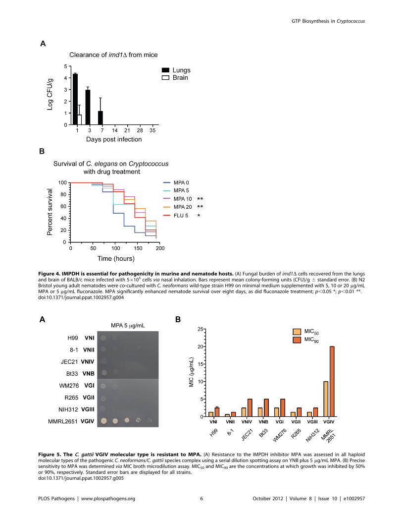
Figure 4. IMPDH is essential for pathogenicity in murine and nematode hosts. (A) Fungal burden of imd1D cells recovered from the lungsand brain of BALB/c mice infected with 56105 cells via nasal inhalation. Bars represent mean colony-forming units (CFU)/g 6 standard error. (B) N2Bristol young adult nematodes were co-cultured with C. neoformans wild-type strain H99 on minimal medium supplemented with 5, 10 or 20 mg/mLMPA or 5 mg/mL fluconazole. MPA significantly enhanced nematode survival over eight days, as did fluconazole treatment; p,0.05 *; p,0.01 **.doi:10.1371/journal.ppat.1002957.g004
Figure 5. The C. gattii VGIV molecular type is resistant to MPA. (A) Resistance to the IMPDH inhibitor MPA was assessed in all haploidmolecular types of the pathogenic C. neoformans/C. gattii species complex using a serial dilution spotting assay on YNB plus 5 mg/mL MPA. (B) Precisesensitivity to MPA was determined via MIC broth microdilution assay. MIC50 and MIC90 are the concentrations at which growth was inhibited by 50%or 90%, respectively. Standard error bars are displayed for all strains.doi:10.1371/journal.ppat.1002957.g005
GTP Biosynthesis in Cryptococcus
PLOS Pathogens | www.plospathogens.org 6 October 2012 | Volume 8 | Issue 10 | e1002957
collection were easily identified by their MPA resistance, while all
others were sensitive (data not shown). VGIV MPA resistance was
uniform and 100% reproducible, supporting this as an inherent
characteristic of the VGIV molecular type.
We reasoned that resistance to MPA could arise from mutations
in the IMD1 ORF, overexpression of Imd1, increased IMD1 copy
number, or changes in drug uptake or efflux from the cell. To
determine if MPA resistance could result from changes in
expression level of IMPDH in C. gattii VGIV, we assessed the
transcriptional response of C. neoformans (strain H99) and C. gattii
(strain MMRL2651) IMD1 to challenge by guanine and MPA via
qRT-PCR. After treatment with guanine, both C. neoformans and C.
gattii IMD1 were significantly downregulated, consistent with de
novo GTP synthesis not being required when abundant salvageable
guanine is present (Figure S4). Importantly, no real difference was
observed between the two strains’ responses to either substrate or
inhibitor, suggesting that resistance to MPA may in fact be an
intrinsic property of the VGIV IMPDH isoform. To determine if
either copy number or a feature of the C. gattii VGIV IMD1 allele
(CgIMD1) confer MPA resistance, we introduced both the C.
neoformans var. grubii VNI allele (CnIMD1) from the MPA-sensitive
strain H99 and CgIMD1 from the MPA-resistant strain
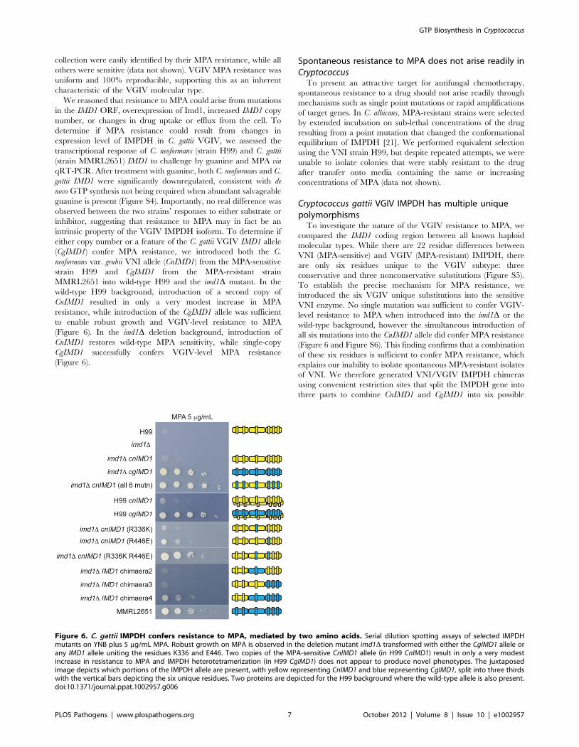
MMRL2651 into wild-type H99 and the imd1D mutant. In the
wild-type H99 background, introduction of a second copy of
CnIMD1 resulted in only a very modest increase in MPA
resistance, while introduction of the CgIMD1 allele was sufficient
to enable robust growth and VGIV-level resistance to MPA
(Figure 6). In the imd1D deletion background, introduction of
CnIMD1 restores wild-type MPA sensitivity, while single-copy
CgIMD1 successfully confers VGIV-level MPA resistance
(Figure 6).
Spontaneous resistance to MPA does not arise readily inCryptococcus
To present an attractive target for antifungal chemotherapy,
spontaneous resistance to a drug should not arise readily through
mechanisms such as single point mutations or rapid amplifications
of target genes. In C. albicans, MPA-resistant strains were selected
by extended incubation on sub-lethal concentrations of the drug
resulting from a point mutation that changed the conformational
equilibrium of IMPDH [21]. We performed equivalent selection
using the VNI strain H99, but despite repeated attempts, we were
unable to isolate colonies that were stably resistant to the drug
after transfer onto media containing the same or increasing
concentrations of MPA (data not shown).
Cryptococcus gattii VGIV IMPDH has multiple uniquepolymorphisms
To investigate the nature of the VGIV resistance to MPA, we
compared the IMD1 coding region between all known haploid
molecular types. While there are 22 residue differences between
VNI (MPA-sensitive) and VGIV (MPA-resistant) IMPDH, there
are only six residues unique to the VGIV subtype: three
conservative and three nonconservative substitutions (Figure S5).
To establish the precise mechanism for MPA resistance, we
introduced the six VGIV unique substitutions into the sensitive
VNI enzyme. No single mutation was sufficient to confer VGIV-
level resistance to MPA when introduced into the imd1D or the
wild-type background, however the simultaneous introduction of
all six mutations into the CnIMD1 allele did confer MPA resistance
(Figure 6 and Figure S6). This finding confirms that a combination
of these six residues is sufficient to confer MPA resistance, which
explains our inability to isolate spontaneous MPA-resistant isolates
of VNI. We therefore generated VNI/VGIV IMPDH chimeras
using convenient restriction sites that split the IMPDH gene into
three parts to combine CnIMD1 and CgIMD1 into six possible
Figure 6. C. gattii IMPDH confers resistance to MPA, mediated by two amino acids. Serial dilution spotting assays of selected IMPDHmutants on YNB plus 5 mg/mL MPA. Robust growth on MPA is observed in the deletion mutant imd1D transformed with either the CgIMD1 allele orany IMD1 allele uniting the residues K336 and E446. Two copies of the MPA-sensitive CnIMD1 allele (in H99 CnIMD1) result in only a very modestincrease in resistance to MPA and IMPDH heterotetramerization (in H99 CgIMD1) does not appear to produce novel phenotypes. The juxtaposedimage depicts which portions of the IMPDH allele are present, with yellow representing CnIMD1 and blue representing CgIMD1, split into three thirdswith the vertical bars depicting the six unique residues. Two proteins are depicted for the H99 background where the wild-type allele is also present.doi:10.1371/journal.ppat.1002957.g006
GTP Biosynthesis in Cryptococcus
PLOS Pathogens | www.plospathogens.org 7 October 2012 | Volume 8 | Issue 10 | e1002957
configurations. Only the construct comprising the first third of
CnIMD1 and the last two-thirds of CgIMD1 (bearing R446E,
G450A and A500V) conferred VGIV-level resistance to MPA
(Figure 6 and Figure S6). Based on this observation we employed
site-directed mutagenesis of the MPA sensitive VNI allele to
introduce the conservative R336K substitution (the only unique
residue in the middle fragment) in turn with each of the three
residues from the last fragment; the combination of K336 and
E446 was sufficient to confer MPA resistance (Figure 6 and Figure
S6). Again, transformation of these constructs into the wild-type
H99 background yielded levels of MPA resistance comparable
with the imd1D deletion strain, suggesting that simple gene
amplification may be insufficient to produce robust resistance,
and the resulting mixed IMPDH heterotetramers in these strains
did not appear to possess novel drug-resistant characteristics.
Steady-state kinetics of CnIMPDH and CgIMPDHThe catalytic mechanism of IMPDH involves a transition from
a dehydrogenase to a hydrolase reaction; initially, the substrate
IMP and cofactor NAD+ bind, IMP is then oxidized and forms a
covalent transition state complex E-XMP* via a catalytic cysteine,
while NAD+ is simultaneously reduced to NADH. The enzyme
then undergoes a conformational change whereby a large mobile
flap folds into the vacant NAD+ site, carrying a catalytic arginine
residue that activates a water molecule to hydrolyze the E-XMP*
covalent complex, releasing XMP [26,42,43]. To investigate if
resistance to MPA is due to functional differences between
CnImd1 and CgImd1, we determined the steady-state kinetic
parameters (Figure S7). Plots of velocity versus IMP concentrations
were best fit by the Michaelis-Menten equation
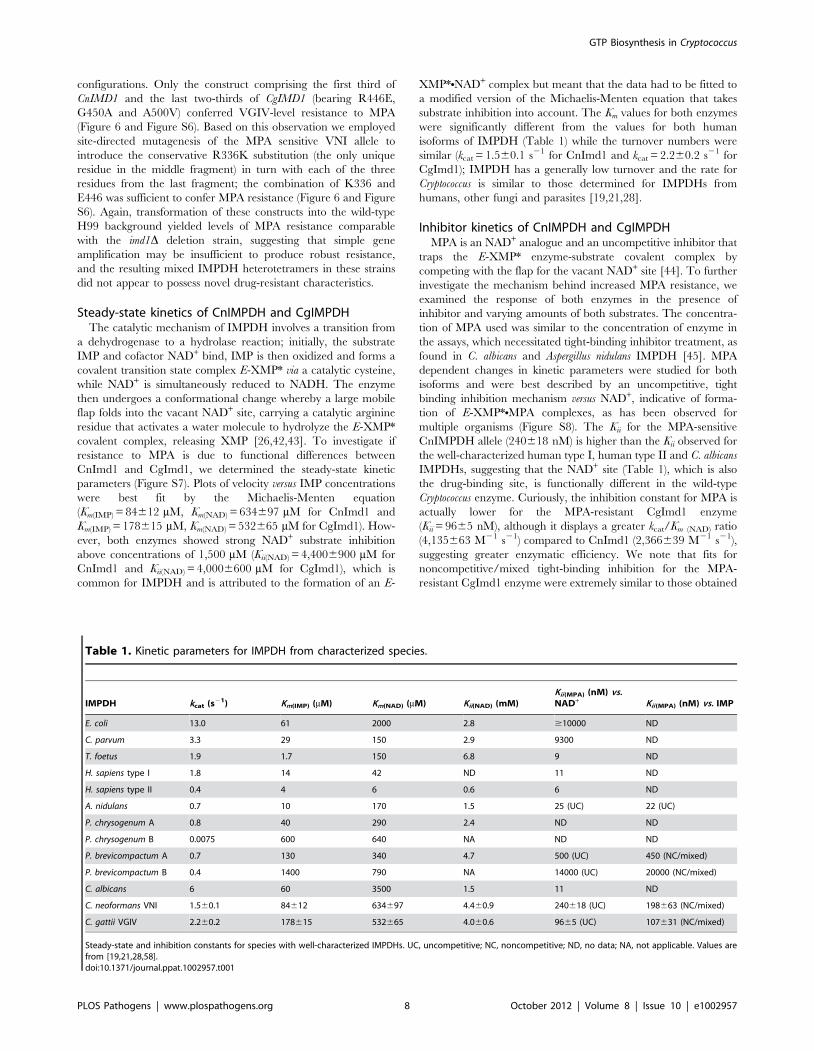
(Km(IMP) = 84612 mM, Km(NAD) = 634697 mM for CnImd1 and
Km(IMP) = 178615 mM, Km(NAD) = 532665 mM for CgImd1). How-
ever, both enzymes showed strong NAD+ substrate inhibition
above concentrations of 1,500 mM (Kii(NAD) = 4,4006900 mM for
CnImd1 and Kii(NAD) = 4,0006600 mM for CgImd1), which is
common for IMPDH and is attributed to the formation of an E-
XMP*NNAD+ complex but meant that the data had to be fitted to
a modified version of the Michaelis-Menten equation that takes
substrate inhibition into account. The Km values for both enzymes
were significantly different from the values for both human
isoforms of IMPDH (Table 1) while the turnover numbers were
similar (kcat = 1.560.1 s21 for CnImd1 and kcat = 2.260.2 s21 for
CgImd1); IMPDH has a generally low turnover and the rate for
Cryptococcus is similar to those determined for IMPDHs from
humans, other fungi and parasites [19,21,28].
Inhibitor kinetics of CnIMPDH and CgIMPDHMPA is an NAD+ analogue and an uncompetitive inhibitor that
traps the E-XMP* enzyme-substrate covalent complex by
competing with the flap for the vacant NAD+ site [44]. To further
investigate the mechanism behind increased MPA resistance, we
examined the response of both enzymes in the presence of
inhibitor and varying amounts of both substrates. The concentra-
tion of MPA used was similar to the concentration of enzyme in
the assays, which necessitated tight-binding inhibitor treatment, as
found in C. albicans and Aspergillus nidulans IMPDH [45]. MPA
dependent changes in kinetic parameters were studied for both
isoforms and were best described by an uncompetitive, tight
binding inhibition mechanism versus NAD+, indicative of forma-
tion of E-XMP*NMPA complexes, as has been observed for
multiple organisms (Figure S8). The Kii for the MPA-sensitive
CnIMPDH allele (240618 nM) is higher than the Kii observed for
the well-characterized human type I, human type II and C. albicans
IMPDHs, suggesting that the NAD+ site (Table 1), which is also
the drug-binding site, is functionally different in the wild-type
Cryptococcus enzyme. Curiously, the inhibition constant for MPA is
actually lower for the MPA-resistant CgImd1 enzyme
(Kii = 9665 nM), although it displays a greater kcat/Km (NAD) ratio
(4,135663 M21 s21) compared to CnImd1 (2,366639 M21 s21),
suggesting greater enzymatic efficiency. We note that fits for
noncompetitive/mixed tight-binding inhibition for the MPA-
resistant CgImd1 enzyme were extremely similar to those obtained
Table 1. Kinetic parameters for IMPDH from characterized species.
IMPDH kcat (s21) Km(IMP) (mM) Km(NAD) (mM) Kii(NAD) (mM)Kii(MPA) (nM) vs.NAD+ Kii(MPA) (nM) vs. IMP
E. coli 13.0 61 2000 2.8 $10000 ND
C. parvum 3.3 29 150 2.9 9300 ND
T. foetus 1.9 1.7 150 6.8 9 ND
H. sapiens type I 1.8 14 42 ND 11 ND
H. sapiens type II 0.4 4 6 0.6 6 ND
A. nidulans 0.7 10 170 1.5 25 (UC) 22 (UC)
P. chrysogenum A 0.8 40 290 2.4 ND ND
P. chrysogenum B 0.0075 600 640 NA ND ND
P. brevicompactum A 0.7 130 340 4.7 500 (UC) 450 (NC/mixed)
P. brevicompactum B 0.4 1400 790 NA 14000 (UC) 20000 (NC/mixed)
C. albicans 6 60 3500 1.5 11 ND
C. neoformans VNI 1.560.1 84612 634697 4.460.9 240618 (UC) 198663 (NC/mixed)
C. gattii VGIV 2.260.2 178615 532665 4.060.6 9665 (UC) 107631 (NC/mixed)
Steady-state and inhibition constants for species with well-characterized IMPDHs. UC, uncompetitive; NC, noncompetitive; ND, no data; NA, not applicable. Values arefrom [19,21,28,58].doi:10.1371/journal.ppat.1002957.t001
GTP Biosynthesis in Cryptococcus
PLOS Pathogens | www.plospathogens.org 8 October 2012 | Volume 8 | Issue 10 | e1002957
for uncompetitive tight-binding inhibition and yield a much higher
Kii (293676 nM; data not shown), which may indicate some
affinity of the inhibitor for other enzyme forms. Surprisingly, fits of
MPA versus IMP for both enzymes were best described by
noncompetitive/mixed patterns of inhibition (Figure S8), with a
Kii = 198663 nM for CnImd1 and a Kii = 107631 nM for
CgImd1. This suggests that in Cryptococcus IMPDH, MPA binds
to additional enzyme forms such as free enzyme or E-IMP. Such a
scenario is also found in MPA-resistant Penicillium brevicompactum
IMPDH, where the initial hydride transfer step (as opposed to the
subsequent hydrolysis step) is either partially or completely rate-
limiting, resulting in the predominate enzyme form being E-IMP-
NAD+ instead of E-XMP*, conferring MPA resistance [46].
Crystal structure of CnImd1To provide a foundation for fungus-specific inhibitor develop-
ment, we determined the crystal structure of the clinically relevant
C. neoformans var. grubii IMPDH in complex with the substrate IMP
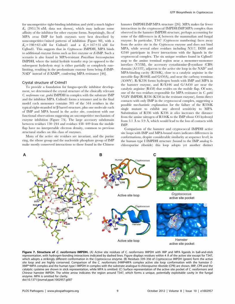
and the inhibitor MPA. CnImd1 forms a tetramer and in the final
model each monomer contains 395 of the 544 residues in the
typical eight-stranded a/b barrel structure, plus one molecule each
of IMP and MPA bound in the active site, consistent with our
functional observations suggesting an uncompetitive mechanism of
enzyme inhibition (Figure 7A). The large accessory subdomain
between residues 130–244 and residues 438–449 from the mobile
flap have no interpretable electron density, common to previous
structural studies on this class of enzymes.
Many of the active site residues are invariant, and the purine
ring, the ribose group and the nucleotide phosphate group of IMP
make mostly conserved interactions to those found in the Chinese
hamster IMPDH:IMP:MPA structure [26]. MPA makes far fewer
interactions in the cryptococcal IMPDH:IMP:MPA complex than
observed in the hamster IMPDH structure, perhaps accounting for
some of the differences in Ki between the mammalian and fungal
enzyme. In particular, T347 (Cryptococcus numbering) faces away
from the active site in the Cryptococcus enzyme and does not bind
MPA, while several other residues including N317, D288 and
G340 participate in fewer interactions with the ligands in the
cryptococcal complex. The six unique residues found in CgImd1
map to the amino terminal region near a monomer-monomer
interface (V55M), the accessory cystathionine-b-synthase (CBS)
domain (A153T), adjacent to the active site loop in the NAD+ and
MPA-binding cavity (R336K), close to a catalytic arginine in the
movable flap (R446E and G450A), and near the carboxy terminus
(A500V). R/K336 forms hydrogen bonds with IMP and MPA in
the hamster enzyme, and R/E446 and G/A450 are near the
catalytic arginine (R458) that resides on the mobile flap. Of note,
one of the two residues responsible for MPA resistance in C. gattii
VGIV IMPDH, R336 (K336 in the resistant enzyme), forms direct
contacts with only IMP in the cryptococcal complex, suggesting a
possible mechanistic explanation for the failure of the R336K
single mutant to exhibit any altered sensitivity to MPA.
Substitution of R336 with K336 in silico increases the distance
from the amine nitrogen of R336K to the IMP ribose O3 hydroxyl
from 3.1 A to 3.9 A, which would lead to the loss of contacts with
IMP.
Comparison of the hamster and cryptococcal IMPDH active
site loops with IMP and MPA-bound states indicates differences in
conformations, despite considerable similarity at sequence level; in
the human type I IMPDH structure (bound to the IMP analog 6-
chloropurine ribotide) this loop adopts yet another distinct
Figure 7. Structure of C. neoformans IMPDH. (A) Active site residues of C. neoformans IMPDH with IMP and MPA ligands in ball-and-stickrepresentation, with hydrogen-bonding interactions indicated by dashed lines. Figure displays residues within 4 A of the active site except for T347,which adopts a strikingly different conformation in the Cryptococcus enzyme. (B) Residues 339–356 of Cryptococcus IMPDH (green) form the activesite loop and are highly conserved. Comparison of the C. neoformans ENIMPNMPA complex active site loop conformation with the hamster E-XMP*NMPA complex and the human type I IMPDH in complex with the substrate analogue 6-chloropurine ribotide (CPR) are shown. IMP, CPR and thecatalytic cysteine are shown in stick representation, while MPA is omitted. (C) Surface representation of the active site pocket of C. neoformans andChinese hamster IMPDH. The white arrow indicates the region around T347, which forms a unique, potentially exploitable cavity in the fungalenzyme. MPA is omitted for clarity.doi:10.1371/journal.ppat.1002957.g007
GTP Biosynthesis in Cryptococcus
PLOS Pathogens | www.plospathogens.org 9 October 2012 | Volume 8 | Issue 10 | e1002957
conformation (Figure 7B). The change in orientation of the loop in
CnImd1 creates a large, Cryptococcus-specific pocket in the active
site (Figure 7C). There is considerable flexibility in the conforma-
tion of the active site loop observed in structures of different
enzyme complexes, which suggests that it can mimic the changes
during the reaction cycle where it may modulate closure of the flap
[47]. While IMP is covalently bound to the catalytic cysteine in the
hamster and human structures, which may be responsible for the
altered orientation, comparison with Tritrichomonas foetus IMPDH
bound to IMP or mizoribine phosphate (where the ligands are not
covalently bound in either structure) suggests the conformation
observed in the cryptococcal complex to be unique, thus offering a
potential avenue for species-specific inhibition. IMPDH crystal
structures have been demonstrated to provide a reliable indication
of the position of the loop at different parts of the reaction cycle,
suggesting the pocket observed in the Cryptococcus structure to be a
legitimate target [47].
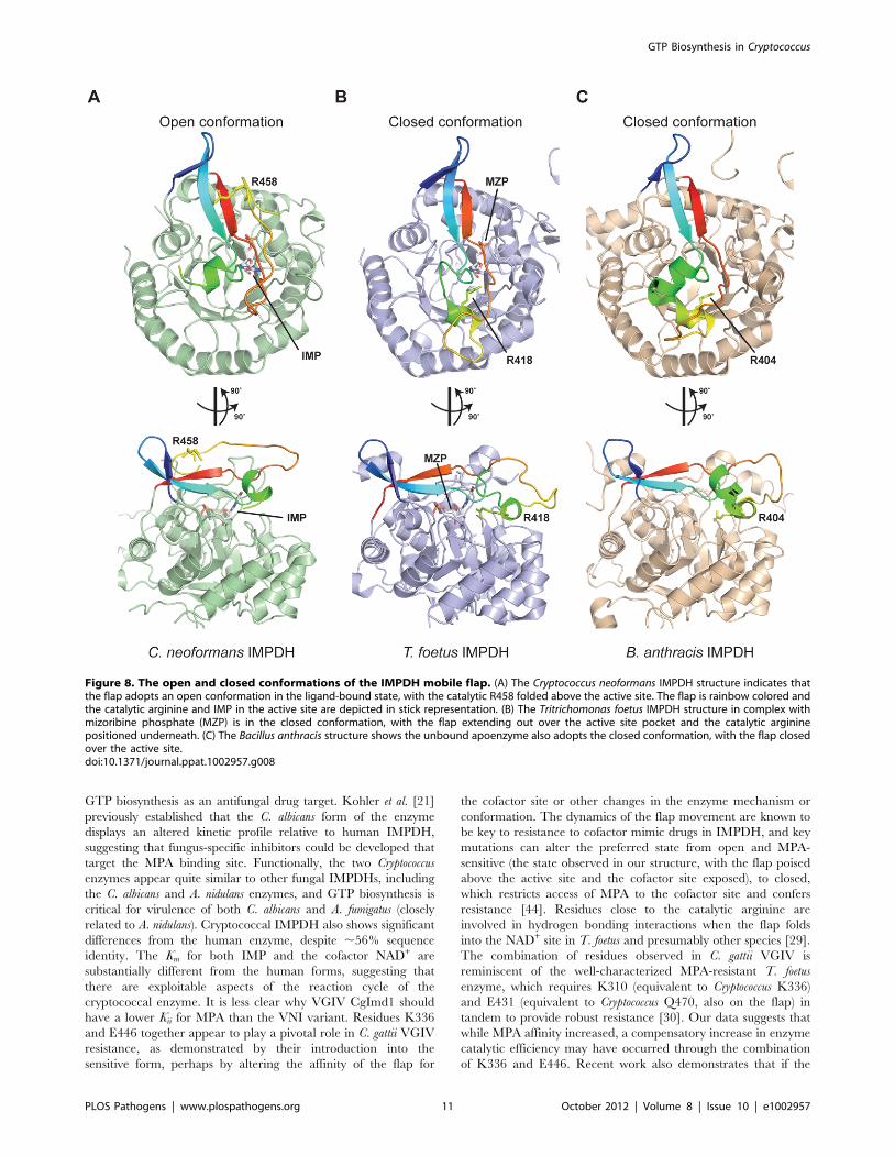
The cryptococcal IMPDH structure is unique in having most of
the mobile flap, except for 12 residues that encompass a small
insertion region unique to Cryptococcus, sufficiently ordered to
permit all but three residues corresponding to the flap in human
IMPDH to be observed. The only comparable structural evidence
for the flap is presented for the parasite T. foetus IMPDH
complexed with mizoribine phosphate, in which the flap is in the
closed conformation within the cofactor site [29] and the Bacillus
anthracis apoenzyme structure [48]. The cryptococcal structure
reveals the flap in the ‘open’ conformation, as would be required
for the initial dehydrogenation before the flap folds into the NAD+
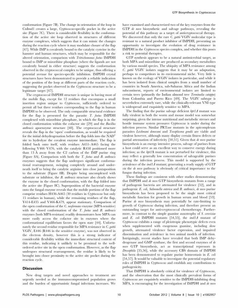
site to hydrolyze the covalent enzyme intermediate. The flap is
folded back onto itself, with residues A455–A465 facing the
following V466–V476, with the catalytic R458 positioned more
than 13 A away from the C2 carbon of the IMP purine ring
(Figure 8A). Comparison with both the T. foetus and B. anthracis
enzymes suggests that the flap undergoes significant conforma-
tional rearrangement, swinging completely around and under-
neath itself to bring the catalytic arginine into close juxtaposition
to the substrate (Figure 8B). Despite being uncomplexed with
substrate or inhibitor, the B. anthracis structure also clearly shows
the enzyme in the closed conformation, with the flap folded into
the active site (Figure 8C). Superposition of the bacterial enzyme
onto the fungal enzyme reveals that the mobile portions of the flap
comprise residues H436-A465 (30 residues in Cryptococcus but only
15 residues in human), while the remaining residues of the flap,
Y414-E435 and V466-K479, appear stationary. Comparison of
the open conformation of the C. neoformans enzyme (MPA-sensitive)
with the closed conformation of the T. foetus and B. anthracis
enzymes (both MPA-resistant) readily demonstrates how MPA can
more easily access the cofactor site in enzymes where the
conformational equilibrium favors the open state [44]. Unfortu-
nately the second residue responsible for MPA resistance in C. gattii
VGIV, E446 (R446 in the sensitive enzyme), was not observed in
the electron density, however this is a strong indicator of
considerable flexibility within the immediate structural context of
this residue, indicating it unlikely to be proximal to the well-
ordered active site in the open conformation. However, as the flap
undergoes structural rearrangement, the residue is likely to be
brought into closer proximity to the active site pocket during the
reaction cycle.
Discussion
New drug targets and novel approaches to treatment are
urgently needed as the immunocompromised population grows
and the burden of opportunistic fungal infections increases. We
have examined and characterized two of the key enzymes from the
GTP de novo biosynthetic and salvage pathways, revealing the
potential of this pathway as a target of anticryptococcal therapy.
We discovered that only the rare C. gattii VGIV molecular type is
resistant to a natural product inhibitor of IMPDH, presenting an
opportunity to investigate the evolution of drug resistance in
IMPDH in the Cryptococcus species complex, and whether this poses
a risk to potential therapies.
GTP synthesis appears to be a natural antimicrobial target, as
both MPA and mizoribine are produced as secondary metabolites
by various mould species. The ubiquity of MPA resistance among
C. gattii VGIV isolates suggests that it may be an adaptation,
perhaps to competitors in its environmental niche. Very little is
known on the ecology of VGIV isolates in particular, and while it
has been isolated from clinical samples from a small number of
countries in South America, sub-Saharan Africa and the Indian
subcontinent, reports of environmental isolates are limited to
certain trees (primarily the Indian almond tree Terminalia catappa)
from Colombia and Puerto Rico [49]. The VGIV subtype is
nevertheless extremely rare, while the clinically-relevant VNI type
is widespread and exquisitely sensitive to MPA.
The finding that the purine salvage deficient hpt1D mutant was
fully virulent in both the worm and mouse model was somewhat
surprising, given the intense nutritional and metabolic stresses and
hostile immune system pressures Cryptococcus is under during the
infection process. Similar PRTase mutants in the apicomplexan
parasites Leishmani donovani and Toxoplasma gondii are viable and
virulent however, although many display certain fitness defects or
partial attenuation of infectivity [50,51]. Given that de novo purine
biosynthesis is an energy intensive process, salvage of purines from
a host could serve as an excellent way to conserve energy during
infection; as the hpt1D mutant is not perturbed for virulence, this
may reflect a generally low concentration of salvageable purines
during the infection process. This model is supported by the
avirulence of the imd1D mutant; generating a ready supply of GTP
via the de novo pathway is obviously of critical importance to the
fungus during infection.
These findings are consistent with other studies demonstrating
that IMPDH and de novo GTP biosynthesis mutants from a range
of pathogenic bacteria are attenuated for virulence [52], and in
pathogenic E. coli, Salmonella enterica and B. anthracis, de novo purine
biosynthesis has been proposed to be the single most critical
metabolic function for growth in human serum and blood [53].
Purine de novo biosynthesis may potentially be rate-limiting to
growth of Cryptococcus during infection, and therefore present an
outstanding target for anti-cryptococcal chemotherapy. Further-
more, in contrast to the simple guanine auxotrophy of S. cerevisiae
and E. coli IMPDH mutants [54,55], the imd1D mutant of
Cryptococcus exhibits a range of pleiotropic phenotypic defects even
when supplemented with exogenous guanine, including slow
growth, attenuated virulence factor expression, and impaired
dissemination and avirulence in two animal models of infection.
Intriguingly, recent studies have revealed that both IMP dehy-
drogenase and GMP synthase, the first and second enzymes of de
novo GTP biosynthesis, act as transcriptional repressors in
Drosophila [35,56], while the accessory CBS domain of IMPDH
has been demonstrated to regulate purine homeostasis in E. coli
[54,57]. It would be valuable to investigate the potential regulatory
roles of IMPDH in Cryptococcus and establish any contributions to
pathogenicity.
That IMPDH is absolutely critical for virulence of Cryptococcus,
and the observation that the most clinically prevalent forms of
Cryptococcus are exquisitely sensitive to the IMPDH-inhibiting drug
MPA, is encouraging for the investigation of IMPDH and de novo
GTP Biosynthesis in Cryptococcus
PLOS Pathogens | www.plospathogens.org 10 October 2012 | Volume 8 | Issue 10 | e1002957
GTP biosynthesis as an antifungal drug target. Kohler et al. [21]
previously established that the C. albicans form of the enzyme
displays an altered kinetic profile relative to human IMPDH,
suggesting that fungus-specific inhibitors could be developed that
target the MPA binding site. Functionally, the two Cryptococcus
enzymes appear quite similar to other fungal IMPDHs, including
the C. albicans and A. nidulans enzymes, and GTP biosynthesis is
critical for virulence of both C. albicans and A. fumigatus (closely
related to A. nidulans). Cryptococcal IMPDH also shows significant
differences from the human enzyme, despite ,56% sequence
identity. The Km for both IMP and the cofactor NAD+ are
substantially different from the human forms, suggesting that
there are exploitable aspects of the reaction cycle of the
cryptococcal enzyme. It is less clear why VGIV CgImd1 should
have a lower Kii for MPA than the VNI variant. Residues K336
and E446 together appear to play a pivotal role in C. gattii VGIV
resistance, as demonstrated by their introduction into the
sensitive form, perhaps by altering the affinity of the flap for
the cofactor site or other changes in the enzyme mechanism or
conformation. The dynamics of the flap movement are known to
be key to resistance to cofactor mimic drugs in IMPDH, and key
mutations can alter the preferred state from open and MPA-
sensitive (the state observed in our structure, with the flap poised
above the active site and the cofactor site exposed), to closed,
which restricts access of MPA to the cofactor site and confers
resistance [44]. Residues close to the catalytic arginine are
involved in hydrogen bonding interactions when the flap folds
into the NAD+ site in T. foetus and presumably other species [29].
The combination of residues observed in C. gattii VGIV is
reminiscent of the well-characterized MPA-resistant T. foetus
enzyme, which requires K310 (equivalent to Cryptococcus K336)
and E431 (equivalent to Cryptococcus Q470, also on the flap) in
tandem to provide robust resistance [30]. Our data suggests that
while MPA affinity increased, a compensatory increase in enzyme
catalytic efficiency may have occurred through the combination
of K336 and E446. Recent work also demonstrates that if the
Figure 8. The open and closed conformations of the IMPDH mobile flap. (A) The Cryptococcus neoformans IMPDH structure indicates thatthe flap adopts an open conformation in the ligand-bound state, with the catalytic R458 folded above the active site. The flap is rainbow colored andthe catalytic arginine and IMP in the active site are depicted in stick representation. (B) The Tritrichomonas foetus IMPDH structure in complex withmizoribine phosphate (MZP) is in the closed conformation, with the flap extending out over the active site pocket and the catalytic argininepositioned underneath. (C) The Bacillus anthracis structure shows the unbound apoenzyme also adopts the closed conformation, with the flap closedover the active site.doi:10.1371/journal.ppat.1002957.g008
GTP Biosynthesis in Cryptococcus
PLOS Pathogens | www.plospathogens.org 11 October 2012 | Volume 8 | Issue 10 | e1002957
initial hydride transfer step becomes rate-limiting, this reduces
accumulation of the enzyme-substrate covalent complex and thus
reduces susceptibility to MPA inhibition. Notably, this has
occurred in MPA-resistant Penicillium brevicompactum IMPDH
without significant residue changes around the substrate/cofactor
binding sites [46,58]. Our work demonstrates that MPA binds to
additional cryptococcal IMPDH forms (an uncompetitive inhib-
itor should bind only to the enzyme-substrate covalent complex),
suggesting the enzyme to be fundamentally functionally different
from the human isoforms.
To provide a solid foundation for chemotherapeutic agent
development, we have determined the structure of C. neoformans
var. grubii IMPDH in complex with IMP and MPA. While the
accessory domain is presumably disordered and was not observed,
we were able to successfully model the residues comprising the
mobile flap domain, which to date had only been structurally
characterized in the closed conformation in the T. foetus and the B.
anthracis enzymes [29,48]. While Cryptococcus IMPDH possesses an
insertion within the flap that was mostly disordered and there is
considerable flexibility associated with the flap region, the
structural evidence suggests that the flap undergoes a drastic
conformational change during the reaction cycle, which has been
demonstrated functionally but not structurally in previous studies
[27,59,60]. In the open enzyme conformation, the catalytic
arginine is positioned away from the active site, with the flap
undergoing a 270u swinging rotation to perform the hydrolysis
reaction. The near-complete flap structure in the open orientation
will aid future drug design efforts, as the flap folds into the cofactor
site – a critical target site for inhibitors. The cofactor site, and the
adenosine subsite in particular, also present an opportunity for
fungus-specific inhibitor development, as there are a number of
critical residues that vary in the Cryptococcus enzyme; for example,
the adenine ring of NAD+ likely stacks between R267 and Y296 in
Cryptococcus IMPDH, and these two residues differ from the
corresponding H253 and F282 in the human enzyme. The
eukaryotic parasite T. foetus shares these two substitutions, which
have been suggested as potential species-specific targets [61]. The
adenosine portion of the cofactor site is also a monomer-monomer
interface, and one of the more variable parts of the enzyme
between species. Recent strategies extend inhibitors from the
nicotinamide subsite (where MPA binds) out to the adenosine
subsite to increase specificity [44,62]. The unique pocket in the
active site loop region of Cryptococcus IMPDH will enable the design
of inhibitors with moieties that occupy the cavity, which should be
selective for the fungal enzyme over the human enzymes.
Overall, we have demonstrated that de novo GTP biosynthesis is
crucial for cryptococcal virulence factor expression and patho-
genesis, and multiple exploitable avenues exist in IMPDH to
develop inhibitors that are specific for the fungal enzyme. This
work lays the foundation for purine biosynthesis as a legitimate
antifungal target, and the functional similarity between the
Cryptococcus and Candida enzymes presents an intriguing opportu-
nity to develop novel IMPDH inhibitors that could lead to
effective therapy against a spectrum of fungal pathogens.
Materials and Methods
Ethics statementThe Cryptococcus murine virulence assay protocol was approved
by the Molecular Biosciences Animal Ethics Committee of the
University of Queensland (AEC# SCMB/473/09/UQ/
NHMRC). Assays were conducted in accordance with the
guidelines in the Australian code of practice for the care and use
of animals for scientific purposes by the National Health and
Medical Research Council. Infection and euthanasia were
performed under methoxyfluorane anesthesia, and all efforts were
made to minimize suffering via strict adherence to the Guidelines
to promote the wellbeing of animals used for scientific purposes by
the National Health and Medical Research Council.
Strains and mediaC. neoformans and C. gattii strains were cultured in YPD or YNB
(Becton Dickinson, Franklin Lakes NJ) media at 30uC. imd1Dmutants are guanine auxotrophs and were grown on minimal
YNB media supplemented with 1 mM guanine for all manipula-
tions; ade2D and hpt1D ade2D mutants are adenine auxotrophs and
were maintained on rich media. Cloning and plasmid propagation
was performed in E. coli strain Mach1 (Life Technologies,
Carlsbad CA). Genomic DNA for E. coli gene isolation was
prepared from strain K12. C. elegans strain N2 Bristol was
maintained at 20uC on lawns of the E. coli uracil auxotroph strain
OP50 on Nematode Growth Medium using standard procedures
[63]. Recombinant Cryptococcus IMPDH was heterologously
expressed in E. coli strain BL21(DE3)pLysS (Merck, Darmstadt,
Germany). MPA, mizoribine, 5-fluoro-29-deoxyuridine, G418,
NAD+, Tris, Bis-Tris, glycine, NaCl, KCl, DTT, EDTA, DMSO,
uric acid, inosine, hypoxanthine, xanthine, xanthosine, guanine,
guanosine, adenine, adenosine, IMP, AMP, XMP and GMP were
purchased from Sigma (St Louis, MO). Nourseothricin (clonNAT)
was purchased from Werner BioAgents (Jena, Germany). MPA
was dissolved in DMSO and was used at a concentration of 5 mg/
mL. Supplemental purines when required were added to a final
concentration of 1 mM [63].
Molecular techniquesStandard molecular techniques were performed as described by
Sambrook et al. [64]. C. neoformans genomic DNA for Southern blot
analysis was prepared as described [65]. Oligonucleotides used are
given in Table S1. For Southern hybridizations, DNA was
digested with appropriate enzymes, electrophoresed on TAE-
agarose gels and blotted onto Hybond-XL membrane (GE
Healthcare, Little Chalfont, United Kingdom) using standard
procedures. Probes were generated using the Rediprime II
Random Prime Labeling Kit (GE Healthcare) with PCR products
from H99 or MMRL2651 template DNA, and a32P dCTP
(PerkinElmer, Waltham MA). Blots were hybridized overnight at
65uC and membranes were exposed onto Fuji Super RX medical
X-ray film (Fujifilm, Tokyo, Japan). Splice overlap PCR, knockout
transformations and gene complementations were performed as
described [66–69], using particle delivery using a BioRad He-1000
Biolistic Device (Bio-Rad, Hercules CA).
Bioinformatic analysisThe unpublished C. neoformans var. grubii genome is available from
the Broad Institute of MIT and Harvard (http://www.
broadinstitute.org/annotation/genome/cryptococcus_neoformans/
MultiHome.html). To amplify genes from C. gattii strain
MMRL2651, primers were designed from the closely related C.
gattii strains WM276 and R265, which are also available from the
Broad Institute (http://www.broadinstitute.org/annotation/
genome/cryptococcus_neoformans_b/MultiHome.html). Purine
biosynthetic genes were identified using reciprocal best-hit BLAST
analysis and annotated using MacVector 9.5 (MacVector, Cary NC)
[70]. Sequencing was performed at the Australian Genome
Research Facility (Brisbane, Australia) and sequence traces were
analyzed using Sequencher 4.7 (Gene Codes, Ann Arbor MI).
Sequences for Cryptococcus IMPDH from strains 8-1 (VNII), Bt33
GTP Biosynthesis in Cryptococcus
PLOS Pathogens | www.plospathogens.org 12 October 2012 | Volume 8 | Issue 10 | e1002957
(VNB), NIH312 (VGIII) and MMRL2651 (VGIV) are available
from GenBank under the accession numbers FJ418778–FJ418781.
Phenotypic and virulence factor assaysFor growth curve analysis, cells were cultured in RPMI 1640
media (Life Technologies) plus 2% glucose, 10% fetal bovine
serum (Life Technologies) and 1 mM guanine. Cells were freshly
cultured on YNB or YNB plus guanine plates, washed, diluted to
an OD600 of 0.05 and grown in 50 mL cultures with shaking at
180 rpm at 30uC for a total of five days and monitored
spectrophotometrically. Melanization assays were performed on
solid or liquid Cryptococcus melanization media containing L-DOPA
(L-3,4-dihydroxyphenylalanine) and 10 mM asparagine at 30uCand 37uC [71]. Spectrophotometric analysis was performed on
culture supernatant at OD475 using a NanoDrop spectrophotom-
eter [72]. Capsule growth was induced via overnight growth in
RPMI 1640 media plus 2% glucose and 10% fetal bovine serum,
and stained with India ink (Becton Dickinson) to visualize capsule.
Capsule sizes were determined from phase contrast images using
CellProfiler image analysis software [73]. An algorithm was
developed to identify the cryptococcal capsule and cell wall
boundaries by segmentation. Clumped cells, those outside size
bounds and those touching the border of the image were excluded.
Geometric parameters were measured from the identified capsule
and cell wall boundaries, and the average of the major and minor
axes from an ellipsoid fit used to estimate both capsule size and
cellular diameter. Capsule diameter was expressed as a relative
percentage of cell size. At least 10 independent images were used
from each sample with at least 50 and typically 100–200 cells
measured from each replicate; all measurements were performed
in biological triplicate [74]. Mating tests were performed on
Murashige-Skoog or V8 media plus 100 mg/mL myo-inositol
(pH 5.0) using the MATa H99 isogenic strain KN99a, as
described [75,76].
Nematode virulence assaysAssessment of cryptococcal virulence in the C. elegans model
system was performed as described [77]. Cryptococcus strains were
grown on either rich (Brain-Heart Infusion media, Becton
Dickinson) or minimal (Nematode Growth Medium plus S.
cerevisiae Synthetic Complete Supplement Mixture) media with or
without supplemental guanine. For C. elegans plus antifungal assays,
5, 10 or 20 mg/mL MPA or 5 mg/mL fluconazole were added to
the minimal media plates. For the control drug assays, worms were
cultured on NGM supplemented with 6 mM 5-fluoro-29-deoxyur-
idine to prevent egg laying, plus either OP50 bacteria or heat-
killed OP50 bacteria, with or without 20 mg/mL MPA.
Murine virulence assays and organ burden analysisAssessment of cryptococcal virulence in the murine model
system was performed as described [78], with slight modification.
Due to the propensity for the imd1D strain to clump in liquid
media, all strains were freshly grown on YNB or YNB plus
guanine plates overnight before washing and counting for
infection. Kaplan-Meier survival curves were plotted using the
software package GraphPad Prism 5.0 (GraphPad Software, San
Diego CA), and statistical significance was assessed using a log-
rank test, with a p value ,0.05 considered significant. For organ
burden analysis, brain, lungs, liver, spleen and kidneys were
harvested from three mice per time point, weighed, homogenized
and plated in serial dilution to determine colony-forming units per
gram organ weight.
Gene expression analysisQuantitative reverse transcription PCR analysis was performed
as described [79]. Overnight cultures of strains H99 and
MMRL2651 were grown for 16 hours to mid-log phase, before
addition of MPA (5 mg/mL) or guanine (1 mM). Cells were grown
for 15 minutes, 60 minutes or 240 minutes before harvesting and
flash freezing in liquid nitrogen. Total RNA was extracted using
TriZOL reagent (Life Technologies) and cDNA was generated
using Superscript III (Life Technologies).
MIC determination and MPA resistanceMIC susceptibility testing for MPA was performed using the
broth microdilution method according to CLSI (CLSI M27-A2)
modified for Cryptococcus neoformans [80]. Strains tested are found in
Table S2. For analysis of spontaneous MPA resistance, 161010
cells each of H99 overnight cultures were plated onto YNB with
sub-lethal concentrations of MPA (1–5 mg/mL) as described [21].
Plates were incubated at 30uC for two months. Putatively resistant
colonies were subcultured onto fresh YNB plus MPA plates.
Cloning, chimera construction and site-directedmutagenesis
All primers used in the study are listed in Table S1. IMPDH
genes were amplified from C. neoformans and C. gattii strains, cloned
into pCR2.1-TOPO (Life Technologies) and sequenced. Wild-
type strains utilized are listed in Table S2. IMD1 from C. neoformans
strain H99 and from C. gattii strain were subcloned into
pBluescript SK- (Agilent, Santa Clara CA), then pPZP-NEO
(containing a G418 resistance cassette) and pPZP-NAT (contain-
ing a nourseothricin resistance cassette) to create complementation
constructs [81]. Complementation constructs for C. neoformans
bearing the E. coli xanthine-guanine phosphoribosyltransferase-
encoding gpt and E. coli IMP dehydrogenase-encoding guaB genes
were generated via overlap PCR using the C. neoformans ACT1
promoter and TRP1 terminator, combined with the E. coli strain
K12 gpt or guaB ORF, cloned into pCR2.1-TOPO and sequenced.
Site-directed mutagenesis was used to introduce new residues
into wild-type CnIMD1. All six variants were subcloned into
pBluescript SK- and further subcloned into pPZP-NEO or directly
into pPZP-NAT. Double mutants and the sextuple mutant were
created using successive rounds of mutagenesis. All mutagenesis
products were sequenced to confirm identity and fidelity. IMPDH
gene chimeras were created by dividing the gene into thirds using
two common restriction sites in CnIMD1 and CgIMD1 (XhoI and
NcoI) and reconstructing all six possible combinations. The first
fragment comprises 207 residues and contains the first two unique
residues V/M55 and A/T153, the second fragment comprises 225
residues and contains only the third unique residue R/K336, and
the final third comprises 111 residues and contains the final three
unique residues R/E446, G/A450 and A/V500. Subsequently, all
chimeras were subcloned into pLITMUS28i (New England
Biolabs, Ipswich MA) before further subcloning into both pPZP-
NEO and pPZP-NAT. All strains created are listed in Table S3.
Expression, purification and crystallization ofCryptococcus IMPDH
In-frame cloning of the cDNA coding for CnImd1 and CgImd1
into expression vectors, heterologous expression in E. coli,
purification of protein and crystallization parameters have been
described previously [82]. For this study, purified protein in
crystallization buffer was mixed with a 10-fold molar excess of the
substrate IMP plus 100 mM of the inhibitor MPA dissolved in
dimethylsulfoxide prior to crystallization. Large crystals were
GTP Biosynthesis in Cryptococcus
PLOS Pathogens | www.plospathogens.org 13 October 2012 | Volume 8 | Issue 10 | e1002957
obtained in wells containing 1.9 M lithium sulfate and 0.09 M
imidazole/HCl pH 6.5 with either no additive or 3.0–12.0%
pentaerythritol ethoxylate (15/04 EO/OH) for CnImd1.
Steady-state and inhibitor kineticsAll enzymatic assays contained standard IMPDH assay buffer
(50 mM Tris-HCl (pH 8.0), 100 mM KCl, 3 mM EDTA, 1 mM
DTT, 500 mM IMP and 250 mM NAD+) and were performed as
described [21]. Enzymatic assays were performed at 25uC in
triplicate by monitoring the production of NADH at 340 nm
(e= 6.22 mM21 cm21) using a Varian Cary 50 spectrophotometer
(Agilent Technologies, Santa Clara CA). For Km determination,
IMP and NAD+ concentrations were varied over a range from
62.5 mM to 5,000 mM, with fixed second substrate concentrations
of 250 mM NAD+ and 500 mM IMP, respectively. Nonlinear
fitting of the data to the Michaelis-Menton equation (Equation 1)
and the uncompetitive substrate inhibition equation (Equation 2)
were performed using SigmaPlot 12.0 (SysStat Software, Chicago
IL). IC50 values for both proteins were determined using fixed
concentrations of IMP (850 mM or 1,800 mM) and NAD+
(1,000 mM) while varying the concentration of MPA (0 nM–
400 nM). The Km and kcat values were then determined in the
presence of concentrations of MPA that produced 0%, 25%, 50%
and 75% inhibition. Velocity data were fitted to the uncompetitive
tight-binding inhibition equation (Equation 3) and the noncom-
petitive/mixed tight-binding inhibition equation (Equation 4) in
SigmaPlot,
v~kcat½E�½S�=(Kmz½S�) ð1Þ
v~kcat½E�=(1zKm=½S�z½S�=Kii) ð2Þ
v~(v0=(2½E�)):(½E�{½I �{Kappz
ffiffiffiffiffiffiffiffiffiffiffiffiffiffiffiffiffiffiffiffiffiffiffiffiffiffiffiffiffiffiffiffiffiffiffiffiffiffiffiffiffiffiffiffiffiffiffiffiffiffiffiffiffiffiffiffiffiffiffiffiffiffi((½E�{½I �{Kapp)2z4½E�Kapp))
q
Kapp~Kii(1zKm=½S�)ð3Þ
v~(v0=(2½E�)):(½E�{½I �{Kappz
ffiffiffiffiffiffiffiffiffiffiffiffiffiffiffiffiffiffiffiffiffiffiffiffiffiffiffiffiffiffiffiffiffiffiffiffiffiffiffiffiffiffiffiffiffiffiffiffiffiffiffiffiffiffiffiffiffiffiffiffiffiffi((½E�{½I �{Kapp)2z4½E�Kapp))
q
Kapp~(½S�zKm)=((KmzKii)z(½S�=aKii))
ð4Þ
where v is the initial velocity; Km is the Michaelis constant; kcat is
the turnover number; [E] is enzyme concentration; [S] is substrate
concentration; [I] is inhibitor concentration; Kii and Kis are the
intercept and slope inhibition constants.
Structure determinationData were collected on the MX2 beamline of the Australian
Synchrotron, Melbourne, Australia, using the Blu-Ice software
[83]. Diffraction data were processed and reduced using XDS and
SCALA [84,85]. Initial crude molecular replacement phases were
obtained using a search model based on the coordinates of the
human type I IMPDH (PDB ID 1jcn) using the fast Fourier
transform molecular replacement method as implemented in
Phaser [86] as implemented in the PHENIX suite [87,88]. After
extensive manual re-building in Coot [89], combined with
maximum-likelihood-based restrained refinement in BUSTER-
TNT [90], the final model had Rwork/Rfree values of 0.17/0.20
(Table S4). The stereochemistry of the structure was assessed and
validated with MolProbity [91]. Data collection and refinement
statistics are listed in Table S4. Figures were generated with
PyMOL (http://www.pymol.org). Atomic coordinates and struc-
ture factors have been deposited in the Protein Data Bank, www.
pdb.org (PDB ID: 4af0).
Supporting Information
Figure S1 Specificity of Cryptococcus Hpt1. (A) The
nucleobase xanthine and the nucleosides guanosine and xantho-
sine rescued the imd1D mutant, though with less robust growth
than guanine. Neither XMP nor GMP were able to complement
the auxotrophy of the imd1D strain. Hypoxanthine also did not
rescue the mutant, suggesting that the Cryptococcus a-ketoglutarate-
dependent dioxygenase is unable to perform the conversion of
hypoxanthine to xanthine found in the Schizosaccharomyces pombe
homolog. (B) All xanthylic and guanylic nucleotides rescue the
MPA phenotype in the wild-type and complemented strains. Only
nucleoside monophosphates after the IMPDH blockage by MPA
rescue the hpt1D phenotype. (C) The ade2D mutant, defective in de
novo purine metabolism before the pathway branchpoint, can
utilize either adenine or hypoxanthine for purine nucleotide
biosynthesis. Only adenine can rescue the ade2D hpt1D phenotype
on minimal media. As hypoxanthine successfully supplements the
purine auxotrophy of the ade2D mutant, Hpt1 can salvage
hypoxanthine.
(TIF)
Figure S2 Quantification of melanin production. Mela-
nin production was measured in liquid L-DOPA medium from
culture supernatant at OD475. (A) imd1D at 30uC. (B) imd1D at
37uC. (C) hpt1D at 30uC. (D) hpt1D at 37uC. Bars represent mean
OD475 from three replicates with standard error shown. No
significant differences were found between strains.
(TIF)
Figure S3 Effect of 20 mg/mL MPA on nematodesurvival. N2 Bristol young adult nematodes were cultivated for
eight days on OP50 or heat-killed OP50 on standard NGM
supplemented with 6 mM 5-fluoro-29-deoxyuridine to prevent egg
laying, plus or minus 20 mg/mL MPA, the highest concentration
of MPA used in the Cryptococcus/MPA nematode virulence assays.
No significant differences were found between treatments.
(TIF)
Figure S4 Quantitative reverse transcription PCR anal-ysis of IMD1 expression. (A) Strain H99 and strain
MMRL2651 were grown in YNB media at 30uC overnight and
treated with 1 mM guanine before harvesting at three timepoints.
(B) Strains grown overnight and treated with 5 mg/mL MPA
before harvesting at three timepoints. Transcript levels of CnIMD1
and CgIMD1 are expressed as a fold change relative to expression
of the housekeeping gene b-tubulin. Standard error bars are
shown. p,0.05 *; p,0.01 **.
(TIF)
Figure S5 Alignment of IMPDH from all eight molecu-lar types of Cryptococcus. IMPDH was amplified and
sequenced from the four molecular types for which there was no
existing sequence data (VNII, VNB, VGIII & VGIV) and aligned
with existing sequence data for VNI, VNIV, VGI and VGII.
MPA-resistant C. gattii VGIV IMPDH has 22 substitutions
compared to MPA-sensitive C. neoformans VNI IMPDH, although
only six of these residues are not shared with another molecular
type. Residues unique to VGIV are highlighted in red. The
IMPDH accessory domain is highlighted in green, the active site
loop in blue and the mobile flap in orange.
(TIF)
GTP Biosynthesis in Cryptococcus
PLOS Pathogens | www.plospathogens.org 14 October 2012 | Volume 8 | Issue 10 | e1002957
Figure S6 Growth of all IMPDH mutants on MPA. Serial
dilution spotting assays of all IMPDH mutants on YNB plus 5 mg/
mL MPA. The juxtaposed images depict which portions of the
IMPDH allele are present, with yellow representing CnIMD1 and
blue representing CgIMD1, split into three thirds with the vertical
bars depicting the six unique residues. Two proteins are depicted
for the H99 background where the wild-type allele is also present.
(A) Transformation of IMPDH variants (point mutants, double
mutants, sextuple mutants, chimeras plus the two wild-type alleles)
into the imd1D deletion background. (B) Transformation of
IMPDH variants (point mutants, double mutants, sextuple mutants,
chimeras plus the two wild-type alleles) into the wild-type H99
background, which possesses one copy of the MPA-sensitive allele.
(TIF)
Figure S7 Steady-state kinetics of Cryptococcus IMPDH.Plots of velocity versus IMP and velocity versus NAD+ concentration
were generated by fixing one substrate (250 mM IMP and 500 mM
NAD+) and varying the other. For both enzymes, velocity versus IMP
plots were best described by the Michaelis-Menton equation, while
velocity versus NAD+ plots were best fit by the uncompetitive
substrate inhibition equation. (A) CnImd1. (B) CgImd1.
(TIF)
Figure S8 Inhibitor kinetics of Cryptococcus IMPDH.Inhibition by MPA was investigated at fixed concentration of IMP
(850 mM for CnImd1 and 1,800 mM for CgImd1) and varying
NAD+ (62.5 mM–2,000 mM) and indicated MPA concentrations.
Initial velocity data was best described by the uncompetitive tight-
binding inhibition equation versus NAD+, while initial velocity data
of MPA versus IMP were best fit by a noncompetitive/mixed tight-
binding model. (A) CnImd1. (B) CgImd1.
(TIF)
Table S1 Primers used in this study.
(DOC)
Table S2 Wild-type strains used in this study.
(DOC)
Table S3 Strains created for this study.
(DOC)
Table S4 Crystallographic data collection and refinement
statistics.
(DOC)
Acknowledgments
The authors would like to thank Dr Drew Titmarsh for creating the
CellProfiler analysis pipeline.
Author Contributions
Conceived and designed the experiments: CAM UK BK JAF. Performed
the experiments: CAM EV AS EWLC IRL AW SJW JMH JTD. Analyzed
the data: CAM EV JAF. Wrote the paper: CAM JAF.
References
1. Liu M, Healy MD, Dougherty BA, Esposito KM, Maurice TC, et al. (2006)
Conserved fungal genes as potential targets for broad-spectrum antifungal drug
discovery. Eukaryot Cell 5: 638–649.
2. Abadio AK, Kioshima ES, Teixeira MM, Martins NF, Maigret B, et al. (2011)
Comparative genomics allowed the identification of drug targets against human
fungal pathogens. BMC Genomics 12: 75.
3. Roemer T, Jiang B, Davison J, Ketela T, Veillette K, et al. (2003) Large-scale
essential gene identification in Candida albicans and applications to antifungal
drug discovery. Mol Microbiol 50: 167–181.
4. Hu W, Sillaots S, Lemieux S, Davison J, Kauffman S, et al. (2007) Essential gene
identification and drug target prioritization in Aspergillus fumigatus. PLOS Pathog
3: e24.
5. Justice MC, Hsu MJ, Tse B, Ku T, Balkovec J, et al. (1998) Elongation factor 2
as a novel target for selective inhibition of fungal protein synthesis. J Biol Chem
273: 3148–3151.
6. Botet J, Rodriguez-Mateos M, Ballesta JP, Revuelta JL, Remacha M (2008) A
chemical genomic screen in Saccharomyces cerevisiae reveals a role for diphthamida-
tion of translation elongation factor 2 in inhibition of protein synthesis by
sordarin. Antimicrob Agents Chemother 52: 1623–1629.
7. Heitman J (2010) Cryptococcus: From human pathogen to model yeast. Heitman J,
Kozel TR, Kwon-Chung KJ, Perfect JR, Casadevall A, editors. Washington,
DC: ASM Press. 646 p.
8. Saag MS, Graybill RJ, Larsen RA, Pappas PG, Perfect JR, et al. (2000) Practice
guidelines for the management of cryptococcal disease. Infectious Diseases
Society of America. Clin Infect Dis 30: 710–718.
9. Park BJ, Wannemuehler KA, Marston BJ, Govender N, Pappas PG, et al. (2009)
Estimation of the current global burden of cryptococcal meningitis among
persons living with HIV/AIDS. AIDS 23: 525–530.
10. Perfect JR, Dismukes WE, Dromer F, Goldman DL, Graybill JR, et al. (2010)
Clinical practice guidelines for the management of cryptococcal disease: 2010
update by the Infectious Diseases Society of America. Clin Infect Dis 50: 291–
322.
11. Sable CA, Strohmaier KM, Chodakewitz JA (2008) Advances in antifungal
therapy. Annu Rev Med 59: 361–379.
12. Elion GB (1989) The purine path to chemotherapy. Science 244: 41–47.
13. Kirsch DR, Whitney RR (1991) Pathogenicity of Candida albicans auxotrophic
mutants in experimental infections. Infect Immun 59: 3297–3300.
14. Jiang L, Zhao J, Guo R, Li J, Yu L, et al. (2010) Functional characterization and
virulence study of ADE8 and GUA1 genes involved in the de novo purine
biosynthesis in Candida albicans. FEMS Yeast Res 10: 199–208.
15. Rodriguez-Suarez R, Xu D, Veillette K, Davison J, Sillaots S, et al. (2007)
Mechanism-of-action determination of GMP synthase inhibitors and target
validation in Candida albicans and Aspergillus fumigatus. Chem Biol 14: 1163–1175.
16. Schiappa D, Gueyikian A, Kakar S, Alspaugh JA, Perfect JR, et al. (2002) An
auxotrophic pigmented Cryptococcus neoformans strain causing infection of the bone
marrow. Med Mycol 40: 1–5.
17. Perfect JR, Toffaletti DL, Rude TH (1993) The gene encoding phosphor-
ibosylaminoimidazole carboxylase (ADE2) is essential for growth of Cryptococcus
neoformans in cerebrospinal fluid. Infect Immun 61: 4446–4451.
18. Chapuis AG, Paolo Rizzardi G, D’Agostino C, Attinger A, Knabenhans C, et al.
(2000) Effects of mycophenolic acid on human immunodeficiency virus infection
in vitro and in vivo. Nat Med 6: 762–768.
19. Umejiego NN, Li C, Riera T, Hedstrom L, Striepen B (2004) Cryptosporidium
parvum IMP dehydrogenase: identification of functional, structural, and dynamic
properties that can be exploited for drug design. J Biol Chem 279: 40320–40327.
20. Gollapalli DR, Macpherson IS, Liechti G, Gorla SK, Goldberg JB, et al. (2010)
Structural determinants of inhibitor selectivity in prokaryotic IMP dehydroge-
nases. Chem Biol 17: 1084–1091.
21. Kohler GA, Gong X, Bentink S, Theiss S, Pagani GM, et al. (2005) The
functional basis of mycophenolic acid resistance in Candida albicans IMP
dehydrogenase. J Biol Chem 280: 11295–11302.
22. McMillan FM, Cahoon M, White A, Hedstrom L, Petsko GA, et al. (2000)
Crystal structure at 2.4 A resolution of Borrelia burgdorferi inosine 59-
monophosphate dehydrogenase: evidence of a substrate-induced hinged-lid
motion by loop 6. Biochemistry 39: 4533–4542.
23. Umejiego NN, Gollapalli D, Sharling L, Volftsun A, Lu J, et al. (2008) Targeting
a prokaryotic protein in a eukaryotic pathogen: identification of lead compounds
against cryptosporidiosis. Chem Biol 15: 70–77.
24. Eells JT, Spector R (1983) Purine and pyrimidine base and nucleoside
concentrations in human cerebrospinal fluid and plasma. Neurochem Res 8:
1451–1457.
25. Cultrone A, Scazzocchio C, Rochet M, Montero-Moran G, Drevet C, et al.
(2005) Convergent evolution of hydroxylation mechanisms in the fungal
kingdom: molybdenum cofactor-independent hydroxylation of xanthine via
alpha-ketoglutarate-dependent dioxygenases. Mol Microbiol 57: 276–290.
26. Sintchak MD, Fleming MA, Futer O, Raybuck SA, Chambers SP, et al. (1996)
Structure and mechanism of inosine monophosphate dehydrogenase in complex
with the immunosuppressant mycophenolic acid. Cell 85: 921–930.
27. Guillen Schlippe YV, Riera TV, Seyedsayamdost MR, Hedstrom L (2004)
Substitution of the conserved Arg-Tyr dyad selectively disrupts the hydrolysis
phase of the IMP dehydrogenase reaction. Biochemistry 43: 4511–4521.
28. Digits JA, Hedstrom L (1999) Kinetic mechanism of Tritrichomonas foetus inosine
59-monophosphate dehydrogenase. Biochemistry 38: 2295–2306.
29. Gan L, Seyedsayamdost MR, Shuto S, Matsuda A, Petsko GA, et al. (2003) The
immunosuppressive agent mizoribine monophosphate forms a transition state
analogue complex with inosine monophosphate dehydrogenase. Biochemistry
42: 857–863.
GTP Biosynthesis in Cryptococcus
PLOS Pathogens | www.plospathogens.org 15 October 2012 | Volume 8 | Issue 10 | e1002957
30. Digits JA, Hedstrom L (1999) Species-specific inhibition of inosine 59-
monophosphate dehydrogenase by mycophenolic acid. Biochemistry 38:
15388–15397.
31. Craig SP, 3rd, Eakin AE (2000) Purine phosphoribosyltransferases. J Biol Chem
275: 20231–20234.
32. Cox GM, Mukherjee J, Cole GT, Casadevall A, Perfect JR (2000) Urease as a
virulence factor in experimental cryptococcosis. Infect Immun 68: 443–448.
33. Olszewski MA, Noverr MC, Chen GH, Toews GB, Cox GM, et al. (2004)
Urease expression by Cryptococcus neoformans promotes microvascular sequestra-
tion, thereby enhancing central nervous system invasion. Am J Pathol 164:
1761–1771.
34. Rodrigues ML, Nakayasu ES, Oliveira DL, Nimrichter L, Nosanchuk JD, et al.
(2008) Extracellular vesicles produced by Cryptococcus neoformans contain protein
components associated with virulence. Eukaryot Cell 7: 58–67.
35. Kozhevnikova EN, van der Knaap JA, Pindyurin AV, Ozgur Z, van Ijcken WF,
et al. (2012) Metabolic enzyme IMPDH is also a transcription factor regulated
by cellular state. Mol Cell 47: 133–139.
36. Ngamskulrungroj P, Chang Y, Sionov E, Kwon-Chung KJ (2012) The primary
target organ of Cryptococcus gattii is different from that of Cryptococcus neoformans in a
murine model. M Bio 3: e00103–12.
37. Chayakulkeeree M, Johnston SA, Oei JB, Lev S, Williamson PR, et al. (2011)
SEC14 is a specific requirement for secretion of phospholipase B1 and
pathogenicity of Cryptococcus neoformans. Mol Microbiol 80: 1088–1101.
38. Baiga TJ, Guo H, Xing Y, O’Doherty GA, Dillin A, et al. (2008) Metabolite
induction of Caenorhabditis elegans dauer larvae arises via transport in the pharynx.
ACS Chem Biol 3: 294–304.
39. Breger J, Fuchs BB, Aperis G, Moy TI, Ausubel FM, et al. (2007) Antifungal
chemical compounds identified using a C. elegans pathogenicity assay. PLOS
Pathog 3: e18.
40. Bovers M, Hagen F, Boekhout T (2008) Diversity of the Cryptococcus neoformans-
Cryptococcus gattii species complex. Rev Iberoam Micol 25: S4–12.
41. Ngamskulrungroj P, Gilgado F, Faganello J, Litvintseva AP, Leal AL, et al.
(2009) Genetic diversity of the Cryptococcus species complex suggests that
Cryptococcus gattii deserves to have varieties. PLOS One 4: e5862.
42. Jackson RC, Morris HP, Weber G (1977) Partial purification, properties and
regulation of inosine 59-phosphate dehydrogenase in normal and malignant rat
tissues. Biochem J 166: 1–10.
43. Guillen Schlippe YV, Hedstrom L (2005) Guanidine derivatives rescue the
Arg418Ala mutation of Tritrichomonas foetus IMP dehydrogenase. Biochemistry
44: 16695–16700.
44. Hedstrom L (2009) IMP dehydrogenase: structure, mechanism, and inhibition.
Chem Rev 109: 2903–2928.
45. Morrison JF (1969) Kinetics of the reversible inhibition of enzyme-catalysed
reactions by tight-binding inhibitors. Biochim Biophys Acta 185: 269–286.
46. Sun XE, Hansen BG, Hedstrom L (2011) Kinetically controlled drug resistance:
how Penicillium brevicompactum survives mycophenolic acid. J Biol Chem 286:
40595–40600.
47. Josephine HR, Ravichandran KR, Hedstrom L (2010) The Cys319 loop
modulates the transition between dehydrogenase and hydrolase conformations
in inosine 59-monophosphate dehydrogenase. Biochemistry 49: 10674–10681.
48. Makowska-Grzyska M, Kim Y, Wu R, Wilton R, Gollapalli DR, et al. (2012)
Bacillus anthracis inosine 59-monophosphate dehydrogenase in action: The first
bacterial series of structures of phosphate ion-, substrate-, and product-bound
complexes. Biochemistry 51: 6148–6163.
49. Escandon P, Sanchez A, Martinez M, Meyer W, Castaneda E (2006) Molecular
epidemiology of clinical and environmental isolates of the Cryptococcus neoformans
species complex reveals a high genetic diversity and the presence of the
molecular type VGII mating type a in Colombia. FEMS Yeast Res 6: 625–635.
50. Chaudhary K, Darling JA, Fohl LM, Sullivan WJ, Jr., Donald RG, et al. (2004)
Purine salvage pathways in the apicomplexan parasite Toxoplasma gondii. J Biol
Chem 279: 31221–31227.
51. Boitz JM, Ullman B (2006) Leishmania donovani singly deficient in HGPRT, APRT
or XPRT are viable in vitro and within mammalian macrophages. Mol Biochem
Parasitol 148: 24–30.
52. Santiago AE, Cole LE, Franco A, Vogel SN, Levine MM, et al. (2009)
Characterization of rationally attenuated Francisella tularensis vaccine strains that
harbor deletions in the guaA and guaB genes. Vaccine 27: 2426–2436.
53. Samant S, Lee H, Ghassemi M, Chen J, Cook JL, et al. (2008) Nucleotide
biosynthesis is critical for growth of bacteria in human blood. PLOS Pathog 4:
e37.
54. Pimkin M, Markham GD (2008) The CBS subdomain of inosine 59-
monophosphate dehydrogenase regulates purine nucleotide turnover. Mol
Microbiol 68: 342–359.
55. Hyle JW, Shaw RJ, Reines D (2003) Functional distinctions between IMP
dehydrogenase genes in providing mycophenolate resistance and guanine
prototrophy to yeast. J Biol Chem 278: 28470–28478.
56. van der Knaap JA, Kozhevnikova E, Langenberg K, Moshkin YM, Verrijzer CP
(2010) Biosynthetic enzyme GMP synthetase cooperates with ubiquitin-specific
protease 7 in transcriptional regulation of ecdysteroid target genes. Mol Cell Biol
30: 736–744.
57. Pimkin M, Pimkina J, Markham GD (2009) A regulatory role of the Bateman
domain of IMP dehydrogenase in adenylate nucleotide biosynthesis. J Biol
Chem 284: 7960–7969.
58. Hansen BG, Sun XE, Genee HJ, Kaas CS, Nielsen JB, et al. (2012) Adaptiveevolution of drug targets in producer and non-producer organisms. Biochem J
441: 219–226.
59. Hedstrom L, Wang CC (1990) Mycophenolic acid and thiazole adeninedinucleotide inhibition of Tritrichomonas foetus inosine 59-monophosphate
dehydrogenase: implications on enzyme mechanism. Biochemistry 29: 849–854.
60. Riera TV, Wang W, Josephine HR, Hedstrom L (2008) A kinetic alignment of
orthologous inosine-59-monophosphate dehydrogenases. Biochemistry 47:
8689–8696.
61. Gan L, Petsko GA, Hedstrom L (2002) Crystal structure of a ternary complex of
Tritrichomonas foetus inosine 59-monophosphate dehydrogenase: NAD+ orients theactive site loop for catalysis. Biochemistry 41: 13309–13317.
62. Pankiewicz KW, Patterson SE, Black PL, Jayaram HN, Risal D, et al. (2004)
Cofactor mimics as selective inhibitors of NAD-dependent inosine monophos-phate dehydrogenase (IMPDH) - the major therapeutic target. Curr Med Chem
11: 887–900.
63. Brenner S (1974) The genetics of Caenorhabditis elegans. Genetics 77: 71–94.
64. Sambrook J, Fritsch EF, Maniatis T (1989) Molecular Cloning: A Laboratory
Manual. 2nd edition. Cold Spring Harbor (New York): Cold Spring HarborLaboratory Press.
65. Pitkin JW, Panaccione DG, Walton JD (1996) A putative cyclic peptide efflux
pump encoded by the TOXA gene of the plant-pathogenic fungus Cochliobolus
carbonum. Microbiology 142: 1557–1565.
66. Davidson RC, Cruz MC, Sia RA, Allen B, Alspaugh JA, et al. (2000) Genedisruption by biolistic transformation in serotype D strains of Cryptococcus
neoformans. Fungal Genet Biol 29: 38–48.
67. Toffaletti DL, Rude TH, Johnston SA, Durack DT, Perfect JR (1993) Genetransfer in Cryptococcus neoformans by use of biolistic delivery of DNA. J Bacteriol
175: 1405–1411.
68. Fraser JA, Subaran RL, Nichols CB, Heitman J (2003) Recapitulation of the
sexual cycle of the primary fungal pathogen Cryptococcus neoformans var. gattii:
implications for an outbreak on Vancouver Island, Canada. Eukaryot Cell 2:1036–1045.
69. McDade HC, Cox GM (2001) A new dominant selectable marker for use inCryptococcus neoformans. Med Mycol 39: 151–154.
70. Altschul SF, Gish W, Miller W, Myers EW, Lipman DJ (1990) Basic local
alignment search tool. J Mol Biol 215: 403–410.
71. D’Souza CA, Alspaugh JA, Yue C, Harashima T, Cox GM, et al. (2001) Cyclic
AMP-dependent protein kinase controls virulence of the fungal pathogen
Cryptococcus neoformans. Mol Cell Biol 21: 3179–3191.
72. Hicks JK, D’Souza CA, Cox GM, Heitman J (2004) Cyclic AMP-dependent
protein kinase catalytic subunits have divergent roles in virulence factorproduction in two varieties of the fungal pathogen Cryptococcus neoformans.
Eukaryot Cell 3: 14–26.
73. Lamprecht MR, Sabatini DM, Carpenter AE (2007) CellProfiler: free, versatilesoftware for automated biological image analysis. Biotechniques 42: 71–75.
74. Zaragoza O, Fries BC, Casadevall A (2003) Induction of capsule growth inCryptococcus neoformans by mammalian serum and CO(2). Infect Immun 71: 6155–
6164.
75. Nielsen K, Cox GM, Wang P, Toffaletti DL, Perfect JR, et al. (2003) Sexualcycle of Cryptococcus neoformans var. grubii and virulence of congenic a and alpha
isolates. Infect Immun 71: 4831–4841.
76. Xue C, Tada Y, Dong X, Heitman J (2007) The human fungal pathogen
Cryptococcus can complete its sexual cycle during a pathogenic association with
plants. Cell Host Microbe 1: 263–273.
77. Mylonakis E, Ausubel FM, Perfect JR, Heitman J, Calderwood SB (2002) Killing
of Caenorhabditis elegans by Cryptococcus neoformans as a model of yeast pathogenesis.Proc Nat Acad Sci U S A 99: 15675–15680.
78. Morrow CA, Lee IR, Chow EW, Ormerod KO, Goldinger A, et al. (2012) A
unique chromosomal rearrangement in the Cryptococcus neoformans var. grubii typestrain enhances key phenotypes associated with virulence. M Bio 3: e:00310–11.
79. Lee IR, Chow EW, Morrow CA, Djordjevic JT, Fraser JA (2011) Nitrogen
metabolite repression of metabolism and virulence in the human fungalpathogen Cryptococcus neoformans. Genetics 188: 309–323.
80. Clinical and Laboratory Standards Institute. (2005) Reference method for brothdilution antifungal susceptibility testing of yeasts; approved standard-second
edition. CLSI document M27-A2. Wayne, PA: Clinical and Laboratory
Standards Institute.
81. Idnurm A, Reedy JL, Nussbaum JC, Heitman J (2004) Cryptococcus neoformans
virulence gene discovery through insertional mutagenesis. Eukaryot Cell 3: 420–429.
82. Morrow CA, Stamp A, Valkov E, Kobe B, Fraser JA (2010) Crystallization and
preliminary X-ray analysis of mycophenolic acid-resistant and mycophenolicacid-sensitive forms of IMP dehydrogenase from the human fungal pathogen
Cryptococcus. Acta Crystallogr Sect F Struct Biol Cryst Commun 66: 1104–1107.
83. McPhillips TM, McPhillips SE, Chiu HJ, Cohen AE, Deacon AM, et al. (2002)
Blu-Ice and the Distributed Control System: software for data acquisition and
instrument control at macromolecular crystallography beamlines. J SynchrotronRadiat 9: 401–406.
84. Kabsch W (2010) XDS. Acta Crystallogr D Biol Crystallogr 66: 125–132.
85. Evans P (2006) Scaling and assessment of data quality. Acta Crystallogr D Biol
Crystallogr 62: 72–82.
86. McCoy AJ, Grosse-Kunstleve RW, Adams PD, Winn MD, Storoni LC, et al.(2007) Phaser crystallographic software. J Appl Crystallogr 40: 658–674.
GTP Biosynthesis in Cryptococcus
PLOS Pathogens | www.plospathogens.org 16 October 2012 | Volume 8 | Issue 10 | e1002957
87. DiMaio F, Terwilliger TC, Read RJ, Wlodawer A, Oberdorfer G, et al. (2011)
Improved molecular replacement by density- and energy-guided proteinstructure optimization. Nature 473: 540–543.
88. Adams PD, Afonine PV, Bunkoczi G, Chen VB, Davis IW, et al. (2010)
PHENIX: a comprehensive Python-based system for macromolecular structuresolution. Acta Crystallogr D Biol Crystallogr 66: 213–221.
89. Emsley P, Cowtan K (2004) Coot: model-building tools for molecular graphics.Acta Crystallogr D Biol Crystallogr 60: 2126–2132.
90. Blanc E, Roversi P, Vonrhein C, Flensburg C, Lea SM, et al. (2004) Refinement
of severely incomplete structures with maximum likelihood in BUSTER-TNT.
Acta Crystallogr D Biol Crystallogr 60: 2210–2221.
91. Chen VB, Arendall WB, 3rd, Headd JJ, Keedy DA, Immormino RM, et al.
(2010) MolProbity: all-atom structure validation for macromolecular crystallog-
raphy. Acta Crystallogr D Biol Crystallogr 66: 12–21.
GTP Biosynthesis in Cryptococcus
PLOS Pathogens | www.plospathogens.org 17 October 2012 | Volume 8 | Issue 10 | e1002957