Embed Size (px)
Citation preview
Structural Determinants of Discrimination of NAD� fromNADH in Yeast Mitochondrial NADH Kinase Pos5*□S
Received for publication, April 10, 2011, and in revised form, July 4, 2011 Published, JBC Papers in Press, July 7, 2011, DOI 10.1074/jbc.M111.249011
Takuya Ando‡1, Kazuto Ohashi‡1, Akihito Ochiai‡, Bunzo Mikami§, Shigeyuki Kawai‡, and Kousaku Murata‡2
From the ‡Laboratory of Basic and Applied Molecular Biotechnology and the §Laboratory of Applied Structural Biology, GraduateSchool of Agriculture, Kyoto University, Uji, Kyoto 611-0011, Japan
NAD kinase catalyzes the phosphorylation of NAD� to syn-thesize NADP�, whereas NADH kinase catalyzes conversion ofNADH to NADPH. The mitochondrial protein Pos5 of Saccha-romyces cerevisiae showsmuch higher NADH kinase than NADkinase activity and is therefore referred to as NADH kinase. Toclarify the structural determinant underlying the high NADHkinase activity of Pos5 and its selectivity for NADH over NAD�,we determined the tertiary structure of Pos5 complexed withNADHat a resolutionof 2.0 A.Detailedanalysis, includingacom-parison of the tertiary structure of Pos5 with the structures ofhuman and bacterial NAD kinases, revealed that Arg-293 of Pos5,corresponding toHis-351ofhumanNADkinase, confersapositivecharge on the surface of NADH-binding site, whereas the corre-spondingHis residuedoesnot.Accordingly, conversionof theArg-293 into aHis residue reduced the ratio ofNADHkinase activity toNAD kinase activity from 8.6 to 2.1. Conversely, simultaneouschangesofAla-330andHis-351ofhumanNADkinase intoSerandArg residues significantly increased the ratio of NADH kinaseactivity toNADkinase activity from 0.043 to 1.39; humanAla-330corresponds toPos5Ser-272,which interactswith the side chainofArg-293. Arg-293 and Ser-272 were highly conserved in Pos5homologs (putative NADH kinases), but not in putative NADkinases. Thus, Arg-293 of Pos5 is a major determinant of NADHselectivity. Moreover, Ser-272 appears to assist Arg-293 in achiev-ing the appropriate conformation.
NAD kinase (NADK; EC 2.7.1.23)3 catalyzes the phosphory-lation of NAD�, yielding NADP�; NADK is a vital enzymerequired for the regulation of cellular concentrations of NAD�
and NADP� (1, 2). All organisms contain at least one NADKortholog gene in their genome, whereas fungi and plants gen-
erally have several (1). In the genome of fungus Saccharomycescerevisiae, three NADK ortholog genes (UTR1, YEF1, andPOS5) are found. Yef1 and cytosolic Utr1 are NADKs with highNADK activity, but low NADH kinase (NADHK, i.e. NADHphosphorylating) activity; however, the mitochondrial enzymePos5 exhibits a higherNADHK activity (3–6). In the genome ofthe plant Arabidopsis thaliana, three NADK ortholog genes(NADK1,NADK2, andNADK3) exist (7, 8); NADK3 also exhib-its higher NADHK activity than NADK activity (8). Thus, Pos5and NADK3 have been referred to as NADHKs (6, 8). In con-trast to these eukaryotic NADHKs, NADKs from Gram-nega-tive bacteria Escherichia coli, Sphingomonas sp. A1, and Salmo-nella enterica exhibit no NADHK activity (3, 6, 9, 10).Phylogenetic tree analysis indicates that fungal Pos5
homologs are distinguishable from other NADKs, includingUtr1, Yef1, and human NADK, and also from NADK3 (supple-mental Fig. S1) (6). Almost all of fungal Pos5 homologs have amitochondria-targeting sequence, implying that fungal Pos5homologs encodemitochondrial NADHKs (6). Among the fun-gal Pos5 homologs, only the NADHK activity of Pos5 has beenexperimentally demonstrated (6, 11).The ability of an enzyme to express NADK orNADHK activity
has a significant impact on the intracellular balance of NAD(H)andNADP(H). Previously,we compared theprimary structures ofNADKswith lowNADHKactivity, such asMycobacterium tuber-culosisNADK(Ppnk),with thoseofNADKs thathavenoNADHKactivity, suchasE. coliNADK(YfjB) (3).On thebasis of the tertiarystructure of Ppnk complexedwithNAD� (Ppnk-NAD�), we havedemonstrated that Arg-170 of YfjB is one structural determinantthat confers a strict specificity for NAD� alone. We succeeded inrelaxing this specificity by changing the Arg-170 into Gly, Thr,Gln, or His (3). However, the mechanism by which an NADHKsuch as Pos5 exhibits higherNADHKactivity thanNADKactivityand the structural basis for discrimination of NADH fromNAD�
remain to be elucidated.To address these issues, we have determined the tertiary
structure of Pos5 complexedwithNADH (Pos5-NADH). Com-parison of the structure of Pos5-NADH with that of Ppnk-NAD� or the recently released apo-form of human NADK(Protein Data Bank code 3pfn) provided a clue regarding amajor structural determinant responsible for discrimination ofNAD� from NADH.
EXPERIMENTAL PROCEDURES
Plasmids—The plasmid pMK2159 carries POS5�MTS,encoding a Pos5 in which the N-terminal 16 residues (residues2–17) have been replaced by an N-terminal sequence,
* This work was supported in part by Grant-in-Aid for Scientific Research21780069 from the Ministry of Education, Culture, Sports, Science andTechnology of Japan (to S. K.) and by the Program for Promotion of BasicResearch Activities for Innovative Biosciences.
□S The on-line version of this article (available at http://www.jbc.org) containssupplemental Figs. S1–S5.
The atomic coordinates and structure factors (code 3AFO) have been deposited inthe Protein Data Bank, Research Collaboratory for Structural Bioinformatics,Rutgers University, New Brunswick, NJ (http://www.rcsb.org/).
1 Both authors contributed equally to this work.2 To whom correspondence should be addressed: Laboratory of Basic and
Applied Molecular Biotechnology, Graduate School of Agriculture, KyotoUniversity, Uji, Kyoto 611-0011, Japan. Tel.: 81-774-38-3766; Fax: 81-774-38-3767; E-mail: [email protected].
3 The abbreviations used are: NADK, NAD kinase; NADHK, NADH kinase; Pos5-NADH, Pos5 complexed with NADH; Ppnk, NADK of M. tuberculosis; YfjB,NADK of E. coli; Ppnk-NAD�, Ppnk complexed with NAD�.
THE JOURNAL OF BIOLOGICAL CHEMISTRY VOL. 286, NO. 34, pp. 29984 –29992, August 26, 2011© 2011 by The American Society for Biochemistry and Molecular Biology, Inc. Printed in the U.S.A.
29984 JOURNAL OF BIOLOGICAL CHEMISTRY VOLUME 286 • NUMBER 34 • AUGUST 26, 2011
by guest on June 3, 2018http://w
ww
.jbc.org/D
ownloaded from
1M18STLDSHS24. The gene is cloned into the NdeI/BamHIsites of pET-28b (Novagen, San Diego, CA) (6). The plasmidpMK2784 carries cDNA encoding human NADK lacking itsN-terminal 87 residues, cloned into the BamHI site of pQE-30(Qiagen) (12). To alter the selected amino acid residues, site-directed mutagenesis was performed by PCR using thepMK2159 or pMK2784 as a template. The mutated plasmidswere confirmed by DNA sequencing.Expression andPurification—Pos5was expressed inMK2162
cells (E. coli RosettaBlue(DE3) (Novagen) carrying pMK2159)as described previously (6). Briefly,MK2162 cells were grown at37 °C in 4 liters of LB medium (1 liter/2-liter flask) containing25 �g/ml kanamycin and 34 �g/ml chloramphenicol to anA600of 0.6–0.7. Then isopropyl thio-�-galactoside was added to afinal concentration of 0.2 mM, and the cultivation was contin-ued at 16 °C for 72 h. The cellswere collected, suspended in lysisbuffer (11), and disrupted by sonication. After centrifugation,the clear supernatant was used as a cell extract, from whichPos5 was purified by using nickel-nitrilotriacetic acid-agarose(1.8 � 9 cm; Qiagen) as described previously (6). The purifiedPos5 was concentrated by ultrafiltration using an Amiconmodel 8200 (Amicon, Lexington, MA) equipped with anUltrafiltration Membrane (nominal molecular weight limit:10,000) (Millipore, Danvers, MA). The concentrated solu-tion was applied to a HiLoadTM16/60 SuperdexTM 200-pgcolumn (1.6 � 60 cm; GE Healthcare) equilibrated with 50 mM
Tris-HCl, pH 7.5, containing 200 mM NaCl, 2.0 mM 2-mercap-toethanol, and 250 mM imidazole. Pos5 was eluted with thesame buffer. Fractions containing Pos5 were combined to atotal volume of 12 ml and dialyzed overnight against 50 mM
Tris-HCl, pH 7.5. After dialysis, Pos5 was concentrated to 11.5mg/ml by ultrafiltration in a Centriprep tube (molecular weightcut-off, 10,000; Millipore). The concentration of Pos5 wasdetermined using A280. A solution containing purified Pos5with A280 of 1.0 was calculated to contain 1.7 mg/ml from themolar extinction coefficient (27,180) and the calculated molec-ularmass (46,284.6 Da) of Pos5 using ExPASy. The final yield ofPos5 was 26 mg. Mutated Pos5 was expressed in E. coli Roset-taBlue(DE3) and purified as described above. Human NADKand mutated human NADK were expressed in E. coli Rosetta-gami(DE3)pLysS (Novagen) and purified as described (12).NADK activity was assayed in a 1.0-ml reactionmixture con-
taining 2.0 mM NAD�, 10 mM ATP, 10 mM MgCl2, 5.0 mM
glucose 6-phosphate, 0.5 unit glucose 6-phosphate dehydro-genase, and 200 mM Tris-HCl, pH 8.0 (6). NADHK activity wasassayed as described (6) in a 1.0-ml reactionmixture containing2.0 mM NADH, 5.0 mM ATP, 5.0 mM MgCl2, and 100 mM Tris-HCl, pH 8.0.Crystallization—Crystals of Pos5-NADH were prepared
using the sitting drop vapor diffusionmethod in a 24-well VDXplate (Hampton Research, Aliso Viejo, CA) at 20 °C. 10 �l ofprotein solution (10.4mg/ml Pos5 and 5.0mMNADH in 50mM
Tris-HCl, pH7.5)wasmixedwith 10�l of the reservoir solution(15% (v/v) 2-methyl 2,4-pentanediol, 5% (w/v) polyethylene gly-col 4000 and 100 mM imidazole-HCl, pH 8.0) to form the drop.In 2 weeks, the crystals grew to a size larger than 0.4 mm.Data Collection—Crystals of Pos5-NADH on a nylon loop
(HamptonResearch)were placed directly in a cold nitrogen-gas
stream at �173 °C, and x-ray diffraction images of the crystalswere collected at �173 °C under the nitrogen-gas stream usinga Jupiter 210 charge-coupled device detector and synchrotronradiation of wavelength 1.0 Å at the BL-38B1 station ofSPring-8 (Hyogo, Japan). 240 diffraction images, with 1.0° oscil-lation, were collected as a series of consecutive data sets. Dif-fraction data were processed using the HKL2000 programpackage (13).Structure Determination and Refinement—The crystal struc-
ture of Pos5-NADH was solved by molecular replacementusing Molrep (14) from the CCP4 package (15); the structuresof five NADKs (Protein Data Bank codes 2i1w, 1y3h, 1z0z, 1yt5,and 2an1) were used as reference models. Coot (16) was usedfor manual modification of the initial model. Initial rigid bodyrefinement, and several rounds of restrained refinement againstthe data set were performed using Refmac5 (17). Water mole-cules were incorporated where the difference in densityexceeded 3.0 � above the mean, and the Fo � Fc omit mapshowed a density of�3.0�. AnNADHmoleculewas also incor-porated. The final model was refined at 2.00 Å resolution.Refinement was performed using 50.0–2.00 Å resolution dataand 2.00–1.93 Å data were truncated to get R-factor below 20.Figures for the protein structure were prepared using PyMOL(18). Electric charge on the molecular surface of Pos5 orNADKs was calculated using Adaptive Poisson-BoltzmannSolver (19).Sequence and Structure Homology—The structure of Pos5-
NADH was compared with other protein structures usingDALI (20). The primary structure of Pos5 was compared withthose in the database using Blastp (21) or the ClustalW (22).Mitochondrial signal sequence was predicted using TargetP(23).Phylogenetic tree and multiple alignment were constructed
as follows. Among 1,533 proteins detected by Blastp searchusing the primary structure of Pos5 as a query, NADK3 wasdetected as the 1,483th most similar protein. Other plant
TABLE 1Data collection and structure refinement statistics
Data collection
Wavelength (Å) 1.000Resolution range (Å) 50�1.93(2.00�1.93)Space group P21212Cell dimension (Å) a � 98.7, b � 132.8, c � 59.6Molecules in asymmetric unit 2Unique reflections 59,896Completeness (%) 99.5 (99.0)Rmerge (%) 6.4 (39.9)I/� 16.8 (5.9)Redundancy 8.0 (7.0)
RefinementDetermination method Molecular replacementResolution range (Å) 50.0–2.00 (2.05–2.00)Number of reflections 50,821 (3,709)R-factor/Rfree (%) 19.7/24.4 (22.9/30.4)Average B-factor (Å2)Protein 30.3NADH 24.2
Root mean square deviationsBond (Å) 0.009Angle (°) 1.187
Ramachandran plotMost favored regions (%) 91.5Additional allowed regions (%) 8.2Generously allowed regions (%) 0.3
Discrimination of NAD� from NADH in Pos5
AUGUST 26, 2011 • VOLUME 286 • NUMBER 34 JOURNAL OF BIOLOGICAL CHEMISTRY 29985
by guest on June 3, 2018http://w
ww
.jbc.org/D
ownloaded from
Discrimination of NAD� from NADH in Pos5
29986 JOURNAL OF BIOLOGICAL CHEMISTRY VOLUME 286 • NUMBER 34 • AUGUST 26, 2011
by guest on June 3, 2018http://w
ww
.jbc.org/D
ownloaded from
NADK3 homologs showed weaker similarity with Pos5 thanNADK3. Using the top 100 proteins homologouswith Pos5 andalso the NADK3 homologs including NADK3 (8 proteins), weconstructed a phylogenetic tree using N-J tree with branchedlength on the Kyoto Encyclopedia of Genes and Genomes web-site. We also constructed a multiple alignment of the top 100proteins using ClustalW (22). Primary structures of NADK3,NADK (MJ0917) from Methanococcus jannaschii, Ppnk, YfjB,and NADK from Sphingomonas sp. A1 were also aligned basedon the results of Blastp.Protein Data Bank Accession Number—Coordinates have
been deposited in the Protein Data Bank as accession code3AFO.
RESULTS AND DISCUSSION
Structure Determination of Pos5-NADH—Pos5 was crystal-lized in the presence of NADH, and crystals of Pos5-NADHwere obtained in 2 weeks at 20 °C. The Pos5-NADH crystalbelonged to group P21212. The tertiary structure of Pos5-NADH was determined at 2.00 Å resolution; this was the firststructure solution for anNADHK. The data collection statisticsare summarized in Table 1. The final R-factor was 19.7%, andRfree was 24.4%. The refinedmodel of Pos5-NADH consisted of701 residues, two NADH, and 478 water molecules per twosubunits of Pos5-NADH (Pos5-NADH-A/B) in an asymmetricunit. TheDNA sequence of thePOS5�MTS encodes a polypep-
FIGURE 1. A, stereo view of a ribbon model of Pos5-NADH. The �-helices and �-strands are colored in salmon pink and green, respectively. These features arenumbered from their N termini and are also shown in Fig. 2. NADH is denoted in green (nitrogen, blue; oxygen, red; phosphorus, orange). B, the stereo view ofthe electron density of NADH bound to Pos5. The electron density is a Fo � Fc omit map contoured at 3 �. NADH is colored as in A. C, ribbon model of quaternary(tetramer) structure of Pos5-NADH. The asymmetric unit contains two subunits (Pos5-NADH-A/B and Pos5-NADH-A�/B�). NADH is indicated as in A. D, stereoview of the ribbon model of the Pos5-NADH-A/B� in Fig. 1C.
FIGURE 2. Multiple alignment of primary structures of NADKs and Pos5 based on their tertiary structures. Alignment was conducted using Dali (20).Hu_NK, Mt_NK, Af_NK, Lm_NK, Tm_NK, and St_NK correspond to NADKs from human (Protein Data Bank code 3pfn, A chain), M. tuberculosis (Protein Data Bankcode 1u0r, C chain), A. fulgidus (Protein Data Bank code 1z0s, B chain), L. monocytogenes (Protein Data Bank code 2i2b, A chain), T. maritima (Protein Data Bankcode 1yt5, A chain), and S. typhimurim (Protein Data Bank code 2an1, A chain) (24 –26, 28). The secondary structural features of Pos5 are labeled as in Fig. 1A. Theresidues interacting with NADH in Pos5 (Fig. 3A, Table 2) are denoted above the primary structure of Pos5 and shown in yellow in the aligned sequences. Thealigned residues corresponding to Arg-293 of Pos5 are also boxed in red. The residues interacting with the Arg-293 of Pos5 (Table 5) are shown in green inthe aligned sequences. The aligned residues corresponding to His-231 of Pos5 are indicated in orange. Residues that are not observed in their tertiary structuresare not shown in this alignment: these include residues 1–26, 53–56, 358 –375, and 409 – 414 of Pos5; residues 1– 4, 41–75, and 307 of Mt_NK; residues 110 –114of Lm_NK; residues 257–258 of Tm_NK; and residues 1–3, 14 –20, and 50 of St_NK. The three additional Pos5-specific structures (c1, c2, and c3) comprisingresidues 27–52, 316 –320, and 350 –377, and the c1� structure, comprising residues 27– 65 and overlapping with c1, are indicated by blue arrows.
Discrimination of NAD� from NADH in Pos5
AUGUST 26, 2011 • VOLUME 286 • NUMBER 34 JOURNAL OF BIOLOGICAL CHEMISTRY 29987
by guest on June 3, 2018http://w
ww
.jbc.org/D
ownloaded from
tide of 398 residues, that lacks the N-terminal 16 residues (res-idues 2–17) of the wild-type protein; the N terminus of a poly-peptide encoded on POS5�MTS is 1M18STLDSHS24 (6). TheN-terminal sequence of the purified Pos5 was determined to be27LQSGSK32 byN-terminal amino acid sequence analysis, indi-cating that the His tag and N-terminal 10 residues(1M18STLDSHSLK26) were absent from purified Pos5. Takentogether, electron density was present for all but residues53–56, 358–375, and 409–414 of Pos5-NADH-A and all butresidues 27–33, 315–322, 350–375, and 409–414 ofPos5-NADH-B.The overall structure of Pos5-NADH consists of an N-do-
main (residues 1–204 and 376–414) and a C-domain (residues205–375). The overall structure of Pos5-NADH is similar tothat of Ppnk (24) (Fig. 1A). Secondary structural elements ofPos5, corresponding to Fig. 1A, are shownon the primary struc-ture of Pos5 (Fig. 2). Electron density corresponding to NADHwas also observed (Fig. 1B). As with NAD� in other solvedNADK structures, NADH in Pos5-NADH displayed a twistedor bent conformation, such that the distance between the ade-nine six-amino group and the nicotinamide amide carbon is10.8 Å (Fig. 1B and supplemental Fig. S2A) (24–27).The two subunits of Pos5-NADH (Pos5-NADH-A/B) in the
asymmetric unit formed a dimer. The dimer contacts an adja-cent dimer (Pos5-NADH-A�/B�), forming a tetramer in thequaternary structure (Fig. 1C). These fourmonomers create sixdifferent contacts, which can be then grouped into three typesaccording to the size of the contact area. The most extensiveinteraction, as judged by the size of the contact area, is createdby pairs A-B or A�-B� and has an area of �2000 Å2 (Fig. 1C andsupplemental Fig. S2B). Less extensive contacts are created bypairs A-B� and B-A�, which create an NADH-binding site asdescribed below and have an area of �930 Å2 (Fig. 1, C and D).The A-A� or B-B� contact represents the weakest interactionand account for �780 Å2 of the surface area (Fig. 1C and sup-plemental Fig. S2C).Comparison of the Tertiary Structure of Pos5 with Those of
Other NADKs—Homology analysis on Dali (20) showed thatPos5 resembled NADKs from human (Protein Data Bank code3pfn; Z-score: 34.2, root mean square deviation: 2.4 Å, com-pared C�s of 294); bacteria,M. tuberculosis (Protein Data Bankcode 1u0t, 27.1, 3.0Å,C�s of 265),Listeriamonocytogenes (Pro-tein Data Bank code 2q5f, 26.4, 2.1 Å, C�s of 251), Thermotogamaritima (Protein Data Bank code 1yt5, 26.1, 2.2 Å, C�s of253), and Salmonella typhimurim (Protein Data Bank code2an1, 28.9, 2.6 Å, C�s of 279), and an archaeon, Archaeoglobusfulgidus (Protein Data Bank code 1suw, 25.8, 2.1 Å, C�s of 244)(24–26, 28).Multiple alignment of the primary structures of Pos5 and
those six NADKs was performed based on their tertiary struc-tures, using Dali (20) (Fig. 2). The primary structure of Pos5contains three additional Pos5-specific structures (c1, c2, andc3) comprising residues 27–52, 316–320, and 350–377; thesestructures are not present in other NADKs (Fig. 2). c1�, whichcomprises residues 27–65 and overlaps with c1, was also iden-tified as a Pos5-specific structure when compared with bacte-rial and archaeal NADKs (Fig. 2). These c1�, c2, and c3 struc-tures are respectively located in the N terminus, a loop
structure in C-domain, and a linker-loop structure between theN- and C-domains (Fig. 2 and supplemental Fig. S3A). Blastpanalysis using the primary structures of c1�, c2, and c3 as querysequences showed that only c1�was homologouswithN-termi-nal regions of Pos5 homologs from yeasts (Candida glabrata,Lachancea thermotolerans, Kluyveromyces lactis, Zygosaccha-romyces rouxii, Vanderwaltozyma polyspora, and Pichia pasto-ris) belonging to the Saccharomycetes family. Multiple align-ment of these homologous N-terminal regions using ClustalW(22) and predictions ofmitochondria targeting sequences usingTargetP (23) indicated that the predicted N-terminal mito-chondria-targeting sequences in these proteins are followed byN-terminal regions that are homologous with c1� (supplemen-tal Fig. S3B). It is tempting to speculate that c1� structure func-tions to guarantee the correct mitochondrial targeting of thesePos5 homologs.NADH-binding Site in Pos5—Aswith theNAD�-binding site
in other solved NADK structures (24–27), the NADH-bindingsite of Pos5 was formed in a deep cleft between the N- andC-domains and in the dimer interface between the two subunits(A-B� or A�-B) (Fig. 1, A and D). The NADH-binding site ofPos5-NADH-A and the interactions with NADH are shown inTable 2 and Fig. 3A. The Asp-256, Cys-291, and Arg-293 inPos5 come from another subunit (B�). The amino acid residuesforming the NADH-binding site of Pos5 are highly conserved(except forCys-291 andArg-293, as discussed below) relative tothe sequences of NADKs whose tertiary structures have beensolved. These NADKs include Ppnk complexed with NAD�
(Ppnk-NAD�) and human NADK (apo-form); the conserva-tion is evident with respect to both the primary (Fig. 2) andtertiary structures (Fig. 3B). The Cys-291 of Pos5 correspondsto Cys-349 of humanNADK in both primary and tertiary struc-tures (Figs. 2 and 3B). The Arg-293 of Pos5 corresponds to
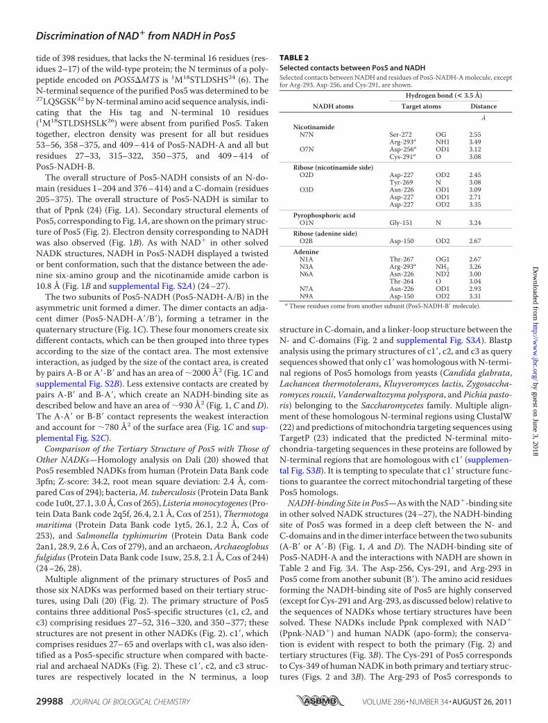
TABLE 2Selected contacts between Pos5 and NADHSelected contacts between NADH and residues of Pos5-NADH-Amolecule, exceptfor Arg-293, Asp-256, and Cys-291, are shown.
NADH atomsHydrogen bond (< 3.5 Å)
Target atoms Distance
ÅNicotinamideN7N Ser-272 OG 2.55
Arg-293a NH1 3.49O7N Asp-256a OD1 3.12
Cys-291a O 3.08Ribose (nicotinamide side)O2D Asp-227 OD2 2.45
Tyr-269 N 3.08O3D Asn-226 OD1 3.09
Asp-227 OD1 2.71Asp-227 OD2 3.35
Pyrophosphoric acidO1N Gly-151 N 3.24
Ribose (adenine side)O2B Asp-150 OD2 2.67
AdenineN1A Thr-267 OG1 2.67N3A Arg-293a NH2 3.26N6A Asn-226 ND2 3.00
Thr-264 O 3.04N7A Asn-226 OD1 2.93N9A Asp-150 OD2 3.31
a These residues come from another subunit (Pos5-NADH-B� molecule).
Discrimination of NAD� from NADH in Pos5
29988 JOURNAL OF BIOLOGICAL CHEMISTRY VOLUME 286 • NUMBER 34 • AUGUST 26, 2011
by guest on June 3, 2018http://w
ww
.jbc.org/D
ownloaded from
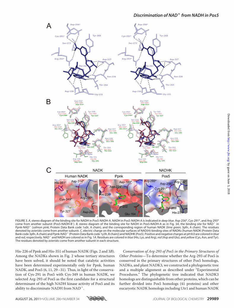
His-226 of Ppnk andHis-351 of humanNADK (Figs. 2 and 3B).Among the NADKs shown in Fig. 2 whose tertiary structureshave been solved, it should be noted that catalytic activitieshave been determined experimentally only for Ppnk, humanNADK, and Pos5 (6, 11, 29–31). Thus, in light of the conserva-tion of Cys-291 in Pos5 with Cys-349 in human NADK, weselected Arg-293 of Pos5 as the first candidate for a structuraldeterminant of the high NADH kinase activity of Pos5 and itsability to discriminate NADH from NAD�.
Conservation of Arg-293 of Pos5 in the Primary Structures ofOther Proteins—To determine whether the Arg-293 of Pos5 isconserved in the primary structures of other Pos5 homologs,NADKs, and plant NADK3, we constructed a phylogenetic treeand a multiple alignment as described under “ExperimentalProcedures.” The phylogenetic tree indicated that NADK3homologs are distinguishable fromother proteins, which can befurther divided into Pos5 homologs (41 proteins) and othereucaryotic NADKhomologs including Utr1 and humanNADK
FIGURE 3. A, stereo diagram of the binding site for NADH in Pos5-NADH-A. NADH in Pos5-NADH-A is indicated in deep blue. Asp-256*, Cys-291*, and Arg-293*come from another subunit (Pos5-NADH-B�). B, stereo diagram of the binding site for NADH in Pos5-NADH-A as in Fig. 3A, the binding site for NAD� inPpnk-NAD� (salmon pink; Protein Data Bank code 1u0r, A chain), and the corresponding region of human NADK (lime green; 3pfn, A chain). The residuesdenoted by asterisks come from another subunit. C, electric charge on the molecular surfaces of NAD(H)-binding sites of NADKs [human NADK (Protein DataBank code 3pfn, A chain) and Ppnk-NAD� (Protein Data Bank code 1y3h, B chain)] and NADHK (Pos5). Positive and negative charges at pH 8.0 are colored in blueand red, respectively. NAD� and NADH are colored as in Fig. 1A. Residues are colored in blue (His, Lys, and Arg), red (Asp and Glu), and yellow (Cys, Asn, and Tyr).The residues denoted by asterisks come from another subunit in each structure.
Discrimination of NAD� from NADH in Pos5
AUGUST 26, 2011 • VOLUME 286 • NUMBER 34 JOURNAL OF BIOLOGICAL CHEMISTRY 29989
by guest on June 3, 2018http://w
ww
.jbc.org/D
ownloaded from

(supplemental Fig. S1). The NADK3 and Pos5 homologs areputative NADHKs, whereas others are putative NADKs (sup-plemental Fig. S1). Multiple alignment demonstrated that theArg-293 of Pos5 is conserved in the primary structures of 39 of41 Pos5 homologs and also in NADK3. In contrast, in 64 of 64NADKhomologs, Arg-293 of Pos5 corresponds to aHis residue(supplemental Fig. S4). TheCys-291 of Pos5 is highly conservedin both Pos5 and NADK homologs (supplemental Fig. S4).Thus, we revealed that the Arg-293 of Pos5 is highly conservedamong the primary structures of the Pos5 homologs, where itcorresponds to a His residue that is conserved among theNADK homologs.Arg-293 of Pos5 Is the Determinant for Discrimination of
NAD� and NADH—To develop hypotheses about the role ofArg-293, we compared the electrostatic level on the molecularsurface of NADH-binding site of Pos5 (NADHK) with those ofNADKs (Ppnk and human NADK). Obviously, the surface ofthe NADH-binding site of Pos5 (NADHK) was positivelycharged at pH 8.0 (Fig. 3C) and pH 9.5 (data not shown),whereas those of NADKs were negatively charged at pH 8.0(Fig. 3C), pH 7.5 (for human NADK; data not shown), and pH6.5 (for Ppnk; data not shown). The optimum pH for NADHKactivity of Pos5 is pH9.5, whereas the optima forNADKactivityof Pos5, human NADK, and Ppnk are pH 8.0, 7.5, and 6.5,respectively (6, 29, 31). The positively charged surface of theNADH-binding site of Pos5 was attributed to Arg-293 (pKa of12). Accordingly, artificial replacement of the Arg-293 with theHis residue (pKa of 6.0) using COOT (16) resulted in a nega-tively charged surface (data not shown).To further explore whether the Arg-293 is a structural deter-
minant of Pos5 high NADH kinase activity and the ability todiscriminate NADH from NAD�, we purified a Pos5 mutant,R293H, in which Arg-293 was converted to a His residue. Weexamined the catalytic properties of R293H and compared themutantwithwild-type Pos5. For thewild-type protein, the ratioof NADHK activity to NADK activity was 8.6 (Table 3). Asexpected, the NADK activity of Pos5 R293H increased by 2.4-fold and NADHK activity deceased by 40%, resulting in thedecrease of the ratio ofNADHKactivity toNADKactivity to 2.1(Table 3). To get more detailed information, we determinedkinetic values (Table 4). Catalytic activities of Pos5 were char-acterized by high NADHK activity, i.e. by high kcat and low Kmfor NADH, and also by lowNADK activity, i.e. low kcat and highKm for NAD� (Table 4). In the case of Pos5 R293H, kcat for
NADH decreased by 52%, and Km for NAD� was also loweredby 71%, as expected. These kinetic values indicated that theArg-293 is at least one of the structural determinants of the highNADHK activity of Pos5 and for the discrimination of NADHfrom NAD�.From the viewpoint of the electrostatics of the relevant
molecular surfaces (Fig. 3C), Pos5 would hinder positivelycharged NAD� from coming close to the NADH-binding site,which is also positively charged because of Arg-293. However,this is not the case inNAD�-binding sites of NADKs (Ppnk andhuman NADK), which are negatively charged. The surfaces ofNAD�-binding sites of other NADKs whose tertiary structureshave been solved, except for NADK of A. fulgidus, are also neg-atively charged (supplemental Fig. S5), although the activities ofthese NADKs have not been reported (25, 26, 28). In the pri-mary structures of these negatively charged NADKs, the resi-dues corresponding to the Arg-293 of Pos5 are His, Ile, or Gln(Fig. 2). Thus, we speculate that these NADKs are not alsoNADHKs.With regard to theNADKofA. fulgidus, the putativeNAD(H)-binding site is positively charged because of Arg-143and Lys-126, although the residue corresponding to Pos5 Arg-293 in theA. fulgidus protein is Phe-182 (Fig. 2 and supplemen-tal Fig. S5). Thus, the possibility remains that the NADK of A.fulgidus also displays high NADHK activity.The Km for NADH of Pos5 R293H was decreased by 83%
(Table 4) relative to the wild-type protein. Based on thedecreased Km, we speculated that Arg-293 decreases the abilityof even NADH to approach the NADH-binding site and thatPos5 substituted Arg-293 for His to completely deny access toNAD� at the sacrifice of optimal affinity for NADH.Role of His-231 of Pos5—Although we revealed the signifi-
cance of Arg-293 in Pos5, the mutant R293H still exhibits highNADHK activity and low NADK activity (Table 3), indicatingthat the other determinants of specificity must exist. Uponfurther inspection of the NADH-binding site of Pos5, we foundthat His-231 also contributes to the positive-charged surface of
TABLE 3NADK and NADHK activities of purified enzymes
NADK NADHKNADHK/NADKSpecific activity Fold Specific activity Fold
units/mg units/mgPos5 0.7 1.0 6.0 1.0 8.6Pos5 R293H 1.7 2.4 3.6 0.6 2.1Pos5 H231D NDa 0.4 0.067Pos5 R293H/H231D NDa NDa
Human NADK 14 1.0 0.6 1.0 0.043Human NADK H351R NDa NDa
Human NADK H351R A330S 0.00192 0.000137 0.00266 0.00443 1.39a ND, not detected. NADK activity was not detected even after a 10-min reaction in the presence of the purified proteins (0.31 mg of Pos5 H231D, 0.42 mg of Pos5 R293HH231D, and 0.35 mg of human NADK H351R). The purified Pos5 (0.31 mg) and human NADK (0.35 mg) theoretically yield �A340 values of 13 and 250, respectively, after a10-min NADK reaction. NADHK activity was not detected even after a 10-min reaction in the presence of the purified proteins (2.4 �g of Pos5 R293H H231D and 0.18 mgof human NADK H351R). Purified Pos5 (2.4 �g) yield ed �A340 values of 0.45 and human NADK (0.18 mg) yields theoretically 7.4 after a 10-min NADHK reaction.
TABLE 4Kinetic values for NAD� and NADH of Pos5 and Pos5 R293H
kcata Km kcat/Km
NAD� NADH NAD� NADH NAD� NADH
s�1 mM
Pos5 7.4 0.9 16.1 2.8 4.5 0.6 0.190 0.04 1.6 85Pos5 R293H 6.2 1.2 7.7 1.9 1.3 0.3 0.032 0.01 4.8 241a Pos5 was taken as homotetramer. kcat was calculated using 185,138.4 Da as themolecular mass of the homotetrameric Pos5.
Discrimination of NAD� from NADH in Pos5
29990 JOURNAL OF BIOLOGICAL CHEMISTRY VOLUME 286 • NUMBER 34 • AUGUST 26, 2011
by guest on June 3, 2018http://w
ww
.jbc.org/D
ownloaded from
the NADH-binding site (Fig. 3C). In contrast, the correspond-ing positions of NADKs are acidic residues (Glu-164 of Ppnkand Asp-289 of human NADK; Fig. 3C). Notably, His-231 ofPos5 was completely conserved in all 41 Pos5 homologs,whereas the corresponding residue is Asp in 60 of the 65NADKhomologs (supplemental Fig. S4). Taken together, these obser-vations imply that His-231 is also a structural determinant ofspecificity. However, both NADK andNADHK activities of the
purified Pos5 mutants H231D and Pos5 R293H H231D weresignificantly diminished or below the detection limit (Table 3).Increasing the Ratio of NADHK Activity to NADK Activity in
Human NADK—Based on our finding that Pos5 Arg-293,which corresponds to the His-351 of human NADK, is one ofthe determinants that we sought, it was expected that wemightconfer high NADHK activity on human NADK by convertingthe His-351 of human NADK into Arg residue. However, puri-fied human NADK H351R lacked both NADK and NADHKactivities (Table 3).We hypothesized that the side chain of Pos5 Arg-293
requires other specific residues to attain a suitable conforma-tion. There are four residues (Thr-254, Thr-267, Ala-268, andSer-272) that interact with Arg-293 (Table 5 and Fig. 4A).Among these, Thr-267, Ala-268, and Ser-272 interact with theside chain of Arg-293, whereas Thr-254 contacts the N atom ofmain chain (Table 5 and Fig. 4A). Thr-267 also interacts withthe NADHmolecule (Fig. 3, A and B, and Table 2).
FIGURE 4. The residues interacting with Arg-293 of Pos5. A, stereo diagram of the residues that interact with Arg-293 in Pos5-NADH-A. NADH in Pos5-NADH-B� is shown in blue. The residues in Pos5-NADH-B� are shown. The residues denoted by asterisks come from subunit A. B, stereo diagram of the residuesshown in Fig. 4A and the corresponding superimposed residues of Ppnk-NAD� (pink, Protein Data Bank code 1u0t, A chain) and human NADK (translucentgreen, Protein Data Bank code 3pfn, A chain). The residues denoted by asterisks come from another subunit.
TABLE 5Selected contacts between Pos5 Arg-293 and Pos5Selected contacts between Arg-293 of Pos5-NADH-A molecule and residues ofPos5-NADH-B� molecule, except for Thr-254 of Pos5-NADH-A molecule.
Hydrogen bond (<3.5 Å)Target atoms Distance
ÅN Thr-254 O 2.78NH1 Thr-267 O 3.20
Ala-268 O 2.76Ser-272 OG 2.96
NH2 Thr-267 O 2.82
Discrimination of NAD� from NADH in Pos5
AUGUST 26, 2011 • VOLUME 286 • NUMBER 34 JOURNAL OF BIOLOGICAL CHEMISTRY 29991
by guest on June 3, 2018http://w
ww
.jbc.org/D
ownloaded from
Among the residues that interact with the side chain of Arg-293, Thr-267 and Ala-268 are highly conserved in both Pos5and NADK homologs, including Ppnk and human NADK (Fig.4B and supplemental Fig. S4). However, Ser-272 of Pos5 is con-served in Pos5 homologs but can be either Ser (e.g. the Ser-205of Ppnk) or Ala (e.g. the Ala-330 of human NADK) in NADKhomologs (Fig. 4B and supplemental Fig. S4). Hence, we attrib-uted the inactivation of human NADK H351R to Ala-330,which does not interact with the converted Arg-351. Accord-ingly, simultaneous conversions of both His-351 and Ala-330into Arg and Ser residues resulted in recovery of measurableNADK and NADHK activities. Furthermore, the ratio ofNADHK activity to NADK activity was markedly increasedfrom 0.043 to 1.39, although the absolute activity levels werelower than wild type (Table 3). These results indicate that theside chain of the converted Arg-351 of human H351R A330Srequires Ser-330 to assist Arg-351 in attaining a suitable sidechain conformation. Furthermore, the data imply that in Pos5,Ser-272 has a critical role in assisting Arg-293 in attaining asuitable side chain confirmation. The significance of Ser-272 isin good agreementwith the high level of conservation of this Serresidue in Pos5 homologs (supplemental Fig. S4).Significance of the Intersubunit Contact—InNADKs, the res-
idue that corresponds to Thr-254 of Pos5 is also critical in rec-ognition of substrate (e.g. E. coli NADK YfjB) and conferringstrict specificity for NAD� (3). Because the residue is derivedfrom another subunit, the intersubunit contact is important forsubstrate recognition in NADKs (3). In this study, we revealedthe significance of Arg-293 to the highNADHK activity of Pos5and its ability to discriminateNADH fromNAD�. TheArg-293also comes from another subunit (Fig. 3A), again demonstrat-ing the significance of the intersubunit contact.
REFERENCES1. Kawai, S., andMurata, K. (2008) Biosci. Biotechnol. Biochem. 72, 919–9302. Pollak, N., Dolle, C., and Ziegler, M. (2007) Biochem. J. 402, 205–2183. Mori, S., Kawai, S., Shi, F., Mikami, B., andMurata, K. (2005) J. Biol. Chem.
280, 24104–241124. Shi, F., Kawai, S., Mori, S., Kono, E., and Murata, K. (2005) FEBS J. 272,
3337–33495. Bieganowski, P., Seidle, H. F., Wojcik, M., and Brenner, C. (2006) J. Biol.
Chem. 281, 22439–224456. Miyagi, H., Kawai, S., andMurata, K. (2009) J. Biol. Chem. 284, 7553–75607. Turner, W. L., Waller, J. C., Vanderbeld, B., and Snedden, W. A. (2004)
Plant Physiol. 135, 1243–1255
8. Turner, W. L., Waller, J. C., and Snedden, W. A. (2005) Biochem. J. 385,217–223
9. Grose, J. H., Joss, L., Velick, S. F., andRoth, J. R. (2006)Proc.Natl. Acad. Sci.U.S.A. 103, 7601–7606
10. Ochiai, A., Mori, S., Kawai, S., and Murata, K. (2004) Protein Expr. Purif.36, 124–130
11. Strand, M. K., Stuart, G. R., Longley, M. J., Graziewicz, M. A., Dominick,O. C., and Copeland, W. C. (2003) Eukaryot. Cell 2, 809–820
12. Ohashi, K., Kawai, S., Koshimizu, M., and Murata, K. (2011) Mol. Cell.Biochem., in press
13. Otwinowski, Z., and Minor, W. (1997)Methods Enzymol. 276, 307–32614. Vagin, A., and Teplyakov, A. (1997) J. Appl. Crystallogr. 30, 1022–102515. Collaborative Computational Project, N. (1994) Acta Crystallogr. D Biol.
Crystallogr. 50, 760–76316. Emsley, P., and Cowtan, K. (2004)Acta Crystallogr. D Biol. Crystallogr. 60,
2126–213217. Murshudov, G. N., Vagin, A. A., and Dodson, E. J. (1997)Acta Crystallogr.
D Biol. Crystallogr. 53, 240–25518. DeLano, W. L. (2004) The PyMOL Molecular Graphics System, DeLano
Scientific LLC, San Carlos, CA19. Baker, N. A., Sept, D., Joseph, S., Holst,M. J., andMcCammon, J. A. (2001)
Proc. Natl. Acad. Sci. U.S.A. 98, 10037–1004120. Holm, L., and Rosenstrom, P. (2010) Nucleic Acids Res. 38,W545–W54921. Altschul, S. F., Madden, T. L., Schaffer, A. A., Zhang, J., Zhang, Z., Miller,
W., and Lipman, D. J. (1997) Nucleic Acids Res. 25, 3389–340222. Thompson, J. D., Higgins, D.G., andGibson, T. J. (1994)Nucleic Acids Res.
22, 4673–468023. Emanuelsson, O., Nielsen, H., Brunak, S., and vonHeijne, G. (2000) J. Mol.
Biol. 300, 1005–101624. Mori, S., Yamasaki,M.,Maruyama, Y.,Momma, K., Kawai, S., Hashimoto,
W., Mikami, B., and Murata, K. (2005) Biochem. Biophys. Res. Commun.327, 500–508
25. Liu, J., Lou, Y., Yokota, H., Adams, P. D., Kim, R., and Kim, S. H. (2005) J.Mol. Biol. 354, 289–303
26. Poncet-Montange, G., Assairi, L., Arold, S., Pochet, S., and Labesse, G.(2007) J. Biol. Chem. 282, 33925–33934
27. Petrelli, R., Sham, Y. Y., Chen, L., Felczak, K., Bennett, E., Wilson, D.,Aldrich, C., Yu, J. S., Cappellacci, L., Franchetti, P., Grifantini, M., Maz-zola, F., Di Stefano, M., Magni, G., and Pankiewicz, K. W. (2009) Bioorg.Med. Chem. 17, 5656–5664
28. Oganesyan, V., Huang, C., Adams, P. D., Jancarik, J., Yokota,H.A., Kim, R.,andKim, S. H. (2005)ActaCrystallogr. Sect. F Struct. Biol. Cryst. Commun.61, 640–646
29. Kawai, S., Mori, S., Mukai, T., Suzuki, S., Yamada, T., Hashimoto,W., andMurata, K. (2000) Biochem. Biophys. Res. Commun. 276, 57–63
30. Raffaelli, N., Finaurini, L., Mazzola, F., Pucci, L., Sorci, L., Amici, A., andMagni, G. (2004) Biochemistry 43, 7610–7617
31. Lerner, F., Niere,M., Ludwig, A., and Ziegler,M. (2001) Biochem. Biophys.Res. Commun. 288, 69–74
Discrimination of NAD� from NADH in Pos5
29992 JOURNAL OF BIOLOGICAL CHEMISTRY VOLUME 286 • NUMBER 34 • AUGUST 26, 2011
by guest on June 3, 2018http://w
ww
.jbc.org/D
ownloaded from

Kousaku MurataTakuya Ando, Kazuto Ohashi, Akihito Ochiai, Bunzo Mikami, Shigeyuki Kawai and
Mitochondrial NADH Kinase Pos5 from NADH in Yeast+Structural Determinants of Discrimination of NAD
doi: 10.1074/jbc.M111.249011 originally published online July 5, 20112011, 286:29984-29992.J. Biol. Chem.
10.1074/jbc.M111.249011Access the most updated version of this article at doi:
Alerts:
When a correction for this article is posted•
When this article is cited•
to choose from all of JBC's e-mail alertsClick here
Supplemental material:
http://www.jbc.org/content/suppl/2011/07/05/M111.249011.DC1
http://www.jbc.org/content/286/34/29984.full.html#ref-list-1
This article cites 29 references, 9 of which can be accessed free at
by guest on June 3, 2018http://w
ww
.jbc.org/D
ownloaded from