Embed Size (px)
Citation preview
Proc. Nati. Acad. Sci. USAVol. 86, pp. 3524-3528, May 1989Biochemistry
Structural significance of an internal water molecule studied bysite-directed mutagenesis of tyrosine-67 in rat cytochrome c
(protein stability/cytochrome c structure-function relationships)
T. L. LUNTZ*, A. SCHEJTERt, E. A. E. GARBER*, AND E. MARGOLIASH**Department of Biochemistry, Molecular Biology and Cell Biology, Northwestern University, Evanston, IL 60208; and tSackler Institute of MolecularMedicine, Sackler Faculty of Medicine, Tel Aviv University, Tel Aviv, Israel 69978
Contributed by E. Margoliash, January 25, 1989
ABSTRACT The tyrosine-67 to phenylalanine mutated ratcytochrome c is similar to the unmutated protein in its spectral,reduction potential, and enzymic electron-transfer properties.However, the loss of the 695-nm band, characteristic of theferric form of the normal low-spin physiologically active con-figuration, occurs 1.2 pH units higher on the alkaline side and0.7 pH unit lower on the acid side. Similarly, the heme iron-methionine-80 sulfur bond is more stable to temperature, withthe midpoint ofthe transition being 300C higher, correspondingto an increase in AH of 5 kcal/mol (1 cal = 4.184 J), partiallymitigated by an increase of 11 entropy units in AS. Urea hasonly slightly different effects on the two proteins. Thesephenomena are best explained by considering that the loss ofone of the three hydrogen-bonding side chains, tyrosine-67,asparagine-52, and threonine-78, which hold an internal watermolecule on the "left, lower front" side of the protein [Takano,T. & Dickerson, R. E. (1981) J. Mol. Biol. 153, 95-115], issufficient to prevent its inclusion in the mutant protein, leadingto a more stable structure, and, as indicated by preliminaryproton NMR two-dimensional phase-sensitive nuclear Over-hauser effect spectroscopy analyses, a reorganization of thisarea. This hypothesis predicts that elimination ofthe hydrogen-bonding ability of residue 52 or 78 would also result incytochromes c having similar properties. It is not obvious whythe space-filling structure involving the internalized watermolecule that leads to a destabilization energy of about 3kcal/mol should be subject to extreme evolutionary conserva-tion, when a more stable and apparently fully functional struc-ture is readily available.
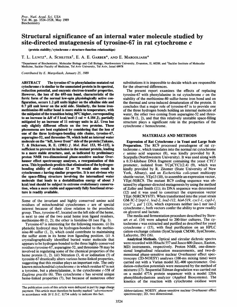
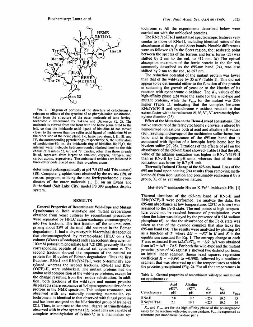
Some of the invariant and highly conserved amino acidresidues of mitochondrial cytochromes c are of specialinterest because of their close relation to the prostheticgroup. Thus, tyrosine-67, located on the left side of the heme,is next to one of the two axial heme iron ligand residues,methionine-80 (1, 2); the other is histidine-18 (see Fig. 1).
Tyrosine-67 is interesting for the following reasons. (i) Itsphenolic hydroxyl may be hydrogen-bonded to the methio-nine-80 sulfur (1, 2), which could contribute to maintainingthe sulfur atom in its heme iron liganding position. (ii) Acrystallographically identified buried water molecule thatappears to be hydrogen-bonded to the three highly conservedresidues tyrosine-67, asparagine-52, and threonine-78 may beinvolved in regulating some of the chemical properties of theheme protein (1, 2). (iii) Nitration (3, 4) or iodination (5) oftyrosirle-67 drastically alters various heme-linked properties,suggesting that this residue plays an important role. The onlyknown mitochondrial cytochrome c in which residue 67 is nota tyrosine, but a phenylalanine, is the cytochrome c-558 ofEuglena gracilis (6). This cytochrome c has several uniqueheme-linked properties (7), but because of numerous other
substitutions it is impossible to decide which are responsiblefor the observed differences.The present report examines the effects of replacing
tyrosine-67 with phenylalanine in rat cytochrome c on thestability of the methionine-80 sulfur-heme iron bond and onthe thermal and urea-induced denaturation of the protein. Itconcludes that a major role of tyrosine-67 is to provide oneof the three hydrogen bonds holding an internal molecule ofwater, the other two coming from asparagine-52 and threo-nine-78 (1, 2), and that this relatively unstable space-fillingstructure plays a significant role in the properties of thecytochrome c hemochrome.
MATERIALS AND METHODSExpression of Rat Cytochrome c in Yeast and Large Scale
Preparation. The RC9 processed pseudogene of rat cy-tochrome c, which translates into the normal rat cytochromec amino acid sequence (8), was kindly provided by R.Scarpulla (Northwestern University). It was used along witha 0.33-kilobase DNA fragment containing the yeast CYCJpromoter, isolated from YCpCYC1(2.4) (9), which waskindly provided by R. Zitomer (State University of NewYork, Albany), and an Escherichia coli-yeast multicopyshuttle vector, YEp13 (10), to assemble an expression vector,YEp(-50)RC9. The mutant RC9 coding sequence was ob-tained by oligomer-directed mutagenesis by using the methodof Zoller and Smith (11); its DNA sequence was determined(12) and it was used to construct the expression vectorYEp(-50)RC9(Y67F). When transformed into the yeast strainGM-3C-2 [trpl-1, leu2-2, leu2-112, his4-519, cycl-1, cyp3-1,(cyc7-), gal-] (13), which expresses neither iso-1 nor iso-2cytochrome c, both vectors confer the ability to grow readilyon glycerol and lactate.The media and fermentation procedure described by Stew-
art et al. (14) were adapted to 200-liter cultures. The cy-tochrome c was extracted and purified as described for yeastcytochrome c (15), with final purification on an HPLCcation-exchange column (SynChropak CM300, SynChrome,Lafayette, IN) (16).
Analytical Methods. Optical and circular dichroic spectrawere recorded with Hitachi 557 and Jasco 600 (Jasco, Easton,MD) instruments, respectively. Proton NMR, one-dimen-sional longitudinal relaxation measurements, and two-di-mensional phase-sensitive nuclear Overhauser effect spec-troscopy (2D-NOESY) analyses (100-ms mixing time) werecarried out with a Varian model XL-400 spectrometer. Oxi-dation-reduction potentials were measured by the method ofmixtures (17). Sequential Edman degradation was carried outon a model 477A protein sequencer with a model 120Aphenylthiohydantoin analyzer (Applied Biosystems). Thekinetics of the reaction with cytochrome oxidase were
Abbreviations: NOESY, phase-sensitive nuclear Overhauser effectspectroscopy; 2D, two dimensional.
3524
The publication costs of this article were defrayed in part by page chargepayment. This article must therefore be hereby marked "advertisement"in accordance with 18 U.S.C. §1734 solely to indicate this fact.
Proc. Natl. Acad. Sci. USA 86 (1989) 3525
FIG. 1. Diagram of portions of the structure of cytochrome crelevant to effects of the tyrosine-67 to phenylalanine substitution,taken from the structure of the outer molecule of tuna ferricy-tochrome c determined by Takano and Dickerson (1, 2). Themolecule is viewed from the front with the heme plane tilted to theleft, so that the imidazole axial ligand of histidine-18 has movedcloser to the viewer than the sulfur axial ligand of methionine-80 onthe other side of the heme plane. Fe, heme iron atom; I, II, III, andIV, the corresponding pyrrole rings, respectively; S, the sulfur atomof methionine-80; im, the imidazole ring of histidine-18; H20, theinternal water molecule hydrogen-bonded (dashed lines) to the sidechains of residues 52, 67, and 78. Circles, other than those alreadylisted, represent from largest to smallest, oxygen, nitrogen, andcarbon atoms, respectively. The amino acid residues are indicated inthree-letter code placed near their a-carbon atoms.
determined polarographically at pH 7.9 (25 mM Tris acetate)(18). Computer graphics were obtained by the HYDRA (19), orFRODO program, utilizing the tuna ferricytochrome c coor-dinates of the outer molecule (1, 2), on an Evans andSutherland (Salt Lake City) model PS 390 graphics displaysystem.
RESULTSGeneral Properties of Recombinant Wild-Type and Mutant
Cytochromes c. Both wild-type and mutant preparationsobtained from yeast cultures by recombinant procedureswere separated by HPLC cation-exchange chromatographyinto two fractions. The first fraction off the column, com-prising about 25% of the total, did not react in the Edmandegradation. It had a chymotryptic N-terminal decapeptidethat chromatographed, by reverse-phase HPLC on a C18column (Waters ,Bondapak) under an acetonitrile gradient in100 mM potassium phosphate (pH 7.2) (20), precisely like thecorresponding peptide of natural rat cytochrome c. Thesecond fraction showed the sequence of the natural ratprotein for 10 cycles of Edman degradation. Thus the firstfractions, RNc-I and RNc(Y67F)-I, were N-terminally ace-tylated, whereas the second fractions, RNc-II and RNc-(Y67F)-II, were unblocked. The mutant proteins had theamino acid composition of the wild-type proteins, except forthe change resulting from the residue substitution. In addi-tion, both fractions of the wild-type and mutant proteinsdisplayed a sharp resonance at 3.4 ppm representative of nineprotons in the NMR spectrum. This unique resonance, notobserved with any naturally occurring mammalian cy-tochrome c, is identical to that observed with fungal proteinsand has been assigned to the Ne-trimethyl group of lysine-72(21). Thus, in contrast to the small degree of trimethylationobserved with in vitro systems (22), yeast cells are capable ofcomplete trimethylation of lysine-72 in a mammalian cy-
tochrome c. All the experiments described below werecarried out with the unblocked proteins.The RNc(Y67F)-II mutant had spectroscopic features very
similar to those of RNc-II, including identical ratios of theabsorbance of the a, /3, and Soret bands. Notable differenceswere as follows: (i) In the Soret region, the isosbestic pointbetween the spectra of the ferrous and ferric forms (23) wasshifted by 2 nm to the red, to 412 nm. (ii) The opticalabsorption maximum of the ferric protein in the far red,commonly described as the 695-nm band (24), was alsoshifted by 2 nm to the red, to 697 nm.The reduction potential of the mutant protein was lower
than that of the wild-type by 35 mV (Table 1). This did notappear to be detrimental either to the function of the proteinin sustaining the growth of yeast or to the kinetics of itsreaction with cytochrome c oxidase. The Km values of thehigh-affinity phase (18) were the same for the wild-type andmutant proteins, while the Vmax for the mutant was 25%higher (Table 1), indicating that the complex betweenRNc(Y67F)-II and cytochrome c oxidase reacted to thatextent faster with the reductant N, N, N', N'-tetramethylphe-nylene diamine (25).
Effect of the Mutation on the Heme-Linked Ionizations. Thenative structure of the ferricytochrome c crevice is altered byheme-linked ionizations both at acid and alkaline pH values(26), resulting in cleavage of the methionine sulfur-heme ironbond and in disappearance of the 695-nm band that isassociated with ligation of a low-spin ferric heme iron bybivalent sulfur (27, 28). Titrations of the effects of pH on theabsorbance of the 695-nm band showed (Table 1) that the pKavalue of the alkaline ionization was higher in RNc(Y67F)-IIthan in RNc-II by 1.2 pH units, whereas that of the acidionization was lower by 0.7 pH unit.Thermally Induced Change of the 695-nm Band. Loss of the
695-nm band upon heating (24) results from removing meth-ionine-80 from iron ligation and presumably replacing it by agroup, X, of as yet unknown nature:
Met-S-Fe3+-imidazole-His z± X-Fe3+-imidazole-His [1]
Thermal titrations of the 695-nm band of RNc-II andRNc(Y67F)-II were performed. To analyze the data, the695-nm absorbance at low temperatures (20°C or lower) wasassigned to the Fe-S state. The end-points at high tempera-ture could not be reached because of precipitation, evenwhen the latter was delayed by the presence of 0.5 M sodiumphosphate (6), so that the absorbance of the Fe-X state wastaken as that of the cyanide complex that also lacks the695-nm band (24). The results were analyzed by plotting AGas a function of T, where AG = -RT In K and K is theequilibrium constant for Eq. 1. The entropy change at eachT was estimated from (dAG/dT)p = -AS; Al was obtainedfrom AG = AH - TAS. For both the wild-type and the mutantproteins, plots of AG against T showed two distinct portions:an initial linear segment (linear least squares regressioncoefficient R = -0.996 to -0.998), followed by a nonlinearsegment that was observed up to the temperatures at whichthe proteins precipitated (Fig. 2). For all the temperatures in
Table 1. General properties of recombinant wild-type and mutantrat cytochromes c
Acid AlkalinepK695, pKs,95 E0, Km,
Cytochrome c pH pH mV nM VmaxRNc-Il 2.8 9.5 +259 10.5 43RNc(Y67F)-II 2.1 10.7 +224 10.5 54Km and Vmax are for the high-affinity phase of the polarographic
assay for the reaction with cytochrome oxidase. Vmax is expressed aselectrons per monomeric oxidase per s.
Biochemistry: Luntz et al.
Proc. Natl. Acad. Sci. USA 86 (1989)
300 320 340 360
Temperature (OK)
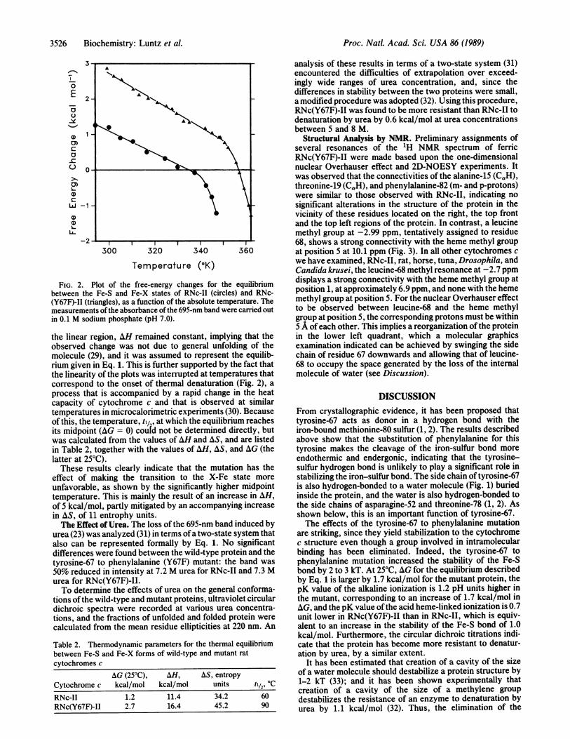
FIG. 2. Plot of the free-energy changes for the equilibriumbetween the Fe-S and Fe-X states of RNc-II (circles) and RNc-(Y67F)-II (triangles), as a function of the absolute temperature. Themeasurements ofthe absorbance of the 695-nm band were carried outin 0.1 M sodium phosphate (pH 7.0).
the linear region, AH remained constant, implying that theobserved change was not due to general unfolding of themolecule (29), and it was assumed to represent the equilib-rium given in Eq. 1. This is further supported by the fact thatthe linearity of the plots was interrupted at temperatures thatcorrespond to the onset of thermal denaturation (Fig. 2), aprocess that is accompanied by a rapid change in the heatcapacity of cytochrome c and that is observed at similartemperatures in microcalorimetric experiments (30). Becauseof this, the temperature, til, at which the equilibrium reachesits midpoint (AG = 0) coutd not be determined directly, butwas calculated from the values of AH and AS, and are listedin Table 2, together with the values of AH, AS, and AG (thelatter at 250C).These results clearly indicate that the mutation has the
effect of making the transition to the X-Fe state moreunfavorable, as shown by the significantly higher midpointtemperature. This is mainly the result of an increase in AH,of 5 kcal/mol, partly mitigated by an accompanying increasein AS, of 11 entrophy units.The Effect of Urea. The loss of the 695-nm band induced by
urea (23) was analyzed (31) in terms ofa two-state system thatalso can be represented formally by Eq. 1. No significantdifferences were found between the wild-type protein and thetyrosine-67 to phenylalanine (Y67F) mutant: the band was50% reduced in intensity at 7.2 M urea for RNc-II and 7.3 Murea for RNc(Y67F)-II.To determine the effects of urea on the general conforma-
tions ofthe wild-type and mutant proteins, ultraviolet circulardichroic spectra were recorded at various urea concentra-tions, and the fractions of unfolded and folded protein werecalculated from the mean residue ellipticities at 220 nm. An
Table 2. Thermodynamic parameters for the thermal equilibriumbetween Fe-S and Fe-X forms of wild-type and mutant ratcytochromes c
AG (250C), AH, AS, entropyCytochrome c kcal/mol kcal/mol units t1/2, 0CRNc-II 1.2 11.4 34.2 60RNc(Y67F)-II 2.7 16.4 45.2 90
analysis of these results in terms of a two-state system (31)encountered the difficulties of extrapolation over exceed-ingly wide ranges of urea concentration, and, since thedifferences in stability between the two proteins were small,a modified procedure was adopted (32). Using this procedure,RNc(Y67F)-II was found to be more resistant than RNc-II todenaturation by urea by 0.6 kcal/mol at urea concentrationsbetween 5 and 8 M.
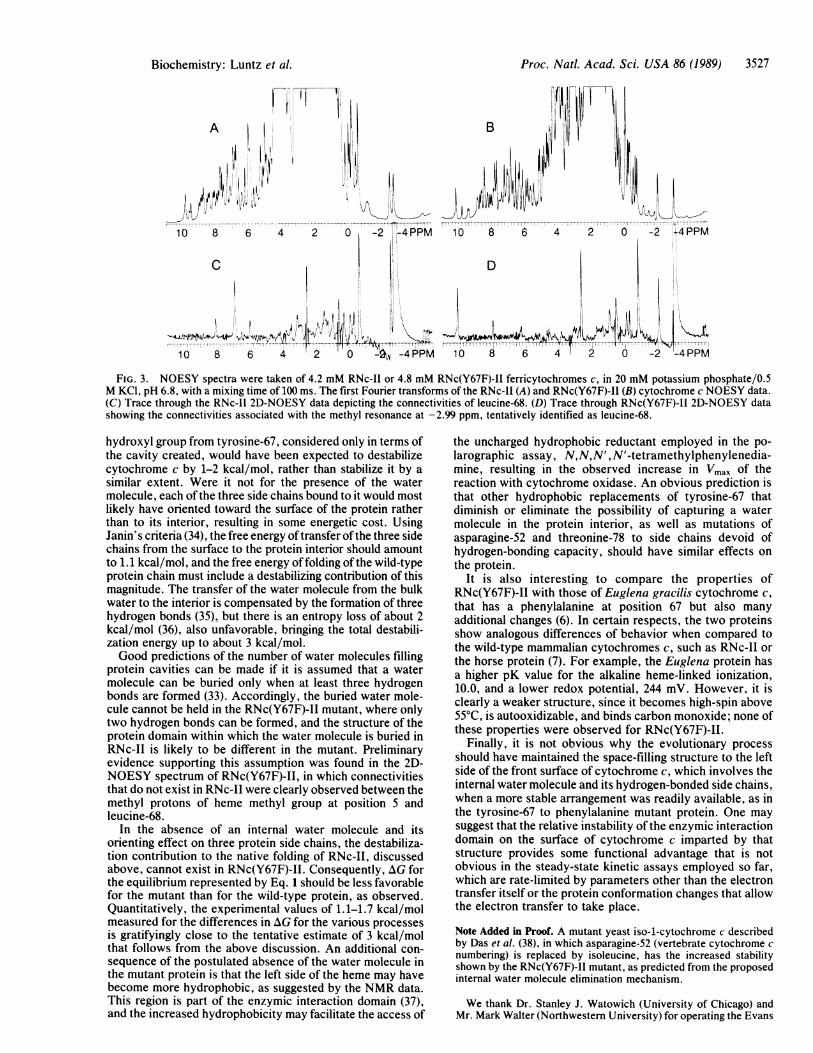
Structural Analysis by NMR. Preliminary assignments ofseveral resonances of the 1H NMR spectrum of ferricRNc(Y67F)-II were made based upon the one-dimensionalnuclear Overhauser effect and 2D-NOESY experiments. Itwas observed that the connectivities of the alanine-15 (CaH),threonine-19 (CQH), and phenylalanine-82 (m- and p-protons)were similar to those observed with RNc-II, indicating nosignificant alterations in the structure of the protein in thevicinity of these residues located on the right, the top frontand the top left regions of the protein. In contrast, a leucinemethyl group at -2.99 ppm, tentatively assigned to residue68, shows a strong connectivity with the heme methyl groupat position 5 at 10.1 ppm (Fig. 3). In all other cytochromes cwe have examined, RNc-II, rat, horse, tuna, Drosophila, andCandida krusei, the leucine-68 methyl resonance at -2.7 ppmdisplays a strong connectivity with the heme methyl group atposition 1, at approximately 6.9 ppm, and none with the hememethyl group at position 5. For the nuclear Overhauser effectto be observed between leucine-68 and the heme methylgroup at position 5, the corresponding protons must be withins A of each other. This implies a reorganization of the proteinin the lower left quadrant, which a molecular graphicsexamination indicated can be achieved by swinging the sidechain of residue 67 downwards and allowing that of leucine-68 to occupy the space generated by the loss of the internalmolecule of water (see Discussion).
DISCUSSIONFrom crystallographic evidence, it has been proposed thattyrosine-67 acts as donor in a hydrogen bond with theiron-bound methionine-80 sulfur (1, 2). The results describedabove show that the substitution of phenylalanine for thistyrosine makes the cleavage of the iron-sulfur bond moreendothermic and endergonic, indicating that the tyrosine-sulfur hydrogen bond is unlikely to play a significant role instabilizing the iron-sulfur bond. The side chain of tyrosine-67is also hydrogen-bonded to a water molecule (Fig. 1) buriedinside the protein, and the water is also hydrogen-bonded tothe side chains of asparagine-52 and threonine-78 (1, 2). Asshown below, this is an important function of tyrosine-67.The effects of the tyrosine-67 to phenylalanine mutation
are striking, since they yield stabilization to the cytochromec structure even though a group involved in intramolecularbinding has been eliminated. Indeed, the tyrosine-67 tophenylalanine mutation increased the stability of the Fe-Sbond by 2 to 3 kT. At 25°C, AG for the equilibrium describedby Eq. 1 is larger by 1.7 kcal/mol for the mutant protein, thepK value of the alkaline ionization is 1.2 pH units higher inthe mutant, corresponding to an increase of 1.7 kcal/mol inAG, and the pK value ofthe acid heme-linked ionization is 0.7unit lower in RNc(Y67F)-II than in RNc-II, which is equiv-alent to an increase in the stability of the Fe-S bond of 1.0kcal/mol. Furthermore, the circular dichroic titrations indi-cate that the protein has become more resistant to denatur-ation by urea, by a similar extent.
It has been estimated that creation of a cavity of the sizeof a water molecule should destabilize a protein structure by1-2 kT (33); and it has been shown experimentally thatcreation of a cavity of the size of a methylene groupdestabilizes the resistance of an enzyme to denaturation byurea by 1.1 kcal/mol (32). Thus, the elimination of the
3526 Biochemistry: Luntz et al.
Proc. Natl. Acad. Sci. USA 86 (1989) 3527
A~~~~~~~~~~~~j1
10 8 6 4
C
1 6, Ii 4i
10 8 6 4
2
,IIia tljil
,IM10
O 1-4 PPM 10 8 6 4 2 0 -2 ;4 PPM
0~~~~--4 PPM 10 8 6 4 2 0 -2-4 PPM
FIG. 3. NOESY spectra were taken of 4.2 mM RNc-II or 4.8 mM RNc(Y67F)-II ferricytochromes c, in 20 mM potassium phosphate/0.5M KCI, pH 6.8, with a mixing time of 100 ms. The first Fourier transforms of the RNc-II (A) and RNc(Y67F)-II (B) cytochrome c NOESY data.(C) Trace through the RNc-II 2D-NOESY data depicting the connectivities of leucine-68. (D) Trace through RNc(Y67F)-II 2D-NOESY datashowing the connectivities associated with the methyl resonance at -2.99 ppm, tentatively identified as leucine-68.
hydroxyl group from tyrosine-67, considered only in terms ofthe cavity created, would have been expected to destabilizecytochrome c by 1-2 kcal/mol, rather than stabilize it by asimilar extent. Were it not for the presence of the watermolecule, each of the three side chains bound to it would mostlikely have oriented toward the surface of the protein ratherthan to its interior, resulting in some energetic cost. UsingJanin's criteria (34), the free energy oftransfer ofthe three sidechains from the surface to the protein interior should amountto 1.1 kcal/mol, and the free energy of folding of the wild-typeprotein chain must include a destabilizing contribution of thismagnitude. The transfer of the water molecule from the bulkwater to the interior is compensated by the formation of threehydrogen bonds (35), but there is an entropy loss of about 2kcal/mol (36), also unfavorable, bringing the total destabili-zation energy up to about 3 kcal/mol.Good predictions of the number of water molecules filling
protein cavities can be made if it is assumed that a watermolecule can be buried only when at least three hydrogenbonds are formed (33). Accordingly, the buried water mole-cule cannot be held in the RNc(Y67F)-II mutant, where onlytwo hydrogen bonds can be formed, and the structure of theprotein domain within which the water molecule is buried inRNc-II is likely to be different in the mutant. Preliminaryevidence supporting this assumption was found in the 2D-NOESY spectrum of RNc(Y67F)-II, in which connectivitiesthat do not exist in RNc-II were clearly observed between themethyl protons of heme methyl group at position 5 andleucine-68.
In the absence of an internal water molecule and itsorienting effect on three protein side chains, the destabiliza-tion contribution to the native folding of RNc-II, discussedabove, cannot exist in RNc(Y67F)-II. Consequently, AG forthe equilibrium represented by Eq. 1 should be less favorablefor the mutant than for the wild-type protein, as observed.Quantitatively, the experimental values of 1.1-1.7 kcal/molmeasured for the differences in AG for the various processesis gratifyingly close to the tentative estimate of 3 kcal/molthat follows from the above discussion. An additional con-sequence of the postulated absence of the water molecule inthe mutant protein is that the left side of the heme may havebecome more hydrophobic, as suggested by the NMR data.This region is part of the enzymic interaction domain (37),and the increased hydrophobicity may facilitate the access of
the uncharged hydrophobic reductant employed in the po-larographic assay, N,N,N', N'-tetramethylphenylenedia-mine, resulting in the observed increase in Vmax of thereaction with cytochrome oxidase. An obvious prediction isthat other hydrophobic replacements of tyrosine-67 thatdiminish or eliminate the possibility of capturing a watermolecule in the protein interior, as well as mutations ofasparagine-52 and threonine-78 to side chains devoid ofhydrogen-bonding capacity, should have similar effects onthe protein.
It is also interesting to compare the properties ofRNc(Y67F)-II with those of Euglena gracilis cytochrome c,that has a phenylalanine at position 67 but also manyadditional changes (6). In certain respects, the two proteinsshow analogous differences of behavior when compared tothe wild-type mammalian cytochromes c, such as RNc-II orthe horse protein (7). For example, the Euglena protein hasa higher pK value for the alkaline heme-linked ionization,10.0, and a lower redox potential, 244 mV. However, it isclearly a weaker structure, since it becomes high-spin above55°C, is autooxidizable, and binds carbon monoxide; none ofthese properties were observed for RNc(Y67F)-II.
Finally, it is not obvious why the evolutionary processshould have maintained the space-filling structure to the leftside of the front surface of cytochrome c, which involves theinternal water molecule and its hydrogen-bonded side chains,when a more stable arrangement was readily available, as inthe tyrosine-67 to phenylalanine mutant protein. One maysuggest that the relative instability of the enzymic interactiondomain on the surface of cytochrome c imparted by thatstructure provides some functional advantage that is notobvious in the steady-state kinetic assays employed so far,which are rate-limited by parameters other than the electrontransfer itself or the protein conformation changes that allowthe electron transfer to take place.
Note Added in Proof. A mutant yeast iso-1-cytochrome c describedby Das et al. (38), in which asparagine-52 (vertebrate cytochrome c
numbering) is replaced by isoleucine, has the increased stabilityshown by the RNc(Y67F)-II mutant, as predicted from the proposedinternal water molecule elimination mechanism.
We thank Dr. Stanley J. Watowich (University of Chicago) andMr. Mark Walter (Northwestern University) for operating the Evans
B
Biochemistry: Luntz et al.
Proc. Natl. Acad. Sci. USA 86 (1989)
and Sutherland PS390 graphics display system; we also thank Mr.Kurt D. Berndt (University of Chicago) for the circular dichroicspectra. This work was supported by Grants GM 29001 and GM19121from the National Institutes of Health.
1. Takano, T. & Dickerson, R. E. (1981) J. Mol. Biol. 153, 79-94.2. Takano, T. & Dickerson, R. E. (1981)J. Mol. Biol. 153,95-115.3. Sokolovsky, M., Aviram, I. & Schejter, A. (1970) Biochemistry
9, 5113-5118.4. Schejter, A., Aviram, I. & Sokolovsky, M. (1970) Biochemistry
9, 5118-5122.5. McGowan, E. B. & Stellwagen, E. (1970) Biochemistry 9,
3047-3052.6. Pettigrew, G. W., Leaver, J. L., Meyer, T. E. & Ryle, A. P.
(1975) Biochem. J. 147, 291-302.7. Pettigrew, G. W., Aviram, I. & Schejter, A. (1975) Biochem. J.
149, 155-162.8. Scarpulla, R. (1984) Mol. Cell. Biol. 4, 2279-2288.9. Lowry, C. V., Weiss, J. L., Walthall, D. A. & Zitomer, R. S.
(1983) Proc. Natl. Acad. Sci. USA 80, 151-155.10. Broach, J., Strathern, J. & Hicks, J. (1979) Gene 8, 121-133.11. Zoller, M. & Smith, M. (1984) DNA 3, 479-488.12. Sanger, F., Nicklen, S. & Coulson, A. R. (1977) Proc. Natl.
Acad. Sci. USA 74, 5463-5467.13. Faye, G., Leung, D. W., Tatchell, K., Hall, B. D. & Smith, M.
(1981) Proc. Natl. Acad. Sci. USA 78, 2258-2262.14. Stewart, J. W., Sherman, F., Shipman, N. & Jackson, M.
(1971) J. Biol. Chem. 246, 7429-7445.15. Sherman, F., Stewart, J. W., Parker, J. H., Inhaber, E.,
Shipman, N., Putterman, G. J., Gardisky, R. L. & Margoliash,E. (1968) J. Biol. Chem. 243, 5446-5456.
16. Theodorakis, J. L., Armes, L. G. & Margoliash, E. (1988) inAdvances in Membrane Biochemistry and Bioenergetics, eds.Kim, C. H., Tedeschi, H., Diwan, J. J. & Salerno, J. C.(Plenum, New York), pp. 185-192.
17. Margalit, R. & Schejter, A. (1973) Eur. J. Biochem. 32, 492-499.18. Brautigan, D. L., Ferguson-Miller, S. & Margoliash, E. (1978)
Methods Enzymol. 53, 371-386.
19. Hubbard, R. E. (1986) in Computer Graphics and MolecularModeling, eds. Fletterick, R. & Zoller, H. (Cold Spring HarborLab., Cold Spring Harbor, NY), pp. 9-11.
20. Vensel, W. H., Fujita, V. S., Tarr, G. E., Margoliash, E. &Kayser, H. (1983) J. Chromatogr. 266, 491-500.
21. Moore, G. R., Ratcliffe, R. G. & Williams, R. J. P. (1983)Essays Biochem. 19, 142-195.
22. Durban, E., Nochumson, S., Kim, S. & Paik, W. K. (1978) J.Biol. Chem. 253, 1427-1435.
23. Margoliash, E. & Frohwirth, N. (1959) Biochem. J. 71, 559-570.
24. Schejter, A. & George, P. (1965) Biochemistry 3, 1045-1049.25. Garber, E. A. E., Luntz, T. L. & Margoliash, E. (1988) in
Oxidases and Related Redox Systems, ed. Mason, H. S. (Liss,New York), pp. 749-769.
26. Theorell, H. & Akesson, A. (1941) J. Am. Chem. Soc. 63,1804-1821.
27. Shechter, E. & Saludjian, P. (1967) Biopolymers 5, 788-790.28. Schejter, A. & Plotkin, B. (1988) Biochem. J. 255, 353-356.29. Creighton, T. E. (1983) Proteins (Freeman, New York).30. Privalov, P. L. & Khechinashvili, N. N. (1974) J. Mol. Biol. 86,
665-684.31. Knapp, J. A. & Pace, C. N. (1974) Biochemistry 13,1289-1296.32. Kellis, J. T., Nyberg, K., Sali, D. & Fersht, A. R. (1988)
Nature (London) 333, 784-786.33. Rashin, A. A., lofin, M. & Honig, B. (1986) Biochemistry 25,
3619-3625.34. Janin, J. (1979) Nature (London) 277, 491-492.35. Fersht, A. R., Leatherbarrow, R. J. & Wells, T. N. C. (1987)
Biochemistry 26, 8031-8037.36. Ptitsyn, 0. B. (1972) Pure Appl. Chem. 31, 227-244.37. Margoliash, E. & Bosshard, H. (1983) Trends Biochem. Sci. 93,
316-320.38. Das, G., Hickey, D. R., McLendon, D., McLendon, G. &
Sherman, F. (1989) Proc. Natl. Acad. Sci. USA 86, 496-499.
3528 Biochemistry: Luntz et al.