Embed Size (px)
Citation preview
Cell Reports
Article
Structural Model for Tubulin Recognitionand Deformation by Kinesin-13Microtubule DepolymerasesAna B. Asenjo,1 Chandrima Chatterjee,1 Dongyan Tan,1 Vania DePaoli,1 William J. Rice,2 Ruben Diaz-Avalos,2
Mariena Silvestry,2 and Hernando Sosa1,*1Department of Physiology and Biophysics, Albert Einstein College of Medicine, Bronx, NY 10461, USA2New York Structural Biology Center, New York, NY 10027, USA
*Correspondence: [email protected]
http://dx.doi.org/10.1016/j.celrep.2013.01.030
SUMMARY
To elucidate the structural basis of the mechanism ofmicrotubule depolymerization by kinesin-13s, weanalyzed complexes of tubulin and the Drosophilamelanogaster kinesin-13 KLP10A by electronmicroscopy (EM) and fluorescence polarizationmicroscopy. We report a nanometer-resolution(1.1 nm) cryo-EM three-dimensional structure of theKLP10A head domain (KLP10AHD) bound to curvedtubulin. We found that binding of KLP10AHD inducesa distinct tubulin configuration with displacement(shear) between tubulin subunits in addition to curva-ture. In this configuration, the kinesin-binding sitediffers from that in straight tubulin, providing anexplanation for the distinct interaction modes ofkinesin-13s with the microtubule lattice or its ends.The KLP10AHD-tubulin interface comprises threeareas of interaction, suggesting a crossbow-typetubulin-bending mechanism. These areas includethe kinesin-13 family conserved KVD residues, andas predicted from the crossbow model, mutatingthese residues changes the orientation and mobilityof KLP10AHDs interacting with the microtubule.
INTRODUCTION
Kinesins are a superfamily of microtubule (MT)-associated
ATPases characterized by the presence of a highly conserved
catalytic or ‘‘motor’’ head domain (HD,�340 residues long) con-
taining MT- and ATP-binding sites (Goldstein and Philp, 1999).
Although most kinesins work as motile molecular motors gener-
ating force and movement along MTs, some kinesins act as MT
depolymerases. The latter group includes the kinesin-13 family
members, which were the first to be clearly recognized as non-
motile kinesinswithMTdepolymerase activity (Desai et al., 1999).
Kinesin-13s have been reported to play important roles in
various cellular processes, including mitosis (Manning et al.,
2007; Rogers et al., 2004), cytokinesis (Rankin and Wordeman,
2010), axonal branching (Homma et al., 2003), and ciliogenesis
(Kobayashi et al., 2011). Kinesin13s have also attracted consid-
erable attention as potential targets for anticancer therapy due to
their effects on MT dynamics and their involvement in mitosis
(Sanhaji et al., 2011).
Although much work has been devoted to elucidating the
mechanism of action of kinesin motors, it is still not fully clear
why some are motile and others are MT depolymerases. The
molecular mechanism of kinesin-13 MT depolymerization likely
involves the stabilization of a curved tubulin structure, which is
incompatible with the formation of lateral interprotofilament
contacts in the MT lattice (Desai et al., 1999). However, how
kinesin-13s induce tubulin curvature is not well understood.
Catalytic activity is coupled to ATP hydrolysis (Hunter et al.,
2003) as in other kinesins, but the interaction of kinesin-13s
with MTs is very different from that of motile kinesins. Motile
kinesins alternate between strong and weak interactions with
tubulin as they translocate along the MT lattice. On the other
hand, kinesin-13s bind to the MT lattice weakly and undergo
unbiased one-dimensional (1D) diffusion until they reach the
end of the MT, where they induce depolymerization (Helenius
et al., 2006). How kinesin-13 distinguishes tubulin in the MT
lattice or at the MT ends is unknown.
As with motile kinesins, many key kinesin-13 functions,
such as ATP hydrolysis and tubulin binding, are located in the
kinesin-13 HD, but additional areas outside the HD are also func-
tionally important (Hertzer et al., 2006; Maney et al., 2001). A
positively charged, �60-residue-long sequence N-terminal to
the HD, known as the neck, promotes MT binding (Cooper
et al., 2010) and is critical for normal in vivo MT depolymerization
(Ovechkina et al., 2002). The neck may also increase MT depo-
lymerization efficiency by inducing the two HDs of a full-length
kinesin-13 dimer to bind adjacent protofilaments (Mulder et al.,
2009). However, the kinesin-13 HD alone has the ability to induce
tubulin curvature and depolymerize MTs (Moores et al., 2002;
Tan et al., 2008). Thus, elucidating how the kinesin-13 HD in
particular binds to and bends tubulin is crucial for understanding
the mechanism of kinesin-13-mediated MT depolymerization.
Previous lower-resolution electron microscopy (EM) studies
have indicated that the kinesin-13 HD binds to curved tubulin
in a configuration that is similar to other kinesin HDs bound to
straight tubulin in the MT lattice (Tan et al., 2008). This similarity
makes it difficult to explain how the kinesin-13 HD binds to
and bends tubulin. To address this problem, we analyzed the
structure of several complexes of tubulin and the Drosophila
Cell Reports 3, 759–768, March 28, 2013 ª2013 The Authors 759
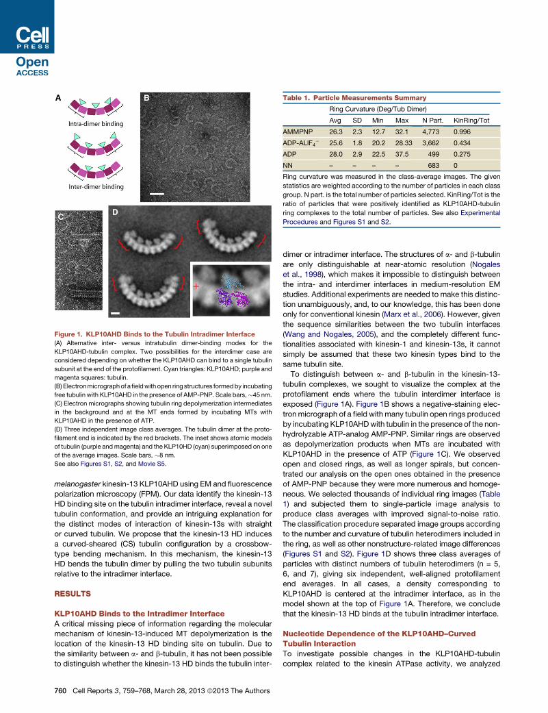
Figure 1. KLP10AHD Binds to the Tubulin Intradimer Interface
(A) Alternative inter- versus intratubulin dimer-binding modes for the
KLP10AHD-tubulin complex. Two possibilities for the interdimer case are
considered depending on whether the KLP10AHD can bind to a single tubulin
subunit at the end of the protofilament. Cyan triangles: KLP10AHD; purple and
magenta squares: tubulin.
(B) Electronmicrographof afieldwith open ring structures formedby incubating
free tubulin with KLP10AHD in the presence of AMP-PNP. Scale bars,�45 nm.
(C) Electron micrographs showing tubulin ring depolymerization intermediates
in the background and at the MT ends formed by incubating MTs with
KLP10AHD in the presence of ATP.
(D) Three independent image class averages. The tubulin dimer at the proto-
filament end is indicated by the red brackets. The inset shows atomic models
of tubulin (purple andmagenta) and the KLP10HD (cyan) superimposed on one
of the average images. Scale bars, �8 nm.
See also Figures S1, S2, and Movie S5.
Table 1. Particle Measurements Summary
Ring Curvature (Deg/Tub Dimer)
Avg SD Min Max N Part. KinRing/Tot
AMMPNP 26.3 2.3 12.7 32.1 4,773 0.996
ADP-ALlF4� 25.6 1.8 20.2 28.33 3,662 0.434
ADP 28.0 2.9 22.5 37.5 499 0.275
NN – – – – 683 0
Ring curvature was measured in the class-average images. The given
statistics are weighted according to the number of particles in each class
group. N part. is the total number of particles selected. KinRing/Tot is the
ratio of particles that were positively identified as KLP10AHD-tubulin
ring complexes to the total number of particles. See also Experimental
Procedures and Figures S1 and S2.
melanogaster kinesin-13 KLP10AHD using EM and fluorescence
polarization microscopy (FPM). Our data identify the kinesin-13
HD binding site on the tubulin intradimer interface, reveal a novel
tubulin conformation, and provide an intriguing explanation for
the distinct modes of interaction of kinesin-13s with straight
or curved tubulin. We propose that the kinesin-13 HD induces
a curved-sheared (CS) tubulin configuration by a crossbow-
type bending mechanism. In this mechanism, the kinesin-13
HD bends the tubulin dimer by pulling the two tubulin subunits
relative to the intradimer interface.
RESULTS
KLP10AHD Binds to the Intradimer InterfaceA critical missing piece of information regarding the molecular
mechanism of kinesin-13-induced MT depolymerization is the
location of the kinesin-13 HD binding site on tubulin. Due to
the similarity between a- and b-tubulin, it has not been possible
to distinguish whether the kinesin-13 HD binds the tubulin inter-
760 Cell Reports 3, 759–768, March 28, 2013 ª2013 The Authors
dimer or intradimer interface. The structures of a- and b-tubulin
are only distinguishable at near-atomic resolution (Nogales
et al., 1998), which makes it impossible to distinguish between
the intra- and interdimer interfaces in medium-resolution EM
studies. Additional experiments are needed to make this distinc-
tion unambiguously, and, to our knowledge, this has been done
only for conventional kinesin (Marx et al., 2006). However, given
the sequence similarities between the two tubulin interfaces
(Wang and Nogales, 2005), and the completely different func-
tionalities associated with kinesin-1 and kinesin-13s, it cannot
simply be assumed that these two kinesin types bind to the
same tubulin site.
To distinguish between a- and b-tubulin in the kinesin-13-
tubulin complexes, we sought to visualize the complex at the
protofilament ends where the tubulin interdimer interface is
exposed (Figure 1A). Figure 1B shows a negative-staining elec-
tronmicrograph of a field with many tubulin open rings produced
by incubating KLP10AHDwith tubulin in the presence of the non-
hydrolyzable ATP-analog AMP-PNP. Similar rings are observed
as depolymerization products when MTs are incubated with
KLP10AHD in the presence of ATP (Figure 1C). We observed
open and closed rings, as well as longer spirals, but concen-
trated our analysis on the open ones obtained in the presence
of AMP-PNP because they were more numerous and homoge-
neous. We selected thousands of individual ring images (Table
1) and subjected them to single-particle image analysis to
produce class averages with improved signal-to-noise ratio.
The classification procedure separated image groups according
to the number and curvature of tubulin heterodimers included in
the ring, as well as other nonstructure-related image differences
(Figures S1 and S2). Figure 1D shows three class averages of
particles with distinct numbers of tubulin heterodimers (n = 5,
6, and 7), giving six independent, well-aligned protofilament
end averages. In all cases, a density corresponding to
KLP10AHD is centered at the intradimer interface, as in the
model shown at the top of Figure 1A. Therefore, we conclude
that the kinesin-13 HD binds at the tubulin intradimer interface.
Nucleotide Dependence of the KLP10AHD–CurvedTubulin InteractionTo investigate possible changes in the KLP10AHD-tubulin
complex related to the kinesin ATPase activity, we analyzed
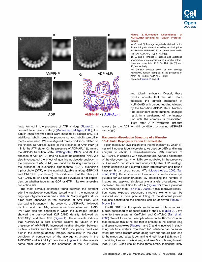
Figure 2. Nucleotide Dependence of
KLP10AHD Binding to Tubulin Protofila-
ments
(A, C, and E) Average negatively stained proto-
filament ring structures formed by incubating free
tubulin with KLP10AHD in the presence of AMP-
PNP (A), ADP-AlF4� (C), or ADP (E).
(B, D, and F) Images of aligned and averaged
asymmetric units (consisting of a tubulin hetero-
dimer and associated KLP10AHD) in (A), (C), and
(E), respectively.
(G) Density contour plots of the average
KLP10AHD-tubulin complex in the presence of
AMP-PNP (red) or ADP-AlF4� (blue).
See also Figures S1 and S2.
rings formed in the presence of ATP analogs (Figure 2). In
contrast to a previous study (Moores and Milligan, 2008), the
tubulin rings analyzed here were induced by kinesin only. No
additional tubulin drugs to promote curved tubulin protofila-
ments were used. We investigated three conditions related to
the kinesin-13 ATPase cycle: (1) the presence of AMP-PNP (to
mimic the ATP state), (2) the presence of ADP-AlF4� (to mimic
the ADP-Pi transition state; Wittinghofer, 1997), and (3) the
absence of ATP or ADP (the no-nucleotide condition [NN]). We
also investigated the effect of guanine nucleotide analogs. In
the presence of AMP-PNP, we found similar ring structures in
the presence of guanosine diphosphate (GDP), guanosine
triphosphate (GTP), or the nonhydrolyzable analogs GTP-Y-S
and GMPCPP (not shown). This indicates that the ability of
KLP10AHD to bind and induce tubulin curvature is not depen-
dent on whether tubulin has GDP or GTP in its exchangeable
nucleotide site.
The most obvious difference found between the different
adenine nucleotide conditions tested was in the number of
ring-type oligomers observed (Table 1). Many ring-like struc-
tures were observed in the presence of AMP-PNP, with
decreasing frequency in the presence of ADP-AlF4� followed
by ADP and then NN, where none were observed. AMP-
PNP was also the condition in which the averaged images
showed the best-defined KLP10AHD density, followed by
ADP-AlF4� and then ADP (Figure 2). These results indicate
that KLP10AHD is best oriented relative to tubulin in the
presence of AMP-PNP. Increased relative disorder between
protein subunits and less KLP10AHD occupancy produced
blur in the average density images, particularly in the ADP
condition. A comparison of the average structures in the
AMP-PNP and ADP-AlF4� conditions (Figure 2G) also reveals
some small changes in the orientation of the KLP10AHD
Cell Reports 3, 759–76
and tubulin subunits. Overall, these
results indicate that the ATP state
stabilizes the tightest interaction of
KLP10AHD with curved tubulin, followed
by the transition ADP-Pi state. Nucleo-
tide-dependent conformational changes
result in a weakening of the interac-
tion until the complex is dissociated,
likely after ATP hydrolysis product
release (in the ADP or NN condition, or during ADP/ATP
exchange).
Nanometer-Resolution Structure of a Kinesin-13-Tubulin Depolymerization IntermediateTo gain molecular-level insight into the mechanism by which ki-
nesin-13 induces tubulin curvature, we used cryo-EM and image
analysis to obtain a three-dimensional (3D) structure of
KLP10AHD in complex with curved tubulin. We took advantage
of the discovery that when MTs are incubated in the presence
of kinesin-13 constructs and nonhydrolyzable ATP analogs,
spirals consisting of a curved tubulin protofilament and bound
kinesin-13s can wrap around MTs (Moores et al., 2006; Tan
et al., 2006). These spirals can form very uniform helical arrays
suitable for 3D reconstruction. By increasing the number of
images and applying single-particle analysis procedures, we
increased the resolution to �11 A (Figure S3) from a previous
28 A resolution map (Tan et al., 2008). At this improved resolu-
tion, some exposed secondary structure elements can be
resolved and a more precise fitting of the different protein
subunits constituting the complex can be achieved (Figure 3;
Movie S1).
The KLP10AHD in the spirals has two areas of interaction with
tubulin positioned at opposite sides of the HD (Figure 3C). We
refer to these areas as Kin-Tub-1 and Kin-Tub-2 (Tan et al.,
2008). We will focus our description here on the Kin-Tub-1 inter-
face because this is the one that is present in the isolated ring
and spiral complexes (Figures 1 and 2) and is involved in stabi-
lizing tubulin curvature. The Kin-Tub-1 interface can be sepa-
rated into three distinct areas going from the tubulin plus end
to the minus end: area 1, containing kinesin loop 8 (L8); area 2,
containing kinesin a-helix 4 (a4); and area 3, containing kinesin
loop 2 (L2). Close-ups of these three areas, indicating likely
8, March 28, 2013 ª2013 The Authors 761
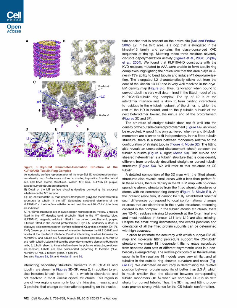
Figure 3. Cryo-EM Nanometer-Resolution Structure of the
KLP10AHD-Tubulin Ring Complex
(A) Isodensity surface representation of the cryo-EM 3D reconstruction elec-
tron density map. Surfaces are colored according to position from the helical
axis and fitted atomic structures. Yellow, MT; blue, KLP10AHD; purple,
outside curved tubulin protofilament.
(B) Detail of the MT surface showing densities contouring the exposed
a-helices on the MT surface.
(C) End-on view of the 3D map density (transparent gray) and the fitted atomic
structures of tubulin in the MT. Secondary structural elements of the
KLP10AHD at the interface with the curved protofilament (Kin-Tub-1 interface)
are indicated.
(C–F) Atomic structures are shown in ribbon representation. Yellow, a-tubulin
fitted in the MT density; gold, b-tubulin fitted in the MT density; blue,
KLP10AHD; magenta, a-tubulin fitted in the curved protofilament; purple,
b-tubulin fitted in the curved protofilament. Cryo-EM isodensity surface is
displayed as a semitransparent surface in (B) and (C), and as a mesh in (D)–(F).
(D–F) Close-up of the three areas of interaction between the KLP10AHD and
tubulin at the Kin-Tub-1 interface. Putative interacting residues between the
KLP10AHD and tubulin (<5 A separation) are colored dark blue in KLP10HD
and red in tubulin. Labels indicate the secondary structure elements (H, tubulin
helix; S, tubulin sheet; a, kinesin helix) where the putative interacting residues
are located. Labels are also color-coded according to location (blue,
KLP10AHD; red, a-tubulin; green, b-tubulin).
See also Figures S3, S5, and Movies S1 and S6.
interacting secondary structure elements in KLP10AHD and
tubulin, are shown in Figures 3D–3F. Area 2, in addition to a4,
also includes kinesin loop 11 (L11), which is disordered and
not resolved in most kinesin crystal structures. This region is
one of two regions commonly found in kinesins, myosins, and
G-proteins that change conformation depending on the nucleo-
762 Cell Reports 3, 759–768, March 28, 2013 ª2013 The Authors
tide species that is present on the active site (Kull and Endow,
2002). L2, in the third area, is a loop that is elongated in the
kinesin-13 family and contains the class-conserved KVD
sequence at the tip. Mutating these three residues severely
disrupts depolymerization activity (Ogawa et al., 2004; Shipley
et al., 2004). We found that KLP10AHD constructs with the
KVD residues mutated to AAA were unable to form tubulin ring
complexes, highlighting the critical role that this area plays in ki-
nesin-13’s ability to bend tubulin and induce MT depolymeriza-
tion. The elongated L2 characteristically sticks out from the
core of the kinesin-13 HD and is very well resolved in the cryo-
EM density map (Figure 3F). Thus, its location when bound to
curved tubulin is very well determined in the fitted model of the
KLP10AHD-tubulin ring complex. The tip of L2 is at the
interdimer interface and is likely to form binding interactions
to residues in the a-tubulin subunit of the dimer, to which the
rest of the HD is bound, and to the b-tubulin subunit of the
next heterodimer toward the minus end of the protofilament
(Figures 3C and 3F).
The structure of straight tubulin does not fit well into the
density of the outside curved protofilament (Figure 4A), as would
be expected. A good fit is only achieved when a- and b-tubulin
monomers are allowed to fit independently. In this fitted tubulin
structure, there is a bend between monomers relative to the
configuration of straight tubulin (Figure 4; Movie S2). The fitting
also reveals an unexpected displacement (shear) between the
tubulin subunits (Figure 4, right; Movie S3). This curved and
sheared heterodimer is a tubulin structure that is considerably
different from previously described straight or curved tubulin
structures (Figure S4). We will refer to this structure as CS
tubulin.
A detailed comparison of the 3D map with the fitted atomic
structures also reveals small areas with a less than perfect fit.
In these areas, there is density in the 3D EM map with no corre-
sponding atomic structures from the fitted atomic structures or
atoms with no corresponding density (Figure 3; Movie S1). At
the present resolution, it cannot be fully established whether
such differences correspond to local conformational changes
or areas that are disordered in the crystal structures becoming
ordered in the complex. In the tubulin atomic structures, there
are 12–16 residues missing (disordered) at the C-terminal end
and most residues in kinesin L11 and L12 are also missing.
Despite the small fitting mismatches, the overall positions and
orientation of all the fitted protein subunits can be determined
with high accuracy.
In order to estimate the accuracy with which our cryo-EM 3D
map and molecular fitting procedure support the CS-tubulin
structure, we made 18 independent fits to maps calculated
from separate data sets or different asymmetric units in a non-
helically averaged map. The relative positions of all the individual
subunits in the resulting 18 models were very similar, and all
tubulins in the outside ring showed curvature and shear (Fig-
ure S5). We estimated an accuracy in determining the relative
position between protein subunits of better than 2.3 A, which
is much smaller than the distance between corresponding
tubulin monomers (16 A) when we compare CS-tubulin with
straight or curved tubulin. Thus, the 3D map and fitting proce-
dure provide strong evidence for the CS-tubulin conformation.
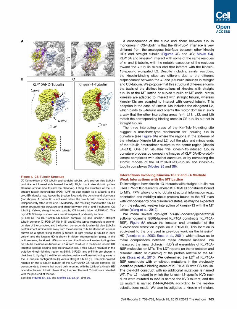
Figure 4. CS-Tubulin Structure
(A) Comparison of CS tubulin and straight tubulin. Left: end-on view (tubulin
protofilament luminal side toward the left). Right: back view (tubulin proto-
filament luminal side toward the observer). Fitting the structure of the a-b
straight tubulin heterodimer (PDB: 1JFF) to best match its a-subunit to the
cryo-EM density map leaves the b-subunit outside the density and vice versa
(not shown). A better fit is achieved when the two tubulin monomers are
independently fitted in the cryo-EM density. The resulting model of the tubulin
dimer structure has curvature and shear between the a- and b-subunits (CS
tubulin). Yellow, straight tubulin; purple, CS tubulin; blue, KLP10AHD. The
cryo-EM 3D map is shown as a semitransparent isodensity surface.
(B and C) The KLP10AHD-CS-tubulin complex (B) and kinesin-1-straight
tubulin complex (C; PDB: 2P4N). In (B) and (C) the top corresponds to an end-
on view of the complex, and the bottom corresponds to a frontal view (tubulin
protofilament luminal side away from the observer). Tubulin atomic structure is
shown as a space-filling model (a-tubulin in light yellow; b-tubulin in dark
yellow) and the kinesin HD is shown in ribbon representation (blue). In the
bottom views, the kinesin HD structure is omitted to show kinesin-binding sites
on tubulin. Residues in tubulin at %5 A from residues in the bound kinesin HD
(putative kinesin-binding site) are shown in red. Three tubulin residues in the
putative kinesin-binding region (a-E415, b-P263, and b-T419) are shown in
dark blue to highlight the different relative positions of kinesin-binding areas in
the CS-tubulin configuration (B) versus straight tubulin (C). The pink-colored
residue on the b-tubulin subunit of the KLP10AHD-CS-tubulin complex (B)
corresponds to the one that would be interacting with the L2 tip of a kinesin HD
bound to the next tubulin dimer along the protofilament. Tubulins are oriented
with the plus end at the top.
See also Figures S4, S5, and Movies S2, S3, S4, and S6.
A consequence of the curve and shear between tubulin
monomers in CS-tubulin is that the Kin-Tub-1 interface is very
different from the analogous interface between other kinesin
HDs and straight tubulin (Figures 4B and 4C; Movie S4).
KLP10A and kinesin-1 interact with some of the same residues
of a- and b-tubulin, with the notable exception of the residues
toward the a-tubulin minus end that interact with the kinesin-
13-specific elongated L2. Despite including similar residues,
the kinesin-binding sites are different due to the different
displacement between the a- and b-tubulin subunits in straight
and CS-tubulin. We propose that this structural difference forms
the basis of the distinct interactions of kinesins with straight
tubulin at the MT lattice or curved tubulin at MT ends. Motile
kinesins are adapted to interact with straight tubulin, whereas
kinesin-13s are adapted to interact with curved tubulin. This
adaption in the case of kinesin-13s includes the elongated L2,
which binds to a-tubulin and orients the motor domain in such
a way that the other interacting areas (a-4, L11, L12, and L8)
match the corresponding binding areas in CS-tubulin but not in
straight tubulin.
The three interacting areas of the Kin-Tub-1-binding site
suggest a crossbow-type mechanism for inducing tubulin
curvature (see Figure 6A) where the regions at the extreme of
the interface (kinesin L8 and L2) pull the plus and minus ends
of the tubulin heterodimer relative to the center region (kinesin
a4-L11). One can visualize this kinesin-13-induced tubulin
curvature process by comparing images of KLP10AHD-protofi-
lament complexes with distinct curvature, or by comparing the
atomic models of the KLP10AHD-CS-tubulin and kinesin-1-
tubulin complexes (Movies S5 and S6).
Interactions Involving Kinesin-13 L2 and a4 MediateWeak Interactions with the MT LatticeTo investigate how kinesin-13 interacts with straight tubulin, we
used FPM of fluorescently labeled KLP10AHD constructs bound
to MTs. FPM allows one to obtain structural information (e.g.,
orientation and mobility) about proteins bound to the MT even
with low occupancy or in disordered states, as may be expected
from the relatively weaker interaction of kinesin-13 with the MT
lattice (Wang et al., 2012).
We made several cys-light bis-((N-iodoacetyl)piperazinyl)
sulfonerhodamine (BSR)-labeled KLP10A constructs (KLP10A-
BSR). Figure 5A shows the location and orientation of the
fluorescence transition dipole on KLP10AHD. This location is
equivalent to the one used in previous work on the kinesin-1
HD (Asenjo et al., 2003; Sosa et al., 2001), which allows us to
make comparisons between these different kinesins. We
measured the linear dichroism (LD0) of ensembles of KLP10A-
BSR molecules on MTs. The LD0 reports on the orientation and
disorder (static or dynamic) of the probes relative to the MT
axis (Sosa et al., 2010). We determined the LD0 of KLP10A-
BSR constructs with or without mutations in the previously
identified putative binding areas of KLP10AHD with CS tubulin.
The cys-light construct with no additional mutations is named
WT. The L2 mutant in which the kinesin-13-specific KVD resi-
dues were mutated to AAA is named the KVD mutant, and the
L8 mutant is named D444A,K446A according to the residue
substitutions made. We also investigated a kinesin a4 mutant
Cell Reports 3, 759–768, March 28, 2013 ª2013 The Authors 763
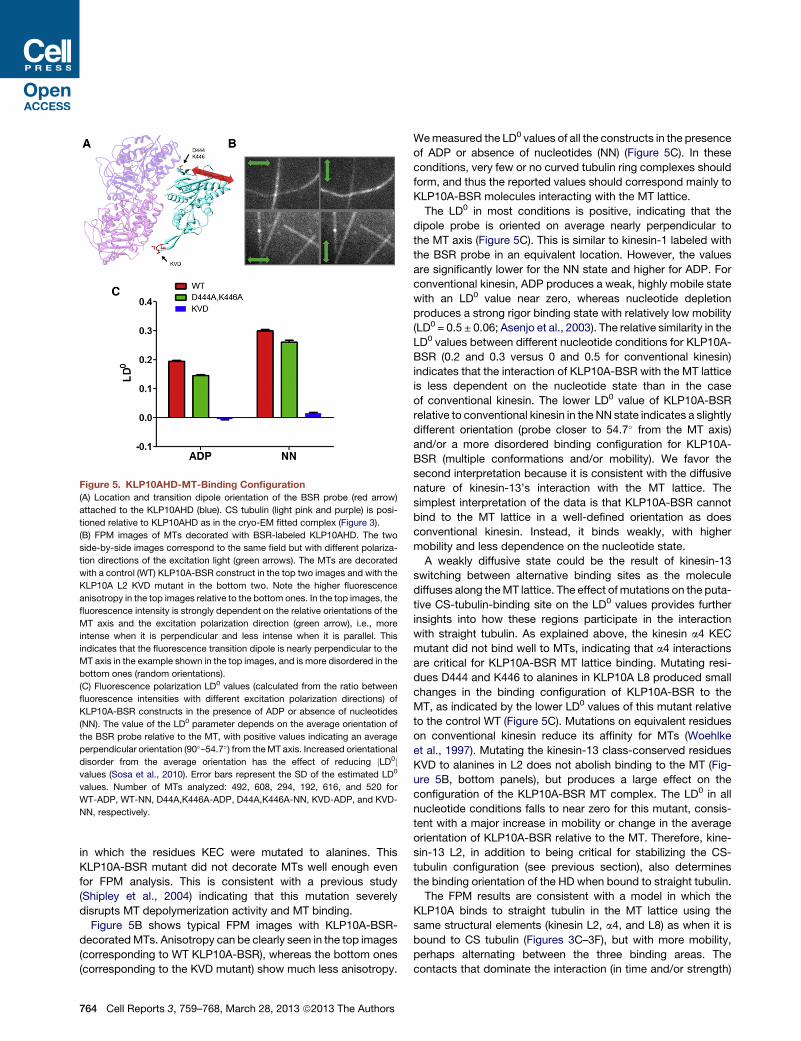
Figure 5. KLP10AHD-MT-Binding Configuration
(A) Location and transition dipole orientation of the BSR probe (red arrow)
attached to the KLP10AHD (blue). CS tubulin (light pink and purple) is posi-
tioned relative to KLP10AHD as in the cryo-EM fitted complex (Figure 3).
(B) FPM images of MTs decorated with BSR-labeled KLP10AHD. The two
side-by-side images correspond to the same field but with different polariza-
tion directions of the excitation light (green arrows). The MTs are decorated
with a control (WT) KLP10A-BSR construct in the top two images and with the
KLP10A L2 KVD mutant in the bottom two. Note the higher fluorescence
anisotropy in the top images relative to the bottom ones. In the top images, the
fluorescence intensity is strongly dependent on the relative orientations of the
MT axis and the excitation polarization direction (green arrow), i.e., more
intense when it is perpendicular and less intense when it is parallel. This
indicates that the fluorescence transition dipole is nearly perpendicular to the
MT axis in the example shown in the top images, and is more disordered in the
bottom ones (random orientations).
(C) Fluorescence polarization LD0 values (calculated from the ratio between
fluorescence intensities with different excitation polarization directions) of
KLP10A-BSR constructs in the presence of ADP or absence of nucleotides
(NN). The value of the LD0 parameter depends on the average orientation of
the BSR probe relative to the MT, with positive values indicating an average
perpendicular orientation (90�–54.7�) from theMT axis. Increased orientational
disorder from the average orientation has the effect of reducing jLD0jvalues (Sosa et al., 2010). Error bars represent the SD of the estimated LD0
values. Number of MTs analyzed: 492, 608, 294, 192, 616, and 520 for
WT-ADP, WT-NN, D44A,K446A-ADP, D44A,K446A-NN, KVD-ADP, and KVD-
NN, respectively.
in which the residues KEC were mutated to alanines. This
KLP10A-BSR mutant did not decorate MTs well enough even
for FPM analysis. This is consistent with a previous study
(Shipley et al., 2004) indicating that this mutation severely
disrupts MT depolymerization activity and MT binding.
Figure 5B shows typical FPM images with KLP10A-BSR-
decoratedMTs. Anisotropy can be clearly seen in the top images
(corresponding to WT KLP10A-BSR), whereas the bottom ones
(corresponding to the KVD mutant) show much less anisotropy.
764 Cell Reports 3, 759–768, March 28, 2013 ª2013 The Authors
Wemeasured the LD0 values of all the constructs in the presence
of ADP or absence of nucleotides (NN) (Figure 5C). In these
conditions, very few or no curved tubulin ring complexes should
form, and thus the reported values should correspond mainly to
KLP10A-BSR molecules interacting with the MT lattice.
The LD0 in most conditions is positive, indicating that the
dipole probe is oriented on average nearly perpendicular to
the MT axis (Figure 5C). This is similar to kinesin-1 labeled with
the BSR probe in an equivalent location. However, the values
are significantly lower for the NN state and higher for ADP. For
conventional kinesin, ADP produces a weak, highly mobile state
with an LD0 value near zero, whereas nucleotide depletion
produces a strong rigor binding state with relatively low mobility
(LD0 = 0.5 ± 0.06; Asenjo et al., 2003). The relative similarity in the
LD0 values between different nucleotide conditions for KLP10A-
BSR (0.2 and 0.3 versus 0 and 0.5 for conventional kinesin)
indicates that the interaction of KLP10A-BSR with the MT lattice
is less dependent on the nucleotide state than in the case
of conventional kinesin. The lower LD0 value of KLP10A-BSR
relative to conventional kinesin in the NN state indicates a slightly
different orientation (probe closer to 54.7� from the MT axis)
and/or a more disordered binding configuration for KLP10A-
BSR (multiple conformations and/or mobility). We favor the
second interpretation because it is consistent with the diffusive
nature of kinesin-13’s interaction with the MT lattice. The
simplest interpretation of the data is that KLP10A-BSR cannot
bind to the MT lattice in a well-defined orientation as does
conventional kinesin. Instead, it binds weakly, with higher
mobility and less dependence on the nucleotide state.
A weakly diffusive state could be the result of kinesin-13
switching between alternative binding sites as the molecule
diffuses along theMT lattice. The effect of mutations on the puta-
tive CS-tubulin-binding site on the LD0 values provides further
insights into how these regions participate in the interaction
with straight tubulin. As explained above, the kinesin a4 KEC
mutant did not bind well to MTs, indicating that a4 interactions
are critical for KLP10A-BSR MT lattice binding. Mutating resi-
dues D444 and K446 to alanines in KLP10A L8 produced small
changes in the binding configuration of KLP10A-BSR to the
MT, as indicated by the lower LD0 values of this mutant relative
to the control WT (Figure 5C). Mutations on equivalent residues
on conventional kinesin reduce its affinity for MTs (Woehlke
et al., 1997). Mutating the kinesin-13 class-conserved residues
KVD to alanines in L2 does not abolish binding to the MT (Fig-
ure 5B, bottom panels), but produces a large effect on the
configuration of the KLP10A-BSR MT complex. The LD0 in all
nucleotide conditions falls to near zero for this mutant, consis-
tent with a major increase in mobility or change in the average
orientation of KLP10A-BSR relative to the MT. Therefore, kine-
sin-13 L2, in addition to being critical for stabilizing the CS-
tubulin configuration (see previous section), also determines
the binding orientation of the HD when bound to straight tubulin.
The FPM results are consistent with a model in which the
KLP10A binds to straight tubulin in the MT lattice using the
same structural elements (kinesin L2, a4, and L8) as when it is
bound to CS tubulin (Figures 3C–3F), but with more mobility,
perhaps alternating between the three binding areas. The
contacts that dominate the interaction (in time and/or strength)
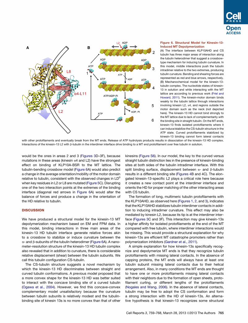
Figure 6. Structural Model for Kinesin-13-
Induced MT Depolymerization
(A) The interface between KLP10AHD and CS
tubulin has three major areas of interaction along
the tubulin heterodimer that suggest a crossbow-
type mechanism for inducing tubulin curvature. In
this model, middle interactions push the tubulin
intradimer relative to the two extremes, producing
tubulin curvature. Bending and shearing forces are
represented as red and blue arrows, respectively.
(B) Mechanochemical model for the kinesin-13-
tubulin complex. The nucleotide states of kinesin-
13 in solution and while interacting with the MT
lattice are according to previous work (Friel and
Howard, 2011). The kinesin-motor domain binds
weakly to the tubulin lattice through interactions
involving kinesin L2, a4, and regions outside the
motor domain such as the neck (not depicted
here). The kinesin-13 HD cannot bind strongly to
the MT lattice due to lack of complementarity with
the binding site in straight tubulin. On theMT ends,
kinesin-13 finds isolated protofilaments where it
can induce/stabilize the CS-tubulin structure in the
ATP state. Curved protofilaments stabilized by
kinesin-13 binding cannot form lateral contacts
with other protofilaments and eventually break from the MT ends. Release of ATP hydrolysis products results in dissociation of the kinesin-13 HD complex.
Interactions of the kinesin-13 L2 with b-tubulin in the interdimer interface drive binding to a MT end protofilament over free tubulin in solution.
would be the ones in areas 2 and 3 (Figures 3D–3F), because
mutations in these areas (kinesin a4 and L2) have the strongest
effect on binding of KLP10A-BSR to the MT lattice. The
tubulin-bending crossbow model (Figure 6A) would also predict
a change in the average orientation/mobility of themotor domain
relative to tubulin, consistent with the observed changes in LD0
when key residues in L2 or L8 aremutated (Figure 5C). Disrupting
one of the two interaction points at the extremes of the binding
interface (diagonal red arrows in Figure 6A) would alter the
balance of forces and produce a change in the orientation of
the HD relative to tubulin.
DISCUSSION
We have produced a structural model for the kinesin-13 MT
depolymerization mechanism based on EM and FPM data. In
this model, binding interactions in three main areas of the
kinesin-13 HD tubulin interface generate relative forces akin
to a crossbow to stabilize or induce curvature between the
a- and b-subunits of the tubulin heterodimer (Figure 6A). A nano-
meter-resolution structure of the kinesin-13 HD tubulin complex
also revealed that in addition to curvature, there is considerable
relative displacement (shear) between the tubulin subunits. We
call this tubulin configuration CS-tubulin.
The CS-tubulin structure suggests a novel mechanism by
which the kinesin-13 HD discriminates between straight and
curved tubulin conformations. A previous model proposed that
a more convex shape for the kinesin-13 HD was better suited
to interact with the concave binding site of a curved tubulin
(Ogawa et al., 2004). However, we find this concave-convex
complementary model unsatisfactory because the curvature
between tubulin subunits is relatively modest and the tubulin-
binding site of kinesin 13s is no more convex than that of other
kinesins (Figure S6). In our model, the key to the curved versus
straight tubulin distinction lies in the presence of kinesin-binding
sites at both sides of the tubulin intradimer interface. With this
split binding surface, displacement between a- and b-tubulin
results in a different binding site (Figures 4B and 4C). The elon-
gated kinesin-13-specific L2 plays a critical role here because
it creates a new contact point at the interdimer interface and
orients the HD for proper matching of the other interacting areas
with CS tubulin.
The formation of long, multimeric tubulin protofilaments with
the KLP10AHD, as observed here (Figures 1, 2, and 3), indicates
that the KLP10AHD stabilizes tubulin interdimer contacts in addi-
tion to inducing intradimer curvature. This effect may also be
mediated by kinesin L2, because its tip is at the interdimer inter-
face (Figures 3C and 3F). This interaction may give kinesin-13s
a higher affinity for isolated protofilaments at the end of the MT
compared with free tubulin, where interdimer interactions would
be missing. This would provide a structural explanation for why
kinesin-13s are efficient MT catastrophe promoters rather than
polymerization inhibitors (Gardner et al., 2011).
A simple explanation for how kinesin-13s specifically recog-
nize and depolymerize MT ends is that they recognize tubulin
protofilaments with missing lateral contacts. In the absence of
capping proteins, the MT ends will always have at least one
tubulin subunit missing lateral contacts due to their helical
arrangement. Also, in many conditions the MT ends are thought
to have one or more protofilaments missing lateral contacts
with their neighbors due to the formation of open sheets, proto-
filament curling, or different lengths of the protofilaments
(Nogales and Wang, 2006). In the absence of lateral contacts,
tubulin may be free to adopt the CS conformation and form
a strong interaction with the HD of kinesin-13s. An alterna-
tive hypothesis is that kinesin-13 recognizes some structural
Cell Reports 3, 759–768, March 28, 2013 ª2013 The Authors 765

feature of GTP-tubulin, which is thought to cap the end of MTs
(Zanic et al., 2009). However, the fact that we observed similar
KLP10AHD tubulin rings in the presence of GDP, GTP, or nonhy-
drolyzable GTP analogs argues against this possibility.
The process of kinesin-13-induced protofilament bending at
the end of the MTs could occur by an induced fit or a conforma-
tional selection-type mechanism (Changeux and Edelstein,
2011). In the first case, interactions of the kinesin-13 HD in the
ATP state with straight tubulin would promote a transition from
straight to CS-tubulin concomitantly with the formation of
a strong binding complex. This transition would only occur on
isolated protofilaments where kinesin-13 could overcome the
straightening force created by lateral tubulin interactions. In the
second view, the kinesin-13 HD would stabilize a pre-existing
CS-tubulin configuration. The probability of finding this pre-ex-
isting configuration would be higher in isolated protofilaments
where the lack of stabilizing lateral protofilament contacts would
allow tubulin to fluctuate between different conformations. In
favor of a conformational selection-type mechanism is the fact
that tubulin with curvature exists in the absence of kinesin-13s
(Nogales and Wang, 2006). However, proving this mechanism
will require determining whether the CS-tubulin structure in
particular can exist outside a kinesin-13-tubulin complex.
Currently, we favor a model that incorporates elements of both
mechanisms (induced fit and conformational selection). A plau-
sible sequence of events is that the kinesin-13 HD initially forms
a complex with slightly curved protofilaments at the MT ends,
and this interaction triggers further tubulin bending and shearing.
The fact that KLP10AHD binds to tubulin protofilaments with
a range of curvatures (Table 1; Movie S4) supports this view.
Our structural data can be combined with previously reported
kinetic data (Friel and Howard, 2011; Wang et al., 2012) to obtain
a mechanochemical cycle model for kinesin-13 (Figure 6B). In
this model, the distinct effects of the MT ends or lattice on kine-
sin-13 activity are explained by the ability/inability to form all the
three kinesin-13-tubulin interactions, as seen in the Kin-Tub1
interface of the KLP10AHD CS-tubulin complex (Figures 3C–
3F). As indicated by our FPM data, the kinesin-13 HD binds to
the MT lattice in a relatively mobile state mediated, at least in
part, by interactions with the kinesin-13 L2 and a4. In this weakly
bound state, kinesin-13 could undergo 1D diffusion toward
either MT end (Helenius et al., 2006). At the MT ends, a kine-
sin-13 could arrive in either the ATP state (from solution) or the
ADP state (by 1D diffusion along the MT lattice). Lack of lateral
protofilament contacts at theMT endswould allow tubulin curva-
ture and the kinesin-13 to form complementary binding at the
three regions of the Kin-Tub-1 interface. ADP/ATP exchange of
kinesins arriving in the ADP state from the MT lattice (Friel and
Howard, 2011) may also be triggered during this process. The re-
sulting stable kinesin-13-CS-tubulin complex would not be able
to straighten spontaneously and reform lateral interprotofilament
contacts, leading to MT end depolymerization. Following ATP
hydrolysis and product release, the complex would dissociate,
as suggested by the very small number or lack of KLP10AHD-
curved tubulin complexes observed in the ADP and NN condi-
tions (Table 1). Separation of individual tubulin dimers from the
MT end protofilament could occur at this step by a yet to be
defined additional conformational change. Alternatively, it could
766 Cell Reports 3, 759–768, March 28, 2013 ª2013 The Authors
occur as the curved protofilaments grow longer and become
prone to breakage.
EXPERIMENTAL PROCEDURES
Protein Constructs
D. melanogaster KLP10A protein constructs were expressed and purified as
previously described (Tan et al., 2006, 2008). The KLP10AHD construct
includes KLP10A residues I279–I615. For FPM, we made several new
constructs that include the KLP10A head and neck domains (residues T198–
I615). Inclusion of the neck residues improvedMT decoration. For site-specific
labeling, all of the surface-exposed cysteines were replaced with alanines or
other residues based on naturally occurring substitutions within the kinesin-
13 family. Accordingly, we made the following substitutions: C282A, C339A,
C389S, C548S, and C594V. Two residues were mutated to cysteines (T455C
and D460C) for labeling with the bifunctional thiol-reactive fluorescence probe
BSR (Molecular Probes). We call this basic construct WT. We verified that this
WT construct retained kinesin-13’s ability to induce tubulin curvature. We
prepared three mutant versions of this construct by replacing the following
class-conserved residues: K317A, V318A, and D319A triple mutant (residues
located in L2); D444A and K446A double mutant (residues located in L8);
and K546A, E547A, C548A triple mutant (residues located in kinesin a4). All
of the mutations were introduced into the plasmid vector using the Quik-
Change Lightning Site-DirectedMutagenesis Kit (Agilent Technologies/Strata-
gene). The attachment location of the probe (crosslinking residues 455–460)
was verified by trypsin digestion followed by mass spectrometry. Pig brain
tubulin was purchased (Cytoskeleton) or purified in the laboratory according
to standard protocols (Miller and Wilson, 2010).
Single-Particle Negative-Staining EM
Tubulin (0.15 mM) was first mixed with the nucleotide species to be tested and
then with an excess amount of KLP10AHD in cold BRB80 buffer (1 mMMgCl2;
1 mM EGTA; 80 mM PIPES, pH = 6.8). The final concentrations of nucleotide
depending on the experimental condition to be tested were as follows:
AMP-PNP, 1 mM AMP-PNP (Sigma Aldrich); ADP, 1 mM ADP (Sigma Aldrich);
NN, 5 U ml�1 apyrase (Sigma Aldrich) and no added nucleotides; and ADP-
AlF4�, 4 mM ADP, 2 mM AlCl3, and 10 mM KF. The mixture was incubated
on ice for 1 min and then loaded onto carbon-coated copper grids. We also
tested the effect of including different GTP analogs (GTP: 1 mM GTP [Sigma
Aldrich]; GDP, 1 mM GDP [Sigma Aldrich]; GMPCPP: 1 mM GMPCPP [Jena
Bioscience]; and GTP-Y-S, 1 mM GTP-Y-S [Cytoskeleton]). The grids with
the KLP10AHD and tubulin mixtures were negatively stained with 1% uranyl
acetate (Electron Microscopy Sciences). Electron micrographs were recorded
on a TVIPS F224HD digital camera in an FEI Tecnai-20microscope operated at
120 kV with a nominal magnification of 50KX (pixel size: 0.27 3 0.27 nm2).
Cryo-EM
A suspension of the KLP10AHD-MT complex was applied onto freshly glow-
discharged EM grids and flash frozen as previously described (Tan et al.,
2008). Electronmicrographs were recorded on film under low-dose conditions
at a nominal magnification of 50,000 or 45,000 with a nominal defocus range of
1–2.5 mm on a Tecnai-F20 FEG cryo-electron microscope operated at 200 kV
or on a JEOL 3200FSC operated at 300 kV. Electron micrographs were re-
corded on film under low-dose conditions and digitized for further analysis.
Image Analysis and 3D Reconstruction
Negatively stained particle images of KLP10AHD tubulin complexes were
digitized and subjected to multivariate statistical analysis, classification, and
averaging (see Extended Experimental Procedures).
We carried out 3D reconstruction of helical 15-pf MTs with wrapped-around
KLP10AHD tubulin protofilament spirals using a single-particle approach (see
Extended Experimental Procedures).
Molecular Fitting
We created atomic models by fitting the atomic structures of tubulin (Protein
Data Bank ID code [PDB]: 1JFF; Lowe et al., 2001) and KLP10AHD into the
asymmetric unit of the 3D map using the fitmap function of UCSF-Chimera
(Pettersen et al., 2004). The atomic coordinates of each protein subunit
were fitted independently as rigid bodies (for further details see Figure S5
and corresponding legend). An atomic model of KLP10AHD was generated
with MMM (Rai and Fiser, 2006) using the sequence of KLP10A and the crystal
structure of the kinesin-13 KIF2C (PDB: 1V8K (Ogawa et al., 2004)).
FPM
Taxol-stabilized MTs were attached to glass coverslip flow chambers, and
solutions with KLP10A fluorescent constructs were then flowed through the
chamber to decorate the MTs as previously described (Asenjo and Sosa,
2009). Nucleotides were added in accordance with the experimental condition
to be interrogated: ADP (2 mM) and NN (5 U ml�1 apyrase with no nucleotide
added) in BRB12 buffer (12 mM PIPES, 2 mMMgCl2, 1 mM EGTA, and 20 mM
Taxol, pH 6.8). Flow chambers were placed in a custom-modified microscope
for fluorescence polarization observation and data recording. Ensemble
fluorescence polarization data were recorded and analyzed as previously
described (Sosa et al., 2010).
ACCESSION NUMBERS
The electron density map has been deposited in the EMDataBank (EMDB)
under accession code EMD-5565, and the atomic model of the complex has
been deposited in the Protein Data Bank under ID code 3J2U.
SUPPLEMENTAL INFORMATION
Supplemental Information includes Extended Experimental Procedures, six
figures, and six movies and can be found with this article online at http://dx.
doi.org/10.1016/j.celrep.2013.01.030.
LICENSING INFORMATION
This is an open-access article distributed under the terms of the Creative
Commons Attribution-NonCommercial-No Derivative Works License, which
permits non-commercial use, distribution, and reproduction in any medium,
provided the original author and source are credited.
ACKNOWLEDGMENTS
We thank J. Hargitai and S. Wang for help with installing and running software
in the high-performance computer cluster at the Albert Einstein College of
Medicine (AECOM); F. Macaluso, L. Gunther, and members of the AECOM
Analytical Imaging Facility for help with EM; and D. Sharp and G. Gerfen for
discussions and critical reading of the manuscript. Part of this investigation
was conducted in a facility constructed with support from the Research Facil-
ities Improvement Program of the National Center for Research Resources,
National Institutes of Health (NIH, grant C06 RR017528-01-CEM). The 300
keV microscope at the New York Structural Biology Center was purchased
with funds from NIH grant S10 RR17291. This project was supported by NIH
grant R01-GM083338 to H.S.
Received: July 24, 2012
Revised: December 12, 2012
Accepted: January 24, 2013
Published: February 21, 2013
REFERENCES
Asenjo, A.B., and Sosa, H. (2009). A mobile kinesin-head intermediate during
the ATP-waiting state. Proc. Natl. Acad. Sci. USA 106, 5657–5662.
Asenjo, A.B., Krohn, N., and Sosa, H. (2003). Configuration of the two kinesin
motor domains during ATP hydrolysis. Nat. Struct. Biol. 10, 836–842.
Changeux, J.P., and Edelstein, S. (2011). Conformational selection or induced
fit? 50 years of debate resolved. F1000 Biol. Rep. 3, 19.
Cooper, J.R., Wagenbach, M., Asbury, C.L., and Wordeman, L. (2010).
Catalysis of the microtubule on-rate is the major parameter regulating the
depolymerase activity of MCAK. Nat. Struct. Mol. Biol. 17, 77–82.
Desai, A., Verma, S., Mitchison, T.J., and Walczak, C.E. (1999). Kin I kinesins
are microtubule-destabilizing enzymes. Cell 96, 69–78.
Friel, C.T., andHoward, J. (2011). The kinesin-13MCAK has an unconventional
ATPase cycle adapted for microtubule depolymerization. EMBO J. 30, 3928–
3939.
Gardner, M.K., Zanic, M., Gell, C., Bormuth, V., and Howard, J. (2011). Depo-
lymerizing kinesins Kip3 andMCAK shape cellular microtubule architecture by
differential control of catastrophe. Cell 147, 1092–1103.
Goldstein, L.S.B., and Philp, A.V. (1999). The road less traveled: emerging prin-
ciples of kinesin motor utilization. Annu. Rev. Cell Dev. Biol. 15, 141–183.
Helenius, J., Brouhard, G., Kalaidzidis, Y., Diez, S., and Howard, J. (2006).
The depolymerizing kinesin MCAK uses lattice diffusion to rapidly target
microtubule ends. Nature 441, 115–119.
Hertzer, K.M., Ems-McClung, S.C., Kline-Smith, S.L., Lipkin, T.G., Gilbert,
S.P., and Walczak, C.E. (2006). Full-length dimeric MCAK is a more efficient
microtubule depolymerase than minimal domain monomeric MCAK. Mol.
Biol. Cell 17, 700–710.
Homma, N., Takei, Y., Tanaka, Y., Nakata, T., Terada, S., Kikkawa, M., Noda,
Y., andHirokawa, N. (2003). Kinesin superfamily protein 2A (KIF2A) functions in
suppression of collateral branch extension. Cell 114, 229–239.
Hunter, A.W., Caplow, M., Coy, D.L., Hancock, W.O., Diez, S., Wordeman, L.,
and Howard, J. (2003). The kinesin-related protein MCAK is a microtubule
depolymerase that forms an ATP-hydrolyzing complex at microtubule ends.
Mol. Cell 11, 445–457.
Kobayashi, T., Tsang, W.Y., Li, J., Lane, W., and Dynlacht, B.D. (2011).
Centriolar kinesin Kif24 interacts with CP110 to remodel microtubules and
regulate ciliogenesis. Cell 145, 914–925.
Kull, F.J., and Endow, S.A. (2002). Kinesin: switch I & II and the motor mecha-
nism. J. Cell Sci. 115, 15–23.
Lowe, J., Li, H., Downing, K.H., and Nogales, E. (2001). Refined structure of
alpha beta-tubulin at 3.5 A resolution. J. Mol. Biol. 313, 1045–1057.
Maney, T., Wagenbach, M., andWordeman, L. (2001). Molecular dissection of
the microtubule depolymerizing activity of mitotic centromere-associated ki-
nesin. J. Biol. Chem. 276, 34753–34758.
Manning, A.L., Ganem, N.J., Bakhoum, S.F., Wagenbach, M., Wordeman, L.,
and Compton, D.A. (2007). The kinesin-13 proteins Kif2a, Kif2b, and Kif2c/
MCAK have distinct roles during mitosis in human cells. Mol. Biol. Cell 18,
2970–2979.
Marx, A., Muller, J., Mandelkow, E.M., Hoenger, A., andMandelkow, E. (2006).
Interaction of kinesin motors, microtubules, and MAPs. J. Muscle Res. Cell
Motil. 27, 125–137.
Miller, H.P., and Wilson, L. (2010). Preparation of microtubule protein and
purified tubulin from bovine brain by cycles of assembly and disassembly
and phosphocellulose chromatography. Methods Cell Biol. 95, 3–15.
Moores, C.A., and Milligan, R.A. (2008). Visualisation of a kinesin-13 motor on
microtubule end mimics. J. Mol. Biol. 377, 647–654.
Moores, C.A., Yu, M., Guo, J., Beraud, C., Sakowicz, R., and Milligan, R.A.
(2002). A mechanism for microtubule depolymerization by KinI kinesins. Mol.
Cell 9, 903–909.
Moores, C.A., Cooper, J., Wagenbach, M., Ovechkina, Y., Wordeman, L., and
Milligan, R.A. (2006). The role of the kinesin-13 neck inmicrotubule depolymer-
ization. Cell Cycle 5, 1812–1815.
Mulder, A.M., Glavis-Bloom, A., Moores, C.A., Wagenbach, M., Carragher, B.,
Wordeman, L., and Milligan, R.A. (2009). A newmodel for binding of kinesin 13
to curved microtubule protofilaments. J. Cell Biol. 185, 51–57.
Nogales, E., and Wang, H.W. (2006). Structural mechanisms underlying
nucleotide-dependent self-assembly of tubulin and its relatives. Curr. Opin.
Struct. Biol. 16, 221–229.
Cell Reports 3, 759–768, March 28, 2013 ª2013 The Authors 767
Nogales, E., Wolf, S.G., and Downing, K.H. (1998). Structure of the alpha beta
tubulin dimer by electron crystallography. Nature 391, 199–203.
Ogawa, T., Nitta, R., Okada, Y., and Hirokawa, N. (2004). A common mecha-
nism for microtubule destabilizers-M type kinesins stabilize curling of the
protofilament using the class-specific neck and loops. Cell 116, 591–602.
Ovechkina, Y., Wagenbach, M., and Wordeman, L. (2002). K-loop insertion
restores microtubule depolymerizing activity of a ‘‘neckless’’ MCAK mutant.
J. Cell Biol. 159, 557–562.
Pettersen, E.F., Goddard, T.D., Huang, C.C., Couch, G.S., Greenblatt, D.M.,
Meng, E.C., and Ferrin, T.E. (2004). UCSF Chimera—a visualization system
for exploratory research and analysis. J. Comput. Chem. 25, 1605–1612.
Rai, B.K., and Fiser, A. (2006). Multiple mapping method: a novel approach
to the sequence-to-structure alignment problem in comparative protein
structure modeling. Proteins 63, 644–661.
Rankin, K.E., and Wordeman, L. (2010). Long astral microtubules uncouple
mitotic spindles from the cytokinetic furrow. J. Cell Biol. 190, 35–43.
Rogers, G.C., Rogers, S.L., Schwimmer, T.A., Ems-McClung, S.C., Walczak,
C.E., Vale, R.D., Scholey, J.M., and Sharp, D.J. (2004). Two mitotic kinesins
cooperate to drive sister chromatid separation during anaphase. Nature 427,
364–370.
Sanhaji, M., Friel, C.T., Wordeman, L., Louwen, F., and Yuan, J. (2011).
Mitotic centromere-associated kinesin (MCAK): a potential cancer drug target.
Oncotarget 2, 935–947.
Shipley, K., Hekmat-Nejad, M., Turner, J., Moores, C., Anderson, R., Milligan,
R., Sakowicz, R., and Fletterick, R. (2004). Structure of a kinesin microtubule
depolymerization machine. EMBO J. 23, 1422–1432.
768 Cell Reports 3, 759–768, March 28, 2013 ª2013 The Authors
Sosa, H., Peterman, E.J., Moerner, W.E., and Goldstein, L.S. (2001).
ADP-induced rocking of the kinesinmotor domain revealed by single-molecule
fluorescence polarization microscopy. Nat. Struct. Biol. 8, 540–544.
Sosa, H., Asenjo, A.B., and Peterman, E.J. (2010). Structure and dynamics of
the kinesin-microtubule interaction revealed by fluorescence polarization
microscopy. Methods Cell Biol. 95, 505–519.
Tan, D., Asenjo, A.B., Mennella, V., Sharp, D.J., and Sosa, H. (2006). Kinesin-
13s form rings around microtubules. J. Cell Biol. 175, 25–31.
Tan, D., Rice, W.J., and Sosa, H. (2008). Structure of the kinesin13-microtu-
bule ring complex. Structure 16, 1732–1739.
Wang, H.W., and Nogales, E. (2005). Nucleotide-dependent bending flexibility
of tubulin regulates microtubule assembly. Nature 435, 911–915.
Wang, W., Jiang, Q., Argentini, M., Cornu, D., Gigant, B., Knossow, M., and
Wang, C. (2012). Kif2C minimal functional domain has unusual nucleotide
binding properties that are adapted to microtubule depolymerization. J. Biol.
Chem. 287, 15143–15153.
Wittinghofer, A. (1997). Signalingmechanistics: aluminum fluoride formolecule
of the year. Curr. Biol. 7, R682–R685.
Woehlke, G., Ruby, A.K., Hart, C.L., Ly, B., Hom-Booher, N., and Vale, R.D.
(1997). Microtubule interaction site of the kinesin motor. Cell 90, 207–216.
Zanic, M., Stear, J.H., Hyman, A.A., and Howard, J. (2009). EB1 recognizes
the nucleotide state of tubulin in the microtubule lattice. PLoS ONE 4,
e7585.


![The Role of c-Tubulin in Centrosomal Microtubule Organization...small complex (c-TuSC) [9]. In metazoans, multiple c-TuSCs associate with additional proteins to form open c-tubulin](https://img.pdfslide.us/doc/110x75/5fe7eab8a1fa371c9b543f4f/the-role-of-c-tubulin-in-centrosomal-microtubule-organization-small-complex.jpg)

![BIOACTIVE SECONDARY METABOLITES: AN … SECONDARY METABOLITES: AN OVERVIEW ... biochemical activities include the tubulin microtubule] ... microorganisms present in our environment](https://img.pdfslide.us/doc/110x75/5aafa36b7f8b9aa8438d8df2/bioactive-secondary-metabolites-an-secondary-metabolites-an-overview-biochemical.jpg)