Embed Size (px)
Citation preview

Structural Insight into the Clostridium difficileEthanolamine Utilisation MicrocompartmentAlison C. Pitts1., Laura R. Tuck1., Alexandra Faulds-Pain2, Richard J. Lewis1, Jon Marles-Wright1,3*
1 Institute for Cell and Molecular Biosciences, Newcastle University, Newcastle upon Tyne, United Kingdom, 2 Department of Pathogen Molecular Biology, London School
of Hygiene and Tropical Medicine, London, United Kingdom, 3 Institute of Structural and Molecular Biology, School of Biological Sciences, University of Edinburgh,
Edinburgh, United Kingdom
Abstract
Bacterial microcompartments form a protective proteinaceous barrier around metabolic enzymes that process unstable ortoxic chemical intermediates. The genome of the virulent, multidrug-resistant Clostridium difficile 630 strain contains anoperon, eut, encoding a bacterial microcompartment with genes for the breakdown of ethanolamine and its utilisation as asource of reduced nitrogen and carbon. The C. difficile eut operon displays regulatory genetic elements and proteinencoding regions in common with homologous loci found in the genomes of other bacteria, including the entericpathogens Salmonella enterica and Enterococcus faecalis. The crystal structures of two microcompartment shell proteins,CD1908 and CD1918, and an uncharacterised protein with potential enzymatic activity, CD1925, were determined by X-raycrystallography. CD1908 and CD1918 display the same protein fold, though the order of secondary structure elements ispermuted in CD1908 and this protein displays an N-terminal b-strand extension. These proteins form hexamers withmolecules related by crystallographic and non-crystallographic symmetry. The structure of CD1925 has a cupin b-barrel foldand a putative active site that is distinct from the metal-ion dependent catalytic cupins. Thin-section transmission electronmicroscopy of Escherichia coli over-expressing eut proteins indicates that CD1918 is capable of self-association into arrays,suggesting an organisational role for CD1918 in the formation of this microcompartment. The work presented provides thebasis for further study of the architecture and function of the C. difficile eut microcompartment, its role in metabolism andthe wider consequences of intestinal colonisation and virulence in this pathogen.
Citation: Pitts AC, Tuck LR, Faulds-Pain A, Lewis RJ, Marles-Wright J (2012) Structural Insight into the Clostridium difficile Ethanolamine UtilisationMicrocompartment. PLoS ONE 7(10): e48360. doi:10.1371/journal.pone.0048360
Editor: Paul C. Driscoll, MRC National Institute for Medical Research, United Kingdom
Received July 14, 2012; Accepted September 24, 2012; Published October 29, 2012
Copyright: � 2012 Pitts et al. This is an open-access article distributed under the terms of the Creative Commons Attribution License, which permits unrestricteduse, distribution, and reproduction in any medium, provided the original author and source are credited.
Funding: This research was supported by a University Vacation Studentship awarded by Newcastle University (ACP) and by a Faculty Development Fellowshipawarded by Newcastle University (JMW). The funders had no role in study design, data collection and analysis, decision to publish, or preparation of themanuscript.
Competing Interests: The authors have declared that no competing interests exist.
* E-mail: [email protected]
. These authors contributed equally to this work.
Introduction
The human gut is a complex and highly competitive ecosystem
that is populated by many different species of bacteria, each
adopting different strategies to survive within the niches they
inhabit [1]. Pathogenic species are usually out-competed by the
commensal species that make up the healthy gut microbiota [2,3],
but they often make use of toxins directed towards other bacterial
species [4], or the host [5] to enable them to colonise
environments that would otherwise be occupied by competitors,
or to create new niches through changes to their host organism.
Salmonella enterica and Escherichia coli species are common causes of
gastroenteritis and diarrheal illness in the healthy population [6,7],
while Clostridium difficile is a major cause of hospital acquired
diarrhoea and has significant risks of morbidity and mortality in
the elderly and immune compromised patients [8,9]. With an
ageing population that is becoming increasingly reliant on hospital
care, there is much interest in understanding the molecular basis of
the metabolism of C. difficile and its role in intestinal colonisation
and virulence.
Nutritional stress induces the expression of the C. difficile toxins,
which act on host cells and induce an inflammatory response
[10,11]. As a consequence of the cellular damage and ensuing
inflammation caused by these toxins large quantities of phospho-
lipids, particularly the abundant phosphatidylethanolamine, are
liberated from the cell membranes of host epithelial cells and other
bacteria [12,13]. Phosphatidylethanolamine is broken down
readily by bacterial phosphodiesterases into glycerol and ethanol-
amine [14], and a number of enteric pathogens, including S.
enterica, Enterococcus faecalis and some species of Clostridia can use
ethanolamine as a sole source of nitrogen and carbon [15–18].
Indeed, an association between ethanolamine metabolism and
virulence in S. enterica is emerging [19,20].
The breakdown of ethanolamine is carried out by a two-subunit
adenosylcobalamin (AdoCbl) cofactor-dependent ethanolamine
ammonia lyase protein complex, which is encoded by the genes
eutB and eutC [21]. These genes are usually associated with a
number of accessory proteins that activate the AdoCbl cofactor
and allow the efficient conversion of the acetaldehyde produced by
this enzyme into acetyl-CoA, which can then be used in various
metabolic processes, such as the TCA cycle in those bacteria
capable of aerobic respiration, lipid biosynthesis, or for substrate
level phosphorylation to generate ATP [22]. The genes associated
with the ethanolamine ammonia lyase vary widely between
PLOS ONE | www.plosone.org 1 October 2012 | Volume 7 | Issue 10 | e48360

species; some bacteria are only able to utilise ethanolamine as
source of reduced nitrogen, as they possess only the lyase genes,
while others possess a long operon encoding regulatory elements
and a number of proteins that are homologous to carboxysome
shell proteins [23–27]. These metabolic compartments allow the
efficient utilisation of various carbon sources and are termed
bacterial microcompartments (BMCs) [28]. The sequestration of
ethanolamine metabolism within a BMC is thought to protect the
cell from the acetaldehyde produced as an intermediate in its
breakdown and to prevent the loss of this volatile compound and
its carbon from the cell [29].
The biochemistry of the ethanolamine utilisation has been
extensively studied in Salmonella species [15,21,30–32] However,
there remain a number of questions about the roles of some of the
enzymes associated with the ethanolamine ammonia lyase
[15,18,32,33]. Work on the structure of this BMC is limited to
X-ray crystal structures of a number of the shell proteins from the
E. coli eut operon [34]. To understand the function of the
ethanolamine utilisation BMC, it is necessary to understand its
architecture, including the features that are unique to this
particular class of BMC and those that are conserved across
carboxysomes and other BMCs. Knowledge of the structure of
the ethanolamine utilisation BMC will form the basis for
exploring its wider impact on bacterial metabolism and virulence.
It is also apparent that BMCs have the potential for exploitation
as protein containers for nano-technology and synthetic biology
[35–37].
The ethanolamine utilisation (eut) operon of the virulent multi-
drug resistant Clostridium difficile strain 630 encodes an ethanol-
amine ammonia lyase and associated accessory proteins; enzymes
required for the utilisation of the organic carbon liberated by the
lyase; regulatory proteins and six proteins with homology to the
carboxysome shell proteins [38] (Fig. 1, Table 1 and Supporting
Information S1for detailed description of the operon). To
understand the structure and function of the C. difficile eut bacterial
microcompartment we have determined the crystal structures of
two proteins with homology to BMC shell proteins, CD1908 and
CD1918, and show that they are well conserved between species.
We have also determined the atomic structure of CD1925, a
member of the EutQ family of cupin barrels. A putative active site
is identified that is metal ion independent and unique in the wider
cupin family. The cytoplasm of E. coli cells over-expressing these
proteins revealed arrays formed by CD1918 in vivo, which are
analogous to the arrays seen for the PduA protein from the
propanediol utilisation microcompartment of Citrobacter freundii and
EtuA from the ethanol utilisation microcompartment from
Citrobacter kluyveri.
Results
Structural analysis of C. difficile eut operon proteinsTo understand the function and macromolecular organisation
of the individual proteins within the C. difficile eut operon, a
number of eut genes were cloned into pET28b for overexpression
and subsequent structural analysis by X-ray crystallography and
transmission electron microscopy (TEM). The genes encoding
CD1908, CD1918 and CD1925 were expressed to high levels both
as C-terminal His6 tagged and untagged variants; the tagged
variants of these proteins were subjected to crystallisation and their
structures determined by X-ray crystallography, while the native
proteins were subjected to TEM to assess the formation of higher-
order structures in vivo.
Crystal structure of CD1908, the C. difficile EutShomologue
The structure of CD1908 was determined by molecular
replacement to 1.51 A resolution. Two molecules are present in
the asymmetric unit, comprising the complete native protein
sequence, less the N-terminal methionine, with a single amino acid
visible from the C-terminal His6 tag in chain A. CD1908 displays a
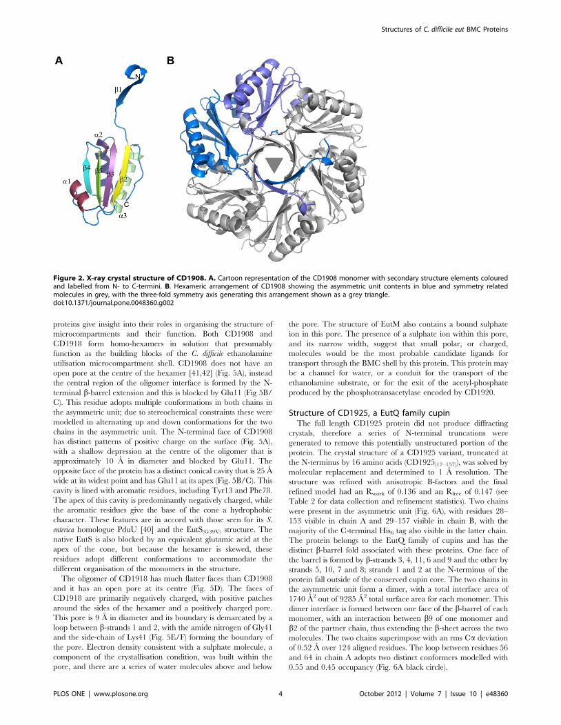
permuted BMC domain fold [39], with a four-stranded anti-
parallel b-sheet flanked by two a-helices on one face and one helix
on the other with an N-terminal b-strand extension (Fig. 2A). The
protein forms a hexamer with pairs of molecules related by the
crystallographic 3-fold axis (Fig. 2B). The two chains in the
asymmetric unit are virtually identical, with an rms Ca deviation
of 0.4 A over 115 aligned residues. A glycerol molecule from the
crystallisation condition was found associated with each chain.
The structure was refined with anisotropic B-factors and the final
refined model had an Rcryst of 0.140 and Rfree of 0.180 (see Table 2
for data collection and refinement statistics).
A single CD1908 monomer superimposes on PduU from S.
enterica (PDBID: 3CGI) [40] and EutS from E. coli (PDBID: 3IA0)
[34] with root mean square Ca deviations of 0.9 A and 1.1 A,
respectively, over 111 aligned residues. The CD1908 hexamer
adopts a conformation almost identical to both PduU and to the
EutS(G39V) mutant (Fig. 3A/B); it superimposes on PduU with an
rms Ca deviation of 0.97 A over 662 residues with 65% sequence
identity, while EutS(G69V) superimposes with an rms Cadeviation of 1.05 A over 657 residues with 53% sequence
identity. The flat CD1908 hexamer is in contrast to the bent and
displaced arrangement seen for the wild type EutS protein
(Fig. 3C/D). In wild type EutS, the hexamer is split into two
trimers that are offset by roughly 17 A and the central axis is
bent by roughly 40u from the conformation seen in EutS(G39V),
PduU and CD1908. The region of the protein containing the
equivalent residues to Gly39 of EutS is an external loop and is
poorly conserved between the three proteins. In CD1908 this
residue is an aspartic acid and PduU has a serine in this position
(Fig. 3E). The structural basis for why changes to this residue
cause a large rearrangement of the EutS multimer is not clear,
and Tanaka et al [34] offer no hypothesis as to why changes at
this surface would affect the formation of the hexamer in such a
way.
Both CD1908 and PduU have longer extensions to their N-
terminal b-barrel than EutS, although at only three and four
residues longer respectively, they do not constitute a significant
increase in length (Fig. 3E). This region is visible in the structure of
CD1908 and loops back over the core of the protein to form a
short 310 helix that contacts the main body of the protein.
Crystal structure of CD1918, the C. difficile EutMhomologue
The structure of CD1918 was determined by molecular
replacement to 1.62 A. Three protein chains were present in the
asymmetric unit, with each chain visible in electron density to
residue ninety of the polymer. The chains superimpose with an
average rms Ca deviation of 0.5 A over the whole protein chain.
The structure was refined with isotropic B-factors and the final
refined model has an Rcryst of 0.166 and Rfree of 0.196 (see Table 2
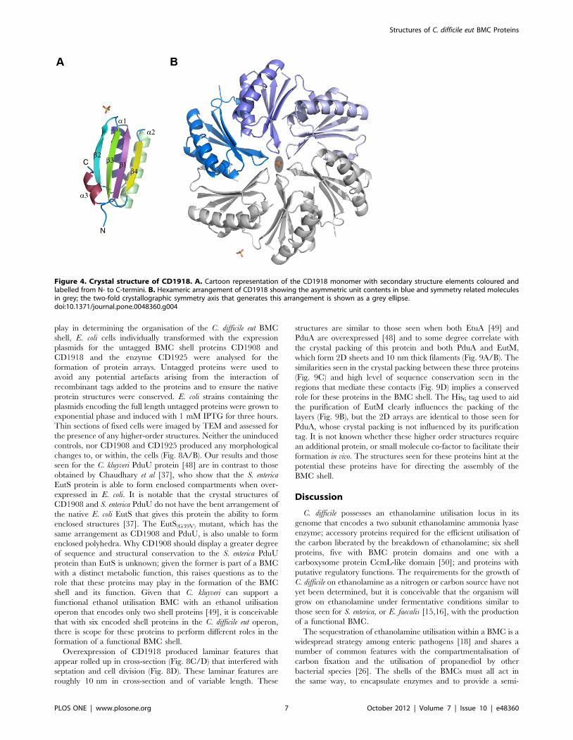
for data collection and refinement statistics). CD1918 displays the
canonical BMC fold with a four-stranded anti-parallel b-sheet
flanked by a-helices (Fig. 4A), and forms a hexamer with molecules
related by a crystallographic 2-fold axis (Fig. 4B). Two sulphate
ions are present at crystallographic symmetry axes. CD1918 shares
77% sequence identity with PduA from S. enterica (PDBID:3NGK)
Structures of C. difficile eut BMC Proteins
PLOS ONE | www.plosone.org 2 October 2012 | Volume 7 | Issue 10 | e48360
[41], and 72% with EutM from E. coli (PDBID: 3MPW) [42]. It
superimposes on these proteins with root mean square Cadeviations of 0.5 and 0.7 A respectively over 86 aligned residues,
forming identical hexameric arrangements to these proteins.
Functional implications for the structures of CD1908 andCD1918
The microcompartment shell must recruit and encapsulate
those enzymes required for its function and allow the passage of
substrates and products through the shell. The structures of BMC
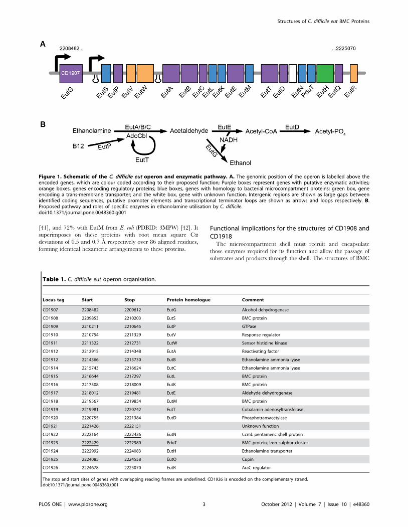
Figure 1. Schematic of the C. difficile eut operon and enzymatic pathway. A. The genomic position of the operon is labelled above theencoded genes, which are colour coded according to their proposed function; Purple boxes represent genes with putative enzymatic activities;orange boxes, genes encoding regulatory proteins; blue boxes, genes with homology to bacterial microcompartment proteins; green box, geneencoding a trans-membrane transporter; and the white box, gene with unknown function. Intergenic regions are shown as large gaps betweenidentified coding sequences, putative promoter elements and transcriptional terminator loops are shown as arrows and loops respectively. B.Proposed pathway and roles of specific enzymes in ethanolamine utilisation by C. difficile.doi:10.1371/journal.pone.0048360.g001
Table 1. C. difficile eut operon organisation.
Locus tag Start Stop Protein homologue Comment
CD1907 2208482 2209612 EutG Alcohol dehydrogenase
CD1908 2209853 2210203 EutS BMC protein
CD1909 2210211 2210645 EutP GTPase
CD1910 2210754 2211329 EutV Response regulator
CD1911 2211322 2212731 EutW Sensor histidine kinase
CD1912 2212915 2214348 EutA Reactivating factor
CD1912 2214366 2215730 EutB Ethanolamine ammonia lyase
CD1914 2215743 2216624 EutC Ethanolamine ammonia lyase
CD1915 2216644 2217297 EutL BMC protein
CD1916 2217308 2218009 EutK BMC protein
CD1917 2218012 2219481 EutE Aldehyde dehydrogenase
CD1918 2219567 2219854 EutM BMC protein
CD1919 2219981 2220742 EutT Cobalamin adenosyltransferase
CD1920 2220755 2221384 EutD Phosphotransacetylase
CD1921 2221426 2222151 Unknown function
CD1922 2222164 2222436 EutN CcmL pentameric shell protein
CD1923 2222429 2222980 PduT BMC protein, Iron sulphur cluster
CD1924 2222992 2224083 EutH Ethanolamine transporter
CD1925 2224085 2224558 EutQ Cupin
CD1926 2224678 2225070 EutR AraC regulator
The stop and start sites of genes with overlapping reading frames are underlined. CD1926 is encoded on the complementary strand.doi:10.1371/journal.pone.0048360.t001
Structures of C. difficile eut BMC Proteins
PLOS ONE | www.plosone.org 3 October 2012 | Volume 7 | Issue 10 | e48360
proteins give insight into their roles in organising the structure of
microcompartments and their function. Both CD1908 and
CD1918 form homo-hexamers in solution that presumably
function as the building blocks of the C. difficile ethanolamine
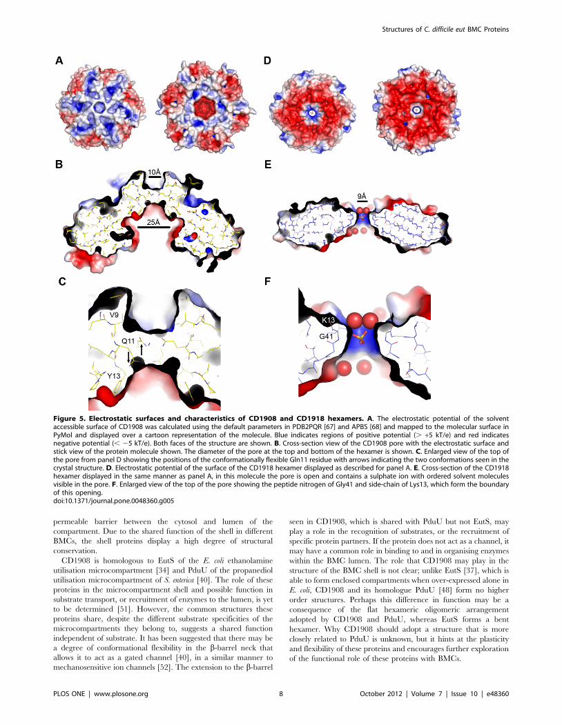
utilisation microcompartment shell. CD1908 does not have an
open pore at the centre of the hexamer [41,42] (Fig. 5A), instead
the central region of the oligomer interface is formed by the N-
terminal b-barrel extension and this is blocked by Glu11 (Fig 5B/
C). This residue adopts multiple conformations in both chains in
the asymmetric unit; due to stereochemical constraints these were
modelled in alternating up and down conformations for the two
chains in the asymmetric unit. The N-terminal face of CD1908
has distinct patterns of positive charge on the surface (Fig. 5A),
with a shallow depression at the centre of the oligomer that is
approximately 10 A in diameter and blocked by Glu11. The
opposite face of the protein has a distinct conical cavity that is 25 A
wide at its widest point and has Glu11 at its apex (Fig. 5B/C). This
cavity is lined with aromatic residues, including Tyr13 and Phe78.
The apex of this cavity is predominantly negatively charged, while
the aromatic residues give the base of the cone a hydrophobic
character. These features are in accord with those seen for its S.
enterica homologue PduU [40] and the EutS(G39V) structure. The
native EutS is also blocked by an equivalent glutamic acid at the
apex of the cone, but because the hexamer is skewed, these
residues adopt different conformations to accommodate the
different organisation of the monomers in the structure.
The oligomer of CD1918 has much flatter faces than CD1908
and it has an open pore at its centre (Fig. 5D). The faces of
CD1918 are primarily negatively charged, with positive patches
around the sides of the hexamer and a positively charged pore.
This pore is 9 A in diameter and its boundary is demarcated by a
loop between b-strands 1 and 2, with the amide nitrogen of Gly41
and the side-chain of Lys41 (Fig. 5E/F) forming the boundary of
the pore. Electron density consistent with a sulphate molecule, a
component of the crystallisation condition, was built within the
pore, and there are a series of water molecules above and below
the pore. The structure of EutM also contains a bound sulphate
ion in this pore. The presence of a sulphate ion within this pore,
and its narrow width, suggest that small polar, or charged,
molecules would be the most probable candidate ligands for
transport through the BMC shell by this protein. This protein may
be a channel for water, or a conduit for the transport of the
ethanolamine substrate, or for the exit of the acetyl-phosphate
produced by the phosphotransacetylase encoded by CD1920.
Structure of CD1925, a EutQ family cupinThe full length CD1925 protein did not produce diffracting
crystals, therefore a series of N-terminal truncations were
generated to remove this potentially unstructured portion of the
protein. The crystal structure of a CD1925 variant, truncated at
the N-terminus by 16 amino acids (CD1925(17–157)), was solved by
molecular replacement and determined to 1 A resolution. The
structure was refined with anisotropic B-factors and the final
refined model had an Rwork of 0.136 and an Rfree of 0.147 (see
Table 2 for data collection and refinement statistics). Two chains
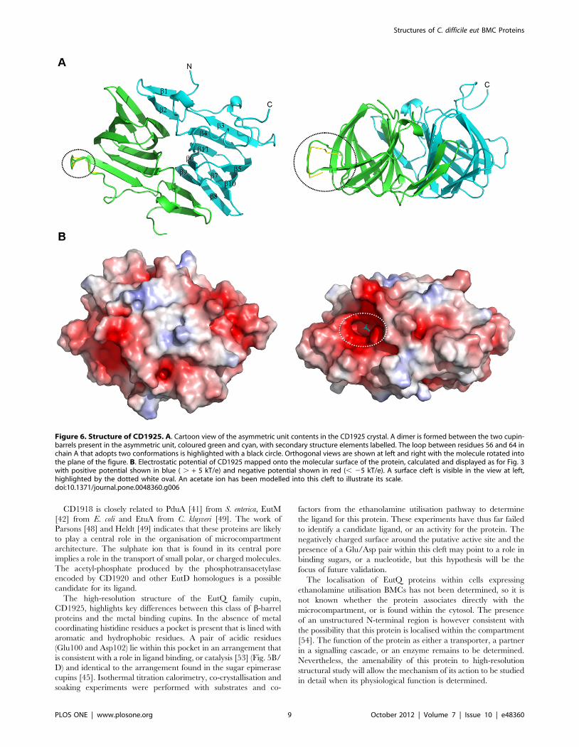
were present in the asymmetric unit (Fig. 6A), with residues 28–
153 visible in chain A and 29–157 visible in chain B, with the
majority of the C-terminal His6 tag also visible in the latter chain.
The protein belongs to the EutQ family of cupins and has the
distinct b-barrel fold associated with these proteins. One face of
the barrel is formed by b-strands 3, 4, 11, 6 and 9 and the other by
strands 5, 10, 7 and 8; strands 1 and 2 at the N-terminus of the
protein fall outside of the conserved cupin core. The two chains in
the asymmetric unit form a dimer, with a total interface area of
1740 A2 out of 9285 A2 total surface area for each monomer. This
dimer interface is formed between one face of the b-barrel of each
monomer, with an interaction between b9 of one monomer and
b2 of the partner chain, thus extending the b-sheet across the two
molecules. The two chains superimpose with an rms Ca deviation
of 0.52 A over 124 aligned residues. The loop between residues 56
and 64 in chain A adopts two distinct conformers modelled with
0.55 and 0.45 occupancy (Fig. 6A black circle).
Figure 2. X-ray crystal structure of CD1908. A. Cartoon representation of the CD1908 monomer with secondary structure elements colouredand labelled from N- to C-termini. B. Hexameric arrangement of CD1908 showing the asymmetric unit contents in blue and symmetry relatedmolecules in grey, with the three-fold symmetry axis generating this arrangement shown as a grey triangle.doi:10.1371/journal.pone.0048360.g002
Structures of C. difficile eut BMC Proteins
PLOS ONE | www.plosone.org 4 October 2012 | Volume 7 | Issue 10 | e48360
The surface of CD1925 does not display the distinctive charge
distribution patterns seen for the oligomeric BMC proteins
CD1908 and CD1918; instead its surface has irregular patches
of positive and negative electrostatic potential (Fig. 6B). A shallow,
negatively charged cleft is visible on the surface of each protein
chain within the cupin b-barrel (Fig. 6B white oval), this cleft
corresponds to the region that coordinates divalent cations and
acts as the active site in the metal binding cupins, such as those
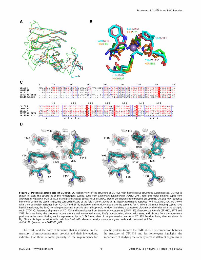
with sugar isomerase activity [43] (Fig 7A/B). CD1925 shares 30%
sequence identity with EutQ from S. typhimurium (PDBID: 2PYT,
unpublished structural genomics output) and superimposes with an
rms Ca deviation of 1.25 A over 116 amino acids. Despite low
sequence identity with metal binding cupins, such as those from
Thermotoga maritima (PDBID: 1VJ2) [44] (14%), and Bacillus subtilis
(2Y0O) [45] (12%), these structures superimpose with rms Cadeviations of 1.88 and 1.74 A respectively, over 98 residues in both
cases. The conservation of the cupin core is evident in these
structures, with the majority of structural differences occurring at
the termini of the proteins and loops that extend from the core
(Fig. 7A).
In contrast to the metal-binding catalytic cupins, CD1925 and
the EutQ family, do not possess the histidine residues that are
responsible for metal coordination in the oxidoreductase [46] and
epimerase [47] classes of cupins. In the place of the histidine
residues are aromatic (Trp94) and hydrophobic residues (Leu 96,
Ile134) and in the fourth position a glutamic acid residue (Glu100)
(Fig. 7B/C), which is also present in the cupins with epimerase
activity. The glutamic acid (Glu100) is within hydrogen bonding
distance of an aspartic acid residue (Asp102), which may act to
alter its pKa. Because of strong sequence conservation seen in this
area for the EutQ family and the fact that this region is solvent
accessible and it corresponds with the position of the catalytic site
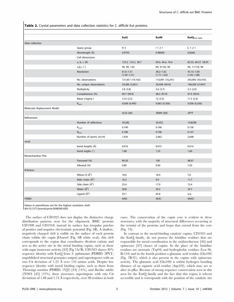
Table 2. Crystal parameters and data collection statistics for C. difficile Eut proteins.
EutS EutM EutQ(17–157)
Data collection
Space group H 3 I 1 2 1 C 1 2 1
Wavelength (A) 0.9793 0.98040 0.8266
Cell dimensions
a, b, c (A) 123.2, 123.2, 38.7 69.4, 46.4, 76.4 82.33, 66.57, 58.95
a,b,c (u) 90, 90, 120 90, 91.56, 90 90, 117.54, 90
Resolution 61.6–1.51(1.59–1.51)
38.2–1.62(1.71–1.62)
43.15–1.00(1.05–1.00)
No. observations 131,851 (19,163) 110,091 (16,241) 343,892 (43,410)
No. unique observations 34,286 (5,001) 30,458 (4416) 148,309 (21947)
Multiplicity 3.8 (3.8) 3.6 (3.7) 2.3 (2.0)
Completeness (%) 99.7 (99.9) 98.3 (97.9) 97.9 (99.4)
Mean I/sigma I 12.4 (2.5) 12 (3.5) 11.5 (2.4)
Rsym 0.058 (0.495) 0.063 (0.356) 0.058 (0.356)
Molecular Replacement Model
3CGI [40] 2EWH [69] 2PYT
Refinement
Number of reflections 34,282 30,452 14,8298
Rcryst 0.140 0.166 0.136
Rfree 0.180 0.196 0.147
Number of atoms (no-H) 1,930 2,062 2,648
rmsd
bond lengths (A) 0.016 0.015 0.014
bond angles (u) 1.68 1.55 1.60
Ramachandran Plot
Favoured (%) 99.20 100 98.97
Allowed (%) 0.80 0.00 1.03
B-factors
Wilson B (A2) 18.6 18.4 7.6
Main chain (A2) 16.2 9.9 11.7
Side chain (A2) 23.4 17.9 15.4
Water (A2) 30.8 30.2 24.7
Ligand (A2) 33.1 45.8 n/a
PDBID 4AXI 4AXJ 4AXO
Values in parentheses are for the highest resolution shell.doi:10.1371/journal.pone.0048360.t002
Structures of C. difficile eut BMC Proteins
PLOS ONE | www.plosone.org 5 October 2012 | Volume 7 | Issue 10 | e48360

of other cupins, this region may act to bind ligands, or act as a
catalytic centre (Fig. 7C, starred residues).
Higher order structures formed by C. difficile Eut proteinsThe interactions between the proteins encoded in BMC loci
produce an enclosed microcompartment that has a shell made
solely of protein [39]. This shell envelops the enzymes required for
the metabolic function of the BMC and allows the passage of
substrates and products, while preventing the escape of metabolic
intermediates. Transmission electron microscopy of thin sections
of E. coli cells overexpressing carboxysome proteins from
Halothiobacillus neapolitanus [35], Citrobacter freundii propanediol
utilisation BMC proteins [48], Clostridium kluyveri ethanol utilisation
BMC [49] and S. enterica ethanolamine utilisation proteins [37],
have revealed the presence of higher-order protein structures. To
understand the possible roles that the proteins in this study may
Figure 3. Alignment of CD1908 and its homologues. A. Ribbon view of a structural alignment of CD1908 (blue), PduU (green) and EutS(G39V)
(red). B. Orthogonal view of A, viewed down the hexameric axis. C. Alignment of CD1908 with the bent EutS hexamer, the orientation of the centralaxes are shown with grey lines. D. Orthogonal view of C, viewed down the multimeric symmetry axis. Rather than being a symmetric hexamer, EutSdisplays a split arrangement of two trimers.doi:10.1371/journal.pone.0048360.g003
Structures of C. difficile eut BMC Proteins
PLOS ONE | www.plosone.org 6 October 2012 | Volume 7 | Issue 10 | e48360
play in determining the organisation of the C. difficile eut BMC
shell, E. coli cells individually transformed with the expression
plasmids for the untagged BMC shell proteins CD1908 and
CD1918 and the enzyme CD1925 were analysed for the
formation of protein arrays. Untagged proteins were used to
avoid any potential artefacts arising from the interaction of
recombinant tags added to the proteins and to ensure the native
protein structures were conserved. E. coli strains containing the
plasmids encoding the full length untagged proteins were grown to
exponential phase and induced with 1 mM IPTG for three hours.
Thin sections of fixed cells were imaged by TEM and assessed for
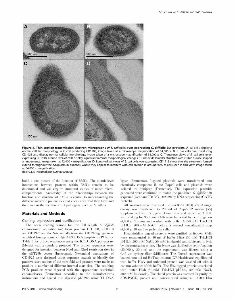
the presence of any higher-order structures. Neither the uninduced
controls, nor CD1908 and CD1925 produced any morphological
changes to, or within, the cells (Fig. 8A/B). Our results and those
seen for the C. kluyveri PduU protein [48] are in contrast to those
obtained by Chaudhary et al [37], who show that the S. enterica
EutS protein is able to form enclosed compartments when over-
expressed in E. coli. It is notable that the crystal structures of
CD1908 and S. enterica PduU do not have the bent arrangement of
the native E. coli EutS that gives this protein the ability to form
enclosed structures [37]. The EutS(G39V) mutant, which has the
same arrangement as CD1908 and PduU, is also unable to form
enclosed polyhedra. Why CD1908 should display a greater degree
of sequence and structural conservation to the S. enterica PduU
protein than EutS is unknown; given the former is part of a BMC
with a distinct metabolic function, this raises questions as to the
role that these proteins may play in the formation of the BMC
shell and its function. Given that C. kluyveri can support a
functional ethanol utilisation BMC with an ethanol utilisation
operon that encodes only two shell proteins [49], it is conceivable
that with six encoded shell proteins in the C. difficile eut operon,
there is scope for these proteins to perform different roles in the
formation of a functional BMC shell.
Overexpression of CD1918 produced laminar features that
appear rolled up in cross-section (Fig. 8C/D) that interfered with
septation and cell division (Fig. 8D). These laminar features are
roughly 10 nm in cross-section and of variable length. These
structures are similar to those seen when both EtuA [49] and
PduA are overexpressed [48] and to some degree correlate with
the crystal packing of this protein and both PduA and EutM,
which form 2D sheets and 10 nm thick filaments (Fig. 9A/B). The
similarities seen in the crystal packing between these three proteins
(Fig. 9C) and high level of sequence conservation seen in the
regions that mediate these contacts (Fig. 9D) implies a conserved
role for these proteins in the BMC shell. The His6 tag used to aid
the purification of EutM clearly influences the packing of the
layers (Fig. 9B), but the 2D arrays are identical to those seen for
PduA, whose crystal packing is not influenced by its purification
tag. It is not known whether these higher order structures require
an additional protein, or small molecule co-factor to facilitate their
formation in vivo. The structures seen for these proteins hint at the
potential these proteins have for directing the assembly of the
BMC shell.
Discussion
C. difficile possesses an ethanolamine utilisation locus in its
genome that encodes a two subunit ethanolamine ammonia lyase
enzyme; accessory proteins required for the efficient utilisation of
the carbon liberated by the breakdown of ethanolamine; six shell
proteins, five with BMC protein domains and one with a
carboxysome protein CcmL-like domain [50]; and proteins with
putative regulatory functions. The requirements for the growth of
C. difficile on ethanolamine as a nitrogen or carbon source have not
yet been determined, but it is conceivable that the organism will
grow on ethanolamine under fermentative conditions similar to
those seen for S. enterica, or E. faecalis [15,16], with the production
of a functional BMC.
The sequestration of ethanolamine utilisation within a BMC is a
widespread strategy among enteric pathogens [18] and shares a
number of common features with the compartmentalisation of
carbon fixation and the utilisation of propanediol by other
bacterial species [26]. The shells of the BMCs must all act in
the same way, to encapsulate enzymes and to provide a semi-
Figure 4. Crystal structure of CD1918. A. Cartoon representation of the CD1918 monomer with secondary structure elements coloured andlabelled from N- to C-termini. B. Hexameric arrangement of CD1918 showing the asymmetric unit contents in blue and symmetry related moleculesin grey; the two-fold crystallographic symmetry axis that generates this arrangement is shown as a grey ellipse.doi:10.1371/journal.pone.0048360.g004
Structures of C. difficile eut BMC Proteins
PLOS ONE | www.plosone.org 7 October 2012 | Volume 7 | Issue 10 | e48360
permeable barrier between the cytosol and lumen of the
compartment. Due to the shared function of the shell in different
BMCs, the shell proteins display a high degree of structural
conservation.
CD1908 is homologous to EutS of the E. coli ethanolamine
utilisation microcompartment [34] and PduU of the propanediol
utilisation microcompartment of S. enterica [40]. The role of these
proteins in the microcompartment shell and possible function in
substrate transport, or recruitment of enzymes to the lumen, is yet
to be determined [51]. However, the common structures these
proteins share, despite the different substrate specificities of the
microcompartments they belong to, suggests a shared function
independent of substrate. It has been suggested that there may be
a degree of conformational flexibility in the b-barrel neck that
allows it to act as a gated channel [40], in a similar manner to
mechanosensitive ion channels [52]. The extension to the b-barrel
seen in CD1908, which is shared with PduU but not EutS, may
play a role in the recognition of substrates, or the recruitment of
specific protein partners. If the protein does not act as a channel, it
may have a common role in binding to and in organising enzymes
within the BMC lumen. The role that CD1908 may play in the
structure of the BMC shell is not clear; unlike EutS [37], which is
able to form enclosed compartments when over-expressed alone in
E. coli, CD1908 and its homologue PduU [48] form no higher
order structures. Perhaps this difference in function may be a
consequence of the flat hexameric oligomeric arrangement
adopted by CD1908 and PduU, whereas EutS forms a bent
hexamer. Why CD1908 should adopt a structure that is more
closely related to PduU is unknown, but it hints at the plasticity
and flexibility of these proteins and encourages further exploration
of the functional role of these proteins with BMCs.
Figure 5. Electrostatic surfaces and characteristics of CD1908 and CD1918 hexamers. A. The electrostatic potential of the solventaccessible surface of CD1908 was calculated using the default parameters in PDB2PQR [67] and APBS [68] and mapped to the molecular surface inPyMol and displayed over a cartoon representation of the molecule. Blue indicates regions of positive potential (. +5 kT/e) and red indicatesnegative potential (, 25 kT/e). Both faces of the structure are shown. B. Cross-section view of the CD1908 pore with the electrostatic surface andstick view of the protein molecule shown. The diameter of the pore at the top and bottom of the hexamer is shown. C. Enlarged view of the top ofthe pore from panel D showing the positions of the conformationally flexible Gln11 residue with arrows indicating the two conformations seen in thecrystal structure. D. Electrostatic potential of the surface of the CD1918 hexamer displayed as described for panel A. E. Cross-section of the CD1918hexamer displayed in the same manner as panel A, in this molecule the pore is open and contains a sulphate ion with ordered solvent moleculesvisible in the pore. F. Enlarged view of the top of the pore showing the peptide nitrogen of Gly41 and side-chain of Lys13, which form the boundaryof this opening.doi:10.1371/journal.pone.0048360.g005
Structures of C. difficile eut BMC Proteins
PLOS ONE | www.plosone.org 8 October 2012 | Volume 7 | Issue 10 | e48360
CD1918 is closely related to PduA [41] from S. enterica, EutM
[42] from E. coli and EtuA from C. kluyveri [49]. The work of
Parsons [48] and Heldt [49] indicates that these proteins are likely
to play a central role in the organisation of microcompartment
architecture. The sulphate ion that is found in its central pore
implies a role in the transport of small polar, or charged molecules.
The acetyl-phosphate produced by the phosphotransacetylase
encoded by CD1920 and other EutD homologues is a possible
candidate for its ligand.
The high-resolution structure of the EutQ family cupin,
CD1925, highlights key differences between this class of b-barrel
proteins and the metal binding cupins. In the absence of metal
coordinating histidine residues a pocket is present that is lined with
aromatic and hydrophobic residues. A pair of acidic residues
(Glu100 and Asp102) lie within this pocket in an arrangement that
is consistent with a role in ligand binding, or catalysis [53] (Fig. 5B/
D) and identical to the arrangement found in the sugar epimerase
cupins [45]. Isothermal titration calorimetry, co-crystallisation and
soaking experiments were performed with substrates and co-
factors from the ethanolamine utilisation pathway to determine
the ligand for this protein. These experiments have thus far failed
to identify a candidate ligand, or an activity for the protein. The
negatively charged surface around the putative active site and the
presence of a Glu/Asp pair within this cleft may point to a role in
binding sugars, or a nucleotide, but this hypothesis will be the
focus of future validation.
The localisation of EutQ proteins within cells expressing
ethanolamine utilisation BMCs has not been determined, so it is
not known whether the protein associates directly with the
microcompartment, or is found within the cytosol. The presence
of an unstructured N-terminal region is however consistent with
the possibility that this protein is localised within the compartment
[54]. The function of the protein as either a transporter, a partner
in a signalling cascade, or an enzyme remains to be determined.
Nevertheless, the amenability of this protein to high-resolution
structural study will allow the mechanism of its action to be studied
in detail when its physiological function is determined.
Figure 6. Structure of CD1925. A. Cartoon view of the asymmetric unit contents in the CD1925 crystal. A dimer is formed between the two cupin-barrels present in the asymmetric unit, coloured green and cyan, with secondary structure elements labelled. The loop between residues 56 and 64 inchain A that adopts two conformations is highlighted with a black circle. Orthogonal views are shown at left and right with the molecule rotated intothe plane of the figure. B. Electrostatic potential of CD1925 mapped onto the molecular surface of the protein, calculated and displayed as for Fig. 3with positive potential shown in blue ( . + 5 kT/e) and negative potential shown in red (, 25 kT/e). A surface cleft is visible in the view at left,highlighted by the dotted white oval. An acetate ion has been modelled into this cleft to illustrate its scale.doi:10.1371/journal.pone.0048360.g006
Structures of C. difficile eut BMC Proteins
PLOS ONE | www.plosone.org 9 October 2012 | Volume 7 | Issue 10 | e48360
This work, and the body of literature that is available on the
structures of microcompartment proteins and their interactions,
indicates that there is some plasticity in the requirements for
specific proteins to form the BMC shell. The comparison between
the structure of CD1908 and its homologues highlights the
importance of studying the same systems in different organisms to
Figure 7. Potential active site of CD1925. A. Ribbon view of the structure of CD1925 with homologous structures superimposed. CD1925 isshown in cyan, the structures of the homologous cupins, EutQ from Salmonella typhimurium (PDBID: 2PYT, red) and metal binding cupin fromThermotoga maritima (PDBID: 1VJ2, orange) and Bacillus subtilis (PDBID: 2Y0O, green), are shown superimposed on CD1925. Despite low sequencehomology within the cupin family, the core architecture of the fold is almost identical. B. Metal coordinating residues from 1VJ2 and 2Y0O are shownwith their equivalent residues from CD1925 and 2PYT, molecule and residue colours are the same as for A. Where the metal binding cupins havehistidine residues, the EutQ homologues possess aromatic and hydrophobic residues and share a conserved glutamic acid residue with the catalyticcupin 2Y0O. C. Sequence alignment of CD1925 and homologues from Listeria monocytogenes (LMO1187), Enterococcus faecalis (EF1617), 2PYT and1VJ2. Residues lining the proposed active site are well conserved among EutQ type proteins, shown with stars, and distinct from the equivalentpositions in the metal binding cupins represented by 1VJ2. D. Stereo view of the proposed active site of CD1925. Residues lining the cleft shown inFig. 6B are displayed as sticks with their final 2mFo-dFc electron density shown as a grey mesh and contoured at 1.5s.doi:10.1371/journal.pone.0048360.g007
Structures of C. difficile eut BMC Proteins
PLOS ONE | www.plosone.org 10 October 2012 | Volume 7 | Issue 10 | e48360
build a true picture of the function of BMCs. The atomic-level
interactions between proteins within BMCs remain to be
determined and will require structural studies of intact micro-
compartments. Knowledge of the relationships between the
function and structure of BMCs is central to understanding the
different substrate preferences and chemistries that they have and
their role in the metabolism of pathogens, such as C. difficile.
Materials and Methods
Cloning, expression and purificationThe open reading frames for the full length C. difficile
ethanolamine utilisation (eut) locus proteins CD1908, CD1918
and CD1925 and the N-terminally truncated CD1925(17–157) were
amplified from genomic C. difficile 630 DNA template by PCR (see
Table 3 for primer sequences) using the KOD DNA polymerase
(Merck) with a standard protocol. The primer sequences were
designed for insertion between NcoI and XhoI restriction sites in
the pET28b vector (Invitrogen). N-terminal truncations of
CD1925 were designed using sequence analysis to identify the
putative start residue of the core fold and primers were made to
produce a number of different internal start sites. The resulting
PCR products were digested with the appropriate restriction
endonucleases (Fermentas) according to the manufacturer’s
instructions and ligated into digested pET28b using T4 DNA
ligase (Fermentas). Ligated plasmids were transformed into
chemically competent E. coli Top10 cells and plasmids were
isolated by miniprep (Fermentas). The expression plasmids
generated were confirmed to match the published C. difficile 630
sequence (Genbank ID: NC_009089) by DNA sequencing (GATC
Biotech).
All constructs were expressed in E. coli B834 (DE3) cells. A single
colony was transferred to 200 ml of Zyp-5052 media [55]
supplemented with 50 mg/ml kanamycin and grown at 310 K
with shaking for 36 hours. Cells were harvested by centrifugation
(4,000 g, 30 min) and washed with buffer A (50 mM Tris.HCl
pH 8.0, 200 mM NaCl) before a second centrifugation step
(4,000 g, 30 min) to pellet the cells.
Hexahistidine tagged proteins were purified as follows. Cells
were resuspended in 40 ml of buffer HisA (50 mM Tris.HCl
pH 8.0, 500 mM NaCl, 50 mM imidazole) and subjected to lysis
by ultrasonication on ice. The lysate was clarified by centrifugation
(35,000 g, 30 min) and the supernatant was filtered using a
0.45 mm syringe filter (Millipore). The filtered supernatant was
loaded onto a 5 ml HisTrap column (GE Healthcare) equilibrated
with buffer HisA and unbound protein was washed off with 5
column volumes of this buffer. The His6-tagged protein was eluted
with buffer HisB (50 mM Tris.HCl pH 8.0, 500 mM NaCl,
500 mM Imidazole). The eluted protein was assessed for purity by
SDS-PAGE, pooled and concentrated using a 10,000 kDa
Figure 8. Thin-section transmission electron micrographs of E. coli cells over-expressing C. difficile Eut proteins. A. All cells display anormal cellular morphology in E. coli producing CD1908, image taken at a microscope magnification of 34,000 x. B. E. coli cells over producingCD1925 also display normal cellular morphology, image taken at a microscope magnification of 34,000 x. C. Transverse views of E. coli cells over-expressing CD1918, around 90% of cells display significant internal morphological changes, 10 nm wide lamellar structures are visible as rose-shapedarrangements, image taken at 92,000 x magnification. D. Longitudinal views of E. coli cells overexpressing CD1918 show that the structures formedextend throughout the cytoplasm in bunches, where they appear to interfere with cell division in around 90% of cells seen in this view, image takenat 64,000 x magnification.doi:10.1371/journal.pone.0048360.g008
Structures of C. difficile eut BMC Proteins
PLOS ONE | www.plosone.org 11 October 2012 | Volume 7 | Issue 10 | e48360

MWCO centrifugal concentrator (Amicon), prior to size exclusion
chromatography using a Superdex S200 HR16/60 column (GE
Healthcare) equilibrated with buffer A. Protein fractions were
assessed by SDS-PAGE.
Untagged proteins were purified as follows. Cells were
resuspended in 40 ml of buffer QA (50 mM Tris.HCl pH 8.0)
and subjected to lysis by ultrasonication on ice. The lysate was
clarified by centrifugation (35,000 g, 30 min) and the supernatant
was filtered using a 0.45 mm syringe filter (Millipore). The filtered
supernatant was loaded onto a 5 ml Q-sepharose column (GE
Healthcare) equilibrated with buffer QA. Unbound sample was
washed off with 10 column volumes of buffer QA and protein was
eluted with a linear gradient of 0–100% buffer QB (50 mM
Tris.HCl pH 8.0, 1 M NaCl) over 25 column volumes. Peak
fractions were analysed by SDS-PAGE and fractions containing
the protein of interest were pooled and subjected to size exclusion
chromatography as described above.
Crystallisation and data collectionPurified hexa-histidine tagged CD1908 (CD1908-His) was
concentrated to 10 mg/ml in Milli-Q H2O and crystallised by
sitting drop vapour diffusion in drops of 100 nl protein plus 100 nl
crystallisation solution, over 100 ml of the latter. Crystals were
obtained in 24% (w/v) PEG 1500, 20% (v/v) glycerol; these were
harvested from the well using a CryoLoop (Hampton Research)
and flash cooled directly in liquid nitrogen. CD1918-His was
concentrated to 10 mg/ml in Milli-Q H2O and crystallised as
described for CD1908 in drops supplemented with crystallisation
solution containing 200 mM Li2SO4, 100 mM phosphate/citrate
buffer pH 4.2, 20% (w/v) PEG 1000. Crystals were harvested
through paratone oil using a CryoLoop and flash cooled in liquid
nitrogen. CD1925(17–157)-His was concentrated to 8 mg/ml in
buffer A and crystallised by hanging drop vapour diffusion in
drops of 1 ml protein plus 1 ml crystallisation solution over 1 ml of
the latter. Crystals were obtained in 100 mM sodium acetate
pH 4.5, 35% (w/v) PEG 6000, 200 mM MgCl2, these were
harvested by transfer to a cryoprotection solution containing the
well solution supplemented with 20% v/v PEG300 and flash
cooled in liquid nitrogen. All crystallographic data were collected
on beamlines I02 and I04 at Diamond Light Source (Didcot, UK)
at 100 K using ADSC CCD detectors. Diffraction data were
Figure 9. Crystal contacts of CD1918 and its homologues. A. Views of the crystal packing of CD1918 along the a,c (top) and b,c (bottom)planes with contact regions highlighted as roman numerals: i, ii, and iii. The crystallographic 2-fold generating the hexamer is shown as a black ellipsefor a single multimer. B. crystal packing for EutM (PDBID: 3MPW), this protein packs into a regular two-dimensional lattice in the a,c plane (top) with asingle conserved crystallographic interface, i. The b,c plane has widely spaced alternating layers distinct to CD1918, due to interactions between theC-terminal his-tag added to the construct for purification. C. Residues mediating CD1918 crystal contacts, panels i, ii and iii correspond to theinterfaces marked in A. Interface i is conserved between CD1908 and its homologues, EutM and PduA, and in the crystal structures includes acoordinated sulphate ion; while interface ii is formed by solvent mediated contacts between chains. Interface iii is formed by non-conserved residuesand forms a tight offset-layer packing between layers of hexamers. D. Sequence alignment of CD1918, PduA (PDBID: 3NGK) and EutM (PDBID: 3MPW).The secondary structure assignment for CD1918 is shown above the alignment. Conserved residues are shown in red, with strict conservationhighlighted with a red background. Residues participating in crystal contacts shown in C are highlighted with i: stars; ii: triangles; iii: open circles.doi:10.1371/journal.pone.0048360.g009
Table 3. Primers.
Name Sequence
CD1908F CTCCCATGGGCTTGACTGAAGAATCTAAGCAAAGAG
CD1908R CTCCTCGAGTTATGTCCTTGTGATTTTTGTAGATGAGAAG
CD1908R-His CTCCTCGAGTGTCCTTGTGATTTTTGTAGATGAGAAG
CD1918F CTCCCATGGGCGCAAGTGCAAACGCATTA
CD1918R CTCCTCGAGTTACTCAGCTGATACCTTTGG
CD1918R-His CTCCTCGAGCTCAGCTGATACCTTTGG
CD1925F CTCCCATGGGCGATATATCAAATATAGATAAAAAT
CD1925R CGCCTCGAGTTAATTTTGAGATGCCCAATCTGCCGG
CD1925(17–157)F CTCCCATGGGCCAAATAATAGAAGAAAAAATAAGT
CD1925(17–157)R-His CGCCTCGAGATTTTGAGATGCCCAATCTGCCGG
Restriction sites are shown in bold face and genome complementary regions initalics.doi:10.1371/journal.pone.0048360.t003
Structures of C. difficile eut BMC Proteins
PLOS ONE | www.plosone.org 12 October 2012 | Volume 7 | Issue 10 | e48360
integrated using iMosflm [56] or XDS [57] and scaled and merged
with Scala [58]. Data collection and refinement statistics are
shown in Table 2.
Structure solution and analysisAll structures were solved by molecular replacement using
Phaser [59], molecular replacement models used are shown in
Table 2. Refinement of the coordinates, TLS parameters and
atomic temperature factors (anisotropic in the case of CD1908 and
CD1925(17–152)) was carried out using Phenix.refine [60]. Model
building was performed using Coot [61]. The secondary structure
and stereochemistry of the models was analysed by MolProbity
[62]. Sequence alignment was performed using ClustalW [63] and
the corresponding figures were generated using ESPript [64].
Oligomerisation states and values of buried surface areas were
calculated using the PISA server [65]. Structural superimpositions
were calculated using Coot. Crystallographic figures were gener-
ated with PyMOL [66].
Thin-section transmission electron microscopyE. coli B834 cells transformed with the plasmids for untagged
CD1908, CD1918 and CD1925 were grown to mid-log phase in
Luria-Bertani media supplemented with 50 mg/ml kanamycin at
310 K and induced with 1 mM final concentration of IPTG and
harvested after 3 hours. 1 ml of these cells and un-induced
controls were fixed in 2.5% (v/v) glutaraldehyde, 50 mM sodium
cacodylate pH 7.0 for 24 hours at 4uC. Cells were subsequently
immobilized in 2% (w/v) water-agar and post-fixed in 1.5% (w/v)
osmium tetroxide in 50 mM sodium cacodylate pH 7.0 for 1 hour
at 4uC followed by dehydration in an ethanol series. The final 70%
dehydration step was supplemented with 1% (w/v) uranyl acetate
and was performed overnight at 21uC. 100 nm sections were post-
stained with 2% (w/v) uranyl acetate and analysed using a Philips
CM100 transmission electron microscope.
Accession CodesRefined coordinates and structure factors have been deposited
at the PDB with the following accession numbers: CD1908 PDB
ID: 4AXI; CD1918 PDB ID: 4AXJ; CD1925 PDB ID: 4AXO.
Supporting Information
Supporting Information S1 Organisation of C. difficile
ethanolamine utilisation operon.
(DOCX)
Acknowledgments
We would like to thank the staff working on beamlines I02 and I04 at
Diamond Light Source and Dr Arnaud Basle for assistance with data
collection. Electron microscopy was performed at the Newcastle University
Electron Microscopy Research Service and we would like to thank Dr
Kathryn White and Tracey Davey for their assistance with sample
preparation.
Author Contributions
Conceived and designed the experiments: ACP LRT JMW. Performed the
experiments: ACP LRT JMW. Analyzed the data: ACP LRT AFP RJL
JMW. Contributed reagents/materials/analysis tools: AFP. Wrote the
paper: AFP RJL JMW.
References
1. Backhed F, Ley RE, Sonnenburg JL, Peterson DA, Gordon JI (2005) Host-bacterial mutualism in the human intestine. Science 307: 1915–1920.
2. Ley RE, Peterson DA, Gordon JI (2006) Ecological and evolutionary forces
shaping microbial diversity in the human intestine. Cell 124: 837–848.
3. Keeney KM, Finlay BB (2011) Enteric pathogen exploitation of the microbiota-generated nutrient environment of the gut. Curr Opin Microbiol 14: 92–98.
4. Riley MA, Wertz JE (2002) Bacteriocins: evolution, ecology, and application.Annu Rev Microbiol 56: 117–137.
5. Kuehne SA, Cartman ST, Heap JT, Kelly ML, Cockayne A, et al. (2010) The
role of toxin A and toxin B in Clostridium difficile infection. Nature 467: 711–713.
6. Rabsch W, Tschape H, Baumler AJ (2001) Non-typhoidal salmonellosis:
emerging problems. Microbes Infect 3: 237–247.
7. Chen HD, Frankel G (2005) Enteropathogenic Escherichia coli: unravelling
pathogenesis. FEMS Microbiol Rev 29: 83–98.
8. Poutanen SM, Simor AE (2004) Clostridium difficile-associated diarrhea inadults. CMAJ 171: 51–58.
9. Elliott B, Chang BJ, Golledge CL, Riley TV (2007) Clostridium difficile-associated diarrhoea. Intern Med J 37: 561–568.
10. Dineen SS, McBride SM, Sonenshein AL (2010) Integration of metabolism and
virulence by Clostridium difficile CodY. J Bacteriol 192: 5350–5362.
11. Antunes A, Martin-Verstraete I, Dupuy B (2011) CcpA-mediated repression ofClostridium difficile toxin gene expression. Mol Microbiol 79: 882–899.
12. Randle CL, Albro PW, Dittmer JC (1969) The phosphoglyceride composition ofGram-negative bacteria and the changes in composition during growth. Biochim
Biophys Acta 187: 214–220.
13. Kawai K, Fujita M, Nakao M (1974) Lipid components of two different regionsof an intestinal epithelial cell membrane of mouse. Biochim Biophys Acta 369:
222–233.
14. Larson TJ, Ehrmann M, Boos W (1983) Periplasmic glycerophosphodiester
phosphodiesterase of Escherichia coli, a new enzyme of the glp regulon. J BiolChem 258: 5428–5432.
15. Roof DM, Roth JR (1988) Ethanolamine utilization in Salmonella typhimurium.
J Bacteriol 170: 3855–3863.
16. Del Papa MF, Perego M (2008) Ethanolamine activates a sensor histidine kinase
regulating its utilization in Enterococcus faecalis. J Bacteriol 190: 7147–7156.
17. Bradbeer C (1965) The clostridial fermentations of choline and ethanolamine. II.Requirement for a cobamide coenzyme by an ethanolamine deaminase. J Biol
Chem 240: 4675–4681.
18. Garsin DA (2010) Ethanolamine utilization in bacterial pathogens: roles and
regulation. Nat Rev Microbiol 8: 290–295.
19. Harvey PC, Watson M, Hulme S, Jones MA, Lovell M, et al. (2011) Salmonella
enterica serovar typhimurium colonizing the lumen of the chicken intestine
grows slowly and upregulates a unique set of virulence and metabolism genes.
Infect Immun 79: 4105–4121.
20. Thiennimitr P, Winter SE, Winter MG, Xavier MN, Tolstikov V, et al. (2011)
Intestinal inflammation allows Salmonella to use ethanolamine to compete with
the microbiota. Proc Natl Acad Sci U S A 108: 17480–17485.
21. Roof DM, Roth JR (1989) Functions required for vitamin B12-dependent
ethanolamine utilization in Salmonella typhimurium. J Bacteriol 171: 3316–
3323.
22. Stojiljkovic I, Baumler AJ, Heffron F (1995) Ethanolamine utilization in
Salmonella typhimurium: nucleotide sequence, protein expression, and muta-
tional analysis of the cchA cchB eutE eutJ eutG eutH gene cluster. J Bacteriol
177: 1357–1366.
23. Tsoy O, Ravcheev D, Mushegian A (2009) Comparative genomics of
ethanolamine utilization. J Bacteriol 191: 7157–7164.
24. Iancu CV, Morris DM, Dou Z, Heinhorst S, Cannon GC, et al. (2010)
Organization, structure, and assembly of alpha-carboxysomes determined by
electron cryotomography of intact cells. J Mol Biol 396: 105–117.
25. Yeates TO, Tsai Y, Tanaka S, Sawaya MR, Kerfeld CA (2007) Self-assembly in
the carboxysome: a viral capsid-like protein shell in bacterial cells. Biochem Soc
Trans 35: 508–511.
26. Yeates TO, Kerfeld CA, Heinhorst S, Cannon GC, Shively JM (2008) Protein-
based organelles in bacteria: carboxysomes and related microcompartments. Nat
Rev Microbiol 6: 681–691.
27. Kerfeld CA, Sawaya MR, Tanaka S, Nguyen CV, Phillips M, et al. (2005)
Protein structures forming the shell of primitive bacterial organelles. Science
309: 936–938.
28. Cheng S, Liu Y, Crowley CS, Yeates TO, Bobik TA (2008) Bacterial
microcompartments: their properties and paradoxes. Bioessays 30: 1084–1095.
29. Penrod JT, Roth JR (2006) Conserving a volatile metabolite: a role for
carboxysome-like organelles in Salmonella enterica. J Bacteriol 188: 2865–2874.
30. Brinsmade SR, Escalante-Semerena JC (2004) The eutD gene of Salmonella
enterica encodes a protein with phosphotransacetylase enzyme activity.
J Bacteriol 186: 1890–1892.
31. Chang GW, Chang JT (1975) Evidence for the B12-dependent enzyme
ethanolamine deaminase in Salmonella. Nature 254: 150–151.
32. Kofoid E, Rappleye C, Stojiljkovic I, Roth J (1999) The 17-gene ethanolamine
(eut) operon of Salmonella typhimurium encodes five homologues of carboxy-
some shell proteins. J Bacteriol 181: 5317–5329.
Structures of C. difficile eut BMC Proteins
PLOS ONE | www.plosone.org 13 October 2012 | Volume 7 | Issue 10 | e48360
33. Brinsmade SR, Paldon T, Escalante-Semerena JC (2005) Minimal functions and
physiological conditions required for growth of salmonella enterica on
ethanolamine in the absence of the metabolosome. J Bacteriol 187: 8039–8046.
34. Tanaka S, Sawaya MR, Yeates TO (2010) Structure and mechanisms of a
protein-based organelle in Escherichia coli. Science 327: 81–84.
35. Bonacci W, Teng PK, Afonso B, Niederholtmeyer H, Grob P, et al. (2012)
Modularity of a carbon-fixing protein organelle. Proc Natl Acad Sci U S A 109:
478–483.
36. Worsdorfer B, Woycechowsky KJ, Hilvert D (2011) Directed evolution of a
protein container. Science 331: 589–592.
37. Choudhary S, Quin MB, Sanders MA, Johnson ET, Schmidt-Dannert C (2012)
Engineered protein nano-compartments for targeted enzyme localization. PLoS
One 7: e33342.
38. Monot M, Boursaux-Eude C, Thibonnier M, Vallenet D, Moszer I, et al. (2011)
Reannotation of the genome sequence of Clostridium difficile strain 630. J Med
Microbiol 60: 1193–1199.
39. Yeates TO, Crowley CS, Tanaka S (2010) Bacterial microcompartment
organelles: protein shell structure and evolution. Annu Rev Biophys 39: 185–
205.
40. Crowley CS, Sawaya MR, Bobik TA, Yeates TO (2008) Structure of the PduU
shell protein from the Pdu microcompartment of Salmonella. Structure 16:
1324–1332.
41. Crowley CS, Cascio D, Sawaya MR, Kopstein JS, Bobik TA, et al. (2010)
Structural insight into the mechanisms of transport across the Salmonella
enterica Pdu microcompartment shell. J Biol Chem 285: 37838–37846.
42. Takenoya M, Nikolakakis K, Sagermann M (2010) Crystallographic insights into
the pore structures and mechanisms of the EutL and EutM shell proteins of the
ethanolamine-utilizing microcompartment of Escherichia coli. J Bacteriol 192:
6056–6063.
43. Dunwell JM, Purvis A, Khuri S (2004) Cupins: the most functionally diverse
protein superfamily? Phytochemistry 65: 7–17.
44. Jaroszewski L, Schwarzenbacher R, von Delft F, McMullan D, Brinen LS, et al.
(2004) Crystal structure of a novel manganese-containing cupin (TM1459) from
Thermotoga maritima at 1.65 A resolution. Proteins 56: 611–614.
45. Marles-Wright J, Lewis RJ (2011) The structure of a D-lyxose isomerase from
the sigmaB regulon of Bacillus subtilis. Proteins 79: 2015–2019.
46. Just VJ, Burrell MR, Bowater L, McRobbie I, Stevenson CE, et al. (2007) The
identity of the active site of oxalate decarboxylase and the importance of the
stability of active-site lid conformations. Biochem J 407: 397–406.
47. van Staalduinen LM, Park CS, Yeom SJ, Adams-Cioaba MA, Oh DK, et al.
(2010) Structure-based annotation of a novel sugar isomerase from the
pathogenic E. coli O157:H7. J Mol Biol 401: 866–881.
48. Parsons JB, Frank S, Bhella D, Liang M, Prentice MB, et al. (2010) Synthesis of
empty bacterial microcompartments, directed organelle protein incorporation,
and evidence of filament-associated organelle movement. Mol Cell 38: 305–315.
49. Heldt D, Frank S, Seyedarabi A, Ladikis D, Parsons JB, et al. (2009) Structure of
a trimeric bacterial microcompartment shell protein, EtuB, associated with
ethanol utilization in Clostridium kluyveri. Biochem J 423: 199–207.
50. Tanaka S, Kerfeld CA, Sawaya MR, Cai F, Heinhorst S, et al. (2008) Atomic-
level models of the bacterial carboxysome shell. Science 319: 1083–1086.51. Parsons JB, Dinesh SD, Deery E, Leech HK, Brindley AA, et al. (2008)
Biochemical and structural insights into bacterial organelle form and biogenesis.
J Biol Chem 283: 14366–14375.52. Koprowski P, Kubalski A (2003) C termini of the Escherichia coli mechan-
osensitive ion channel (MscS) move apart upon the channel opening. J BiolChem 278: 11237–11245.
53. Harris TK, Turner GJ (2002) Structural basis of perturbed pKa values of
catalytic groups in enzyme active sites. IUBMB Life 53: 85–98.54. Fan C, Cheng S, Liu Y, Escobar CM, Crowley CS, et al. (2010) Short N-
terminal sequences package proteins into bacterial microcompartments. ProcNatl Acad Sci U S A 107: 7509–7514.
55. Studier FW (2005) Protein production by auto-induction in high density shakingcultures. Protein Expr Purif 41: 207–234.
56. Battye TG, Kontogiannis L, Johnson O, Powell HR, Leslie AG (2011)
iMOSFLM: a new graphical interface for diffraction-image processing withMOSFLM. Acta Crystallogr D Biol Crystallogr 67: 271–281.
57. Kabsch W (2010) Xds. Acta Crystallogr D Biol Crystallogr 66: 125–132.58. Evans P (2006) Scaling and assessment of data quality. Acta Crystallogr D Biol
Crystallogr 62: 72–82.
59. McCoy AJ, Grosse-Kunstleve RW, Adams PD, Winn MD, Storoni LC, et al.(2007) Phaser crystallographic software. J Appl Crystallogr 40: 658–674.
60. Adams PD, Afonine PV, Bunkoczi G, Chen VB, Davis IW, et al. (2010)PHENIX: a comprehensive Python-based system for macromolecular structure
solution. Acta Crystallogr D Biol Crystallogr 66: 213–221.61. Emsley P, Lohkamp B, Scott WG, Cowtan K (2010) Features and development
of Coot. Acta Crystallogr D Biol Crystallogr 66: 486–501.
62. Chen VB, Arendall WB 3rd, Headd JJ, Keedy DA, Immormino RM, et al.(2010) MolProbity: all-atom structure validation for macromolecular crystallog-
raphy. Acta Crystallogr D Biol Crystallogr 66: 12–21.63. Larkin MA, Blackshields G, Brown NP, Chenna R, McGettigan PA, et al. (2007)
Clustal W and Clustal X version 2.0. Bioinformatics 23: 2947–2948.
64. Gouet P, Robert X, Courcelle E (2003) ESPript/ENDscript: Extracting andrendering sequence and 3D information from atomic structures of proteins.
Nucleic Acids Res 31: 3320–3323.65. Krissinel E, Henrick K (2007) Inference of macromolecular assemblies from
crystalline state. J Mol Biol 372: 774–797.66. De Lano WL (2002) The PyMol Molecular Graphics System. De Lano
Scientific.
67. Dolinsky TJ, Czodrowski P, Li H, Nielsen JE, Jensen JH, et al. (2007)PDB2PQR: expanding and upgrading automated preparation of biomolecular
structures for molecular simulations. Nucleic Acids Res 35: W522–525.68. Baker NA, Sept D, Joseph S, Holst MJ, McCammon JA (2001) Electrostatics of
nanosystems: application to microtubules and the ribosome. Proc Natl Acad
Sci U S A 98: 10037–10041.69. Tsai Y, Sawaya MR, Cannon GC, Cai F, Williams EB, et al. (2007) Structural
analysis of CsoS1A and the protein shell of the Halothiobacillus neapolitanuscarboxysome. PLoS Biol 5: e144.
Structures of C. difficile eut BMC Proteins
PLOS ONE | www.plosone.org 14 October 2012 | Volume 7 | Issue 10 | e48360