Embed Size (px)
Citation preview
Structural Fingerprints of Development atthe Intersection of Evo-Devo and the FossilRecord
Gar W. Rothwell and Alexandru M.F. Tomescu
ContentsIntroduction . . . . . . . . . . . . . . . . . . . . . . . . . . . . . . . . . . . . . . . . . . . . . . . . . . . . . . . . . . . . . . . . . . . . . . . . . . . . . . . . . . . . . . . 2Beyond Principles: What Has the Inclusion of Data from the Fossil Record Contributed toEvo-Devo Plant Biology . . . . . . . . . . . . . . . . . . . . . . . . . . . . . . . . . . . . . . . . . . . . . . . . . . . . . . . . . . . . . . . . . . . . . . . . . . 3
Growth Patterns and Dynamics . . . . . . . . . . . . . . . . . . . . . . . . . . . . . . . . . . . . . . . . . . . . . . . . . . . . . . . . . . . . . . . 4Fundamental Plant Growth Responses . . . . . . . . . . . . . . . . . . . . . . . . . . . . . . . . . . . . . . . . . . . . . . . . . . . . . . . 7Homology and Sporophyte Body Plans . . . . . . . . . . . . . . . . . . . . . . . . . . . . . . . . . . . . . . . . . . . . . . . . . . . . . . 9Sequence of Character Evolution . . . . . . . . . . . . . . . . . . . . . . . . . . . . . . . . . . . . . . . . . . . . . . . . . . . . . . . . . . . . . 11Developmental Regulation . . . . . . . . . . . . . . . . . . . . . . . . . . . . . . . . . . . . . . . . . . . . . . . . . . . . . . . . . . . . . . . . . . . . 12Life Cycles, Reproductive Systems . . . . . . . . . . . . . . . . . . . . . . . . . . . . . . . . . . . . . . . . . . . . . . . . . . . . . . . . . . . 14
Emblematic Case Studies . . . . . . . . . . . . . . . . . . . . . . . . . . . . . . . . . . . . . . . . . . . . . . . . . . . . . . . . . . . . . . . . . . . . . . . . . 16Gravitropism . . . . . . . . . . . . . . . . . . . . . . . . . . . . . . . . . . . . . . . . . . . . . . . . . . . . . . . . . . . . . . . . . . . . . . . . . . . . . . . . . . 16Polar Auxin Transport . . . . . . . . . . . . . . . . . . . . . . . . . . . . . . . . . . . . . . . . . . . . . . . . . . . . . . . . . . . . . . . . . . . . . . . . 19Euphyllophyte Leaf Evolution . . . . . . . . . . . . . . . . . . . . . . . . . . . . . . . . . . . . . . . . . . . . . . . . . . . . . . . . . . . . . . . . 22The Equisetum Strobilus: A Case of Reciprocal Illumination . . . . . . . . . . . . . . . . . . . . . . . . . . . . . . . 25
Conclusions and Future Outlook . . . . . . . . . . . . . . . . . . . . . . . . . . . . . . . . . . . . . . . . . . . . . . . . . . . . . . . . . . . . . . . . . 27Cross-References . . . . . . . . . . . . . . . . . . . . . . . . . . . . . . . . . . . . . . . . . . . . . . . . . . . . . . . . . . . . . . . . . . . . . . . . . . . . . . . . . 28References . . . . . . . . . . . . . . . . . . . . . . . . . . . . . . . . . . . . . . . . . . . . . . . . . . . . . . . . . . . . . . . . . . . . . . . . . . . . . . . . . . . . . . . . 29
AbstractThe plant body preserves diagnostic structural features that develop as the resultof specific regulatory genes and growth regulators. When recognized in extinctspecies, those features serve as structural fingerprints for the regulatory programs
G. W. RothwellDepartment of Botany and Plant Pathology, Oregon State University, Corvallis, OR, USA
Department of Environmental and Plant Biology, Ohio University, Athens, OH, USAe-mail: [email protected]; [email protected]
A. M. F. Tomescu (*)Department of Biological Sciences, Humboldt State University, Arcata, CA, USAe-mail: [email protected]
# Springer International Publishing AG 2018L. Nuño de la Rosa, G.B. Müller (eds.), Evolutionary Developmental Biology,https://doi.org/10.1007/978-3-319-33038-9_169-1
1
by which they were produced. We review the contributions of the fossil record tounderstanding the evolution of plant development in a temporal (geologic time)and a structural perspective (morphology, anatomy), and we highlight majortopics in plant evolution in which integration of data from fossil and living plantshas yielded significant resolution. Up to the present, the most ubiquitous growthregulator, auxin, has been documented as essential to the regulation of secondarygrowth and wood formation not only in seed plants, but also in several othermajor groups in which living species are no longer characterized by secondarygrowth. Additional fingerprints of growth regulation reveal the occurrence ofgravitropic responses in fossils that extend back in time 400 million years andexplain the evolution of equisetacean reproductive morphologies, living andextinct, by the interaction of modular regulatory programs. Still other fingerprintsdocument parallel evolution of stem/leaf organography in several clades of livingplants (e.g., ferns, Equisetum, and seed plants) and of substantial rooting systemsthat facilitated evolution of giant trees in extinct lycophytes and seed plants.Future application of techniques for identifying and interpreting the significanceof structural fingerprints to a much broader spectrum of developmental processesholds tremendous potential for the paleontological record to substantially illumi-nate and enhance understanding of systematics and evolution within the contextof plant development.
KeywordsAnatomy · Auxin · Body plan · Developmental regulation · Fossil · Leaf ·Morphology · Paleo-evo-devo · Phytomer · Rhizomorph · Root · Secondarygrowth · Strobilus · Structural fingerprint
Introduction
Paleontology has a long history of illuminating patterns of evolution, but not theprocesses that underpin evolution. Until relatively recently, evolutionary processeshave been investigated primarily within the realm of classical and population genetictheory. Nevertheless, our understanding of such processes has remained frustratinglyincomplete. This situation has begun to change with the rise of molecular biology(ca. 1980s), which is providing a platform for a rapidly increasing number oftechniques by which a deeper understanding of gene regulatory processes is beingforged. The relatively new discipline of developmental molecular biology, in partic-ular, presents exciting potential for the rapid advancement of knowledge on theprocesses that underpin evolution at the organismal level.
Developmental molecular studies characterize evolution within the context ofdifferential developmental trajectories under the control of gene regulation, includ-ing the activities of developmental gene networks and growth regulators. Thisfruitful approach also provides, for the first time, an opportunity for ontogeneticstudies of extinct plants to begin to contribute to our growing understanding ofevolutionary processes (Rothwell et al. 2014; Spencer et al. 2015; Tomescu et al.
2 G. W. Rothwell and A. M. F. Tomescu
2017). The rationale that underlies such paleontological studies is simple. In plantsthere are ontogenetically diagnostic structural features that result from the activity ofspecific regulators of development (genes, hormones), and such features can beregarded as fingerprints for the specific regulatory pathways by which they havedeveloped (Rothwell et al. 2014). Furthermore, by mapping on phylogenetic trees ofliving plants the earliest occurrences of genetic regulatory pathways that producesuch fingerprints, the tempo of evolution of structural innovations can bedocumented and correlated with the evolution of gene regulation (e.g., Langdale2008; Harrison 2016). As is also true for the emerging discipline of paleogenomics,when employed as reciprocal hypothesis tests, these combined approaches comprisepowerful methodologies for integrating pattern and process in plant evolution.
The purpose of this contribution is to characterize plant paleo-developmentalevolutionary biology, to explain the rationale for and scope of such studies, tohighlight studies that integrate patterns of plant evolution and the fossil recordwith rapidly developing understanding of the role of regulatory genetics in organis-mal ontogeny, and thereby to illuminate the developmental foundations of plantevolution in an updated perspective of F.O. Bower’s and W.N. Stewart’s upwardoutlook.
Beyond Principles: What Has the Inclusion of Data from the FossilRecord Contributed to Evo-Devo Plant Biology
Fossils provide direct evidence for the process of evolution. As bearers of morpho-logical and anatomical characters, fossils are best integrated into evolutionarystudies within an evo-devo framework. Inclusion of fossils in evolutionary hypoth-eses pre-dated and foreshadowed the modern evo-devo paradigm. Classic transfor-mational series, such as those proposed for the evolution of the conifer bract-scalecomplex or the sphenopsid sporangiophore, were elaborated based on fossils longbefore the rise of evo-devo molecular biology. Such paleontological data illustratemorphological (and, implicitly, developmental) change through time, the veryagenda of evo-devo.
The types of data contributed by fossils range from basic observations on theshape or position of organs, to interpretations of plant development, and to compar-ative datasets including complex anatomical or morphological relationships betweenplant parts, tissues, or cells. Crucial for the latter are anatomical and morphologicalfingerprints that allow for the recognition of developmental and physiologicalprocesses in extinct plants and, thus, can bridge the gap between molecular biologyand hundred-million-year old fossils. These different types of data illuminate diverseaspects of the evolution of plant features including growth patterns and dynamics(topology, tempo, and modes of meristematic growth; developmental domainpartitioning; tissue-level positional patterning of cells and cell types); mechanismsof growth regulation and growth responses; organization of the plant body; andreproductive biology. In turn, these diverse plant features and their temporal (strat-igraphic), taxonomic, and phylogenetic context address several categories of
Structural Fingerprints of Development at the Intersection of Evo-Devo and. . . 3
knowledge relevant to the evo-devo agenda: tempo and mode of evolution (mini-mum ages for the evolution of specific features, processes, or regulators; sequence ofcharacter evolution), evidence for homology, and phylogenetic relationships.
Growth Patterns and Dynamics
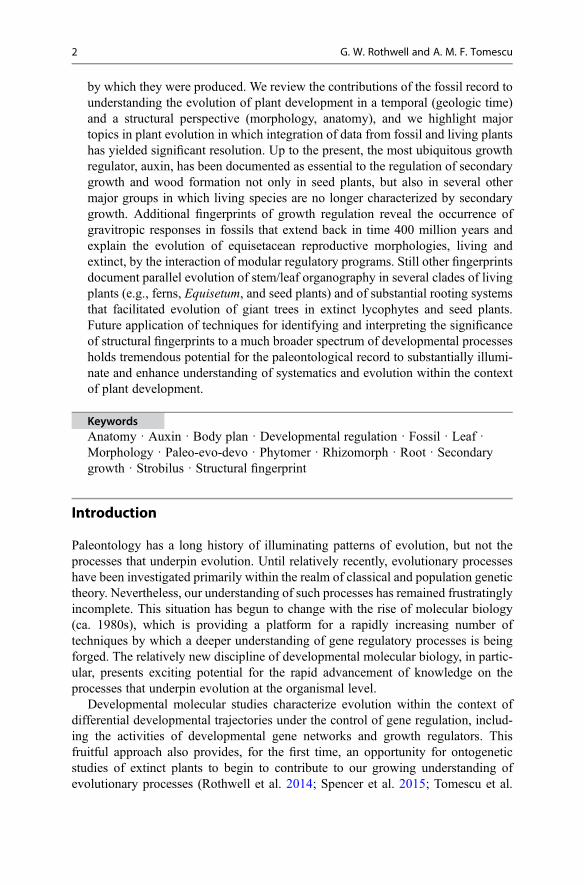
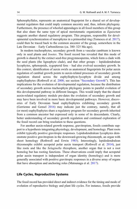
The precisely structured anatomy of plants is the result of spatially and temporallycoordinated sequences of cell division, growth, and differentiation. One aspect ofsuch developmental sequences is the early partitioning of meristematic tissues intodomains with distinct developmental trajectories, i.e., developmental domainpartitioning, such as the specification of protoderm versus ground meristem versusprocambium in apical meristems. In the root apical meristem, another aspect ofdevelopmental domain partitioning involves the early establishment of the Körper(body) and Kappe (cap) domains, characterized by distinct patterns of cell division.The two domains cover different extents of the root apical meristem and give rise todifferent tissues of the root in different plant lineages; therefore, this partitioningbears a phylogenetic signal. Importantly, because they are identified based on patternsof cell division, the Körper and Kappe domains can be recognized in fossils withanatomical preservation, and not just in live, developing plants. This has allowed forrecognition of a type of gymnospermous Körper-Kappe organization in a Carbonif-erous (ca. 320 Ma) root apical meristem (Fig. 1) that is different from those of allextant gymnosperms (Hetherington et al. 2016a) and, thus, reveals structural diversitypreviously unaccounted for, that could be used in phylogenetic inference.
Plant reproductive structures are often produced as a result of expression of areproductive regulatory module in meristems otherwise responsible for vegetativegrowth. Reproductive regulatory modules likely conserved across embryophytesinvolve LEAFY genes, the AP2 gene subfamily, MIKC MADS-box genes, andPolycomb group genes (Tomescu et al. 2017). In all known cases, the reproductivegrowth mode is activated in apical meristems. However, Paleozoic and Mesozoicsphenopsid fossils have recently been shown to exhibit patterns of size and positioningof reproductive structures (sporangiophores) consistent with activation of a reproduc-tive regulatory module in intercalary meristems. This has implications for the topol-ogy and mode of meristematic growth, suggesting that growth in reproductive modecan be effected not only by apical meristems, but also by intercalarymeristems. This isthe first example of reproductive growth arising from intercalarymeristems, a mode ofgrowth that could not have been predicted from the modern flora alone, and which hasdeep implications for the homology and evolution of sphenopsid reproductive struc-tures (see section “The Equisetum strobilus: A Case of Reciprocal Illumination”).
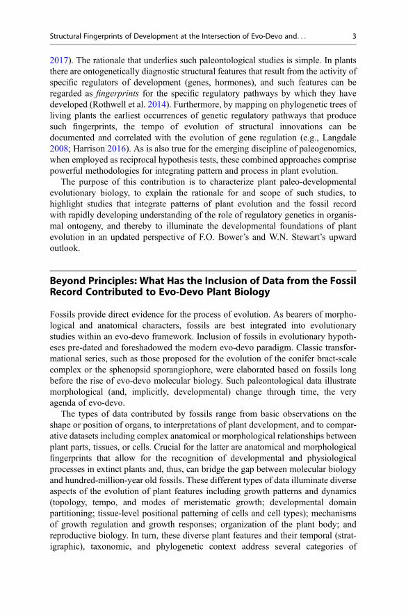
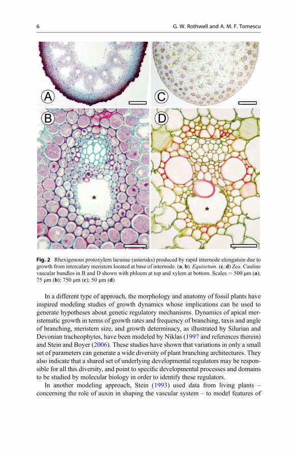
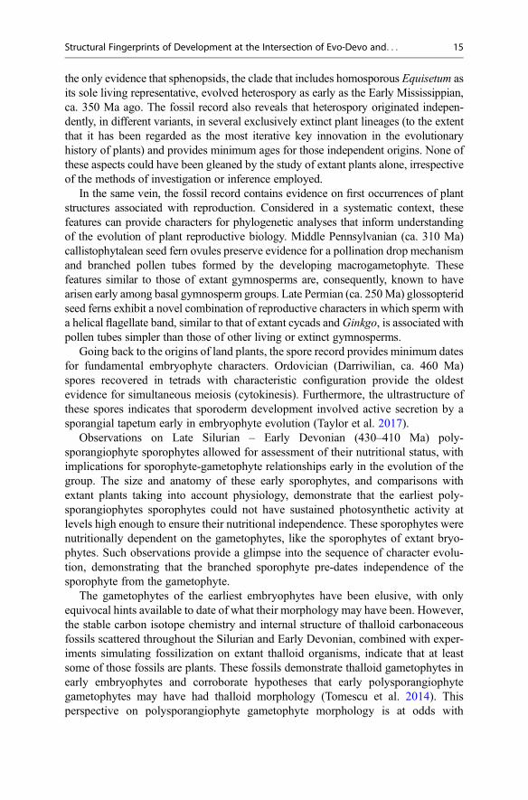
Also associated with intercalary meristematic growth, rapid internode elongationthat exceeds the tensional capacity of mature protoxylem cells generates rhexigenousprotoxylem lacunae. Such lacunae found in Equisetum and grasses (Fig. 2) indicatethat rapid growth from intercalary meristems evolved independently in distant plantlineages. The lacunae also provide a fingerprint for this topology (position) andtempo of meristematic growth that can be identified in the fossil record. If the
4 G. W. Rothwell and A. M. F. Tomescu
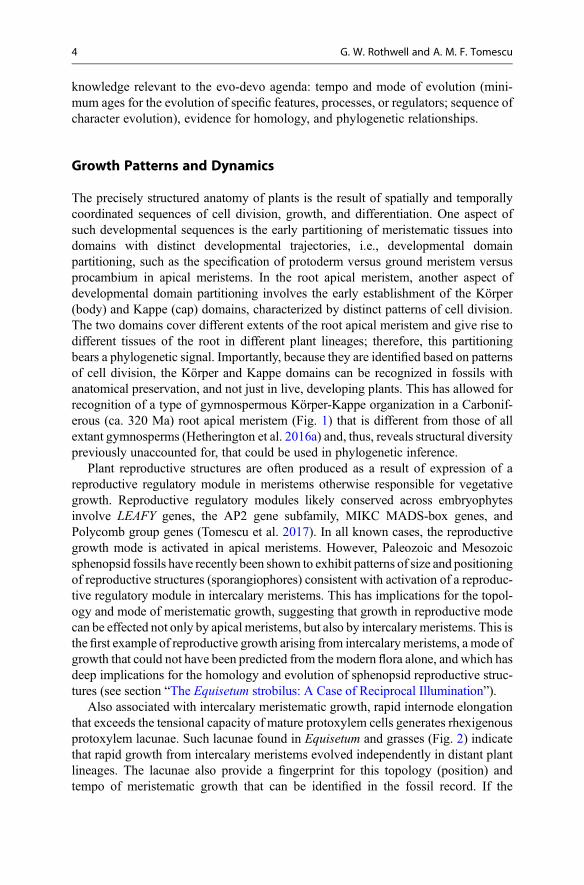
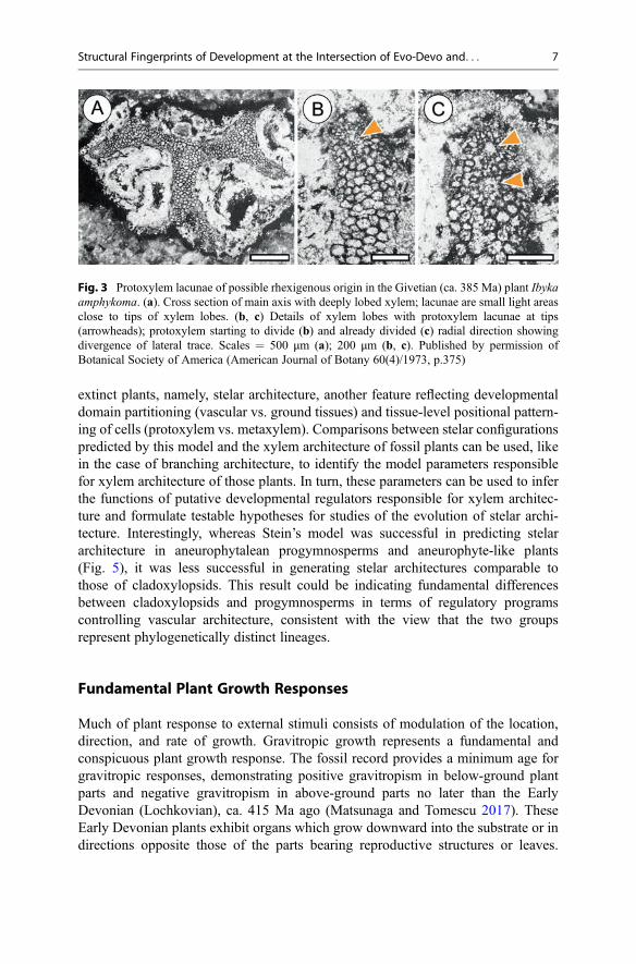
Givetian (ca. 385 Ma) plant Ibyka (Fig. 3) does indeed include rhexigenous proto-xylem lacunae (as opposed to areas of incomplete preservation of protoxylemparenchyma), such rapid intercalary meristematic growth may have evolved asearly as the Middle Devonian.
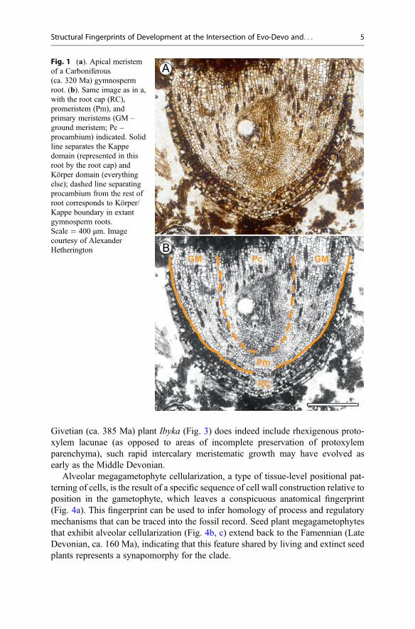
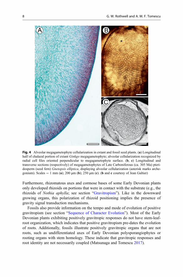
Alveolar megagametophyte cellularization, a type of tissue-level positional pat-terning of cells, is the result of a specific sequence of cell wall construction relative toposition in the gametophyte, which leaves a conspicuous anatomical fingerprint(Fig. 4a). This fingerprint can be used to infer homology of process and regulatorymechanisms that can be traced into the fossil record. Seed plant megagametophytesthat exhibit alveolar cellularization (Fig. 4b, c) extend back to the Famennian (LateDevonian, ca. 160 Ma), indicating that this feature shared by living and extinct seedplants represents a synapomorphy for the clade.
Fig. 1 (a). Apical meristemof a Carboniferous(ca. 320 Ma) gymnospermroot. (b). Same image as in a,with the root cap (RC),promeristem (Pm), andprimary meristems (GM –ground meristem; Pc –procambium) indicated. Solidline separates the Kappedomain (represented in thisroot by the root cap) andKörper domain (everythingelse); dashed line separatingprocambium from the rest ofroot corresponds to Körper/Kappe boundary in extantgymnosperm roots.Scale = 400 μm. Imagecourtesy of AlexanderHetherington
Structural Fingerprints of Development at the Intersection of Evo-Devo and. . . 5
In a different type of approach, the morphology and anatomy of fossil plants haveinspired modeling studies of growth dynamics whose implications can be used togenerate hypotheses about genetic regulatory mechanisms. Dynamics of apical mer-istematic growth in terms of growth rates and frequency of branching, taxis and angleof branching, meristem size, and growth determinacy, as illustrated by Silurian andDevonian tracheophytes, have been modeled by Niklas (1997 and references therein)and Stein and Boyer (2006). These studies have shown that variations in only a smallset of parameters can generate a wide diversity of plant branching architectures. Theyalso indicate that a shared set of underlying developmental regulators may be respon-sible for all this diversity, and point to specific developmental processes and domainsto be studied by molecular biology in order to identify these regulators.
In another modeling approach, Stein (1993) used data from living plants –concerning the role of auxin in shaping the vascular system – to model features of
Fig. 2 Rhexigenous protoxylem lacunae (asterisks) produced by rapid internode elongation due togrowth from intercalary meristem located at base of internode. (a, b). Equisetum. (c, d) Zea. Caulinevascular bundles in B and D shown with phloem at top and xylem at bottom. Scales = 500 μm (a);75 μm (b); 750 μm (c); 50 μm (d)
6 G. W. Rothwell and A. M. F. Tomescu
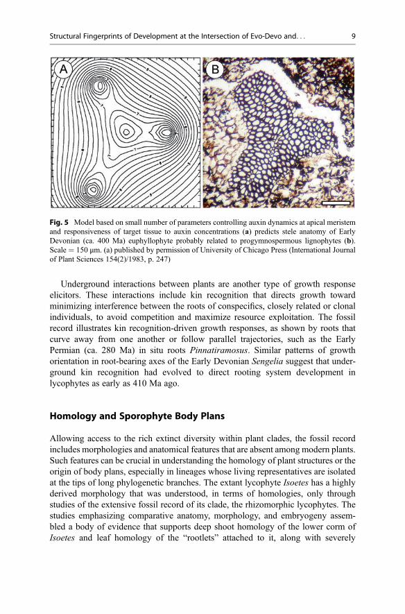
extinct plants, namely, stelar architecture, another feature reflecting developmentaldomain partitioning (vascular vs. ground tissues) and tissue-level positional pattern-ing of cells (protoxylem vs. metaxylem). Comparisons between stelar configurationspredicted by this model and the xylem architecture of fossil plants can be used, likein the case of branching architecture, to identify the model parameters responsiblefor xylem architecture of those plants. In turn, these parameters can be used to inferthe functions of putative developmental regulators responsible for xylem architec-ture and formulate testable hypotheses for studies of the evolution of stelar archi-tecture. Interestingly, whereas Stein’s model was successful in predicting stelararchitecture in aneurophytalean progymnosperms and aneurophyte-like plants(Fig. 5), it was less successful in generating stelar architectures comparable tothose of cladoxylopsids. This result could be indicating fundamental differencesbetween cladoxylopsids and progymnosperms in terms of regulatory programscontrolling vascular architecture, consistent with the view that the two groupsrepresent phylogenetically distinct lineages.
Fundamental Plant Growth Responses
Much of plant response to external stimuli consists of modulation of the location,direction, and rate of growth. Gravitropic growth represents a fundamental andconspicuous plant growth response. The fossil record provides a minimum age forgravitropic responses, demonstrating positive gravitropism in below-ground plantparts and negative gravitropism in above-ground parts no later than the EarlyDevonian (Lochkovian), ca. 415 Ma ago (Matsunaga and Tomescu 2017). TheseEarly Devonian plants exhibit organs which grow downward into the substrate or indirections opposite those of the parts bearing reproductive structures or leaves.
Fig. 3 Protoxylem lacunae of possible rhexigenous origin in the Givetian (ca. 385 Ma) plant Ibykaamphykoma. (a). Cross section of main axis with deeply lobed xylem; lacunae are small light areasclose to tips of xylem lobes. (b, c) Details of xylem lobes with protoxylem lacunae at tips(arrowheads); protoxylem starting to divide (b) and already divided (c) radial direction showingdivergence of lateral trace. Scales = 500 μm (a); 200 μm (b, c). Published by permission ofBotanical Society of America (American Journal of Botany 60(4)/1973, p.375)
Structural Fingerprints of Development at the Intersection of Evo-Devo and. . . 7
Furthermore, rhizomatous axes and cormose bases of some Early Devonian plantsonly developed rhizoids on portions that were in contact with the substrate (e.g., therhizoids of Nothia aphylla; see section “Gravitropism”). Like in the downwardgrowing organs, this polarization of rhizoid positioning implies the presence ofgravity signal transduction mechanisms.
Fossils also provide information on the tempo and mode of evolution of positivegravitropism (see section “Sequence of Character Evolution”). Most of the EarlyDevonian plants exhibiting positively gravitropic responses do not have stem-leaf-root organization, which indicates that positive gravitropism pre-dates the evolutionof roots. Additionally, fossils illustrate positively gravitropic organs that are notroots, such as undifferentiated axes of Early Devonian polysporangiophytes orrooting organs with stem homology. These indicate that gravitropic responses androot identity are not necessarily coupled (Matsunaga and Tomescu 2017).
Fig. 4 Alveolar megagametophyte cellularization in extant and fossil seed plants. (a) Longitudinalhalf of chalazal portion of extant Ginkgo megagametophyte; alveolar cellularization recognized byradial cell files oriented perpendicular to megagametophyte surface. (b, c) Longitudinal andtransverse sections (respectively) of megagametophytes of Late Carboniferous (ca. 305 Ma) pteri-dosperm (seed fern) Gnetopsis elliptica, displaying alveolar cellularization (asterisk marks arche-gonium). Scales = 1 mm (a); 200 μm (b); 250 μm (c). (b and c courtesy of Jean Galtier)
8 G. W. Rothwell and A. M. F. Tomescu
Underground interactions between plants are another type of growth responseelicitors. These interactions include kin recognition that directs growth towardminimizing interference between the roots of conspecifics, closely related or clonalindividuals, to avoid competition and maximize resource exploitation. The fossilrecord illustrates kin recognition-driven growth responses, as shown by roots thatcurve away from one another or follow parallel trajectories, such as the EarlyPermian (ca. 280 Ma) in situ roots Pinnatiramosus. Similar patterns of growthorientation in root-bearing axes of the Early Devonian Sengelia suggest that under-ground kin recognition had evolved to direct rooting system development inlycophytes as early as 410 Ma ago.
Homology and Sporophyte Body Plans
Allowing access to the rich extinct diversity within plant clades, the fossil recordincludes morphologies and anatomical features that are absent among modern plants.Such features can be crucial in understanding the homology of plant structures or theorigin of body plans, especially in lineages whose living representatives are isolatedat the tips of long phylogenetic branches. The extant lycophyte Isoetes has a highlyderived morphology that was understood, in terms of homologies, only throughstudies of the extensive fossil record of its clade, the rhizomorphic lycophytes. Thestudies emphasizing comparative anatomy, morphology, and embryogeny assem-bled a body of evidence that supports deep shoot homology of the lower corm ofIsoetes and leaf homology of the “rootlets” attached to it, along with severely
Fig. 5 Model based on small number of parameters controlling auxin dynamics at apical meristemand responsiveness of target tissue to auxin concentrations (a) predicts stele anatomy of EarlyDevonian (ca. 400 Ma) euphyllophyte probably related to progymnospermous lignophytes (b).Scale = 150 μm. (a) published by permission of University of Chicago Press (International Journalof Plant Sciences 154(2)/1983, p. 247)
Structural Fingerprints of Development at the Intersection of Evo-Devo and. . . 9
diminished elongation growth and branching capacities of the main axes, comparedto extinct relatives (e.g., Lepidodendrales; see section “Lepidodendralean RootingStructures”).
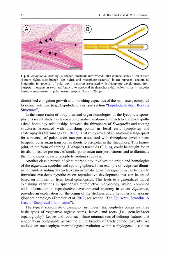
In the same realm of body plan and organ homologies of the lycophyte sporo-phyte, a recent study has taken a comparative anatomy approach to address hypoth-esized homology relationships between the rhizophore of Selaginella and rootingstructures associated with branching points in fossil early lycophytes andzosterophylls (Matsunaga et al. 2017). That study revealed an anatomical fingerprintfor a reversal of polar auxin transport associated with rhizophore development:basipetal polar auxin transport in shoots to acropetal in the rhizophore. This finger-print, in the form of arching (U-shaped) tracheids (Fig. 6), could be sought for infossils, to test for presence of similar polar auxin transport patterns and to illuminatethe homologies of early lycophyte rooting structures.
Another classic puzzle of plant morphology involves the origin and homologiesof the Equisetum strobilus and sporangiophore. In an example of reciprocal illumi-nation, understanding of vegetative meristematic growth in Equisetum can be used toformulate evo-devo hypotheses on reproductive development that can be testedbased on information from fossil sphenopsids. This leads to a generalized modelexplaining variations in sphenopsid reproductive morphology, which, combinedwith information on reproductive developmental anatomy in extant Equisetum,provides an explanation for the origin of the strobilus and a hypothesis of sporan-giophore homology (Tomescu et al. 2017; see section “The Equisetum Strobilus: ACase of Reciprocal Illumination”).
The typical sporophyte organization in modern tracheophytes comprises threebasic types of vegetative organs: stems, leaves, and roots (i.e., stem-leaf-rootorganography). Leaves and roots each share minimal sets of defining features thatrender them comparable across the entire breadth of tracheophyte diversity. Anoutlook on tracheophyte morphological evolution within a phylogenetic context
Fig. 6 Selaginella. Arching (U-shaped) tracheids (arrowheads) that connect steles of main stem(bottom right), side branch (top right), and rhizophore (asterisk) in (a) represent anatomicalfingerprint for reversal of polar auxin transport associated with rhizophore development: frombasipetal transport in stem and branch, to acropetal in rhizophore (b); yellow strips = vasculartissue; orange arrows = polar auxin transport. Scale = 200 μm
10 G. W. Rothwell and A. M. F. Tomescu
that excludes the fossil record can easily take these features and their ubiquity inextant plants as indicating that leaves and roots are each homologous across allvascular plants. Conversely, inclusion of the fossil record in such a broad outlookplays a crucial role in resolving major aspects of the evolution of this basic body planand the homologies of leaves and roots. Specifically, Late Silurian and Devoniantracheophytes characterized by simple body plans (undifferentiated branching axesbearing sporangia) form paraphyletic grades at the base of major branches oftracheophyte phylogeny, demonstrating that stem-leaf-root organography evolvedindependently in different lineages (Rothwell et al. 2014). This implies that neitherleaves nor roots are homologous across different lineages. Furthermore, several linesof evidence reveal that leaves and roots almost certainly evolved independently morethan twice (Boyce and Knoll 2002; Tomescu 2009; see section “Euphyllophyte LeafEvolution”).
Sequence of Character Evolution
Plant phylogenies can be used to infer the mode of morphological evolution.Character distribution on phylogenetic trees can be and has been used to infersequences of character evolution and ancestral character states. However, becausephylogenetic trees represent hypotheses of relationships, sequences of characterevolution predicted based on them are just as hypothetical. This is particularlyevident in systematic trees that exclude extinct taxa (Rothwell and Nixon 2006).Within this context, fossils provide the only direct means for testing sequences ofcharacter evolution. Presence or absence of structures and anatomical features infossils of different ages within a lineage provide direct evidence for the order ofappearance of those features. An example is the sequential evolution of characters inorgans we call leaves, during their independent parallel evolution in ferns and seedplants. Fossils demonstrate that whereas seed plants evolved determinate growth andbroad pinnules before adaxial-abaxial polarity in the leaves, in filicalean fern leavesevolution of adaxial-abaxial polarity preceded broad pinnules and determinacy(Sanders et al. 2009; see section “Euphyllophyte Leaf Evolution”).
Lycophyte rooting structures are diverse and so are their homologies, some ofwhich are not fully resolved (Rothwell and Erwin 1985; Tomescu 2011; Matsunagaet al. 2017). The oldest unequivocal lycophyte roots were described in the EarlyDevonian plant Sengelia, which produced roots on specialized axes of the branchingsystem that are stem homologs. Sengelia rooting systems consist of horizontal ordownward-growing root-bearing axes with laterally diverging roots. In all cases, theroots expand in a horizontal plane, irrespective of the orientation of subtending root-bearing axes. These observations indicate that, in lycophytes, root identity wasuncoupled from positive gravitropism, a feature fundamentally associated withmodern plant roots – in Sengelia, the organs that exhibit a gravitropic response arethe root-bearing axes and not the roots. The roots of Sengelia also provide evidencefor the sequence of character evolution: roots acquired positive gravitropism afterthey evolved as distinct organs, in lycophytes (Matsunaga and Tomescu 2017).
Structural Fingerprints of Development at the Intersection of Evo-Devo and. . . 11
Early Devonian strata have yielded euphyllophytes as old as 407 Ma that exhibitsecondary growth (wood production) from a vascular cambium (Gerrienne andGensel 2016) (euphyllophytes are the sister clade to lycophytes and includepsilotophytes, ferns, sphenopsids, and seed plants, along with diverse related line-ages). These fossils provide a minimum age for the evolution of this importantstructural feature. Furthermore, the fact that these early wood producers have simplesporophyte organization (undifferentiated axes) indicates that secondary growthpre-dates the evolution of complex body plans with stem-leaf-root differentiation,in euphyllophytes. The small size of these wood-producing sporophytes suggeststhat secondary growth evolved primarily in response to selective pressures related tomaximizing hydraulic conductance and not mechanical stiffness (Gerrienne andGensel 2016; see sections “Developmental Regulation” and “Secondary Growth”).
Developmental Regulation
Plant fossils exhibit combinations of characters unknown in modern plants andpreserve anatomical and morphological fingerprints for developmental processesand physiological mechanisms. Aside from implications for sequences of characterevolution (see section “Sequence of Character Evolution”), when considered in theirstratigraphic (temporal) and taxonomic context, these types of data provide glimpsesinto the systems biology of developmental regulation and its evolution. In manyinstances, the resulting perspectives inform understanding of the modularity ofdevelopmental regulatory networks, hierarchy of regulatory modules, synchroniza-tion in developmental processes, or relationships between physiology anddevelopment.
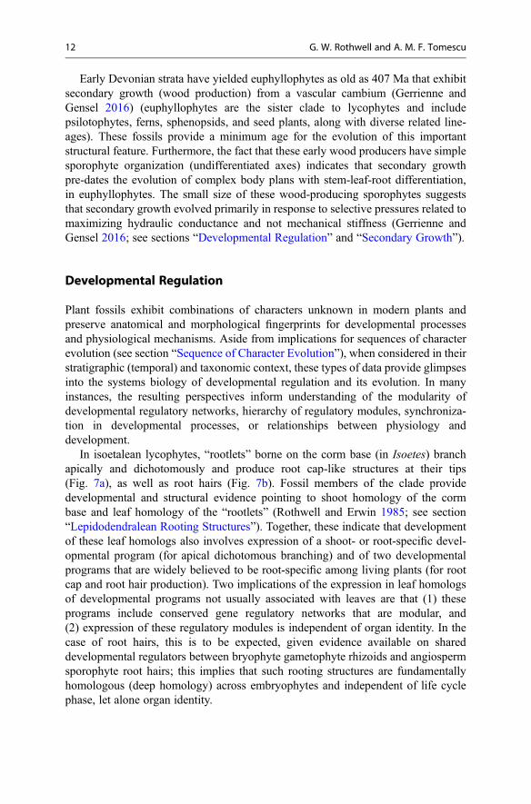
In isoetalean lycophytes, “rootlets” borne on the corm base (in Isoetes) branchapically and dichotomously and produce root cap-like structures at their tips(Fig. 7a), as well as root hairs (Fig. 7b). Fossil members of the clade providedevelopmental and structural evidence pointing to shoot homology of the cormbase and leaf homology of the “rootlets” (Rothwell and Erwin 1985; see section“Lepidodendralean Rooting Structures”). Together, these indicate that developmentof these leaf homologs also involves expression of a shoot- or root-specific devel-opmental program (for apical dichotomous branching) and of two developmentalprograms that are widely believed to be root-specific among living plants (for rootcap and root hair production). Two implications of the expression in leaf homologsof developmental programs not usually associated with leaves are that (1) theseprograms include conserved gene regulatory networks that are modular, and(2) expression of these regulatory modules is independent of organ identity. In thecase of root hairs, this is to be expected, given evidence available on shareddevelopmental regulators between bryophyte gametophyte rhizoids and angiospermsporophyte root hairs; this implies that such rooting structures are fundamentallyhomologous (deep homology) across embryophytes and independent of life cyclephase, let alone organ identity.
12 G. W. Rothwell and A. M. F. Tomescu
In light of the homologies of the body plan of Isoetes, as resolved by data from thefossil record, the presence of root cap-like structures in the “rootlets” of this plantprovides interesting phylogenetic perspectives. These root cap-like structures, pre-sent on organs that are not root homologues, could imply that the root cap pre-datesroots and evolved on less specialized axes with rooting function, if rhizomorphiclycophytes evolved directly from ancestors devoid of roots. However, becauseamong modern tracheophytes the root cap is known exclusively in roots, anotherpossible explanation, namely, that Isoetes descends from ancestors that had trueroots with root caps, seems more probable. This hypothesis has implications forlycophyte phylogeny and character evolution, consistent with previous ideas that theclade of root-less lycophytes that includes Isoetes (rhizomorphic clade) occupies aderived position in the lycophyte clade.
In equisetacean sphenopsids, the fossil record yielded several Paleozoic fossilsexhibiting character combinations that fill important gaps in terms of morphologicalevolution between modern Equisetum and ancestral forms. Considered in the devel-opmental context provided by modern Equisetum, these fossils were crucial in thedevelopment of hypotheses that explain the morphology of equisetacean reproduc-tive structures as the result of a hierarchic system of modular regulatory programs.Nested within this set of hypotheses are also implications for the developmentalprogram of the sporangiophore, which may represent a conserved regulatory moduleresponsible for the development of basic fertile lateral branching systems, and fortiming of the evolution of this module, which may have preceded the evolution ofstem-leaf-root organography (Tomescu et al. 2017; see section “The EquisetumStrobilus: A Case of Reciprocal Illumination”).
At the scale of the sphenopsid group, the presence of intercalary meristems at thebase of each internode in Equisetaceae, and in fossil Calamitaceae and
Fig. 7 Appendages (“rootlets”) of Isoetes corm base (rhizomorph) bear structures typical of roots –a protective cap on the apical meristem (a) and absorptive hairs (b) – even though they are leafhomologs; note incipient isotomous branching of the “rootlet” apical meristem. Scales = 75 μm
Structural Fingerprints of Development at the Intersection of Evo-Devo and. . . 13
Sphenophyllales, represents an anatomical fingerprint for a shared set of develop-mental regulators that could imply common ancestry and, thus, inform phylogeny.Furthermore, the presence of whorled appendages in all fossil members of this groupthat also share the same type of apical meristematic organization as Equisetumsuggests another shared regulatory program. This program, responsible for devel-opmental synchronization of merophytes in a primordial ring (Tomescu et al. 2017),could then be traced back to the common ancestor of the group, somewhere in theLate Devonian – Early Carboniferous (ca. 380–325 Ma ago).
In modern tracheophytes, secondary growth from a vascular cambium is knownonly in seed plants and Isoetes. The fossil record has revealed that this mode ofgrowth is shared by the extinct seed-free progymnosperms, which form a clade withthe seed plants (the lignophyte clade), and that other groups – lepidodendraleanlycophytes, sphenopsids, zygopterid fens – had also evolved secondary growth. Inthis context, identification of auxin swirls as anatomical fingerprints for polar auxinregulation of cambial growth points to auxin-related processes of secondary growthregulation shared across the euphyllophyte-lycophyte divide and amongeuphyllophytes (Rothwell et al. 2008; see section “Secondary Growth”). This hasimplications for the evolution of developmental regulation, because the distributionof secondary growth across tracheophyte phylogeny points to parallel evolution ofthis developmental pathway in different lineages. This would imply that the sharedauxin-related regulatory module pre-dates the evolution of secondary growth andmay have been involved in more basic developmental pathways. However, discov-eries of Early Devonian basal euphyllophytes exhibiting secondary growth(Gerrienne and Gensel 2016) may indicate just the contrary, namely, that all(or most) euphyllophytes share a regulatory program for secondary growth inheritedfrom a common ancestor but expressed only in some of its descendants. Clearly,better understanding of secondary growth regulation and continued exploration ofthe fossil record can bring resolution to these questions.
For another auxin-related growth response, gravitropism, fossils contribute sup-port to a hypothesis integrating physiology, development, and homology. Plant rootsexhibit typically positive gravitropic responses. Lepidodendralean lycophytes dem-onstrate positive gravitropism in the downward-growing rhizomorph, an organ withshoot homology (Rothwell and Erwin 1985). Interestingly, lepidodendraleanrhizomorphs exhibit acropetal polar auxin transport (Rothwell et al. 2014), justlike roots and like the Selaginella rhizophore, another organ that is not a roothomolog but has rooting functions. These observations could imply that acropetalpolar auxin transport is independent of organ identity (homology) and is moregenerally associated with positive gravitropic responses in a diverse array of organsthat have absorption and anchoring roles (Matsunaga et al. 2017).
Life Cycles, Reproductive Systems
The fossil record has provided direct and indirect evidence for the timing and mode ofevolution of reproductive biology and plant life cycles. For instance, fossils provide
14 G. W. Rothwell and A. M. F. Tomescu
the only evidence that sphenopsids, the clade that includes homosporous Equisetum asits sole living representative, evolved heterospory as early as the Early Mississippian,ca. 350 Ma ago. The fossil record also reveals that heterospory originated indepen-dently, in different variants, in several exclusively extinct plant lineages (to the extentthat it has been regarded as the most iterative key innovation in the evolutionaryhistory of plants) and provides minimum ages for those independent origins. None ofthese aspects could have been gleaned by the study of extant plants alone, irrespectiveof the methods of investigation or inference employed.
In the same vein, the fossil record contains evidence on first occurrences of plantstructures associated with reproduction. Considered in a systematic context, thesefeatures can provide characters for phylogenetic analyses that inform understandingof the evolution of plant reproductive biology. Middle Pennsylvanian (ca. 310 Ma)callistophytalean seed fern ovules preserve evidence for a pollination drop mechanismand branched pollen tubes formed by the developing macrogametophyte. Thesefeatures similar to those of extant gymnosperms are, consequently, known to havearisen early among basal gymnosperm groups. Late Permian (ca. 250Ma) glossopteridseed ferns exhibit a novel combination of reproductive characters in which sperm witha helical flagellate band, similar to that of extant cycads andGinkgo, is associated withpollen tubes simpler than those of other living or extinct gymnosperms.
Going back to the origins of land plants, the spore record provides minimum datesfor fundamental embryophyte characters. Ordovician (Darriwilian, ca. 460 Ma)spores recovered in tetrads with characteristic configuration provide the oldestevidence for simultaneous meiosis (cytokinesis). Furthermore, the ultrastructure ofthese spores indicates that sporoderm development involved active secretion by asporangial tapetum early in embryophyte evolution (Taylor et al. 2017).
Observations on Late Silurian – Early Devonian (430–410 Ma) poly-sporangiophyte sporophytes allowed for assessment of their nutritional status, withimplications for sporophyte-gametophyte relationships early in the evolution of thegroup. The size and anatomy of these early sporophytes, and comparisons withextant plants taking into account physiology, demonstrate that the earliest poly-sporangiophytes sporophytes could not have sustained photosynthetic activity atlevels high enough to ensure their nutritional independence. These sporophytes werenutritionally dependent on the gametophytes, like the sporophytes of extant bryo-phytes. Such observations provide a glimpse into the sequence of character evolu-tion, demonstrating that the branched sporophyte pre-dates independence of thesporophyte from the gametophyte.
The gametophytes of the earliest embryophytes have been elusive, with onlyequivocal hints available to date of what their morphology may have been. However,the stable carbon isotope chemistry and internal structure of thalloid carbonaceousfossils scattered throughout the Silurian and Early Devonian, combined with exper-iments simulating fossilization on extant thalloid organisms, indicate that at leastsome of those fossils are plants. These fossils demonstrate thalloid gametophytes inearly embryophytes and corroborate hypotheses that early polysporangiophytegametophytes may have had thalloid morphology (Tomescu et al. 2014). Thisperspective on polysporangiophyte gametophyte morphology is at odds with
Structural Fingerprints of Development at the Intersection of Evo-Devo and. . . 15
predictions based on phylogenetic studies that place mosses, which have leafygametophytes, as the sister group of polysporangiophytes.
Morphological evolution in the gametophytes of early polysporangiophytes has astory of which we have uncovered only some parts, and those thanks to the fossilrecord. Unlike the thalloid gametophytes of early polysporangiophytes, the nextoldest known gametophytes, belonging to Early Devonian (Lochkovian–Pragian,ca. 408 Ma) protracheophytes and tracheophytes of the Rhynie chert, exhibit mor-phologies with no counterpart in the modern flora. Their morphology is similar tothat of sporophytes, with axial organization, branched architecture, and vasculartissues. The only approximation to this morphology in living plants are the subter-ranean gametophytes of Psilotum and Tmesipteris, which are also axial, can branch,and can be vascularized. However, compared to these, Rhynie chert gametophyteswere larger, highly branched, and developed above-ground. Currently, it is not clearhow polysporangiophyte gametophytes evolved from a basic thalloid morphology tothe axial forms seen in the Rhynie chert, and then back to the primarily thalloidforms seen in living seed-free vascular plants. These present questions in evo-devowhose answers will require additional data from the fossil record.
The Early Devonian Rhynie chert plants also provide the oldest direct evidence ofanisospory (segregation of micro- and megaspores within the same sporangium), areproductive system currently known only in a subset of bryophytes, in a few Isoetesspecies and, potentially, in Equisetum and Ceratopteris. Specifically, gametophytesof the Rhynie chert protracheophyte Aglaophyton are found forming densepopulations, when preserved in situ. Because Aglaophyton spores were dispersedas masses representing the contents of whole sporangia, such dense gametophytetufts probably represent the product of individual sporangia. Although spore sizeshows no bimodal distribution within Aglaophyton sporangia and gametophytes areexclusively unisexual, the gametophyte tufts always include mixtures of both sexes.These have been interpreted as indicating anisospory of Aglaophyton and otherRhynie chert plants (Taylor et al. 2005). Like in the case of gametophyte morphol-ogy, it is not yet clear what the anisospory of Rhynie chert plants means for thecomplex picture of plant life cycle evolution.
Emblematic Case Studies
Gravitropism
Tropisms play a major role in plant development and evidence is accumulating thattropic responses influenced growth of the earliest land plants. Fossils from the EarlyDevonian reveal that rooting organs of lycophytes appeared long before those of theother major clade of vascular plants (euphyllophytes), for which roots are not knownuntil the Middle Devonian. Evidence for tropic responses in the aerial and rootingstructures of Early Devonian lycophytes is provided by several plants, includingDrepanophycus and Sengelia (Matsunaga and Tomescu 2017). These plants haveleafy axes that extend in one direction (horizontally or upwards) and smaller smooth
16 G. W. Rothwell and A. M. F. Tomescu
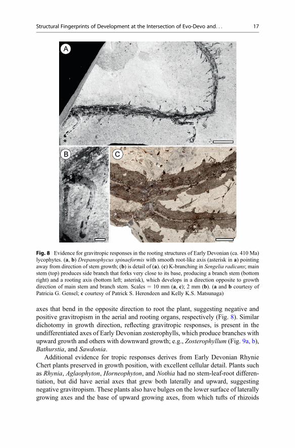
axes that bend in the opposite direction to root the plant, suggesting negative andpositive gravitropism in the aerial and rooting organs, respectively (Fig. 8). Similardichotomy in growth direction, reflecting gravitropic responses, is present in theundifferentiated axes of Early Devonian zosterophylls, which produce branches withupward growth and others with downward growth; e.g., Zosterophyllum (Fig. 9a, b),Bathurstia, and Sawdonia.
Additional evidence for tropic responses derives from Early Devonian RhynieChert plants preserved in growth position, with excellent cellular detail. Plants suchas Rhynia, Aglaophyton, Horneophyton, and Nothia had no stem-leaf-root differen-tiation, but did have aerial axes that grew both laterally and upward, suggestingnegative gravitropism. These plants also have bulges on the lower surface of laterallygrowing axes and the base of upward growing axes, from which tufts of rhizoids
Fig. 8 Evidence for gravitropic responses in the rooting structures of Early Devonian (ca. 410 Ma)lycophytes. (a, b) Drepanophycus spinaeformis with smooth root-like axis (asterisk in a) pointingaway from direction of stem growth; (b) is detail of (a). (c) K-branching in Sengelia radicans; mainstem (top) produces side branch that forks very close to its base, producing a branch stem (bottomright) and a rooting axis (bottom left; asterisk), which develops in a direction opposite to growthdirection of main stem and branch stem. Scales = 10 mm (a, c); 2 mm (b). (a and b courtesy ofPatricia G. Gensel; c courtesy of Patrick S. Herendeen and Kelly K.S. Matsunaga)
Structural Fingerprints of Development at the Intersection of Evo-Devo and. . . 17
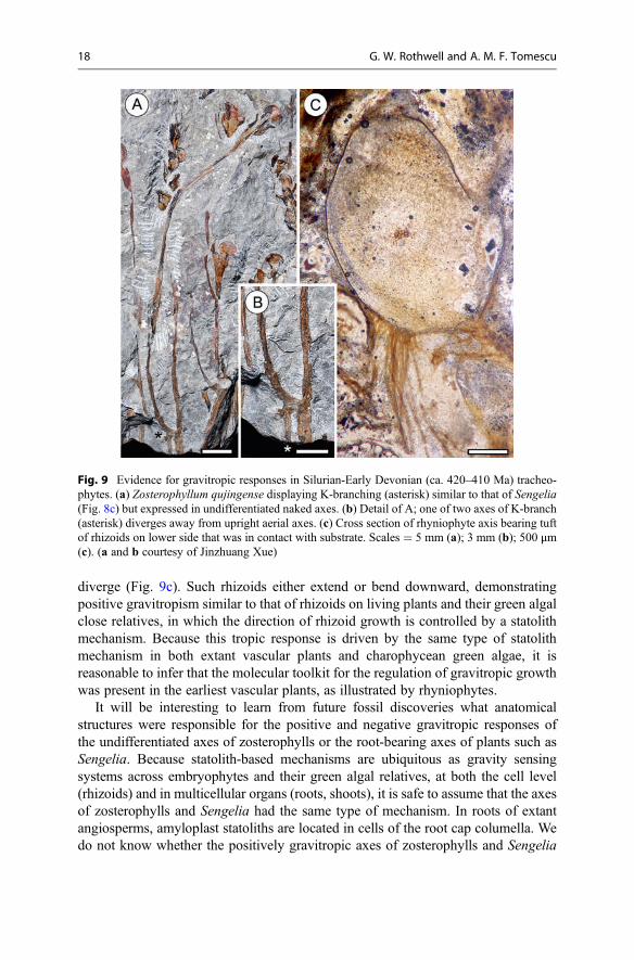
diverge (Fig. 9c). Such rhizoids either extend or bend downward, demonstratingpositive gravitropism similar to that of rhizoids on living plants and their green algalclose relatives, in which the direction of rhizoid growth is controlled by a statolithmechanism. Because this tropic response is driven by the same type of statolithmechanism in both extant vascular plants and charophycean green algae, it isreasonable to infer that the molecular toolkit for the regulation of gravitropic growthwas present in the earliest vascular plants, as illustrated by rhyniophytes.
It will be interesting to learn from future fossil discoveries what anatomicalstructures were responsible for the positive and negative gravitropic responses ofthe undifferentiated axes of zosterophylls or the root-bearing axes of plants such asSengelia. Because statolith-based mechanisms are ubiquitous as gravity sensingsystems across embryophytes and their green algal relatives, at both the cell level(rhizoids) and in multicellular organs (roots, shoots), it is safe to assume that the axesof zosterophylls and Sengelia had the same type of mechanism. In roots of extantangiosperms, amyloplast statoliths are located in cells of the root cap columella. Wedo not know whether the positively gravitropic axes of zosterophylls and Sengelia
Fig. 9 Evidence for gravitropic responses in Silurian-Early Devonian (ca. 420–410 Ma) tracheo-phytes. (a) Zosterophyllum qujingense displaying K-branching (asterisk) similar to that of Sengelia(Fig. 8c) but expressed in undifferentiated naked axes. (b) Detail of A; one of two axes of K-branch(asterisk) diverges away from upright aerial axes. (c) Cross section of rhyniophyte axis bearing tuftof rhizoids on lower side that was in contact with substrate. Scales = 5 mm (a); 3 mm (b); 500 μm(c). (a and b courtesy of Jinzhuang Xue)
18 G. W. Rothwell and A. M. F. Tomescu
had root cap-like structures. Considering that zosterophylls did not possess stem-leaf-root differentiation and that the root-bearing axes of Sengelia are not root homologs,it is likely that none of these axes had root cap-like structures, unless regulation ofroot cap development is independent of organography (as it appers to be in Isoetes). Ifa root cap was absent from these positively gravitropic axes, could such axes havehoused statoliths in boundary layers (e.g., endodermis, starch sheath) like thoseresponsible for negative gravitropism in the shoots of extant angiosperms? If so,how would the statolith-based mechanisms responsible for positive gravitropism inthe below-ground axes of zosterophylls and for negative gravitropism in the above-ground axes of those plants have been different? Could they have differed only in thepolarity of the response to a gravitropic stimulus sensed in a shared type of structure?And would these tropic responses have involved redistribution of polar auxin fluxes,as seen in positive and negative gravitropic responses of angiosperm roots andshoots? Answers to all of these questions and their integration into a more completepicture of the evolution of gravitropism will also require understanding of theincompletely explored structural, developmental, and physiological underpinningsof gravitropism in many lineages of extant seed-free plants (the roots and shoots oflycophytes, ferns, Equisetum, or the rhizophores of Selaginella).
Polar Auxin Transport
Auxin is among the most prominent of growth regulators, and polar auxin transportfrom developing leaf primordia in the apical meristem toward the base of the stem,and then toward the apical meristem(s) of the root system, regulates a wide spectrumof developmental processes. Among the most important of those processes is thepatterning of primary vascular architecture and of tracheary elements in the second-ary xylem.
Secondary GrowthVascular tissue differentiation of living plants is under the control of several plantgrowth regulators, including gibberellins, cytokinins, and ethylene, among whichauxin is the most prominent. Moreover, at least some aspects of polar auxin transporthave been identified as far down the green lineage as bryophytes and charophyceanalgae.
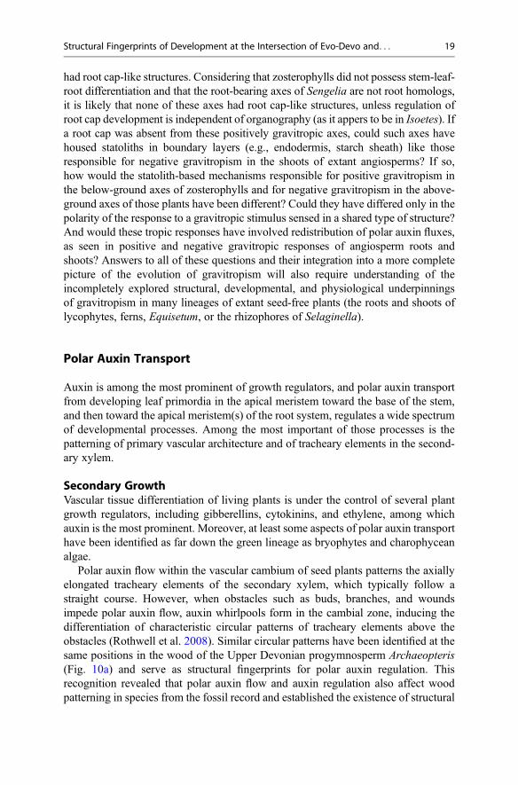
Polar auxin flow within the vascular cambium of seed plants patterns the axiallyelongated tracheary elements of the secondary xylem, which typically follow astraight course. However, when obstacles such as buds, branches, and woundsimpede polar auxin flow, auxin whirlpools form in the cambial zone, inducing thedifferentiation of characteristic circular patterns of tracheary elements above theobstacles (Rothwell et al. 2008). Similar circular patterns have been identified at thesame positions in the wood of the Upper Devonian progymnosperm Archaeopteris(Fig. 10a) and serve as structural fingerprints for polar auxin regulation. Thisrecognition revealed that polar auxin flow and auxin regulation also affect woodpatterning in species from the fossil record and established the existence of structural
Structural Fingerprints of Development at the Intersection of Evo-Devo and. . . 19
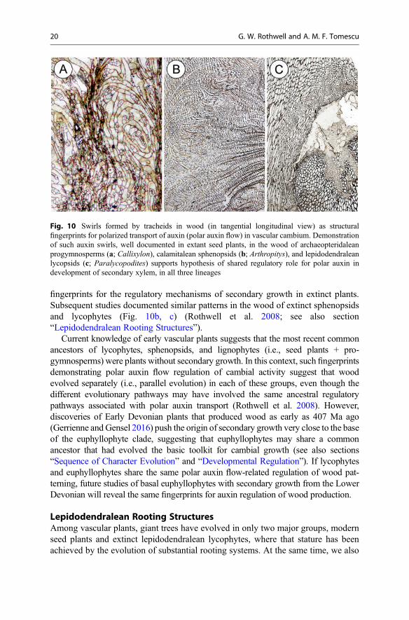
fingerprints for the regulatory mechanisms of secondary growth in extinct plants.Subsequent studies documented similar patterns in the wood of extinct sphenopsidsand lycophytes (Fig. 10b, c) (Rothwell et al. 2008; see also section“Lepidodendralean Rooting Structures”).
Current knowledge of early vascular plants suggests that the most recent commonancestors of lycophytes, sphenopsids, and lignophytes (i.e., seed plants + pro-gymnosperms) were plants without secondary growth. In this context, such fingerprintsdemonstrating polar auxin flow regulation of cambial activity suggest that woodevolved separately (i.e., parallel evolution) in each of these groups, even though thedifferent evolutionary pathways may have involved the same ancestral regulatorypathways associated with polar auxin transport (Rothwell et al. 2008). However,discoveries of Early Devonian plants that produced wood as early as 407 Ma ago(Gerrienne and Gensel 2016) push the origin of secondary growth very close to the baseof the euphyllophyte clade, suggesting that euphyllophytes may share a commonancestor that had evolved the basic toolkit for cambial growth (see also sections“Sequence of Character Evolution” and “Developmental Regulation”). If lycophytesand euphyllophytes share the same polar auxin flow-related regulation of wood pat-terning, future studies of basal euphyllophytes with secondary growth from the LowerDevonian will reveal the same fingerprints for auxin regulation of wood production.
Lepidodendralean Rooting StructuresAmong vascular plants, giant trees have evolved in only two major groups, modernseed plants and extinct lepidodendralean lycophytes, where that stature has beenachieved by the evolution of substantial rooting systems. At the same time, we also
Fig. 10 Swirls formed by tracheids in wood (in tangential longitudinal view) as structuralfingerprints for polarized transport of auxin (polar auxin flow) in vascular cambium. Demonstrationof such auxin swirls, well documented in extant seed plants, in the wood of archaeopteridaleanprogymnosperms (a; Callixylon), calamitalean sphenopsids (b; Arthropitys), and lepidodendraleanlycopsids (c; Paralycopodites) supports hypothesis of shared regulatory role for polar auxin indevelopment of secondary xylem, in all three lineages
20 G. W. Rothwell and A. M. F. Tomescu
recognize that the rooting structures of the two groups have distinctly differenthomologies and arose by divergent evolutionary pathways, and that those differ-ences are understood only because the lycophytes have a rich fossil record.
In seed plants, giant trees are supported by a rooting system that arises from theradicle of a cotyledonary embryo, thus establishing bipolar growth via a system oftrue roots. By contrast, lycophytes do not have cotyledonary embryos and the onlyliving descendent of the giant lepidodendraleans is the tiny quillwort, Isoetes.Interestingly, Isoetes appears to have distinctly unusual structure and growth, unlessinterpreted with reference to extinct relatives (Rothwell and Erwin 1985). Amonglycophytes, the fossil record reveals that giant lepidodendralean trees are rooted by ashoot that is modified for rooting (known as a rhizomorph), rather than by a systemof true roots. As recently emphasized by Hetherington et al. (2016b), homologies ofthe lepidodendralean rooting system are with the shoot system, as originally hypoth-esized more than a century ago.
Interest in similarities between the developmental morphology and anatomy ofIsoetes and the lepidodendralean rooting system (e.g., the fossil genera Stigmariaand Protostigmaria) was rekindled by Stewart (1947), who emphasized thatStigmaria axes have anatomical features that agree more closely with stems thanroots. Stewart also detailed that the leaf-like anatomy and arrangement of stigmarianlateral appendages, referred to as stigmarian rootlets, compare closely to both theleaves and rootlets of Isoetes. Likewise, the elongated branched rooting systems ofStigmaria and the cormose lobed rooting systems of Protostigmaria and Isoetes arenow recognized as growth variations of a common organography (Rothwell andErwin 1985). However, such paleontological evidence was not fully understood orappreciated until much later, despite several, additional paleontological discoveries.Frankenberg and Eggert (1969) reconstructed the overall morphology and anatomyof stigmarian rooting systems, reemphasizing both anatomical and developmentalsimilarities of stigmarian axes to the lepidodendralean stems. The authors furtherdemonstrated that stigmarian rootlets abscised as if they were leaves, and providedadditional support for anatomical and developmental similarities between stigmarianappendages and Isoetes leaves. Concurrently, Jennings (1975) recognized that somelepidodendralean trees were rooted by a cormose Isoetes-like rooting system, thusstrengthening the homologies between living and extinct rhizomorphic lycophytes.Subsequent characterizations of both embryogeny and apical development forrelated lycophytes clarified meristematic activity and embryogeny in the clade, andlaid the groundwork for a comprehensive summary of homologies among lycophyteshoots and stigmarian rooting systems (Rothwell and Erwin 1985).
Most recently, the developmental significance of the overwhelming morpholog-ical, anatomical, developmental, and embryological evidence that stigmarian rootingsystems of lepidodendralean lycophytes (and Isoetes) are homologous to the above-ground shoot systems (Rothwell et al. 2014) has been explained by the discovery offingerprints for polar auxin patterning of xylem in such plants. Polar auxin swirls arenow known to occur in the wood of both the stems and stigmarian rooting axes oflepidodendraleans, but polar auxin flows in opposite directions in these above- andbelow ground systems. In stems of the lepidodendralean Paralycopodites, such
Structural Fingerprints of Development at the Intersection of Evo-Devo and. . . 21
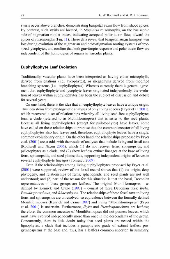
swirls occur above branches, demonstrating basipetal auxin flow from shoot apices.By contrast, such swirls are located, in Stigmaria rhizomorphs, on the basiscopicside of stigmarian rootlet traces, indicating acropetal polar auxin flow, toward theapices of rhizomorphs (Fig. 11). These data reveal that basipetal auxin transport waslost during evolution of the stigmarian and protostigmarian rooting systems of tree-sized lycophytes, and confirm that both gravitropic response and polar auxin flow areindependent of the homologies of organs in vascular plants.
Euphyllophyte Leaf Evolution
Traditionally, vascular plants have been interpreted as having either microphylls,derived from enations (i.e., lycophytes), or megaphylls derived from modifiedbranching systems (i.e., euphyllophytes). Whereas currently there is general agree-ment that euphyllophyte and lycophyte leaves originated independently, the evolu-tion of leaves within euphyllophytes has been the subject of discussion and debatefor several years.
On one hand, there is the idea that all euphyllophyte leaves have a unique origin.This idea stems from phylogenetic analyses of only living species (Pryer et al. 2001),which recovered a set of relationships whereby all living seed-free euphyllophytesform a clade (referred to as Moniliformopses) that is sister to the seed plants.Because all living euphyllophytes (except for psilotophytes) have leaves, somehave called on these relationships to propose that the common ancestor of all livingeuphyllophytes also had leaves and, therefore, euphyllophyte leaves have a single,common evolutionary origin. On the other hand, the relationships proposed by Pryeret al. (2001) are at odds with the results of analyses that include living and fossil taxa(Rothwell and Nixon 2006), which (1) do not recover ferns, sphenopsids, andpsilotophytes as a clade, and (2) show leafless extinct lineages at the base of livingferns, sphenopsids, and seed plants; thus, supporting independent origins of leaves inseveral euphyllophyte lineages (Tomescu 2009).
Even if the relationships among living euphyllophytes proposed by Pryer et al.(2001) were supported, review of the fossil record shows that (1) the origin, deepphylogeny, and relationships of ferns, sphenopsids, and seed plants are not wellunderstood; and (2) part of the reason for this situation is that the basal, Devonianrepresentatives of these groups are leafless. The original Moniliformopses – asdefined by Kenrick and Crane (1997) – consist of three Devonian taxa: Ibyka,Pseudosporochnus, and Rhacophyton. The relationships of these fossil taxa to livingferns and sphenopsids are unresolved, so equivalence between the formally definedMoniliformopses (Kenrick and Crane 1997) and living “Moniliformopses” (Pryeret al. 2001) is uncertain. Furthermore, Ibyka and Pseudosporochnus are leafless;therefore, the common ancestor of Moniliformopses did not possess leaves, whichmust have evolved independently more than once in the descendants of the group.Concurrently, there is little doubt today that seed plants are nested within thelignophytes, a clade that includes a paraphyletic grade of extinct leafless pro-gymnosperms at the base and, thus, has a leafless common ancestor. In summary,
22 G. W. Rothwell and A. M. F. Tomescu
Fig.1
1Polarauxintransportinvascularcambium
ofarbo
rescentlepidod
endraleanlycopsid(left).Instem
s(top
panel),aux
inflow
sbasipetally,i.e.,aw
ayfrom
shoo
ttip
.Anob
stacle
inthecambium
,representedby
base
ofdivergingbranch
(br),disrup
tsflow
ofauxinleadingto
form
ationof
auxinsw
irls
inwoo
dacroscop
icallywith
respecttobranch
trace(abo
vetrace),butno
tinabasiscop
icpo
sitio
n(below
trace).Inpo
sitiv
elygravitrop
icrootingstructures
(rhizomorph
s;bo
ttompanel),sub
tleauxinsw
irlsareform
edin
woo
dbasiscop
ically
with
respectto“roo
tlet”traces
(asterisks),i.e.,abov
etraces,butno
tinacroscop
icpo
sitio
n(below
traces).Thisdemon
stratesthat
polarauxinflow
isdirected
acropetally
inrhizom
orph
,i.e.,towardtip
.Woo
danatom
yillustrated
byParalycop
odites
(stem)andStigmaria
(rhizomorph
)
Structural Fingerprints of Development at the Intersection of Evo-Devo and. . . 23
from a phylogenetic perspective, the fossil record unequivocally supports a mini-mum of three independent origins of leaves among euphyllophytes – in ferns,sphenopsids, and seed plants.
The fossil record also contributes evidence for multiple origins of euphyllophyteleaves in an evo-devo perspective. One line of evidence is the demonstration that leafevolution followed different trajectories, in terms of sequence of character evolution,in ferns and seed plants (Sanders et al. 2009; see also section “Sequence of CharacterEvolution”). Another line of evidence is provided by fossils demonstrating that theevolution of leaf venation followed similar trajectories, from simpler to morecomplex architectures, in different euphyllophyte lineages (Boyce and Knoll2002). This corroborates the evidence for leaf evolution from leafless ancestors ineach of those lineages, indicating parallel evolution of leaf venation in distincteuphyllophyte lineages, as opposed to inheritance from a common ancestor thathad leaves with complex venation.
Partial homology has been proposed for the leaves of different euphyllophytelineages at the level of their precursor structures, i.e., the lateral branching systems oftheir leafless Devonian ancestors (Kenrick and Crane 1997). However, because thebranching architectures of Devonian tracheophytes cover a continuous range ofmorphologies from lateral subordinate (overtopped) branching systems all the wayto the branched sporophyte axes of the ancestral polysporangiophyte, statements ofhomology are difficult to formulate, let alone demonstrate, along this morphologicalcontinuum. In a similar vein, Boyce and Knoll (2002) hypothesized that the inde-pendent origins of euphyllophyte leaves could have been based on modifications of acommon underlying developmental system. Tomescu (2009) reviewed the geneticregulation of leaf development and concluded that interactions between sharedregulatory genes are too diverse among (and sometimes within) major lineages tosupport a common underlying regulatory system.
Vasco et al. (2016) proposed another form of deep homology. They demonstratedexpression of Class III HD-Zip transcription factors (HD-Zip III) in the sporangia ofSelaginella (lycophyte), Psilotum (psilotophyte), and Ophioglossum (fern). BecauseHD-Zip III genes have also been shown to be expressed in the sporangia ofPhyscomitrella (bryophyte) and Arabidopsis (angiosperm), Vasco et al. hypothe-sized deep homology of leaves across all tracheophyte lineages, resulting fromindependent co-option of an ancestral sporangium developmental program thatinvolved III HD-Zip III transcription factors. However, it is also possible that theshared expression of HD-Zip III genes in plant sporangia is not directly relevant toleaf homologies. HD-Zip III genes also have a role in vascular tissue development inall tracheophytes (Floyd and Bowman 2010), possibly evolved after duplication ofthe ancestral HD-Zip III, which regulated sporangium development. Therefore, it islikely that HD-Zip III expression patterns in the leaves of lycophytes andeuphyllophytes have more to do with vascular tissue identity and the regulation ofradial (and adaxial-abaxial) polarity in vascular tissues, than with leaf identity andhomology (Floyd and Bowman 2010).
In summary, in the debate of euphyllophyte leaf evolution, the fossil record addsphylogenetic resolution by revealing leafless taxa at the base of major euphyllophyte
24 G. W. Rothwell and A. M. F. Tomescu
lineages, as well as morpho-anatomical resolution, by showing plants with combi-nations of characters (determinacy, adaxial-abaxial polarity, venation) that could notbe predicted from studies of extant plant diversity alone. When considered alongsidethe living plants, these reveal patterns of phylogeny and character evolution thatsupport multiple independent origins of leaves among euphyllophytes.
The Equisetum Strobilus: A Case of Reciprocal Illumination
The strobilus of Equisetum, a highly condensed structure, and the sporangiophores itcomprises have presented a puzzle in terms of evolution and homology for manyyears. Equisetum is the only living representative of the sphenophytes, a diverseclade with a rich fossil record, and as such provides an excellent example of a longphylogenetic branch on which homology issues can only be resolved by queryingthe fossil record. At the same time, information from fossils is only relevant in thecontext of development and developmental regulation, as understood based onstudies of extant plants (including Equisetum), in an example of reciprocal illumi-nation between data on fossils and living plants (Fig. 12).
Transformational series assembled during the mid-twentieth century based on thesphenophyte fossil record suggest that both the leaves and the sporangiophores ofEquisetum evolved from lateral branching systems. This implies equivalencebetween leaves and sporangiophores and, consequently, equivalence of their loca-tions on shoots, which were regarded as nodes for both types of organs. However, asubset of sphenophyte fossils demonstrate quite the contrary by possessing whorls ofsporangiophores attached along internodes. These seemingly irreconcilable inter-pretations, based on two distinct datasets, led to a deadlock in homology interpre-tations that was not resolved until the realization that a node-internode view may notbe the appropriate paradigm within which to interpret homologies of Equisetumstrobilus structure (Tomescu et al. 2017), cleared the way for a solution to theconundrum.
Studies of vegetative development in extant Equisetum provide a framework forhypothesis generation (Fig. 12) by showing that shoot development in this genusowes to the combined activity of the apical meristem (which generates phytomers)and intercalary meristems (responsible for growth of individual phytomers byinternode elongation). Our growing understanding of the molecular regulatorymechanisms responsible for meristematic growth suggests that plant meristems ofall types are equivalent in their fundamental capacities (Tomescu et al. 2017). Theseinclude the capacity to transition to reproductive growth, and molecular programsregulating this transition in meristems are shared broadly among tracheophytes.Together, these developmental capabilities of living plants suggest the hypothesisthat the switch to a reproductive developmental program in the intercalary meristemscould lead to production of sporangiophore whorls along internodes, as has beenobserved in fossil equisetaleans. Predictions based on this hypothesis for the devel-opment internodal sporangiophore whorls were tested against the anatomy andmorphology of fossils. These tests confirm that extinct sphenophytes had the same
Structural Fingerprints of Development at the Intersection of Evo-Devo and. . . 25
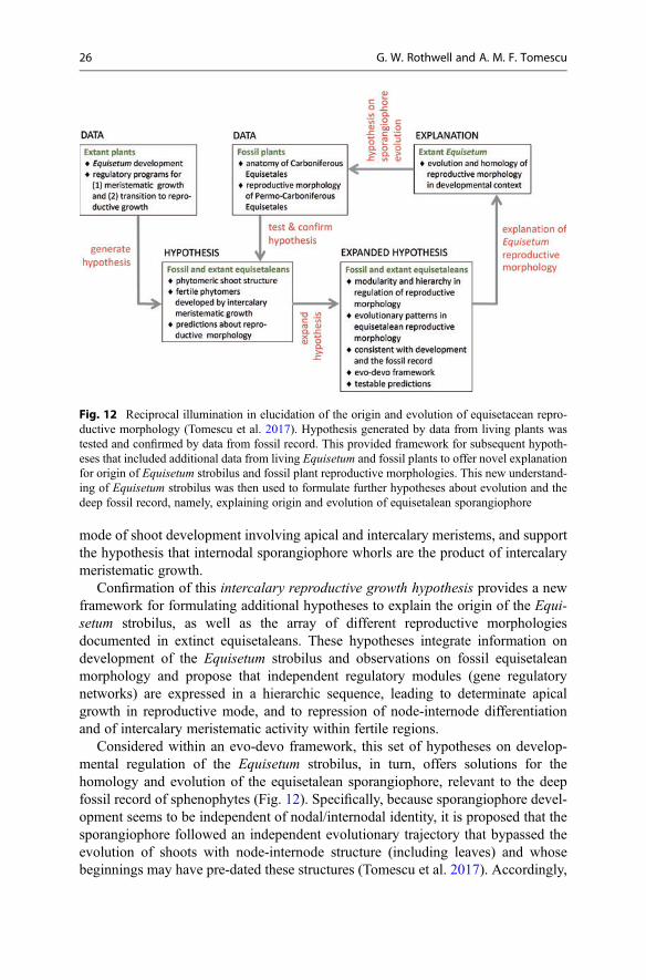
mode of shoot development involving apical and intercalary meristems, and supportthe hypothesis that internodal sporangiophore whorls are the product of intercalarymeristematic growth.
Confirmation of this intercalary reproductive growth hypothesis provides a newframework for formulating additional hypotheses to explain the origin of the Equi-setum strobilus, as well as the array of different reproductive morphologiesdocumented in extinct equisetaleans. These hypotheses integrate information ondevelopment of the Equisetum strobilus and observations on fossil equisetaleanmorphology and propose that independent regulatory modules (gene regulatorynetworks) are expressed in a hierarchic sequence, leading to determinate apicalgrowth in reproductive mode, and to repression of node-internode differentiationand of intercalary meristematic activity within fertile regions.
Considered within an evo-devo framework, this set of hypotheses on develop-mental regulation of the Equisetum strobilus, in turn, offers solutions for thehomology and evolution of the equisetalean sporangiophore, relevant to the deepfossil record of sphenophytes (Fig. 12). Specifically, because sporangiophore devel-opment seems to be independent of nodal/internodal identity, it is proposed that thesporangiophore followed an independent evolutionary trajectory that bypassed theevolution of shoots with node-internode structure (including leaves) and whosebeginnings may have pre-dated these structures (Tomescu et al. 2017). Accordingly,
Fig. 12 Reciprocal illumination in elucidation of the origin and evolution of equisetacean repro-ductive morphology (Tomescu et al. 2017). Hypothesis generated by data from living plants wastested and confirmed by data from fossil record. This provided framework for subsequent hypoth-eses that included additional data from living Equisetum and fossil plants to offer novel explanationfor origin of Equisetum strobilus and fossil plant reproductive morphologies. This new understand-ing of Equisetum strobilus was then used to formulate further hypotheses about evolution and thedeep fossil record, namely, explaining origin and evolution of equisetalean sporangiophore
26 G. W. Rothwell and A. M. F. Tomescu
the sporangiophore could represent the direct expression of a conserved regulatorymodule originally responsible for development of fertile lateral branching systems, amodule that underwent its own evolution, which included heterotopic change, fromexpression along undifferentiated axes, to expression on specialized shoot segments,the internodes.
Summarizing this case of reciprocal illumination: a hypothesis generated by datafrom living plants was tested and confirmed by data from the fossil record. Thisprovided a framework for subsequent hypotheses that included additional data fromliving Equisetum and fossil plants, to offer a novel explanation of the origin of theEquisetum strobilus and fossil reproductive morphologies. This new understandingof the Equisetum strobilus was then used to formulate further hypotheses aboutevolution and the deep fossil record, explaining the origin and evolution of theequisetalean sporangiophore.
Conclusions and Future Outlook
Fossils are quintessential witnesses of evolution. Study of the fossil record hascontributed tremendously toward resolving plant evolution, systematics, and phy-logeny, and gaining a fuller understanding of the role and workings of developmentin evolution. Living biodiversity represents only a small fraction of the diversity oflife that spans Earth’s history; therefore, most of the history of plant life is revealedexclusively by the fossil record. The fossil record provides access to an extensivediversity of plant structure that allows for higher resolution in the understanding ofevolutionary processes and events in deep time.
Understanding the indispensable role of fossils in addressing questions of plantevolution and phylogeny also provides a powerful argument for a much widersystematic spectrum of genomic sequencing (i.e., a species of Lycopodiaceae,Psilotum, Equisetum, a species of Ophioglossales, a species of Marattiales). Onlywith such data available to test hypotheses of phylogeny will we be able to resolvecurrently recalcitrant relationships among seed plants, euphyllophytes, ferns, and inseveral regions of the angiosperm clade.
Plant fossils are invaluable in documenting the pattern of evolution (for tissues,organs, modes of growth, life cycles, etc.), which illuminates structural and develop-mental homologies and provides a test for hypotheses that have been generated fromother disciplines. Focused queries of data from the physiology, developmental molec-ular biology, and comparative developmental anatomy of extant plants will identifyadditional fingerprints like the ones discussed here. These fingerprints provide asmany additional bridges over the gaps that separate living plants, in which develop-ment, physiology, genetics, and molecular biology can be studied directly, from thefossils, in which only morphology and anatomy can be observed.
Conversely, the fossil record provides data for the formulation of hypotheses thatcan be tested with genetic and developmental regulatory experiments. Pressingquestions that are currently apparently insoluble and could be addressed by thesemethodologies regard patterns of evolution for plant vegetative organs (e.g., stele
Structural Fingerprints of Development at the Intersection of Evo-Devo and. . . 27
types, axillary branching, intercalary growth), fertile organs, and life cycles (e.g., theseed, the flower, the fruit, heterospory, angiospermous fertilization). It would beinteresting to test, for example, if enhanced polar auxin transport in Isoetes wouldlead to elongation and branching in the rhizomorph and stem, and to more activesecondary growth, lepidodendralean-style. Or if discovery and silencing of theregulatory module that represses node-internode differentiation in Equisetum spo-rangiophore phytomers would lead to reproductive morphologies like those ofextinct Peltotheca. And if further induction of indeterminacy in the fertile shootsof such plants would produce Cruciaetheca-like morphologies. We could also testwhether abaxialized leaves of loss-of-function HD-Zip III mutants would grow andbranch like the undifferentiated axes of early polysporangiophytes, if they wereinduced into indeterminacy.
The examples highlighted here encompass only a small number of insightfulstudies where hypotheses generated either from the fossil record or from regulatorydevelopmental genetics serve as reciprocal hypothesis tests. Nevertheless, theydemonstrate the exciting potential for such approaches to dramatically improveour ability to address many evolutionary questions that have thus far eluded resolu-tion through the application of either paleontological, systematic, or regulatorygenetic/developmental approaches alone. These examples also highlight the valueof developmental fingerprints for employing data from the fossil record to enhanceour understanding of the role of regulatory genetics in the evolution of plant structureand the origin of major clades. Because these techniques have thus far been appliedto such a small number of studies distributed across a narrow sample of potentiallyfruitful approaches, we are optimistic that the rapidly expanding application ofcoordinated developmental genetic–paleontological studies will, for the first time,allow us to address some of the most poorly understood events and processes ofevolutionary biology.
Cross-References
▶A Process-based Approach to the Study of Flower Morphological Variation▶Developmental homology▶Evo-devo of the Origin of Flowering Plants▶Macroevolution▶Methods and Practices in Paleo-evo-devo▶Novelty and Innovation▶The Evolution of Branching in Land Plants: Between Conservation and Disparity
Acknowledgments We are indebted to Jean Galtier, Patricia Gensel, Alexander Hetherington,Jinzhuang Xue, Patrick Herendeen, and Kelly Matsunaga, who kindly provided images for illus-trations. Nevin Cullen, Kyla Garten, and Dennis Walker are thanked for preparing some of thesections. Space constraints precluded us from fully referencing this work, and we apologize to themany authors whose publications are not included.
28 G. W. Rothwell and A. M. F. Tomescu
References
Boyce CK, Knoll AH (2002) Evolution of developmental potential and the multiple independentorigins of leaves in Paleozoic vascular plants. Paleobiology 28:70–100
Floyd SK, Bowman JL (2010) Gene expression patterns in seed plant shoot meristems and leaves:homoplasy or homology? J Plant Res 123:43–55
Frankenberg JM, Eggert DA (1969) Petrified Stigmaria from North America: part I. Stigmariaficoides, the underground portions of Lepidodendraceae. Palaeontogr B 128:1–47
Gerrienne P, Gensel PG (2016) New data about anatomy, branching, and inferred growth patterns inthe early Devonian plant Armoricaphyton chateaupannense, Montjean-sur-Loire, France. RevPalaeobot Palynol 224:38–53
Harrison CJ (2016) Auxin transport in the evolution of branching forms. New Phytol. https://doi.org/10.1111/nph.14333
Hetherington AJ, Dubrovski JG, Dolan L (2016a) Unique cellular organization in the oldest rootmeristem. Curr Biol 26:1629–1633
Hetherington AJ, Berry CM, Dolan L (2016b) Networks of highly branched stigmarian rootletsdeveloped on the first giant trees. Proc Natl Acad Sci U S A 113:6695–6700
Jennings JR (1975) Protostigmaria, a new plant organ from the lower Mississippian of Virginia.Palaeontology 18:19–24
Kenrick P, Crane PR (1997) The origin and early diversification of land plants. SmithsonianInstitution Press, Washington, DC
Langdale JA (2008) Evolution of developmental mechanisms in plants. Curr Opin Genet Dev18:368–373
Matsunaga KKS, Tomescu AMF (2017) An organismal concept for Sengelia radicans gen. etsp. nov. – morphology and natural history of an early Devonian lycophyte. Ann Bot117:1097–1113
Matsunaga KKS, Cullen NP, Tomescu AMF (2017) Vascularization of the Selaginella rhizophore:anatomical fingerprints of polar auxin transport with implications for the deep fossil record. NewPhytol. https://doi.org/10.1111/nph.14478
Niklas KJ (1997) The evolutionary biology of plants. Chicago University Press, ChicagoPryer KM, Schneider H, Smith AR et al (2001) Horsetails and ferns are a monophyletic group and
the closest living relatives to seed plants. Nature 409:618–622Rothwell GW, Erwin DM (1985) The rhizomorph apex of Paurodendron: implications for homol-
ogies among the rooting organs of Lycopsida. Am J Bot 72:86–98Rothwell GW, Nixon K (2006) How does the inclusion of fossil data change our conclusions about
the phylogenetic history of euphyllophytes? Int J Plant Sci 167:737–749Rothwell GW, Sanders H, Wyatt SE et al (2008) A fossil record for growth regulation: the role of
auxin in wood evolution. Ann Mo Bot Gard 95:121–134Rothwell GW,Wyatt SE, Tomescu AMF (2014) Plant evolution at the interface of paleontology and
developmental biology: an organism-centered paradigm. Am J Bot 101:899–913Sanders H, Rothwell GW, Wyatt SE (2009) Key morphological alterations in the evolution of
leaves. Int J Plant Sci 170:860–868Spencer ART, Mapes G, Hilton J et al (2015) Middle Jurassic evidence for the origin of
Cupressaceae: a paleobotanical context for the roles of regulatory genetics and developmentin the evolution of conifer seed cones. Am J Bot 102:942–961
Stein WE (1993) Modeling the evolution of stelar architecture in vascular plants. Int J Plant Sci154:229–263
Stein WE, Boyer JS (2006) Evolution of land plant architecture: beyond the telome theory.Paleobiology 32:450–482
Stewart WN (1947) A comparative study of stigmarian appendages and Isoetes roots. Am J Bot34:315–324
Taylor TN, Kerp H, Hass H (2005) Life history biology of early land plants: deciphering thegametophyte phase. Proc Natl Acad Sci U S A 102:5892–5897
Structural Fingerprints of Development at the Intersection of Evo-Devo and. . . 29

Taylor WA, Strother PK, Vecoli M et al (2017) Wall ultrastructure of the oldest embryophyticspores: implications for early land plant evolution. Rev Micropaleontol. https://doi.org/10.1016/j.revmic.2016.12.002
Tomescu AMF (2009) Megaphylls, microphylls and the evolution of leaf development. TrendsPlant Sci 14:5–12
Tomescu AMF (2011) The sporophytes of seed-free vascular plants – major vegetative develop-mental features and molecular genetic pathways. In: Fernandez H, Kumar A, Revilla MA (eds)Working with ferns. Issues and applications. Springer, New York, pp 67–94
Tomescu AMF, Wyatt SE, Hasebe M et al (2014) Early evolution of the vascular plant body plan –the missing mechanisms. Curr Opin Plant Biol 17:126–136
Tomescu AMF, Escapa IH, Rothwell GWet al (2017) Developmental programmes in the evolutionof Equisetum reproductive morphology: a hierarchical modularity hypothesis. Ann Bot119:489–505
Vasco A, Smalls TL, Graham SWet al (2016) Challenging the paradigms of leaf evolution: Class IIIHD-Zips in ferns and lycophytes. New Phytol 212:745–758
30 G. W. Rothwell and A. M. F. Tomescu