Embed Size (px)
Citation preview
Planta (Berl.) 80, 227~236 (1968)
The Production of Auxin by Autolysing Tissues A. R. Sg~LD~AK~ and D. H. NO~THCOT~
Department of Biochemistry, University of Cambridge, England
Received December 11, 1967
Summary. Autolysing plant tissues are known to produce auxin when extracted with ether. It has been shown that autolysing plant, yeast and rat liver tissues produce auxin in vitro," this suggests that relatively unspecific mechanisms are involved. Furthermore, sterile plant and animal tissues which have been killed by freezing and thawing induce nodules of differentiated cells in a previously undiffe- rentiated callus of Phaseolus vulgaris. The callus tissue is known to differentiate in response to applied gradients of auxin. Plant and animal tissues killed by boiling were considerably less effective in inducing differentiation in the tissue. The evi- dence indicates that auxin is a normal product of autolysing cells. It is suggested that dying cells are an important source of auxin in the plant.
Introduction I t is well known that plant tissues under continuous extraction with
wet ether, in the cold, release or produce auxin for many months. Only the auxin extracted in the first few hours is considered to represent the free auxin in the tissue. Since the continued release of auxin is abolished by boiling the tissue it is probably a result of enzymic activity, and the kinetics of release also point to this conclusion. The addition of proteolytic enzymes to unboiled and to boiled tissue results in a release of auxin, but the amount released from boiled tissue is considerably less than that pro- duced from unboiled tissue (LINK and EGG~aS, 1941; THIMANN, SKOOG and BY~RS, 1942). These results suggest that with autolysing tissue auxin is produced both by its release from a protein-bound form and by its formation from a precursor which is available in greater amounts in the presence of exogenous proteolytic enzymes. Tryptophan can be converted to indolyl acetic acid (IAA) by many plant tissues (Ku- LESC~A, 1952; GO~DO~, 1961) and is also released from proteins as they are hydrolysed: t ryptophan could therefore be one of the sources of the auxin produced by autolysing tissues under the conditions of ether extraction.
The production of auxin by plant tissues during ether extraction suggests that in the plant dying and dead cells may be a normal source of auxin. Both by direct estimation of auxin production and by using a model system which employs the technique of sterile tissue culture we have obtained evidence which supports this conclusion. The model 16 Planta (Berl.), :Bd. 80
228 A. ~ . SHELDRAKE and D. H. NORTHCOTE:
s y s t e m is b a s e d o n t h e e x p e r i m e n t s w h i c h h a v e s h o w n t h a t d i f f e r e n t i a - t i o n c a n be i n d u c e d in c e r t a i n u n d i f f e r e n t i a t e d ca l lus c u l t u r e s b y a p p l i e d g r a d i e n t s of a u x i n (W~TMOI~S a n d ]~IER, 1963; JEFFS a n d NORT~COT~, 1966, 1967). W e h a v e i n v e s t i g a t e d w h e t h e r a u t o l y s i n g t i s sue s c a n sub- s t i t u t e fo r a u x i n i n e x p e r i m e n t s of t h i s t y p e .
Ma te r i a l s a n d M e t h o d s Extraction and Estimation o] Auxin. The methods used were based on those
described by L•RSEN (1955). The tissue homogenates were acidified to pH 3 using methyl orange as an internal indicator and then extracted twice with peroxide free ether for 2 hours at 0 ~ C. The ether extract was concentrated, and the acidic auxins separated by shaking with sodium bicarbonate solution, which was then acidified to methyl orange and re-extracted with peroxide free ether. This ether extract, containing acidic ether-soluble substances, was evaporated to dryness, taken up in a small volume of ethanol and applied to chromatograms on W h a t m a n paper No. 1. These were developed in a descending system with isopropanol: ammonia :wate r (10 :1 : ! v.v). Marker strips were run with IAA the position of which was revealed by the ferric chloride-perehloric acid spray (LA~ss._~, 1955). The zones of the paper ehromatograms corresponding to the marker IAA were eluted with water and diluted to 5 mls with phosphate buffer (final concentration 0.01 M; pH 5). These solutions were placed in specimen tubes for the bioassay. S tandard solutions were made using IAA (0.01--2.0 ~g).
Seeds of Avena sativa var Padarn (Ison Ltd., Cambridge) were grown on sand in dim red light for 3 days before being used. The top 2 mm of the seedlings was removed and sections 10 mm long were prepared using a special cutt ing instrument. The leaves were left inside the coleoptiles (BENTLEY and HOVSLS~Y, 1954). All operations were carried out in red light. Ten sections were floated on each tes t solution and left in the dark a t 25 ~ C for 20 hours. They were then removed, placed on a glass plate in a photographic enlarger, and photographic images of them were prepared, magnified 6 times. The lengths of the sections on these photographs were measured. Standard curves showed a linear relationship between extension and the log of the IAA concentration over the range used (0.01--2.0 ~xg).
Tissue Homogenates. Pea leaves (25 g) from plants of Pisum sativum (var. Laxton superb) grown in a greenhouse for 3 weeks were homogenized in a blendor with 0.1 3/[ phosphate buffer (50 ml) a t pH 8. The homogenate was filtered through muslin and samples of equal volume were placed in boiling tubes together with toluene and stoppered with cotton wool, covered by aluminium foil. They were incubated a t 37~ in a water ba th with constant shaking.
Rats (Wistar strain) were killed by decapitation, their liver tissue (20 g) was rapidly removed, placed in water (40 ml) and homogenized in a blendor. The homogenate was filtered through muslin and incubated with toluene a t 37 ~ C as described above.
Bakers' yeast was suspended in distilled water and samples were incubated with toluene at 37~ as described above.
Samples from all the experiments were taken a t various t imes and stored in the deep freeze.
Tissue Culture Experiments. The callus of Phaseolus vulgaris, the media on which it was grown, and the sterile methods employed, have all been described by JEFFS and NORT~COTE (1966, 1967). The Phaseolus callus was normally grown on a full nu t r ien t medium bu t for the experiments described here on the induction of
The Product ion of Auxin by Autolysing Tissues 229
differentiation a maintenance medium was used which contained only salts and vitamins, and naphthylaect ic acid (50[xg/1) and sucrose (0.5%) (JEFFS and NORT~COTE, 1966).
In the experiments of JEF•S and NO~T~eOTE an approximate 1 em cube of callus had a groove cut in the top and a wedge shaped piece of agar gel containing the sub- stances to be tested was placed in this groove. In the experiments reported here, killed tissues were placed in a similar groove in the top of the callus block and a small amount of 1% agar gel was used to cement them into tile groove.
The tissues tested for their ability to induce differentiation were placed in sterile boiling tubes and killed either by being placed in a boiling water ba th for 10 minutes or by being frozen twice to - - 3 0 ~ and then thawed. Microscopic examinat ion of similarly t reated tissues showed tha t they were killed by these methods.
The tissues tested were: (a) Phaseolus vulgaris eMlus. The killed samples were taken from the same callus growth t ha t was used for the experimental induction of differentiation. (b) Parthenocissus tricuspidata crown gall callus. This callus has been mainta ined in culture in this laboratory for several years on a medium con- gaining no growth factors. The culture was originally supplied by Dr. KASSANIS of Rothamstcad Experimental Station. (c) R a t Liver tissue. Rats were killed by decapitation, the skin was swabbed with alcohol and liver tissue was rapidly removed using sterile instruments.
After the experimental cultures had been set up they were left for 1 month at 25 ~ C. They were then fixed, dehydrated, embedded in wax, sectioned and stained with safranin and picrie-analinc blue and mounted as described by JEFFS and NORT~COT]~ (1966). They were examined on an optical microscope (Zeiss Ultraphot) ; polarized optics were used to detect xylem cells and ultraviolet fluorescence micro- scopy of aniline blue stained sections was used to detect phloem (CunRIER, 1957).
For each tes t system a t least five callus blocks were examined, and the experi- ments were carried out on three separate occasions.
Fresh sections of callus blocks from each experiment were also examined; they were stained with phloroglucin/HC1 (J~?cSEN, 1962). Microscopical examinat ion of these enabled an estimate to be made of the relative amounts of differentiation which had been induced by the various t reatments . The traeheids in each nodule of differentiated tissue stained red and could clearly be seen when the section was examined a t low magnification.
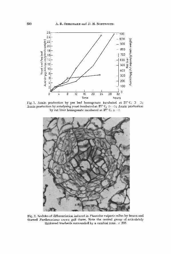
Results I A A Production by Autolysing Tissues. T h e a m o u n t s of I A A in
t i s sues w h i c h h a d b e e n a u t o l y s i n g for v a r i o u s t i m e s a t 37 ~ C a re s h o w n in Fig. 1, for p e a leaf t i s sue , y e a s t a n d r a t l iver . I n al l cases t h e a m o u n t of I A A i n c r e a s e s w i t h t i m e . T h i s p r o d u c t i o n of I A A c a n n o t be a t t r i b u t e d to b a c t e r i a l g r o w t h s ince al l s a m p l e s we re i n c u b a t e d in t h e p r e s e n c e of t o l u e n e .
Induction o] Di//erentiation in Phaseolus vulgaris Callus by Autolysing Tissues
1. Parthenocissus Crown Gall. F r o z e n a n d t h a w e d c r o w n gal l t i s s u e i n d u c e d t h e d i f f e r e n t i a t i o n of n u m e r o u s g r o u p s of t r a c h e i d s . T h e y were s o m e t i m e s s u r r o u n d e d b y a n o r g a n i z e d c a m b i a l z o n e (Fig. 2) b u t n o p h l o e m was d e t e c t a b l e w h e n t h e t i s s u e w as s t a i n e d w i t h d i l u t e a n i l i n e
16"
230 A. 1:~. SHELDRAKE and D. H. NOR~HCOTE:
26 24.
~: 20 , ~ 1 8 ~ 1 6 ~ .~14
1o
6
< 2
1100
1000 y: 900 ~
800
700 "~
600 >_> 500 ~5 rw< 400 <
300 E ._ 200 x
100 <
4 8 20 24 0 0 12 16 28 32 l ime hours
Fig. l. Auxin production by pea leaf homogenate incubated ~t 37 ~ C, [ ] - - ~ ; Auxin production by autolysing yeast incubated at 37 ~ C, o - - o ; Auxin production
by rat liver homogenate incubated at 37 ~ C, A--A
Fig. 2. Nodules of differentiation induced in Phaseolus vulgaris callus by frozen and thawed Parthenocissus crown gall tissue. Note the central group of reticulately
thickened tracheids surrounded by a eambial zone. • 225
The Production of Auxin by Autolysing Tissues 231
blue and v iewed under the UV fluorescence sys tem (Fig. 5). Boiled crown gall t issue also induced the d i f ferent ia t ion of groups of t rache ids in most of the callus blocks t h a t were examined. However there were roughly one th i rd to one qua r t e r as m a n y as when frozen and t hawed tissue was used.
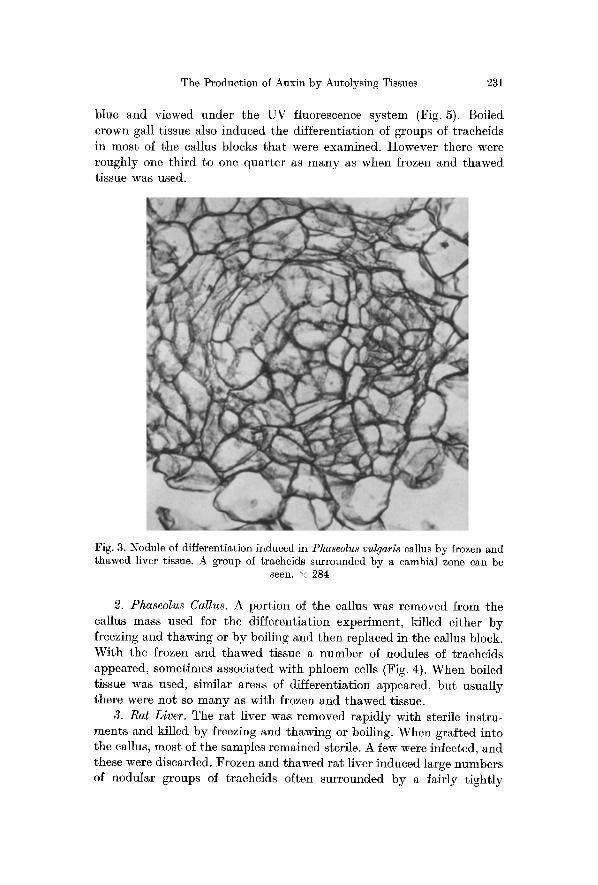
Fig. 3. Nodule of differentiation induced in Pha~eolus vulgaris callus by frozen and thawed liver tissue. A group of traeheids surrounded by a cambial zone can be
seen. • 284
2. Phaseolus Callus. A por t ion of the callus was r emoved from the callus mass used for the d i f ferent ia t ion exper iment , ki l led e i ther b y freezing and thawing or b y boil ing and then replaced in the callus block. W i t h the frozen and t hawed tissue a number of nodules of t r aehe ids appeared , somet imes associa ted wi th ph loem cells (Fig. 4). W h e n boi led t issue was used, s imilar areas of d i f ferent ia t ion appeared , bu t usua l ly there were no t so m a n y as wi th frozen and t hawed tissue.
3. Rat Liver. The r a t l iver was r emoved r ap id ly wi th steri le ins t ru- men t s and ki l led b y freezing and thawing or boiling. W h e n gra f ted in to the callus, mos t of the samples r emained sterile. A few were infected, and these were discarded. F rozen and t hawed r a t l iver induced large number s of nodula r groups of t rache ids often sur rounded b y a fa i r ly t i gh t l y
232 A. ~. SHELDRAKE and D.H. NonTncoT~:
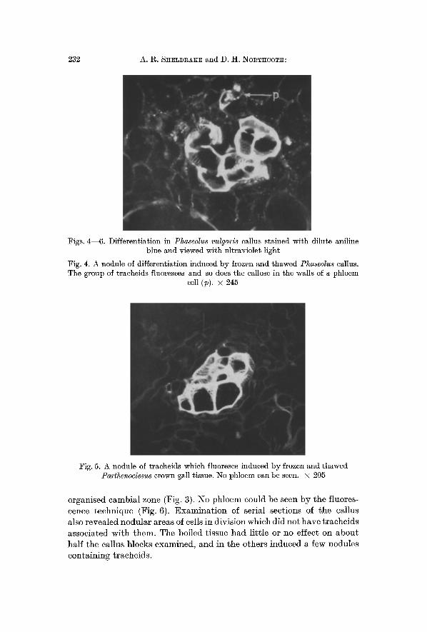
Figs. 4--6. Differentiation in Phaseolus vulgaris callus stained with dilute aniline blue and viewed with ultraviolet light
Fig. 4. A nodule of differentiation induced by frozen and thawed Phaseolus callus. The group of tracheids fluoresces and so does the callose in the walls of a phloem
cell (p). x 245
Fig. 5. A nodule of tracheids which fluoresce induced by frozen and thawed Parthenocissus crown gall tissue. No phloem can be seen. • 205
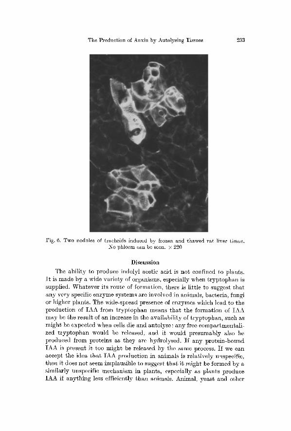
organised cambial zone (Fig. 3). No phloem could be seen by the fluores- cence technique (Fig. 6). Examina t i on of serial sections of the callus also revealed nodular areas of cells in division which did not have t raeheids associated with them. The boiled tissue had li t t le or no effect on about half the callus blocks examined, and in the others induced a few nodules containing tracheids.
The Production of Auxin by Autolysing Tissues 233
Fig. 6. Two nodules of tracheids induced by frozen and thawed rat liver tissue. No phloem can be seen. • 220
Discussion The ability to produce indolyl acetic acid is not confined to plants.
I t is made by a wide variety of organisms, especially when tryptophan is supplied. Whatever its route of formation, there is little to suggest that any very specific enzyme systems are involved in animals, bacteria, fungi or higher plants. The wide-spread presence of enzymes which lead to the production of IAA from tryptophan means that the formation of IAA may be the result of an increase in the availability of tryptophan, such as might be expected when cells die and autolyse: any free compartmentali- zed tryptophan would be released, and it would presumably also be produced from proteins as they are hydrolysed. If any protein-bound IAA is present it too might be released by the same process. If we can accept the idea that IAA production in animals is relatively unspecific, then it does not seem implausible to suggest that it might be formed by a similarly unspecific mechanism in plants, especially as plants produce IAA if anything less efficiently than animals. Animal, yeast and other
234 A.R. SHELDRAKE and D. H. NORT~COT~:
plant tissues all produce IAA as they autolyse, and the processes involved may be very similar.
Differentiation can be induced in Phaseolus vulgaris callus by applied gradients of auxin ( J E ~ s and NOICTHCOTE, 1966). Although no doubt many substances are released by autolysing tissues, the simplest explana- tion of the observed induction of differentiation by autolysing tissues is tha t they act as a source of auxin. That crown gall tissue should have this effect is not surprising: crown gall tissues are known to produce and contain auxin (KuLI~SCHA, 1952). Some of the effects of killed Phaseolus callus could also be due to the release of the synthetic auxin, 2.4 dichlorophenoxy acetic acid stored in the cells in some form, since it had previously been grown on a medium containing this auxin. But in both these cases the role of enzymic activity in producing auxin is implied by the fact tha t tissues killed by freezing and thawing were more active in inducing differentiation than boiled tissues. The same is true of the rat liver tissue, but here the difference between boiled tissue, which had little or no effect, and unboiled autolysing tissue was much more pronounced.
Auxin may thus be a normal product of autolysing cells. Since the death of cells is a normal and important feature of plant growth and differentiation, dying cells may therefore be important sites of auxin formation in the plant. They could, indeed, account for most of the known sites of auxin production (SHELD~AK]~ and NORTHCOT~, 1968 a, b).
The production of auxin by damaged, dying cells might also help to explain the wound response. Damaged cells provide substances which stimulate cell division (BLocH, 1941), and apart from observations on the wound response following physical wounding, there are many others which also suggest that dying eells release such substances. In his studies on embryo development, NUTMAN (1939) observed tha t "the initiation of each new phase of development occurs by the degeneration of some previously formed tissue". In many virus diseases, cells adjacent to the necrotic area undergo hyperplasia and hypertrophy (ESAu, 1938). In cell aggregates in tissue cultures, cell division is found adjacent to areas of necrosis (BLAKELY and STEWARD, 1961). In groups of tobacco callus cells grown in microculture, growth takes place in surges; when some of the mature cells become senescent and die, some of the smaller cells are stimulated to divide and produce cells which enlarge and differentiate (JoNEs, HILD]~B~ANDT, RIKE~ and Wu, 1960). The association between cell division and areas of necrosis has also been reported in genetic tumours (IIAGSN, GUNCK]~L and SPArrow, 1961) and in crown gall (BAN]fIELD, 1935). Since cell division, studied in tissue culture, has been found to be triggered off by a combination of auxins and kinins (SKooe and MILLER, 1957) all these phenomena, which seem to be variations
The Production of Auxin by Autolysing Tissues 235
on the wound response, might be explicable in terms of a release of auxins and also kinins from dying cells. Dying cells might produce kinins as the nucleic acids break down (SHELDI~AKE and IqOnT~COT~, ]968a). They may also l iberate auxin, and there is evidence t ha t auxin really is formed at wound surfaces (1LIEMBEI~G, 1943). The idea of auxin being a normal product of dying cells enables the wound response and most of the major sites of auxin product ion in the p lan t to be unders tood as different aspects of the same phenomenon.
The hypothesis tha t much of the auxin in the p lan t is formed as a consequence of cell death does not mean tha t auxin product ion by this mechanism would be haphazard and uncontrolled. Cells within the in tac t p lan t do not normal ly die at r andom : cell death is itself controlled. The amounts and avai labi l i ty of auxin produced by dying cells would be regulated by enzymes bringing about its dest ruct ion and by the auxin t ranspor t system.
A. 12~. SHELDRAKE gratefully acknowledges a grant from the Science Research Council.
References BANFIELD, W. M. : Effects of cane gall bacteria upon gall tissue cells of the black
raspberry. Bot. Gaz. 97, 193 (1935). BENTLEY, J. A., and S. HOUSLEY: Bio-assay of plant growth hormones. Physiol.
Plant. 7, 405 (1954). BLAKELY, L. M., and F. C. STEWARD : Growth induction in cultures of Haplopappus
gracilis Amer. J. Bot. 48, 351 (1961). BLOCH, R. : Wound healing in higher plants. Bot. l%ev. 7, 110 (1941). CUI~IER, H. B. : Callose substance in plant cells. Amer. J. Bot. 44, 479 (1957). ESAU, K. : Some anatomical aspects of plant virus disease problems. Bot. Rev.
4, 548 (1938). Go~Do~, S. A.: The biogenesis of auxin. Handbuch der Pflanzenphysiologie,
Bd. XIV, S. 620. Berlin-GSttingen-Heidelberg: Springer 1961. HAGEN, G. L., J. E. GU~eKEL, and A. H. SeAnROW: Morphology and histology of
tumor types induced by X, gamma and beta irradiation of a tobacco hybrid. Amer. J. Rot. 48, 691 (1961).
HEMBERr T. : (Jber das Vorkommen wachstumhemmender Stoffe in Kartoffel- knollen und die Bildung wachstumfSrdernder Stoffe in Wundfl/~chen derselben. Arkiv Rot. 80 B, 7 (1943).
JEFES, 1~. A., and D. H. NORTHeOTE: Experimental induction of vascular tissue in an undifferentiated plant callus. Biochem. J. 101, 146 (1966).
- - - - The influence of Indol-3yl acetic acid and sugar on the pattern of induced differentiation in plant tissue culture. J. Cell. Sci. 2, 77 (1967).
JENS~N, W. A.: Botanical histochemistry. San Francisco and London: W.H. Fremann & Co. 1962.
JONES, L. E., A. C. HILDEBRANDT, A. J. RIKER, and J. H. Wv: Growth of somatic tobacco cel]s in microculture. Amer. J. Bot. 47, 468 (1960).
KULESC~A, Z. : l~@cherches sur l'@laboration de substances de croissance par les tissues v~g@taux. Rev. g@n. Bot. 59, 19 (1952).
236 SI-I:ELDRAK:E and NOI~T~ICOT~ : The Production of Auxin by Autolysing Tissues
LAI~SEN, P. : Growth substances in higher plants. Moderne Methoden der Pflanzen- analyse, Bd. III , S. 565. Berlin-GSttingen-Heidelberg: Springer 1955.
LINK, G. K. K., and V. EGGERS: Hyperauxiny in crown gall of tomato. Bet. Gaz. 108, 87 (1941).
NUT~A~, P. S. : The anatomical and cytological evidence for the formation of growth promoting substances in the developing grain of rye. Ann. Bet., N.S. 8, 731 (1939).
SHELDRAKE, A. R., and D. H. NOI~TI~COTE: The production of auxin by tobacco internode tissues. New Phytol. 67, 1 (1968a).
- - - The production of auxin by detached leaves. Nature (Lend.) 217, 195 (19685).
S]~ooG, F., and C. 0. MILLER: Chemical regulation of growth and organ formation in plant tissue cultures in vitro. Symp. See. exp. Biol. 11, 118 (1957).
TI~IM~NN, K. V., F. SI;ooG, and A. C. BY~I~S: The extraction of auxin from plant tissues. Amer. J. Bet. 29, 598 (1942).
W~TMO~, 1~. H., and J . P. RI~R : ExperimentM induction of vascular tissue in the callus of angiosperms. Amer. J. Bet. 50, 418 (1963).
A. i~. SIIELDI~AKE, D. H. NOI~THCOTE Department of Biochemistry, University of Cambridge Cambridge, England