Embed Size (px)
Citation preview

Nucleic Acids Research, 1994, Vol. 22, No. 18 3671 -3680
Structural characterization of intrinsically curved AT-richDNA sequences
Pilar Carrera and Fernando Azorin *Departament de Biologia Molecular Cellular, Centre d'Investigacio Desenvolupament-CSIC,Jordi Girona Salgado 18-26, 08034 Barcelona, Spain
Received June 30, 1994; Revised and Accepted August 10, 1994
ABSTRACTAT-rich DNA sequences other than AnTm tracts(n+m .4) are known to be intrinsically curved. TheAATAT-element constitutes one known example ofthese sequences. In this paper, the elucidation of thestructural basis of the curvature induced by thissequence element was addressed. As judged by thepatterns of cleavage by the hydroxyl radical and DNase1, the AATAT sequence shows a narrow minor groove.Furthermore, the 5' adenine residue of the AAdinucleotide contained within the sequence ishyperreactive to diethylpyrocarbonate. Similarstructural properties are shown by several sequencesinducing intrinsic DNA curvature, such as an A5-tractor the closely related ATAAT, AATATA and TAATATsequences, which are also shown here to inducecurvature. On the other hand, other related sequences,such as TATAA and ATATA, that do not inducecurvature, show different structural characteristics.
INTRODUCTION
Intrinsic curvature is a sequence dependent property of the DNAmolecule. Intrinsically curved DNA is nearly ubiquitous andelements of curved DNA have been found to occur in many DNAmolecules. Intrinsic DNA curvature is generally associated withthe presence of AnTm tracts (n+m.4), periodically spacedalong the DNA molecule with a period close to the helical repeat(see 1-3 for reviews). More recently, DNA sequences containingother combinations of adenine and thymine residues were alsoshown to be curved (4-6). In particular, the sequence elementAATAT was determined to be curved, contributing significantlyto the intrinsic curvature of the mouse satellite DNA repeat (6).Finally, some GC-rich DNA sequences have also been foundcurved (4, 5, 7, 8).
Essentially two types of models were proposed to account forintrinsic DNA curvature. In a first group of models, curvaturearises from the differential structural conformation of thesequence elements inducing the curvature (9-12). In thesemodels deflection of the helical axis occurs principally at thejunction between the structurally altered curving elements andthe surrounding B-type DNA, and is manifested by changes in
either tilt or roll. In the second group of models, DNA curvatureis the consequence of the vectorial addition of the deflections ofthe helical axis occurring at each individual dinucleotide step (1,4, 5, 13-16). A particular wedge angle, which has componentsof tilt and roll, is associated with each type of dinucleotide step.Depending on the precise nucleotidic sequence, this vectorial summay or may not result in a net deviation of the helical axis.
In this paper, the structural characteristics of the AATATsequence element were investigated through the determinationof its patterns of reactivity with the hydroxyl radical (-OH),diethylpyrocarbonate (DEPC) and DNase I. Our results indicatethat this sequence element shows an altered structuralconformation which is manifested by a narrowing of the minorgroove and a distorted conformation of the AA dinucleotide. Theextent to which these structural properties determine the curvatureinduced by this sequence element was also investigated.
MATERIALS AND METHODSDNAsOligonucleotides were synthesized in an Applied Biosystemsautomatic synthesizer and they were purified by polyacrylamidegel electrophoresis. For cloning into pUC19, oligonucleotideswere self-ligated with T4 DNA ligase and the ligated productswere cloned into the unique SmaI site ofpUC19 after treatmentwith the Klenow enzyme. Cloned oligonucleotides weresequenced according to (17).
Polyacrylamide gel electrophoresis analysisThe electrophoretic analysis of the different oligonucleotidesstudied here was performed as described before (6). Briefly,synthetic oligonucleotides were annealed, 5'-end labelled with['y-32P]ATP and T4 polynucleotide kinase, self-ligated andresolved in non denaturing 8% polyacrylamide gels run in 44.5mM Tris-borate, 1.25 mM EDTA, pH 8.3. Electrophoresiswere performed in a water-jacketed apparatus at either 40C or400C. After electrophoresis gels were dried and autoradiographswere recorded on Hyperfilm (Amersham). For quantitativeanalysis of the results, autoradiographs were scanned with aMolecularDynamics laser densitometer. Oligomers of the 18-merof sequence d(TTAGGG-AACCCT)3 were used as molecular
*To whom correspondence should be addressed
k.. 1994 Oxford University Press
3672 Nucleic Acids Research, 1994, Vol. 22, No. 18
weight standards and the apparent molecular weights, in basepairs, were determined from the corresponding regression curves.
The RL values are expressed as apparent length/ actual length.
Hydroxyl radical cleavageCloned oligonucleotides were liberated from pUC19 by cleavagewith EcoRI+HindElI which produces DNA fragments carryingseveral repeats of the corresponding synthetic oligonucleotide.These fragments were labelled at either the EcoRI or the Hindmsite with [a-32P]dATP and the Klenow enzyme before cleavagewith the second restriction endonuclease. The labelled fragmentswere purified by gel electrophoresis and then subjected to thereaction with the hydroxyl radical (*OH) essentially as describedbefore (6). About 50 ng of purified fragment were subjected to-OH cleavage for 90 sec at 4°C in the presence of 500 ng ofE.coli DNA in 10 mM Tris, 20 mM NaCl, pH 7.4. Whencleavage was performed at 20°C, the reaction was allowed toproceed for 30 sec. Reactions were stopped by the addition of20 ,ul of 0.1 M thiourea. DNAs were then precipitated withethanol and analysed on 8% polyacrylamide-7 M urea
denaturing gels. Autoradiographs were recorded in Hyperfilm(Amersham) and scans obtained in a MolecularDynamics laserdensitometer.
DNase I digestionFor DNase I digestion, labelled fragments carrying several copiesof the corresponding synthetic oligonucleotide were obtained as
described above and then subjected to DNase I (Boehringer)digestion at 4°C in a final volume of 8 Al at differentenzyme/DNA ratios for 1 min in the presence of 500 ng of calfthymus DNA in a buffer containing 20 mM MgCl2, 20 mMNaCl, 4 mM MnCl2, 10 mM Tris-HCI, pH 7.4. The reactionwas stopped by the addition of 2 1l of 80% formamide, 20 mMEDTA, pH 8. Cleavage products were resolved on denaturingpolyacrylamide gels and autoradiographs recorded as describedbefore. For quantitative analysis of the results, scans wereobtained in a MolecularDynamics laser densitometer. Theintensity of each band (I) was determined as the area underneaththe corresponding peak on the densitometer scan and normalizedwith respect to the sum of the intensities of all bands (I). Therelative frequency of cleavage at each phosphodiester bond was
then expressed as the ln(I/11). The three-bond running averageof cleavage was obtained by averaging the frequency of cleavageof each individual step with those of its nearest neighbours.
DEPC modificationFor DEPC modification fragments were obtained as describedbefore and then subjected to reaction with 2 ul of DEPC (Fluka)at 4°C for 90 min in a final volume of 25 of itl in the presenceof 100 ng of calf thymus DNA in a buffer containing 10 mMTris -HCI, 20 mM NaCl, pH 7.4. The reaction was stopped byethanol precipitation. Modified fragments were then subjectedto cleavage with 1M piperidine at 90°C for 30 min and thecleavage products analysed on denaturing polyacrylamide gelsas described before.
RESULTSCurvature induced by the AATAT sequence depends stronglyon its precise nucleotidic sequence
To study the influence of the nucleotidic sequence on the degreeof curvature induced by the AATAT sequence element, we
analyze the electrophoretic behaviour of oligonucleotides in-whichthe position of the AA dinucleotide within the sequence was
permutated (Table I). Oligo(AATAT), in which the AAdinucleotide occupies the further most 5' position on the AT-rich sequence, shows the highest electrophoretic retardation. At4°C, the RL value of 160 bp long oligomers of thisoligonucleotide is 1.47 which, as expected for a curved DNAmolecule, decreases strongly as temperature is increased to 40°C.Similar results were obtained earlier (6). On the other hand,oligo(TAATA) and oligo(TATAA), in which the ApAdinucleotide was moved in the 3' direction one or three stepsrespectively, do not appear to be significantly curved. They show,at 4°C, RL values close to one which decrease only slightlywhen the electrophoresis is carried out at 40°C. Furthermore,a very similar RL value is observed in the case ofoligo(ATATA), which contains an alternating AT sequence whichis known not to induce any intrinsic curvature. An intermediatesituation is found in the case of oligo(ATAAT). In this case theRL value at 4°C is 1.31, lower than that corresponding tooligo(AATAT), but significantly higher than those correspondingto oligo(TAATA) and oligo(TATAA). The degree of thecurvature induced by the ATAAT sequence appears to besignificantly lower than that induced by the AATAT-element as
reflected by its smaller RL value. In agreement with thisinterpretation, oligo(ATAAT) does not show any significantretardation when the electrophoresis is carried out at 20°C, while
Table I. RL Values of the oligonucleotides used in these experiments
RLOligonucleotide Sequence 40C 400C
oligo(AATAT) (tggAATATgatggAATATga)n 1.47 (±0.06) 1.09 (±0.01)oligo(TAATA) (tggTAATAgatggTAATAga)n 1.13 (±0.02) 1.03 (±0.03)oligo(ATAAT) (tggATAATgatggATAATga)n 1.31 (±0.01) 1.02 (±0.02)oligo(TATAA) (tggTATAAgatggTATAAga)n 1.12 (±0.01) 1.04 (0.02)oligo(ATATA) (tggATATAgatggATATAga)n 1.12 (±40.01) 1.04 (±+0.02)oligo(A5-AATAT) (tggAATATgacAAAAAcggc)n 1.741 (i0.06) 1.171 (X0.02)oligo(AATATA) (tggAATATAgtggAATATAg)n 1.50 (±0.02) 1.14 (+0.02)oligo(TAATAT) (tggTAATATgtggTAATATg)n 1.42 (±0.01) 1.08 (±0.03)oligo(TTAGGG)2 (ftagggttagggttaggg)n 1.00 1.00
The RL values (apparent length/actual length), at 4°C and 40°C, corresponding to 160 bp long oligomers of the indicated oligonucleotides are shown. Numbersin within parenthesis correspond to the error bars. The AT-region of each oligonucleotide is shown in bold-face.IRL values corresponding to oligo(A5-AATAT) are taken from reference 6.2Oligo(TTAGGG) corresponds to the 18-mer used as molecular weight standard.
Nucleic Acids Research, 1994, Vol. 22, No. 18 3673
oligo(AATAT) still shows some retardation at this temperature(not shown). At 40°C, the RL value of oligo(ATAAT) is alsoclose to one.
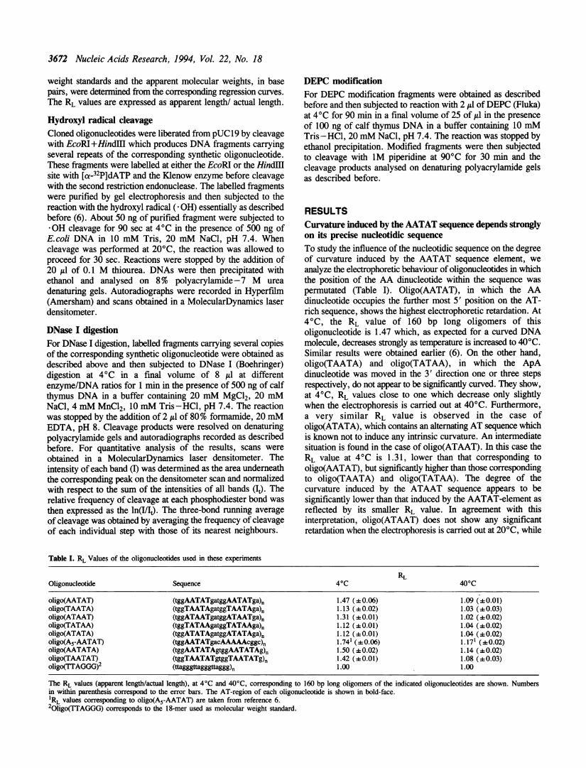
Oligonucleotides detected as intrinsically curved bypolyacrylamide gel electrophoresis, but not those showing RLvalues close to 1, show a sinusoidal pattern of hydroxyl radical(-OH) cleavageIntrinsically curved DNA molecules are known to have adifferential sensitivity to the hydroxyl radical ( *OH), showinga characteristic sinusoidal pattern of -OH cleavage (6, 18-20).In general, the waves of *OH reactivity are phased with respectto the sequence elements introducing the curvature, the frequencyof cleavage decreasing in the 5' to 3' direction. Instead, straightB-DNA is cleaved by * OH nearly equally at each base step (21).Figure 1, shows the patterns of *OH cleavage corresponding tothe oligonucleotides described in Table I. Oligo(AATAT) showsa pattern of cleavage which is clearly sinusoidal (Figure 1B).The maxima of cleavage occur three or four bases upstream fromthe AT-rich sequence. Minimum frequency of cleavage is alwaysobserved at the thymine residue located immediately 3' from theAA dinucleotide. A very similar pattern of *OH cleavage of theAATAT sequence was obtained for oligo(A5-AATAT), whichcontains an A5-tract spaced approximately one helical repeatfrom the AATAT sequence (Table I). Oligomers ofoligo(A5-AATAT) were shown to have a higher electrophoreticretardation than oligomers of oligo(AATAT) (6). In this case,the pattern of -OH reactivity of the AATAT-sequence is alsosinusoidal (Figure IA). Cleavage of the A5-tract is alsosinusoidal. Oligo(ATAAT), which is also found to be intrinsicallycurved by gel electrophoresis shows a similar, though lesspronounced, sinusoidal pattern of *OH cleavage (Figure IC,upper part). In this case, the maximum of reactivity also occurseither three or four bases upstream from the AT-rich elementwhile the minimum is located at the 5' adenine residue of theAA dinucleotide. When the reaction is performed at 20°C, thepattern of *OH cleavage of oligo(ATAAT) becomes fairlyuniform (Figure IC, lower part). As discussed above,oligo(ATAAT) does not show any significant degree of curvatureat this temperature. Similarly, oligo(TAATA), oligo(TATAA)and oligo(ATATA), which are not intrinsically curved as judgedby gel electrophoresis, show uniform patterns of *OH cleavageeven at 4°C (Figure ID-F).
The AATAT sequence shows a decreased sensitivity tocleavage by DNase IA decrease in -OH cleavage has been generally interpreted asindicative of a local narrowing of the minor groove. Therefore,the results reported in Figure 1 suggest that the AT-rich regionof the intrinsically curved oligonucleotides has a narrow minorgroove. The patterns of DNase I cleavage shown in Figure 2are in agreement with this interpretation. Cleavage by the nucleaseDNase I is sensitive to changes in minor groove width (22-24).Resistance to DNase I cleavage at AT-rich sequences is generallyinterpreted as reflecting the presence of a narrow minor groove.The pattern of DNase I cleavage of oligo(A5-AATAT) showstwo regions of low frequency of cleavage (Figure 2A). A firstregion of protection is centred around the A5-tract, extendingfrom the second adenine of the tract to the cytosine immediatelydownstream from it (Figure 2D). The DNA region around theAATAT-element is also resistant to DNase I cleavage. Protection
in this case involves the two guanine residues upstream from thetract and extends up to the first thymine residue of the AATAT-element (Figure 2D). These two regions of low frequency ofDNase I cleavage correspond to the same regions also showinga decreasing frequency of *OH reactivity (Figure 1A).Oligo(AATAT), which solely contains the AATAT-element,
shows a similar though not identical pattern of DNase I cleavage.Also in this case, the 5' region of the AATAT sequence isresistant to cleavage by DNase I (Figure 2B). However, the GpGstep upstream from the AATAT-element is cleaved moreefficiently in oligo(AATAT) than in oligo(A5-AATAT). As aconsequence, the region of low frequency of cleavage is one
A
B
CATAA
TAA ATAA T T A T
D
ATAAT TATT
AAAT
TT TA ATA T
ETA
A~~~~~~~~~~~~~~
F
Figure 1. Patterns of hydroxyl radical cleavage of oligo(A5-AATAT) (A),oligo(AATAT) (B), oligo(ATAAT) (C), oligo(TAATA) (D), oligo(TATAA) (E)and oligo(ATATA) (F). Cleavage by the hydroxyl radical was always performedat 4°C, except in the case of oligo(ATAAT), where the reaction was also carriedout at 20°C (C, lower part). Shown are the densitometer scans correspondingto the gel electrophoretic analysis of the cleavage products of each oligonucleotide.The position corresponding to the AT-rich sequences is indicated in each case.The 5'-to-3' direction is left-to-right. Similar results were obtained when thecomplementary strands were studied.
3674 Nucleic Acids Research, 1994, Vol. 22, No. 18
AL1 2
TA
A.- .
A__0__G_:_sC
CBL 1 2
5 _
T_3_.A_
TIGLA::.
L. W-0
3 i
0
'ni/lt
Oiigo(A -AATAT)I~ 1 -II
I-1 0
3k,P & .. P
:5r
Oligo(AATAT)
Tni/I- F T
-50 0
-7
TGGAATATGATGGAATATGA TGAATATGA
Ini/I-1
-3
-5
-7
W -
MW
'00
aw
AW --T ,s
l . -
3,-9
Oligo(ATAAT)
0 0 0 0
0 00 0 00I ~ ~~~ 0
0
0°TGGATAATCATGGATAATGATGCATAATGA
Figure 2. Patterns of DNase I cleavage of oligo(A5-AATAT) (A), oligo(AATAT) (B) and oligo(ATAAT) (C). DNase I digestion was performed at 5 x 10⁻³ enzymeunits/p (lanes 1) and 2.5 x 10⁻² enzyme units/Al (anes 2). Lanes L correspond to the G+A sequencing ladders of each oligonucleotide and the sequence correspondingto the repeating unit is indicated in each case. The position of adjacent repeats is indicated by the brackets. The 5'-to-3' direction is indicated. Quantitative analysisof the results are shown in (D). The actual relative frequency of cleavage (0) as well as the three-bond running average of cleavage (O -0) are shown throughthree consecutive repeats for each oligonucleotide. Similar results were obtained when the complementary strands were studied.
nucleotide shorter (Figure 2D). Again, the same region showsa decreasing frequency of -OH reactivity (Figure IB). A seconddifference between the patterns of DNase I cleavage ofoligo(AATAT) and oligo(A5-AATAT) is found at the regiondownstream from the AATAT-element. This region issignificantly less sensitive to DNase I in oligo(AATAT) than inoligo(A5-AATAT) (compare Figures 2A and 2B). As aconsequence, in the case of oligo(AATAT), a second region oflow frequency of cleavage is found downstream from the AATATsequence. As discussed later, this downstream region of lowDNase I cutting is likely to arise from the sequence preferencesof DNase I cleavage (25).
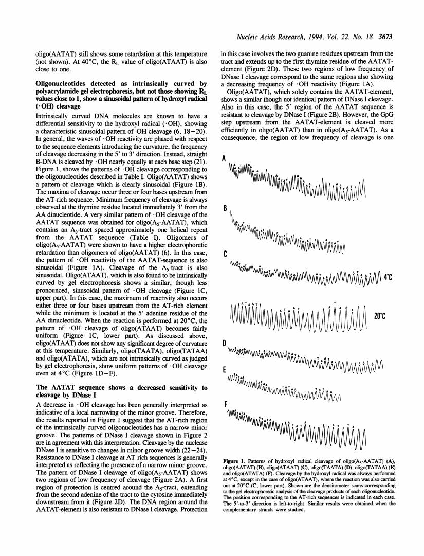
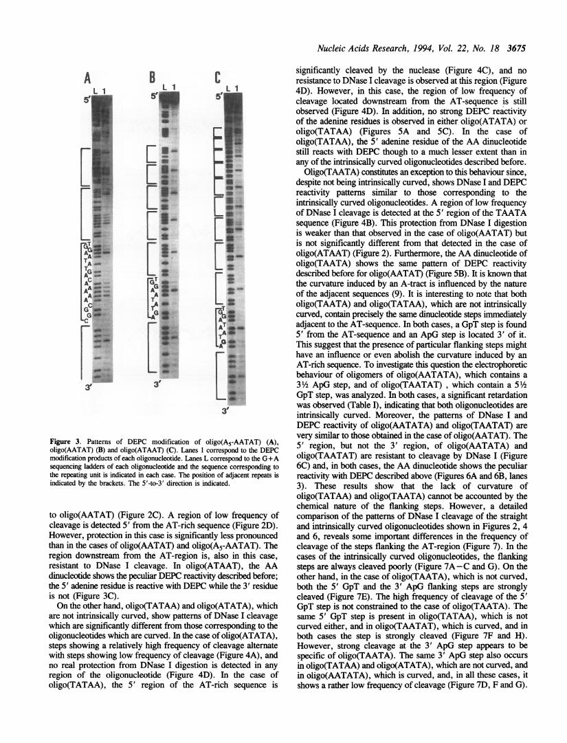
The AA dinucleotide of the AATAT sequence shows a peculiarreactivity with DEPCThe low *OH and DNase I cleavage rates shown by the AATAT-sequence indicate that this sequence has an altered structuralconformation. Additional evidence in favour of this hypothesiscomes from the DEPC modification experiments described inFigure 3. DEPC reacts with adenine residues at their N7-group.In general, regular right-handed B-DNA is not reactive to DEPC.However, a strong DEPC reactivity of the 5' adenine residue
of the AA dinucleotide of the AATAT sequence is observed bothin oligo(AATAT) and in oligo(A5-AATAT), the 3' adenineresidue being totally unreactive (Figures 3A and 3B). TheA5-tract of oligo(A5-AATAT) is also reactive with DEPC. Alsoin this case, the further most 3' adenine residue of the tract isnot reactive with DEPC while the remaining four residues of thetract show a strong DEPC reactivity (Figure 3A). This patternof reactivity is not observed in random sequence DNA (33).
Several intrinsically curved sequences share similar patternsof DNase I and DEPC reactivityAs shown before, the patterns of -OH, DNase I and DEPCreactivity of the intrinsically curved AATAT sequence showsspecific features which are similar to those corresponding to anA5-tract. The question then arises as to what extent thesecommon features are characteristic of intrinsically curved DNA.To address this question the patterns of DNase I and DEPCreactivity of several straight and intrinsically curvedoligonucleotides were determined. Oligo(ATAAT) which, asjudged by its retarded electrophoretic migration in polyacrylamidegels (Table I), is also intrinsically curved shows a pattern ofDNase I cleavage which is very similar to that corresponding
Nucleic Acids Research, 1994, Vol. 22, No. 18 3675
A
TA__TGA -IAI-am;AL
_ib
G..........
! ...... ... ...
......... .
BL 1
_
_
.
r.
_ _O_
=.......MXw:_ 3_W:.:;.....-.. }}__._.
.._.-
_ ,_..*_
_ C_
.-s.__*_
_ *rrGatAA .w_
^ > .::.:..: .::::.
..:._ _
LAG .*:...','::.
.. ::W.. :::M-'22 :.
.^,j,:,@B; ....._.......;_ .,.g..._:.
.w;_....:..::i
_....aw,|, .........
. ......:H:.:}
L ,^i....* :*::, j;..,::
3'
CL 1
5 -5'
S_ f
...
..
f
eX3
X-TteA=AT _se<-
P_ , :._ :!: :}:.:
.X:.-..D .w,SE,:.:.. ::. .':.':::::::..::.:.-... ,.. ..X.es
.. "^:M'.: .':.:.:::':::
_''.......Z;:'.'.X.......................,:. :. .::..
_;''''.*:
3'
Figure 3. Patterns of DEPC modification of oligo(A5-AATAT) (A),oligo(AATAT) (B) and oligo(ATAAT) (C). Lanes 1 correspond to the DEPCmodification products of each oligonucleotide. Lanes L correspond to the G+Asequencing ladders of each oligonucleotide and the sequence corresponding tothe repeating unit is indicated in each case. The position of adjacent repeats isindicated by the brackets. The 5'-to-3' direction is indicated.
to oligo(AATAT) (Figure 2C). A region of low frequency ofcleavage is detected 5' from the AT-rich sequence (Figure 2D).However, protection in this case is significantly less pronouncedthan in the cases of oligo(AATAT) and oligo(A5-AATAT). Theregion downstream from the AT-region is, also in this case,resistant to DNase I cleavage. In oligo(ATAAT), the AAdinucleotide shows the peculiar DEPC reactivity described before;the 5' adenine residue is reactive with DEPC while the 3' residueis not (Figure 3C).On the other hand, oligo(TATAA) and oligo(ATATA), which
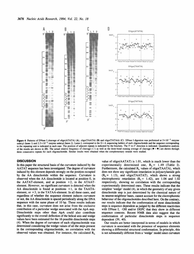
are not intrinsically curved, show patterns of DNase I cleavagewhich are significantly different from those corresponding to theoligonucleotides which are curved. In the case of oligo(ATATA),steps showing a relatively high frequency of cleavage alternatewith steps showing low frequency of cleavage (Figure 4A), andno real protection from DNase I digestion is detected in anyregion of the oligonucleotide (Figure 4D). In the case ofoligo(TATAA), the 5' region of the AT-rich sequence is
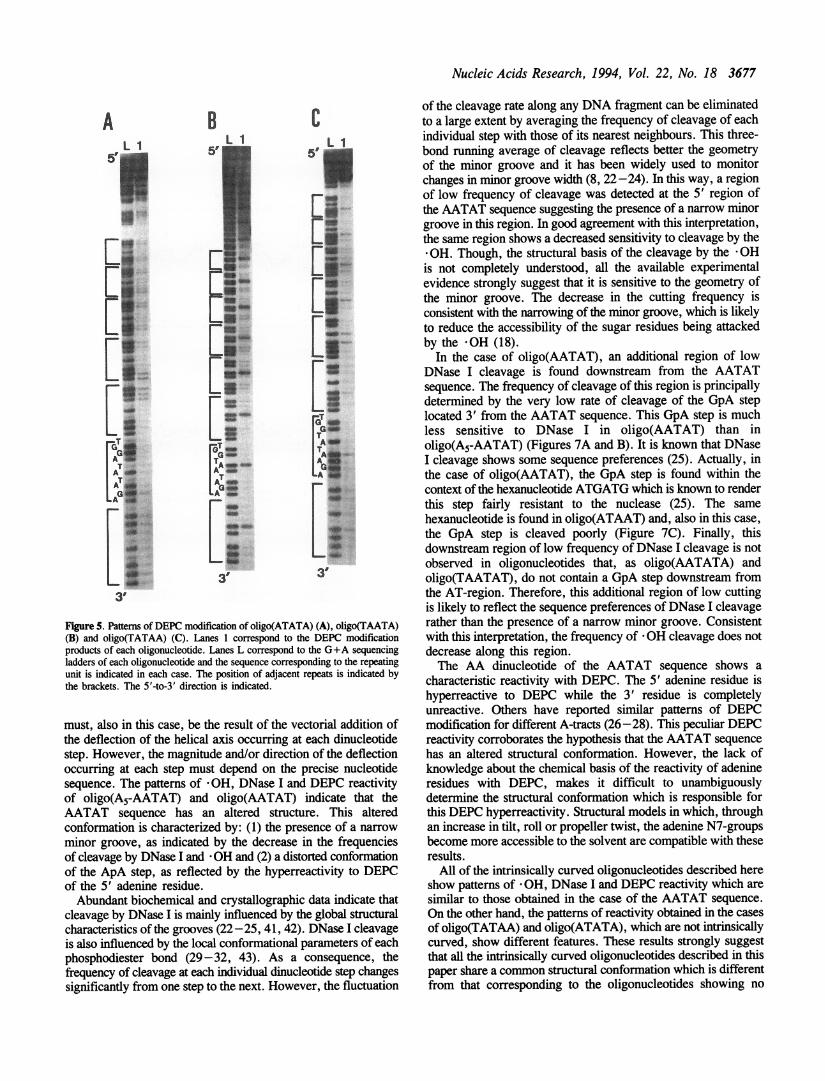
significantly cleaved by the nuclease (Figure 4C), and noresistance to DNase I cleavage is observed at this region (Figure4D). However, in this case, the region of low frequency ofcleavage located downstream from the AT-sequence is stillobserved (Figure 4D). In addition, no strong DEPC reactivityof the adenine residues is observed in either oligo(ATATA) oroligo(TATAA) (Figures 5A and 5C). In the case ofoligo(TATAA), the 5' adenine residue of the AA dinucleotidestill reacts with DEPC though to a much lesser extent than inany of the intrinsically curved oligonucleotides described before.Oligo(TAATA) constitutes an exception to this behaviour since,
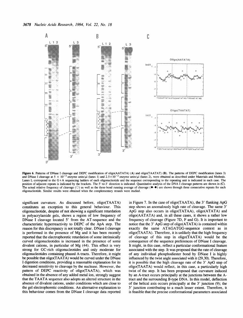
despite not being intrinsically curved, shows DNase I and DEPCreactivity patterns similar to those corresponding to theintrinsically curved oligonucleotides. A region of low frequencyof DNase I cleavage is detected at the 5' region of the TAATAsequence (Figure 4B). This protection from DNase I digestionis weaker than that observed in the case of oligo(AATAT) butis not significantly different from that detected in the case ofoligo(ATAAT) (Figure 2). Furthermore, the AA dinucleotide ofoligo(TAATA) shows the same pattern of DEPC reactivitydescribed before for oligo(AATAT) (Figure SB). It is known thatthe curvature induced by an A-tract is influenced by the natureof the adjacent sequences (9). It is interesting to note that botholigo(TAATA) and oligo(TATAA), which are not intrinsicallycurved, contain precisely the same dinucleotide steps immediatelyadjacent to the AT-sequence. In both cases, a GpT step is found5' from the AT-sequence and an ApG step is located 3' of it.This suggest that the presence of particular flanking steps mighthave an influence or even abolish the curvature induced by anAT-rich sequence. To investigate this question the electrophoreticbehaviour of oligomers of oligo(AATATA), which contains a31/2 ApG step, and of oligo(TAATAT) , which contain a 51/2GpT step, was analyzed. In both cases, a significant retardationwas observed (Table I), indicating that both oligonucleotides areintrinsically curved. Moreover, the patterns of DNase I andDEPC reactivity of oligo(AATATA) and oligo(TAATAT) arevery similar to those obtained in the case of oligo(AATAT). The5' region, but not the 3' region, of oligo(AATATA) andoligo(TAATAT) are resistant to cleavage by DNase I (Figure6C) and, in both cases, the AA dinucleotide shows the peculiarreactivity with DEPC described above (Figures 6A and 6B, lanes3). These results show that the lack of curvature ofoligo(TATAA) and oligo(TAATA) cannot be accounted by thechemical nature of the flanking steps. However, a detailedcomparison of the patterns of DNase I cleavage of the straightand intrinsically curved oligonucleotides shown in Figures 2, 4and 6, reveals some important differences in the frequency ofcleavage of the steps flanking the AT-region (Figure 7). In thecases of the intrinsically curved oligonucleotides, the flankingsteps are always cleaved poorly (Figure 7A-C and G). On theother hand, in the case of oligo(TAATA), which is not curved,both the 5' GpT and the 3' ApG flanking steps are stronglycleaved (Figure 7E). The high frequency of cleavage of the 5'GpT step is not constrained to the case of oligo(TAATA). Thesame 5' GpT step is present in oligo(TATAA), which is notcurved either, and in oligo(TAATAT), which is curved, and inboth cases the step is strongly cleaved (Figure 7F and H).However, strong cleavage at the 3' ApG step appears to bespecific of oligo(TAATA). The same 3' ApG step also occursin oligo(TATAA) and oligo(ATATA), which are not curved, andin oligo(AATATA), which is curved, and, in all these cases, itshows a rather low frequency of cleavage (Figure 7D, F and G).
3676 Nucleic Acids Research, 1994, Vol. 22, No. 18
Ini/It
.3
-5
-7
-9
O igo(ATATA)
TGGA TAT A GATGGA TATAGAT GGAT A TAG A
Oiigo(TAATA)
T GGTAATAGATGGTAATAGATGGTAATAGA
Oligo(TATAA)
0 T
0 00 0
0 0 0
.io a 0v
TGGT AT A AGATGGTAT A AGA TGGTATA AGA
Figure 4. Patterns of DNase I cleavage of oligo(ATATA) (A), oligo(TAATA) (B) and oligo(TATAA) (C). DNase I digestion was performed at 5 x 10⁻³ enzymeunits/ul Oanes 1) and 2.5 x 10⁻² enzyme units/il (lanes 2). Lanes L correspond to the G+A sequencing ladders of each oligonucleotide and the sequence correspondingto the repeating unit is indicated in each case. The position of adjacent repeats is indicated by the brackets. The 5'-to-3' direction is indicated. Quantitative analysisof the results are shown in (D). The actual relative frequency of cleavage (0) as well as the three-bond running average of cleavage (0-0) are shown throughthree consecutive repeats for each oligonucleotide. Similar results were obtained when the complementary strands were studied.
DISCUSSIONIn this paper the structural basis of the curvature induced by theAATAT sequence has been investigated. The degree of curvatureinduced by this element depends strongly on the position occupiedby the AA dinucleotide within the sequence. Curvature isobserved when the AA dinucleotide is located at position 0, inthe AATAT-element, and at position +2, in the ATAAT-element. However, no significant curvature is detected when theAA dinucleotide is found at positions +1, in the TAATA-element, or + 3, in the TATAA-element. In all these cases, andregardless of whether the sequence element induces curvatureor not, the AA dinucleotide is spaced periodically along the DNAsequence with the same phase of 10 bp. These results indicatethat, in this case, curvature does not arise from the exclusivecontribution of a particular wedge associated with the ApA step.Dinucleotide steps other than ApA could also contributesignificantly to the overall deflection of the helical axis and wedgevalues have been estimated for the 16 possible dinucleotide steps(4). When the degree of curvature of each oligonucleotide wasdetermined considering the wedge values of all the steps presentin the corresponding oligonucleotide, no correlation with theobserved values was obtained. For instance, the calculated RL
value of oligo(AATAT) is 1.01, which is much lower than theexperimentally determined one, RL= 1.49 (Table I).Furthermore, the calculated RL values of oligo(TAATA), whichdoes not show any significant retardation in polyacrylamide gels(RL= 1.13), and oligo(TAATAT), which shows a strongelectrophoretic retardation (RL= 1.42), are 1.04 and 1.02respectively, showing no correlation with the correspondingexperimentally determined ones. These results indicate that thesimplest 'wedge' model (4), in which the geometry of any givendinucleotide step is just determined by the chemical nature ofits nearest-neighbour bases, cannot account for the electrophoreticbehaviour of the oligonucleotides described here. On the contray,our results indicate that the conformation of most dinucleotidesteps is sequence dependent as judged by the different reactivitieswith DNase I, OH and/or DEPC that they show in differentsequence contexts. Recent NMR data also suggest that theconformation of particular dinucleotide steps is sequencedependent (39, 40).Our results are better interpreted in terms of models in which
curvature is associated with the presence of sequence elementsshowing a differential structural conformation. In principle, thisis not substantially different from a 'wedge' model since curvature
B CCL 1 2Li 25._
0Ini/i -1
-3
-5
-7
-9
LKr
A
AT
L 1 2
AT
Af -
GAA
In I/ t
a
ow
IT
A...
A a"I'
FA at*
I%p
rmr^flow
I WSS?L 44i:ii
0 a a 0 0
.7 1
9 .... ..
K'iITAo
sEsa
L AG3IA ,Ar-I -..1}
"
I _..::3
Nucleic Acids Research, 1994, Vol. 22, No. 18 3677
A
[GF
3,
B C
5
m
AT
3,I
LA
... ...
Figure S. Patterns of DEPC modification of oligo(ATATA) (A), oligo(TAATA)
(B) and oligo(TATAA) (C). Lanes correspond to the DEPC modification
products of each oligonucleotide. Lanes L correspond to the G+A sequencing
ladders of each oligonucleotide and the sequence corresponding to the repeating
unit is indicated in each case. The position of adjacent repeats is indicated by
the brackets. The 5'-to-3' direction is indicated.
must, also in this case, be the result of the vectorial addition of
the deflection of the helical axis occurring at each dinucleotide
step. However, the magnitude and/or direction of the deflection
occurring at each step must depend on the precise nucleotide
sequence. The patterns of -OH, DNase I and DEPC reactivity
of oligo(A5-AATAT) and oligo(AATAT) indicate that the
AATAT sequence has an altered structure. This altered
conformation is characterized by: (1) the presence of a narrow
minor groove, as indicated by the decrease in the frequencies
of cleavage by DNase I and OH and (2) a distorted conformation
of the ApA step, as reflected by the hyperreactivity to DEPC
of the 5' adenine residue.
Abundant biochemical and crystallographic data indicate that
cleavage by DNase I is mainly influenced by the global structural
characteristics of the grooves (22-25, 41, 42). DNase I cleavage
is also influenced by the local conformational parameters of each
phosphodiester bond (29-32, 43). As a consequence, the
frequency of cleavage at each individual dinucleotide step changes
significantly from one step to the next. However, the fluctuation
of the cleavage rate along any DNA fragment can be eliminatedto a large extent by averaging the frequency of cleavage of eachindividual step with those of its nearest neighbours. This three-bond running average of cleavage reflects better the geometryof the minor groove and it has been widely used to monitorchanges in minor groove width (8, 22-24). In this way, a regionof low frequency of cleavage was detected at the 5' region ofthe AATAT sequence suggesting the presence of a narrow minorgroove in this region. In good agreement with this interpretation,the same region shows a decreased sensitivity to cleavage by the* OH. Though, the structural basis of the cleavage by the OHis not completely understood, all the available experimentalevidence strongly suggest that it is sensitive to the geometry ofthe minor groove. The decrease in the cutting frequency isconsistent with the narrowing of the minor groove, which is likelyto reduce the accessibility of the sugar residues being attackedby the -OH (18).
In the case of oligo(AATAT), an additional region of lowDNase I cleavage is found downstream from the AATATsequence. The frequency of cleavage of this region is principallydetermined by the very low rate of cleavage of the GpA steplocated 3' from the AATAT sequence. This GpA step is muchless sensitive to DNase I in oligo(AATAT) than inoligo(A5-AATAT) (Figures 7A and B). It is known that DNaseI cleavage shows some sequence preferences (25). Actually, inthe case of oligo(AATAT), the GpA step is found within thecontext of the hexanucleotide ATGATG which is known to renderthis step fairly resistant to the nuclease (25). The samehexanucleotide is found in oligo(ATAAT) and, also in this case,the GpA step is cleaved poorly (Figure 7C). Finally, thisdownstream region of low frequency of DNase I cleavage is notobserved in oligonucleotides that, as oligo(AATATA) andoligo(TAATAT), do not contain a GpA step downstream fromthe AT-region. Therefore, this additional region of low cuttingis likely to reflect the sequence preferences of DNase I cleavagerather than the presence of a narrow minor groove. Consistentwith this interpretation, the frequency of -OH cleavage does notdecrease along this region.The AA dinucleotide of the AATAT sequence shows a
characteristic reactivity with DEPC. The 5' adenine residue ishyperreactive to DEPC while the 3' residue is completelyunreactive. Others have reported similar patterns of DEPCmodification for different A-tracts (26-28). This peculiar DEPCreactivity corroborates the hypothesis that the AATAT sequencehas an altered structural conformation. However, the lack ofknowledge about the chemical basis of the reactivity of adenineresidues with DEPC, makes it difficult to unambiguouslydetermine the structural conformation which is responsible forthis DEPC hyperreactivity. Structural models in which, throughan increase in tilt, roll or propeller twist, the adenine N7-groupsbecome more accessible to the solvent are compatible with theseresults.
All of the intrinsically curved oligonucleotides described hereshow patterns of OH, DNase I and DEPC reactivity which aresimilar to those obtained in the case of the AATAT sequence.On the other hand, the patterns of reactivity obtained in the casesof oligo(TATAA) and oligo(ATATA), which are not intrinsicallycurved, show different features. These results strongly suggestthat all the intrinsically curved oligonucleotides described in thispaper share a common structural conformation which is differentfrm ht orepndn to% them oligonucleotides showingy no
3678 Nucleic Acids Research, 1994, Vol. 22, No. 18
1 2 L 3_ 5' sEl i* _ B S
- K .
_ 11! ?a:.,#
.: "
r"..q ,. eS;
"Z| w.C':
"r, .BX;}.W}it
SriR
,t
L^<*s....svz}qfSs
ss:...:
I s-.>ir.}isx
_ f ,
iseRYe!fI ..'.vde
:}}i si Lr ;i;:k #S
o-/ r^U.:e:: YX;;e si:.. I ei . 0.: :::
e L !:.:
e 3'
BL 1 2 L 3
'5 ',5 It
!ta,
.....
.9 w,,i C
Inill
r-
.:.
L
'n%
I :::e:~.
rwltr*.W.sO.
;*V..
.WS*{
rtI b.SU. :-P:
BJX
I it;
- - - rX
* sew
...r..:.::
- L
*i....
oI ......:
.......L W.:
OIigo(AATATA)
,.,AA'TATAr,r,GAA TATAGTGGAAT ATAG
Oligo(TAATAT)
InI/IW
r > <> ° ° °
|1SF O
.- S
r'G AATA' GT(;GTAATATGTGGTAATAT AG
Figure 6. Patterns of DNase I cleavage and DEPC modification of oligo(AATATA) (A) and oligo(TAATAT) (B). The patterns of DEPC modification (lanes 3)and DNase I cleavage at 5 x 10⁻³ enzyme units/Il (lanes 1) and 2.5 x 10⁻² enzyme units/gl (lanes 2), were obtained as described under Materials and Methods.Lanes L correspond to the G+A sequencing ladders of each oligonucleotide and the sequence corresponding to the repeating unit is indicated in each case. Theposition of adjacent repeats is indicated by the brackets. The 5'-to-3' direction is indicated. Quantitative analysis of the DNA I cleavage patterns are shown in (C).The actual relative frequency of cleavage (0) as well as the three-bond running average of cleavage (0-0) are shown through three consecutive repeats for eacholigonucleotide. Similar results were obtained when the complementary strands were studied.
significant curvature. As discussed before, oligo(TAATA)constitutes an exception to this general behaviour. Thisoligonucleotide, despite of not showing a significant retardationin polyacrylamide gels, shows a region of low frequency ofDNase I cleavage located 5' from the AT-sequence and thecharacteristic hyperreactivity to DEPC of the ApA step. Thereason for this discrepancy is not totally clear. DNase I cleavageis performed in the presence of Mg and it has been recentlyreported that the electrophoretic retardation of some intrinsicallycurved oligonucleotides is increased in the presence of somedivalent cations, in particular of Mg (44). This effect is verystrong for GC-rich oligonucleotides and only moderate foroligonucleotides containing phased A-tracts. Therefore, it mightbe possible that oligo(TAATA) would be curved under the DNaseI digestion conditions, providing a reasonable explanation for itsdecreased sensitivity to cleavage by the nuclease. However, thepattern of DEPC reactivity of oligo(TAATA), which wasobtained in the absence of any added metal ion, strongly suggestthat the TAATA sequence also adopts an altered structure in theabsence of divalent cations, under conditions which are close tothe gel electrophoretic conditions. An alternative explanation tothis behaviour comes from the DNase I cleavage data reported
in Figure 7. In the case of oligo(TAATA), the 3' flanking ApGstep shows an anomalously high rate of cleavage. The same 3'ApG step also occurs in oligo(TATAA), oligo(ATATA) andoligo(AATATA) and, in all these cases, it shows a rather lowfrequency of cleavage (Figure 7D, F and G). It is important tonotice that the 3' ApG step of oligo(ATATA) is contained withinexactly the same ATAGATGG-sequence context as inoligo(TAATA). Therefore, it is unlikely that the high frequencyof cleavage of this step in oligo(TAATA) would be theconsequence of the sequence preferences of DNase I cleavage.It might, in this case, reflect a particular conformational featureassociated with the step. It was proposed that the rate of cleavageof any individual phosphodiester bond by DNase I is highlyinfluenced by the twist angle associated with it (29,30). Therefore,it is possible that the high cleavage rate of the 3' ApG step ofoligo(TAATA) would reflect, in this case, a particularly hightwist of the step. It has been proposed that curvature inducedby an A-tract occurs principally at the junctions between the A-tract and the surrounding B-type DNA. In this model, deflectionof the helical axis occurs principally at the 3' junction (9); the5' junction contributing to a much lesser extent. Therefore, itis feasible that the precise conformational parameters associated
A5
.
A...
r
.'
.3
m
Nucleic Acids Research, 1994, Vol. 22, No. 18 3679
B
,J., ,I4V _TT11111TGA ATGGAATATGAGA ATA TGATG A ATA T AG
C
F
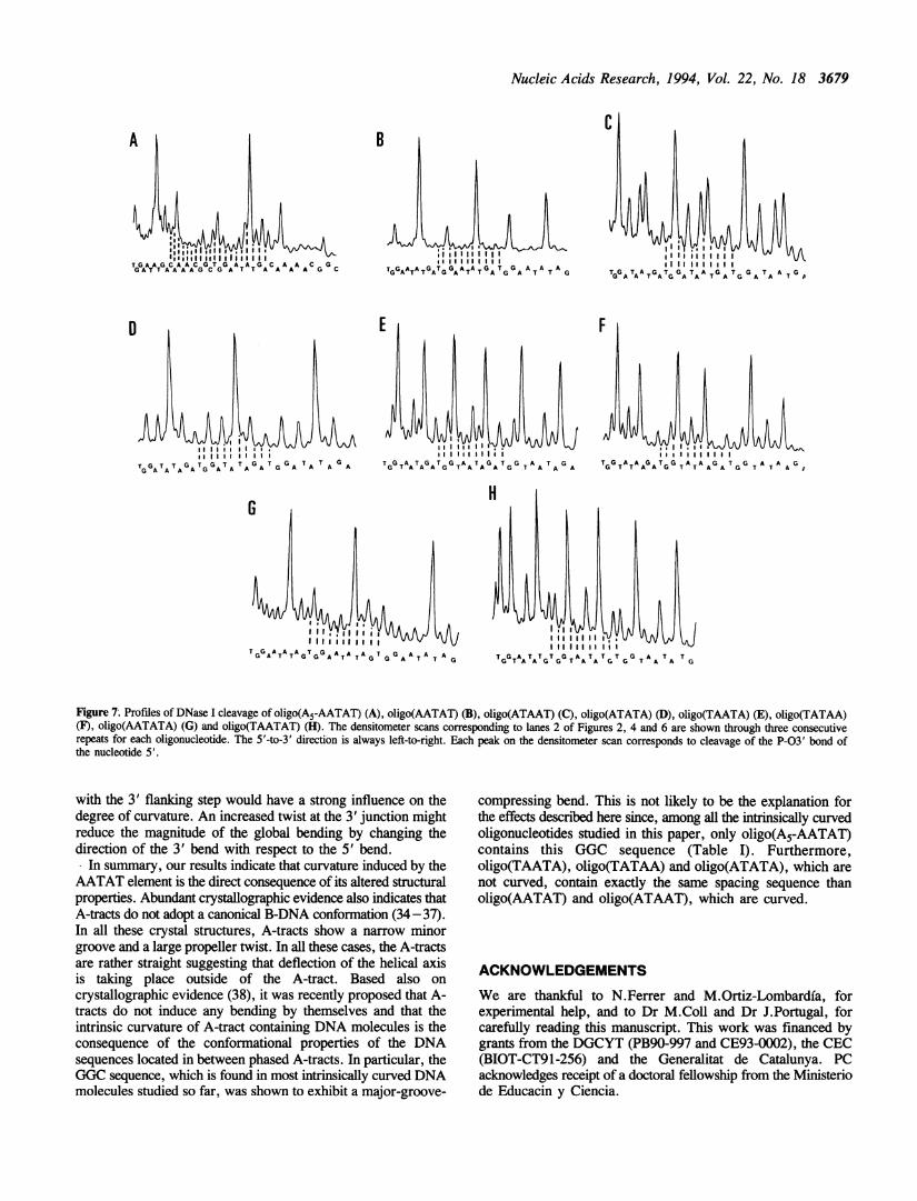
Figure 7; Profiles of DNase I cleavage of oligo(A5-AATAT) (A), oligo(AATAT) (B), oligo(ATAAT) (C), oligo(ATATA) (D), oligo(TAATA) (E), oligo(TATAA)(F), oligo(AATATA) (G) and oligo(TAATAT) (H). The densitometer scans corresponding to lanes 2 of Figures 2, 4 and 6 are shown through three consecutiverepeats for each oligonucleotide. The 5'-to-3' direction is always left-to-right. Each peak on the densitometer scan corresponds to cleavage of the P-03' bond ofthe nucleotide 5'.
with the 3' flanking step would have a strong influence on thedegree of curvature. An increased twist at the 3' junction mightreduce the magnitude of the global bending by changing thedirection of the 3' bend with respect to the 5' bend.
In summary, our results indicate that curvature induced by theAATAT element is the direct consequence of its altered structuralproperties. Abundant crystallographic evidence also indicates thatA-tracts do not adopt a canonical B-DNA conformation (34 -37).In all these crystal structures, A-tracts show a narrow minorgroove and a large propeller twist. In all these cases, the A-tractsare rather straight suggesting that deflection of the helical axisis taking place outside of the A-tract. Based also on
crystallographic evidence (38), it was recently proposed that A-tracts do not induce any bending by themselves and that theintrinsic curvature of A-tract containing DNA molecules is theconsequence of the conformational properties of the DNAsequences located in between phased A-tracts. In particular, theGGC sequence, which is found in most intrinsically curved DNAmolecules studied so far, was shown to exhibit a major-groove-
compressing bend. This is not likely to be the explanation forthe effects described here since, among all the intrinsically curvedoligonucleotides studied in this paper, only oligo(A5-AATAT)contains this GGC sequence (Table I). Furthermore,oligo(TAATA), oligo(TATAA) and oligo(ATATA), which arenot curved, contain exactly the same spacing sequence thanoligo(AATAT) and oligo(ATAAT), which are curved.
ACKNOWLEDGEMENTS
We are thankful to N.Ferrer and M.Ortiz-Lombardia, forexperimental help, and to Dr M.Coll and Dr J.Portugal, forcarefully reading this manuscript. This work was financed bygrants from the DGCYT (PB90-997 and CE93-0002), the CEC(BIOT-CT91-256) and the Generalitat de Catalunya. PCacknowledges receipt of a doctoral fellowship from the Ministeriode Educacin y Ciencia.
A
0
G
TGGATATAGTGGATATAGT G G AT T G
3680 Nucleic Acids Research, 1994, Vol. 22, No. 18
REFERENCES1. Trifonov, E. N. (1985) CRC Crit. Rev. Biochem. 19, 89-106.2. Hagerman, P. J. (1990) Annu. Rev. Biochem. 59, 755-781.3. Hagerman, P. J. (1992) Biochim. Biophys. Acta 1131, 125-132.4. Bolshoy, A., McNamara, P., Harrington, R.E., and Trifonov, E.N. (1991)
Proc. Natl. Acad. Sci. USA 88, 2312-2316.5. McNamara, P. T., and Harrington, R. E. (1991) J. Biol. Chem. 266,
12548-12554.6. Carrera, P., Martfnez-Balbas, M. A., Portugal, J., and Azorin, F. (1991)
Nucleic Acids Res. 19, 5639-5644.7. Bruckner, I., Jurukovski, V., Konstantinovic, M., and Savic, A. (1991)
Nucleic Acids Res. 19, 3549- 355 1.8. Bruckner, I., Dlakic, M., Savic, A., Susic, S., Pongor, S., and Suck, D.
(1993) Nucleic Acids Res. 21, 1025-1029.9. Koo, H.-S., Wu, H.-M., and Crothers, D.M. (1986) Nature 320, 501-506.
10. Chuprina, V.P. (1987) Nucleic Acids Res. 15, 293 -31111. Koo, H.-S., and Crothers, D. M. (1988) Proc. Natl. Acad. Sci. USA 85,
1763-1767.12. Nadeau, J.G., and Crothers, D.M. (1989) Proc. Natl. Acad. Sci. USA 86,
2622-2626.13. Ulanovsky, L. E., Bodner, M., Trifonov, E. N., and Choder, M. (1986)
Proc. Natl Acad. Sci. USA 83, 862-866.14. Ulanovsky, L. E., and Trifonov, E. N. (1987) Nature 326, 720-722.15. Calladine, C. R., Drew, H.R., and McCall, M. J. (1988) J. Mo. Biol. 201,
127-137.16. Zurkhin, V. B., Ulyanov, N. B., Gorin, A. A., and Jernigan, R. L. (1991)
Proc. Natl. Acad. Sci. USA 88, 7046-7050.17. Sanger, F., Nicklen, S., and Coulson, A. R. (1977) Proc. Natl. Acad. Sci.
USA 74, 5463-5476.18. Burkhoff, A. M., and Tullius, T. D. (1987) Cell 48, 935 -943.19. Burkhoff, A. M., and Tullius, T. D. (1988) Nature 331, 445-457.20. Price, M. A., and Tullius, T. D. (1993) Biochemistry 32, 127-136.21. Tullius, T. D., and Dombrosky, B. A. (1985) Science 230, 679-681.22. Drew, H. R. (1984) J. Mol. Biol. 176, 535-557.23. Drew, H. R., and Travers, A. A. (1984) Cell 37, 491-502.24. Lahm, A., and Suck, D. (1991) J. Mo. Biol. 221, 645-667.25. Herrera, J. E., and Chaires, J. B. (1994) J. Mol. Biol. 236, 405-411.26. McCarthy, J. G., Williams, L. D., and Rich, A. (1990) Biochemistry 29,
6071-6081.27. McCarthy, J. G., and Rich, A. (1991) Nucleic Acids Res. 19, 3421-3429.28. McCarthy, J. G., Frederick, C. A., and Nicolas, A. (1993) Nucleic Acids
Res. 14, 3309-3317.29. Lomonosoff, G. P., Butler, P. J. G., and Klug, A. (1981) J. Mol. Biol.
149, 745-760.30. Dickerson, R. E., and Drew, H. R. (1981) J. Mol. Biol. 149, 761-786.31. Hogan, M. E., Roberson, M. W., and Austin, R. H. (1989) Proc. Natl.
Acad. Sci. USA 86, 9273-9277.32. Brukner, I., Jurukovski, V., and Savic, A. (1990) Nucleic Acids Res. 18,
891 -894.33. Portugal, J., Fox, K.R., McLean, M.J., Richenberg, J.L., and Waring, M.J.
(1988) Nucleic Acids Res. 16, 3655-3670.34. Wing, R.M., Drew, H.R., Takano, T., Broke, C., Tanaka, S., Itakura, K.,
and Dickerson, R.E. (1980) Nature 287, 755-758.35. Nelson, H.C.M., Finch, J.T., Luisi, B.F., and Klug, A. (1987) Nature 330,
221 -226.36. Coll, M., Frederick, C.A., Wang, A.H.-J., and Rich A. (1987) Proc Natl.
Acad. Sci. USA 86, 8385-8389.37. DiGabriele, A.D., Sanderson, M.R., and Steitz, T.A. (1989) Proc. Natl.
Acad. Sci. USA 86, 1816-1820.38. Goodsell, D.S., Kopka, M.L., Cascio, D., and Dickerson, R.E. (1993) Proc.
Natl. Acad. Sci. USA 90, 2930-2934.39. Mujeeb, A., Kerwin, S.M., Kenyon, G.L., and James, T.L. (1993)
Biochemistry 32, 13419-13431.40. Weisz, K., Shafer, R.H., Egan, W., and James, T.L. (1994) Biochemistry
33, 354-366.41. Suck, D., Lahm, A., and Oefner, C. (1988) Nature 332, 462-468.42. Weston, S.A., Lahm, A., and Suck, D. (1992) J.Mol.Biol. 226, 1237-1256.43. Fox, K.R. (1992) Nucleic Acids Res.20, 6487-6493.44. Brukner, I., Susic, S., Dlakic, M., Savic, A., and Pongor, S. (1994)
J.Mol.Biol. 236, 26-28.


![Complexi cations of nonnegatively curved manifolds - …totaro/papers/public_html/complex.pdf · Ramanujam’s topological characterization of C2 [21] and related results by ... is](https://img.pdfslide.us/doc/110x75/5b5eda827f8b9a164b8d46fd/complexi-cations-of-nonnegatively-curved-manifolds-totaropaperspublichtml.jpg)