Embed Size (px)
Citation preview
ARTICLE
Received 3 Jul 2015 | Accepted 12 Oct 2015 | Published 16 Nov 2015
Structural basis for cpSRP43 chromodomainselectivity and dynamics in Alb3 insertaseinteractionAnnemarie Horn1,*, Janosch Hennig2,3,*, Yasar L. Ahmed1, Gunter Stier1, Klemens Wild1, Michael Sattler2,3
& Irmgard Sinning1
Canonical membrane protein biogenesis requires co-translational delivery of ribosome-
associated proteins to the Sec translocase and depends on the signal recognition particle
(SRP) and its receptor (SR). In contrast, high-throughput delivery of abundant light-harvesting
chlorophyll a,b-binding proteins (LHCPs) in chloroplasts to the Alb3 insertase occurs
post-translationally via a soluble transit complex including the cpSRP43/cpSRP54 hetero-
dimer (cpSRP). Here we describe the molecular mechanisms of tethering cpSRP to the Alb3
insertase by specific interaction of cpSRP43 chromodomain 3 with a linear motif in the Alb3
C-terminal tail. Combining NMR spectroscopy, X-ray crystallography and biochemical
analyses, we dissect the structural basis for selectivity of chromodomains 2 and 3 for their
respective ligands cpSRP54 and Alb3, respectively. Negative cooperativity in ligand binding
can be explained by dynamics in the chromodomain interface. Our study provides a model for
membrane recruitment of the transit complex and may serve as a prototype for a functional
gain by the tandem arrangement of chromodomains.
DOI: 10.1038/ncomms9875 OPEN
1 Heidelberg University Biochemistry Center (BZH), INF 328, Heidelberg D-69120, Germany. 2 Center for Integrated Protein Science Munich at BiomolecularNMR Spectroscopy, Department Chemie, Technische Universitat Munchen, Lichtenbergstrasse 4, Garching DE-85747, Germany. 3 Institute of StructuralBiology, Helmholtz Center Munich, Ingolstadter Landstrasse 1, Neuherberg D-85764, Germany. * These authors contributed equally to this work.Correspondence and requests for materials should be addressed to I.S. (email: [email protected]) or to M.S.(email: [email protected]).
NATURE COMMUNICATIONS | 6:8875 | DOI: 10.1038/ncomms9875 | www.nature.com/naturecommunications 1
& 2015 Macmillan Publishers Limited. All rights reserved.
Co-translational membrane protein delivery relies on thesignal recognition particle (SRP) machinery found in thecytosol of all prokaryotes and eukaryotes1–3. The SRP core
is universally conserved and consists of SRP54 (Ffh in bacteria)and the SRP RNA. In contrast to cytosolic SRPs, SRP in thechloroplasts of higher plants (cpSRP) has lost its RNA fortargeting proteins to the thylakoid membrane4,5 and acts both ina co- and post-translational manner. Co-translational targeting ofchloroplast-encoded cargo proteins is carried out by cpSRP54alone, whereas the post-translational cpSRP transport pathway ismediated by cpSRP54 in complex with cpSRP43, a 43-kDaprotein that is unique to chloroplasts6–9.
Post-translational cpSRP-dependent protein transport is adedicated pathway for members of the light-harvesting chloro-phyll a,b-binding protein family (LHCPs)10, the most highlyabundant membrane protein in chloroplasts. LHCPs serve asantenna systems in photosynthesis11 to funnel absorbed lightenergy to the photoreaction centres. They contain threetransmembrane helices (TM1 to TM3) to which chlorophyllsand carotenoids bind during insertion into the thylakoidmembrane12. Synthesized on cytosolic ribosomes, the nuclear-encoded LHCPs are guided to the chloroplast envelope by acleavable transit peptide and are imported into the chloroplaststroma via interaction with the TOC/TIC import machinery13,14.In the stroma, the LHCPs are transferred from the envelope viathe small LTD protein15 to a soluble transit complex with cpSRP7.The transit complex is guided to the thylakoid membrane, wherethe LHCPs are inserted by the interaction with the membrane-bound SRP receptor cpFtsY and the C terminus of the membraneinsertase Alb3 (refs 16,17) (Fig. 1a). Alb3 belongs to theYidC/Oxa1/Alb3 family of membrane insertases18,19, which areresponsible for insertion and folding of membrane proteins andtheir assembly into larger protein complexes18. Escherichia coliYidC represents the best characterized member of this familywith its C terminus being involved in ribosome interaction20–22.
The C terminus of Alb3 is intrinsically disordered, recruitscpSRP43 to the thylakoid membrane, and participates in cpSRP-dependent post-translational membrane targeting23. CpSRP43 is amodular protein with a unique arrangement of threechromodomains (CD1–3) and four ankyrin repeats (Ank1–4)24,25
(Fig. 1a,b). Chromodomains and ankyrin repeats are versatileprotein–protein interaction modules and allow cpSRP43 toparticipate in numerous, specific interactions with linear motifs.Chromodomains are typically found in the nucleus where they playa key role in chromatin remodelling and gene expression26,27
like the heterochromatin protein 1 and polycomb28. Thechromodomains of these proteins accommodate methylatedlysines within an ARKS signature sequence of histone tails in an‘aromatic cage’. In contrast, cpSRP43 CD2 binds a positivelycharged arginine-rich motif at the C-terminal tail of cpSRP54 in atwinned and modified cage and thereby recruits cpSRP54 into thetransit complex25. Ankyrin repeats are typical tandem-arrays of 33residues that bind linear motifs in different biological contexts andare also used for the design of specific binding proteins(DARPins)29. In cpSRP43, the ankyrin repeats bind a conservedregion in the loop between LHCP TM2 and TM3 (the L18motif)24,30. This enables cpSRP43 to act as a specific chaperone thatprevents LHCP aggregation31,32. Recent studies showed cpSRP43 toexhibit significant inter-domain dynamics, which is reduced oncpSRP54 binding33.
We previously showed that the two C-terminal chromodo-mains CD2 and CD3 of cpSRP43 are important for binding twopositively charged motifs in the Alb3 C-terminal tail (A3CT)23.However, the molecular details of the cpSRP43–A3CT interactionand the selectivity, dynamics and cooperativity with respect tocpSRP54 binding remained unknown. Here we present a detailed
structural and biochemical analysis of the cpSRP43 CD2CD3interaction with A3CT, demonstrating that CD3 binds to a linearmotif of A3CT and that LHCP targeting is regulated by a serialconnection of ankyrin repeats and chromodomains. Our dataprovide the structural basis for transit complex tethering to thethylakoid membrane by the Alb3 insertase.
ResultsAlb3 binds specifically to cpSRP43 CD3. Recruitment of thetransit complex to the thylakoid membrane involves the con-served interaction of the A3CT with the two C-terminal chro-modomains of cpSRP43 (in the following denoted with CD2 andCD3 only)23 (Supplementary Fig. 1a,b). To test whether A3CT isable to discriminate between CD2 and CD3, we used nuclearmagnetic resonance (NMR) spectroscopy to characterize theinteraction with a cpSRP43 construct comprising CD2 and CD3(CD2CD3) (Fig. 1b). Titration of unlabelled A3CT into 15N and13C labelled CD2CD3 shows NMR chemical shift perturbations(CSPs) for residues located almost exclusively in CD3, while CD2was only affected at its C-terminal end (residues 313 and 316)(Fig. 1c). These data show that A3CT binds to cpSRP43 CD3 anddoes not directly compete with cpSRP54 for CD2. To quantify thebinding event, we performed isothermal titration calorimetry(ITC, Supplementary Table 1a). CD2CD3 binds A3CT with adissociation constant of 20.6 mM in contrast to 5.1 mM observedfor full-length cpSRP43 (Fig. 1d), indicating a stabilizing effect ofthe ankyrin repeats on the chromodomains.
In a crystal structure of a cpSRP43–cpSRP54 complex, wepreviously reported how CD2 binds the C-terminal tail ofcpSRP54 harbouring a RRKR motif25. In a titration of CD2CD3with the same RRKR peptide (RRKRp) (Fig. 1b) monitored byNMR, we find that this ligand binds exclusively to CD2 also inpresence of CD3, although some CSPs are observed forN-terminal residues in CD3 (Gly316 to Glu318) (Fig. 1e). NMRCSPs for RRKRp binding to CD2 are much stronger than in theNMR titration for A3CT binding to CD3, consistent with theB10 times higher binding affinity of the cpSRP54 tail incomparison with A3CT as determined by ITC25. The data alsoexplain our previous observations that cpSRP43 is able to bindboth ligands at the same time23 and show that CD2 and CD3provide specific interaction sites for the tails of cpSRP54 and Alb3despite the high sequence similarity between the twochromodomains and the ligands25.
CD3 binds the A3CT motif IV. Having established that A3CTinteracts specifically with CD3, we tested whether CD3 dis-criminates between similar interaction motifs (II and IV) withinA3CT. The A3CT harbours three ARKS-like signature sequencesthat could potentially bind to chromodomains (SupplementaryFig. 1b). From the cpSRP43DCD3–RRKRp complex it was knownthat CD2 accommodates the second arginine in the ARRK sig-nature sequence of cpSRP54 in a modified cage25. However, thesignature is extended to 531-PPGTARRKR and all three argininesare read-out by cpSRP43. Motif II of A3CT contains a 375-AKRSsequence flanked by predominantly small and unchargedresidues, whereas motif IV is highly positively charged andcontains two putative overlapping binding sequences (453-SKRSand 456-SKRK). Therefore, it was a priori not clear, which of thethree signature sequences would bind, whether binding is specificor if there is any promiscuity.
Titration of unlabelled CD2CD3 into 15N,13C labelled A3CTshowed CSPs most pronounced for the C-terminal region ofA3CT harbouring motif IV (Fig. 1f). The CSPs are considerablysmaller than for the previous titrations, reflecting the loweraffinity of A3CT23. Based on these data, we designed an A3CT
ARTICLE NATURE COMMUNICATIONS | DOI: 10.1038/ncomms9875
2 NATURE COMMUNICATIONS | 6:8875 | DOI: 10.1038/ncomms9875 | www.nature.com/naturecommunications
& 2015 Macmillan Publishers Limited. All rights reserved.
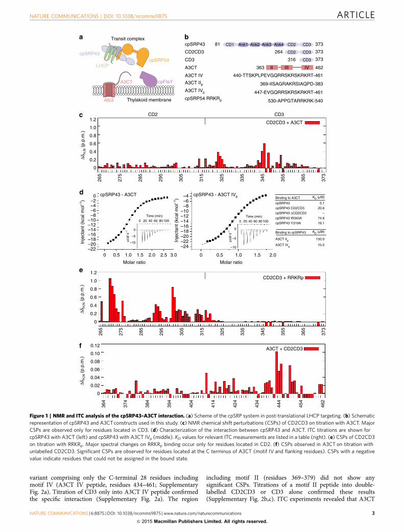
variant comprising only the C-terminal 28 residues includingmotif IV (A3CT IV peptide, residues 434–461; SupplementaryFig. 2a). Titration of CD3 only into A3CT IV peptide confirmedthe specific interaction (Supplementary Fig. 2a). The region
including motif II (residues 369–379) did not show anysignificant CSPs. Titrations of a motif II peptide into double-labelled CD2CD3 or CD3 alone confirmed these results(Supplementary Fig. 2b,c). ITC experiments revealed that A3CT
cpSRP43 CD3Ank1 Ank2 Ank3 Ank4 CD2CD181 373
II III IV363 462A3CT
A3CT IV 440-TTSKPLPEVGQRRSKRSKRKRT-461
A3CT IIp 369-IISAGRAKRSIAQPD-383A3CT IVp 447-EVGQRRSKRSKRKRT-461cpSRP54 RRKRp 530-APPGTARRKRK-540
CD3CD2 373264CD2CD3
CD3 373316CD3
CD2CD3 + A3CT
0
0.2
0.4
0.6
0.8
1.0
1.2
265
275
285
295
305
315
325
335
345
355
365
373
CD2 CD3
0
0.2
0.4
0.6
0.8
1.0
1.2
A3CT + CD2CD3
0
0.02
0.04
0.06
0.08
0.10
0.12
364
374
384
394
404
414
424
434
444
454
462
0 1.5 2.00.5 1.0 2.5 3.0
0–2–4–6–8
–10–12–14–16–18–20–22
Inje
ctan
t (kc
al m
ol–1
)
0
–5
–10μcal
s–1
0 20 40 60 80 100Time (min)
–4–6–8
–20–22–24
Inje
ctan
t (kc
al m
ol–1
)
0 1.00.5 1.5
0 20 40 60 80100Time (min)
0
–5
μcal
s–1
–10
Molar ratio Molar ratio
cpSRP43
LHCPcpSRP54
cpFtsY
Alb3
A3CT
Transit complex
Thylakoid membrane
ΔδH
,N (
p.p.
m.)
ΔδH
,N (
p.p.
m.)
ΔδH
,N (
p.p.
m.)
CD2CD3 + RRKRp
265
275
285
295
305
315
325
335
345
355
365
373
cpSRP43 - A3CT
–18–16–14–12–10
2.0
cpSRP43 - A3CT IVp Binding to A3CT KD (μM)
cpSRP43
cpSRP43 CD2CD3cpSRP43 ΔCD2CD3
cpSRP43 W343A
cpSRP43 Y319A
Binding to cpSRP43
A3CT IIpA3CT IVp
5.1
20.6
–
74.6
19.1
KD (μM)
150.0
15.0
Figure 1 | NMR and ITC analysis of the cpSRP43–A3CT interaction. (a) Scheme of the cpSRP system in post-translational LHCP targeting. (b) Schematic
representation of cpSRP43 and A3CT constructs used in this study. (c) NMR chemical shift perturbations (CSPs) of CD2CD3 on titration with A3CT. Major
CSPs are observed only for residues located in CD3. (d) Characterization of the interaction between cpSRP43 and A3CT. ITC titrations are shown for
cpSRP43 with A3CT (left) and cpSRP43 with A3CT IVp (middle). KD values for relevant ITC measurements are listed in a table (right). (e) CSPs of CD2CD3
on titration with RRKRp. Major spectral changes on RRKRp binding occur only for residues located in CD2. (f) CSPs observed in A3CT on titration with
unlabelled CD2CD3. Significant CSPs are observed for residues located at the C terminus of A3CT (motif IV and flanking residues). CSPs with a negative
value indicate residues that could not be assigned in the bound state.
NATURE COMMUNICATIONS | DOI: 10.1038/ncomms9875 ARTICLE
NATURE COMMUNICATIONS | 6:8875 | DOI: 10.1038/ncomms9875 | www.nature.com/naturecommunications 3
& 2015 Macmillan Publishers Limited. All rights reserved.
motif II and IV bind to CD2CD3 with dissociation constants KD
of 150 mM and 15 mM, respectively (Fig. 1d), while the affinity tothe complete A3CT corresponds to a KD of 5mM. Thus thecpSRP43–A3CT interaction is in a similar range as canonicalchromodomain interactions with histone tails, which typicallyexhibit KD values of 1–10 mM (ref. 34).
Taken together, our NMR and ITC experiments show that thecpSRP43–Alb3 complex is based on the specific interaction ofCD3 with the C-terminal region of A3CT harbouring motif IV,and neither CD2 nor A3CT motif II significantly contribute to theinteraction.
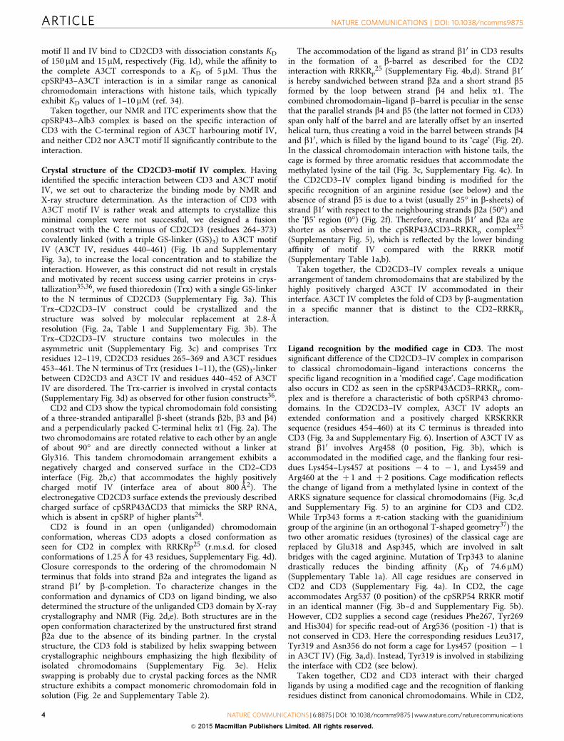
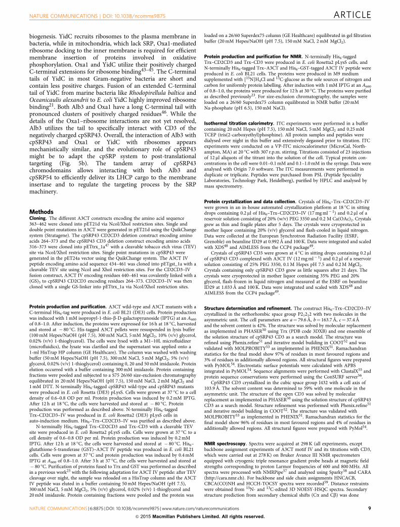
Crystal structure of the CD2CD3-motif IV complex. Havingidentified the specific interaction between CD3 and A3CT motifIV, we set out to characterize the binding mode by NMR andX-ray structure determination. As the interaction of CD3 withA3CT motif IV is rather weak and attempts to crystallize thisminimal complex were not successful, we designed a fusionconstruct with the C terminus of CD2CD3 (residues 264–373)covalently linked (with a triple GS-linker (GS)3) to A3CT motifIV (A3CT IV, residues 440–461) (Fig. 1b and SupplementaryFig. 3a), to increase the local concentration and to stabilize theinteraction. However, as this construct did not result in crystalsand motivated by recent success using carrier proteins in crys-tallization35,36, we fused thioredoxin (Trx) with a single GS-linkerto the N terminus of CD2CD3 (Supplementary Fig. 3a). ThisTrx–CD2CD3–IV construct could be crystallized and thestructure was solved by molecular replacement at 2.8-Åresolution (Fig. 2a, Table 1 and Supplementary Fig. 3b). TheTrx–CD2CD3–IV structure contains two molecules in theasymmetric unit (Supplementary Fig. 3c) and comprises Trxresidues 12–119, CD2CD3 residues 265–369 and A3CT residues453–461. The N terminus of Trx (residues 1–11), the (GS)3-linkerbetween CD2CD3 and A3CT IV and residues 440–452 of A3CTIV are disordered. The Trx-carrier is involved in crystal contacts(Supplementary Fig. 3d) as observed for other fusion constructs36.
CD2 and CD3 show the typical chromodomain fold consistingof a three-stranded antiparallel b-sheet (strands b2b, b3 and b4)and a perpendicularly packed C-terminal helix a1 (Fig. 2a). Thetwo chromodomains are rotated relative to each other by an angleof about 90� and are directly connected without a linker atGly316. This tandem chromodomain arrangement exhibits anegatively charged and conserved surface in the CD2–CD3interface (Fig. 2b,c) that accommodates the highly positivelycharged motif IV (interface area of about 800 Å2). Theelectronegative CD2CD3 surface extends the previously describedcharged surface of cpSRP43DCD3 that mimicks the SRP RNA,which is absent in cpSRP of higher plants24.
CD2 is found in an open (unliganded) chromodomainconformation, whereas CD3 adopts a closed conformation asseen for CD2 in complex with RRKRp25 (r.m.s.d. for closedconformations of 1.25 Å for 43 residues, Supplementary Fig. 4d).Closure corresponds to the ordering of the chromodomain Nterminus that folds into strand b2a and integrates the ligand asstrand b10 by b-completion. To characterize changes in theconformation and dynamics of CD3 on ligand binding, we alsodetermined the structure of the unliganded CD3 domain by X-raycrystallography and NMR (Fig. 2d,e). Both structures are in theopen conformation characterized by the unstructured first strandb2a due to the absence of its binding partner. In the crystalstructure, the CD3 fold is stabilized by helix swapping betweencrystallographic neighbours emphasizing the high flexibility ofisolated chromodomains (Supplementary Fig. 3e). Helixswapping is probably due to crystal packing forces as the NMRstructure exhibits a compact monomeric chromodomain fold insolution (Fig. 2e and Supplementary Table 2).
The accommodation of the ligand as strand b10 in CD3 resultsin the formation of a b-barrel as described for the CD2interaction with RRKRp
25 (Supplementary Fig. 4b,d). Strand b10
is hereby sandwiched between strand b2a and a short strand b5formed by the loop between strand b4 and helix a1. Thecombined chromodomain–ligand b–barrel is peculiar in the sensethat the parallel strands b4 and b5 (the latter not formed in CD3)span only half of the barrel and are laterally offset by an insertedhelical turn, thus creating a void in the barrel between strands b4and b10, which is filled by the ligand bound to its ‘cage’ (Fig. 2f).In the classical chromodomain interaction with histone tails, thecage is formed by three aromatic residues that accommodate themethylated lysine of the tail (Fig. 3c, Supplementary Fig. 4c). Inthe CD2CD3–IV complex ligand binding is modified for thespecific recognition of an arginine residue (see below) and theabsence of strand b5 is due to a twist (usually 25� in b-sheets) ofstrand b10 with respect to the neighbouring strands b2a (50�) andthe ‘b5’ region (0�) (Fig. 2f). Therefore, strands b10 and b2a areshorter as observed in the cpSRP43DCD3–RRKRp complex25
(Supplementary Fig. 5), which is reflected by the lower bindingaffinity of motif IV compared with the RRKR motif(Supplementary Table 1a,b).
Taken together, the CD2CD3–IV complex reveals a uniquearrangement of tandem chromodomains that are stabilized by thehighly positively charged A3CT IV accommodated in theirinterface. A3CT IV completes the fold of CD3 by b-augmentationin a specific manner that is distinct to the CD2–RRKRp
interaction.
Ligand recognition by the modified cage in CD3. The mostsignificant difference of the CD2CD3–IV complex in comparisonto classical chromodomain–ligand interactions concerns thespecific ligand recognition in a ‘modified cage’. Cage modificationalso occurs in CD2 as seen in the cpSRP43DCD3–RRKRp com-plex and is therefore a characteristic of both cpSRP43 chromo-domains. In the CD2CD3–IV complex, A3CT IV adopts anextended conformation and a positively charged KRSKRKRsequence (residues 454–460) at its C terminus is threaded intoCD3 (Fig. 3a and Supplementary Fig. 6). Insertion of A3CT IV asstrand b10 involves Arg458 (0 position, Fig. 3b), which isaccommodated in the modified cage, and the flanking four resi-dues Lys454–Lys457 at positions � 4 to � 1, and Lys459 andArg460 at the þ 1 and þ 2 positions. Cage modification reflectsthe change of ligand from a methylated lysine in context of theARKS signature sequence for classical chromodomains (Fig. 3c,dand Supplementary Fig. 5) to an arginine for CD3 and CD2.While Trp343 forms a p-cation stacking with the guanidiniumgroup of the arginine (in an orthogonal T-shaped geometry37) thetwo other aromatic residues (tyrosines) of the classical cage arereplaced by Glu318 and Asp345, which are involved in saltbridges with the caged arginine. Mutation of Trp343 to alaninedrastically reduces the binding affinity (KD of 74.6 mM)(Supplementary Table 1a). All cage residues are conserved inCD2 and CD3 (Supplementary Fig. 4a). In CD2, the cageaccommodates Arg537 (0 position) of the cpSRP54 RRKR motifin an identical manner (Fig. 3b–d and Supplementary Fig. 5b).However, CD2 supplies a second cage (residues Phe267, Tyr269and His304) for specific read-out of Arg536 (position -1) that isnot conserved in CD3. Here the corresponding residues Leu317,Tyr319 and Asn356 do not form a cage for Lys457 (position � 1in A3CT IV) (Fig. 3a,d). Instead, Tyr319 is involved in stabilizingthe interface with CD2 (see below).
Taken together, CD2 and CD3 interact with their chargedligands by using a modified cage and the recognition of flankingresidues distinct from canonical chromodomains. While in CD2,
ARTICLE NATURE COMMUNICATIONS | DOI: 10.1038/ncomms9875
4 NATURE COMMUNICATIONS | 6:8875 | DOI: 10.1038/ncomms9875 | www.nature.com/naturecommunications
& 2015 Macmillan Publishers Limited. All rights reserved.
a twinned cage reads out two neighbouring arginines, in CD3respective residues of the second cage are involved in stabilizationof the CD2–CD3 interface.
Flanking residues determine selectivity. From thecpSRP43DCD3–RRKRp complex it was known that CD2 recog-nizes the extended 531-PPGTARRKR sequence of cpSRP54 andthat the residues flanking the arginine (0) and especially the argi-nine at the last position (þ 2) are important for binding25. Ourbiochemical and structural data for the CD2CD3–IV complexshow that also here the extended 454-KRSKRKR sequence of Alb3is specifically recognized. To understand how the twochromodomains discriminate between similar linear motifs fromcpSRP54 and Alb3, we mutated residues from the � 7 to the þ 2position in A3CT IV to alanine and determined the KD values ofsingle and double point mutants by ITC (Supplementary Table 1a).Residues involved in salt bridges (Arg455, Lys457, Arg458)contribute the most to the binding, which is typical for anentropically unfavourable interaction and reflects the ordering ofA3CT IV by formation of strand b10 during binding. Mutation ofthe central Lys457 and Arg458 together decreases the dissociationconstant by about ninefold (KD of 44mM) compared with the wild-type interaction. This highlights the importance of Arg458 (0),which is recognized within the modified cage, as the key residue inA3CT IV. Arg455 (� 3) forms a salt bridge with Asp273 in CD2
(Fig. 4a) and replacement by alanine results in a sixfold reductionin binding affinity, while the mutation of Ser456 (� 2) had only aminor effect (threefold reduction), probably as the small side chainof alanine still fits into place. In general, for sterical reasonschromodomains need an alanine at the � 2 position of the ligand,which holds true for the cpSRP54 tail binding to CD2 and forhistone tails binding to canonical chromodomains38 (Fig. 3b). Thisrestriction is, however, not valid for the CD3–A3CT IV interactiondue to the ‘super-twist’ of strand b10, which creates extra space fora serine residue. The importance of the � 2 position for bindingspecificity is underlined by the recent finding that cpSRP54 tails ofgreen algae of the chlorophyte division have a valine at thisposition, which specifically inhibits binding to cpSRP43 (ref. 39).
The introduction of alanine mutations at the � 7 to � 4positions all show detrimental effects according to their distanceto the 0 position, even though Arg451 (� 7) to Ser453 (� 5) arenot visible in the X-ray structure. Interestingly, the correspondingregion in the cpSRP54 tail (530-APP) forms a tight turn whenbound to CD2, which would clash with Asp358 of CD3 (alaninein CD2) (Supplementary Fig. 5a,b). The double mutation ofLys459 and Arg460 (þ 1 and þ 2) results in a fourfold reductionof binding affinity (KD of 20 mM). Their binding modes differslightly in the two complexes present in the crystallographicasymmetric unit. Either the side chain of Lys459 is involved in asalt bridge with CD3 Glu352 or the guanidinium group of Arg460is positioned on the negative dipole of the CD2 C-terminal a1
CD
2C
D3
A3CT IVβ1’
β2a
β2b
β3
β4
α1
β2b
β3β4
Gly316
N
C
α1
CD3
CD3 symmetry mate
Trp343
Asp345
Glu318
Arg458
Turn
β4
‘β5’
β2a
Cage50°
β1’
‘β2a’‘β2a’
OpenOpen
Figure 2 | Structural characterization of the cpSRP43–A3CT complex. (a) Crystal structure of cpSRP43 CD2CD3 (blue) with bound A3CT IV (salmon) as
part of the fusion construct with thioredoxin (not shown). (b) The electrostatic surface potential of CD2CD3 is shown with positive charges in blue and
negative charges in red (contoured at±5 kT). (c) ConSURF analysis showing the degree of conservation mapped on the surface of the CD2CD3 structure.
Highly conserved residues are represented in dark red, partially conserved residues in magenta and less conserved residues in cyan. (d) Crystal structure of
cpSRP43 CD3 in the open conformation with an unstructured ‘b2a’ strand. CD3 is stabilized by helix swapping between two CD3 molecules. (e) Solution
structure of cpSRP43 CD3. A superposition of the 10 lowest energy structures is shown. (f) Close-up view of CD3 bound to A3CT IV highlighting the gap
between strands b4 and b10 induced by a helical turn (green) and forming the cage for substrate recognition. The increased b-sheet twist in the b-barrel,
resulting in the loss of strand ‘b5’ is indicated (50�).
NATURE COMMUNICATIONS | DOI: 10.1038/ncomms9875 ARTICLE
NATURE COMMUNICATIONS | 6:8875 | DOI: 10.1038/ncomms9875 | www.nature.com/naturecommunications 5
& 2015 Macmillan Publishers Limited. All rights reserved.
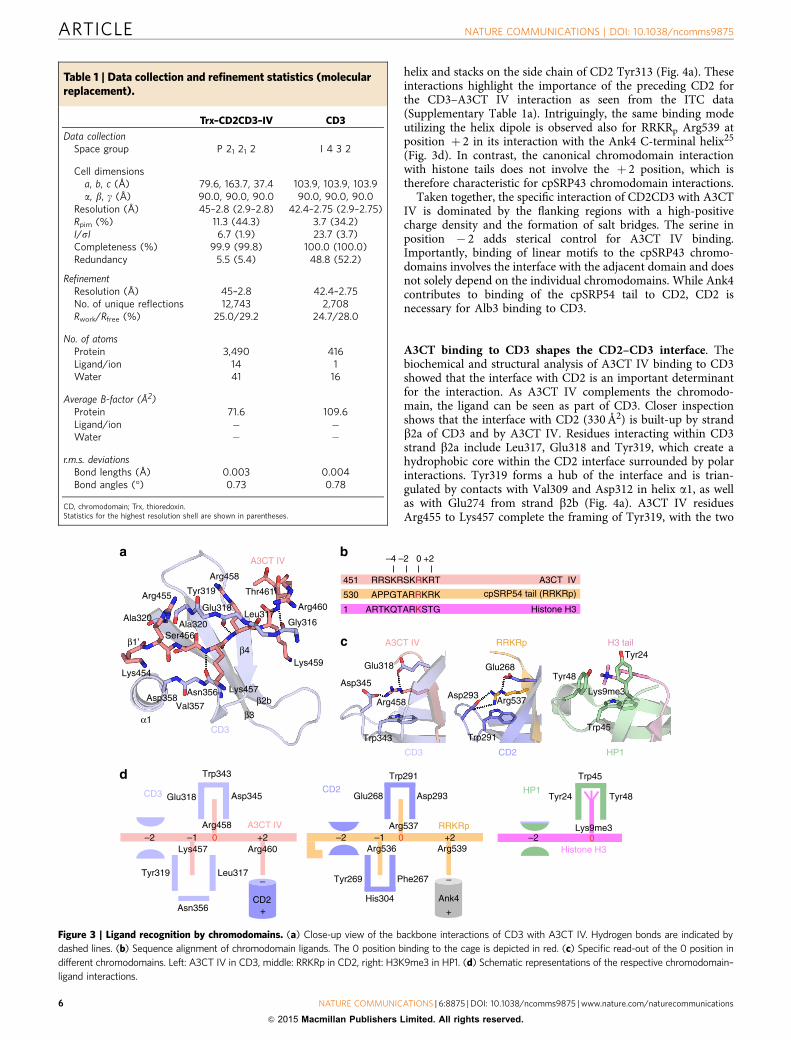
helix and stacks on the side chain of CD2 Tyr313 (Fig. 4a). Theseinteractions highlight the importance of the preceding CD2 forthe CD3–A3CT IV interaction as seen from the ITC data(Supplementary Table 1a). Intriguingly, the same binding modeutilizing the helix dipole is observed also for RRKRp Arg539 atposition þ 2 in its interaction with the Ank4 C-terminal helix25
(Fig. 3d). In contrast, the canonical chromodomain interactionwith histone tails does not involve the þ 2 position, which istherefore characteristic for cpSRP43 chromodomain interactions.
Taken together, the specific interaction of CD2CD3 with A3CTIV is dominated by the flanking regions with a high-positivecharge density and the formation of salt bridges. The serine inposition � 2 adds sterical control for A3CT IV binding.Importantly, binding of linear motifs to the cpSRP43 chromo-domains involves the interface with the adjacent domain and doesnot solely depend on the individual chromodomains. While Ank4contributes to binding of the cpSRP54 tail to CD2, CD2 isnecessary for Alb3 binding to CD3.
A3CT binding to CD3 shapes the CD2–CD3 interface. Thebiochemical and structural analysis of A3CT IV binding to CD3showed that the interface with CD2 is an important determinantfor the interaction. As A3CT IV complements the chromodo-main, the ligand can be seen as part of CD3. Closer inspectionshows that the interface with CD2 (330 Å2) is built-up by strandb2a of CD3 and by A3CT IV. Residues interacting within CD3strand b2a include Leu317, Glu318 and Tyr319, which create ahydrophobic core within the CD2 interface surrounded by polarinteractions. Tyr319 forms a hub of the interface and is trian-gulated by contacts with Val309 and Asp312 in helix a1, as wellas with Glu274 from strand b2b (Fig. 4a). A3CT IV residuesArg455 to Lys457 complete the framing of Tyr319, with the two
Table 1 | Data collection and refinement statistics (molecularreplacement).
Trx–CD2CD3–IV CD3
Data collectionSpace group P 21 21 2 I 4 3 2
Cell dimensionsa, b, c (Å) 79.6, 163.7, 37.4 103.9, 103.9, 103.9a, b, g (Å) 90.0, 90.0, 90.0 90.0, 90.0, 90.0
Resolution (Å) 45–2.8 (2.9–2.8) 42.4–2.75 (2.9–2.75)Rpim (%) 11.3 (44.3) 3.7 (34.2)I/sI 6.7 (1.9) 23.7 (3.7)Completeness (%) 99.9 (99.8) 100.0 (100.0)Redundancy 5.5 (5.4) 48.8 (52.2)
RefinementResolution (Å) 45–2.8 42.4–2.75No. of unique reflections 12,743 2,708Rwork/Rfree (%) 25.0/29.2 24.7/28.0
No. of atomsProtein 3,490 416Ligand/ion 14 1Water 41 16
Average B-factor (Å2)Protein 71.6 109.6Ligand/ion � �Water � �
r.m.s. deviationsBond lengths (Å) 0.003 0.004Bond angles (�) 0.73 0.78
CD, chromodomain; Trx, thioredoxin.Statistics for the highest resolution shell are shown in parentheses.
RRSKRSKRKRT451 A3CT IV
ARTKQTARKSTG
APPGTARRKRK530 cpSRP54 tail (RRKRp)
Histone H31
0–2–4 +2
Glu318
Asp345
Trp343
Arg458
A3CT IV
CD3
RRKRp
CD2
Trp291
Asp293Arg537
Glu268
H3 tail
HP1
Tyr48
Tyr24
Trp45
Lys9me3
Glu268 Asp293
Trp291
Phe267
His304
Tyr269
Arg536
Arg537
+
–
Ank4
0–1 +2Arg539
Tyr24 Tyr48
Trp45
Lys9me30
Glu318 Asp345
Trp343
CD3
Leu317
Asn356
Tyr319
Lys457
Arg458 A3CT IV0–1 +2
Arg460
Thr461
Arg460
Lys459
Arg458
Lys457
Ser456
Arg455
Lys454
A3CT IV
CD3
Asn356
Val357Asp358
Gly316Leu317
Glu318
Tyr319
Ala320Ala320
β2b
β1’
β3
β4
α1
CD2
RRKRp
Histone H3
HP1
+
–
CD2
–2–2–2
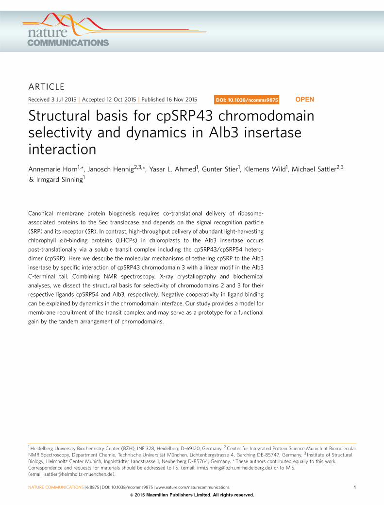
Figure 3 | Ligand recognition by chromodomains. (a) Close-up view of the backbone interactions of CD3 with A3CT IV. Hydrogen bonds are indicated by
dashed lines. (b) Sequence alignment of chromodomain ligands. The 0 position binding to the cage is depicted in red. (c) Specific read-out of the 0 position in
different chromodomains. Left: A3CT IV in CD3, middle: RRKRp in CD2, right: H3K9me3 in HP1. (d) Schematic representations of the respective chromodomain–
ligand interactions.
ARTICLE NATURE COMMUNICATIONS | DOI: 10.1038/ncomms9875
6 NATURE COMMUNICATIONS | 6:8875 | DOI: 10.1038/ncomms9875 | www.nature.com/naturecommunications
& 2015 Macmillan Publishers Limited. All rights reserved.
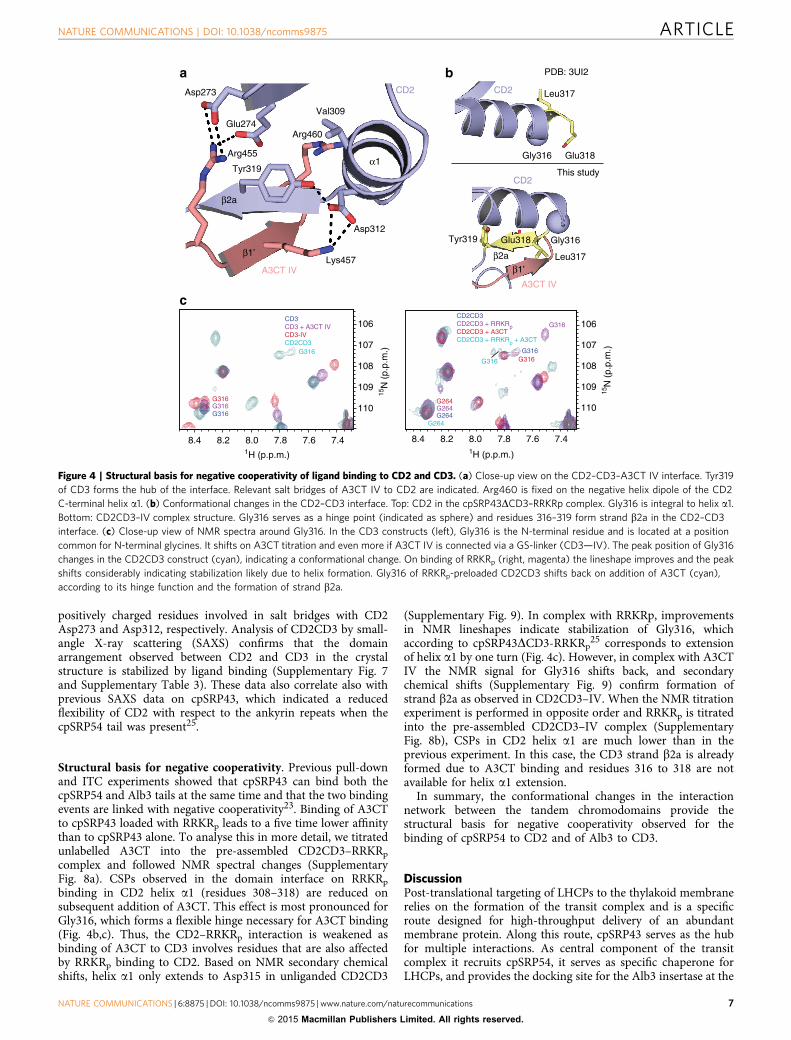
positively charged residues involved in salt bridges with CD2Asp273 and Asp312, respectively. Analysis of CD2CD3 by small-angle X-ray scattering (SAXS) confirms that the domainarrangement observed between CD2 and CD3 in the crystalstructure is stabilized by ligand binding (Supplementary Fig. 7and Supplementary Table 3). These data also correlate also withprevious SAXS data on cpSRP43, which indicated a reducedflexibility of CD2 with respect to the ankyrin repeats when thecpSRP54 tail was present25.
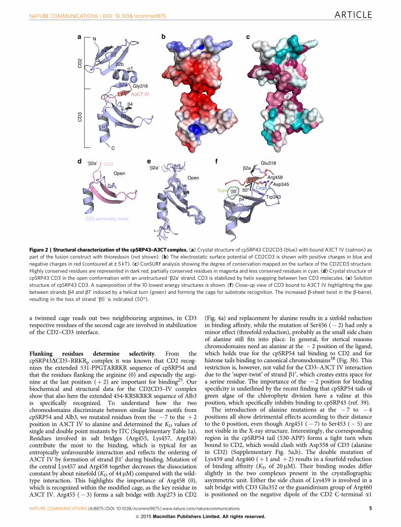
Structural basis for negative cooperativity. Previous pull-downand ITC experiments showed that cpSRP43 can bind both thecpSRP54 and Alb3 tails at the same time and that the two bindingevents are linked with negative cooperativity23. Binding of A3CTto cpSRP43 loaded with RRKRp leads to a five time lower affinitythan to cpSRP43 alone. To analyse this in more detail, we titratedunlabelled A3CT into the pre-assembled CD2CD3–RRKRp
complex and followed NMR spectral changes (SupplementaryFig. 8a). CSPs observed in the domain interface on RRKRp
binding in CD2 helix a1 (residues 308–318) are reduced onsubsequent addition of A3CT. This effect is most pronounced forGly316, which forms a flexible hinge necessary for A3CT binding(Fig. 4b,c). Thus, the CD2–RRKRp interaction is weakened asbinding of A3CT to CD3 involves residues that are also affectedby RRKRp binding to CD2. Based on NMR secondary chemicalshifts, helix a1 only extends to Asp315 in unliganded CD2CD3
(Supplementary Fig. 9). In complex with RRKRp, improvementsin NMR lineshapes indicate stabilization of Gly316, whichaccording to cpSRP43DCD3-RRKRp
25 corresponds to extensionof helix a1 by one turn (Fig. 4c). However, in complex with A3CTIV the NMR signal for Gly316 shifts back, and secondarychemical shifts (Supplementary Fig. 9) confirm formation ofstrand b2a as observed in CD2CD3–IV. When the NMR titrationexperiment is performed in opposite order and RRKRp is titratedinto the pre-assembled CD2CD3–IV complex (SupplementaryFig. 8b), CSPs in CD2 helix a1 are much lower than in theprevious experiment. In this case, the CD3 strand b2a is alreadyformed due to A3CT binding and residues 316 to 318 are notavailable for helix a1 extension.
In summary, the conformational changes in the interactionnetwork between the tandem chromodomains provide thestructural basis for negative cooperativity observed for thebinding of cpSRP54 to CD2 and of Alb3 to CD3.
DiscussionPost-translational targeting of LHCPs to the thylakoid membranerelies on the formation of the transit complex and is a specificroute designed for high-throughput delivery of an abundantmembrane protein. Along this route, cpSRP43 serves as the hubfor multiple interactions. As central component of the transitcomplex it recruits cpSRP54, it serves as specific chaperone forLHCPs, and provides the docking site for the Alb3 insertase at the
Lys457
Asp273
Glu274Val309
Arg460
Tyr319
Arg455
Asp312
A3CT IV
β2a
α1
β1’
CD2 CD2
CD2
Leu317
Gly316 Glu318
Gly316
Leu317
A3CT IV
Tyr319 Glu318
This study
PDB: 3UI2
CD3 CD3 + A3CT IVCD3-IVCD2CD3
G316
G316
G316G316
15N
(p.
p.m
.)
108
106
1H (p.p.m.)
8.4 8.2 8.0
CD2CD3CD2CD3 + RRKRpCD2CD3 + A3CTCD2CD3 + RRKRp + A3CT
7.8 7.6 7.4 8.4 8.2 8.0 7.8 7.6 7.4
109
108
107
106
110G264
G264
G264G264
G316
G316
G316G316
β2a
β1’
107
109
110
15N
(p.
p.m
.)
1H (p.p.m.)
Figure 4 | Structural basis for negative cooperativity of ligand binding to CD2 and CD3. (a) Close-up view on the CD2–CD3–A3CT IV interface. Tyr319
of CD3 forms the hub of the interface. Relevant salt bridges of A3CT IV to CD2 are indicated. Arg460 is fixed on the negative helix dipole of the CD2
C-terminal helix a1. (b) Conformational changes in the CD2–CD3 interface. Top: CD2 in the cpSRP43DCD3–RRKRp complex. Gly316 is integral to helix a1.
Bottom: CD2CD3–IV complex structure. Gly316 serves as a hinge point (indicated as sphere) and residues 316–319 form strand b2a in the CD2–CD3
interface. (c) Close-up view of NMR spectra around Gly316. In the CD3 constructs (left), Gly316 is the N-terminal residue and is located at a position
common for N-terminal glycines. It shifts on A3CT titration and even more if A3CT IV is connected via a GS-linker (CD3—IV). The peak position of Gly316
changes in the CD2CD3 construct (cyan), indicating a conformational change. On binding of RRKRp (right, magenta) the lineshape improves and the peak
shifts considerably indicating stabilization likely due to helix formation. Gly316 of RRKRp-preloaded CD2CD3 shifts back on addition of A3CT (cyan),
according to its hinge function and the formation of strand b2a.
NATURE COMMUNICATIONS | DOI: 10.1038/ncomms9875 ARTICLE
NATURE COMMUNICATIONS | 6:8875 | DOI: 10.1038/ncomms9875 | www.nature.com/naturecommunications 7
& 2015 Macmillan Publishers Limited. All rights reserved.
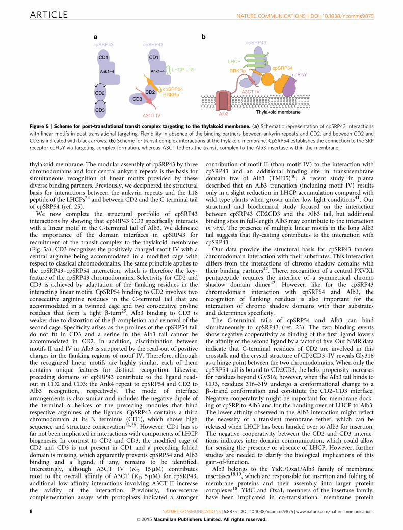
thylakoid membrane. The modular assembly of cpSRP43 by threechromodomains and four central ankyrin repeats is the basis forsimultaneous recognition of linear motifs provided by thesediverse binding partners. Previously, we deciphered the structuralbasis for interactions between the ankyrin repeats and the L18peptide of the LHCPs24 and between CD2 and the C-terminal tailof cpSRP54 (ref. 25).
We now complete the structural portfolio of cpSRP43interactions by showing that cpSRP43 CD3 specifically interactswith a linear motif in the C-terminal tail of Alb3. We delineatethe importance of the domain interfaces in cpSRP43 forrecruitment of the transit complex to the thylakoid membrane(Fig. 5a). CD3 recognizes the positively charged motif IV with acentral arginine being accommodated in a modified cage withrespect to classical chromodomains. The same principle applies tothe cpSRP43–cpSRP54 interaction, which is therefore the key-feature of the cpSRP43 chromodomains. Selectivity for CD2 andCD3 is achieved by adaptation of the flanking residues in theinteracting linear motifs. CpSRP54 binding to CD2 involves twoconsecutive arginine residues in the C-terminal tail that areaccommodated in a twinned cage and two consecutive prolineresidues that form a tight b-turn25. Alb3 binding to CD3 isweaker due to distortion of the b-completion and removal of thesecond cage. Specificity arises as the prolines of the cpSRP54 taildo not fit in CD3 and a serine in the Alb3 tail cannot beaccommodated in CD2. In addition, discrimination betweenmotifs II and IV in Alb3 is supported by the read-out of positivecharges in the flanking regions of motif IV. Therefore, althoughthe recognized linear motifs are highly similar, each of themcontains unique features for distinct recognition. Likewise,preceding domains of cpSRP43 contribute to the ligand read-out in CD2 and CD3: the Ank4 repeat to cpSRP54 and CD2 toAlb3 recognition, respectively. The mode of interfacearrangements is also similar and includes the negative dipole ofthe terminal a helices of the preceding modules that bindrespective arginines of the ligands. CpSRP43 contains a thirdchromodomain at its N terminus (CD1), which shows highsequence and structure conservation24,25. However, CD1 has sofar not been implicated in interactions with components of LHCPbiogenesis. In contrast to CD2 and CD3, the modified cage ofCD2 and CD3 is not present in CD1 and a preceding foldeddomain is missing, which apparently prevents cpSRP54 and Alb3binding and a ligand, if any, remains to be identified.Interestingly, although A3CT IV (KD 15mM) contributesmost to the overall affinity of A3CT (KD 5 mM) for cpSRP43,additional low affinity interactions involving A3CT-II increasethe avidity of the interaction. Previously, fluorescencecomplementation assays with protoplasts indicated a stronger
contribution of motif II (than motif IV) to the interaction withcpSRP43 and an additional binding site in transmembranedomain five of Alb3 (TMD5)40. A recent study in plantadescribed that an Alb3 truncation (including motif IV) resultsonly in a slight reduction in LHCP accumulation compared withwild-type plants when grown under low light conditions41. Ourstructural and biochemical study focused on the interactionbetween cpSRP43 CD2CD3 and the Alb3 tail, but additionalbinding sites in full-length Alb3 may contribute to the interactionin vivo. The presence of multiple linear motifs in the long Alb3tail suggests that fly-casting contributes to the interaction withcpSRP43.
Our data provide the structural basis for cpSRP43 tandemchromodomain interaction with their substrates. This interactiondiffers from the interactions of chromo shadow domains withtheir binding partners42. There, recognition of a central PXVXLpentapeptide requires the interface of a symmetrical chromoshadow domain dimer42. However, like for the cpSRP43chromodomain interaction with cpSRP54 and Alb3, therecognition of flanking residues is also important for theinteraction of chromo shadow domains with their substratesand determines specificity.
The C-terminal tails of cpSRP54 and Alb3 can bindsimultaneously to cpSRP43 (ref. 23). The two binding eventsshow negative cooperativity as binding of the first ligand lowersthe affinity of the second ligand by a factor of five. Our NMR dataindicate that C-terminal residues of CD2 are involved in thiscrosstalk and the crystal structure of CD2CD3–IV reveals Gly316as a hinge point between the two chromodomains. When only thecpSRP54 tail is bound to CD2CD3, the helix propensity increasesfor residues beyond Gly316; however, when the Alb3 tail binds toCD3, residues 316–319 undergo a conformational change to ab-strand conformation and constitute the CD2–CD3 interface.Negative cooperativity might be important for membrane dock-ing of cpSRP to Alb3 and for the handing over of LHCP to Alb3.The lower affinity observed in the Alb3 interaction might reflectthe necessity of a transient membrane tether, which can bereleased when LHCP has been handed over to Alb3 for insertion.The negative cooperativity between the CD2 and CD3 interac-tions indicates inter-domain communication, which could allowfor sensing the presence or absence of LHCP. However, furtherstudies are needed to clarify the biological implications of thisgain-of-function.
Alb3 belongs to the YidC/Oxa1/Alb3 family of membraneinsertases18,19, which are responsible for insertion and folding ofmembrane proteins and their assembly into larger proteincomplexes18. YidC and Oxa1, members of the insertase family,have been implicated in co-translational membrane protein
cpSRP43cpSRP43
CD1
CD2
CD3
Ank1–4
CD1
CD2
CD3
Ank1–4 LHCP L18
cpSRP54RRKRp
A3CT IV Alb3
cpSRP43
A3CT IV
Thylakoid membrane
cpSRP54cpFtsY
LHCP
RRKRp
Figure 5 | Scheme for post-translational transit complex targeting to the thylakoid membrane. (a) Schematic representation of cpSRP43 interactions
with linear motifs in post-translational targeting. Flexibility in absence of the binding partners between ankyrin repeats and CD2, and between CD2 and
CD3 is indicated with black arrows. (b) Scheme for transit complex interactions at the thylakoid membrane. CpSRP54 establishes the connection to the SRP
receptor cpFtsY via targeting complex formation, whereas A3CT tethers the transit complex to the Alb3 insertase within the membrane.
ARTICLE NATURE COMMUNICATIONS | DOI: 10.1038/ncomms9875
8 NATURE COMMUNICATIONS | 6:8875 | DOI: 10.1038/ncomms9875 | www.nature.com/naturecommunications
& 2015 Macmillan Publishers Limited. All rights reserved.
biogenesis. YidC recruits ribosomes to the plasma membrane inbacteria, while in mitochondria, which lack SRP, Oxa1-mediatedribosome docking to the inner membrane is required for efficientmembrane insertion of proteins involved in oxidativephosphorylation. Oxa1 and YidC utilize their positively chargedC-terminal extensions for ribosome binding43–45. The C-terminaltails of YidC in most Gram-negative bacteria are short andcontain less positive charges. Fusion of an extended C-terminaltail of YidC from marine bacteria like Rhodopirellula baltica andOceanicaulis alexandrii to E. coli YidC highly improved ribosomebinding21. Both Alb3 and Oxa1 have a long C-terminal tail withpronounced clusters of positively charged residues46. While thedetails of the Oxa1–ribosome interactions are not yet resolved,Alb3 utilizes the tail to specifically interact with CD3 of thenegatively charged cpSRP43. Overall, the interaction of Alb3 withcpSRP43 and Oxa1 or YidC with ribosomes appearsmechanistically similar, and the evolutionary role of cpSRP43might be to adapt the cpSRP system to post-translationaltargeting (Fig. 5b). The tandem array of cpSRP43chromodomains allows interacting with both Alb3 andcpSRP54 to efficiently deliver its LHCP cargo to the membraneinsertase and to regulate the targeting process by the SRPmachinery.
MethodsCloning. The different A3CT constructs encoding the amino acid sequence363–462 were cloned into pET21d via NcoI/XhoI restriction sites. Single anddouble point mutations in A3CT were generated in pET21d using the QuikChangesystem (Stratagene). The cpSRP43 CD2CD3 deletion construct encoding aminoacids 264–373 and the cpSRP43 CD3 deletion construct encoding amino acids316–373 were cloned into pETtrx_1a47 with a cleavable tobacco etch virus (TEV)site via NcoI/XhoI restriction sites. Single point mutations in cpSRP43 weregenerated in the pET24a vector using the QuikChange system. The A3CT IVpeptide encoding amino acid sequence 434–461 was cloned into pETgst_1a with acleavable TEV site using NcoI and XhoI restriction sites. For the CD2CD3–IVfusion construct, A3CT IV encoding residues 440–461 was covalently linked with a(GS)3 to cpSRP43 CD2CD3 encoding residues 264–373. CD2CD3–IV was thencloned with a single GS-linker into pETtrx_1a via NcoI/XhoI restriction sites.
Protein production and purification. A3CT wild-type and A3CT mutants with aC-terminal His6-tag were produced in E. coli BL21 (DE3) cells. Protein productionwas induced with 1 mM isopropyl-1-thio-b-D-galactopyranoside (IPTG) at an A600
of 0.8–1.0. After induction, the proteins were expressed for 16 h at 18 �C, harvestedand stored at � 80 �C. His-tagged A3CT pellets were resuspended in lysis buffer(100 mM Hepes/NaOH (pH 7.5), 300 mM NaCl, 5 mM MgCl2, 10% (v/v) glycerol,0.02% (v/v) 1-thioglycerol). The cells were lysed with a M1–10L microfluidizer(microfluidics), the lysate was clarified and the supernatant was applied onto a1-ml HisTrap HP column (GE Healthcare). The column was washed with washingbuffer (50 mM Hepes/NaOH (pH 7.5), 300 mM NaCl, 5 mM MgCl2, 5% (v/v)glycerol, 0.02% (v/v) 1-thioglycerol) containing 0, 20 and 50 mM imidazole. Proteinelution occurred with a buffer containing 300 mM imidazole. Protein containingfractions were pooled and subjected to a S75 26/60 size-exclusion chromatographyequilibrated in 20 mM Hepes/NaOH (pH 7.5), 150 mM NaCl, 2 mM MgCl2 and1 mM DTT. N-terminally His6-tagged cpSRP43 wild-type and cpSRP43 mutantswere produced in E. coli Rosetta (DE3) pLysS. Cells were grown at 37 �C to a celldensity of 0.6–0.8 OD per ml. Protein production was induced by 0.2 mM IPTG.After 12 h at 18 �C, the cells were harvested and stored at � 80 �C. Proteinproduction was performed as described above. N-terminally His6-taggedTrx–CD2CD3–IV was produced in E. coli Rosetta2 (DE3) pLysS cells inauto-induction medium. His6–Trx–CD2CD3–IV was purified as described above.
N-terminally His6-tagged Trx–CD2CD3 and Trx–CD3 with a cleavable TEVsite were produced in E. coli Rosetta2 pLysS cells. Cells were grown at 37 �C to acell density of 0.6–0.8 OD per ml. Protein production was induced by 0.2 mMIPTG. After 12 h at 18 �C, the cells were harvested and stored at � 80 �C. His6–glutathione-S-transferase (GST)–A3CT IV peptide was produced in E. coli BL21cells. Cells were grown at 37 �C and protein production was induced by 0.4 mMIPTG at A600 of 0.8–1.0. After 3 h at 37 �C, the cells were harvested and stored at� 80 �C. Purification of proteins fused to Trx and GST was performed as describedin a previous work23 with the following adaptation for A3CT IV peptide: after TEVcleavage over night, the sample was reloaded on a HisTrap column and the A3CTIV peptide was eluted in a buffer containing 50 mM Hepes/NaOH (pH 7.5),300 mM NaCl, 5 mM MgCl2, 5% (v/v) glycerol, 0.02% (v/v) 1-thioglycerol and20 mM imidazole. Protein containing fractions were pooled and the protein was
loaded on a 26/60 Superdex75 column (GE Healthcare) equilibrated in gel filtrationbuffer (20 mM Hepes/NaOH (pH 7.5), 150 mM NaCl, 2 mM MgCl2).
Protein production and purification for NMR. N-terminally His6-taggedTrx–CD2CD3 and Trx–CD3 were produced in E. coli Rosetta2 pLysS cells, andN-terminally His6-tagged Trx–A3CT and His6–GST-tagged A3CT IV peptide wereproduced in E. coli BL21 cells. The proteins were produced in M9 mediumsupplemented with [15N]H4Cl and 13C-glucose as the sole sources of nitrogen andcarbon for uniformly protein labelling. After induction with 1 mM IPTG at an A600
of 0.8–1.0, the proteins were produced for 12 h at 30 �C. The proteins were purifiedas described previously23. For size-exclusion chromatography, the samples wereloaded on a 26/60 Superdex75 column equilibrated in NMR buffer (20 mMNa-phosphate (pH 6.5), 150 mM NaCl).
Isothermal titration calorimetry. ITC experiments were performed in a buffercontaining 20 mM Hepes (pH 7.5), 150 mM NaCl, 5 mM MgCl2 and 0.25 mMTCEP (tris(2-carboxyethyl)phosphine). All protein samples and peptides weredialysed over night in this buffer and extensively degassed prior to titration. ITCexperiments were conducted on a VP-ITC microcalorimeter (MicroCal, North-ampton, MA) at 20 �C with 307 r.p.m. stirring. Titrations consisted of 23 injectionsof 12ml aliquots of the titrant into the solution of the cell. Typical protein con-centrations in the cell were 0.01–0.1 mM and 0.1–1.0 mM in the syringe. Data wereanalysed with Origin 7.0 software. The ITC measurements were performed induplicate or triplicate. Peptides were purchased from PSL (Peptide SpecialityLaboratories, Technology Park, Heidelberg), purified by HPLC and analysed bymass spectrometry.
Protein crystallization and data collection. Crystals of His6–Trx–CD2CD3–IVwere grown in an in-house automated crystallization platform at 18 �C in sittingdrops containing 0.2 ml of His6–Trx–CD2CD3–IV (17 mg ml� 1) and 0.2 ml of areservoir solution consisting of 20% (w/v) PEG 3350 and 0.2 M Ca(OAc)2. Crystalsgrew as thin and fragile plates after 5 days. The crystals were cryoprotected inmother liquor containing 20% (v/v) glycerol and flash-cooled in liquid nitrogen.Data were collected at the European Synchrotron Radiation Facility (ESRF,Grenoble) on beamline ID29 at 0.992 Å and 100 K. Data were integrated and scaledwith XDS48 and AIMLESS from the CCP4 package49.
Crystals of cpSRP43 CD3 were grown at 4 �C in sitting drops containing 0.2 mlof cpSRP43 CD3 complexed with A3CT IV (12 mg ml� 1) and 0.2 ml of a reservoirsolution consisting of 25% PEG 3350, 0.1 M Hepes pH 7.5 and 0.2 M MgCl2.Crystals containing only cpSRP43 CD3 grew as little squares after 21 days. Thecrystals were cryoprotected in mother liquor containing 35% PEG and 20%glycerol, flash-frozen in liquid nitrogen and measured at the ESRF on beamlineID29 at 1.033 Å and 100 K. Data were integrated and scaled with XDS48 andAIMLESS from the CCP4 package49.
Structure determination and refinement. The construct His6–Trx–CD2CD3–IVcrystallized in the orthorhombic space group P21212 with two molecules in theasymmetric unit. The cell parameters are a¼ 79.6 Å, b¼ 163.7 Å, c¼ 37.4 Åand the solvent content is 42%. The structure was solved by molecular replacementas implemented in PHASER50 using Trx (PDB code 3DXB) and one ensemble ofthe solution structure of cpSRP43 CD3 as a search model. The structure wasrefined using Phenix.refine51 and iterative model building in COOT52 and wasvalidated with MOLPROBITY53 as implemented in PHENIX51. Ramachandranstatistics for the final model show 97% of residues in most favoured regions and3% of residues in additionally allowed regions. All structural figures were preparedwith PyMOL54. Electrostatic surface potentials were calculated with APBSintegrated in PyMOL54. Sequence alignments were performed with ClustalX55 andprotein sequence conservations were performed using the ConSURF server56.
CpSRP43 CD3 crystallized in the cubic space group I432 with a cell axis of103.9 Å. The solvent content was determined to 59% with one molecule in theasymmetric unit. The structure of the open CD3 was solved by molecularreplacement as implemented in PHASER50 using the solution structure of cpSRP43CD3 as a search model. Structure refinement was performed with Phenix.refine51
and iterative model building in COOT52. The structure was validated withMOLPROBITY53 as implemented in PHENIX51. Ramachandran statistics for thefinal model show 96% of residues in most favoured regions and 4% of residues inadditionally allowed regions. All structural figures were prepared with PyMol54.
NMR spectroscopy. Spectra were acquired at 298 K (all experiments, exceptbackbone assignment experiments of A3CT motif IV and its titrations with CD3,which were carried out at 278 K) on Bruker Avance III NMR spectrometersequipped with cryogenic triple resonance gradient probe heads at magnetic fieldstrengths corresponding to proton Larmor frequencies of 600 and 800 MHz. Allspectra were processed with NMRPipe57 and analysed using Sparky58 and CARA(http://cara.nmr.ch). For backbone and side chain assignments HNCACB,CBCA(CO)NH and HCCH-TOCSY spectra were recorded59. Distance restraintswere obtained from 15N- and 13C-edited 3D NOESY-HSQC spectra. Secondarystructure prediction from secondary chemical shifts (Ca and Cb) was done
NATURE COMMUNICATIONS | DOI: 10.1038/ncomms9875 ARTICLE
NATURE COMMUNICATIONS | 6:8875 | DOI: 10.1038/ncomms9875 | www.nature.com/naturecommunications 9
& 2015 Macmillan Publishers Limited. All rights reserved.
according to the study by Wishart and Sykes60. NMR CSPs of CD3, CD2CD3,A3CT and motif II/IV on titration with each other and RRKRp were observed bytitrating the ligand stepwise until saturation was reached. A series of 1H, 15NHSQC spectra were recorded for 15N-labelled CD3, CD2CD3, A3CT or motif IVwith various amounts of binding partner. The following stoichiometries were usedfor diverse interactions (with the 15N-labelled partner being first): CD2CD3:A3CT(1:0.5, 1:1, 1:1.5, 1:2.7, 1:3.9); CD2CD3:RRKR (1:0.25, 1:0.35, 1:0.7, 1:1.1, 1:1.9,1:2.8, 1:3.8); A3CT:CD2CD3 (1:0.55, 1:1.2, 1:1.8, 1:3.2); CD2CD3:A3CT:RRKR(1:3.9:0.7, 1:3.9:2.2, 1:3.9:5.2); CD2CD3:RRKR:A3CT (1:3.8:2.8); CD3:motif IV(1:0.9, 1:2.7, 1:7); motif IV:CD3 (1:1, 1:2.1, 1:3.2, 1:5.7, 1:8.4); CD3:motif II (1:0.9,1:2.7, 1:7); CD2CD3:motif IV (1:0.9, 1:2.3, 1:7) and CD2CD3:motif II (1:0.9, 1:2.3,1:7). All samples used in the titrations were in the same buffer to prevent CSPsbased on buffer or pH effects. Affinities from NMR data could not be obtained inmost cases as interactions were in the intermediate exchange regime. Interactingresidues were identified by manually tracing peaks in the NMR spectra andconfirmed by recording and analysing backbone assignment experiments forthe bound form. Atom-specific chemical shift weighting was done accordingto Mulder et al.61.
The NMR structure of CD3 was calculated by automated NOE cross-peakassignment and torsion angle dynamics, using the software CYANA 3.0 (ref. 62).Automatically assigned NOEs and completeness of the NOE cross-peaks weremanually inspected. Distance restraints from the CYANA calculation andTALOSþ derived63 were used in a water refinement calculation64 using ARIA 1.2(ref. 65). Structural quality of the final ensemble of 10 structures with lowestenergy was validated using the iCING web server66. Ramachandran statistics forthe final NMR structure of CD3 show 90.9% of residues in most favoured regionsand 9.1% in additionally allowed regions. The structural statistics are shown inSupplementary Table 2.
Small-angle X-ray scattering. The constructs cpSRP43 CD2CD3 (residues 264–373) and cpSRP43 CD3–IV were produced and purified as described above. About50ml of CD2CD3 (5, 10, 20 mg ml� 1) and CD2CD3 with 7� molar excess ofA3CT IV (2.5, 5, 10 mg ml� 1), as well as buffer and buffer with the same amountof A3CT IV were measured at 298 K on a Rigaku BioSAXS1000 using a Pilatusdetector. Six frames with 900-s exposure time per frame were recorded for eachsample and buffer using an X-ray wavelength of l¼ 1.5418 Å. Frames showingradiation damage were removed prior to data analysis. For SAXS data collectionand processing, the software SAXSLab 3.0.1r1 was used. The one-dimensionalscattering intensities of samples and buffers were expressed as a function of themodulus of the scattering vector Q¼ (4p/l)siny. Buffer intensities were subtractedfrom CD2CD3 samples, and buffer plus peptide intensities were subtracted fromCD2CD3 plus A3CT IV samples using the software PRIMUS67. The radii ofgyration Rg of all samples were extracted by the Guinier approximation with thesame programme. Rg and Dmax were also calculated from pairwise distributionfunctions using GNOM68. CRYSOL69 was used to fit the experimental scatteringdensities of CD2CD3 plus A3CT IV with the back-calculated scattering densitiesfrom the crystal structure. All statistics are summarized according to the study byJacques et al.70 in Supplementary Table 3.
References1. Cross, B. C., Sinning, I., Luirink, J. & High, S. Delivering proteins for export
from the cytosol. Nat. Rev. Mol. Cell Biol. 10, 255–264 (2009).2. Pool, M. R. Signal recognition particles in chloroplasts, bacteria, yeast and
mammals (Review). Mol. Membr. Biol. 22, 3–15 (2005).3. Saraogi, I. & Shan, S. O. Molecular mechanism of co-translational protein
targeting by the signal recognition particle. Traffic 12, 535–542 (2011).4. Li, X., Henry, R., Yuan, J., Cline, K. & Hoffman, N. E. A chloroplast homologue
of the signal recognition particle subunit SRP54 is involved in theposttranslational integration of a protein into thylakoid membranes. Proc. NatlAcad. Sci. USA 92, 3789–3793 (1995).
5. Schunemann, D. Mechanisms of protein import into thylakoids of chloroplasts.Biol. Chem. 388, 907–915 (2007).
6. Franklin, A. E. & Hoffman, N. E. Characterization of a chloroplast homologueof the 54-kDa subunit of the signal recognition particle. J. Biol. Chem. 268,22175–22180 (1993).
7. Schunemann, D. et al. A novel signal recognition particle targets light-harvesting proteins to the thylakoid membranes. Proc. Natl Acad. Sci. USA 95,10312–10316 (1998).
8. Klimyuk, V. I. et al. A chromodomain protein encoded by the arabidopsis CAOgene is a plant-specific component of the chloroplast signal recognition particlepathway that is involved in LHCP targeting. Plant Cell 11, 87–99 (1999).
9. Groves, M. R. et al. Functional characterization of recombinant chloroplastsignal recognition particle. J. Biol. Chem. 276, 27778–27786 (2001).
10. Ruban, A. V. et al. Plasticity in the composition of the light harvesting antennaof higher plants preserves structural integrity and biological function. J. Biol.Chem. 281, 14981–14990 (2006).
11. Barros, T. & Kuhlbrandt, W. Crystallisation, structure and function of plantlight-harvesting Complex II. Biochim. Biophys. Acta 1787, 753–772 (2009).
12. Kuhlbrandt, W., Wang, D. N. & Fujiyoshi, Y. Atomic model of plant light-harvesting complex by electron crystallography. Nature 367, 614–621 (1994).
13. Soll, J. & Schleiff, E. Protein import into chloroplasts. Nat. Rev. Mol. Cell Biol. 5,198–208 (2004).
14. Kessler, F. & Schnell, D. Chloroplast biogenesis: diversity and regulation of theprotein import apparatus. Curr. Opin. Cell Biol. 21, 494–500 (2009).
15. Ouyang, M. et al. LTD is a protein required for sorting light-harvestingchlorophyll-binding proteins to the chloroplast SRP pathway. Nat. Commun. 2,277 (2011).
16. Moore, M., Goforth, R. L., Mori, H. & Henry, R. Functional interaction ofchloroplast SRP/FtsY with the ALB3 translocase in thylakoids: substrate notrequired. J. Cell Biol. 162, 1245–1254 (2003).
17. Schunemann, D. Structure and function of the chloroplast signal recognitionparticle. Curr. Genet. 44, 295–304 (2004).
18. Driessen, A. J. Protein translocation across the bacterial cytoplasmicmembrane. Annu. Rev. Biochem. 77, 643–667 (2008).
19. Yi, L. & Dalbey, R. E. Oxa1/Alb3/YidC system for insertion of membraneproteins in mitochondria, chloroplasts and bacteria (review). Mol. Membr. Biol.22, 101–111 (2005).
20. Kedrov, A. et al. Elucidating the native architecture of the YidC: ribosomecomplex. J. Mol. Biol. 425, 4112–4124 (2013).
21. Seitl, I., Wickles, S., Beckmann, R., Kuhn, A. & Kiefer, D. The C-terminalregions of YidC from Rhodopirellula baltica and Oceanicaulis alexandrii bind toribosomes and partially substitute for SRP receptor function in Escherichia coli.Mol. Microbiol. 91, 408–421 (2014).
22. Kumazaki, K. et al. Crystal structure of Escherichia coli YidC, a membraneprotein chaperone and insertase. Sci. Rep. 4, 7299 (2014).
23. Falk, S., Ravaud, S., Koch, J. & Sinning, I. The C terminus of the Alb3membrane insertase recruits cpSRP43 to the thylakoid membrane. J. Biol.Chem. 285, 5954–5962 (2009).
24. Stengel, K. F. et al. Structural basis for specific substrate recognition by thechloroplast signal recognition particle protein cpSRP43. Science 321, 253–256(2008).
25. Holdermann, I. et al. Chromodomains read the arginine code of post-translational targeting. Nat. Struct. Mol. Biol. 19, 260–263 (2012).
26. Brehm, A., Tufteland, K. R., Aasland, R. & Becker, P. B. The many colours ofchromodomains. Bioessays 26, 133–140 (2004).
27. Grewal, S. I. & Jia, S. Heterochromatin revisited. Nat. Rev. Genet. 8, 35–46(2007).
28. Paro, R. & Hogness, D. S. The Polycomb protein shares a homologous domainwith a heterochromatin-associated protein of Drosophila. Proc. Natl Acad. Sci.USA 88, 263–267 (1991).
29. Pluckthun, A. Designed ankyrin repeat proteins (DARPins): binding proteinsfor research, diagnostics, and therapy. Annu. Rev. Pharmacol. Toxicol. 55,489–511 (2015).
30. Tu, C. J., Peterson, E. C., Henry, R. & Hoffman, N. E. The L18 domain of light-harvesting chlorophyll proteins binds to chloroplast signal recognition particle43. J. Biol. Chem. 275, 13187–13190 (2000).
31. Falk, S. & Sinning, I. cpSRP43 is a novel chaperone specific for light-harvestingchlorophyll a,b-binding proteins. J. Biol. Chem. 285, 21655–21661 (2010).
32. Jaru-Ampornpan, P. et al. ATP-independent reversal of a membrane proteinaggregate by a chloroplast SRP subunit. Nat. Struct. Mol. Biol. 17, 696–702(2010).
33. Gao, F. et al. Regulation of structural dynamics within a signal recognitionparticle promotes binding of protein targeting substrates. J. Biol. Chem. 290,15462–15474 (2015).
34. Fischle, W. et al. Molecular basis for the discrimination of repressive methyl-lysine marks in histone H3 by Polycomb and HP1 chromodomains. Genes Dev.17, 1870–1881 (2003).
35. Corsini, L., Hothorn, M., Scheffzek, K., Sattler, M. & Stier, G. Thioredoxin as afusion tag for carrier-driven crystallization. Protein Sci. 17, 2070–2079 (2008).
36. Bassler, J. et al. A network of assembly factors is involved in remodeling rRNAelements during preribosome maturation. J. Cell Biol. 207, 481–498 (2014).
37. Dougherty, D. A. The cation-pi interaction. Acc. Chem. Res. 46, 885–893(2013).
38. Jacobs, S. A. & Khorasanizadeh, S. Structure of HP1 chromodomain bound to alysine 9-methylated histone H3 tail. Science 295, 2080–2083 (2002).
39. Dunschede, B. et al. Chloroplast SRP54 was recruited for posttranslationalprotein transport via complex formation with chloroplast SRP43 during landplant evolution. J. Biol. Chem. 290, 13104–13114 (2015).
40. Dunschede, B., Bals, T., Funke, S. & Schunemann, D. Interaction studiesbetween the chloroplast signal recognition particle subunit cpSRP43 and thefull-length translocase Alb3 reveal a membrane-embedded binding region inAlb3 protein. J. Biol. Chem. 286, 35187–35195 (2011).
41. Urbischek, M. et al. The extreme Albino3 (Alb3) C terminus is required forAlb3 stability and function in Arabidopsis thaliana. Planta 242, 733–746(2015).
42. Thiru, A. et al. Structural basis of HP1/PXVXL motif peptide interactions andHP1 localisation to heterochromatin. EMBO J. 23, 489–499 (2004).
ARTICLE NATURE COMMUNICATIONS | DOI: 10.1038/ncomms9875
10 NATURE COMMUNICATIONS | 6:8875 | DOI: 10.1038/ncomms9875 | www.nature.com/naturecommunications
& 2015 Macmillan Publishers Limited. All rights reserved.
43. Preuss, M., Ott, M., Funes, S., Luirink, J. & Herrmann, J. M. Evolution ofmitochondrial oxa proteins from bacterial YidC. Inherited and acquiredfunctions of a conserved protein insertion machinery. J. Biol. Chem. 280,13004–13011 (2005).
44. Jia, L. et al. Yeast Oxa1 interacts with mitochondrial ribosomes: the importanceof the C-terminal region of Oxa1. EMBO J. 22, 6438–6447 (2003).
45. Kohler, R. et al. YidC and Oxa1 form dimeric insertion pores on the translatingribosome. Mol. Cell 34, 344–353 (2009).
46. Jiang, F. et al. Defining the regions of Escherichia coli YidC that contribute toactivity. J. Biol. Chem. 278, 48965–48972 (2003).
47. Bogomolovas, J., Simon, B., Sattler, M. & Stier, G. Screening of fusion partnersfor high yield expression and purification of bioactive viscotoxins. Protein Expr.Purif. 64, 16–23 (2009).
48. Kabsch, W. XDS. Acta Crystallogr. D Biol. Crystallogr. 66, 125–132 (2010).49. Winn, M. D. et al. Overview of the CCP4 suite and current developments. Acta
Crystallogr. D Biol. Crystallogr. 67, 235–242 (2011).50. Bunkoczi, G. et al. Phaser.MRage: automated molecular replacement. Acta
Crystallogr. D Biol. Crystallogr. 69, 2276–2286 (2013).51. Adams, P. D. PHENIX: a comprehensive Python-based system for
macromolecular structure solution. Acta Crystallogr. D Biol. Crystallogr. 66,213–221 (2010).
52. Emsley, P., Lohkamp, B., Scott, W. G. & Cowtan, F. Feature and development ofCoot. Acta Crystallogr. D Biol. Crystallogr. 66, 486–501 (2010).
53. Chen, V. B. MolProbity: all-atom structure validation for macromolecularcrystallography. Acta Crystallogr. D Biol. Crystallogr. 66, 12–21 (2010).
54. The PyMol Molecular Graphics System, Version 1.7.6.6 Schrodinger, LLC.55. Thompson, J. D., Gibson, T. J., Plewniak, F., Jeanmougin, F. & Higgins, D. G.
The CLUSTAL_X windows interface: flexible strategies for multiple sequencealignment aided by quality analysis tools. Nucleic Acids Research 25, 4876–4882(1997).
56. Landau, M. et al. ConSurf 2005: the projection of evolutionary conservationscores of residues on protein structures. Nucleic Acids Res. 33, W299–W302(2005).
57. Delaglio, F. et al. NMRPipe: a multidimensional spectral processing systembased on UNIX pipes. J. Biomol. NMR 6, 277–293 (1995).
58. Lee, W., Westler, W. M., Bahrami, A., Eghbalnia, H. R. & Markley, J. L. PINE-SPARKY: graphical interface for evaluating automated probabilistic peakassignments in protein NMR spectroscopy. Bioinformatics 25, 2085–2087(2009).
59. Sattler, M., Schleucher, J. & Griesinger, C. Heteronuclear multidimensional NMRexperiments for the structure determination of proteins in solution emplyingpulsed field gradients. Prog. Nucl. Magn. Res. Spectrosc. 34, 93–158 (1999).
60. Wishart, D. S. & Sykes, B. D. The 13C chemical-shift index: a simple methodfor the identification of protein secondary structure using 13C chemical-shiftdata. J. Biomol. NMR 4, 171–180 (1994).
61. Mulder, F. A., Schipper, D., Bott, R. & Boelens, R. Altered flexibility in thesubstrate-binding site of related native and engineered high-alkaline Bacillussubtilisins. J. Mol. Biol. 292, 111–123 (1999).
62. Guntert, P. Automated structure determination from NMR spectra. Eur.Biophys. J. 38, 129–143 (2009).
63. Shen, Y., Delaglio, F., Cornilescu, G. & Bax, A. TALOSþ : a hybrid method forpredicting protein backbone torsion angles from NMR chemical shifts.J. Biomol. NMR 44, 213–223 (2009).
64. Linge, J. P., Williams, M. A., Spronk, C. A., Bonvin, A. M. & Nilges, M.Refinement of protein structures in explicit solvent. Proteins 50, 496–506(2003).
65. Rieping, W. et al. ARIA2: automated NOE assignment and data integration inNMR structure calculation. Bioinformatics 23, 381–382 (2007).
66. Doreleijers, J. F. et al. CING: an integrated residue-based structure validationprogram suite. J. Biomol. NMR 54, 267–283 (2012).
67. Konarev, P. V., Volkov, V. V., Sokolova, A. V., Koch, M. H. J. & Svergun, D. I.PRIMUS—a Windows-PC based system for small-angle scattering dataanalysis. J. Appl. Cryst. 36, 1277–1282 (2003).
68. Svergun, D. I. Determination of the regularization parameter in indirect-transform methods using perceptual criteria. J. Appl. Cryst. 25, 495–503 (1992).
69. Svergun, D. I., Barberato, C. & Koch, M. H. J. CRYSOL—program to evaluateX-ray solution scattering of biological macromolecules from atomiccoordinates. J. Appl. Cryst. 28, 768–773 (1995).
70. Jacques, D. A., Guss, J. M., Svergun, D. I. & Trewhella, J. Pulication guidlinesmodelling of small-angle scattering data from biomolecules in solution. ActaCrystallogr. D Biol. Crystallogr. 68, 620–626 (2012).
AcknowledgementsWe gratefully acknowledge support of J. Kopp, C. Siegmann and G. Muller from theBZH/Cluster of Excellence: CellNetworks crystallization platform and Ralf Stehle(TU Munchen) for support with SAXS measurements. We thank A. Griessner andK. Haubrich for their contributions in the early stages of the project. Data collection wasperformed at the European Synchrotron Radiation Facility (ESRF), Grenoble, France. Weacknowledge SAXS measurements at the facility of the SFB1035 at Department Chemie,Technische Universitat Munchen and the Bavarian NMR Centre for NMR measurementtime. J.H. gratefully acknowledges the European Molecular Biology Organization(EMBO) for a long-term fellowship (ALTF 276–2010). I.S. is an investigator of theCluster of Excellence: CellNetworks and acknowledges support by the Deutsche For-schungsgemeinschaft (SFB 638). M.S. acknowledges support by the Deutsche For-schungsgemeinschaft (SFB 1035).
Author contributionsA.H., J.H., M.S. and I.S. designed the experiments; A.H., J.H., Y.L.A. and G.S. performedexperiments; A.H., J.H., M.S., K.W. and I.S. analysed the data and wrote the manuscript.All authors discussed the experiments and commented on the manuscript.
Additional informationAccession codes: Protein Data Bank: coordinates and structure factors have beendeposited under the accession codes 5E4W, 5E4X and 2N88. The NMR chemical shiftsare deposited in the Biological Magnetic Resonance Data Bank, entries 25837 and 25838.
Supplementary Information accompanies this paper at http://www.nature.com/naturecommunications
Competing financial interests: The authors declare no competing financial interests.
Reprints and permission information is available online at http://npg.nature.com/reprintsandpermissions/
How to cite this article: Horn, A. et al. Structural basis for cpSRP43 chromodomainselectivity and dynamics in Alb3 insertase interaction. Nat. Commun. 6:8875doi: 10.1038/ncomms9875 (2015).
This work is licensed under a Creative Commons Attribution 4.0International License. The images or other third party material in this
article are included in the article’s Creative Commons license, unless indicated otherwisein the credit line; if the material is not included under the Creative Commons license,users will need to obtain permission from the license holder to reproduce the material.To view a copy of this license, visit http://creativecommons.org/licenses/by/4.0/
NATURE COMMUNICATIONS | DOI: 10.1038/ncomms9875 ARTICLE
NATURE COMMUNICATIONS | 6:8875 | DOI: 10.1038/ncomms9875 | www.nature.com/naturecommunications 11
& 2015 Macmillan Publishers Limited. All rights reserved.