Embed Size (px)
Citation preview
Frontiers in Neuroendocrinology 20, 49–70 (1999)Article ID frne.1998.0173, available online at http://www.idealibrary.com on
Stress and the Aging Hippocampus
Bruce S. McEwen
Harold and Margaret Milliken Hatch Laboratory of Neuroendocrinology, Rockefeller University,1230 York Avenue, New York, New York 10021
The ‘‘glucocorticoid cascade hypothesis’’ of hippocampal aging has stimulated a greatdeal of research into the neuroendocrine aspects of aging and the role of glucocorticoids,in particular. Besides strengthening the methods for investigating the aging brain, thisresearch has revealed that the interactions between glucocorticoids and hippocampalneurons are far more complicated than originally envisioned and involve the participa-tion of neurotransmitter systems, particularly the excitatory amino acids, as well ascalcium ions and neurotrophins. New information has provided insights into the role ofearly experience in determining individual differences in brain and body aging by settingthe reactivity of the hypothalamopituitary–adrenal axis and the autonomic nervoussystem. As a result of this research and advances in neuroscience and the study of aging,we now have a far more sophisticated view of the interactions among genes, earlydevelopment, and environmental influences, as well as a greater appreciation of events atthe cellular and molecular levels which protect neurons, and a greater appreciation ofpathways of neuronal damage and destruction. While documenting the ultimate vulner-ability of the brain to stressful challenges and to the aging process, the net result of thisresearch has highlighted the resilience of the brain and offered new hope for treatmentstrategies for promoting the health of the aging brain. KEY WORDS: stress; hippocampalaging; glucocorticoid cascade hypothesis. r 1999 Academic Press
INTRODUCTION
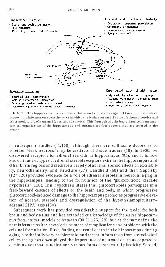
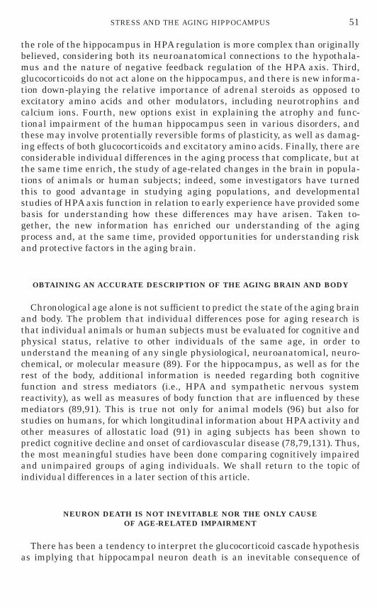
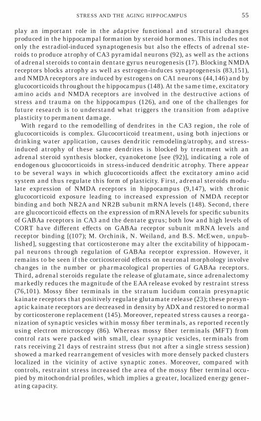
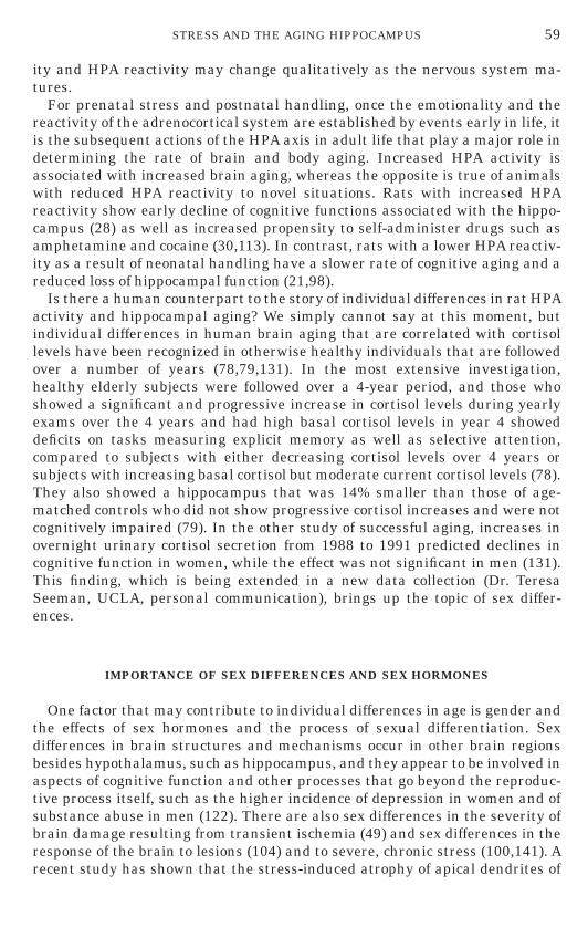
The hippocampus is a particularly vulnerable and sensitive region of thebrain that is also very important for declarative and spatial learning andmemory (36). Hippocampal neurons are vulnerable to seizures, strokes, andhead trauma, as well as responding to stressful experiences (27,92,130). At thesame time they show remarkable plasticity, involving long-term synapticpotentiation and depression, dendritic remodeling, synaptic turnover, andneurogenesis in the case of the dentate gyrus (Fig 1) (17,27,92).
The work of aus der Muhlen and Ockenfels (3) first drew attention topotentially toxic actions of adrenal steroids. The reported darkly stainedneurons in the hippocampus of guinea pigs exposed to high levels of glucocorti-coids, an observation that has been confirmed and extended to repeated stress
Address correspondence and reprint requests to Bruce S. McEwen at Laboratory of Neuroendo-crinology, Rockefeller University, 1230 York Avenue, New York, NY 10021. Fax: 212 327 8634E-mail:[email protected].
49 0091-3022/99 $30.00Copyright r 1999 by Academic Press
All rights of reproduction in any form reserved.
frne 0173@xyserv2/disk3/CLS jrnl/GRP frne/JOB frne20-1/DIV 149a01 rich
in subsequent studies (41,100), although there are still some doubts as towhether ‘‘dark neurons’’ may be artifacts of tissue trauma (18). In 1968, wediscovered receptors for adrenal steroids in hippocampus (95), and it is nowknown that two types of adrenal steroid receptors exist in the hippocampus andother brain regions and mediate a variety of adrenal steroid effects on excitabil-ity, neurochemistry, and structure (27). Landfield (68) and then Sapolsky(127,128) provided evidence for a role of adrenal steroids in neuronal aging inthe hippocampus, leading to the formulation of the ‘‘glucocorticoid cascadehypothesis’’ (130). This hypothesis states that glucocorticoids participate in afeed-forward cascade of effects on the brain and body, in which progressiveglucocorticoid-induced damage to the hippocampus promotes progressive eleva-tion of adrenal steroids and dysregulation of the hypothalamopituitary–adrenal (HPA) axis (130).
Subsequent work has provided considerable support for the model for bothbrain and body aging and has extended our knowledge of the aging hippocam-pus from animal models to humans (90,91,126,129), but at the same time thenew information has revealed a number of complications and problems with theoriginal formulation. First, finding neuronal death in the hippocampus during
FIG. 1. The hippocampal formation is a plastic and vulnerable region of the adult brain whichis providing information about the ways in which the brain ages and the role of adrenal steroids andother modulators of neuronal function and survival. This figure shows the basic three-cell neuroana-tomical organization of the hippocampus and summarizes four aspects that are covered in thearticle.
50 BRUCE S. MCEWEN
aging is technically very problematic, and recent information from stereologicalcell counting has down-played the importance of neuronal death as opposed todeclining neuronal function and various forms of structural plasticity. Second,
frne 0173@xyserv2/disk3/CLS jrnl/GRP frne/JOB frne20-1/DIV 149a01 rich
the role of the hippocampus in HPA regulation is more complex than originallybelieved, considering both its neuroanatomical connections to the hypothala-mus and the nature of negative feedback regulation of the HPA axis. Third,glucocorticoids do not act alone on the hippocampus, and there is new informa-tion down-playing the relative importance of adrenal steroids as opposed toexcitatory amino acids and other modulators, including neurotrophins andcalcium ions. Fourth, new options exist in explaining the atrophy and func-tional impairment of the human hippocampus seen in various disorders, andthese may involve protentially reversible forms of plasticity, as well as damag-ing effects of both glucocorticoids and excitatory amino acids. Finally, there areconsiderable individual differences in the aging process that complicate, but atthe same time enrich, the study of age-related changes in the brain in popula-tions of animals or human subjects; indeed, some investigators have turnedthis to good advantage in studying aging populations, and developmentalstudies of HPA axis function in relation to early experience have provided somebasis for understanding how these differences may have arisen. Taken to-gether, the new information has enriched our understanding of the agingprocess and, at the same time, provided opportunities for understanding riskand protective factors in the aging brain.
OBTAINING AN ACCURATE DESCRIPTION OF THE AGING BRAIN AND BODY
Chronological age alone is not sufficient to predict the state of the aging brainand body. The problem that individual differences pose for aging research isthat individual animals or human subjects must be evaluated for cognitive andphysical status, relative to other individuals of the same age, in order tounderstand the meaning of any single physiological, neuroanatomical, neuro-chemical, or molecular measure (89). For the hippocampus, as well as for therest of the body, additional information is needed regarding both cognitivefunction and stress mediators (i.e., HPA and sympathetic nervous systemreactivity), as well as measures of body function that are influenced by thesemediators (89,91). This is true not only for animal models (96) but also forstudies on humans, for which longitudinal information about HPA activity andother measures of allostatic load (91) in aging subjects has been shown topredict cognitive decline and onset of cardiovascular disease (78,79,131). Thus,the most meaningful studies have been done comparing cognitively impairedand unimpaired groups of aging individuals. We shall return to the topic ofindividual differences in a later section of this article.
NEURON DEATH IS NOT INEVITABLE NOR THE ONLY CAUSEOF AGE-RELATED IMPAIRMENT
51STRESS AND THE AGING HIPPOCAMPUS
There has been a tendency to interpret the glucocorticoid cascade hypothesisas implying that hippocampal neuron death is an inevitable consequence of
frne 0173@xyserv2/disk3/CLS jrnl/GRP frne/JOB frne20-1/DIV 149a01 rich
brain aging and that age-related impairments in cognitive function are solelyrelated to such neuronal loss. This notion has given way to a more flexible viewof brain aging, in which impairments in hippocampal functioning can bestudied in terms of potentially reversible, as well as irreversible, changes inneuronal structure, neurochemistry, and function (69,70,90,103).
The first challenge to the notion of neuronal loss is from a methodologicalproblem in estimating neuron number in the brain. Because of the gradual timecourse of brain aging, even in rapidly-aging small animals like rats, observingneuronal death by counting dying neurons is futile because it would have to besuch a slow process to account for the gradual changes in function. For thatreason, studies of pyramidal neuron damage and the role and mechanism ofaction of glucocorticoids and excitatory amino acids have utilized kainic aciddamage or transient ischemia models (126). In the aging brain, counting ofneurons to determine the average decline in neuronal content of the hippocam-pus is also fraught with technical problems. The initial reports of the aginghippocampus identified a reduced density of pyramidal neurons in aging ratsthat also showed impairments of spatial and other memory tasks (64,128).However, Mark West introduced a stereological counting procedure (73) andreported a failure to find reduced hippocampal neuron numbers in aging ratswith memory impairment compared to aging rats without impairment (120). Asimilar negative finding was also published by Rapp and Gallagher in anotherstudy of aging cognitively impaired versus unimpaired rats (119).
Even while the issue of age-related neuronal loss is controversial, and by nomeans resolved, there is evidence that the aging hippocampus undergoesprogressive changes with age in calcium homeostasis, the plasticity of responseto glucocorticoids, and in the expression of markers related to neuroprotectionand damage. The activity of L-type calcium channels increases in hippocampalCA1 pyramidal neurons of aging rats and results in an increased after-hyperpolarization (70). In cultured embryonic hippocampal neurons that aremaintained for 28 days, there is an increase in calcium channel activity and inafter-hyperpolarization that is accompanied by decreased neuronal survival;blocking L-type calcium channels increased neuronal survival (116). It isinteresting to note that the increased after-hyperpolarization is associated withan enhanced induction of long-term depression in CA1 pyramidal neurons andan impaired induction of long-term potentiation (105). Thus, insofar as LTPand LTD may be related to synaptic plasticity during learning (87), theseage-related changes suggest a possible basis for cognitive impairment in agingrats (105).
Glucocorticoids enhance calcium channel activity and after-hyperpolariza-tion (60,70) and glucocorticoid receptor expression shows a progressive failureof negative feedback regulation in old versus young rats. In young rats,repeated stress causes a down-regulation of glucocorticoid receptor levels, thusdecreasing glucocorticoid efficacy on various target genes, whereas this down-
52 BRUCE S. MCEWEN
regulation is lost with increasing age, thus preserving glucocorticoid actions(64). Thus, there is a natural mechanism in the young hippocampus forrepeated stress to reduce the magnitude of the glucocorticoid feedback signal
frne 0173@xyserv2/disk3/CLS jrnl/GRP frne/JOB frne20-1/DIV 149a01 rich
and thus reduce the impact of glucocorticoids on calcium channel activity,among other effects. This may be protective, insofar as increased calciumchannel activity contributes to free radical generation and other processes thatmay damage neurons (75,88). With the loss of stress-induced down-regulationof glucocorticoid receptors, older rats appear to lose this protective device andmay be more vulnerable to increased levels of glucocorticoids which do appearto accompany aging, particularly in cognitively impaired rats (64).
Even if outright neuronal loss is not a major event in the aging hippocampusof cognitively impaired rats, there are indications that gene products associ-ated with neurodegeneration and damage are differentially regulated in theaging-impaired brain compared to unimpaired aging rats and young rats,although the interpretation of the results is very complex (139). In aging,cognitively impaired rats, the levels of mRNA for the 695-amino-acid form ofthe b-amyloid precursor protein (bAPP) and for magnesium-dependent super-oxide dismutase (Mg-SOD) were both elevated throughout the hippocampuscompared with young rats; at the same time the levels of the bAPP andMg-SOD proteins were both depressed (139). Levels of mRNA for glial fibrillaryacidic protein (GFAP), a marker of astrocytes which increases with damage,were elevated in the hippocampus of aging, cognitively impaired rats, althoughthe level of the GFAP protein was not elevated (139). Since bAPP gives rise toboth a toxic b-amyloid protein and a protective secreted form, the reducedlevels of bAPP expression in aging, cognitively impaired rats is difficult tointerpret without a separate measurement of the two forms of the protein. Onthe other hand, lower Mg-SOD protein is consistent with a lower capacity forfree radical scavenging and an increased risk of free radical-induced neuraldamage (22).
Another aspect of the aging hippocampus is alteration in glutamate releaseassociated with an age-related increase in dynorphin content of the hippocam-pus (155). These changes are associated with impairments of spatial learning.Since dynorphin is present in the mossy fiber pathway as a cotransmitter withglutamate, the increased levels of this peptide may have an inhibitory autoregu-latory function at the mossy fiber synapse in blocking the release of glutamate(155).
THERE ARE MULTIPLE MECHANISMS OF PLASTICITY
The hippocampus is not only a brain structure vulnerable to damage pro-duced by seizures, ischemia, and head trauma, but its vulnerability is a signthat it is also a very plastic region of the brain. Adrenal steroids, which have abad reputation as concerns their role in exacerbating these forms of damage(126), are also involved in three types of adaptive plasticity in the hippocampalformation. First, they reversibly and biphasically modulate the excitability of
53STRESS AND THE AGING HIPPOCAMPUS
hippocampal neurons and influence the magnitude of long-term potentiation,as well as producing long-term depression (27,63,109–112). These effects maybe involved in biphasic effects of adrenal secretion on excitability and cognitive
frne 0173@xyserv2/disk3/CLS jrnl/GRP frne/JOB frne20-1/DIV 149a01 rich
function and memory during the diurnal rhythm and after stress (8,24,31,34).In particular, acute nonpainful novelty stress inhibits primed-burst potentia-tion and memory (32,33).
Second, adrenal steroids participate along with excitatory amino acids inregulating the neurogenesis of dentate gyrus granule neurons (17), in whichacute stressful experiences can suppress the ongoing neurogenesis (43,47). Webelieve that these effects may be involved in fear-related learning and memory,because of the anatomical and functional connections between the dentategyrus and the amygdala (57), a brain area important in the memory of aversiveand fear-producing experiences (71).
Third, adrenal steroids participate along with excitatory amino acids in areversible stress-induced atrophy, or remodeling, of dendrites in the CA3 regionof hippocampus of male rats (92) and tree shrews (85), a process that affectsonly the apical dendrites and results in cognitive impairment in the learning ofspatial and short-term memory tasks (92). Although this type of plasticity doesimpair cognitive function at least temporarily, it may be beneficial to the brainin the long run if the remodeling of dendrites reduces the impact of excitatoryamino acids and glucocorticoids in causing more permanent damage. This is anhypothesis that remains to be rigorously tested.
Besides what stress does to change the hippocampal structure, there areother forms of plasticity in the hippocampus, including reversible synaptogen-esis that is regulated by ovarian steroids and excitatory amino acids via NMDAreceptors in female rats and occurs in the CA1 region (44,150,152) and areversible atrophy of dendrites of CA3 neurons during hibernation in groundsquirrels and hamsters (114,115). The estrogen-regulated CA1 synaptic plastic-ity is a rapid event, occurring during the female rats’ 5-day estrous cycle, withthe synapses taking several days to be induced under the influence of estrogensand endogenous glutamic acid and then disappearing within 12 h under theinfluence of the proestrus surge of progesterone (94).
In contrast, the CA3 atrophy found in rats and noted in the precedingparagraph is a relatively slow process, normally taking at least 3 weeks todevelop under daily stress and a week or so to disappear. However, dendriticatrophy in hibernating ground squirrels and hamsters develops as fast as thehibernating state and can be reversed rapidly within several hours [(114,115);A.-M. Magarinos, B.S. McEwen, and P. Pevet, unpublished]. Although anatomi-cally similar to the stress-induced atrophy in rats and tree shrews, it is not yetclear if this process involves the same mechanisms; however, if this is the case,the question becomes what factors make the atrophy process so rapid inhibernation and so slow in the case of repeated stress.
GLUCOCORTICOIDS DO NOT WORK ALONE
54 BRUCE S. MCEWEN
Many of the above-mentioned hormone effects on morphology and function ofthe hippocampus do not occur alone but rather in the context of ongoingneuronal activity. In particular, excitatory amino acids and NMDA receptors
frne 0173@xyserv2/disk3/CLS jrnl/GRP frne/JOB frne20-1/DIV 149a01 rich
play an important role in the adaptive functional and structural changesproduced in the hippocampal formation by steroid hormones. This includes notonly the estradiol-induced synaptogenesis but also the effects of adrenal ste-roids to produce atrophy of CA3 pyramidal neurons (92), as well as the actionsof adrenal steroids to contain dentate gyrus neurogenesis (17). Blocking NMDAreceptors blocks atrophy as well as estrogen-induces synaptogenesis (83,151),and NMDA receptors are induced by estrogens on CA1 neurons (44,146) and byglucocorticoids throughout the hippocampus (148). At the same time, excitatoryamino acids and NMDA receptors are involved in the destructive actions ofstress and trauma on the hippocampus (126), and one of the challenges forfuture research is to understand what triggers the transition from adaptiveplasticity to permanent damage.
With regard to the remodelling of dendrites in the CA3 region, the role ofglucocorticoids is complex. Glucocorticoid treatment, using both injections ordrinking water application, causes dendritic remodeling/atrophy, and stress-induced atrophy of these same dendrites is blocked by treatment with anadrenal steroid synthesis blocker, cyanoketone [see (92)], indicating a role ofendogenous glucocorticoids in stress-induced dendritic atrophy. There appearto be several ways in which glucocorticoids affect the excitatory amino acidsystem and thus regulate this form of plasticity. First, adrenal steroids modu-late expression of NMDA receptors in hippocampus (9,147), with chronicglucocorticoid exposure leading to increased expression of NMDA receptorbinding and both NR2A and NR2B subunit mRNA levels (148). Second, thereare glucocorticoid effects on the expression of mRNA levels for specific subunitsof GABAa receptors in CA3 and the dentate gyrus; both low and high levels ofCORT have different effects on GABAa receptor subunit mRNA levels andreceptor binding [(107); M. Orchinik, N. Weiland, and B.S. McEwen, unpub-lished], suggesting that corticosterone may alter the excitability of hippocam-pal neurons through regulation of GABAa receptor expression. However, itremains to be seen if the corticosteroid effects on neuronal morphology involvechanges in the number or pharmacological properties of GABAa receptors.Third, adrenal steroids regulate the release of glutamate, since adrenalectomymarkedly reduces the magnitude of the EAA release evoked by restraint stress(76,101). Mossy fiber terminals in the stratum lucidum contain presynaptickainate receptors that positively regulate glutamate release (23); these presyn-aptic kainate receptors are decreased in density by ADX and restored to normalby corticosterone replacement (145). Moreover, repeated stress causes a reorga-nization of synaptic vesicles within mossy fiber terminals, as reported recentlyusing electron microscopy (86). Whereas mossy fiber terminals (MFT) fromcontrol rats were packed with small, clear synaptic vesicles, terminals fromrats receiving 21 days of restraint stress (but not after a single stress session)showed a marked rearrangement of vesicles with more densely packed clusterslocalized in the vicinity of active synaptic zones. Moreover, compared with
55STRESS AND THE AGING HIPPOCAMPUS
controls, restraint stress increased the area of the mossy fiber terminal occu-pied by mitochondrial profiles, which implies a greater, localized energy gener-ating capacity.
frne 0173@xyserv2/disk3/CLS jrnl/GRP frne/JOB frne20-1/DIV 149a01 rich
THE HIPPOCAMPUS AND HPA REGULATION
The hippocampus is involved in the regulation of HPA activity (58), althoughthe nature of this regulation is more complex than originally suspected. Ingeneral, the hippocampus has an inhibitory role (58), whereas the amygdalahas a generally facilitative role (14,118,124). Hippocampal lesions producedelevated cortisol secretion under a variety of stressful and nonstressful condi-tions (58), although the results reported in the literature are not entirelyconsistent [e.g., see (16,58)]. Glucocorticoid implants into the hippocampusaffect HPA activity in ways that are consistent with a feedback role of thehippocampus in HPA regulation [see (58)]. However, lesion and steroid implantexperiments also reveal an equally important role for the medial prefrontalcortex in HPA regulation (35). The anatomical links from the hippocampus andmedial prefrontal cortex to the hypothalamus are postulated to be via the bednucleus of the stria terminalis and preoptic area, with an output from thesestructures to the paraventricular nucleus via inhibitory GABAergic projections(53).
Considerable data have accumulated showing that elevated HPA activity iscorrelated with reduced levels of Type I or Type II receptors in the hippocampus(58) or prefrontal cortex (53). In general, hippocampal Type I receptors areassociated with the maintenance of basal ACTH and glucocorticoid levels(15,27,136,137); forebrain Type II receptors are also associated with the contain-ment ofACTH secretion (15,137). It is unclear whether correlations of hippocam-pal Type I and Type II receptor levels with HPA activity indicate a feedbackaction of adrenal steroids on the hippocampus or a priming role for glucocorti-coids to make the HPA axis optimally reactive to turning on and off a stressresponse by neural signals, as suggested by recent studies by Dallman andcolleagues (2). It is this last aspect of HPA regulation that is the most intrigu-ing, namely, that the role of the hippocampus may be as an adrenal steroid-primed modulator of neural activity that is involved in regulating hypotha-lamic output of CRF and vasopressin. In other words, ‘‘shutoff’’ of the HPAstress response may be due to steroid-modulated neural input, e.g., increasinginhibitory input to the PVN (53), rather than due exclusively to rapid anddelayed steroid feedback at the level of the PVN neuron or pituitary cortico-troph.
Many but not all studies show increased levels of glucocorticoids in aging ratsand humans (89,125,132,144,149). The reasons for these differences are likelyto relate to individual differences in brain aging and in the differences in thedistribution of aging, impaired individuals in populations of animals andhuman subjects (89). Particularly useful have been studies of basal cortisollevels and cognitive deficits in human aging (78,79). Aged subjects followedover a 4-year period, who showed a significant increase in cortisol levels overthe 4 years and had high basal cortisol levels in year 4, showed deficits on tasks
56 BRUCE S. MCEWEN
measuring explicit memory as well as selective attention compared to subjectswith either decreasing cortisol levels over 4 years or subjects with increasingbasal cortisol but moderate current cortisol levels (78). They also showed a
frne 0173@xyserv2/disk3/CLS jrnl/GRP frne/JOB frne20-1/DIV 149a01 rich
hippocampus that was 14% smaller than those of age-matched controls who didnot show progressive cortisol increases and were not cognitively impaired (79).
Elevated and disregulated HPA activity is also seen in depressive illness(46,154), but the reasons for this elevation are not entirely clear. Disregulationof the HPA axis associated with major depression is revealed by the dexametha-sone suppression test (DST) (19,20). Although the DST is most likely workingat the pituitary level (99), the underlying disregulation is undoubtedly of CNSorigin and reflects increased drive upon the CRH and AVP systems of thehypothalamus (45) and constitutes a form of endogenously driven stress. Alessening of the adrenal steroid feedback effects on the hippocampus might be acontributing factor to elevated HPA activity in depression, and recent studies ofBarden and co-workers with a transgenic mouse strain (7,56) have suggestedthat decreased forebrain Type II receptor expression might be a contributingfactor to depression and a potential target of antidepressant therapy (56,102).It is not clear, however, how much hippocampal Type II receptor expression, asopposed to receptor expression in other brain regions, is a key factor.
There is another linkage of the hippocampus with elevated cortisol levels.Recent studies using MRI imaging have indicated that elevated cortisol levelsare associated with some shrinkage of the hippocampal formation and mildcognitive impairment (4,79,133,138). This will be discussed more extensivelybelow. Such findings raise the ‘‘chicken-and-egg’’ question, whether hippocam-pal atrophy is both a cause and a result of the elevated glucocorticoids andwhether the cognitive impairment accompanying these conditions is due to thehippocampal atrophy or to elevated glucocorticoids affecting neuronal excitabil-ity or both. The acute effects of glucocorticoids on memory are recognized andhave been reviewed recently (81). In general, an acute elevation of glucocorti-coids by injection causes acute cognitive impairment of declarative memory inhuman subjects [see (66); S. Lupien, personal communication]. Nevertheless, inspite of one positive report (66), it is not as certain whether acute stress-induced elevations of corticosteroids can do the same [see (80)]. This may be aquestion of the magnitude of the stress-induced cortisol rise, since a bolusinjection of cortisol not only impairs declarative memory but also suppressestemporal lobe uptake of glucose (26).
THE PROBLEMS AND OPPORTUNITIES OF STUDYINGINDIVIDUAL DIFFERENCES
We now return to the question of individual differences in brain and bodyaging and discuss the possible determinants of these individual differences.There are two major factors: genetic constitution and environmental influ-ences, and we know that gene expression is regulated by environmental factorsand that hormones play a major role in the regulation of gene expression.
57STRESS AND THE AGING HIPPOCAMPUS
Therefore, the discussion of the determinants of individual differences can beframed, at least in part, as a question of how experience influences braindevelopment and adult function.
frne 0173@xyserv2/disk3/CLS jrnl/GRP frne/JOB frne20-1/DIV 149a01 rich
The vulnerability of many systems of the body to stress is influenced byexperiences early in life. In animal models, unpredictable prenatal stresscauses increased emotionality and increased reactivity of the HPA axis andautonomic nervous system and these effects last throughout the lifespan.Postnatal handling in rats, a mild stress involving brief daily separation fromthe mother, counteracts the effects of prenatal stress and results in reducedemotionality and reduced reactivity of the HPA axis and autonomic nervoussystem (1,54,72). The vulnerability of the hippocampus to age-related loss offunction parallels these effects—prenatal stress increasing and postnatal han-dling decreasing the rate of brain aging (28,96). Concurrently, age-relateddecline of gonadal function reduces the beneficial and protective actions ofthese hormones on brain function. At the same time, age-related increases inadrenal steroid activity promote age-related changes in brain cells that canculminate in neuronal damage or cell death. Lifelong patterns of adrenocorticalfunction, determined by early experience, contribute to rates of brain aging, atleast in experimental animals.
Unpredictable or uncontrollable stressful experiences of a pregnant ratincrease emotionality and stress hormone reactivity in offspring that last forthe lifetime of the individual, whereas the gentle and repeated stimulation ofnewborn rat pups known as postnatal handling produces reductions in emotion-ality and stress hormone reactivity that also last a lifetime (1,29,38,72,96,142). These effects appear to involve mediation by both the mother’sbehavior and by adrenal and thyroid hormone actions. More is known about themechanism of neonatal handling. Handling involves separating the pups fromthe mother for 10 min per day for the first 2 weeks of neonatal life, and thelicking of the pup by the mother appears to be an important determinant of thepostnatal handling effect (74). At the same time, increasing corticosteronelevels in the mother’s milk mimic some of the effects of neonatal handling (21),and thyroid hormone elevations have been suggested as a possible mediator ofthe neonatal handling effect, particularly regarding the elevated expression ofglucocorticoid receptors in the hippocampus (97).
Studies in which both prenatal stress and postnatal handling were comparedindicate that these two procedures have opposite effects on food intake, bodyweight, and anxiety, as well as HPA activity (142,143). However, the twoprocesses interact, in that prenatal stress effects on HPA activity and emotion-ality are reversed by early postnatal ‘‘adoption’’ or cross-fostering of pups tonew mothers (6,82), which is most likely a form of postnatal handling involvingintense licking of the pup by the mother (74). Prenatal stress during the lastweek of gestation in rats increases reactivity of the HPA axis and reducesexpression of the Type I adrenal steroid receptor in hippocampus, which help tocontain basal levels of HPA activity (52,143). Prenatal stress also increasesanxiety in an open field test and decreases basal food intake and body weight(142,143). It is important to note that some of the these prenatal stress effects
58 BRUCE S. MCEWEN
may involve a mediation by adrenal steroids (5). Taken together with the factthat postnatal handling effects may also be mimicked by adrenal steroids (21),the specific effects of adrenal steroids on the neural development of emotional-
frne 0173@xyserv2/disk3/CLS jrnl/GRP frne/JOB frne20-1/DIV 149a01 rich
ity and HPA reactivity may change qualitatively as the nervous system ma-tures.
For prenatal stress and postnatal handling, once the emotionality and thereactivity of the adrenocortical system are established by events early in life, itis the subsequent actions of the HPA axis in adult life that play a major role indetermining the rate of brain and body aging. Increased HPA activity isassociated with increased brain aging, whereas the opposite is true of animalswith reduced HPA reactivity to novel situations. Rats with increased HPAreactivity show early decline of cognitive functions associated with the hippo-campus (28) as well as increased propensity to self-administer drugs such asamphetamine and cocaine (30,113). In contrast, rats with a lower HPA reactiv-ity as a result of neonatal handling have a slower rate of cognitive aging and areduced loss of hippocampal function (21,98).
Is there a human counterpart to the story of individual differences in rat HPAactivity and hippocampal aging? We simply cannot say at this moment, butindividual differences in human brain aging that are correlated with cortisollevels have been recognized in otherwise healthy individuals that are followedover a number of years (78,79,131). In the most extensive investigation,healthy elderly subjects were followed over a 4-year period, and those whoshowed a significant and progressive increase in cortisol levels during yearlyexams over the 4 years and had high basal cortisol levels in year 4 showeddeficits on tasks measuring explicit memory as well as selective attention,compared to subjects with either decreasing cortisol levels over 4 years orsubjects with increasing basal cortisol but moderate current cortisol levels (78).They also showed a hippocampus that was 14% smaller than those of age-matched controls who did not show progressive cortisol increases and were notcognitively impaired (79). In the other study of successful aging, increases inovernight urinary cortisol secretion from 1988 to 1991 predicted declines incognitive function in women, while the effect was not significant in men (131).This finding, which is being extended in a new data collection (Dr. TeresaSeeman, UCLA, personal communication), brings up the topic of sex differ-ences.
IMPORTANCE OF SEX DIFFERENCES AND SEX HORMONES
One factor that may contribute to individual differences in age is gender andthe effects of sex hormones and the process of sexual differentiation. Sexdifferences in brain structures and mechanisms occur in other brain regionsbesides hypothalamus, such as hippocampus, and they appear to be involved inaspects of cognitive function and other processes that go beyond the reproduc-tive process itself, such as the higher incidence of depression in women and ofsubstance abuse in men (122). There are also sex differences in the severity of
59STRESS AND THE AGING HIPPOCAMPUS
brain damage resulting from transient ischemia (49) and sex differences in theresponse of the brain to lesions (104) and to severe, chronic stress (100,141). Arecent study has shown that the stress-induced atrophy of apical dendrites of
frne 0173@xyserv2/disk3/CLS jrnl/GRP frne/JOB frne20-1/DIV 149a01 rich
CA3 pyramidal neurons occurs in male rats but not in female rats (42). What isnot yet clear in this case is whether this atrophy, which is reversible (see above),increases or decreases the vulnerability of the male and female brain topermanent damage.
Estrogens appear to have protective effects on the brain in relation to aging,as well as acute effects on verbal memory and other cognitive functions linkedto the hippocampus. In animal experiments, it has been difficult to detectcyclicity of performance in spatial tasks, with no effect reported (11), ordifferences reported in motivational or attentional parameters (13), or animpairment reported in performance on proestrus (40). This lack of agreementand paucity of effects may be a reflection of the relative insensitivity of themeasures used to detect behaviors that female rats actually use in their naturalenvironments at the time of mating. However, some success has resulted fromstudying longer term effects of ovariectomy and estrogen replacement onhippocampal-dependent learning and memory, for which there are unfortu-nately no good morphological or physiological correlates at the present time.Three types of effects have been reported in animal models. First, estrogentreatment of ovariectomized female rats has been reported to improve acquisi-tion on a radial maze task as well as in a reinforced T-maze alternation task(25,37). Second, sustained estrogen treatment is reported to improve perfor-mance in a working memory task (106) as well as in the radial arm maze(25,77). Third, estrogen treatment is reported to promote a shift in the strategythat female rats use to solve an appetitive two-choice discrimination, with Etreatment increasing the probability of using a response strategy as opposed toa spatial strategy (67). Fourth, aging female rats that have low plasmaestradiol levels in the ‘‘estropause’’ are reported to perform significantly worsein a Morris water maze than female rats with high estradiol levels (61).
The effects of estrogen replacement in rats are reminiscent of the effects ofestrogen treatment in women whose ovarian function has been eliminated bysurgical menopause or by an GnRH antagonist used to shrink the size offibroids prior to surgery (123,134,135). In general, these effects are seen withina number of weeks and are reversible. Another aspect of estrogen action in theaging brain is that estrogen treatment of postmenopausal women appears tohave a protective effect on the brain reAlzheimer’s disease (12,50,51,62,108,140).It is likely that these protective actions involve a number of other actions ofestrogens besides inducing synapses. These include suppression of the produc-tion of the toxic form of b-amyloid protein (59,153) and inhibition of freeradical-induced toxicity (10,48). It should also be noted that estrogens affectmany regions of the brain, including the basal forebrain cholinergic systems,the serotonergic system, the dopaminergic system, and the noradrenergicsystem [for review, see (93,94)], so that the hippocampus is not the only targetthat may be involved in these cognitive changes.
60 BRUCE S. MCEWEN
Finally, it is important to note that virtually nothing is known about protec-tive effects toward Alzheimer’s disease in men, either by estrogens or bytestosterone; this is an area deserving of investigation.
frne 0173@xyserv2/disk3/CLS jrnl/GRP frne/JOB frne20-1/DIV 149a01 rich
ARE AGE-RELATED MEMORY IMPAIRMENTS TREATABLE?
It is evident that adrenal steroids affect the structure and function of thehippocampus in a variety of ways, and we have also seen that, in humansubjects, there is evidence for both cognitive impairment and hippocampalatrophy associated with altered levels of adrenal steroids and traumatic stress,as in Cushing’s disease, recurrent depressive illness, posttraumatic stressdisorder, as well as individual differences in aging (90). Besides permanentdamage and loss of neurons, what are the possible mechanisms for the atrophyand are they treatable? Permanent loss of neurons is one possibility, but thisarticle has summarized various alternatives, including atrophy of neuronalprocesses and a decreased number of dentate gyrus granule neurons. For theseprocesses, we have also seen that adrenal steroids do not act alone. In the caseof dendritic atrophy in hippocampus, both excitatory amino acids and serotoninrelease, possibly facilitated by circulating glucocorticoids, play a key role. Infact, the final common path for CA3 dendritic atrophy in rats treated witheither corticosterone or by restraint stress involves processes that are blockedby blocking glutamate release or actions using phenytoin or an NMDA antago-nist, respectively, or by facilitating serotonin reuptake using tianeptine. Theefficacy of these agents raises the attractive possibility of treating individuals—perhaps the depressed elderly—with agents such as phenytoin or tianeptine asa means of improving cognitive function (90). If such studies are carried out, itwill be important to determine the degree to which hippocampal volume may beincreased by such treatments and whether long-term treatment protects thesesame individuals from dementia.
HOW TO DEFINE PROTECTIVE FACTORS IN THE BRAIN
The fact that the brain is normally resilient in the face of acute and repeatedstress indicates that there are protective factors that promote resilience ofbrain cells in the face of stressful challenges. There are at least four approachesto identify protective factors. The first is to manipulate genes that are likely toprovide protection, such as the neurotrophins or superoxide dismutase; micewith deficiencies in these genes should be more vulnerable to stress-induceddamage of hippocampal structure and function, and studies are under way totest the validity of this strategy. The second approach is to manipulate meta-bolic factors, e.g., by making rats diabetic or by stressing animals that havegenetic risk for either Type I or Type II diabetes. Some initial results indicatethat diabetes may accelerate stress-induced dendritic atrophy in the hippocam-pus and promote stress-induced neuronal damage (84,121). A third approach isto use hippocampal cell culture models and study the interaction of androgens,estrogens, glucocorticoids, and excitatory amino acids in producing excitotoxic
61STRESS AND THE AGING HIPPOCAMPUS
damage (48,116,117,126). As noted above, the vulnerability to excitotoxicity inhippocampal neurons has been related to increased calcium channel activitythat develops with increasing age in culture (116). The cell culture approach
frne 0173@xyserv2/disk3/CLS jrnl/GRP frne/JOB frne20-1/DIV 149a01 rich
has been extended recently to demonstrate the protective effects of anothersteroid that declines with age in humans, namely, dehydroepiandrosterone(DHEA), toward NMDA-induced neurotoxicity (65). A fourth strategy is to usetargeted delivery of genetic material in viral vectors in order to overcome therestrictions of energy supply in the face of excitotoxic challenge using localenhancement of glucose transporter activity (55).
CONCLUSIONS
The glucocorticoid cascade hypothesis of hippocampal aging, published in1986 (130), has served extremely well to promote research into the factors thatcause individual differences in rates of brain aging. This research, togetherwith advances in many other aspects of neuroscience in the past 12 years, hasled to a far more sophisticated view of the interactions between genes, earlydevelopment, and environmental influences, as well as strengthening themethods that are used to study the aging brain. It has also led to a greaterappreciation of events at the cellular and molecular level which protect neu-rons, as well as pathways for neuronal damage and destruction. While document-ing the ultimate vulnerability of the brain to stressful challenges and to theaging process, the net result of this research is an optimistic view of theresilience of the brain and new hope to develop treatment strategies to main-tain this resilience in the aging brain.
ACKNOWLEDGMENTS
Research in the author’s laboratory on some of the topics discussed in this article is supported byNIH Grants NS07080 and MH41256 and by the Health Foundation (New York), Servier (France),and UCB (Belgium).
REFERENCES
1. Ader R. Effects of early experiences on emotional and physiological reactivity in the rat. JComp Physiol Psychol 1968; 66: 264–268.
2. Akana SF, Jacobson L, Cascio CS, Shinsako J, Dallman MF. Constant corticosteronereplacement normalizes basal adrenocorticotropin (ACTH) but permits sustained ACTHhypersecretion after stress in adrenalectomized rats. Endocrinology 1988; 122: 1337.
3. aus der Muhlen K, Ockenfels H. Morphologische veranderungen im diencephalon undtelencephalon: Storungen des regelkreises adenohypophysenebennierenrinde. Z ZellforschMikrosck Anat 1969; 93: 126–141.
4. Axelson D, Doraiswamy A, McDonald W, Boyko O, Typler L, Patterson L, Nemeroff CB,Ellinwood EH, Krishan KRR. Hypercortisolemia and hippocampal changes in depression.
62 BRUCE S. MCEWEN
Psychiatr Res 1993; 47: 163–173.5. Barbazanges A, Piazza VP, Le Moal M, Maccari S. Maternal glucocorticoid secretion medi-
ates long-term effects of prenatal stress. J Neurosci 1996; 15: 3943–3949.
frne 0173@xyserv2/disk3/CLS jrnl/GRP frne/JOB frne20-1/DIV 149a01 rich
6. Barbazanges A, Vallee M, Mayo W, Day J, Simon H, Le Moal M, Maccari S. Early and lateradoptions have different long-term effects on male rat offspring. J Neurosci 1996; 16:7783–7790.
7. Barden N, Stec ISM, Montkowski A, Holsboer F, Reul JMHM. Endocrine profile andneuroendocrine challenge tests in transgenic mice expressing antisense RNA against theglucocorticoid receptor. Neuroendocrinology 1997; 66: 212–220.
8. Barnes C, McNaughton B, Goddard G, Douglas R, Adamec R. Circadian rhythm of synapticexcitability in rat and monkey central nervous system. Science 1977; 197: 91–92.
9. Bartanusz V, Aubry JM, Pagliusi S, Jezova D, Baffi J, Kiss JZ. Stress-induced changes inmessenger RNA levels of N-methyl-D-aspartate and Ampa receptor subunits in selectedregions of the rat hippocampus and hypothalamus. Neuroscience 1995; 66: 247–252.
10. Behl C, Skutella T, Lezoualc’h F, Post A, Widmann M, Newton CJ, Holsboer F. Neuroprotec-tion against oxidative stress by estrogens: Structure-activity relationship. Mol Pharm 1997;51: 535–541.
11. Berry B, McMahan R, Gallagher M. The effects of estrogen on performance of a hippocampal-dependent task. Soc Neurosci Abstr 1996; 22: No. 547.8-P1386. [abstract].
12. Birge SJ. The role of estrogen deficiency in the aging central nervous system. In: Lobo RA,Ed. Treatment of the Postmenopausal Woman: Basic and Clinical Aspects. New York: RavenPress, 1994: 153–157.
13. Blasberg ME, Stackman RW, Langan CJ, Clark AS. Dynamics of working memory across theestrous cycle. Soc Neurosci Abstr 1996; 22: No. 547.5-P1386. [abstract].
14. Bohus B, Koolhaas JM, Luiten PGM, Korte SM, Roozendaal B, Wiersma A. The neurobiologyof the central nucleus of the amygdala in relation to neuroendocrine and autonomic outflow.Prog Brain Res 1996; 107: 447–460.
15. Bradbury MJ, Akana SF, Dallman MF. Roles of Type I and II corticosteroid receptors inregulation of basal activity in the hypthalamo-pituitary-adrenal axis during the diurnaltrough and peak: Evidence for a nonadditive effect of combined receptor occupation. Endocri-nology 1994; 134: 1286–1296.
16. Bradbury MJ, Strack AM, Dallman MF. Lesions of the hippocampal efferent pathway(fimbria-fornix) do not alter sensitivity of adrenocorticotropin to feedback inhibition bycorticosterone in rats. Neuroendocrinology 1993; 58: 396–407.
17. Cameron HA, Gould E. The control of neuronal birth and survival. In: Shaw CA, Ed. ReceptorDynamics in Neural Development. New York: CRC Press, 1996: 141–157.
18. Cammermeyer J. Is the solitary dark neuron a manifestation of postmortem trauma to thebrain inadequately fixed by perfusion? Histochemistry 1978; 56: 97–115.
19. Carroll B, Curtis G, Mendels J. Neuroendocrine regulation in depression. Arch Gen Psychol1976; 33: 1051–1058.
20. Carroll BJ, Martin FIR, Davies B. Resistance to suppression by dexamethasone of plasma11-O.H.C.S. levels in severe depressive illness. Br Med J 1968; 3: 285–287.
21. Catalani A, Marinelli M, Scaccianoce S, Nicolai R, Muscolo LAA, Porcu A, Koranyi L, PiazzaPV, Angelucci L. Progeny of mothers drinking corticosterone during lactation has lowerstress-induced corticosterone secretion and better cognitive performance. Brain Res 1993;624: 209–215.
22. Chan PH. Role of oxidants in ischemic brain damage. Stroke 1996; 27: 1124–1129.23. Chittajallu R, Vignes M, Dev KK, Barnes JM, Collingridge GL, Henley JM. Regulation of
glutamate release by presynaptic kainate receptors in the hippocampus. Nature 1996; 379:78–81.
24. Dana RC, Martinez JL. Effect of adrenalectomy on the circadian rhythm of LTP. Brain Res
63STRESS AND THE AGING HIPPOCAMPUS
1984; 308: 392–395.25. Daniel JM, Fader AJ, Spencer A, Wee BEF. Effects of estrogen and environment on radial
maze acquisition. Soc Neurosci Abstr 1996; 22: No. 547.7-P1386. [abstract].
frne 0173@xyserv2/disk3/CLS jrnl/GRP frne/JOB frne20-1/DIV 149a01 rich
26. DeKloet ER, Vreugdenhil E, Oitzl MS, Joels M. Brain corticosteroid receptor balance inhealth and disease. Endocr Rev 1998; 19: 269–301.
27. DeLeon MJ, McRae T, Rusinek H, Convit A, De Santi S, Tarshish C, Golomb J, Volkow N,Daisley K, Orentreich N, McEwen BS. Cortisol reduces hippocampal glucose metabolism innormal elderly, but not in Alzheimer’s disease. J Clin Endocrinol Metab 1997; 82: 3251–3259.
28. Dellu F, Mayo W, Vallee M, LeMoal M, Simon H. Reactivity to novelty during youth as apredictive factor of cognitive impairment in the elderly: A longitudinal study in rats. BrainRes 1994; 653: 51–56.
29. Denenberg VH, Haltmeyer GC. Test of the monotonicity hypothesis concerning infantilestimulation and emotional reactivity. J Comp Physiol Psychol 1967; 63: 394–396.
30. Deroche V, Piazza PV, LeMoal M, Simon H. Individual differences in the psychomotor effectsof morphine are predicted by reactivity to novelty and influenced by corticosterone secretion.Brain Res 1993; 623: 341–344.
31. Diamond DM, Bennett MC, Fleshner M, Rose GM. Inverted-U relationship between the levelof peripheral corticosterone and the magnitude of hippocampal primed burst potentiation.Hippocampus 1992; 2: 421–430.
32. Diamond DM, Fleshner M, Ingersoll N, Rose GM. Psychological stress impairs spatialworking memory: Relevance to electrophysiological studies of hippocampal function. BehavNeurosci 1996; 110: 661–672.
33. Diamond DM, Fleshner M, Rose GM. Psychological stress repeatedly blocks hippocampalprimed burst potentiation in behaving rats. Behav Brain Res 1994; 62: 1–9.
34. Diamond DM, Fleshner M, Rose GM. Psychological stress impairs spatial working memory.Behav Neurosci 1996; 110: 661–672.
35. Diorio D, Viau V, Meaney MJ. The role of the medial prefrontal cortex (cingulate gyrus) in theregulation of hypothalamic-pituitary-adrenal responses to stress. J Neurosci 1993; 13:3839–3847.
36. Eichenbaum H. How does the brain organize memories? Science 1997; 277: 330–332.37. Fader AJ, Hendricson AW, Dohanich GP. Effects of estrogen treatment on T-maze alternation
in female and male rats. Soc Neurosci Abstr 1996; 22: No. 547.6-P1386. [abstract].38. Fride E, Dan Y, Feldon J, Halevy G, Weinstock M. Effects of prenatal stress on vulnerability
to stress and prepubertal and adult rats. Physiol Behav 1986; 37: 681–687.39. Deleted in proof.40. Frye CA. Estrus-associated decrements in a water maze task are limited to acquisition.
Physiol Behav 1995; 57: 5–14.41. Fuchs E, Uno H, Flugge G. Chronic psychosocial stress induces morphological alterations in
hippocampal pyramidal neurons of the tree shrew. Brain Res 1995; 673: 275–282.42. Galea LAM, McEwen BS, Tanapat P, Deak T, Spencer RL, Dhabhar FS. Sex differences in
dendritic atrophy of CA3 pyramidal neurons in response to chronic restraint stress. Neurosci-ence 1997; 81: 689–697.
43. Galea LAM, Tanapat P, Gould E. Exposure to predator odor suppresses cell proliferation inthe dentate gyrus of adult rats via a cholinergic mechanism. Soc Neurosci Abstr 1996; 22: No.474.8–1196.
44. Gazzaley AH, Weiland NG, McEwen BS, Morrison JH. Differential regulation of NMDAR1mRNA and protein by estradiol in the rat hippocampus. J Neurosci 1996; 16: 6830–6838.
45. Gold PW, Licinio J, Wong ML, Chrousos GP. Corticotropin releasing hormone in thepathophysiology of melancholic and atypical depression and in the mechanism of action ofantidepressant drugs. Ann N Y Acad Sci 1995; 771: 716–729.
64 BRUCE S. MCEWEN
46. Gold PW, Wong ML, Chrousos GP, Licinio J. Stress system abnormalities in melancholic andatypical depression: Molecular, pathophysiological, and therapeutic implications. Mol Psychi-atr 1996; 1: 257–264.
frne 0173@xyserv2/disk3/CLS jrnl/GRP frne/JOB frne20-1/DIV 149a01 rich
47. Gould E, McEwen BS, Tanapat P, Galea LAM, Fuchs E. Neurogenesis in the dentate gyrus ofthe adult tree shrew is regulated by psychosocial stress and NMDA receptor activation. JNeurosci 1997; 17: 2492–2498.
48. Grindley KE, Green PS, Simpkins JW. Low concentrations of estradiol reduce b-amyloid(25–35) induced toxicity, lipid peroxidation and glucose utilization in human SK-N-SHneuroblastoma cells. Brain Res 1997; 778: 158–165.
49. Hall ED, Pazara KE, Linseman KL. Sex differences in postischemic neuronal necrosis ingerbils. J Cereb Blood Flow Metab 1991; 11: 292–298.
50. Henderson VW, Paganini-Hill A, Emanuel CK, Dunn ME, Buckwalter JG. Estrogen replace-ment therapy in older women: Comparisons between Alzheimer’s Disease cases and nonde-mented control subjects. Arch Neurol 1994; 51: 896–900.
51. Henderson VW, Watt L, Buckwalter JG. Cognitive skills associated with estrogen replace-ment in women with Alzheimer’s Disease. Psychoneuroendocrinology 1996; 12: 421–430.
52. Henry C, Kabbaj M, Simon H, LeMoal M, Maccari S. Prenatal stress increases the hypo-thalamo-pituitary-adrenal axis response in young and adult rats. J Neuroendocrinol 1994; 6:341–345.
53. Herman JP, Cullinan WE. Neurocircuitry of stress: Central control of the hyopthalamo-pituitary-adrenocortical axis. Trends Neurosci 1997; 20: 78–84.
54. Hess JL, Denenberg VH, Zarrow MX, Pfeifer WD. Modification of the corticosterone responsecurve as a function of handling in infancy. Physiol Behav 1968; 4: 109–111.
55. Ho DY, Saydam TC, Fink SL, Lawrence MS, Sapolsky RM. Defective herpes simplex virusvectors expressing the rat brain glucose transporter protect cultured neurons from necroticinsults. J Neurochem 1995; 65: 842–850.
56. Holsboer F, Barden N. Antidepressants and hyptohalamic-pituitary-adrenocortical regula-tion. Endocr Rev 1996; 17: 187–205.
57. Ikegaya Y, Saito H, Abe K. The basomedial and basolateral amygdaloid nuclei contribute tothe induction of long-term potentiation in the dentate gyrus in vivo. Eur J Neurosci 1997; 8:1833–1839.
58. Jacobson L, Sapolsky R. The role of the hippocampus in feedback regulation of the hypotha-lamic-pituitary-adrenocortical axis. Endocr Rev 1991; 12: 118–134.
59. Jaffe AB, Toran-Allerand CD, Greengard P, Gandy SE. Estrogen regulates metabolism ofAlzheimer amyloid b precursor protein. J Biol Chem 1994; 269: 13065–13068.
60. Joels M. Steroid hormones and excitability in the mammalian brain. Front Neuroendocrinol1997; 18: 2–48.
61. Juraska JM, Warren SG. Spatial memory decline in aged, non-cycling female rats varieswith the phase of estropause. Soc Neurosci Abstr 1996; 22: No. 547.12-P1387. [abstract].
62. Kawas C, Resnick S, Morrison A, Brookmeyer R, Corrada M, Zonderman A, Bacal C, LingleDD, Metter E. A prospective study of estrogen replacement therapy and the risk of develop-ing Alzheimer’s disease: The Baltimore Longitudinal Study of Aging. Neurology 1997; 48:1517–1521.
63. Kerr DS, Huggett AM, Abraham WC. Modulation of hippocampal long-term potentiation andlong-term depression by corticosteroid receptor activation. Psychobiology 1994; 22: 123–133.
64. Kerr S, Campbell L, Applegate M, Brodish A, Landfield P. Chronic stress-induced accelera-tion of electrophysiologic and morphometric biomarkers of hippocampal aging. J Neurosci1991; 11: 1316–1324.
65. Kimonides VG, Khatibi NH, Sofroniew MV, Herbert J. Dehydroepiandrosterone (DHEA) andDHEA-sulfate (DHEAS) protect hippocampal neurons against excitatory amino acid-induced neurotoxicity. Proc Natl Acad Sci USA 1998; 95: 1852–1857.
65STRESS AND THE AGING HIPPOCAMPUS
66. Kirschbaum C, Wolf OT, May M, Wippich W, Hellhammer DH. Stress- and treatment-induced elevations of cortisol levels associated with impaired verbal and spatial declarativememory in healthy adults. Life Sci 1996; 58: 1475–1483.
frne 0173@xyserv2/disk3/CLS jrnl/GRP frne/JOB frne20-1/DIV 149a01 rich
67. Korol DL, Couper JM, McIntyre CK, Gold PE. Strategies for learning across the estrous cyclein female rats. Soc Neurosci Abstr 1996; 22: No. 547.4-P1386. [abstract].
68. Landfield P, Waymire J, Lynch G. Hippocampal aging and adrenocorticoids: Quantitativecorrelation. Science 1978; 202: 1098–1101.
69. Landfield PW. Modulation of brain aging correlates by long-term alterations of adrenalsteroids and neurally-active peptides. Prog Brain Res 1987; 72: 279–300.
70. Landfield PW, Eldridge JC. Evolving aspects of the glucocorticoid hypothesis of brain aging:Hormonal modulation of neuronal calcium homeostasis. Neurobiol Aging 1994; 15: 579–588.
71. LeDoux JE. In search of an emotional system in the brain: leaping from fear to emotion andconsciousness. In: Gazzaniga M, Ed. The Cognitive Neurosciences. Cambridge: MIT Press,1995: 1049–1061.
72. Levine S, Haltmeyer GC, Karas GG, Denenberg VH. Physiological and behavioral effects ofinfantile stimulation. Physiol Behav 1967; 2: 55–59.
73. Lindvall-Axelsson M, Hedner P, Owman C. Corticosteroid action on choroid plexus: Reduc-tion in Na1–K1–ATPase activity, choline transport capacity, and rate of CSF formation. ExpBrain Res 1989; 77: 605–610.
74. Liu D, Diorio J, Tannenbaum B, Caldji C, Francis D, Freedman A, Sharma S, Pearson D,Plotsky PM, Meaney MJ. Maternal care, hippocampal glucocorticoid receptors, and hypotha-lamic-pituitary-adrenal responses to stress. Science 1997; 277: 1659–1662.
75. Liu J, Wang X, Shigenaga MK, Yeo HC, Mori A, Ames BN. Immobilization stress causesoxidative damage of lipid, protein and DNA in the brain or rats. FASEB J 1996; 10:1532–1538.
76. Lowy MT, Gault L, Yamamoto BK. Adrenalectomy attenuates stress-induced elevations inextracellular glutamate concentrations in the hippocampus. J Neurochem 1993; 61: 1957–1960.
77. Luine VN, Rentas J, Sterbank L, Beck K. Estradiol effects on rat spatial memory. SocNeurosci Abstr 1996; 22: No. 547.9-P1387. [abstract].
78. Lupien S, Lecours AR, Lussier I, Schwartz G, Nair NPV, Meaney MJ. Basal cortisol levelsand cognitive deficits in human aging. J Neurosci 1994; 14: 2893–2903.
79. Lupien SJ, DeLeon MJ, De Santi S, Convit A, Tarshish C, Nair NPV, Thakur M, McEwen BS,Hauger RL, Meaney MJ. Cortisol levels during human aging predict hippocampal atrophyand memory deficits. Nature Neurosci 1998; 1: 69–73.
80. Lupien SJ, Gaudreau S, Tchiteya BM, Maheu F, Sharma S, Nair NPV, Hauger RL, McEwenBS, Meaney MJ. Stress-induced declarative memory impairment in healthy elderly subjects—Relationship to cortisol reactivity. J Clin Endocrinol Metab 1997; 82: 2070–2075.
81. Lupien SJ, McEwen BS. The acute effects of corticosteroids on cognition: Integration ofanimal and human model studies. Brain Res Rev 1997; 24: 1–27.
82. Maccari S, Piazza PV, Kabbaj M, Barbazanges A, Simon H, LeMoal M. Adoption reverses thelong-term impairment in glucocorticoid feedback induced by prenatal stress. J Neurosci1995; 15: 110–116.
83. Magarinos AM, McEwen BS. Stress-induced atrophy of apical dendrites of hippocampalCA3c neurons: Involvement of glucocorticoid secretion and excitatory amino acid receptors.Neuroscience 1995; 69: 89–98.
84. Magarinos AM, McEwen BS. The hippocampal morphology of diabetic rats shows anincreased vulnerability to repeated stress. Soc Neurosci 1998. [abstract].
85. Magarinos AM, McEwen BS, Flugge G, Fuchs E. Chronic psychosocial stress causes apicaldendritic atrophy of hippocampal CA3 pyramidal neurons in subordinate tree shrews. J
66 BRUCE S. MCEWEN
Neurosci 1996; 16: 3534–3540.
86. Magarinos AM, Verdugo Garcia JM, McEwen BS. Chronic restraint stress alters synapticterminal structure in hippocampus. Proc Natl Acad Sci USA 1997; 94: 14002–14008.
frne 0173@xyserv2/disk3/CLS jrnl/GRP frne/JOB frne20-1/DIV 149a01 rich
87. Maren S. Properties and mechanisms of long-term synaptic plasticity in the mammalianbrain: Relationships to learning and memory. Neurobiol Learning Memory 1995; 63: 1–18.
88. McCord J. Oxygen-derived free radicals in postischemic tissue injury. N Engl J Med 1985;312: 159–163.
89. McEwen BS. Re-examination of the glucocorticoid cascade hypothesis of stress and aging. In:Swaab D, Hoffman M, Mirmiran R, Ravid F, van Leeuwen F, Eds. Progress in BrainResearch. Amsterdam: Elsevier, 1992: 365–383.
90. McEwen BS. Possible mechanisms for atrophy of the human hippocampus. Mol Psychiatr1997; 2: 255–262.
91. McEwen BS. Protective and damaging effects of stress mediators. N Engl J Med 1998; 338:171–179.
92. McEwen BS, Albeck D, Cameron H, Chao HM, Gould E, Hastings N, et al. Stress and thebrain: A paradoxical role for adrenal steroids. In: Litwack GD, Ed. Vitamins and Hormones.New York: Academic Press, 1995: 371–402.
93. McEwen BS, Alves SE, Bulloch K, Weiland NG. Ovarian steroids and the brain: implicationsfor cognition and aging. Neurology 1997; 48: S8–S15.
94. McEwen BS, Gould E, Orchinik M, Weiland NG, Woolley CS. Oestrogens and the structuraland functional plasticity of neurons: Implications for memory, ageing and neurodegenerativeprocesses. In: Goode J, Ed. Ciba Foundation Symposium No. 191: The Non-reproductiveActions of Sex Steroids. London: CIBA Found, 1995: 52–73.
95. McEwen BS, Weiss J, Schwartz L. Selective retention of corticosterone by limbic structuresin rat brain. Nature 1968; 220: 911–912.
96. Meaney M, Aitken D, Berkel H, Bhatnager S, Sapolsky R. Effect of neonatal handling ofage-related impairments associated with the hippocampus. Science 1988; 239: 766–768.
97. Meaney M, Aitken D, Sapolsky R. Thyroid hormones influence the development of hippocam-pal glucocorticoid receptors in the rat: a mechanism for the effects of postnatal handling onthe development of the adrenocortical stress response. Neuroendocrinology 1987; 45: 278–285.
98. Meaney MJ, Tannenbaum B, Francis D, Bhatnagar S, Shanks N, Viau V, O’Donnell D,Plotsky PM. Early environmental programming hypothalamic-pituitary-adrenal responsesto stress. Semin Neurosci 1994; 6: 247–259.
99. Miller AH, Spencer R, Pulera M, Kang S, McEwen BS, Stein M. Adrenal steroid receptoractivation in rat brain and pituitary following dexamethasone: implications for the dexa-methasone suppression test. Biol Psychol 1992; 32: 850–869.
100. Mizoguchi K, Kunishita T, Chui DH, Tabira T. Stress induces neuronal death in thehippocampus of castrated rats. Neurosci Lett 1992; 138: 157–160.
101. Moghaddam B, Boliano ML, Stein-Behrens B, Sapolsky R. Glucocorticoids mediate thestress-induced extracellular accumulation of glutamate. Brain Res 1994; 655: 251–254.
102. Montkowski A, Barden N, Wotjak C, Stec I, Ganster J, Meaney M, Engelmann M, ReulJMHM, Landgraf R, Holsboer F. Long-term antidepressant treatment reduces behaviouraldeficits in transgenic mice with impaired glucocorticoid receptor function. J Neuroendocrinol1995; 7: 841–845.
103. Morrison JH, Hof PR. Life and death of neurons in the aging brain. Science 1997; 278:412–419.
104. Morse JK, Dekosky ST, Scheff SW. Neurotrophic effects of steroids on lesion-induced growthin the hippocampus. Exp Neurol 1992; 118: 47–52.
105. Norris CM, Halpain S, Foster TC. Reversal of age-related alterations in synaptic plasticityby blockade of L-type Ca21 channels. J Neurosci 1998; 18: 3171–3179.
67STRESS AND THE AGING HIPPOCAMPUS
106. O’Neal MF, Means LW, Poole MC, Hamm RJ. Estrogen affects performance of ovariectomizedrats in a two-choice water-escape working memory task. Psychoneuroendocrinology 1996; 21:51–65.
frne 0173@xyserv2/disk3/CLS jrnl/GRP frne/JOB frne20-1/DIV 149a01 rich
107. Orchinik M, Weiland NG, McEwen BS. Adrenalectomy selectively regulates GABAa receptorsubunit expression in the hippocampus. Mol Cell Neurosci 1994; 5: 451–458.
108. Paganini-Hill A, Henderson VW. Estrogen deficiency and risk of Alzheimer’s Disease inwomen. Am J Epidemil 1994; 3: 3–16.
109. Pavlides C, Kimura A, Magarinos AM, McEwen BS. Type I adrenal steroid receptors prolonghippocampal long-term potentiation. NeuroReport 1994; 5: 2673–2677.
110. Pavlides C, Kimura A, Magarinos AM, McEwen BS. Hippocampal homosynaptic long-termdepression/depotentiation induced by adrenal steroids. Neuroscience 1995; 68: 379–385.
111. Pavlides C, Ogawa S, Kimura A, McEwen BS. Role of adrenal steroid mineralcorticoid andglucocorticoid receptors in long-term potentiation in the CA1 field of hippocampal slices.Brain Res 1996; 738: 229–235.
112. Pavlides C, Watanabe Y, Magarinos AM, McEwen BS. Opposing role of adrenal steroid Type Iand Type II receptors in hippocampal long-term potentiation. Neuroscience 1995; 68: 387–394.
113. Piazza PV, Marinelli M, Jodogne C, Deroche V, Rouge-Pont F, Maccari S, LeMoal M, SimonH. Inhibition of corticosterone synthesis by metyrapone decreases cocaine-induced locomo-tion and relapse of cocaine self-administration. Brain Res 1994; 658: 259–264.
114. Popov VI, Bocharova LS. Hibernation-induced structural changes in synaptic contactsbetween mossy fibres and hippocampal pyramidal neurons. Neuroscience 1992; 48: 53–62.
115. Popov VI, Bocharova LS, Bragin AG. Repeated changes of dendritic morphology in thehippocampus of ground squirrels in the course of hibernation. Neuroscience 1992; 48: 45–51.
116. Porter NM, Thibault O, Thibault V, Chen K, Landfield PW. Calcium channel density andhippocampal cell death with age in long-term culture. J Neurosci 1997; 17: 5629–5639.
117. Pouliot wA, Handa RJ, Beck SG. Androgen modulates N-methyl-D-aspartate-mediateddepolarization in CA1 hippocampal pyramidal cells. Synapse 1996; 23: 10–19.
118. Prewitt CMF, Herman JP. Hypothalamo-pituitary-adrenocortical regulation following le-sions of the central nucleus of the amygdala. Stress 1997; 1: 263–279.
119. Rapp PR, Gallagher M. Preserved neuron number in the hippocampus of aged rats withspatial learning deficits. Proc Natl Acad Sci USA 1996; 93: 9926–9930.
120. Rasmussen T, Schliemann T, Sorensen JC, Zimmer J, West MJ. Memory impaired aged rats:No loss of principal hippocampal and subicular neurons. Neurobiol Aging 1996; 14: 143–147.
121. Reagan LP, Magarinos AM, McEwen BS. Molecular changes induced by stress in streptozoto-cin (STZ) diabetic rats. Soc Neurosci 1998. [abstract].
122. Regier DA, Boyd JH, Burke JD, Rae DS, Myers JK, Kramer M, Robbins LN, George LK,Karno M, Locke BZ. One-month prevalence of mental disorders in the U.S. Arch GenPsychiatr 1988; 45: 977–986.
123. Robinson D, Friedman L, Marcus R, Tinklenberg J, Yesavage J. Estrogen replacementtherapy and memory in older women. J Am Geriatr Soc 1994; 42: 919–922.
124. Roozendaal B, Koolhaas JM, Bohus B. Central amygdaloid involvement in neuroendocrinecorrelates of conditioned stress responses. J. Neuroendocrinol 1997; 4: 483–489.
125. Rowe W, Steverman A, Walker M, Sharma S, Barden N, Seckl JR, Meaney MJ. Antidepres-sants restore hypothalamic-pituitary-adrenal feedback function in aged, cognitively im-paired rats. Neubiol Aging 1997; 18: 527–533.
126. Sapolsky R. Stress, the aging brain and the mechanisms of neuron death. Cambridge: MITPress, 1992: 1–423.
127. Sapolsky R, Krey L, McEwen BS. Glucocorticoid-sensitive hippocampal neurons are involvedin terminating the adrenocortical stress response. Proc Natl Acad Sci USA 1984; 81:6174–6177.
68 BRUCE S. MCEWEN
128. Sapolsky R, Krey L, McEwen BS. Prolonged glucocorticoid exposure reduces hippocampalneuron number: Implications for aging. J Neurosci 1985; 5: 1222–1227.
129. Sapolsky RM. Why stress is bad for your brain. Science 1996; 273: 749–750.
frne 0173@xyserv2/disk3/CLS jrnl/GRP frne/JOB frne20-1/DIV 149a01 rich
130. Sapolsky RM, Krey LC, McEwen BS. The neuroendocrinology of stress and aging: Theglucocorticoid cascade hypothesis. Endocr Rev 1986; 7: 284–301.
131. Seeman TE, McEwen BS, Singer BH, Albert MS, Rowe JW. Increase in urinary cortisolexcretion and memory declines: MacArthur studies of successful aging. J Clin EndocrinolMetab 1997; 82: 2458–2465.
132. Seeman TE, Robbins RJ. Aging and hypothalamic-pituitary-adrenal response to challenge inhumans. Endocr Rev 1994; 15: 233–260.
133. Sheline YI, Wang PW, Gado MH, Csernansky JC, Vannier MW. Hippocampal atrophy inrecurrent major depression. Proc Natl Acad Sci USA 1996; 93: 3908–3913.
134. Sherwin BB. Estrogenic effects on memory in women. Ann NY Acad Sci 1994; 743: 213–231.
135. Sherwin BB, Tulandi T. ‘‘Add-back’’ estrogen reverses cognitive deficits induced by a gonado-tropin-releasing hormone agonist in women with leiomyomata uteri. J Clin EndocrinolMetab 1996; 81: 2545–2549.
136. Spencer RL, Kim PJ, Kalman BA, Cole MA. Evidence for mineralocorticoid receptor facilita-tion of glucocorticoid receptor-dependent regulation of hypothalamic-pituitary-adrenal axisactivity. Endocrinology 1998; 139: 2718–2726.
137. Spencer RL, Moday HJ, Miller AH. Maintenance of basal ACTH levels of corticosterone andRu28362, but not aldosterone: Relationship to available Type I and Type II corticosteroidreceptor levels in brain and pituitary. Stress 1997; 2: 51–64.
138. Starkman MN, Gebarski SS, Berent S, Schteingart DE. Hippocampal formation volume,memory dysfunction, and cortisol levels in patients with Cushing’s Syndrome. Biol.Psychiatr1992; 32: 756–765.
139. Sugaya K, Chouinard M, Greene R, Robbins M, Personett D, Kent C, Gallagher M,McKinney M. Molecular indices of neuronal and glial plasticity in the hippocampal forma-tion in a rodent model of age-induced spatial learning impairment. J Neurosci 1996; 16:3427–3443.
140. Tang MX, Jacobs D, Stern Y, Marder K, Schofield P, Gurland B, Andrews H, Mayeux R. Effectof oestrogen during menopause on risk and age at onset of Alzheimer’s disease. Lancet 1996;348: 429–432.
141. Uno H, Ross T, Else J, Suleman M, Sapolsky R. Hippocampal damage associated withprolonged and fatal stress in primates. J Neurosci 1989; 9: 1709–1711.
142. Valee M, Mayo W, Dellu F, Le Moal M, Simon H, Maccari S. Prenatal stress induces highanxiety and postnatal handling induces low anxiety in adult offspring: Correlation withstress-induced corticosterone secretion. J Neurosci 1997; 17: 2626–2636.
143. Vallee M, Mayo W, Maccari S, Le Moal M, Simon H. Long-term effects of prenatal stress andhandling on metabolic parameters: Relationship to corticosterone secretion response. BrainRes 1996; 712: 287–292.
144. Van Cauter E, Leproult R, Kupfer DJ. Effects of gender and age on the levels and circadianrhythmicity of plasma cortisol. J Clin Endocrinol Metab 1996; 81: 2468–2473.
145. Watanabe Y, Weiland NG, McEwen BS. Effects of adrenal steroid manipulations andrepeated restraint stress on dynorphin mRNA levels and excitatory amino acid receptorbinding in hippocampus. Brain Res 1995; 680: 217–225.
146. Weiland NG. Estradiol selectively regulates agonist binding sites on the N-methyl-D-aspartate receptor complex in the CA1 region of the hippocampus. Endocrinology 1992; 131:662–668.
147. Weiland NG, Orchinik M, McEwen BS. Corticosterone regulates mRNA levels of specificsubunits of the NMDA receptor in the hippocampus but not in cortex of rats. Soc NeurosciAbstr 1995; 21: No. 207.12, 502.
69STRESS AND THE AGING HIPPOCAMPUS
148. Weiland NG, Orchinik M, Tanapat P. Chronic corticosterone treatment induces parallelchanges in N-methyl-D-aspartate receptor subunit messenger RNA levels and antagonistbinding sites in the hippocampus. Neuroscience 1997; 78: 653–662.
frne 0173@xyserv2/disk3/CLS jrnl/GRP frne/JOB frne20-1/DIV 149a01 rich
149. Wilkinson CW, Peskind ER, Raskind MA. Decreased hypothalamo-pituitary-adrenal axissensitivity to cortisol feedback inhibition in human aging. Neuroendocrinology 1997; 65:79–90.
150. Woolley C, Gould E, Frankfurt M, McEwen BS. Naturally occurring fluctuation in dendriticspine density on adult hippocampal pyramidal neurons. J Neurosci 1990; 10: 4035–4039.
151. Woolley C, McEwen BS. Estradiol regulates hippocampal dendritic spine density via anN-methyl-D-aspartate receptor dependent mechanism. J Neurosci 1994; 14: 7680–7687.
152. Woolley CS, Weiland NG, McEwen BS, Schwartzkroin PA. Estradiol increases the sensitivityof hippocampal CA1 pyramidal cells to NMDA receptor-mediated synaptic input: Correlationwith dendritic spine density. J Neurosci 1997; 17: 1848–1859.
153. Xu H, Gouras GK, Greenfield JP, Vincent B, Naslund J, Mazzarelli L, Fried G, Jovanovic JN,Seeger M, Relkin NR, Liao F, Checler F, et al. Estrogen reduces neuronal generation ofAlzheimer b-amyloid peptides. Nature Med 1998; 4: 447–451.
154. Young EA, Haskett RF, Grunhaus L, Pande A, Weinberg M, Watson SJ, Akil H. Increasedevening activation of the hypothalamic-pituitary-adrenal axis in depressed patients. ArchGen Psychiatr 1994;51: 701–707.
155. Zhang W, Mundy WR, Thai L, Hudson PM, Gallagher M, Tilson HA, Hong JS. Decreasedglutamate release correlates with elevated dynorphin content in the hippocampus of agedrats with spatial learning deficits. Hippocampus 1991; 1: 391–398.
70 BRUCE S. MCEWEN
frne 0173@xyserv2/disk3/CLS jrnl/GRP frne/JOB frne20-1/DIV 149a01 rich