Embed Size (px)
Citation preview
S
V
a
Ub
Ac
A
A
tpvm–tfsa
©
K
I
sftn
nfar
h0
Available online at www.sciencedirect.com
ScienceDirect
European Journal of Protistology 55 (2016) 39–49
tress and Protists: No life without stress
era Slaveykovaa, Bettina Sonntagb, Juan Carlos Gutiérrezc,∗
Environmental Biogeochemistry and Ecotoxicology, Institute F.-A. Forel, Earth and Environmental Sciences, Faculty of Sciences,niversity of Geneva, 66, Bvd Carl-Vogt, CH-1211 Geneva, Switzerland
Ciliate Taxonomy and Ecology Group, Research Institute for Limnology, Mondsee, University of Innsbruck, Mondseestrasse 9,-5310 Mondsee, Austria
Dpto. Microbiología-III, Facultad de Biologia, C/ José Antonio Novais, 12, Universidad Complutense (UCM), 28040 Madrid, Spain
vailable online 15 June 2016
bstract
We report a summary of the symposium “Stress and Protists: No life without stress”, which was held in September 2015 onhe VII European Congress of Protistology in partnership with the International Society of Protistologists (Seville, Spain). Weresent an overview on general comments and concepts on cellular stress which can be also applied to any protist. Generally,arious environmental stressors may induce similar cell responses in very different protists. Two main topics are reported in thisanuscript: (i) metallic nanoparticles as environmental pollutants and stressors for aquatic protists, and (ii) ultraviolet radiation
induced stress and photoprotective strategies in ciliates. Model protists such as Chlamydomonas reinhardtii and Tetrahymenahermophila were used to assess stress caused by nanoparticles while stress caused by ultraviolet radiation was tested withree living planktonic ciliates as well as with the symbiont-bearing model ciliate Paramecium bursaria. For future studies, weuggest more intensive analyses on protist stress responses to specific environmental abiotic and/or biotic stressors at molecular
nd genetic levels up to ecological consequences and food web dynamics.2016 Elsevier GmbH. All rights reserved.
tive st
aloi
eywords: Environmental stress; Epigenetic; Nanoparticles; Oxida
ntroduction
All living beings may have been under some kind oftress throughout their life. In fact, the existence of dif-
erent environmental stress forms has been a key piece ofhe evolutionary machinery. Stress and life are closely con-ected. In general, the concept of “stress” can be consideredAbbreviations: ENM, engineered nanomaterial; ENPs, engineeredanoparticles; miRNA, microRNA; MOPS, 3-(N-morpholino) propanesul-onic acid; NPs, nanoparticles; OS, oxidative stress; PAR, photosyntheticallyctive radiation; PER, photoenzymatic repair; QDs, quantum dots; ROS,eactive oxygen species; UVR, ultraviolet radiation.
∗Corresponding author. Tel.: +34 913844968; fax: +34 913944964.E-mail address: [email protected] (J.C. Gutiérrez).
tbicsbs2
tA
ttp://dx.doi.org/10.1016/j.ejop.2016.06.001932-4739/© 2016 Elsevier GmbH. All rights reserved.
ress; Protists; (Solar) radiation
s any harmful environmental factor that induces cellu-ar physiological changes, disturbing the homeostasis of anrganism. Therefore, it has a negative connotation because itnduces damage or homeostatic disturbance on the living sys-em. However, the initial negative connotation of stress canecome a positive one after cellular acclimatization (recover-ng the cell homeostasis) and later selection of acclimatizedells, or in other words; “what does not kill it can make ittronger”. If acclimatization fails and homeostasis cannote recovered, cells die (unregulated cell death) or undergouicide (regulated cell death or apoptosis) (Galluzzi et al.
016).From a biological point of view, we can distinguishwo types of stresses; abiotic and biotic ones (Fig. 1).biotic stress includes all exogenous physicochemical
40 V. Slaveykova et al. / European Journal of Protistology 55 (2016) 39–49
Fig. 1. Schematic representation of the main environmental abiotic and bany parasite (living stressor). For details see text.
Fi
ei(nttstab,isogss
awS
abstramadcpmtcscetrsirt
garioTi
ig. 2. Schematic representation of the different strategies involvedn the cellular stress response.
nvironmental factors that may trigger a damage to any liv-ng organism, for instance; pH, temperature, osmotic stress,solar) radiation, inorganic (metals, metalloids, metallicanoparticles) and organic (xenobiotic) compounds, starva-ion, drought, etc. On the other hand, biotic stress involveshe presence of whole cells or organisms acting as the stressource, and the interaction between the living stressor andhe living receptor is the real cause of the stress, and, as theyre living beings, both can be stressed. Some examples ofiotic stresses are parasitical interactions (virus-, bacteria-
or protist-host), predator–prey interactions, or symbioticnteractions (Schwartzman and Ruby 2016). These types oftresses have convergent points in the signaling networks and
verlapping gene clusters (Fujita et al. 2006) (Fig. 2). Theeneration of reactive oxygen species (ROS) has been con-idered as a key process present in both abiotic and biotictress (Apel and Hirt 2004). Likewise, MAP-kinase cascadestnt
iotic stressors on an organism (big circle). Small circle represents
re another convergence point involved in the signaling net-ork of abiotic and biotic stresses (Nakagami et al. 2005;wiecilo 2016).Cell mechanisms to respond to environmental changes
re universal, so, in general, they are present in all livingeings (including protists). The continuous or regular expo-ure to a specific stressor involves a cell acclimatization tohat environmental stressor. This adaptive change can beeversible returning to the non-acclimatized cellular stagefter the stressor is removed or disappears from the environ-ent. When the stressor agent appears in the environment,
cell recognition mechanism carries out a chemical trans-uction by specific or unspecific receptors, indicating theell the presence of that stressor. From this point, a com-lex signaling network connects the initial receptor with theolecular mechanism involved in the cell response against
hat specific stressor (Fig. 2). The cell response can be spe-ific to only one stressor or general (common response) toeveral different stressors. In general, both cellular responsesan co-exist, because cross-protection exists among differ-nt environmental stressors (Swiecilo 2016). Depending onhe nature of stressor, cell adaptive responses may consist in aeadjustment of metabolism or induction of new gene expres-ion (Ruis 1997). In some cases, the new gene expressionnvolves a cell differentiation process inducing a stressor-esistant cellular stage. All these cell alterations are focusedo maintain cell survival under the stress conditions.
Both abiotic and biotic environmental stressors can modifyene activities via epigenetic mechanisms, so representing
connection between environmental change and genomeesponse. In fact, several epigenetic control events (open-ng or closing gene expression) have been reported inrganisms undergoing environmental stress (Meyer 2015).hree main epigenetic mechanisms seem to be involved
n environmental stress acclimatization; DNA methyla-
ion, histone modifications (acetylation or methylation) andon-coding microRNAs (miRNAs). Before transcription ini-iation the gene expression can be regulated by the chemical
ournal o
mlebtb2ttbi2war(2aTp
eurtcMi2GetaMaTt(tbraeg
ssoS
Es
t
eebiigmrulaateAad(tlttidamc
araaicidgst(icFd(od
aCma
V. Slaveykova et al. / European J
odification of DNA or associate histone proteins; unmethy-ated DNA or acetylated histones promote the genexpression, while methylated DNA or deacetylated histoneslock the gene expression. Likewise, after gene transcriptionhe expression of mRNAs can be regulated by miRNAs, bylocking or inducing the transduction (Nolte-’t Hoen et al.015). Several examples of epigenetic processes connectedo environmental stresses are: down-regulation of methyl-ransferase enzymes and reduction of DNA methylationy cadmium stress (Bishak et al. 2015), different miRNAsnvolved in the regulation of heat stress in plants (Liu et al.015), or those involved in metal stresses, such as; miR398hich is essential to maintain the Cu homeostasis, or miR393
nd miR717 which play an important role in the Cd stressesponse in plant cells (Ding and Zhu 2009). Oxidative stressOS), hypoxia, cold stress or starvation (Hudder and Novak008) and stress by radiation (Josson et al. 2008) seem to belso related with post-transcriptional regulation by miRNAs.o the best of our knowledge, there are no studies involvingrotists, environmental stress and epigenetics.In protists, several types of stress responses to differ-
nt environmental factors have been studied, for instance;ltraviolet radiation stress in ciliates (see the section “stressesponses and photoprotective strategies of ciliates exposedo ultraviolet radiation”, below), starvation stress inducingell differentiation (encystment) in ciliates (Gutierrez andartin-Gonzalez, 2002; Gutierrez et al. 2001), cell stress
nduced by metal(loid)s (Dondero et al. 2004; Ferro et al.015; Gutierrez et al. 2008, 2011; Kim et al. 2014; Martin-onzalez et al. 2005; Mendoza-Cózatl et al. 2005; Rico
t al. 2009), stress by metal-containing nanoparticles (sec-ion “Engineered nanomaterials as environmental stressorsnd effects on aquatic protists”, below) (Gonzalo et al. 2014;ortimer et al. 2014b; Navarro et al. 2015; Zou et al. 2013),
nd OS by xenobiotics (Díaz et al. 2016; Prado et al. 2012;rielli et al. 2006), among others. Although, in the last years
he study of cellular stress using different model-protistsmainly Tetrahymena thermophila, Paramecium spp., Dic-yostelium discoideum or Chlamydomonas reinhardtii) haseen increased, there is still much to learn about the stressesponse of these eukaryotic microorganisms against bothbiotic and biotic stresses. Mainly, molecular and genetic (orpigenetic) studies on this topic are still in their infancy, andreater efforts are needed to cover these gaps.
The present manuscript is based on two contributions pre-ented in the symposium “Stress and Protists: No life withouttress” held in September 2015 on the VII European Congressf Protistology (ECOP) in partnership with the Internationalociety of Protistologists (ISOP) (Seville, Spain).
ngineered nanomaterials as environmental
tressors and effects on aquatic protistsEngineered nanomaterial (ENM) use in numerous applica-ions is rapidly rising and their concomitant release into the
attE
f Protistology 55 (2016) 39–49 41
nvironment is inevitable and therefore could bear importantnvironmental implications (Nowack et al. 2012). ENM areeing produced at industrial volumes with significant releasesnto the aquatic environment already occurring and likely toncrease further. Among them the material based on inor-anic nanoparticles such as metals, metal composites andetal oxides, represent about one third of all the nanomate-
ials (OECD 2015). The properties of ENMs that make themseful in manufacturing may also make them potentially bio-ogically disruptive and of risk for the environment if theyre in contact with biota. Therefore, ENMs can be considereds emerging stressors of anthropogenic origin with a poten-ial to persist and to induce the detrimental alterations in thenvironment (Peijnenburg et al. 2015; von Moos et al. 2014).lthough the increasing efforts to assess and classify the alter-
tion potential of ENMs in the aquatic environment, the majorrivers and underlaying mechanisms still need to be exploredIvask et al. 2014). Here, we focus on the impact of ENMs onwo representative aquatic protists: the photosynthetic plant-ike (autotrophic) protist Chlamydomonas reinhardtii, andhe ingestive animal-like (phagotrophic) protist Tetrahymenahermophyla. More specifically, the processes governing thempact of the metal containing ENMs on these protists areiscussed first, and the applicability of the OS paradigm tossess the alteration potential in environmental settings andodifying factors is then illustrated with examples of metal
ontaining nanoparticles.To interact with the biota (e.g. protists) and thus to induce
n effect, the ENMs suspended in the water column have toeach (1, Fig. 3) the vicinity of the microorganism. In thembient medium, ENMs can interact with different organicnd inorganic components, agglomerate and release metalons (2, Fig. 3). As a result, the biota is in contact with aomplex mixture containing different ENM forms includ-ng agglomerates, free/complexed dissolved and partiallyissolved ENM species. Once in the vicinity of the microor-anism, different ENM forms react with sensitive receptorites (adsorption, 3, Fig. 3) on the biological membrane, andhen (but not necessarily) can diffuse through the membraneinternalization, 4, Fig. 3). Once inside the cell, ENMs cannteract with different intracellular components and affectellular processes; ENMs can be transformed or excreted (5,ig. 3). There is a consensus that three major phenomenarive the biological effects of the ENMs (Ivask et al. 2014):i) their dissolution, (ii) organism dependent cellular uptakef ENPs and (iii) induction of OS and consequent cellularamages.
The above general concept of ENM – microorganism inter-ctions is illustrated with the example of the green alga. reinhardtii and the ciliated protist T. thermophyla. Weay anticipate that ENMs with a tendency to agglomer-
te will have lower biological availability to C. reinhardtii
nd thus reduced toxic potential, while the ENMs with aendency to dissolve, both the dissolved and particulate frac-ions would contribute to the bioavailability. Additionally,NMs and their agglomerates are expected to be taken up by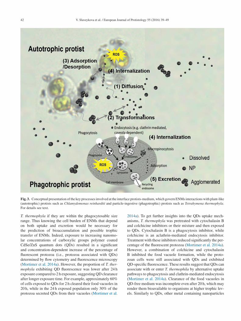
42 V. Slaveykova et al. / European Journal of Protistology 55 (2016) 39–49
F nterfac( icle-inF
TrottlCafld(meao2p
2aatcTcHBzQap(
ig. 3. Conceptual presentation of the key processes involved at the iautotrophic) protists such as Chlamydomonas reinhardtii and partor details see text.
. thermophyla if they are within the phagocytosable sizeange. Thus knowing the cell burden of ENMs that dependn both uptake and excretion would be necessary forhe prediction of bioaccumulation and possible trophicransfer of ENMs. Indeed, exposure to increasing nanomo-ar concentrations of carboxylic groups polymer coateddSe/ZnS quantum dots (QDs) resulted in a significantnd concentration-dependent increase of the percentage ofuorescent protozoa (i.e., protozoa associated with QDs)etermined by flow cytometry and fluorescence microscopyMortimer et al. 2014a). However, the proportion of T. ther-ophyla exhibiting QD fluorescence was lower after 24 h
xposure compared to 2 h exposure, suggesting QD clearancefter longer exposure time. For example, approximately 60%
f cells exposed to QDs for 2 h cleared their food vacuoles in0 h, while in the 24 h exposed population only 30% of therotozoa secreted QDs from their vacuoles (Mortimer et al.Qre
e protists-medium, which govern ENMs interactions with plant-likegestive (phagotrophic) protists such as Tetrahymena thermophyla.
014a). To get further insights into the QDs uptake mech-nisms, T. thermophyla was pretreated with cytochalasin Bnd colchicine inhibitors or their mixture and then exposedo QDs. Cytochalasin B is a phagocytosis inhibitor, whileolchicine is an aclathrin-mediated endocytosis inhibitor.reatment with these inhibitors reduced significantly the per-entage of the fluorescent protozoa (Mortimer et al. 2014a).owever, a combination of colchicine and cytochalasin
inhibited the food vacuole formation, while the proto-oan cells were still associated with QDs and exhibitedD-specific fluorescence. These results suggest that QDs can
ssociate with or enter T. thermophila by alternative uptakeathways to phagocytosis and clathrin-mediated endocytosisMortimer et al. 2014a). Clearance of the food vacuoles in
D-free medium was incomplete even after 20 h, which mayender them bioavailable to organisms at higher trophic lev-ls. Similarly to QDs, other metal containing nanoparticles
ournal o
st2atastltCu(diils(NFid2
stowwta
phrpEMgtfaEcv(bsm2eaat(
2tIoefmOect5idpfawotomeiCtwe
osaampECueowtm2rS
aO(bf
V. Slaveykova et al. / European J
uch as Ag, Au, CuO-NPs and TiO2-NPs, accumulated in T.hermophyla by different uptake pathways (Mortimer et al.014b). The ENMs agglomerates are found in food vacuolesnd the cytoplasm, but the accumulation varied accordingo the ENM concentration and exposure time. Indeed, themount of accumulated ENMs was higher after 2 h of expo-ure than at 24 h, and at higher concentrations. Moreover,he studied ENMs induced increased intracellular ROS andipid peroxidation in protozoa as revealed by flow cytome-ry and staining with CellROX® Green and BODIPY®581/591
11. Nonetheless, no quantitative relationship between ENMptake by T. thermophila and OS and damage was foundMortimer et al. 2014b). Therefore, the contribution of theissolved metal fraction from NPs and the release of Agons form the ENMs into food vacuoles have to be takennto account. The above observations are consistent with theimited literature demonstrating higher toxicity of the dis-olved species to T. thermophyla as compared with Ag-NPsBondarenko et al. 2013; Burkart et al. 2015) and CuO-Ps (Bondarenko et al. 2013; Mortimer et al. 2010, 2011).urthermore, in case of TiO2-NPs exposure the level of
ntracellular ROS generation in Tetrahymena pyriformis wasependent on the light-illumination conditions (Zou et al.013).
Most of the microalgae do not exhibit cellular mechanismsuch as endocytosis, for the transport of ENPs (Moore 2006),hus the role of the cell wall is significant in the modulationf their interactions with ENMs. Indeed, a comparison of theild type of C. reinhardtii and wall-less mutant revealed a cellall-dependent association of QDs demonstrating the impor-
ant role of the cell wall as a protective barrier (Slaveykovand Startchev 2009; Worms et al. 2012).
Among different biological responses used to evaluate theotential effects of ENMs on biota, the generation of theighly ROS, disturbing the pro- and antioxidant equilib-ium and thus inducing OS is currently the best-acceptedaradigm to assess and compare the toxicity of differentNMs in the environment (Burello and Worth 2011; vonoos and Slaveykova 2014). Although nonspecific for a
iven stressor, the OS paradigm provides insights into theoxic relevance of different ENMs and is a useful paradigmor their ranking and development of the structure–reactivitynd dose–response relationships. The OS (and toxicity ofNMs, in general) is a result of a complex interplay of the spe-ific features of the target organism (e.g., particle ingestings. non-ingesting), the specific characteristics of the ENMse.g., composition, size, surface coating and function, solu-ility) and environmental factors (e.g., pH, water hardness,alinity, presence of organic molecules and dissolved organicatter, ultraviolet radiation, etc.) (von Moos and Slaveykova
014; von Moos et al. 2014). Here, the influence of the ambi-nt medium composition and a combined exposure to ENMs
nd other environmental stressors such as ultraviolet radi-tion (UVR) illustrated below are shown exemplarily forhe microalga C. reinhardtii exposed to CuO nanoparticlesCuO-NPs). CuO-NPs are widely-used biocides (Ingle et al.lTbp
f Protistology 55 (2016) 39–49 43
014; Shi et al. 2012), however, they are more toxic to non-argeted than to targeted species (Bondarenko et al. 2013;vask et al. 2014). The influence of the media compositionn the potential of CuO-NPs to induce OS and damage wasxplored at 2 h and 24 h in 3-(N-morpholino) propanesul-onic acid (MOPS) Good’s buffer, standard testing OECDedium and Lake Geneva water (von Moos et al. 2015).xidative stress occurred in all tested media but after 24 h
xposure in OECD medium the percentage of the stressedells increased 50 times with respect to the non-exposed con-rols, while in MOPS and lake water this increase was only
and 3 times, respectively. The medium composition mod-fied the CuO-NPs and thus (sub)-toxic effects altering theirissolution, surface charge and aggregation, as well as theirotential to generate ROS appeared. Indeed, the dissolvedraction in the 10 mg L−1 CuO-NP suspension increased frombout 5% in MOPS to about 80% in OECD medium and lakeater 24 h after NP suspension (von Moos et al. 2015). Thisbservation shows the importance of the medium composi-ion in CuO-NPs dissolution and the significant contributionf the dissolved Cu to the OS response of the algae in OECDedium and lake water. Furthermore, OS occurred also after
xposure to dissolved Cu only but the response was lessmportant in OECD medium and lake water, suggesting thatuO-NPs exerted a particle-specific effect and/or modified
he bioavailability of dissolved Cu, e.g., by complexationith dissolved organic matter present in lake water (von Moos
t al. 2015).In the environment, biota are exposed to a combination
f interacting stressors. For example, the interplay betweenolar radiation and ENMs could affect the protists by: (i)ltering the ENMs dissolution, aggregation and ROS gener-tion; (ii) modifying the chemical speciation of the releasedetal ions; and (iii) affecting the vital cellular functions of
rotists. To explore the combined effect of two stressors –NMs and UVR, C. reinhardtii was exposed to 800 �g L−1
uO-NPs (concentration inducing 50% effect) and to sim-lated solar radiation with altered PAR/UVR ratio (Chelonit al. 2016). Synergistic interactions between the two stress-rs with higher OS in combined than in individual treatmentsere found (Cheloni et al. 2016). The above examples illus-
rated that the potential of CuO-NPs to induce OS was alsoodulated by the test medium composition (von Moos et al.
015), as well as the interactions with other varying envi-onmental factors, including solar radiation (Cheloni andlaveykova, 2013; Cheloni et al. 2016).Overall, no ENPs uptake by C. reinhardtii was observed
nd no clear relationship between ENP concentration andS response was found. By contrast, all studied nanoparticles
Ag, Au, CuO, TiO2 and QDs) accumulated in T. thermophylay different uptake pathways. The ENPs aggregates wereound in food vacuoles and in the cytoplasm, but the accumu-
ation varied according to concentration and exposure time.he OS and damage in aquatic protists induced by metal-ased NPs can be triggered directly, promoted by particleroperties at the nanoscale, or indirectly by dissolved, toxic
4 ournal o
mae
Ssr
aaoi(otatrSfpUbeUp
geabptTbo1WtlM
ttcsfeBsrgt
smttHccc
sapipamiaasUtbU(2itietascttw2tatteutPaamp(
4 V. Slaveykova et al. / European J
etal ions from NPs. Therefore, both the oxidative potentialnd NP dissolution have to be taken into consideration whenvaluating the toxicity potential of ENMs.
tress responses and photoprotectivetrategies of ciliates exposed to ultravioletadiation
Solar radiation is a daily stress factor for almostny free-living organism in the world. The biologicallyctive radiation reaching the Earth’s surface comprises notnly PAR (400–700 nm) that stimulates live processes butncludes also the potentially harmful ultraviolet wavelengths280–400 nm). Generally, we expect negative implicationsf UVR on an organism and many studies indeed showedhat these short wavelengths can easily pass cell membranesnd cause severe direct damages to nucleic acids and pro-eins. In protists, after the impact of UVR, modified shapes,educed movements and retarded growth rates are observed.uch direct effects may indirectly affect aquatic microbialood webs; for instance, when a predator dies off the formerrey can rise. Other indirect effects under the exposure toVR are the production of ROS and consequently OS mayurden an organism. Though, UVR can also have positiveffects on an organism, e.g., such as longer wavelengths ofV-A radiation (315–400 nm) as well as of PAR can inducehotorepair mechanisms (Sinha and Häder 2002).
Almost nothing is known about the reactions and strate-ies of free-living protists how they cope with UVR as annvironmental stress factor. Ciliated protists, in general, play
key role in the microbial food webs of oceans and lakesecause they are one of the major consumers of phyto-lankton and bacteria and hence an essential link to higherrophic levels (e.g., Azam et al. 1983; Sommer et al. 2012).heir abundance and diversity in freshwater plankton rangesetween 2.0 and 130 ind. mL−1, comprising either a fewr wide more than 100 morphospecies (e.g., Müller et al.991; Sonntag et al. 2006, 2011a; Van Wichelen et al. 2013;ille et al. 1999). Moreover, recent phylogenetic investiga-
ions revealed a generally high genetic protist diversity inakes (Kammerlander et al. 2015; Stoeck et al. 2014; Triadó-
argarit and Casamajor 2012).Taken into consideration the available studies so far, we see
hat effects of UVR on protists reveal species-specific reac-ions and strategies. Difficulties in studying natural protistommunities from lakes arise because virtually no cultivatedpecies are available in public culture collections to per-orm relevant experiments in the laboratory. This lack of, forxample, limnetic planktonic species of the common generaalanion, Rimostrombidium, Urotricha or Askenasia force
cientists to cultivate the desired species in their laborato-ies often including multitudinous approaches to optimizerowing conditions in terms of medium, food and tempera-ure; alternatively, commercially available (model) speciesafpo
f Protistology 55 (2016) 39–49
uch as Paramecium or Tetrahymena are used in experi-ents. As these model ciliates derive from other habitats
han the pelagic, different stress responses and photoprotec-ive mechanisms can be expected from UVR experiments.ow different the impact of UVR on varying ciliate species
an be is elucidated in the following examples on Parame-ium bursaria and investigations on free-living planktonicommunities (Fig. 4).
Paramecium bursaria has a mixotrophic lifestyle, i.e. thepecies lives in symbiosis with autotrophic green algae andlso grazes phagotrophically on bacteria and other smallerrotists. Paramecium bursaria is wide-spread and foundn the littoral zone of lakes or in stagnant waters such asonds where it commonly lives attached to surfaces suchs submersed leaves and stones. This species is a suitableodel ciliate because it can be relatively easily maintained
n the laboratory, symbiont-free cell lines can be established,nd many populations and strains from all over the worldre available. To test for the stress responses of P. bur-aria to UVR, the ciliates were exposed under an artificialVR and PAR source in laboratory experiments. In contrast
o their aposymbiotic (algal-free) counterparts, symbiont-earing individuals of P. bursaria were more resistant toVR and PAR, and also (photo-) oxidative stress was lower
Hörtnagl and Sommaruga 2007; Summerer et al. 2007,009). Apparently, the algal symbionts are somehow involvedn the photoprotection of the species. In a second approach,wo interesting phenomena emerged: under UVR and PARndividual ciliates accumulated to dense green spots that wereven visible to the naked eye and formed a kind of ‘collec-ive shield’ (Sommaruga and Sonntag 2009). When taking
closer look onto these aggregated individuals, the algalymbionts within the cells were dislocated to the posteriorell end. The assumption was that the individuals adjustedheir rear end that was densely stuffed with algae againsthe irradiation source and that the ciliates’ nuclear materialas effectively shaded from harmful UVR (Summerer et al.009). The fact that several algal layers significantly reducehe transmission of UVR at 320 nm can be calculated withn optical model developed by Garcia-Pichel (1994). Takenogether, the algal symbionts obviously play a major role inhe stress balance of these mixotrophic ciliates when they arexposed to UVR and PAR. Though, several questions remainnsolved as it is still unknown what triggers the rapid disloca-ion of the algae within the host under exposure to UVR andAR. Besides, the phenomenon is reversible within minutess soon as P. bursaria is not exposed to the irradiation sourceny more. Another question is how the individual ciliatesanage their spot aggregation under the irradiation source. A
hotosensitive area in P. bursaria might be involved somehowNakaoka 1989).
Commonly, another stress factor is accelerated by UVR
nd PAR, which is (photo-)oxidative stress by inducing theormation of ROS. Photo-oxidative stress is defined as theroduction and accumulation of ROS beyond the capacityf an organism to quench them (Lesser 2006). All respiring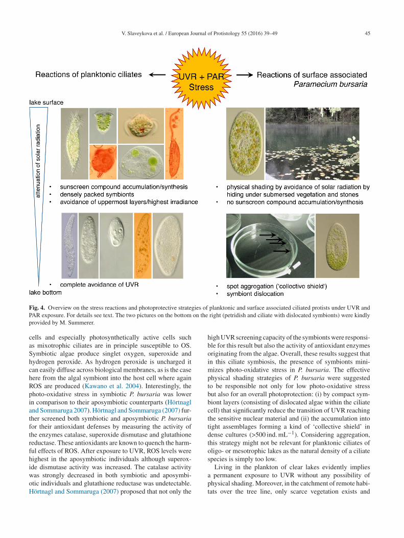
V. Slaveykova et al. / European Journal of Protistology 55 (2016) 39–49 45
F ies of
P on thep
caShchRpiatftrfhiwoH
hboimptbbcttdtos
ig. 4. Overview on the stress reactions and photoprotective strategAR exposure. For details see text. The two pictures on the bottom
rovided by M. Summerer.
ells and especially photosynthetically active cells suchs mixotrophic ciliates are in principle susceptible to OS.ymbiotic algae produce singlet oxygen, superoxide andydrogen peroxide. As hydrogen peroxide is uncharged itan easily diffuse across biological membranes, as is the caseere from the algal symbiont into the host cell where againOS are produced (Kawano et al. 2004). Interestingly, thehoto-oxidative stress in symbiotic P. bursaria was lowern comparison to their aposymbiotic counterparts (Hörtnaglnd Sommaruga 2007). Hörtnagl and Sommaruga (2007) fur-her screened both symbiotic and aposymbiotic P. bursariaor their antioxidant defenses by measuring the activity ofhe enzymes catalase, superoxide dismutase and glutathioneeductase. These antioxidants are known to quench the harm-ul effects of ROS. After exposure to UVR, ROS levels wereighest in the aposymbiotic individuals although superox-de dismutase activity was increased. The catalase activity
as strongly decreased in both symbiotic and aposymbi-tic individuals and glutathione reductase was undetectable.örtnagl and Sommaruga (2007) proposed that not only theapt
planktonic and surface associated ciliated protists under UVR and right (petridish and ciliate with dislocated symbionts) were kindly
igh UVR screening capacity of the symbionts were responsi-le for this result but also the activity of antioxidant enzymesriginating from the algae. Overall, these results suggest thatn this ciliate symbiosis, the presence of symbionts mini-
izes photo-oxidative stress in P. bursaria. The effectivehysical shading strategies of P. bursaria were suggestedo be responsible not only for low photo-oxidative stressut also for an overall photoprotection: (i) by compact sym-iont layers (consisting of dislocated algae within the ciliateell) that significantly reduce the transition of UVR reachinghe sensitive nuclear material and (ii) the accumulation intoight assemblages forming a kind of ‘collective shield’ inense cultures (>500 ind. mL−1). Considering aggregation,his strategy might not be relevant for planktonic ciliates ofligo- or mesotrophic lakes as the natural density of a ciliatepecies is simply too low.
Living in the plankton of clear lakes evidently implies
permanent exposure to UVR without any possibility ofhysical shading. Moreover, in the catchment of remote habi-ats over the tree line, only scarce vegetation exists and
4 ournal o
cmhtfmpthUCeSldh2
peaiaefettso(sacgUBbaTite2msiSpnf
Urte
waMieatd(tei
C
eReaifretamessp(tstttasos
A
oorFw
6 V. Slaveykova et al. / European J
onsequently the input of dissolved chromophoric organicatter attenuating UVR is low and UVR transparency is
igh. As studies on P. bursaria showed, a mutualistic rela-ionship with symbionts obviously provided an advantageor ciliates when exposed to UVR. Though, irrespective of aixotrophic or heterotrophic lifestyle results from a ‘trans-
lantation’ study where the ciliate assemblage of a less UVransparent lake was exposed in a highly UV transparentigh mountain lake revealed that mortality under differentVR intensities was species-specific (Sonntag et al. 2011b).onsequently, not only single cells are affected under UVRxposure but also whole food webs (Mostajir et al. 1999;ommaruga et al. 1999; Wickham and Carstens 1998). Fol-
owing the exposure to (elevated) UVR, generally lowerivision and growth rates, retarded swimming or cell deathave been observed (e.g., Giese et al. 1963; Sanders et al.005).Life under daily UVR exposure as it is the case for
lanktonic organisms in lakes or oceans certainly leads tonormous stress and demands for effective photoprotectionnd -repair strategies. For example, phyto- and zooplanktonn clear alpine lakes perform vertical downward migrationsround noontime (e.g., Tilzer 1973; Alonso et al. 2004). Inter-stingly, this kind of UV avoidance strategy was not observedor the (only) three ciliate species colonizing such an extremenvironment in summer (Sonntag et al. 2011a). The UV sensi-ive Balanion planctonicum persisted in the deepest area nearhe sediment whereas the algal-bearing mixotrophic Askena-ia chlorelligera thrived all over the water column and wasbviously well protected by specific sunscreen compoundsmycosporine-like amino acids; MAAs) synthesized by itsymbionts. Besides, in an A. chlorelligera population from
less UV transparent lake, none of these photoprotectiveompounds were detected (Summerer et al. 2008). MAAs, ineneral, are water-soluble, colorless sunscreens that absorbVR between 309 and 362 nm (Carreto and Carignan 2011).iochemical pathways involved in MAAs synthesis haveeen recently identified but are unknown for ciliates (Balskusnd Walsh 2010; Osborn et al. 2015; Singh et al. 2010).he MAAs detected in several algal-bearing freshwater cil-
ates originated from the synthesis by the symbionts buthe uptake from algal food was also demonstrated in a het-rotrophic species (Sonntag et al. 2007; Summerer et al.008; Sonntag unpubl.). Sunscreen compounds (MAAs andycosporine–glutaminol–glucoside) were neither synthe-
ized by the algal symbionts of P. bursaria nor accumulatedn aposymbiotic individuals from food (Pérez et al. 2006;ummerer et al. 2009). Preliminary experiments with B.lanctonicum also showed that this heterotrophic species wasot able to accumulate MAAs from offered MAA-rich algalood (Sonntag and Pöll unpubl.).
Other efficient mechanisms to cope with stress caused by
VR are DNA-repair strategies including photoenzymaticepair (PER) and nucleotide excision repair (‘dark repair’)hat can be stimulated by UV-A and PAR (e.g., Karentzt al. 1991; Mitchell and Karentz 1993). Both strategies are
CWo(
f Protistology 55 (2016) 39–49
idespread and found in bacteria, phyto- and zooplanktonnd in ciliated protists (Buma et al. 1997; Joux et al. 1999;alloy et al. 1997; Sanders et al. 2005). For example, two cil-
ates from an oligotrophic lake responded differently to UVRxposure: Glaucoma sp. was able to recover under photore-ctive radiation whereas Cyclidium sp. was not. Moreover,he efficiency of PER in Glaucoma sp. was temperature-ependent and significantly reduced at low temperaturesSanders et al. 2005). Yet, only some few ciliate species wereested for the presence of PER. Future studies will have tolucidate which DNA-repair strategies are generally presentn (planktonic) ciliates.
oncluding remarks
From the foregoing, we can highlight some important gen-ral points. Oxidative stress (from the production of excessiveOS) seems to be a general cellular response to manifoldnvironmental stressors (UVR or inorganic nanoparticles,mong others) in very diverse protists. Therefore, an increasen ROS can be considered as a key process found in dif-erent types of stress. A general updated view from studieselated to “environmental stress and protists” leads us to sev-ral conclusions: (i) the scarcity of stress-related studies, (ii)he absence of molecular approaches and genetic analysesnd (iii) the necessity to increase physiological analyses. Stillany open questions on how ciliates cope with UVR as an
nvironmental stressor remain and future investigations fromingle species up to food webs are needed. Model protistsuch as Chlamydomonas reinhardtii (among photosyntheticrotists), Tetrahymena thermophila or Paramecium speciesamong free living phagotrophic protists) are up to nowhe most manageable and well known protists (with alreadyequenced genomes). Although they are very good candidateso carry out analyses on environmental stress-protist interac-ions, studies have to consider other common protist specieshat are relevant for example in lake food web studies (seelso Weisse et al. 2016). Overall, we would like to encouragecientists to use protists as eukaryotic model organisms moreften for investigating the cellular responses to environmentaltress at any level.
cknowledgements
The authors wish thank the president of the congressrganizing committee Dr. Aurelio Serrano for giving us thepportunity to celebrate this symposium. The research car-ied out by VS was supported by Swiss National Scienceoundation grants 406440-131280 and 200021-134627, asell as the Sciex grant 11.270. The contribution of Giulia
heloni, Monika Mortimer, Nadia von Moos and Isabelleorms, is warmly acknowledge by VS. Research carriedut by BS was supported by the Austrian Science FundFWF), grants P21013-B03, I2238-B25, P28333-B25 and
ournal o
PKa
R
A
A
A
B
B
B
B
B
B
C
C
C
D
D
D
F
F
G
G
G
G
G
G
G
G
H
H
I
I
V. Slaveykova et al. / European J
16559-B06 (PI Ruben Sommaruga). BS thanks Barbaraammerlander for constructive input on the ‘stress response
nd photoprotection of ciliates exposed to UVR’ chapter.
eferences
lonso, C., Rocco, V., Barriga, J.P., Battini, M.A., Zagarese, H.,2004. Surface avoidance by freshwater zooplankton: field evi-dence on the role of ultraviolet radiation. Limnol. Oceanogr. 49,225–232.
pel, K., Hirt, H., 2004. Reactive oxygen species: metabolism,oxidative stress, and signal transduction. Annu. Rev. Plant Biol.55, 373–399.
zam, F.T., Fenchel, T., Field, J.G., Gray, J.S., Meyer-Reil, L.A.,Thingstad, F., 1983. The ecological role of water-columnmicrobes in the sea. Mar. Ecol. Prog. Ser. 10, 257–263.
alskus, E.P., Walsh, C.T., 2010. The genetic and molecularbasis for sunscreen biosynthesis in cyanobacteria. Science 329,1653–1656.
ishak, Y.K., Payahoo, L., Osatdrahimi, A., Nourazarian, A., 2015.Mechanisms of cadmium carcinogenicityin the gastrointestinaltract. Asian Pac. J. Cancer Prev. 16, 9–21.
ondarenko, O., Juganson, K., Ivask, A., Kasemets, K., Mortimer,M., Kahru, A., 2013. Toxicity of Ag, CuO and ZnO nanopar-ticles to selected environmentally relevant test organisms andmammalian cells in vitro: a critical review. Arch. Toxicol. 87,1181–1200.
uma, A.G.J., Engelen, A.H., Gieskes, W.W.C., 1997. Wavelength-dependent induction of thymine-dimers and growth ratereduction in the marine diatom Cyclotella sp. exposed to ultra-violet radiation. Mar. Ecol. Progr. Ser. 153, 91–97.
urello, E., Worth, A.P., 2011. A theoretical framework for pre-dicting the oxidative stress potential of oxide nanoparticles.Nanotoxicology 5, 228–235.
urkart, C., Tümpling, W., Berendonk, T., Jungmann, D., 2015.Nanoparticles in wastewater treatment plants: a novel acute tox-icity test for ciliates and its implementation in risk assessment.Environ. Sci. Pollut. Res. 22, 7485–7494.
arreto, J.I., Carignan, M.O., 2011. Mycosporine-like aminoacids: relevant secondary metabolites. Chemical and ecologicalaspects. Mar. Drugs 9, 387–446.
heloni, G., Slaveykova, V.I., 2013. Optimization of the C11-BODIPY581/591 dye for the determination of lipid oxidationin Chlamydomonas reinhardtii by flow cytometry. Cytometry A83A, 952–961.
heloni, G., Marti, E., Slaveykova, V.I., 2016. Interactive effectsof copper oxide nanoparticles and light to green alga Chlamy-domonas reinhardtii. Aquat. Toxicol. 170, 120–128.
íaz, S., Martin-Gonzalez, A., Cubas, L.L., Ortega, R., Amaro, F.,Rodriguez-Martin, D., Gutierrez, J.C., 2016. High resistance ofTetrahymena thermophila to paraquat: mitochondrial alterations,oxidative stress and antioxidant genes expression. Chemosphere144, 909–917.
ing, Y.-F., Zhu, C., 2009. The role of microRNAs in copper andcadmium homeostasis. Biochem. Biophys. Res. Commun. 386,
6–10.ondero, F., Cavaletto, M., Ghezzi, A.R., La Terza, A., Banni,M., Viarengo, A., 2004. Biochemical characterization and quan-titative gene expression analysis of the multi-stress inducible
f Protistology 55 (2016) 39–49 47
metallothionein from Tetrahymena thermophila. Protist 155,157–168.
erro, D., Bakiu, R., De Pittà, C., Boldrin, F., Cattalini, F., Puc-ciarelli, S., Miceli, C., Santovito, G., 2015. Cu, Zn superoxidedismutases from Tetrahymena thermophila: molecular evolutionand gene expression of the first line of antioxidant defenses.Protist 166, 131–145.
ujita, M., Fujita, Y., Noutoshi, Y., Takahashi, F., Narusaka,Y., Yamaguchi-Shinozaki, K., Schinozaki, K., 2006. Crosstalkbetween abiotic and biotic stress responses: a current view fromthe points of convergence in the stress signaling networks. Curr.Opin. Plant Biol. 9, 436–442.
alluzzi, L., Bravo-San Pedro, J.M., Kepp, O., Kroemer, G., 2016.Regulated cell death and adaptive stress responses. Cell. Mol.Life Sci., http://dx.doi.org/10.1007/s00018-016-2209-y.
arcia-Pichel, F., 1994. A model for internal self-shading in plank-tonic organisms and its implications for the usefulness ofultraviolet sunscreens. Limnol. Oceanogr. 39, 1704–1717.
iese, A.C., McCaw, B., Cornell, R., 1963. Retardation of divi-sion of three ciliates by intermittent and continuous ultravioletradiations at different temperatures. J. Gen. Physiol. 46, 1095–1108.
onzalo, S., Llaneza, V., Pulido-Reyes, G., Fernández-Pinas, F.,Bonzongo, J.C., Leganes, F., Rosal, R., Garcia-Calvo, E., Rodea-Palomares, I., 2014. A colloidal singularity reveals the crucialrole of colloidal stability for nanomaterials in vitro toxicitytesting: nZVI-Microalgae colloidal system as a case study.PLOS ONE 9 (10), e109645, http://dx.doi.org/10.1371/journal.pone.0109645.
utierrez, J.C., Martin-Gonzalez, A., 2002. Ciliateencystment–excystment cycle: a response to environmen-tal stress. In: Gutierrez, J.C. (Ed.), Microbial DevelopmentUnder Environmental Stress. Research Signpost, India, pp.29–49.
utierrez, J.C., Amaro, F., Díaz, S., de Francisco, P., Cubas, L.L.,Martín-González, A., 2011. Ciliate metallothioneins: uniquemicrobial eukaryotic heavy-metal-binder molecules. J. Biol.Inorg. Chem. 16, 1025–1034.
utierrez, J.C., Callejas, S., Borniquel, S., Benitez, L., Martin-Gonzalez, A., 2001. Ciliate cryptobiosis: a microbial strategyagainst environmental starvation. Int. Microbiol. 4, 151–157.
utierrez, J.C., Martin-Gonzalez, A., Díaz, S., Amaro, F., Ortega,R., Gallego, A., de Lucas, M.P., 2008. Ciliates as cellular toolsto study the eukaryotic cell-heavy metal interactions. In: Brown,S.E., Welton, W.C. (Eds.), Heavy Metal Pollution. Nova SciencePublishers, Inc., USA, pp. 1–44.
oertnagl, P.H., Sommaruga, R., 2007. Photo-oxidative stress insymbiotic and aposymbiotic strains of the ciliate Parameciumbursaria. Photochem. Photobiol. Sci. 6, 842–847.
udder, A., Novak, R.F., 2008. miRNAs: effectors of environmentalinfluences on gene expression and disease. Toxicol. Sci. 103,228–240.
ngle, A., Duran, N., Rai, M., 2014. Bioactivity, mechanism ofaction, and cytotoxicity of copper-based nanoparticles: a review.Appl. Microbiol. Biotechnol. 98, 1001–1009.
vask, A., Juganson, K., Bondarenko, O., Mortimer, M., Aruoja, V.,Kasemets, K., Blinova, I., Heinlaan, M., Slaveykova, V., Kahru,A., 2014. Mechanisms of toxic action of Ag, ZnO and CuO
nanoparticles to selected ecotoxicological test organisms andmammalian cells in vitro: a comparative review. Nanotoxicology8 (Suppl. 1), 57–71.
4 ournal o
J
J
K
K
K
K
L
L
M
M
M
M
M
M
M
M
M
M
M
M
N
N
N
N
N
O
O
P
P
P
R
R
S
8 V. Slaveykova et al. / European J
osson, S., Sung, S.-Y., Lao, K., Chung, L.W.K., Johnstone, P.A.S.,2008. Radiation modulation of microRNA in prostate cancer celllines. Prostate 68, 1599–1606.
oux, E., Jeffrey, W.H., Lebaron, P., Mitchell, D., 1999. Marinebacterial isolates display diverse responses to UV-B radiation.Appl. Environ. Microbiol. 65, 3820–3827.
ammerlander, B., Breiner, H.-W., Filker, S., Sommaruga, R., Son-ntag, B., Stoeck, T., 2015. High diversity of protistan planktoncommunities in remote high mountain lakes in the EuropeanAlps and the Himalaya mountains. FEMS Microbiol. Ecol. 91,fiv010, http://dx.doi.org/10.1093/femsec/fiv010.
arentz, D., Cleaver, J.E., Mitchell, D.L., 1991. DNA damage inthe Antarctic. Nature 350, 28.
awano, T., Kadono, T., Kosaka, T., Hosoya, H., 2004. Greenparamecia as an evolutionary winner of oxidative symbiosis: ahypothesis and supportive data. Z. Naturforsch. 59, 538–542.
im, S.H., Kim, S.J., Lee, J.S., Lee, Y.M., 2014. Acute effects ofheavy metals on the expression of glutathione-related antioxidantgenes in the marine ciliate Euplotes crassus. Mar. Pollut. Bull.85, 455–462.
esser, M.P., 2006. Oxidative stress in marine environment: bio-chemistry and physiological ecology. Annu. Rev. Physiol. 68,278–583.
iu, J., Feng, L., Li, J., He, Z., 2015. Genetic and epigeneticcontrol of plant heat responses. Front. Plant Sci. 6, 267,http://dx.doi.org/10.3389/fpls.2015.00267.
alloy, K.D., Holman, M.A., Mitchell, D., Dietrich, H.W., 1997.Solar UV-B induced DNA damage and photoenzymatic DNArepair in Antarctic zooplankton. Proc. Natl. Acad. Sci. U. S. A.94, 1258–1263.
artin-Gonzalez, A., Borniquel, S., Díaz, S., Ortega, R., Gutierrez,J.C., 2005. Ultrastructural alterations in ciliated protozoa underheavy metal exposure. Cell Biol. Int. 29, 119–126.
endoza-Cózatl, D., Loza-Tavera, H., Hernández-Navarro, A.,Moreno-Sanchez, R., 2005. Sulfur assimilation and glutathionemetabolism under cadmium stress in yeast, protists and plants.FEMS Microbiol. Rev. 29, 653–671.
eyer, P., 2015. Epigenetic variation and environmental change. J.Exp. Bot. 66, 3541–3548.
itchell, D.L., Karentz, D., 1993. The induction and repair of DNAphotodamage in the environment. In: Young, A.R., Björn, L.O.,Moan, J., Nultsch, W. (Eds.), Environmental UV Photobiology.Plenum Press, New York, pp. 345–377.
oore, M.N., 2006. Do nanoparticles present ecotoxicological risksfor the health of the aquatic environment? Environ. Int. 32,967–976.
ortimer, M., Kasemets, K., Kahru, A., 2010. Toxicity of ZnOand CuO nanoparticles to ciliated protozoa Tetrahymena ther-mophila. Toxicology 269, 182–189.
ortimer, M., Kasemets, K., Vodovnik, M., Marinsek-Logar, R.,Kahru, A., 2011. Exposure to CuO nanoparticles changes thefatty acid composition of protozoa Tetrahymena thermophila.Environ. Sci. Technol. 45, 6617–6624.
ortimer, M., Kahru, A., Slaveykova, V.I., 2014a. Uptake, local-ization and clearance of quantum dots in ciliated protozoaTetrahymena thermophila. Environ. Pollut. 190, 58–64.
ortimer, M., Gogos, A., Bartolome, N., Kahru, A., Bucheli, T.D.,
Slaveykova, V.I., 2014b. Potential of hyperspectral imagingmicroscopy for semi-quantitative analysis of nanoparticle uptakeby protozoa. Environ. Sci. Technol. 48, 8760–8767.S
f Protistology 55 (2016) 39–49
ostajir, B., Demers, S., de Mora, S., Belzile, C., Chanut, J.-P.,Gosselin, M., Roy, S., Villegas, P.Z., Fauchot, J., Bouchard, J.,Bird, D., Monfort, P., Levasseur, M., 1999. Experimental test ofthe effect of ultraviolet-B radiation in a planktonic community.Limnol. Oceanogr. 44, 586–596.
üller, H., Schöne, A., Pinto-Coelho, R.M., Schweizer, A., Weisse,T., 1991. Seasonal succession of ciliates in Lake Constance.Microb. Ecol. 21, 119–138.
akagami, H., Pitzschke, A., Hirt, H., 2005. Emerging MAP kinasepathways in plant stress signaling. Trends Plant Sci. 10, 339–346.
akaoka, Y., 1989. Localization of photosensitivity in Parameciumbursaria. J. Comp. Physiol. A 165, 637–641.
avarro, E., Wagner, B., Odzak, N., Sigg, L., Behra, R., 2015.Effects of differently coated silver nanoparticles on the photo-synthesis of Chlamydomonas reinhardtii. Environ. Sci. Technol.,8041–8047.
olte-’t Hoen, E.N.M., Van Rooij, E., Bushell, M., Zhang, C.-Y.,Dashwood, R.H., James, W.P.T., Harris, C., Baltimore, D., 2015.The role of microRNA in nutritional control. J. Intern. Med.,http://dx.doi.org/10.1111/joim.12372.
owack, B., Ranville, J.F., Diamond, S., Gallego-Urrea, J.A., Met-calfe, C., Rose, J., Horne, N., Koelmans, A.A., Klaine, S.J.,2012. Potential scenarios for nanomaterial release and subse-quent alteration in the environment. Environ. Toxicol. Chem.31, 50–59.
ECD, 2015. Database on Research into the Safety of ManufacturedNanomaterials, www.oecd.org/env/nanosafety/database.
sborn, A.R., Almabruk, K.H., Holzwarth, G., Asamizu, S., LaDu,J., Kean, K.M., Karplus, P.A., Tanguay, R.L., Bakalinsky, A.T.,Mahmud, T., 2015. De novo synthesis of a sunscreen compoundin vertebrates. eLife 4, e05919.
eijnenburg, W.J.G.M., Baalousha, M., Chen, J., Chaudry, Q., Vonder Kammer, F., Kuhlbusch, T.A.J., Lead, J., Nickel, C., Quik,J.T.K., Renker, M., Wang, Z., Koelmans, A.A., 2015. A review ofthe properties and processes determining the fate of engineerednanomaterials in the aquatic environment. Crit. Rev. Environ.Sci. Technol. 45, 2084–2134.
érez, P., Libkind, D., Diéguez, M.d.C., Summerer, M., Sonntag,B., Sommaruga, R., van Broock, M., Zagarese, H.E., 2006.Mycosporines from freshwater yeasts: a trophic cul-de-sac? Pho-tochem. Photobiol. Sci. 5, 25–30.
rado, R., Rioboo, C., Herrero, C., Suarez-Bregua, P., Cid, A., 2012.Flow cytometry analysis to evaluate physiological alterations inherbicide-exposed Chlamydomonas moewusii cells. Ecotoxicol-ogy 21, 409–420.
ico, D., Martin-Gonzalez, A., Díaz, S., de Lucas, P., Gutierrez, J.C.,2009. Heavy metals generate reactive oxygen species in terres-trial and aquatic ciliated protozoa. Comp. Biochem. Physiol. PartC 149, 90–96.
uis, H., 1997. Yeast stress responses: achievements, goals anda look beyond yeast. In: Hohmann, S., Mager, W.H. (Eds.),Yeast Stress Responses. R.G. Landes Company, California, pp.231–247.
anders, R.W., Macaluso, A.L., Sardina, T.J., Mitchell, D.L.,2005. Photoreactivation in two freshwater ciliates: differentialresponses to variations in UV-B flux and temperature. Aquat.Microb. Ecol. 40, 283–292.
hi, M., Kwon, H.S., Peng, Z., Elder, A., Yang, H., 2012. Effects ofsurface chemistry on the generation of reactive oxygen speciesby copper nanoparticles. ACS Nano 6, 2157–2164.
ournal o
S
S
S
S
S
S
S
S
S
S
S
S
S
S
S
S
T
T
T
V
V
V
V
W
W
W
W
V. Slaveykova et al. / European J
chwartzman, J.A., Ruby, E.G., 2016. Stress as a nor-mal cue in the symbiotic environment. Trends Microbiol.,http://dx.doi.org/10.1016/j.tim.2016.02.012.
ingh, S.P., Klisch, M., Sinha, R.P., Haeder, D.P., 2010. Genomemining of mycosporine-like amino acids (MAA) synthesiz-ing and non-synthesizing cyanobacteria: a bioinformatics study.Genomics 95, 120–128.
inha, R.P., Häder, D.-P., 2002. UV-induced DNA damage andrepair: a review. Photochem. Photobiol. Sci. 1, 225–236.
laveykova, V.I., Startchev, K., 2009. Effect of natural organicmatter and green microalga on carboxyl-polyethylene glycolcoated CdSe/ZnS quantum dots stability and transformationsunder freshwater conditions. Environ. Pollut. 157, 3445–3450.
ommaruga, R., Sattler, B., Oberleiter, A., Wille, A., Wögrath-Sommaruga, S., Psenner, R., Felip, M., Camarero, L., Pina, S.,Gironés, R., Catalán, J., 1999. An in situ enclosure experiment totest the solar UVB impact on plankton in a high-altitude moun-tain lake. II. Effects on the microbial food web. J. Plankton Res.21, 859–876.
ommaruga, R., Sonntag, B., 2009. Photobiological aspects ofthe mutualistic association between Paramecium bursaria andChlorella. In: Fujishima, M. (Ed.), Endosymbionts of Parame-cium. Microbiology Monographs 12. Springer Verlag, Berlin,Germany, pp. 111–130.
ommer, U., Adrian, R., De Senerpont Domis, L., Elser, J.J.,Gaedke, U., Ibelings, B., Jeppesen, E., Lurling, M., Molinero,J.C., Mooij, W.M., van Donk, E., Winder, M., 2012. Beyondthe Plankton Ecology Group (PEG) model: mechanisms driv-ing plankton succession. Annu. Rev. Ecol. Evol. Syst. 43, 429–448.
onntag, B., Posch, T., Klammer, S., Teubner, K., Psenner, R.,2006. Phagotrophic ciliates and flagellates in an oligotrophicdeep alpine lake: contrasting variability with seasons and depths.Aquat. Microb. Ecol. 43, 193–207.
onntag, B., Summerer, M., Sommaruga, R., 2007. Sources ofmycosporine-like amino acids in planktonic Chlorella-bearingciliates (Ciliophora). Freshw. Biol. 52, 1476–1485.
onntag, B., Summerer, M., Sommaruga, R., 2011a. Factorsinvolved in the distribution pattern of ciliates in the water columnof a transparent alpine lake. J. Plankton Res. 33, 541–546.
onntag, B., Summerer, M., Sommaruga, R., 2011b. Are freshwatermixotrophic ciliates less sensitive to solar UV radiation thanheterotrophic ones? J. Euk. Microbiol. 58, 196–202.
toeck, T., Breiner, H.-W., Filker, S., Ostermaier, V., Kammer-lander, B., Sonntag, B., 2014. A morpho-genetic survey onciliate plankton from a mountain lake pinpoints the necessity oflineage-specific barcode markers in microbial ecology. Environ.Microbiol. 16, 430–444.
ummerer, M., Sonntag, B., Sommaruga, R., 2007. An experimentaltest of the symbiosis specificity between the ciliate Parame-cium bursaria and strains of the unicellular green alga Chlorella.Environ. Microbiol. 9, 2117–2122.
Z
f Protistology 55 (2016) 39–49 49
ummerer, M., Sonntag, B., Sommaruga, R., 2008. Host-symbiontspecificity of freshwater endosymbiotic Chlorella (Trebouxio-phyceae, Chlorophyta). J. Phycol. 44, 77–87.
ummerer, M., Sonntag, B., Hoertnagl, P., Sommaruga, R., 2009.Symbiotic ciliates receive protection against UV damage fromtheir algae: a test with Paramecium bursaria and Chlorella. Pro-tist 160, 233–243.
wiecilo, A., 2016. Cross-stress resistance in Saccharomyces cere-visiae yeast-new insight into an old phenomenon. Cell StressChaperones 21, 187–200.
ilzer, M.M., 1973. Diurnal periodicityin the phytoplankton assem-blage of a high mountain lake. Limnol. Oceanogr. 18, 15–30.
riadó-Margarit, X., Casamayor, E.O., 2012. Genetic diversity ofplanktonic eukaryotes in high mountain lakes (Central Pyrenees,Spain). Environ. Microbiol. 14, 2445–2456.
rielli, F., Chessa, M.G., Amaroli, A., Ognibene, M., Delmonte Cor-rado, M.U., 2006. Effects of organophosphate compounds on asoil protist, Colpoda inflata (Ciliophora, Colpodidae). Chemo-sphere 65, 1731–1737.
an Wichelen, J., Johansson, L.S., Vanormelingen, P., Declerck,S.A.J., Lauridsen, T.L., De Meester, L., Jeppesen, E., Vyverman,W., 2013. Planktonic ciliate community structure in shallowlakes of lowland Western Europe. Eur. J. Protistol. 49, 538–551.
on Moos, N., Bowen, P., Slaveykova, V.I., 2014. Bioavail-ability of inorganic nanoparticles to planktonic bacteria andaquatic microalgae in freshwater. Environ. Sci.: Nano 1,214–232.
on Moos, N., Maillard, L., Slaveykova, V.I., 2015. Dynamicsof sub-lethal effects of nano-CuO on the microalga Chlamy-domonas reinhardtii during short-term exposure. Aquat. Toxicol.161, 267–275.
on Moos, N., Slaveykova, V.I., 2014. Oxidative stress induced byinorganic nanoparticles in bacteria and aquatic microalgae – stateof the art and knowledge gaps. Nanotoxicology 8, 605–630.
eisse, T., Anderson, R., Arndt, H., Calbet, A., Hansen, P.J., Mon-tagnes, D.J.S., 2016. Functional ecology of aquatic phagotrophicprotists – Concepts, limitations, and perspectives. Eur. J. Protis-tol., http://dx.doi.org/10.1016/j.ejop.2016.03.003 (this issue).
ickham, S., Carstens, M., 1998. Effects of ultraviolet-B radiationon two arctic microbial food webs. Aquat. Microb. Ecol. 16,163–171.
ille, A., Sonntag, B., Sattler, B., Psenner, R., 1999. Abundance,biomass and size structure of the microbial assemblage in thehigh mountain lake Gossenköllesee (Tyrol, Austria) during theice-free period. J. Limnol. 58, 117–126.
orms, I.A.M., Boltzman, J., Garcia, M., Slaveykova, V.I., 2012.Cell-wall-dependent effect of carboxyl-CdSe/ZnS quantum dotson lead and copper availability to green microalgae. Environ.Pollut. 167, 27–33.
ou, X.-Y., Xu, B., Yu, C.-P., Zhang, H.-W., 2013. Imbalancebetween oxidative and antioxidative systems: toward an under-standing of visible light-induced titanium dioxide nanoparticlestoxicity. Chemosphere 93, 2451–2457.