Embed Size (px)
Citation preview

Western UniversityScholarship@Western
Biochemistry Publications Biochemistry Department
9-1-2011
Stomatin-like protein 2 binds cardiolipin andregulates mitochondrial biogenesis and function.Darah [email protected]
Caitlin D Lemke
Isaac M Elias
Luan A Chau
Mark G Kirchhof
See next page for additional authors
Follow this and additional works at: https://ir.lib.uwo.ca/biochempub
Part of the Biochemistry Commons
Citation of this paper:Christie, Darah; Lemke, Caitlin D; Elias, Isaac M; Chau, Luan A; Kirchhof, Mark G; Li, Bo; Ball, Eric H; Dunn, Stanley D; Hatch,Grant M; and Madrenas, Joaquín, "Stomatin-like protein 2 binds cardiolipin and regulates mitochondrial biogenesis and function."(2011). Biochemistry Publications. 169.https://ir.lib.uwo.ca/biochempub/169

AuthorsDarah Christie, Caitlin D Lemke, Isaac M Elias, Luan A Chau, Mark G Kirchhof, Bo Li, Eric H Ball, Stanley DDunn, Grant M Hatch, and Joaquín Madrenas
This article is available at Scholarship@Western: https://ir.lib.uwo.ca/biochempub/169

Published Ahead of Print 11 July 2011. 10.1128/MCB.05393-11.
2011, 31(18):3845. DOI:Mol. Cell. Biol. Grant M. Hatch and Joaquín Madrenas
Dunn,Chau, Mark G. Kirchhof, Bo Li, Eric H. Ball, Stanley D. Darah A. Christie, Caitlin D. Lemke, Isaac M. Elias, Luan A. and Functionand Regulates Mitochondrial Biogenesis Stomatin-Like Protein 2 Binds Cardiolipin
http://mcb.asm.org/content/31/18/3845Updated information and services can be found at:
These include:
REFERENCEShttp://mcb.asm.org/content/31/18/3845#ref-list-1at:
This article cites 43 articles, 21 of which can be accessed free
CONTENT ALERTS more»articles cite this article),
Receive: RSS Feeds, eTOCs, free email alerts (when new
http://journals.asm.org/site/misc/reprints.xhtmlInformation about commercial reprint orders: http://journals.asm.org/site/subscriptions/To subscribe to to another ASM Journal go to:
on March 19, 2013 by U
niversity of Western O
ntariohttp://m
cb.asm.org/
Dow
nloaded from

MOLECULAR AND CELLULAR BIOLOGY, Sept. 2011, p. 3845–3856 Vol. 31, No. 180270-7306/11/$12.00 doi:10.1128/MCB.05393-11Copyright © 2011, American Society for Microbiology. All Rights Reserved.
Stomatin-Like Protein 2 Binds Cardiolipin and RegulatesMitochondrial Biogenesis and Function�
Darah A. Christie,1 Caitlin D. Lemke,1 Isaac M. Elias,1 Luan A. Chau,1 Mark G. Kirchhof,1Bo Li,1 Eric H. Ball,2 Stanley D. Dunn,2 Grant M. Hatch,3 and Joaquín Madrenas1*
The Centre for Human Immunology, Robarts Research Institute, Department of Microbiology and Immunology, and Department ofMedicine,1 and Department of Biochemistry,2 The University of Western Ontario, London, Ontario, Canada N6A 5K8, and
Departments of Pharmacology and Therapeutics and of Biochemistry and Medical Genetics,University of Manitoba, Winnipeg, Manitoba, Canada R3E 0T63
Received 22 March 2011/Returned for modification 2 May 2011/Accepted 1 July 2011
Stomatin-like protein 2 (SLP-2) is a widely expressed mitochondrial inner membrane protein of unknownfunction. Here we show that human SLP-2 interacts with prohibitin-1 and -2 and binds to the mitochondrialmembrane phospholipid cardiolipin. Upregulation of SLP-2 expression increases cardiolipin content and theformation of metabolically active mitochondrial membranes and induces mitochondrial biogenesis. In humanT lymphocytes, these events correlate with increased complex I and II activities, increased intracellular ATPstores, and increased resistance to apoptosis through the intrinsic pathway, ultimately enhancing cellularresponses. We propose that the function of SLP-2 is to recruit prohibitins to cardiolipin to form cardiolipin-enriched microdomains in which electron transport complexes are optimally assembled. Likely through theprohibitin functional interactome, SLP-2 then regulates mitochondrial biogenesis and function.
The demands for energy-generating substrates in a eukary-otic cell rise as the cell transitions from a resting state to anactivated state. This process is well documented in lympho-cytes undergoing mitogenic responses. In these cells, theincreasing energy requirements are met by increased pro-duction of ATP either by glycolysis or by oxidative phos-phorylation (OXPHOS); both processes are regulated bysignaling from antigen receptors and costimulatory molecules(14–16). Scattered evidence suggests that these demands oncellular bioenergetics correlate with augmentation of the mi-tochondrial membrane surface and of mitochondrial numbers(8, 12, 34), a finding consistent with the key role played bythese organelles in supplying most cellular ATP (14). However,the molecules that regulate mitochondrial biogenesis in re-sponse to cell activation remain mostly unknown.
Recent evidence suggests that the biogenesis of mitochon-drial membranes may be regulated by the functional interac-tome of prohibitin 1 (PHB-1) and PHB-2 (35), but it is notknown how prohibitins sense the need to increase mitochon-drial membrane biogenesis. We have previously reported thatSLP-2, an evolutionarily conserved mitochondrial protein be-longing to the stomatin family (19, 37, 43) and enriched indetergent-insoluble microdomains of T lymphocytes, is up-regulated during the activation of these cells and enhances Tcell responses in vivo and in vitro (28). The function of SLP-2is unknown. Since SLP-2 is located mostly in mitochondrialmembranes and has been shown to interact with PHB-1 andPHB-2 (11), we hypothesized that upregulation of SLP-2 ex-pression may act as a linker between PHBs and mitochondrial
biogenesis. Here we show that SLP-2 binds cardiolipin (CL)and facilitates the assembly of respiratory chain components.Upregulation of SLP-2 expression then translates into en-hanced mitochondrial biogenesis and function.
MATERIALS AND METHODS
Cells. Jurkat T cells were obtained from the American Type Culture Collec-tion (Manassas, VA). The LG2 B lymphoblastoid cell line used for antigen-presenting cells was provided by Eric Long (NIAID, NIH, Rockville, MD).Peripheral blood mononuclear cells (PBMCs) were isolated from normal donorsusing Ficoll-Hypaque (Amersham Pharmacia Biotech, Uppsala, Sweden). Cellswere washed and resuspended at 1 � 106/ml. PBMC blasts were generatedwith phorbol myristate acetate (PMA; 1 ng/ml) and ionomycin (100 ng/ml) at37°C for 72 h.
Plasmids, small interfering RNA (siRNA), and T cell transfectants. Full-length human SLP-2 cDNA was subcloned into the pEGFPN1 expression vector(Clontech Inc., Palo Alto, CA) to create an in-frame translational fusion ofSLP-2 with green fluorescent protein (GFP) at the 3� end (SLP-2-GFP) asdescribed previously (28). The full-length SLP-2-GFP construct was placed in thedoxycycline-inducible pBig2i vector (5). A doxycycline-inducible mutant of hu-man SLP-2 lacking the amino-terminal domain tagged with GFP was also con-structed. Stable transfectants were generated by nucleofection.
Upregulation of SLP-2 expression. For induction of high levels of SLP-2expression, doxycycline (Sigma, St. Louis, MO) was added in culture at 1 �g/mlfor 18 to 24 h. Parental cells, which express low levels of SLP-2, are referred toas low expressors (SLP-2lo), whereas SLP-2-GFP-transfected T cells cultured inthe presence of doxycycline are referred to as high expressors (SLP-2hi). Trans-fection of pBig2iGFP, pBig2iSLP2GFP, or pmaxGFP (Amaxa, Gaithersburg,MD), or mock transfection, into human PBMCs or isolated primary CD4� Thelper cells was conducted using the Human T Cell Nucleofector kit (Amaxa).After transfection, cells were cultured for 24 h before stimulation.
Antibodies. The following monoclonal antibodies (MAb) were used in thesestudies: OPA-1 and DLP1 (Drp1) (BD Biosciences, Mississauga, Ontario, Can-ada), cytochrome c, complex I subunit NDUFS3, the 70-kDa subunit of complexII, complex III cor-2 subunit II of complex IV, and the alpha subunit of complexV (MitoSciences, Eugene, OR), �-actin (Santa Cruz Biotechnology, Santa Cruz,CA), and glyceraldehyde-3-phosphate dehydrogenase (GAPDH) (Chemicon In-ternational, Temecula, CA). Rabbit polyclonal antibodies against Mfn2 (Sigma,St. Louis, MO), PGC-1� (Santa Cruz Biotechnology, Santa Cruz, CA), andSLP-2 (Proteintech Group, Inc., Chicago, IL) were also used.
* Corresponding author. Present address: Duff Medical Building,Room 511, McGill University, 3775 University St., Montreal, Que-bac H3A 2B4, Canada. Phone: (514) 398-3914. Fax: (514) 398-7052.E-mail: [email protected].
� Published ahead of print on 11 July 2011.
3845
on March 19, 2013 by U
niversity of Western O
ntariohttp://m
cb.asm.org/
Dow
nloaded from

Reagents. The following working concentrations were used: 4 �M oligomycinand 1 �g/ml ionomycin (both from Sigma). [3H]acetate and [1-14C]linoleic acidwere obtained from Amersham (Oakville, Ontario, Canada). Lipid standardswere obtained from Serdary Research Laboratories, Englewood Cliffs, NJ. Thin-layer chromatographic plates (silica gel G; thickness, 0.25 mm) were obtainedfrom Fisher Scientific (Winnipeg, Manitoba, Canada). Ecolite scintillant wasobtained from ICN Biochemicals (Montreal, Quebec, Canada). Primers for realtime-PCR were obtained from Invitrogen. All other chemicals were certifiedACS grade or better and were obtained from Sigma Chemical Company orFisher Scientific.
Confocal microscopy. Confocal microscopy was performed with a Zeiss LSM510 microscope (Carl Zeiss, Inc.) (2). Photobleaching experiments to test formitochondrial fusion were carried out by targeting an area of MitoTracker RedCMXRos-labeled mitochondria within a cell for repeated exposure to a high-power HeNe1 laser. Loss of fluorescence at the targeted point was then com-pared with the fluorescence at an unrelated point within the same cell or anothercell. A similar loss in fluorescent intensity from the unrelated point within thesame cell indicated mitochondrial fusion between the two points.
Transmission electron microscopy. Cells were fixed in 2.5% glutaraldehyde in0.1 M sodium cacodylate buffer for 2 h, washed in the buffer, fixed in 1% osmiumtetroxide in 0.1 M sodium cacodylate buffer for 1 h, washed in the buffer again,and enrobed in Noble agar. Following a wash in distilled water, cells were stainedwith 2% uranyl acetate for 2 h, dehydrated through a graded series of ethanol,cleared in propylene oxide, infiltrated with Epon resin, and embedded. Thinsections were stained with 2% uranyl acetate and lead citrate and were viewedwith a Philips 410 transmission electron microscope.
Measurement of mitochondrial mass. Human PBMCs were labeled with 100nM MitoTracker Red CMXRos (Invitrogen) for 20 min at 37°C in completeRPMI 1640. Fluorescence was detected by flow cytometry and was analyzed withFlowJo flow cytometry analysis software (Tree Star, Inc., Ashland, OR). Toinhibit cardiolipin synthase during in vitro activation, human PBMCs were stim-ulated with 1 ng/ml PMA and 100 ng/ml ionomycin in the presence of 4 �Mtriacsin C at 37°C for 24 h (22).
Measurement of cardiolipin. Human PBMCs, resting or blasted, were labeledwith 10 nM nonyl acridine orange (Sigma) for 15 min at 37°C. Fluorescence wasdetected by flow cytometry.
Measurement of cardiolipin mass in stable SLP-2-GFP-transfected Jurkatcells. Jurkat cells stably transfected with doxycycline-inducible SLP-2-GFP wereincubated without (SLP-2lo) or with (SLP-2hi) 1 �g/ml doxycycline for 72 h. Afterincubation, the medium was aspirated, and the dishes were washed twice with 2ml of ice-cold phosphate-buffered saline. Cells were harvested in 2 ml of meth-anol-water (1:1, by volume), and 25-�l aliquots were taken for protein determi-nation (31). Subsequently, 0.5 ml of water and 2 ml chloroform were added toinitiate phase separation. Samples were centrifuged at 2,000 rpm for 10 min, andthe upper phase was aspirated. Two milliliters of the theoretical upper phase(methanol–0.9% NaCl–chloroform) (48:47:3, by volume) was added, and cen-trifugation was repeated for 5 min. The organic phase was removed, dried undernitrogen gas, and resuspended in chloroform-methanol (2:1, by volume). A 50-�laliquot was spotted onto thin-layer plates for the separation of cardiolipin fromother phospholipids as described elsewhere (35). Phospholipids were removedfrom the plate, and the phospholipid phosphorus content of cardiolipin wasdetermined as described previously (39).
Radiotracer studies of de novo cardiolipin synthesis. SLP-2lo or SLP-2hi cellswere grown in Dulbecco’s modified Eagle medium (DMEM) with 10% fetalbovine serum (FBS), 100 U penicillin, and 100 �g streptomycin in 60-mm-diameter dishes and were incubated at 37°C under a humidified atmosphere of5% CO2. Cells were then incubated immediately after doxycycline treatmentwith 0.1 �M [3H]acetate (10 �Ci/dish) for 16 h or with 0.1 mM [1-14C]linoleicacid (3 �Ci/dish) bound to albumin (molar ratio, 1:1) for 8 h. Cells were har-vested, and the amount of radioactivity incorporated into cardiolipin was deter-mined as described previously (21).
Real time-PCR. The cycler protocol for real-time PCR consisted of a 30-minreverse transcription (RT) step at 50°C, followed by a 15-min Taq activation stepat 95°C, followed by a 1-min separation at 95°C and a melting curve thatincreased in temperature incrementally from 60°C to 95°C over the course of 20min. The primers used for real time-PCR were Invitrogen’s D-LUX 6-carboxy-fluorescein (FAM)-labeled primer sets: for cardiolipin synthase, reverse primerCGAACCGTGGTGTTGGAAGAGTT-FAM-G and forward primer CGAGAGATGTAATGTTGATTGCTG; for phosphatidylglycerol phosphate synthase,reverse primer CGGTGAGTCACTCAGGTTTGCACC-FAM-G and forwardprimer TCGGCCTCCAGCACATTAAG; for PGC-1�, reverse primer TGCTTC GTC GTC AAA AAC AG and forward primer TCA GTC CTC ACT GGTGGA CA; and for 18S rRNA, reverse primer CGGGTGCTCTTAGCTGAG
TGTCC-FAM-G and forward primer CTCGGGCCTGCTTTGAACAC.Changes in mRNAs were analyzed on an Eppendorf Mastercycler ep Realplexsystem with software version 1.5.474, and data are presented as the mean foldchange (2���CT) in mRNA expression relative to 18S rRNA expression (30).
Mitochondrial DNA quantification. Mitochondrial cytochrome c oxidase I(CCO) and the nuclear polymerase accessory subunit were amplified from totalcellular DNA (10). Relative levels of mitochondrial DNA were determined bycomparing the threshold cycle (CT) detection values of CCO in SLP-2hi andSLP-2lo cells and were normalized to nuclear DNA levels by the ��CT method(30).
Cell lysate preparation. Cells were lysed in a 1% Triton X-100-containingbuffer (1% Triton X-100, 150 mM NaCl, 10 mM Tris [pH 7.6], 5 mM EDTA, 1mM sodium orthovanadate, 10 �g/ml leupeptin, 10 �g/ml aprotinin, 25 �Mp-nitrophenyl-p�-guanidinobenzoate) at 4°C for 30 min. Lysates were cleared bycentrifugation (10,700 � g, 4°C, 10 min), mixed with sample buffer containing�-mercaptoethanol, boiled, and blotted (7).
Mitochondrion isolation. Intact mitochondria were isolated from 5 � 106 to10 � 106 Jurkat T cells using the Qproteome Mitochondria Isolation kit(Qiagen). Mitochondrial preparations were mixed with sample buffer con-taining �-mercaptoethanol, boiled, and analyzed by sodium dodecyl sulfate-polyacrylamide gel electrophoresis (SDS-PAGE).
Prohibitin and SLP-2 membrane association. Mitochondria were isolated andfractionated with 0.1 M Na2CO3 as described previously (20). Fractions treatedwith H2O were used as an input control. Soluble and membrane fractions wereanalyzed by immunoblotting for PHB-1 and SLP-2.
NADH dehydrogenase activity. NADH dehydrogenase activity from whole-celllysates was measured using the complex I enzyme activity microplate assay kit(MitoSciences).
ATP quantitation. Mitochondrial and total-cell-lysate ATP levels were mea-sured using the ATP determination kit (Molecular Probes, Eugene, OR). Briefly,10 �l of ATP standards (ranging from 1.95 to 1,000 nM), mitochondrial fractions,or cell lysates were added in triplicate to 90 �l of a standard reaction solution ina 96-well Chromalux luminescent assay microplate (Dynex Technologies, Chan-tilly, VA). Sample luminescence was measured using an MLX microtiter plateluminometer (Dynex Technologies). Sample ATP concentrations were calcu-lated from the standard curve and were normalized to cell equivalents.
T cell stimulation. Jurkat T cells were stimulated as described previously (2).A CD4� T cell isolation kit and MACS (magnetic cell sorting) columns (MiltenyiBiotec, Inc., Auburn, CA) were used to isolate primary CD4� T helper cells fromPBMCs. pBig2iGFP-, pBig2iSLP2GFP-, pmaxGFP-, or mock-transfectedPBMCs (0.2 � 106 cells/group) in the presence or absence of 1 �g/ml doxycyclinewere plated in triplicate on 96-well plates with various concentrations of staph-ylococcal enterotoxin E (SEE) at 37°C under 5% CO2 for 24 h. Supernatantswere collected, and interleukin-2 (IL-2) was measured by an enzyme-linkedimmunosorbent assay (ELISA) (9).
Oxygen consumption. Cells were incubated in a 96-well oxygen biosensorsystem (Becton Dickinson [BD], San Jose, CA). Background fluorescence wasmeasured before plating of cells, and specific fluorescence due to oxygen con-sumption was measured after 24 h of incubation at 37°C. The fluorescent signalwas measured on a Safire fluorescent plate reader (Tecan, Switzerland). Tocalculate [O2] from fluorescence intensity, we followed the protocol supplied byBD: [O2] [(DR/NRF) � 1]/KSV, where NRF is normalized relative fluores-cence, calculated as the fluorescence intensity of cells (I) divided by the fluores-cence intensity of a blank well (IB); DR is calculated as the fluorescence intensity of100 mM sodium sulfite (IS) divided by IB; and KSV is calculated as (DR � 1)/[O2]A,where [O2]A equals 195 �M at 37°C with 5% CO2.
Induction and detection of apoptosis. T cells were treated with 5 �g/ml ofactinomycin D (Sigma) for 3 h to induce apoptosis, which was measured byflow cytometry as mitochondrial transmembrane potential (��m) loss usingthe cell-permeant probe MitoTracker Red CMXRos (Invitrogen), or as anincrease in annexin V staining.
Generation of hrSLP-2. We generated a human recombinant SLP-2 (hrSLP-2)cDNA coding for amino acids 36 to 356 of human SLP-2 downstream of asequence coding for a 6�His tag, a thrombin cleavage site, thioredoxin, and atobacco etch virus (TEV) cleavage site in a pET-28 plasmid. Rosetta strainEscherichia coli containing plasmids was streaked, and single colonies werepicked and grown to a A600 of 0.5 to 0.6. The culture was cooled to 15°C and wasinduced with 0.1 mM isopropyl-�-D-thiogalactopyranoside (IPTG) overnight.Bacteria were pelleted, scraped into preweighed 50-ml centrifuge tubes, resus-pended in 25-ml 50/10 buffer (50 mM Tris [pH 8.0], 10 mM MgCl2), andcentrifuged at 10,000 rpm for 10 min. The supernatant was decanted, and thepellet was weighed and resuspended in 10� (wet weight) 50/10 buffer. The bufferwas fortified with 1 mM phenylmethylsulfonyl fluoride (PMSF; Sigma), 5 mM
3846 CHRISTIE ET AL. MOL. CELL. BIOL.
on March 19, 2013 by U
niversity of Western O
ntariohttp://m
cb.asm.org/
Dow
nloaded from

p-aminobenzamidine (Sigma), and Complete protease inhibitor cocktail(Roche). The bacteria were lysed by two passes at 10,000 lb/in2 in a Frenchpressure cell, and the lysates were centrifuged for 10 min at 10,000 rpm at 4°C.The supernatant was decanted into 60-ml ultracentrifuge tubes, spun at 38,000rpm at 4°C for 90 min, decanted again, and stored at �80°C.
Chromatography was run using Chelating Sepharose Fast Flow resin (GEBioScience). Two column volumes of NiSO4 were applied to the column in orderto bind the nickel. The column was equilibrated with 10 column volumes ofbuffer A (20 mM Tris-HCl [pH 8.0], 100 mM NaCl, 5 mM 2-mercaptoethanol,10% glycerol, 20 mM imidazole). The sample was added to the column and wasallowed to flow through the resin at a rate of approximately 0.5 ml/min. Thecolumn was washed with 10 column volumes of buffer A, followed by 2 columnvolumes of buffer B (20 mM Tris-HCl [pH 8.0], 1 M NaCl, 5 mM 2-mercapto-ethanol, 10% glycerol) and a further 2 volumes of buffer A. Bound proteins wereeluted by competitive binding with buffer C (20 mM Tris-HCl [pH 8.0], 100 mMNaCl, 5 mM 2-mercaptoethanol, 10% glycerol, 0 to 500 mM imidazole) and werecollected in 1.5-ml or 5-ml fractions. Fractions were stained with Coomassiebrilliant blue and were analyzed by SDS-PAGE.
Protein samples were dialyzed into 10 mM Tris (pH 8.0) overnight at 4°C. Theprotein solution was then brought up to 2 mM EDTA and 1 mM dithiothreitol(DTT) and was quantitated by the bicinchoninic acid (BCA) method (BCAprotein assay kit; Pierce). The SLP-2 construct was then incubated with TEVprotease (ratio, 1:10) at room temperature for 2 h. After cleavage, the samplewas dialyzed into 10 mM Tris (pH 8.0) overnight at 4°C to remove EDTAand DTT.
Phospholipid vesicle coprecipitation assay. The phospholipid vesicle copre-cipitation assay was performed as described previously (4). The requiredamounts of phospholipids were transferred to glass vials and were dried under agentle stream of nitrogen gas. The required amount of binding buffer (20 mMTris [pH 7.5], 150 mM NaCl, 1 mM EDTA) was added, and samples wereincubated for 3 h at 52°C. After this incubation, hrSLP-2 protein was added tothe lipid vesicle solution at a 5:1 ratio of lipid to protein and was incubated for30 min at 37°C. Samples were centrifuged at 40,000 rpm for 10 min at roomtemperature; the supernatant was removed; and pellets were resuspended inbinding buffer. Laemmli sample buffer was added to the supernatant andpellet fractions, boiled for 7 min, and run on an SDS-PAGE gel. Bovine heartcardiolipin was purchased from Sigma; chicken egg L-�-phosphatidylcholine(PC), L-�-phosphatidylethanolamine (PE), and L-�-phosphatidylglycerol,porcine brain L-�-phosphatidylserine, and soy L-�-phosphatidylinositol werepurchased from Avanti Polar Lipids, Inc. (Alabaster, AL).
RESULTS
SLP-2 is localized mostly in mitochondria. Studies by dif-ferent groups, including our own, have shown that SLP-2 inhuman cells is expressed in two pools (11, 20, 28). The majorpool of SLP-2 is associated with the mitochondrial innermembrane, whereas a quantitatively minor pool is associ-ated with the plasma membrane. This compartmentalizationwas corroborated biochemically by subcellular fractionationof human T cells (Fig. 1a). SLP-2 was detected in the mem-brane and organelle/cytoskeleton fractions but not in the nu-clear or cytosolic compartment. Porin (voltage-dependent an-ion channel [VDAC]), a protein detected in mitochondrial andplasma membranes, showed a partitioning pattern similar tothat of SLP-2.
To study the role of SLP-2, we developed stable transfectedcells in which SLP-2 expression can be tightly regulated usinga doxycycline-inducible promoter coding for a C-terminallyGFP tagged SLP-2 protein (SLP-2-GFP). This system allowedus to control the levels of SLP-2 in human cells (T cells forthese experiments) from very low (basal levels of expression byJurkat T cells stably transfected with this cDNA in the absenceof doxycycline; referred to below as SLP-2lo) to high (10- to100-times-higher expression upon addition of doxycycline; re-ferred to below as SLP-2hi). With these cells, we corroboratedthe predominant localization of SLP-2 in mitochondria both by
biochemical fractionation (Fig. 1b) and by confocal micros-copy, in which SLP-2-GFP colocalized with the mitochondrialdye MitoTracker Red (Fig. 1c and d). Such an intracellulardistribution was determined by an N-terminal mitochondrialtargeting sequence (11, 20), as evidenced by the fact that SLP-2-GFP deletion mutants lacking the N terminus abrogatedmitochondrial localization, whereas expression of the SLP-2N-terminal domain fused to GFP was sufficient to determinemitochondrial localization of this protein (data not shown)(11).
Upregulation of SLP-2 expression increases mitochondrialbiogenesis. To learn about the function of SLP-2, given itsmitochondrial localization, we first examined the effect of in-duction of SLP-2 expression on mitochondrial mass by usingthe system mentioned above. We found that upregulation ofSLP-2 expression, but not of a control protein, induced a sig-nificant increase in the mass of metabolically active mitochon-dria (P � 0.01), as determined with the intact mitochondriallabel MitoTracker Red (Fig. 2a and b), and a significant in-crease in the number of mitochondria per T cell (P � 0.001)(Fig. 2c and d). Such mitochondrial biogenesis required ap-propriate targeting of SLP-2 to mitochondria, as evidenced bythe fact that overexpression of a SLP-2-GFP mutant lackingthe N-terminal mitochondrial targeting sequence (�NSLP-2)did not induce a significant increase in the number of mito-chondria (Fig. 2c and d), although a slight increase over thebaseline was observed, likely due to limited transport of themutant as a dimer with endogenous full-length SLP-2. Further-more, the increased mitochondrial mass observed after up-regulation of SLP-2 expression was corroborated by flow cy-tometry for primary human leukocytes from peripheral bloodin the transition from the resting state (in which they expresslow levels of SLP-2) to the primed blasted state (in which theyexpress high levels of SLP-2) (Fig. 2e and f).
Mitochondrial biogenesis requires de novo synthesis ofcardiolipin, a phospholipid in the mitochondrial membrane.Since upregulation of SLP-2 expression was associated withincreased mitochondrial biogenesis, we next tested whetherSLP-2 overexpression increased cardiolipin synthesis. A 37%increase in cardiolipin content (as a percentage of the totalphospholipid phosphorus mass) was detected in SLP-2hi cells(Fig. 3a). This finding was corroborated in human PBMCstransitioning from the resting state (i.e., expressing low levelsof SLP-2) to the activated state (i.e., expressing high levels ofSLP-2) by flow cytometry with the cardiolipin dye nonylacridine orange (Fig. 3b). Since the loading and retention of thisdye are sensitive to changes in mitochondrial potential (25), weverified that the increase in cardiolipin content was due to denovo synthesis of this phospholipid by using radiolabeling as-says incubating SLP-2lo and SLP-2hi cells with [3H]acetate and[14C]linoleic acid, both of which are incorporated into newlysynthesized cardiolipin. SLP-2hi cells showed higher levels ofradiolabeled cardiolipin under both incubation conditions thandid SLP-2lo cells (P � 0.05) (Fig. 3c and d). Finally, we usedreal time RT-PCR to look at the levels of phosphatidylglycerolphosphate synthase mRNA and cardiolipin synthase mRNA,since these resulting enzymes are involved in the final steps ofcardiolipin biosynthesis. As expected, the SLP-2hi cells hadconsistently higher transcript levels of both enzymes (Fig. 3eand f), consistent with a state of activation of lipid biosynthesis
VOL. 31, 2011 SLP-2 AND MITOCHONDRIAL BIOGENESIS 3847
on March 19, 2013 by U
niversity of Western O
ntariohttp://m
cb.asm.org/
Dow
nloaded from

at the transcriptional level. Together, these data led us toconclude that SLP-2 regulates cardiolipin synthesis, which isrequired for the formation of mitochondrial membranes. Toexamine the requirement of cardiolipin synthase upregulationfor mitochondrial biogenesis in SLP-2hi cells, PBMCs wereactivated with 1 ng/ml PMA and 100 ng/ml ionomycin in thepresence of 4 �M triacsin C, an inhibitor of cardiolipin syn-thase. Under these conditions, the increase in mitochondrialmass upon T cell activation was blocked (Fig. 3g), indicating arequirement for cardiolipin synthesis in mitochondrial biogen-esis induced by SLP-2 upregulation.
In addition to membrane formation, mitochondrial biogen-esis also requires activation of a nuclear transcription programthat encodes the majority of mitochondrial proteins. This nu-clear transcriptional program is dependent on PGC-1�, a tran-scriptional coactivator and master regulator of mitochondrialbiogenesis (40). Thus, we performed real-time RT-PCR tomeasure the levels of PGC-1� mRNA and found that these
were increased in SLP-2hi cells (Fig. 3h), supporting the ideathat upregulation of SLP-2 expression is associated with anincrease in the expression of mitochondrially targeted genes.Of interest, induction of SLP-2 expression also resulted insignificant increases in the levels of OPA-1 and mitofusin-2(P � 0.05), both integral mitochondrial membrane proteinsassociated with mitochondrial fusion, but not in the levels ofDrp1 (involved in mitochondrial fission), GAPDH, or cyto-chrome c (data not shown). However, the increases in OPA-1and mitofusin-2 levels did not translate functionally into en-hanced mitochondrial fusion.
Mitochondrial biogenesis requires replication of mitochon-drial DNA. As expected from the data presented above, wefound that upregulation of SLP-2 correlated with a 70% in-crease in the level of mitochondrial DNA (Fig. 3i). Takentogether, these findings lead us to conclude that higher levelsof SLP-2 stimulate cardiolipin synthesis and mitochondrial bio-genesis.
FIG. 1. The major pool of SLP-2 in human T cells is located in mitochondria. (a) Subcellular fractions of Jurkat T cells were isolated bydifferential centrifugation and were immunoblotted for SLP-2. Serial blotting for GAPDH, histone H1, CD45, and talin was performed to controlfor the quality of the fractions. Blotting of porin (VDAC), another protein with a major subcellular pool in the mitochondrial fraction and a smallerpool in the plasma membrane, is also shown. (b) Mitochondrial and cytosolic fractions of stable T cell SLP-2-GFP transfectants expressing low(SLP-2lo) and high (SLP-2hi) levels of SLP-2-GFP were isolated by differential centrifugation and were immunoblotted for SLP-2, electrontransport complex III, and �-actin. Note that in addition to the endogenous SLP-2 and SLP-2-GFP forms, we detected bands likely reflectingintermediate SLP-2 degradation fragments. (c) Stably expressed SLP-2-GFP colocalizes with MitoTracker Red CMXRos by confocal microscopy.Bars, 5 �m. (d) Three-dimensional reconstitution image analysis. A lateral view of a T cell attached to a glass slide is shown. The yellow unitsrepresent mitochondria and the mitochondrial network. Images are representative of more than 100 cells from more than 5 independentexperiments.
3848 CHRISTIE ET AL. MOL. CELL. BIOL.
on March 19, 2013 by U
niversity of Western O
ntariohttp://m
cb.asm.org/
Dow
nloaded from
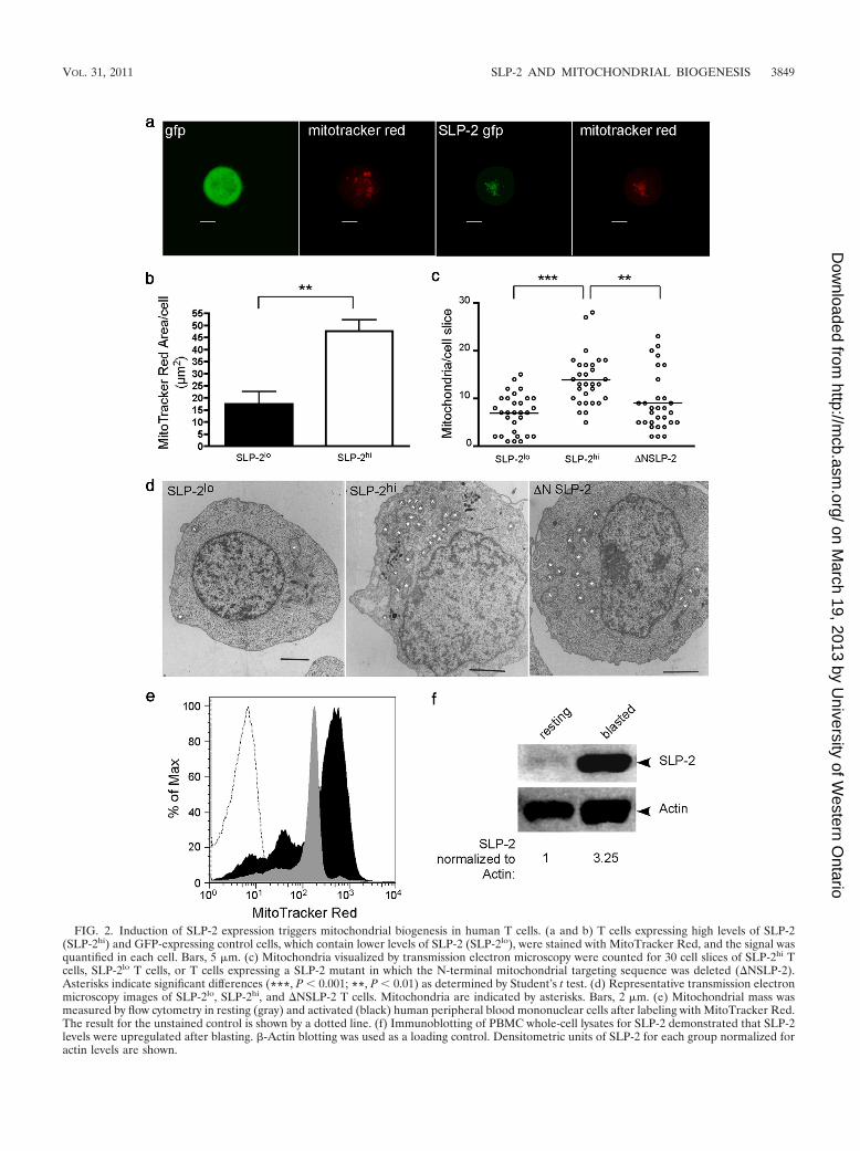
FIG. 2. Induction of SLP-2 expression triggers mitochondrial biogenesis in human T cells. (a and b) T cells expressing high levels of SLP-2(SLP-2hi) and GFP-expressing control cells, which contain lower levels of SLP-2 (SLP-2lo), were stained with MitoTracker Red, and the signal wasquantified in each cell. Bars, 5 �m. (c) Mitochondria visualized by transmission electron microscopy were counted for 30 cell slices of SLP-2hi Tcells, SLP-2lo T cells, or T cells expressing a SLP-2 mutant in which the N-terminal mitochondrial targeting sequence was deleted (�NSLP-2).Asterisks indicate significant differences (***, P � 0.001; **, P � 0.01) as determined by Student’s t test. (d) Representative transmission electronmicroscopy images of SLP-2lo, SLP-2hi, and �NSLP-2 T cells. Mitochondria are indicated by asterisks. Bars, 2 �m. (e) Mitochondrial mass wasmeasured by flow cytometry in resting (gray) and activated (black) human peripheral blood mononuclear cells after labeling with MitoTracker Red.The result for the unstained control is shown by a dotted line. (f) Immunoblotting of PBMC whole-cell lysates for SLP-2 demonstrated that SLP-2levels were upregulated after blasting. �-Actin blotting was used as a loading control. Densitometric units of SLP-2 for each group normalized foractin levels are shown.
VOL. 31, 2011 SLP-2 AND MITOCHONDRIAL BIOGENESIS 3849
on March 19, 2013 by U
niversity of Western O
ntariohttp://m
cb.asm.org/
Dow
nloaded from

To determine the kinetics of mitochondrial biogenesis inresponse to SLP-2 upregulation, MitoTracker Red was used tomeasure mitochondrial mass in PBMCs stimulated with PMAand ionomycin for 48 h. During the first 18 h of stimulation, thecells had a slight decrease in mitochondrial mass, which wasrecovered by 24 h, and mitochondrial mass was elevated by48 h of stimulation (Fig. 4a). During this time, SLP-2 andPHB-1 were both found to be upregulated as early as 1 h afterstimulation, while PGC-1� was elevated by 18 h poststimula-tion (Fig. 4b). This preceded the increase in mitochondrialbiogenesis detected by MitoTracker Red at 24 h poststimula-tion, implying that SLP-2 upregulation in response to T cellactivation precedes PGC-1� upregulation and the subsequentincrease in mitochondrial biogenesis.
SLP-2 interacts with prohibitins and binds cardiolipin-enriched microdomains. Next, we examined how upregula-tion of SLP-2 expression may induce mitochondrial biogen-esis. Emerging data suggest that mitochondrial biogenesis iscoordinated by a mechanism involving PHB-1 and -2 (17, 35).These proteins localize within phospholipid-enriched, detergent-insoluble microdomains in mitochondrial membranes, and theirfunctional interactome includes genes such as Ups1 and Gep1,which are linked to the synthesis of the mitochondrial membranephospholipids cardiolipin and phosphatidylethanolamine, respec-tively (17, 35). We noticed that Saccharomyces cerevisiae PHB-2has a putative transmembrane domain (as predicted by thetransmembrane hidden mark or model [TMHMM] algorithm[29]), but neither mouse nor human PHBs have such a domain.
FIG. 3. Induction of SLP-2 expression increases de novo cardiolipin biosynthesis, nuclear transcription programs, and mitochondrial DNAreplication. (a) Upregulation of SLP-2 expression in Jurkat T cells induces cardiolipin synthesis. Upregulation of SLP-2 expression in T cells(SLP-2hi) was induced with doxycycline and was compared with baseline expression of SLP-2 (SLP-2lo). The pool size of cardiolipin was determinedand was expressed as a percentage of the total phospholipid phosphorus mass. (b) Cardiolipin levels in naïve (gray) and in vitro-activated (black)PBMCs were measured by flow cytometry after staining with nonyl acridine orange. The result for the unstained control is shown by a dotted line.(c and d) Incorporation of [3H]acetate (c) and [14C]linoleic acid (d) into cardiolipin in SLP-2lo and SLP-2hi T cells. Data are shown as dpm/mgof total cellular protein. (e and f) Real-time PCR analysis of the transcript levels of phosphatidylglycerol phosphate synthase (e) and cardiolipinsynthase (f) in SLP-2lo and SLP-2hi T cells. (g) The mitochondrial mass in naïve (shaded histogram) and in vitro-activated PBMC in the absence(thin line) or presence (thick line) of triacsin C was measured by flow cytometry after staining with MitoTracker Red. The result for the unstainedcontrol is shown by a dotted line. (h and i) Real-time PCR analysis of the transcript levels of PGC-1� (h) and of the mitochondrial DNA content(i) in SLP-2lo and SLP-2hi T cells. The results shown here are representative of at least 3 independent experiments. *, P � 0.05.
3850 CHRISTIE ET AL. MOL. CELL. BIOL.
on March 19, 2013 by U
niversity of Western O
ntariohttp://m
cb.asm.org/
Dow
nloaded from

Of interest, S. cerevisiae does not have a homolog of SLP-2, theclosest being PHB-1, with a Clustal W score of 15 (41). Thissuggested that, in mammals, SLP-2 may act to link PHBs andphospholipid-enriched detergent-insoluble microdomains inmitochondrial membranes. Such a possibility is consistent withthe observations that PHBs are upregulated in parallel withSLP-2 during activation (38) (Fig. 4b) and that SLP-2 interactswith PHBs (11).
We first confirmed that human SLP-2 interacted with PHB-1and PHB-2 in T cells by using standard coprecipitation exper-iments (Fig. 5a). We found that immunoprecipitates of SLP-2contained both PHB-1 and PHB-2, as previously reported (11).Furthermore, coprecipitation of PHB-1, PHB-2, and SLP-2increased as the expression of SLP-2 increased with increasinglevels of doxycycline in the culture (Fig. 5b). To demonstratethat SLP-2 plays a role in linking prohibitins to phospholipid-enriched detergent-insoluble microdomains, we isolated mi-tochondria from SLP-2lo and SLP-2hi cells and extractedmitochondrial membranes with Na2CO3. We found a higherassociation of PHB-1 with the mitochondrial membrane frac-tion in SLP-2hi cells, although the total levels of PHB-1 weresimilar in the SLP-2lo and SLP-2hi cells (Fig. 5c).
Next, we generated human recombinant SLP-2 (hrSLP-2)and examined the profile of interaction of this protein withdifferent phospholipids by using a phospholipid vesicle co-
precipitation assay (4). In this assay, molecules that interactwith phospholipids cosediment with the vesicles, whereas non-interacting proteins remain in the supernatant. We found thathrSLP-2 coprecipitated with cardiolipin vesicles (Fig. 6a, P[pellet fraction]), whereas most of the hrSLP-2 remained insolution when vesicles made of other phospholipids found inmitochondria (phosphatidylcholine and phosphatidylethanol-amine) or vesicles made of mostly nonmitochondrial phospho-lipids (phosphatidylinositol and phosphatidylserine) were used(Fig. 6a, S [supernatant fraction]). Of interest, hrSLP-2 boundto phosphatidylglycerol vesicles in smaller amounts, a findinglikely due to the fact that cardiolipin is a composite of twophosphatidylglycerol molecules. To further investigate the as-sociation of hrSLP-2 with cardiolipin, we prepared phospho-lipid vesicles containing cardiolipin along with the other twomajor phospholipids in mitochondria, phosphatidylcholine andphosphatidylethanolamine, at ratios of 1:1:1 (33% CL, 33%PC, 33% PE), 2.5:0.25:0.25 (83% CL, 8% PC, 8% PE), and2.75:0.12:0.12 (92% CL, 4% PC, 4% PE). The interactionbetween hrSLP-2 and cardiolipin was selective and titratedwith the enrichment of this phospholipid in vesicles also contain-ing phosphatidylcholine and phosphatidylethanolamine (Fig. 6b).These data demonstrate that SLP-2 binds to the prohibitincomplex as well as to cardiolipin-enriched membrane microdo-
FIG. 4. SLP-2 upregulation precedes PGC-1� upregulation and mitochondrial biogenesis during T cell activation. (a) PBMCs were stimulatedwith 1 ng/ml PMA and 100 ng/ml ionomycin for as long as 48 h. At the indicated time points, cells were stained with MitoTracker Red and wereanalyzed for mitochondrial mass by flow cytometry. Gray, unstimulated cells; black, stimulated cells. (b) Whole-cell lysates were made at eachindicated time point during stimulation with PMA and ionomycin. The lysates were separated by SDS-PAGE and were serially blotted for PGC-1�,PHB-1, SLP-2, and actin.
VOL. 31, 2011 SLP-2 AND MITOCHONDRIAL BIOGENESIS 3851
on March 19, 2013 by U
niversity of Western O
ntariohttp://m
cb.asm.org/
Dow
nloaded from

mains, potentially mediating the association between prohib-itins and mitochondrial membranes.
Upregulation of SLP-2 expression is associated with in-creased mitochondrial functions. To examine the functionaleffects of upregulation of SLP-2 expression, we first deter-mined the activity of citrate synthase, an enzyme of the citricacid cycle, which is localized to the mitochondrial matrix. Wefound that SLP-2hi cells showed significantly increased citratesynthase activity (P � 0.01) (Fig. 7a). Next, we measured theactivity of the electron transport complexes in the respiratorychain. We found that SLP-2hi cells had significantly higheractivities of NADH dehydrogenase and succinate dehydroge-nase (P � 0.05), complexes I and II of the electron transportchain (Fig. 7b and c). In addition, mitochondrial and whole-cellATP levels in resting SLP-2hi T cells were significantly higherthan those in SLP-2lo T cells (P � 0.05) (Fig. 7d). Such an
FIG. 6. SLP-2 binds cardiolipin. (a) Coprecipitation of hrSLP-2with cardiolipin (CL) vesicles but not with other phospholipid vesiclesas shown by Western blotting of pellet (P) and supernatant (S) frac-tions. PC, phosphatidylcholine; PE, phosphatidylethanolamine; PI,phosphatidylinositol; PS, phosphatidylserine; PG, phosphatidylglyc-erol; Total protein, hrSLP-2 aliquot as used for coprecipitation. (b)Titration of hrSLP-2 coprecipitation with CL in mixed CL-PC-PEvesicles at the indicated ratios. An additional group containing twicethe amount of hrSLP-2 (CL � 2� SLP-2) was added.
FIG. 5. SLP-2 interacts with prohibitins. (a) Immunoprecipitates with an antiserum against human SLP-2 or with a preimmune serumfrom SLP-2-expressing Jurkat T cells were serially blotted for prohibitin 1 (PHB-1), PHB-2, or SLP-2. WCL, whole-cell lysate; IP,immunoprecipitating antiserum; Beads, no-cell-lysate control; Pre-Imm, control immunoprecipitation with preimmune serum. (b) Immu-noprecipitates and whole-cell lysates from SLP-2-GFP cells induced to express increasing amounts of SLP-2 by increasing concentrations ofdoxycycline were collected and blotted as for Fig. 4b. (c) Mitochondria were isolated from SLP-2hi and SLP-2lo cells and were treated withNa2CO3 in order to examine the membrane association of prohibitins in the presence of elevated SLP-2 expression. The fractions wereserially blotted for PHB-1 and SLP-2.
3852 CHRISTIE ET AL. MOL. CELL. BIOL.
on March 19, 2013 by U
niversity of Western O
ntariohttp://m
cb.asm.org/
Dow
nloaded from

increase in ATP levels in SLP-2hi T cells correlated with in-creased resistance to depletion of mitochondrial ATP witholigomycin, as evidenced by increased T-cell responses, indi-cated by increased IL-2 production. In these experiments,
oligomycin-treated SLP-2lo T cells and SLP-2hi T cells werestimulated with antigen-presenting cells and the superantigenstaphylococcal enterotoxin E (SEE), and the production ofinterleukin-2 (IL-2) was determined after 24 h of stimulation.
FIG. 7. Induction of SLP-2 expression is associated with higher mitochondrial activity. (a) Citrate synthase (CS) activity was measured inSLP-2lo and SLP-2hi Jurkat T cells. Data are means and standard deviations for triplicate samples; **, P � 0.01. Data are expressed as nmol/min/mgmitochondrial protein. (b) NADH dehydrogenase was measured in SLP-2lo and SLP-2hi Jurkat T cells, plotted as in panel a. *, P � 0.05. (c)Succinate dehydrogenase (SDH) was measured in SLP-2lo and SLP-2hi Jurkat T cells, plotted as in panel a. *, P � 0.05. Data are expressed asnmol/min/mg mitochondrial protein. (d) Mitochondrial and total cellular ATP levels were measured in SLP-2lo and SLP-2hi Jurkat T cells. Meansand standard deviations for triplicate samples are plotted. *, P � 0.05; **, P � 0.01. (e and f) IL-2 levels in culture supernatants of SLP-2lo (e)and SLP-2hi (f) Jurkat T cells pretreated or not with oligomycin (4 �M) and stimulated with antigen-presenting cells and SEE for 24 h. Means andstandard deviations for triplicate samples are plotted. Asterisks indicate significant differences (*, P � 0.05; ***, P � 0.001) as determined byanalysis of variance (ANOVA) with a Bonferroni posttest. (g) Oxygen consumption was measured in SLP-2lo and SLP-2hi T cells in mediacontaining high (11 mM) or low (0.4 mM) glucose levels. Means and standard deviations for triplicate samples are plotted. Asterisks indicatesignificant differences (*, P � 0.05; **, P � 0.01) as determined by ANOVA with a Bonferroni posttest. (h) Mitochondria were isolated fromSLP-2lo and SLP-2hi cells, separated by SDS-PAGE, and serially blotted for subunits of each complex of the respiratory chain as well as SLP-2.
VOL. 31, 2011 SLP-2 AND MITOCHONDRIAL BIOGENESIS 3853
on March 19, 2013 by U
niversity of Western O
ntariohttp://m
cb.asm.org/
Dow
nloaded from
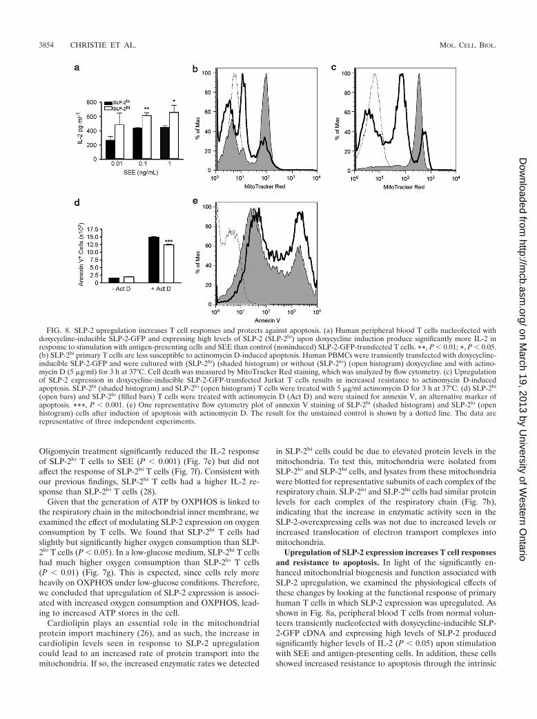
Oligomycin treatment significantly reduced the IL-2 responseof SLP-2lo T cells to SEE (P � 0.001) (Fig. 7e) but did notaffect the response of SLP-2hi T cells (Fig. 7f). Consistent withour previous findings, SLP-2hi T cells had a higher IL-2 re-sponse than SLP-2lo T cells (28).
Given that the generation of ATP by OXPHOS is linked tothe respiratory chain in the mitochondrial inner membrane, weexamined the effect of modulating SLP-2 expression on oxygenconsumption by T cells. We found that SLP-2hi T cells hadslightly but significantly higher oxygen consumption than SLP-2lo T cells (P � 0.05). In a low-glucose medium, SLP-2hi T cellshad much higher oxygen consumption than SLP-2lo T cells(P � 0.01) (Fig. 7g). This is expected, since cells rely moreheavily on OXPHOS under low-glucose conditions. Therefore,we concluded that upregulation of SLP-2 expression is associ-ated with increased oxygen consumption and OXPHOS, lead-ing to increased ATP stores in the cell.
Cardiolipin plays an essential role in the mitochondrialprotein import machinery (26), and as such, the increase incardiolipin levels seen in response to SLP-2 upregulationcould lead to an increased rate of protein transport into themitochondria. If so, the increased enzymatic rates we detected
in SLP-2hi cells could be due to elevated protein levels in themitochondria. To test this, mitochondria were isolated fromSLP-2lo and SLP-2hi cells, and lysates from these mitochondriawere blotted for representative subunits of each complex of therespiratory chain. SLP-2lo and SLP-2hi cells had similar proteinlevels for each complex of the respiratory chain (Fig. 7h),indicating that the increase in enzymatic activity seen in theSLP-2-overexpressing cells was not due to increased levels orincreased translocation of electron transport complexes intomitochondria.
Upregulation of SLP-2 expression increases T cell responsesand resistance to apoptosis. In light of the significantly en-hanced mitochondrial biogenesis and function associated withSLP-2 upregulation, we examined the physiological effects ofthese changes by looking at the functional response of primaryhuman T cells in which SLP-2 expression was upregulated. Asshown in Fig. 8a, peripheral blood T cells from normal volun-teers transiently nucleofected with doxycycline-inducible SLP-2-GFP cDNA and expressing high levels of SLP-2 producedsignificantly higher levels of IL-2 (P � 0.05) upon stimulationwith SEE and antigen-presenting cells. In addition, these cellsshowed increased resistance to apoptosis through the intrinsic
FIG. 8. SLP-2 upregulation increases T cell responses and protects against apoptosis. (a) Human peripheral blood T cells nucleofected withdoxycycline-inducible SLP-2-GFP and expressing high levels of SLP-2 (SLP-2hi) upon doxycycline induction produce significantly more IL-2 inresponse to stimulation with antigen-presenting cells and SEE than control (noninduced) SLP-2-GFP-transfected T cells. **, P � 0.01; *, P � 0.05.(b) SLP-2hi primary T cells are less susceptible to actinomycin D-induced apoptosis. Human PBMCs were transiently transfected with doxycycline-inducible SLP-2-GFP and were cultured with (SLP-2hi) (shaded histogram) or without (SLP-2lo) (open histogram) doxycycline and with actino-mycin D (5 �g/ml) for 3 h at 37°C. Cell death was measured by MitoTracker Red staining, which was analyzed by flow cytometry. (c) Upregulationof SLP-2 expression in doxycycline-inducible SLP-2-GFP-transfected Jurkat T cells results in increased resistance to actinomycin D-inducedapoptosis. SLP-2hi (shaded histogram) and SLP-2lo (open histogram) T cells were treated with 5 �g/ml actinomycin D for 3 h at 37°C. (d) SLP-2hi
(open bars) and SLP-2lo (filled bars) T cells were treated with actinomycin D (Act D) and were stained for annexin V, an alternative marker ofapoptosis. ***, P � 0.001. (e) One representative flow cytometry plot of annexin V staining of SLP-2hi (shaded histogram) and SLP-2lo (openhistogram) cells after induction of apoptosis with actinomycin D. The result for the unstained control is shown by a dotted line. The data arerepresentative of three independent experiments.
3854 CHRISTIE ET AL. MOL. CELL. BIOL.
on March 19, 2013 by U
niversity of Western O
ntariohttp://m
cb.asm.org/
Dow
nloaded from

pathway (Fig. 8b). Such resistance to apoptosis was also ob-served in response to actinomycin D in Jurkat SLP-2hi T cells(Fig. 8c). As an additional marker of apoptosis, actinomycinD-induced cell death was also measured by an increase inannexin V labeling. In agreement with the loss of transmem-brane potential as measured by MitoTracker Red, SLP-2hi
cells also showed lower annexin V staining, which is indicativeof protection against the induction of the intrinsic apoptosispathway (Fig. 8d and e). Thus, consistent with the increasedmitochondrial biogenesis and function, upregulation of SLP-2expression enhances cell function.
DISCUSSION
The function of stomatins and other proteins sharing thestomatin/prohibitin/flotillin/HflK (SPFH) domain remain enig-matic. It has been proposed that through this domain, stom-atins, PHBs, and flotillins interact with different cell mem-branes and facilitate signaling. We report here that SLP-2, amitochondrial member of the stomatin family, binds cardio-lipin and may help to recruit PHBs to cardiolipin-enrichedmicrodomains. Furthermore, upregulation of SLP-2 expressionis associated with increased mitochondrial biogenesis and func-tion, ultimately resulting in enhanced effector cell function.
Our data show that SLP-2 binds cardiolipin-enriched mi-crodomains in a way that titrates with the content of cardiolipinin phospholipid membranes. The mitochondrial membrane iscomposed of 40% phosphatidylcholine, 30% phosphatidyl-ethanolamine, and 10 to 15% phosphatidylinositol and cardio-lipin (36). But the cardiolipin content in the mitochondrialmembrane is not homogeneously distributed. Our data suggestthat SLP-2 can bind to clusters of cardiolipin, i.e., microdo-mains with a much higher localized cardiolipin concentration.This selective binding of SLP-2 to cardiolipin is reminiscentof the observation that two other members of the stomatin/PHB/flotillin/HflK superfamily (MEC-2 and podocin) bindcholesterol (24) and leads us to propose that SPFH domain-containing proteins bind to selective phospholipid-enrichedmembrane microdomains and may act as markers that recruitother proteins to the lipid microdomains. How these proteinsbind lipids is still unknown. The binding of SLP-2 to cardiolipinin the mitochondrial inner membrane is likely independent oftransmembrane insertion, since SLP-2 lacks a putative trans-membrane domain (43).
Upregulation of SLP-2 expression increases cardiolipin syn-thesis and mitochondrial biogenesis. Recent evidence indicatesthat these two processes are tightly coordinated (17), likelythrough the PHB functional interactome, which includes genesinvolved in phospholipid synthesis, such as Ups1, whose dele-tion, in yeast, translates into a marked decrease in cardiolipincontent (35). Although we have not shown a direct interactionamong PHBs, SLP-2, and cardiolipin, our data suggest thatSLP-2 may bring PHBs to cardiolipin and may contribute tothe organization of cardiolipin-enriched microdomains in themitochondrial inner membrane (3). Data from our laboratoryusing T cell-specific SLP-2 knockout mice seem to corroboratethis model (D. A. Christie et al., unpublished data). This pos-sible mechanism accommodates the upregulation of bothSLP-2 and PHBs during T cell activation (38) and the detec-tion of both proteins in detergent-insoluble microdomains of
cell membranes (i.e., cardiolipin-enriched microdomains in themitochondrial inner membrane and cholesterol-enriched mi-crodomains in the plasma membrane) (13, 27, 28, 33). In thiscontext, an increase in SLP-2 levels as a result of cell activationwould increase the interaction of PHBs with cardiolipin in themitochondrial inner membrane and activate the PHB func-tional interactome, leading to de novo cardiolipin synthesis.
Using human T cell activation as a model, we have shownthat upregulation of SLP-2 expression is a molecular step be-tween mitochondrial biogenesis and cell activation and func-tion. SLP-2 does not seem to be critical under resting condi-tions, as suggested by the absence or very low level of SLP-2expression in resting naïve T cells. However, during T cellactivation, SLP-2 expression is dramatically upregulated, andthis increases T cell function, leading to increased IL-2 pro-duction and increased resistance to apoptosis through theintrinsic pathway. PHBs are also upregulated during T cellactivation (38), whereas PHB-2 deletion decreases the abil-ity of cells to proliferate and increases their sensitivity toapoptosis through the intrinsic pathway (32). These obser-vations corroborate the model proposed here by which SLP-2–PHB complexes would act in concert to regulate mito-chondrial biogenesis and function during T cell activation.Of interest, modulation of PHB-2 levels also alters OPA-1levels (e.g., cells lacking PHB-2 have impaired processing ofOPA-1 [3]), and in our experiments, T cells overexpressingSLP-2 have increased OPA-1 levels.
The increased mitochondrial function observed in SLP-2-overexpressing cells is in line with the observation that SLP-2controls the stability and the assembly of electron transportcomplexes (1, 23). This is further corroborated by emergingdata from our laboratory using T cell-specific SLP-2-deficientmice, showing that a lack of SLP-2 is associated with decreasedrecruitment of PHB-1 to cardiolipin-enriched mitochondrialmembranes, as well as a loss of complex I subunits (D. A.Christie et al., unpublished data). Optimal assembly of thesecomplexes may require the formation of specialized membranemicrodomains, and this would correlate with increased respi-ratory chain function. Of interest, SLP-2 is required for mito-chondrial hyperfusion (42), a phenomenon linked with in-creased metabolic demands and cellular stress. Levels of bothOPA-1 and mitofusin-2, proteins required for this process,were increased upon SLP-2 upregulation, but cytochrome clevels were not. It is possible that SLP-2 expression specificallyincreases the expression of proteins involved in mitochondrialfusion, or prevents these proteins from degradation, withoutaltering the expression of other mitochondrial proteins.
Taken together, the evidence presented here leads us topropose that the binding of SLP-2 to cardiolipin recruits PHBs,helping to form cardiolipin-enriched membrane microdomainsin which the respiratory chain components are optimally as-sembled. The formation of these microdomains is likely theresult of the self-oligomerizing properties of SLP-2 (43). Theincrease in respiratory chain function may then induce furthercardiolipin synthesis through its effect on the pH gradientacross the mitochondrial inner membrane (18), enhancing mi-tochondrial biogenesis. The result of this proposed mechanismis the provision of the enhanced mitochondrial function re-quired for an optimal T cell response.
How the increased mitochondrial membrane formation
VOL. 31, 2011 SLP-2 AND MITOCHONDRIAL BIOGENESIS 3855
on March 19, 2013 by U
niversity of Western O
ntariohttp://m
cb.asm.org/
Dow
nloaded from

increases nuclear transcription and mitochondrial DNA rep-lication, both required to increase mitochondrial numbers,remains to be determined. A retrograde nuclear signalingpathway in yeast is starting to emerge, but little is known aboutthis pathway in mammalian cells. Specific signals may involvemitochondrial stress, since it has been shown that mitochon-drial dysfunction increases nuclear transcription (40). In thiscontext, SLP-2 may play a role by regulating mitochondrialmembrane biogenesis and its subsequent effect of calcium-dependent signaling, which has been linked to the activation ofretrograde signaling (6).
In summary, we have shown that increased levels of SLP-2,seen, for example, during lymphocyte activation, stimulate mi-tochondrial biogenesis and function, providing for the ener-getic requirements of activation. Such a function involves thebinding of SLP-2 and its interacting partners (PHBs) to cardio-lipin to form membrane microdomains in which respiratorycomplexes are optimally assembled. The ultimate result of thisfunction is the net enhancement of effector cell functions.
ACKNOWLEDGMENTS
We thank Heidi McBride, Jean-Claude Martinou, Fred Possmayer,and the members of the Madrenas laboratory for helpful discussions inthe course of this work.
This research was supported by the Canadian Institutes of HealthResearch. D.A.C. holds a CIHR Doctoral Studentship. G.M.H. holdsa Canada Research Chair in Molecular Cardiolipin Metabolism, andJ.M. holds a Canada Research Chair in Immunobiology.
REFERENCES
1. Acin-Perez, R., P. Fernandez-Silva, M. L. Peleato, A. Perez-Martos, and J. A.Enriquez. 2008. Respiratory active mitochondrial supercomplexes. Mol. Cell32:529–539.
2. Arp, J., et al. 2003. Regulation of T-cell activation by phosphodiesterase 4B2requires its dynamic redistribution during immunological synapse formation.Mol. Cell. Biol. 23:8042–8057.
3. Artal-Sanz, M., and N. Tavernarakis. 2009. Prohibitin and mitochondrialbiology. Trends Endocrinol. Metab. 20:394–401.
4. Bakolitsa, C., J. M. de Pereda, C. R. Bagshaw, D. R. Critchley, and R. C.Liddington. 1999. Crystal structure of the vinculin tail suggests a pathway foractivation. Cell 99:603–613.
5. Baroja, M. L., et al. 2000. The inhibitory function of CTLA-4 does notrequire its tyrosine phosphorylation. J. Immunol. 164:49–55.
6. Biswas, G., et al. 1999. Retrograde Ca2� signaling in C2C12 skeletal myo-cytes in response to mitochondrial genetic and metabolic stress: a novelmode of inter-organelle crosstalk. EMBO J. 18:522–533.
7. Bueno, C., et al. 2006. Bacterial superantigens bypass Lck-dependent T cellreceptor signaling by activating a G�11-dependent, PLC-�-mediated path-way. Immunity 25:67–78.
8. Carpentieri, U., and L. A. Sordahl. 1980. Respiratory and calcium transportfunctions of mitochondria isolated from normal and transformed humanlymphocytes. Cancer Res. 40:221–224.
9. Chau, T. A., et al. 2009. Toll-like receptor 2 ligands on the staphylococcal cellwall downregulate superantigen-induced T cell activation and prevent toxicshock syndrome. Nat. Med. 15:641–648.
10. Cote, H. C., et al. 2002. Changes in mitochondrial DNA as a marker ofnucleoside toxicity in HIV-infected patients. N. Engl. J. Med. 346:811–820.
11. Da Cruz, S., et al. 2008. SLP-2 interacts with prohibitins in the mitochondrialinner membrane and contributes to their stability. Biochim. Biophys. Acta1783:904–911.
12. D’Souza, A. D., N. Parikh, S. M. Kaech, and G. S. Shadel. 2007. Convergenceof multiple signaling pathways is required to coordinately up-regulatemtDNA and mitochondrial biogenesis during T cell activation. Mitochon-drion 7:374–385.
13. Foster, L. J., C. L. De Hoog, and M. Mann. 2003. Unbiased quantitativeproteomics of lipid rafts reveals high specificity for signaling factors. Proc.Natl. Acad. Sci. U. S. A. 100:5813–5818.
14. Fox, C. J., P. S. Hammerman, and C. B. Thompson. 2005. Fuel feedsfunction: energy metabolism and the T-cell response. Nat. Rev. Immunol.5:844–852.
15. Frauwirth, K. A., et al. 2002. The CD28 signaling pathway regulates glucosemetabolism. Immunity 16:769–777.
16. Frauwirth, K. A., and C. B. Thompson. 2004. Regulation of T lymphocytemetabolism. J. Immunol. 172:4661–4665.
17. Gohil, V. M., and M. L. Greenberg. 2009. Mitochondrial membrane biogen-esis: phospholipids and proteins go hand in hand. J. Cell Biol. 184:469–472.
18. Gohil, V. M., et al. 2004. Cardiolipin biosynthesis and mitochondrial respi-ratory chain function are interdependent. J. Biol. Chem. 279:42612–42618.
19. Green, J. B., and J. P. Young. 2008. Slipins: ancient origin, duplication anddiversification of the stomatin protein family. BMC Evol. Biol. 8:44.
20. Hajek, P., A. Chomyn, and G. Attardi. 2007. Identification of a novel mito-chondrial complex containing mitofusin 2 and stomatin-like protein 2.J. Biol. Chem. 282:5670–5681.
21. Hatch, G. M., and G. McClarty. 1996. Regulation of cardiolipin biosynthesisin H9c2 cardiac myoblasts by CTP. J. Biol. Chem. 271:25810–25816.
22. Hauff, K., D. Linda, and G. M. Hatch. 2009. Mechanism of the elevation incardiolipin during HeLa cell entry into the S-phase of the human cell cycle.Biochem. J. 417:573–582.
23. Houtkooper, R. H., and F. M. Vaz. 2008. Cardiolipin, the heart of mitochon-drial metabolism. Cell. Mol. Life Sci. 65:2493–2506.
24. Huber, T. B., et al. 2006. Podocin and MEC-2 bind cholesterol to regulatethe activity of associated ion channels. Proc. Natl. Acad. Sci. U. S. A.103:17079–17086.
25. Jacobson, J., M. R. Duchen, and S. J. Heales. 2002. Intracellular distributionof the fluorescent dye nonyl acridine orange responds to the mitochondrialmembrane potential: implications for assays of cardiolipin and mitochondrialmass. J. Neurochem. 82:224–233.
26. Joshi, A. S., J. Zhou, V. M. Gohil, S. Chen, and M. L. Greenberg. 2009.Cellular functions of cardiolipin in yeast. Biochim. Biophys. Acta 1793:212–218.
27. Kim, K. B., et al. 2006. Oxidation-reduction respiratory chains and ATPsynthase complex are localized in detergent-resistant lipid rafts. Proteomics6:2444–2453.
28. Kirchhof, M. G., et al. 2008. Modulation of T cell activation by stomatin-likeprotein 2. J. Immunol. 181:1927–1936.
29. Krogh, A., B. Larsson, G. von Heijne, and E. L. Sonnhammer. 2001. Pre-dicting transmembrane protein topology with a hidden Markov model: ap-plication to complete genomes. J. Mol. Biol. 305:567–580.
30. Livak, K. J., and T. D. Schmittgen. 2001. Analysis of relative gene expressiondata using real-time quantitative PCR and the 2���CT method. Methods25:402–408.
31. Lowry, O. H., N. J. Rosebrough, A. L. Farr, and R. J. Randall. 1951. Proteinmeasurement with the Folin phenol reagent. J. Biol. Chem. 193:265–275.
32. Merkwirth, C., et al. 2008. Prohibitins control cell proliferation and apop-tosis by regulating OPA1-dependent cristae morphogenesis in mitochondria.Genes Dev. 22:476–488.
33. Mielenz, D., et al. 2005. Lipid rafts associate with intracellular B cell recep-tors and exhibit a B cell stage-specific protein composition. J. Immunol.174:3508–3517.
34. Nagy, G., M. Barcza, N. Gonchoroff, P. E. Phillips, and A. Perl. 2004. Nitricoxide-dependent mitochondrial biogenesis generates Ca2� signaling profileof lupus T cells. J. Immunol. 173:3676–3683.
35. Osman, C., et al. 2009. The genetic interactome of prohibitins: coordinatedcontrol of cardiolipin and phosphatidylethanolamine by conserved regula-tors in mitochondria. J. Cell Biol. 184:583–596.
36. Osman, C., D. R. Voelker, and T. Langer. 2011. Making heads or tails ofphospholipids in mitochondria. J. Cell Biol. 192:7–16.
37. Owczarek, C. M., et al. 2001. A novel member of the STOMATIN/EPB72/mec-2 family, stomatin-like 2 (STOML2), is ubiquitously expressed andlocalizes to HSA chromosome 9p13.1. Cytogenet. Cell Genet. 92:196–203.
38. Ross, J. A., Z. S. Nagy, and R. A. Kirken. 2008. The PHB1/2 phosphocom-plex is required for mitochondrial homeostasis and survival of human T cells.J. Biol. Chem. 283:4699–4713.
39. Rouser, G., S. Fleischer, and A. Yamamoto. 1970. Two dimensional thinlayer chromatographic separation of polar lipids and determination of phos-pholipids by phosphorus analysis of spots. Lipids 5:494–496.
40. Scarpulla, R. C. 2008. Transcriptional paradigms in mammalian mitochon-drial biogenesis and function. Physiol. Rev. 88:611–638.
41. Thompson, J. D., D. G. Higgins, and T. J. Gibson. 1994. CLUSTAL W:improving the sensitivity of progressive multiple sequence alignment throughsequence weighting, position-specific gap penalties and weight matrix choice.Nucleic Acids Res. 22:4673–4680.
42. Tondera, D., et al. 2009. SLP-2 is required for stress-induced mitochondrialhyperfusion. EMBO J. 28:1589–1600.
43. Wang, Y., and J. S. Morrow. 2000. Identification and characterization ofhuman SLP-2, a novel homologue of stomatin (band 7.2b) present in eryth-rocytes and other tissues. J. Biol. Chem. 275:8062–8071.
3856 CHRISTIE ET AL. MOL. CELL. BIOL.
on March 19, 2013 by U
niversity of Western O
ntariohttp://m
cb.asm.org/
Dow
nloaded from


![OPEN ACCESS viruses - Semantic Scholar...III alpha (PIKIII α) binds to and regulates th e phosphorylation status of NS 5A [14]. The phosphorylation state likely plays an important](https://img.pdfslide.us/doc/110x75/60f6ad79a1e0ef6b36059fb3/open-access-viruses-semantic-scholar-iii-alpha-pikiii-binds-to-and-regulates.jpg)