Embed Size (px)
Citation preview

STEROIDS IN NEUROSURGERY
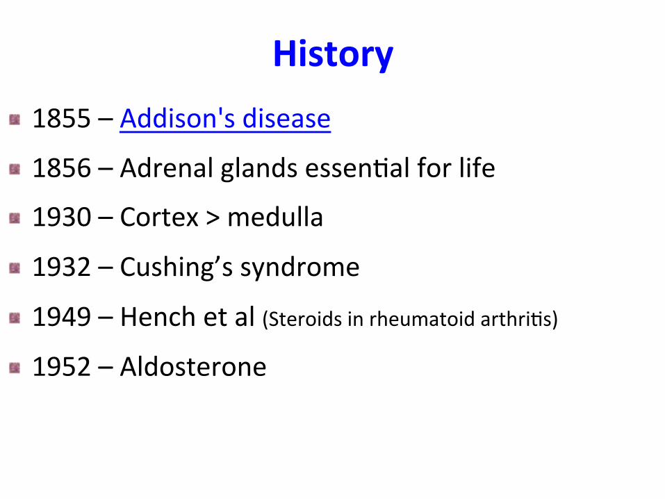
History ! 1855 – Addison's disease
! 1856 – Adrenal glands essen3al for life
! 1930 – Cortex > medulla
! 1932 – Cushing’s syndrome
! 1949 – Hench et al (Steroids in rheumatoid arthri3s)
! 1952 – Aldosterone
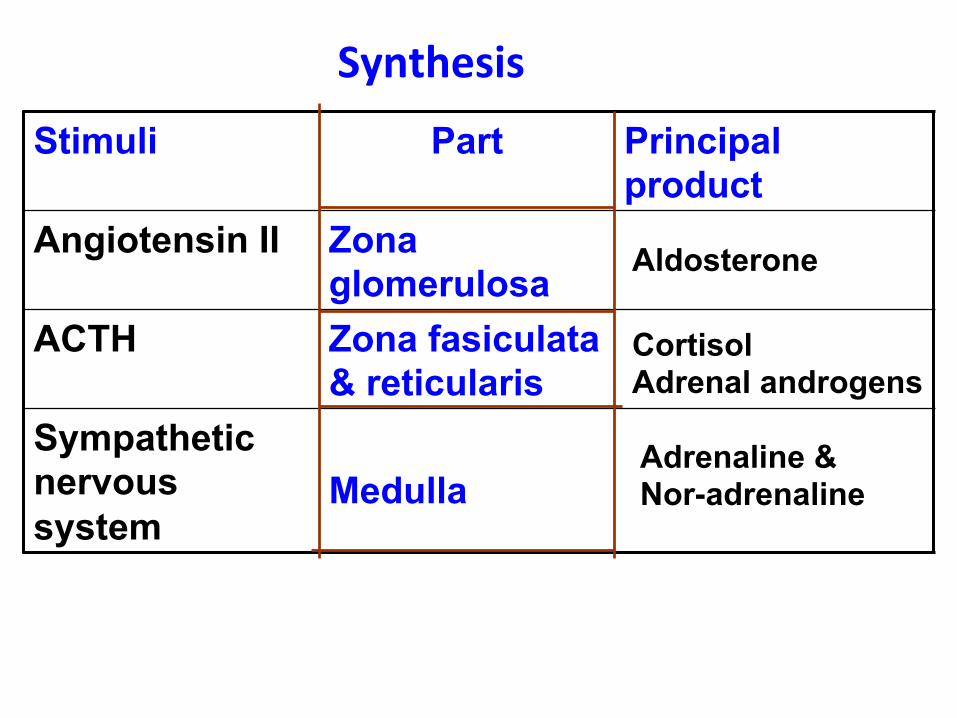
Stimuli Part Principal product
Angiotensin II Zona glomerulosa
ACTH Zona fasiculata & reticularis
Sympathetic nervous system
Medulla
Synthesis
Aldosterone
Cortisol Adrenal androgens
Adrenaline & Nor-adrenaline
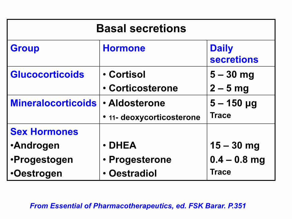
Basal secretions Group Hormone Daily
secretions Glucocorticoids • Cortisol
• Corticosterone 5 – 30 mg 2 – 5 mg
Mineralocorticoids • Aldosterone • 11- deoxycorticosterone
5 – 150 µg Trace
Sex Hormones • Androgen • Progestogen • Oestrogen
• DHEA • Progesterone • Oestradiol
15 – 30 mg 0.4 – 0.8 mg Trace
From Essential of Pharmacotherapeutics, ed. FSK Barar. P.351

Preparations Drug Anti-inflam. Salt retaining Topical
Cortisol 1 1.0 1 Cortisone 0.8 0.8 0 Prednisone 4 0.8 0 Prednisolone 5 0.3 4 Methylpredni- solone
5 0 5
Intermediate acting Triamcinolone 5 0 5 Paramethasone 10 0 - Fluprednisolone 15 0 7
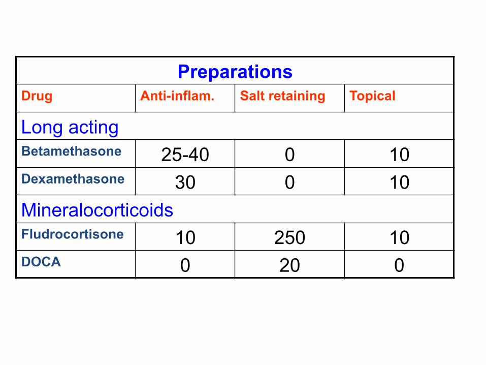
Preparations Drug Anti-inflam. Salt retaining Topical
Long acting Betamethasone 25-40 0 10 Dexamethasone 30 0 10 Mineralocorticoids Fludrocortisone 10 250 10 DOCA 0 20 0
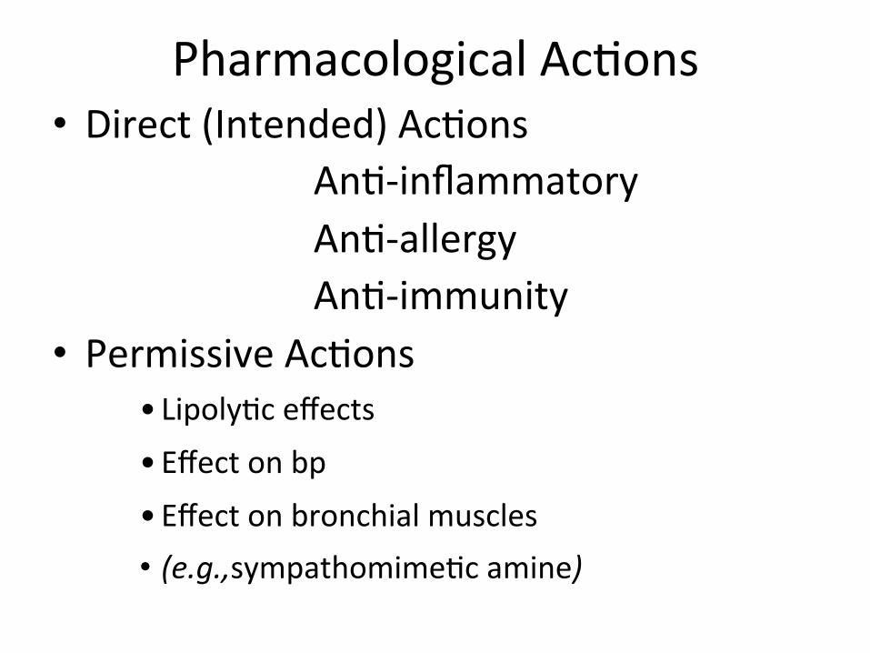
Pharmacological Ac3ons • Direct (Intended) Ac3ons An3-‐inflammatory An3-‐allergy An3-‐immunity
• Permissive Ac3ons • Lipoly3c effects • Effect on bp • Effect on bronchial muscles • (e.g.,sympathomime3c amine)
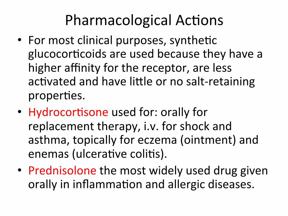
Pharmacological Ac3ons • For most clinical purposes, synthe3c glucocor3coids are used because they have a higher affinity for the receptor, are less ac3vated and have liWle or no salt-‐retaining proper3es.
• Hydrocor3sone used for: orally for replacement therapy, i.v. for shock and asthma, topically for eczema (ointment) and enemas (ulcera3ve coli3s).
• Prednisolone the most widely used drug given orally in inflamma3on and allergic diseases.
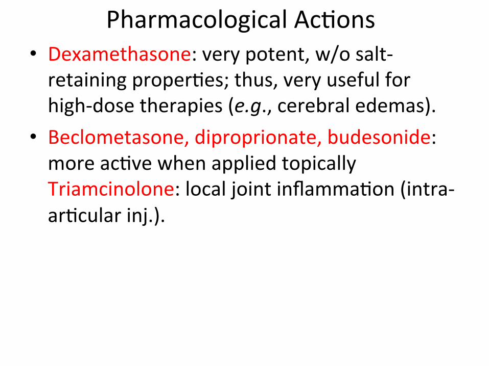
Pharmacological Ac3ons • Dexamethasone: very potent, w/o salt-‐retaining proper3es; thus, very useful for high-‐dose therapies (e.g., cerebral edemas).
• Beclometasone, diproprionate, budesonide: more ac3ve when applied topically Triamcinolone: local joint inflamma3on (intra-‐ar3cular inj.).

• Recruitment of WBC and monocyte-‐ macrophage into affected area & elaboraGon
of chemotacGc substances
• LipocorGn • ELAM1 and ICAM-‐1 in endothelial cells
• TNF from phagocyGc cells
• IL1 from monocyte-‐macrophage
• FormaGon of Plasminogen AcGvator
• AcGon of MIF and fibroblasGc acGvity
• Expression of COX II
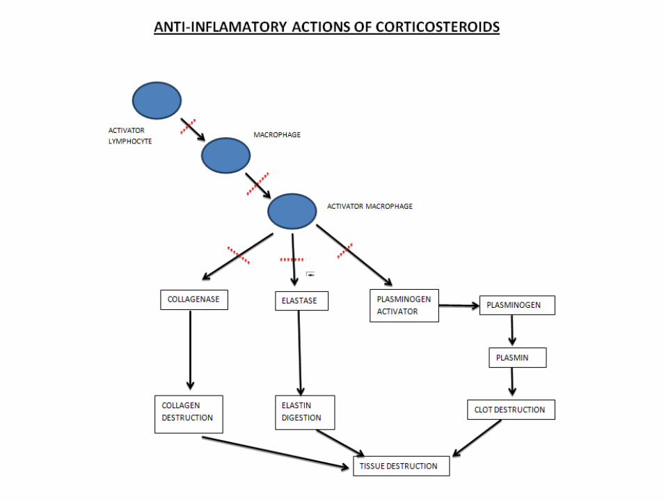
AcGons: AnG-‐inflammatory
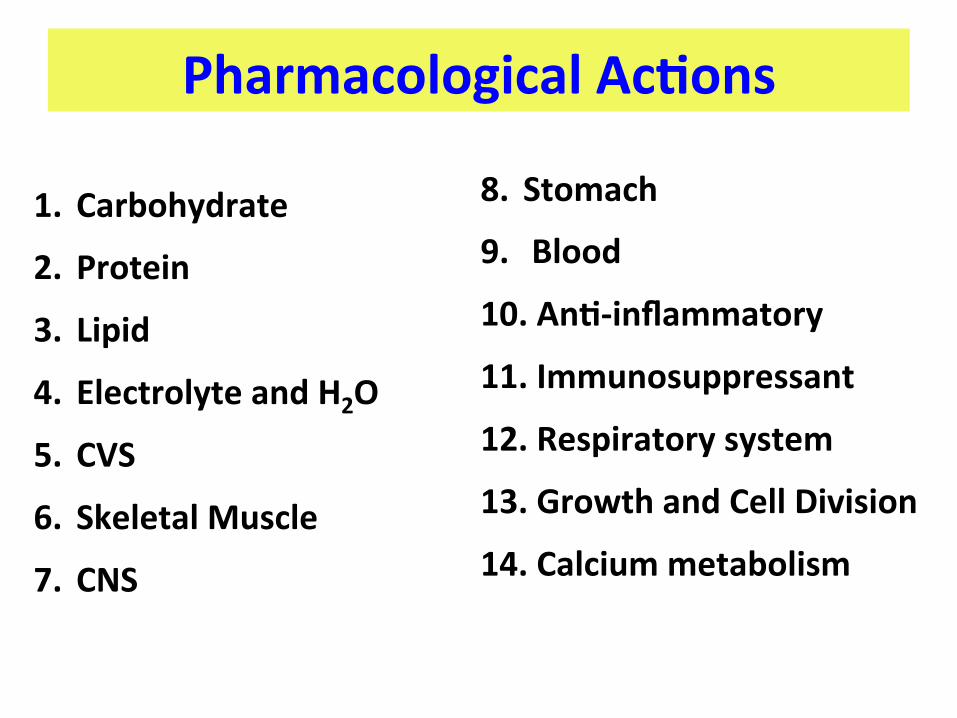
Pharmacological AcGons
1. Carbohydrate 2. Protein 3. Lipid 4. Electrolyte and H2O
5. CVS 6. Skeletal Muscle
7. CNS
8. Stomach
9. Blood 10. AnG-‐inflammatory
11. Immunosuppressant
12. Respiratory system
13. Growth and Cell Division 14. Calcium metabolism
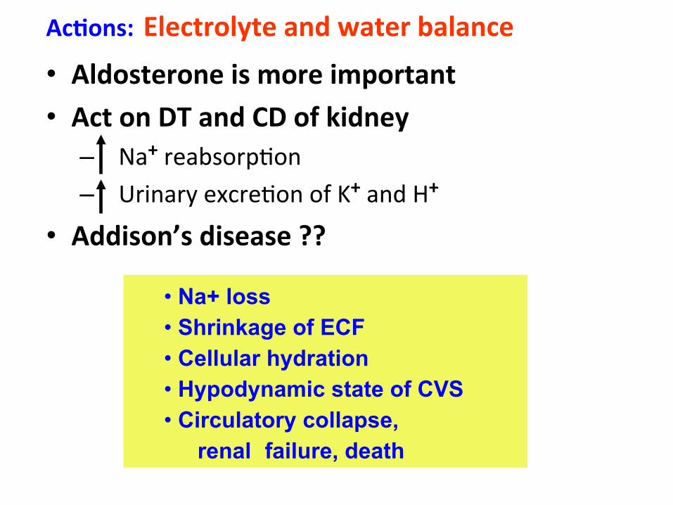
AcGons: Electrolyte and water balance • Aldosterone is more important • Act on DT and CD of kidney
– Na+ reabsorp3on – Urinary excre3on of K+ and H+
• Addison’s disease ??
• Na+ loss • Shrinkage of ECF • Cellular hydration • Hypodynamic state of CVS • Circulatory collapse,
renal failure, death
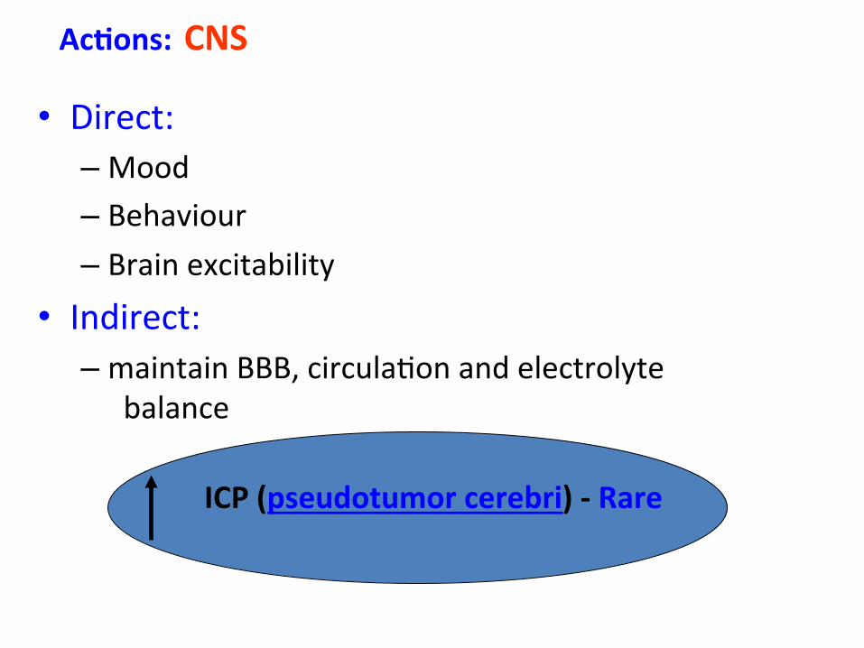
• Direct: – Mood – Behaviour – Brain excitability
• Indirect: – maintain BBB, circula3on and electrolyte
balance
AcGons: CNS
ICP (pseudotumor cerebri) -‐ Rare
Role in neurosurgery

Role in trauma3c spine and head injuries

Pathophysiology of Spinal Cord injury
Primary mechanisms Ini3al crush, shear impingement of cord with the inci3ng trauma.
Secondary mechanisms Vascular insults/insufficiency Edema Cell toxicity Apoptosis
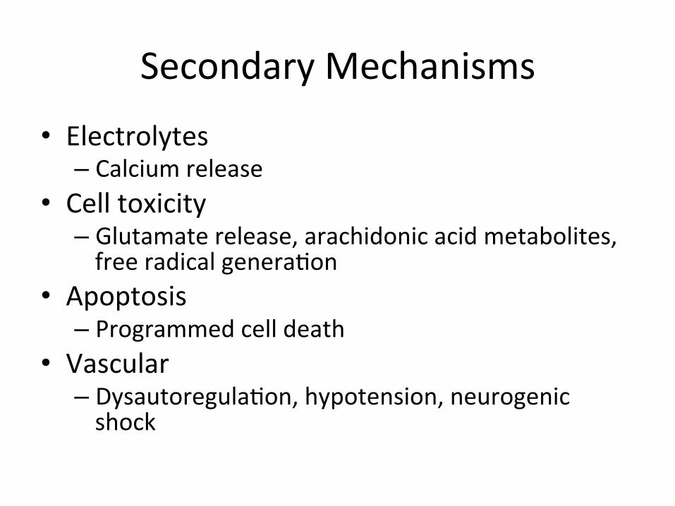
Secondary Mechanisms
• Electrolytes – Calcium release
• Cell toxicity – Glutamate release, arachidonic acid metabolites, free radical genera3on
• Apoptosis – Programmed cell death
• Vascular – Dysautoregula3on, hypotension, neurogenic shock
Secondary mechanisms
• Numerous mediators of spinal cord damage have been iden3fied experimentally.
• The hope is that through simple pharmacologic interven3ons, the secondary damage can be limited, or even poten3ally reversed.
• Unfortunately very liWle clinical progress has been made to date.
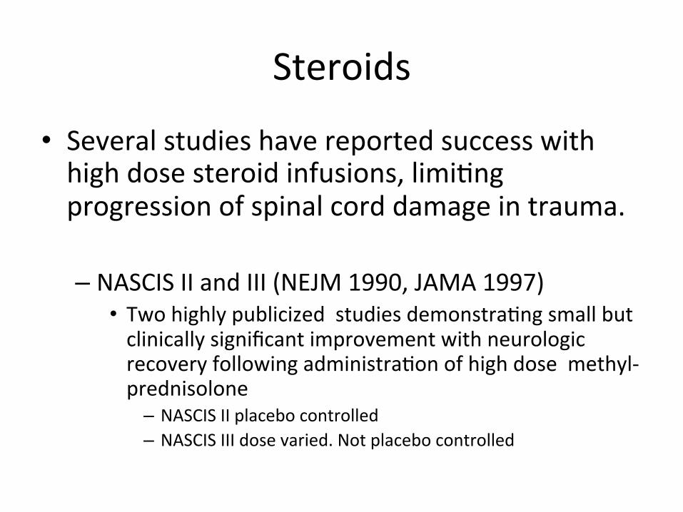
Steroids
• Several studies have reported success with high dose steroid infusions, limi3ng progression of spinal cord damage in trauma.
– NASCIS II and III (NEJM 1990, JAMA 1997) • Two highly publicized studies demonstra3ng small but clinically significant improvement with neurologic recovery following administra3on of high dose methyl-‐prednisolone
– NASCIS II placebo controlled – NASCIS III dose varied. Not placebo controlled
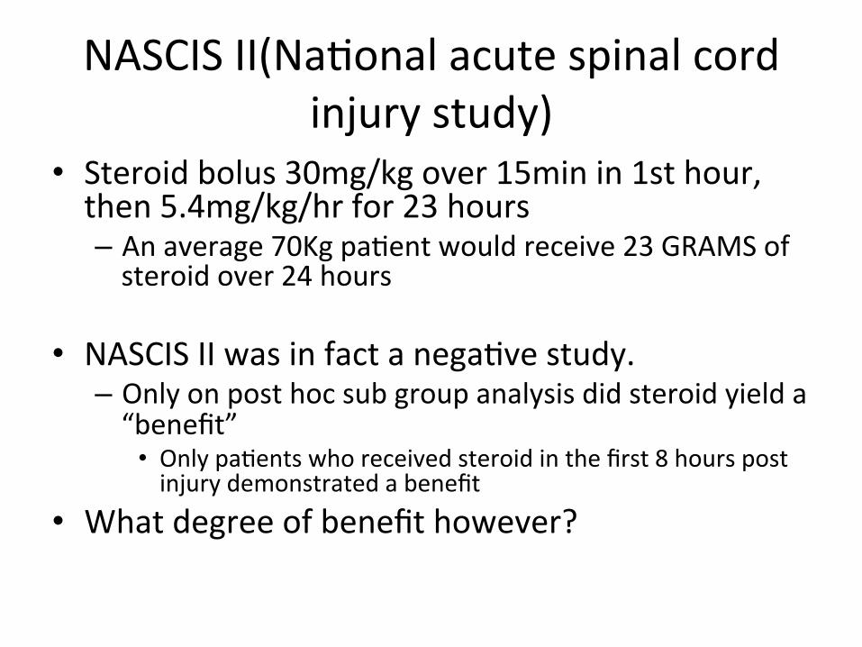
NASCIS II(Na3onal acute spinal cord injury study)
• Steroid bolus 30mg/kg over 15min in 1st hour, then 5.4mg/kg/hr for 23 hours – An average 70Kg pa3ent would receive 23 GRAMS of steroid over 24 hours
• NASCIS II was in fact a nega3ve study. – Only on post hoc sub group analysis did steroid yield a “benefit”
• Only pa3ents who received steroid in the first 8 hours post injury demonstrated a benefit
• What degree of benefit however?
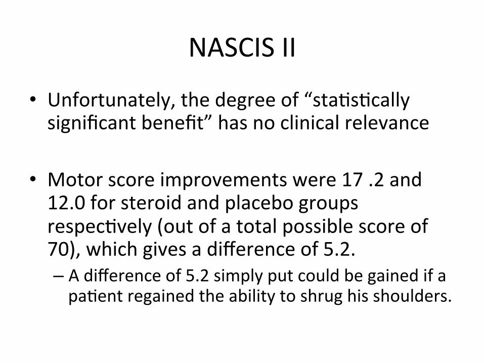
NASCIS II
• Unfortunately, the degree of “sta3s3cally significant benefit” has no clinical relevance
• Motor score improvements were 17 .2 and 12.0 for steroid and placebo groups respec3vely (out of a total possible score of 70), which gives a difference of 5.2. – A difference of 5.2 simply put could be gained if a pa3ent regained the ability to shrug his shoulders.

NASCIS III
MPSS 24 HR GROUP MPSS 48 HR GROUP TIRILAZAD GROUP
5.4 Mg/Kg/Hr. X 24 Hr. 5.4Mg/Kg/Hr. X 48Hr 2.5 Mg/Kg Bolus
6Hrly. X 48Hr
Improved motor fnxn
Same as MPSS 24 Hr. Group
Improved motor fnxn
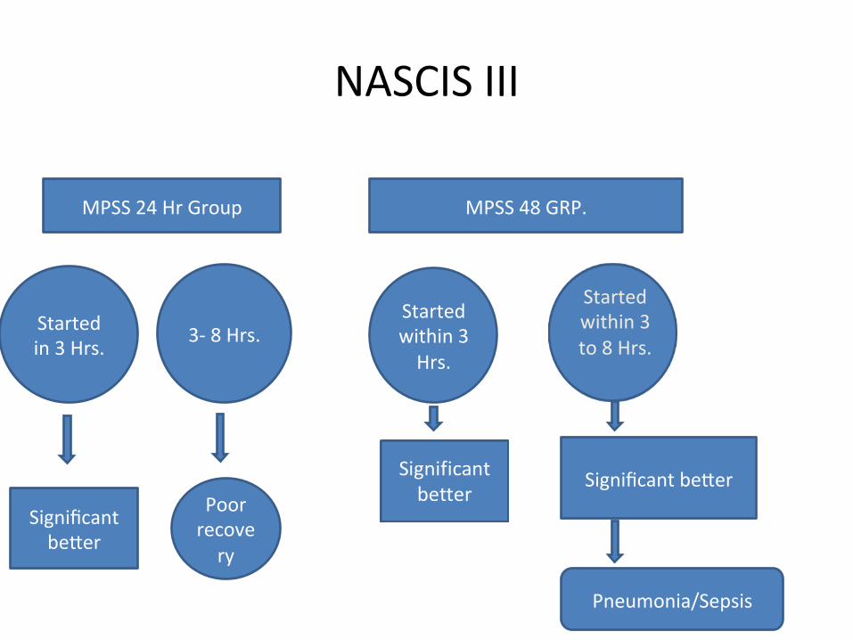
NASCIS III
MPSS 48 GRP.
Started within 3 Hrs.
Started within 3 to 8 Hrs.
MPSS 24 Hr Group
Started in 3 Hrs. 3-‐ 8 Hrs.
Poor recovery
Significant beWer
Significant beWer
Pneumonia/Sepsis

Results
• Ini3a3on of treatment within the first 3 hours is op3mal
• The nonglucocor3coid 3rilazad is as effec3ve as 24-‐hour MP therapy; and
• If treatment is ini3ated more than 3 hours post-‐SCI, extension of the MP dosing regimen is indicated, from 24 hours to 48 hours.
Results
• However, in comparison with the 24-‐hour dosing regimen, significantly more glucocor3oid-‐related immunosuppressive side effects were seen with more prolonged dosing
• In contrast, 3rilazad showed no evidence of steroid-‐related side effects, sugges3ng that this nonglucocor3coid 21-‐aminosteroid would be safer for extension of dosing beyond the 48-‐hour limit used in NASCIS III.
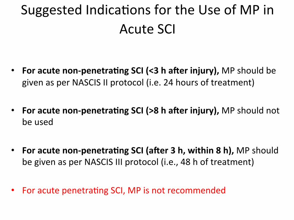
Suggested Indica3ons for the Use of MP in
Acute SCI
• For acute non-‐penetraGng SCI (<3 h ager injury), MP should be given as per NASCIS II protocol (i.e. 24 hours of treatment)
• For acute non-‐penetraGng SCI (>8 h ager injury), MP should not be used
• For acute non-‐penetraGng SCI (ager 3 h, within 8 h), MP should
be given as per NASCIS III protocol (i.e., 48 h of treatment) • For acute penetra3ng SCI, MP is not recommended
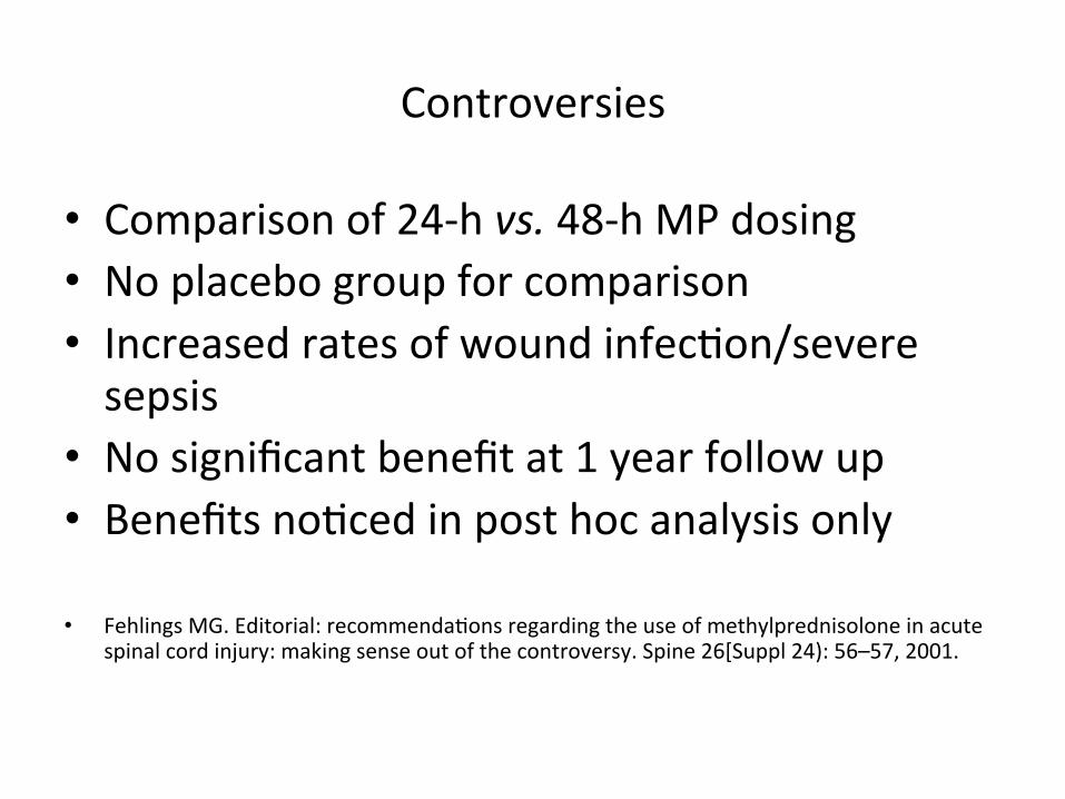
Controversies
• Comparison of 24-‐h vs. 48-‐h MP dosing • No placebo group for comparison • Increased rates of wound infec3on/severe sepsis
• No significant benefit at 1 year follow up • Benefits no3ced in post hoc analysis only
• Fehlings MG. Editorial: recommenda3ons regarding the use of methylprednisolone in acute spinal cord injury: making sense out of the controversy. Spine 26[Suppl 24): 56–57, 2001.
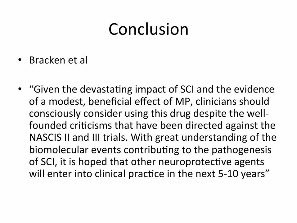
Conclusion • Bracken et al
• “Given the devasta3ng impact of SCI and the evidence of a modest, beneficial effect of MP, clinicians should consciously consider using this drug despite the well-‐founded cri3cisms that have been directed against the NASCIS II and III trials. With great understanding of the biomolecular events contribu3ng to the pathogenesis of SCI, it is hoped that other neuroprotec3ve agents will enter into clinical prac3ce in the next 5-‐10 years”

Important Papers
• NASCIS II – NEJM 1990 322:1405-‐11
• NASCIS III – JAMA 1997 277:1597-‐1604
• Revisi3ng NASCIS II & III – J. Trauma 1998 45:6 1088-‐93
• Methylprednisolone for acute spinal injury…. – J. Neurosurg (Spine 1) 2000:93:1-‐7

• Head injury
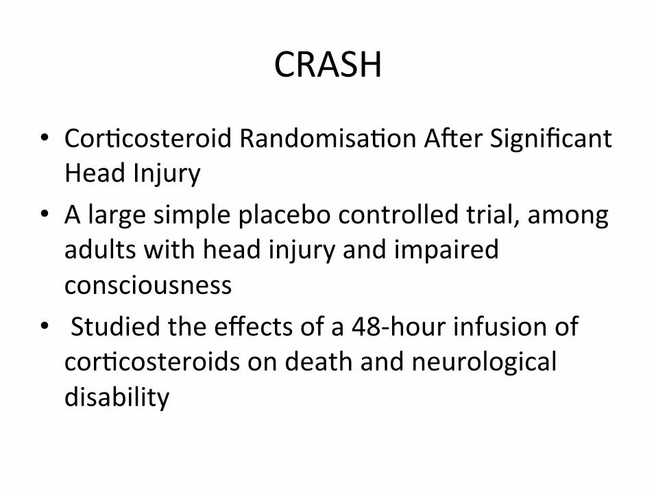
CRASH
• Cor3costeroid Randomisa3on Aver Significant Head Injury
• A large simple placebo controlled trial, among adults with head injury and impaired consciousness
• Studied the effects of a 48-‐hour infusion of cor3costeroids on death and neurological disability
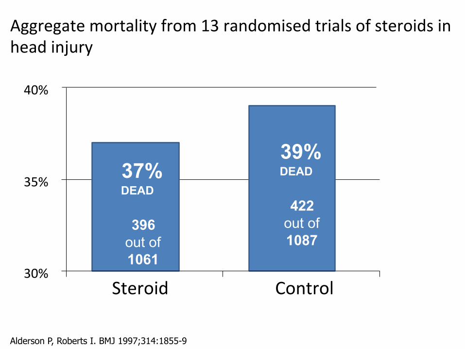
Aggregate mortality from 13 randomised trials of steroids in head injury
39% DEAD
422 out of 1087
37% DEAD
396 out of 1061
30%
35%
40%
Steroid Control
Alderson P, Roberts I. BMJ 1997;314:1855-9
Aim
• To determine reliably the effects of high dose cor3costeroid infusion on: – death and disability aver head injury
– risk of infec3on and gastrointes3nal bleeding • Primary outcomes
– Death within 2 weeks – Death or disability at 6 months
Results • 10,008 adults with head injury and a Glasgow Coma Scale score of 14 or less, within 8 h of injury
• 48-‐h infusion of cor3costeroid (methylprednisolone) or placebo.
• Data at 6 months were obtained for 9673 (96.7%) pa3ents.
• .

Results • The risk of death was higher in the cor3costeroid group than in the placebo group (1248 [25.7%] vs 1075 [22.3%] deaths; rela3ve risk 1.15, 95% CI 1.07-‐1.24; p=0.0001)
• There was no evidence that the effect of cor3costeroids differed by injury severity or 3me since injury.
• These results lend support to earlier conclusion that cor3costeroids should not be used rou3nely in the treatment of head injury

Discussion
• Landmark for head injury research • Also a red flag warning clinical trials • Planned to randomize 20,000 pa3ents • Powered to detect a drop in mortality from 15% to 13% (?? Only 2%)
• Document rela3vely small but important improvements in outcome

• CRASH data safety monitoring board asked that the trial be stopped aver 10,008 pa3ents from 239 hospitals in 49 countries had been randomized
WHY ??
• No evidence that MPSS improved survival • Mortality was greater in the MPSS treated pa3ents
• 1248 [25.7%] vs 1075 [22.3%] • Rela3ve risk 1.15, p=0.0001 • Risk could not be aWributed to injury
References • Final results of MRC CRASH, a randomized placebo-‐controlled trial of
intravenous cor3costeroid in adults with head injury—outcomes at 6 months. Lancet 365: 1957–1959, 2005
• Alderson P, Roberts I: Cor3costeroids for acute trauma3c brain injury. Cochrane Database Syst. Rev 2:CD000196, 2000.
• CRASH (CORTICOSTEROID RANDOMIZATION AFTER SIGNIFICANT HEAD INJURY TRIAL): LANDMARK AND STORM WARNING.Neurosurgery
57:1300-‐1302, 2005

• Brain tumors – Pituitary adenoma – Gliomas – Meningiomas
• Brain edema • Spinal pathologies • Vascular

• Pituitary adenoma
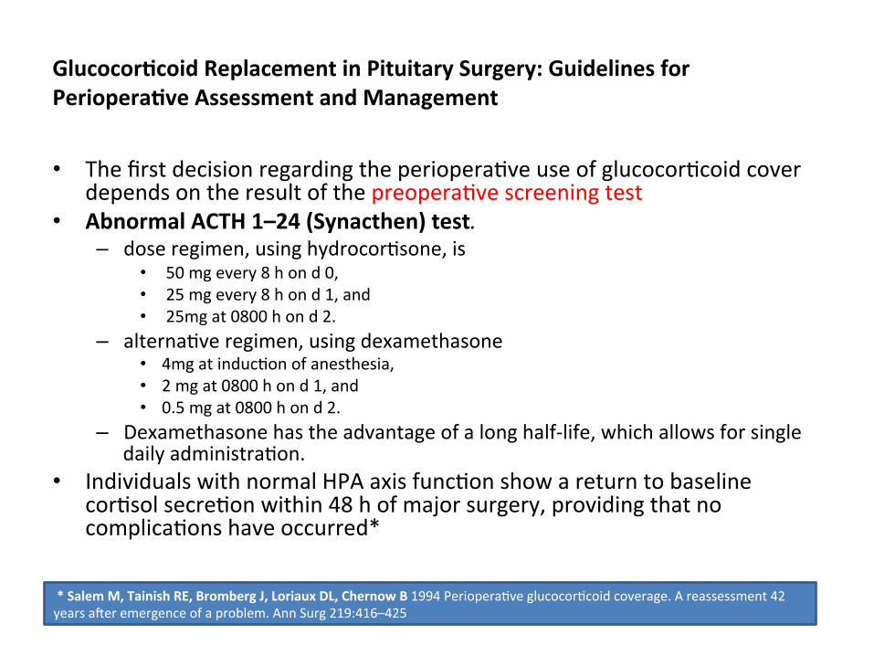
GlucocorGcoid Replacement in Pituitary Surgery: Guidelines for PerioperaGve Assessment and Management
• The first decision regarding the periopera3ve use of glucocor3coid cover depends on the result of the preopera3ve screening test
• Abnormal ACTH 1–24 (Synacthen) test. – dose regimen, using hydrocor3sone, is
• 50 mg every 8 h on d 0, • 25 mg every 8 h on d 1, and • 25mg at 0800 h on d 2.
– alterna3ve regimen, using dexamethasone • 4mg at induc3on of anesthesia, • 2 mg at 0800 h on d 1, and • 0.5 mg at 0800 h on d 2.
– Dexamethasone has the advantage of a long half-‐life, which allows for single daily administra3on.
• Individuals with normal HPA axis func3on show a return to baseline cor3sol secre3on within 48 h of major surgery, providing that no complica3ons have occurred*
* Salem M, Tainish RE, Bromberg J, Loriaux DL, Chernow B 1994 Periopera3ve glucocor3coid coverage. A reassessment 42 years aver emergence of a problem. Ann Surg 219:416–425
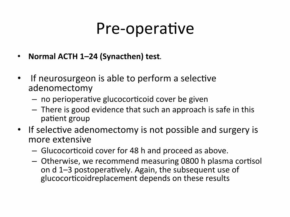
Pre-‐opera3ve • Normal ACTH 1–24 (Synacthen) test. • If neurosurgeon is able to perform a selec3ve
adenomectomy – no periopera3ve glucocor3coid cover be given – There is good evidence that such an approach is safe in this pa3ent group
• If selec3ve adenomectomy is not possible and surgery is more extensive – Glucocor3coid cover for 48 h and proceed as above. – Otherwise, we recommend measuring 0800 h plasma cor3sol on d 1–3 postopera3vely. Again, the subsequent use of glucocor3coidreplacement depends on these results

Post-‐opera3ve • Many pa3ents show a rapid increase in pituitary hormone secre3on
aver pituitary adenomectomy
• Result of increased flow of hypothalamic releasing hormones through the hypothalamic-‐hypophysial portal system
• A pa3ent with hypopituitarism may gain full func3onal recovery, providing that viable normal pituitary 3ssue remains in situ.
• Prolonged treatment with high doses of glucocor3coids postopera3vely may result in adrenal suppression and mask those who spontaneously recover func3on.
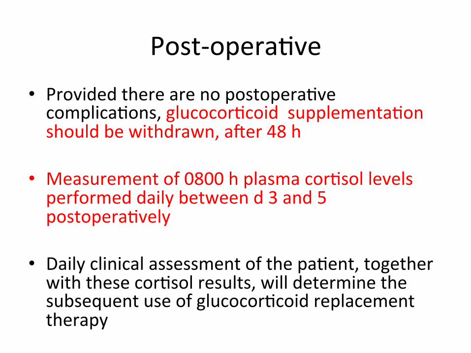
Post-‐opera3ve • Provided there are no postopera3ve complica3ons, glucocor3coid supplementa3on should be withdrawn, aver 48 h
• Measurement of 0800 h plasma cor3sol levels performed daily between d 3 and 5 postopera3vely
• Daily clinical assessment of the pa3ent, together with these cor3sol results, will determine the subsequent use of glucocor3coid replacement therapy
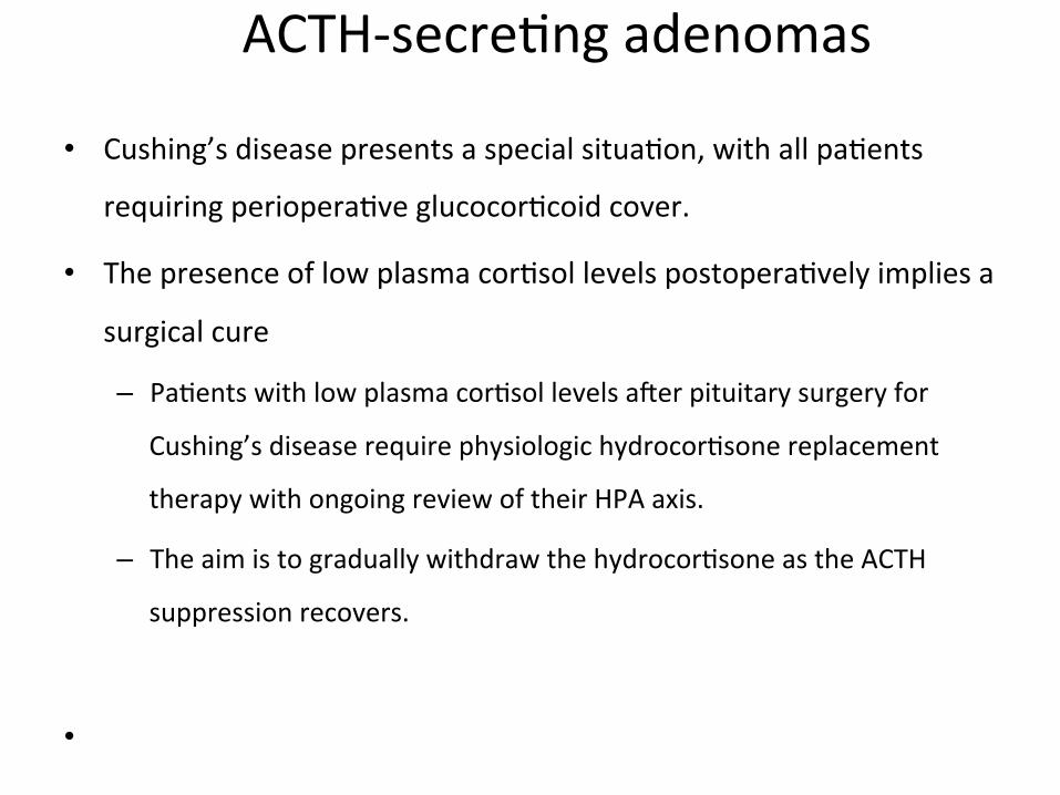
ACTH-‐secre3ng adenomas
• Cushing’s disease presents a special situa3on, with all pa3ents
requiring periopera3ve glucocor3coid cover.
• The presence of low plasma cor3sol levels postopera3vely implies a
surgical cure
– Pa3ents with low plasma cor3sol levels aver pituitary surgery for
Cushing’s disease require physiologic hydrocor3sone replacement
therapy with ongoing review of their HPA axis.
– The aim is to gradually withdraw the hydrocor3sone as the ACTH
suppression recovers.
•
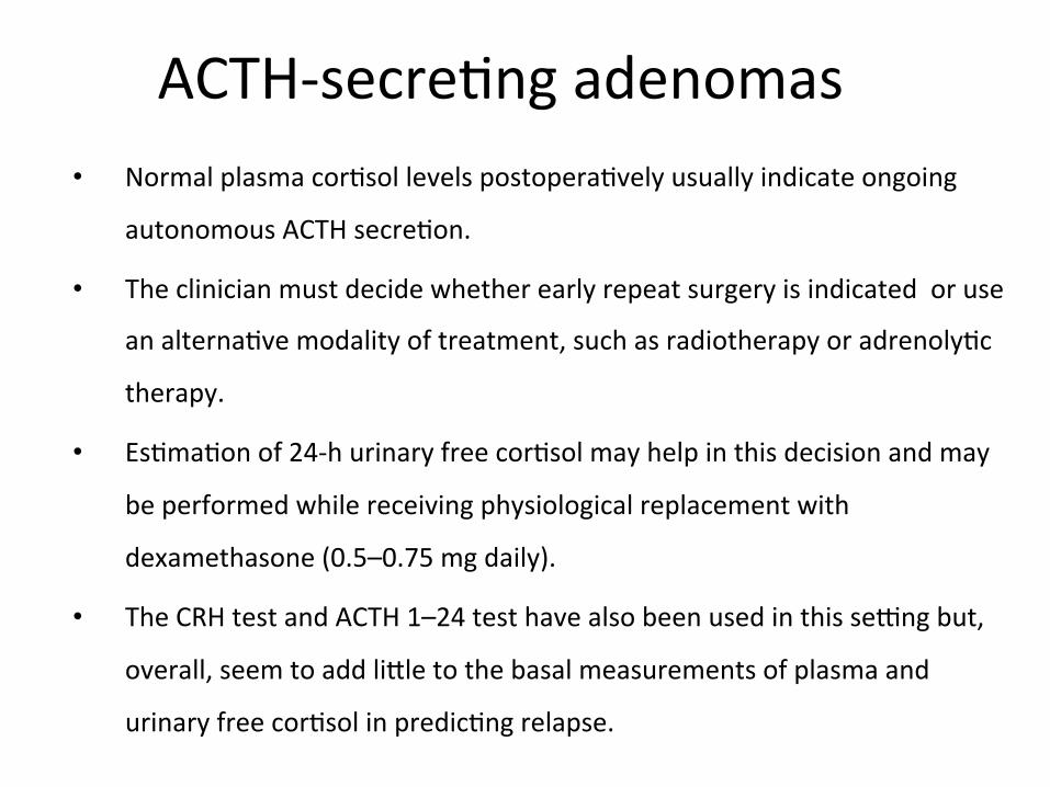
ACTH-‐secre3ng adenomas • Normal plasma cor3sol levels postopera3vely usually indicate ongoing
autonomous ACTH secre3on.
• The clinician must decide whether early repeat surgery is indicated or use
an alterna3ve modality of treatment, such as radiotherapy or adrenoly3c
therapy.
• Es3ma3on of 24-‐h urinary free cor3sol may help in this decision and may
be performed while receiving physiological replacement with
dexamethasone (0.5–0.75 mg daily).
• The CRH test and ACTH 1–24 test have also been used in this se~ng but,
overall, seem to add liWle to the basal measurements of plasma and
urinary free cor3sol in predic3ng relapse.

Early postopera3ve assessment
• Based on the levels of 0800 h plasma cor3sol in the early postopera3ve period
• These measurements should be made on – d 1–3 in pa3ents not treated with glucocor3coids and
– d 3–5 in pa3ents covered with glucocor3coids for the ini3al 48 h
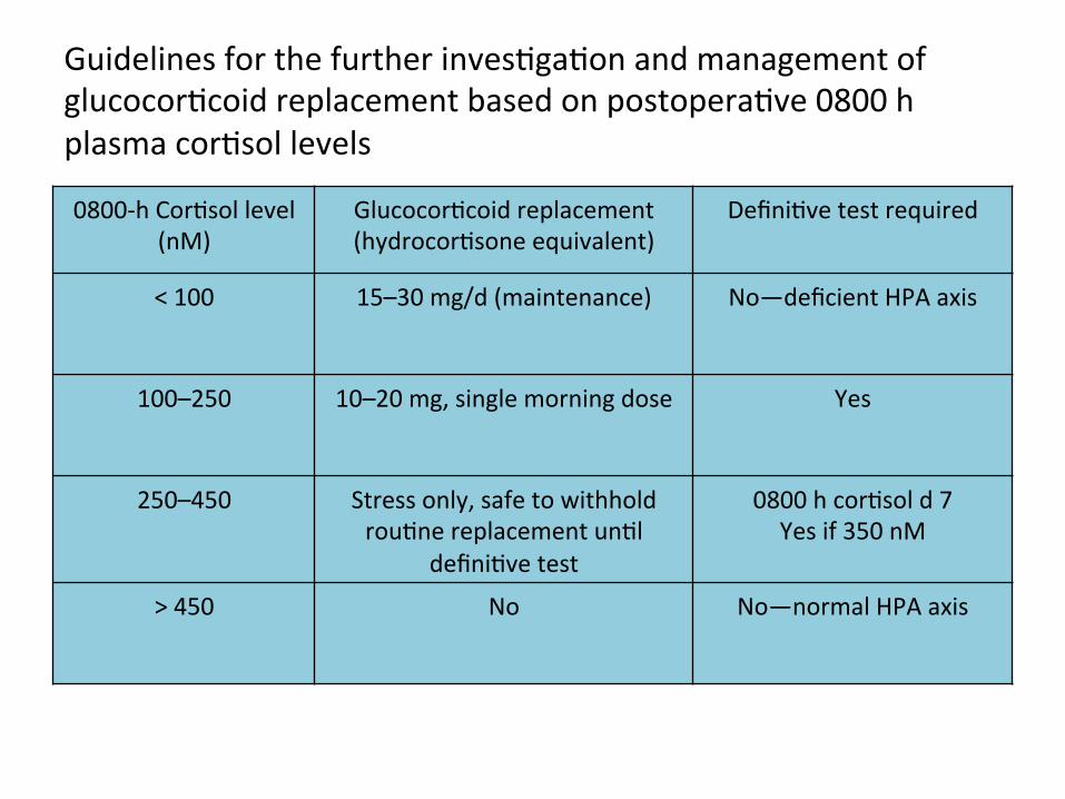
Guidelines for the further inves3ga3on and management of glucocor3coid replacement based on postopera3ve 0800 h plasma cor3sol levels
0800-‐h Cor3sol level (nM)
Glucocor3coid replacement (hydrocor3sone equivalent)
Defini3ve test required
< 100 15–30 mg/d (maintenance) No—deficient HPA axis
100–250 10–20 mg, single morning dose Yes
250–450 Stress only, safe to withhold rou3ne replacement un3l
defini3ve test
0800 h cor3sol d 7 Yes if 350 nM
> 450 No No—normal HPA axis
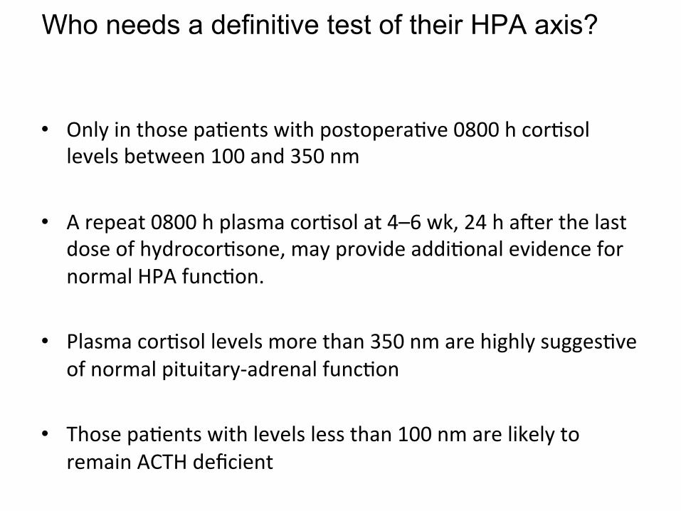
Who needs a definitive test of their HPA axis?
• Only in those pa3ents with postopera3ve 0800 h cor3sol levels between 100 and 350 nm
• A repeat 0800 h plasma cor3sol at 4–6 wk, 24 h aver the last
dose of hydrocor3sone, may provide addi3onal evidence for normal HPA func3on.
• Plasma cor3sol levels more than 350 nm are highly sugges3ve
of normal pituitary-‐adrenal func3on
• Those pa3ents with levels less than 100 nm are likely to remain ACTH deficient
Who needs a defini3ve test of their HPA axis?
• There are cases where late recovery of the HPAaxis is documented
• A repeat 0800h cor3sol, 24 h aver the last dose of
hydrocor3sone, performed between 4 and 6 wk postopera3vely, should iden3fy such pa3ents.
• Those s3ll less than 100 nm require lifelong replacement
therapy, whereas those whose cor3sol levels have risen to more than 100 nm should undergo a defini3ve test and replacement
Pituitary apoplexy • Steroid therapy in pituitary apoplexy • Pa3ents with pituitary apoplexy, who are haemodynamically unstable • Empirical steroid therapy.
• In adults hydrocor3sone 100-‐200mg as an intravenous bolus is appropriate, followed by
• 2-‐4mg per hour by con3nuous intravenous infusion or 50-‐100mg six hourly by intramuscular injec3on, aver drawing blood samples for random cor3sol, FT4, TSH, PRL, IGF1, GH, LH, FSH, testosterone in men, oestradiol in women, electrolytes, renal func3on, liver func3on, full blood count and clo~ng screen;
• Indica3ons for empirical steroid therapy in pa3ents with pituitary apoplexy are – haemodynamic instability, – Altered consciousness level, – reduced visual acuity and – severe visual field defects
• Pa3ents who do not fulfil the criteria for urgent empirical steroid therapy should be considered for treatment with steroids, if their 09.00 serum cor3sol is less than 550nmol/l;
Orders in neurosurgery
• Post-‐op
• Inj phenytoin • Inj clox • Inj amikacin • Inj wymesone • Inj rantac • Inj ketonov
• Pre-‐op
• T eptoin • T decdac • T rantac • C b/c
History of use of steroid in tumor
• Steroids were introduced into the care of brain cancer pa3ents nearly 50 years ago based on their powerful effects on tumor-‐induced edema
• Ingraham pioneered the use of cor3sone to treat postopera3ve
cerebral edema in neurosurgical pa3ents in 1952 • Kofman first used prednisone for peritumoral edema from brain
metastases in 1957
• More than 40 years ago, Galicich demonstrated that dexamethasone effec3vely alleviated cerebral edema due to brain tumors
• Dexamethasone, which was first synthesized in 1958, to date has remained the most favorable drug for brain cancer pa3ents.
• Dexamethasone has become the drug of choice in neuro-‐
oncology, in part owing to its long half-‐life, low mineralocor3coid ac3vity, and a rela3vely low tendency to induce psychosis.
• Subsequently, prednisone, prednisolone, dexamethasone and methylprednisolone have demonstrated an3neoplas3c effects against malignancies

Molecular Mechanisms
• At transcrip3onal levels, glucocor3coids suppress synthesis of several cytokines and chemokines, such as GM CSF; IL-‐1β, -‐4, -‐5, -‐8 and -‐10; as well as eotaxin and lipocorCn 1, which are involved in the regula3on of the inflammatory reac3on.
• Interact with other transcrip3on factors such as NF-‐κB, ac3va3ng protein 1, p53, CRE-‐binding protein, signal transducer & the STAT3, STAT5 and others, indirectly influencing the ac3vity of the laWer on their own target genes
• NF-‐κB ac3vates many of the inflammatory pathways through its regula3on of the produc3on of proinflammatory cytokines and chemokines. Inhibi3on of NF-‐κB results in an an3-‐inflammatory effect
Disrup3on of the BBB
• Trauma • Inflammatory and autoimmune disease, • Infec3on • Cerebrovascular disease • Neurodegenera3ve disease • Epilepsy, and • Neoplasia
Disrup3on of the BBB
• Klatzo first characterized brain edema as a cytotoxic versus a vasogenic process
• BBB remains intact in cytotoxic edema
• Cor3costeroid therapy aver brain tumor surgery helps re-‐establish the BBB integrity.
• Cairncross JG, Macdonald DR, Pexman JH, et al: Steroid-‐induced CT changes in pa3ents with recurrent malignant glioma. Neurology 1988; 38:724-‐726
Cerebral edema
1. Vasogenic edema 2. Cytotoxic edema 3. Inters33al edema
Steroids • Glucocor3coids primarily reduce vascular permeability of
vessels rather than reduced VEGF produc3on
• This inhibi3on of the effects of VPF/VEGF on the vasculature is associated with interference of VEGF's ac3on on vessels and requires the glucocor3coid receptor
• Thus, the effects of steroids in tumor-‐induced vasogenic edema is to restrict permeability of the BBB to macromolecules
• By contrast, steroids are not effec3ve when the BBB is not func3onal
Heiss JD, Papavassiliou E, Merrill MJ, et al: Mechanism of dexamethasone suppression of brain tumor-‐associated vascular permeability in rats. Involvement of the glucocor3coid receptor and vascular permeability factor. J Clin Invest 1996; 98:1400-‐1408. . Merrill MJ, Oldfield EH: A reassessment of vascular endothelial growth factor in central nervous system pathology. J Neurosurg 2005; 103:853-‐868.
Cranial surgery: Brain tumor • Several days before surgery when there is symptoma3c brain edema or mass effect
• Typical dose is 16 mg/day or 4 mg four 3mes a day • In brain edema, there appears to be a dose threshold that must be surpassed before symptoma3c benefit is derived
• Therefore, dexamethasone should be started at a high dose in pa3ents with large amounts of brain edema and then reduced aver neurological improvement has occurred.
Pre-‐op
• Pa3ents respond within 24 hours of beginning steroid treatment
• Decrease tumor capillary permeability and tumor blood volume
• In pa3ents undergoing more elec3ve surgery, such as those with schwannomas and meningiomas, may be given steroids beginning 3 days before surgery
• However, if there is only minimal edema, steroids are typically first given at 3me of surgery
Post-‐op • Pa3ents at risk for the development of significant postopera3ve edema are: – deep intrinsic tumors in which only minimal resec3on was possible
– infiltra3ng tumors involving a large amount of white maWer, and
– extensive edema before surgery. – progressive cerebral edema (can also be treatment related)
• Maximal postopera3ve swelling occurs between days 1 and 5 aver surgery
Post -‐op
• Regardless of the cause of the cerebral edema, management is similar
• These pa3ents should receive maximal medical treatment of the edema and elevated ICP
• Steroids should be given at maximal doses (20 mg every 4 hours).
Yamada K, Ushio Y, Hayakawa T, et al: Effects of methylprednisolone on peritumoral brain edema. J Neurosurg 1983; 59:612-‐619. Leenders KL, Beaney RP, Brooks DJ, et al: Dexamethasone treatment of brain tumor pa3ents: effects on regional cerebral blood flow, blood volume, and oxygen u3liza3on. Neurology 1985; 35:1610-‐1616.
Metastasis • Believe to exert their effect by reducing cerebral edema surrounding
tumor,and to lesser extent there may be direct oncoly3c effect on tumor cells
• Ameliorate sign and symptom in 2/3rd of pa3ents • Benefit evident within 24 hrs.
• Minimum effec3ve dose is not known but in prac3ce loading dose equivalent to 10 mg of dexamethasone intravenously, followed by 16 mg orally/IM (divided in 3-‐4 doses)
• Steroids tends to stabilise pa3ent,aver which more defini3ve treatment (surgery/radiotherapy) may be undertaken. May be con3nued through ini3al phase and tapered and finally discon3nued aver 1-‐2 weeks.
• • Relief is only temporary.pa3ent tends to relapse despite con3nued
steroid administra3on
Brain metastasis
• All pa3ents should receive steroids. • Rapid improvement within 6-‐24 hours. • Maximum effect by 3-‐7 days. • Benefit is temporary.
Dose of Steroids
• Most common dose dexamethasone 4 mg qid. • Doses as low as 4 mg qd if not symptoma3c. • Up to 100 mg per day if symptoms are progressing.
• Taper as tolerated.
Brain abscess
• Concurrent treatment with cor3costeroids, radia3on therapy, and chemotherapy may alter the radiographic progression of abscess development
• Cor3costeroids have been shown to reduce the thickness of the abscess capsule and the extent of contrast enhancement on both CT and MRI
• Cavusoglu H, Kaya RA, Turkmenoglu ON, et al: Brain abscess: analysis of results in a series of 51 pa3ents with a combined surgical and medical approach during an 11-‐year period. Neurosurg Focus 2008; 24(6):E9
Spinal Indica3ons
• Control of pain in low back ache :Epidural injec3on in lumbar pain
• Epidural metastasis (acute phase) • Neurological deteriora3on aver implant migra3on and /or compression
• Rheumatoid arthri3s
Miscellenous
• Carpal syndrome • Bell’s palsy • Cranial n. damage • Chronic SDH • Dexamethasone suppression test • Neurodegenera3ve ds. • Meningi3s
Meningi3s
• Reported in the 1950s
• Individuals with bacterial meningi3s treated with an3bio3cs plus steroids within 12-‐24 h of admission had beWer outcomes
• Interes3ngly, these benefits were not observed if steroid treatment was delayed un3l 5 days aver admission.
• Bacterial lysis induced by an3bio3cs causes inflamma3on in the subarachnoid space, and this response is aWenuated by steroid treatment, thereby improving the outcome
• Hoh TK and Mong CT (1962) The treatment of purulent meningi3s with adrenal cor3co-‐steroids. SingaporeMed J 3: 73-‐77

Current Guidelines and RecommendaGons
• Should be administered just before or in conjunc3on with an3bio3c therapy.*
• Dexamethasone (10 mg) should be administered intravenously every 6 h for 4 days in adults
• At present, children in low-‐income countries do not seem to derive appreciable benefit from steroid use (??)
• In most childhood studies, a 4-‐day regimen of dexamethasone(0.4 or 0.6 mg/kg per day) divided into four daily doses was used.
• We suggest that clinicians consider administering treatment with
this 4-‐day regimen, as most published studies have used this dura3on of treatment without observing increased adverse effects rela3ve to the shorter dura3ons.
*van de Beek D and de Gans J (2006) Dexamethasone in adults with community-‐acquired bacterial meninigi3s. Drugs 66: 415-‐427
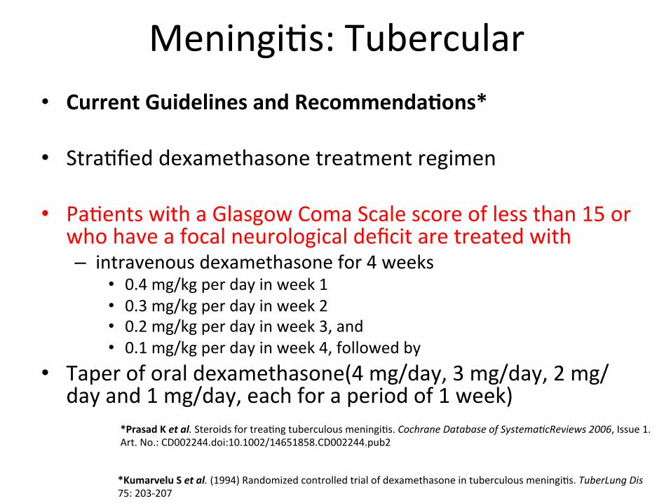
Meningi3s: Tubercular • Current Guidelines and RecommendaGons*
• Stra3fied dexamethasone treatment regimen • Pa3ents with a Glasgow Coma Scale score of less than 15 or
who have a focal neurological deficit are treated with – intravenous dexamethasone for 4 weeks
• 0.4 mg/kg per day in week 1 • 0.3 mg/kg per day in week 2 • 0.2 mg/kg per day in week 3, and • 0.1 mg/kg per day in week 4, followed by
• Taper of oral dexamethasone(4 mg/day, 3 mg/day, 2 mg/day and 1 mg/day, each for a period of 1 week)
*Prasad K et al. Steroids for trea3ng tuberculous meningi3s. Cochrane Database of SystemaCcReviews 2006, Issue 1. Art. No.: CD002244.doi:10.1002/14651858.CD002244.pub2
*Kumarvelu S et al. (1994) Randomized controlled trial of dexamethasone in tuberculous meningi3s. TuberLung Dis 75: 203-‐207

Meningi3s: Tubercular
• Pa3ents with a normal mental status and no neuro logical findings receive – intravenous dexamethasone for 2 weeks
• 0.2 mg/kg per day in week 1, then • 0.1 mg/kg per day in week 2, followed by
• The same oral taper as described above. • steroid treatment should start as soon as possible aver ini3a3on of appropriate first-‐line an3 tuberculosis drugs.
Conclusion
• Cor3costeroids have varied roles in the pathologies of the brain and spine especially in neuro-‐oncology. However need further evalua3on w.r.t.role in trauma

Thank you