Embed Size (px)
Citation preview

Endurant™ II
Endurant™ IIs Stent Graft System
Instructions for Use

© 2017 Medtronic. All rights reserved. Medtronic and Medtronic logo are trademarks of Medtronic. ™* Third party brands are trademarks of their respective owners. All other brands are trademarks of a Medtronic company.

i
EXPLANATION OF SYMBOLS ON PRODUCT LABELING
Refer to the device labeling to see which symbols apply to this product.
Contents: One Device
Do not use if package is damaged
Non-pyrogenic
Peel here
MR Conditional
CAUTION: Federal (USA) law restricts this device for sale by or on order of a physician.
Sterilized using irradiation
Catalogue number
Serial number
Use by
Do not reuse
Manufacturer
Manufactured In
Consult instructions for use at: www.medtronic.com/manuals

ii
Table of Contents 1
1.0 Device Description ........................................................................................................... 3 1.1 Stent Graft ........................................................................................................................ 3
1.1.1 Bifurcated Configuration .............................................................................................. 5 1.1.2 Aorto-Uni-Iliac (AUI) Configuration ............................................................................ 5 1.1.3 Limb Configuration ...................................................................................................... 6 1.1.4 Iliac Extension Configuration ....................................................................................... 6 1.1.5 Aortic Extension Configuration .................................................................................... 6
1.2 Delivery System ................................................................................................................ 7 1.3 Heli-FX™ EndoAnchor™ System ................................................................................... 8
2.0 Indications for Use ............................................................................................................ 9 3.0 Contraindications ............................................................................................................ 10 4.0 Warnings and Precautions ............................................................................................... 11
4.1 General............................................................................................................................ 11 4.2 Patient Selection ............................................................................................................. 11 4.3 Before the Implant Procedure ......................................................................................... 14 4.4 During the Implant Procedure ........................................................................................ 14 4.5 Treatment and Follow-up ................................................................................................ 16 4.6 MRI Safety Information ................................................................................................. 16
5.0 Adverse Events ............................................................................................................... 17 5.1 Observed Adverse Events ............................................................................................... 17 5.2 Potential Adverse Events ................................................................................................ 17 5.3 Device-Related Adverse Events Reporting .................................................................... 18
6.0 Summary of Clinical Studies .......................................................................................... 19 6.1 Bifurcated Study Arm ..................................................................................................... 21
6.1.1 Subject Accountability and Follow-up ....................................................................... 21 6.1.2 Study Demographics and Baseline Medical History .................................................. 23 6.1.3 Baseline Aneurysm Characteristics ............................................................................ 24 6.1.4 Devices Implanted ...................................................................................................... 26 6.1.5 Study Results: Safety Endpoints................................................................................. 27 6.1.6 Study Results: Effectiveness Endpoints ..................................................................... 33
6.2 AUI Study Arm .............................................................................................................. 38 6.2.1 Subject Accountability and Follow-up ....................................................................... 38 6.2.2 Study Demographics and Baseline Medical History .................................................. 39 6.2.3 Baseline Aneurysm Characteristics ............................................................................ 42 6.2.4 Devices Implanted ...................................................................................................... 44 6.2.5 Study Results: Safety Endpoints................................................................................. 46 6.2.6 Study Results: Effectiveness Endpoints ..................................................................... 53
6.3 ANCHOR Registry Short Neck Cohort.......................................................................... 57 6.3.1 Subject Accountability and Follow-up ....................................................................... 57 6.3.2 Study Demographics and Baseline Medical History .................................................. 58 6.3.3 Baseline Aneurysm Characteristics ............................................................................ 60 6.3.4 Devices Implanted ...................................................................................................... 62 6.3.5 Acute Procedural Data ................................................................................................ 64 6.3.6 Study Results: Safety Outcomes ................................................................................. 66

iii
6.3.7 Study Results: Effectiveness Outcomes ..................................................................... 73 6.3.8 Data Post 12 Months................................................................................................... 80
7.0 Patient Selection and Treatment ...................................................................................... 81 7.1 Individualization of Treatment ....................................................................................... 81
8.0 Patient Counseling Information ...................................................................................... 83 9.0 How Supplied.................................................................................................................. 84
9.1 Sterility ........................................................................................................................... 84 9.2 Contents .......................................................................................................................... 84 9.3 Storage ............................................................................................................................ 84
10.0 Clinical Use Information ................................................................................................ 85 10.1 Physician Training Requirements ................................................................................... 85 10.2 Recommended Device Sizing ......................................................................................... 85
10.2.1 Endurant II/IIs stent graft system ............................................................................... 85 10.3 Device Inspection ........................................................................................................... 87 10.4 Additional Required Equipment ..................................................................................... 88 10.5 Additional Recommended Equipment ........................................................................... 88 10.6 MRI Information............................................................................................................. 88
11.0 Implant Instructions ........................................................................................................ 90 11.1 Vascular Access and Device Preparation ....................................................................... 90
11.1.1 Vascular Access .......................................................................................................... 90 11.1.2 Device Preparation ...................................................................................................... 90
11.2 Delivery Procedure ......................................................................................................... 90 11.2.1 Introduction of Main Body Configuration .................................................................. 91 11.2.2 Confirm Position (Endurant II and Endurant IIs Bifurcated Configurations Only) .... 92 11.2.3 Deploy Proximal End of the Stent Graft Configuration .............................................. 92 11.2.4 Deploy Contralateral Leg (Endurant II and Endurant IIs Bifurcated Configurations
Only) ........................................................................................................................... 93 11.2.5 Deploy Covered Portion (AUI Configuration Only) ................................................... 93 11.2.6 Release Proximal End of Suprarenal Stent .................................................................. 94 11.2.7 Deploy Distal End of the Stent Graft Configuration (Endurant II and Endurant IIs
Bifurcated Configurations Only) ................................................................................ 96 11.2.8 Recapture Spindle in Tapered Tip (Endurant II Bifurcated and AUI Configurations
Only) ........................................................................................................................... 96 11.2.9 Remove Delivery System (Endurant II Bifurcated and AUI Configurations Only) ... 97 11.2.10 Deploy Limb Stent Graft Configuration Into Contralateral Leg (Endurant II and
Endurant IIs Bifurcated Configurations Only) ............................................................ 98 11.2.11 Deploy Limb Stent Graft Configuration (AUI Configuration Only) ........................... 99 11.2.12 Recapture Spindle in Tapered Tip for Delivery System in the Ipsilateral Leg
(Endurant IIs Bifurcated Configuration Only) .......................................................... 101 11.2.13 Remove Delivery System (Endurant IIs Bifurcated Configuration Only) ................ 102 11.2.14 Deploy Limb Stent Graft Into Ipsilateral Leg (Endurant IIs Bifurcated Configuration
Only) ......................................................................................................................... 103 11.2.15 Iliac or Aortic Extension Stent Graft Configurations ................................................ 106 11.2.16 Smoothing Stent Graft Fabric and Modeling Stent Graft .......................................... 106 11.2.17 Additional fixation and sealing with the Heli-FX EndoAnchor system (infrarenal
neck > 4 mm and < 10 mm in length) ...................................................................... 107 11.2.18 Verify Placement and Seal ........................................................................................ 107

iv
11.2.19 Seal Entry Sites ......................................................................................................... 108 12.0 Bail-Out Techniques ...................................................................................................... 109
12.1 Screw Gear Handle Disassembly ................................................................................. 109 12.2 Ballooning .................................................................................................................... 109 12.3 Back-End Handle Disassembly .................................................................................... 109 12.4 Snare the Tapered Tip ................................................................................................... 110
13.0 Follow-up Imaging Recommendations ......................................................................... 111 13.1 General.......................................................................................................................... 111 13.2 X-ray ............................................................................................................................. 111 13.3 CT with Contrast ........................................................................................................... 111 13.4 Noncontrast CT ............................................................................................................. 112 13.5 Duplex Ultrasound ........................................................................................................ 112 13.6 MRI or MRA ................................................................................................................ 112 13.7 Imaging Tests ................................................................................................................ 112 13.8 Supplemental Imaging .................................................................................................. 113
14.0 Additional Surveillance and Treatment ......................................................................... 114 15.0 Device Registration....................................................................................................... 115 16.0 Disclaimer of Warranty ................................................................................................. 116

3
1.0 Device Description
The Endurant™ II/Endurant™ IIs stent graft system (hereinafter referred to as Endurant II/IIs stent graft system) is to treat infrarenal abdominal aortic or aortoiliac aneurysms using an endovascular approach. When placed within the aneurysm, the stent graft provides an alternative conduit for blood flow within the patient’s vasculature.
The stent graft system is comprised of 2 main components: the implantable stent graft and the disposable delivery system. The stent graft is preloaded into the delivery system and advanced to the aneurysm using fluoroscopic guidance. Upon deployment, the stent graft self-expands to conform to the shape and size of the seal zones above and below the aneurysm.
The Endurant II/IIs stent graft can also be used with the Heli-FX™ EndoAnchor™ system (available separately). The Heli-FX EndoAnchor system is designed to provide fixation and augment sealing between the Endurant II/IIs stent graft and the native artery. The system consists of an EndoAnchor™ implant that is delivered using the Heli-FX™ applier through the steerable Heli-FX™ guide. 1.1 Stent Graft There are 2 main body stent graft configurations: a bifurcated main body stent graft and an aorto-uni-iliac (AUI) main body stent graft. First, a bifurcated device is implanted into the patient’s aorta. If a bifurcated device cannot be implanted, an AUI device can be used. After placement of the bifurcated or AUI device, limb stent graft configurations are introduced separately into the vessel and mated with the implanted main body stent graft configuration. Depending on the patient’s anatomy, a limb configuration may not be required in the AUI configuration. If additional distal or proximal coverage is needed, an iliac or aortic extension configuration is introduced separately into the vessel and mated with the implanted main body stent graft configuration.
All configurations are composed of nitinol stents sewn to a fabric graft with nonresorbable sutures. Radiopaque markers are sewn onto the stent graft to aid in visualization and to facilitate accurate placement. The nitinol stents may also be visible under fluoroscopy.
Stent grafts should be oversized to be larger than the measured vessel inner diameter (aortic components are oversized approximately 10–20%; limb components are oversized approximately 10–25%). Recommended Device Sizing (Section 10.2) contains detailed sizing information for all stent graft components, including available ranges of length and diameter. Table 1 contains a summary of the stent graft materials.

4
Figure 1: Stent Graft Configurations and Locations of RO Markers
1. Radiopaque Marker 6. Endurant IIs Bifurcated Configuration
2. ‘e’ Marker 7. Endurant II Iliac Extension Configuration
3. Radiopaque Gate Marker 8. Endurant II Limb Configuration
4. Endurant II Aortic Extension Configuration 9. Endurant II Aorto-Uni-Iliac Configuration
5. Endurant II Bifurcated Configuration 10. Overlap Marker
Note: This and all other product graphics appearing in this manual are not drawn to scale.
Table 1: Stent Graft Materials
Component Material
Stents Nickel-Titanium (Nitinol) Alloy Button Radiopaque Markers Platinum-Iridium Alloy “e” Radiopaque Marker Platinum Contralateral Gate Marker Platinum-Iridium Alloy Graft Material Polyester Suture Polyester and Polyethylene

5
The Endurant II/IIs stent graft system does not contain natural rubber latex. However, during the manufacturing/assembly process, it may have incidental contact with latex-containing products. 1.1.1 Bifurcated Configuration The bifurcated stent graft is available in 2 configurations: the Endurant II bifurcated configuration and the Endurant IIs bifurcated configuration. The Endurant II bifurcated configuration is an aortoiliac stent graft that is available in 3 lengths. The Endurant IIs bifurcated configuration is an aortic configuration available in a single, shorter length (Figure 1). The proximal end of both bifurcated configurations deploy into the proximal neck and upper section of the aneurysm. The proximal end of the bifurcated configuration is composed of nitinol stents sewn to a fabric graft. The suprarenal portion of the proximal end is not covered with graft fabric (Figure 1). The suprarenal stent also contains anchor pins to fix the stent graft in place inside the aorta above the renal arteries without obstructing them with graft fabric. The diameters of the available proximal aortic section of the bifurcated stent graft configurations range from 23 mm to 36 mm, and the covered length of the bifurcated stent graft configurations range from 124 mm to 166 mm (Endurant II bifurcated stent graft) or 103 mm (Endurant IIs bifurcated stent graft). The aortic sections of the bifurcated stent graft configurations should be oversized 10% to 20% in relation to the actual measured inner vessel diameter. The available sizes can be used in aortas with diameters ranging from 19 mm to 32 mm.
The aortic section bifurcates distally into 2 smaller tubes: an ipsilateral leg and a shorter contralateral leg. In the Endurant II bifurcated configuration, all stents on the ipsilateral leg are sewn to the outside of the fabric creating a smooth inner lumen. In the Endurant IIs bifurcated configuration, the 4 distal stents are sewn to the inside of the ipsilateral leg graft fabric. For all sizes, the stents on the contralateral leg are sewn to the inside of the graft fabric (Figure 1). The Endurant II bifurcated stent graft configurations have an ipsilateral leg that range in diameters from 13 mm to 20 mm and may be used in iliac arteries with diameters ranging from 10 mm to 18 mm. The Endurant II bifurcated stent graft configurations have a contralateral leg that range in diameters from 12 mm to 14 mm. The Endurant IIs bifurcated stent graft ipsilateral and contralateral legs are available in 14 mm diameter and are not intended to extend to the iliac arteries.
Note: The bifurcated configurations are always used with the limb configuration (Section 1.1.3). 1.1.2 Aorto-Uni-Iliac (AUI) Configuration The AUI device is indicated for the endovascular treatment of infrarenal abdominal aortic or aortoiliac aneurysms only in patients whose anatomy does not allow the use of a bifurcated device. The proximal end of the aorto-uni-iliac (AUI) configuration is deployed into the proximal neck and upper section of the aneurysm. The proximal section of the AUI configuration is composed of nitinol stents sewn to a fabric graft. The suprarenal portion of the proximal end is not covered with fabric (Figure 1). The suprarenal stent includes anchor pins to fix the stent graft in place inside the aorta above the renal arteries without obstructing them with graft fabric. The AUI configurations are available in proximal diameters ranging from 23 mm to 36 mm, with a 102 mm covered length. The aortic sections of the AUI configurations should be oversized 10% to 20% in relation to the actual measured inner vessel

6
diameter. The available sizes can be used in aortas with diameters ranging from 19 mm to 32 mm. For additional sizing information, see Recommended Device Sizing (Section 10.2). Distally, the aortic section tapers down to a smaller diameter tube. In the distal end of the tapered AUI device, the stents are sewn to the inside of the graft fabric (Figure 1). The distal end has a diameter of 14 mm.
Note: Depending on the patient’s anatomy, a limb configuration may not be required for all AUI device implantations.
Note: A femoral-femoral artery bypass may be performed in conjunction with implantation of the AUI device to maintain perfusion to the contralateral femoral artery. In order to prevent backflow into the aneurysm sac, most patients will require occlusion of the iliac artery. This may be accomplished through methods selected by the physician. The Talent™ occluder system (available separately) is an optional component that is often used in conjunction with the AUI configuration. It is closed at both ends to stop retrograde blood flow into the aneurysm sac. For details on the Talent occluder system, refer to the instructions for use. If complete atherosclerotic obstruction of the vessel is already present, occluding the iliac artery may not be necessary. 1.1.3 Limb Configuration The proximal end of the limb configuration deploys within the legs of the bifurcated configuration, while the distal end deploys into the iliac artery. For the AUI device, the proximal end of the limb configuration deploys within the distal end of the AUI configuration while the distal end deploys into the iliac artery on the ipsilateral side. The proximal end of the limb configuration has an open web configuration, which contains no graft material in its stent valleys (Figure 1). The distal diameter of limb configurations range from 10 mm to 28 mm with lengths ranging from 82 mm to 199 mm. The proximal diameter is 16 mm for all sizes of limb configuration, so they can dock with all available bifurcated stent graft configurations. The limb configuration should be oversized 10% to 25% in relation to the inner vessel diameter and can be used in iliac arteries ranging from 8 mm to 25 mm.
Note: A limb device is implanted on both the ipsilateral and contralateral legs of an Endurant IIs bifurcated configuration. See Deploy Limb Stent Graft Into Ipsilateral Leg (Endurant IIs Bifurcated Configuration Only) (Section 11.2.14). 1.1.4 Iliac Extension Configuration An iliac extension configuration is available if additional distal stent graft length is needed. It has an open web configuration on the proximal section (Figure 1). The diameters of iliac extension configurations range from 10 mm to 28 mm with a covered length of 82 mm. Similar to the limb configuration, the iliac extension configuration is designed for oversizing 10% to 25% and can be used in iliac arteries ranging from 8 mm to 25 mm in diameter.
Note: An appropriately sized limb configuration can be used as an iliac extension configuration. 1.1.5 Aortic Extension Configuration An aortic extension configuration is available if additional proximal stent graft length is needed. The aortic extension stent graft has a bare proximal suprarenal stent with anchor pins

7
(Figure 1). The diameters of available aortic extension configurations range from 23 mm to 36 mm with the aortic extension having a covered length from 49 mm to 70 mm. Similar to the bifurcated and AUI configurations, the aortic extension configuration is designed for oversizing of 10% to 20% in relation to the actual measured inner vessel diameter. The available sizes can be used in aortas with diameters ranging from 19 mm to 32 mm. 1.2 Delivery System The Endurant™ II delivery system, which delivers all stent graft configurations, consists of a single-use, disposable catheter with an integrated handle to provide controlled deployment. It is available in 14 French, 16 French, 18 French, and 20 French graft cover diameters and a working length of 57 cm ± 2 cm. The catheter assembly is flexible and compatible with a 1.35 in (0.89 mm) guidewire. There are 2 types of delivery systems: the aortic (Figure 2) and the iliac (Figure 3) delivery systems. The aortic delivery system delivers the bifurcated, aortic extension, and AUI configurations. The iliac delivery system delivers the limb and iliac extension stent graft configurations. The aortic delivery system features a tip capture mechanism, which is not present in the iliac delivery system.
Figure 2: Aortic Delivery System
1. Rear Handle 7. Graft Cover
2. Back-End Wheel 8. Markerband
3. Screw Gear 9. Spindle
4. External Slider 10. Sleeve
5. Trigger 11. Tapered Tip
6. Front Grip

8
Figure 3: Iliac Delivery System
1. Rear Handle 5. Front Grip
2. Screw Gear 6. Graft Cover
3. External Slider 7. Markerband
4. Trigger 8. Tapered Tip
1.3 Heli-FX™ EndoAnchor™ System
The Endurant II/IIs stent graft can also be used with the Heli-FX™ EndoAnchor™ system (available separately). The system consists of an EndoAnchor™ implant that is delivered using the Heli-FX™ applier through the steerable Heli-FX™ guide. The EndoAnchor implant is designed to provide fixation and augment sealing between the Endurant II/IIs stent graft and the native artery. The EndoAnchor implant may be implanted at the time of the initial stent graft implantation or during a secondary (ie, repair) procedure.
Note: For additional information about using this system with the Endurant II/IIs stent graft, refer to Section 11.2.17. For additional information about the Heli-FX EndoAnchor system, refer to the instructions for use provided with the system.

9
2.0 Indications for Use
The Endurant II/IIs bifurcated stent graft is indicated for the endovascular treatment of infrarenal abdominal aortic or aortoiliac aneurysms. They may be utilized in conjunction with the Heli-FX EndoAnchor system when augmented radial fixation and/or sealing is required; in particular, in the treatment of abdominal aortic aneurysms with short (> 4 mm and < 10 mm) infrarenal necks (see Neck length definition below). The Endurant II aorto-uni-iliac (AUI) stent graft is indicated for the endovascular treatment of infrarenal abdominal aortic or aortoiliac aneurysms in patients whose anatomy does not allow the use of a bifurcated stent graft. The Endurant II/IIs stent graft system is indicated for use in patients with the following characteristics:
Adequate iliac or femoral access that is compatible with vascular access techniques, devices, or accessories
Proximal neck length of
≥10 mm; or
≥4 mm and <10 mm when used in conjunction with the Heli-FX EndoAnchor system (bifurcated stent graft only)
Note: Neck length is defined as the length over which the aortic diameter remains within 10% of the infrarenal diameter.
Infrarenal neck angulation of ≤60°
Aortic neck diameters with a range of 19 to 32 mm
Distal fixation length(s) of ≥15 mm
Iliac diameters with a range of 8 to 25 mm
Morphology suitable for aneurysm repair

10
3.0 Contraindications
The Endurant II/IIs stent graft system is contraindicated in:
patients who have a condition that threatens to infect the graft
patients with known sensitivities or allergies to the device materials listed in Table 1
When used with the Heli-FX EndoAnchor system, the Endurant II/IIs Stent Graft System is also contraindicated in:
patients with known sensitivities to the EndoAnchor implant materials
For contraindications regarding ancillary devices used with the Endurant II/IIs stent graft system, refer to the instructions for use provided with each device.

11
4.0 Warnings and Precautions
Caution: Read all instructions carefully. Failure to properly follow the instructions, warnings, and precautions may lead to serious consequences or injury to the patient. 4.1 General The Endurant II/IIs stent graft system should only be used by physicians and teams
trained in vascular interventional techniques, including training in the use of this device. Specific training expectations are described in Physician Training Requirements (Section 10.1).
When the Endurant II/IIs stent graft system is used with the Heli-FX EndoAnchor system, it should only be used by physicians and teams who are also trained in the use of the EndoAnchor System.
The Heli-FX EndoAnchor system should be used when augmented radial fixation and/or sealing is required; in particular, in the treatment of abdominal aortic aneurysms with short (≥ 4 mm and < 10 mm) infrarenal necks.
Always have a vascular surgery team available during implantation or reintervention procedures in the event that conversion to open surgical repair is necessary.
4.2 Patient Selection Inappropriate patient selection may result in poor device performance or device
performance not otherwise in accordance with the specifications.
Do not use the Endurant II/IIs stent graft system in patients unable to undergo, or who will not be compliant with, the necessary preoperative and postoperative imaging and implantation procedures (Sections 9.0 to 12.0).
The Endurant II/IIs stent graft system is not recommended in patients who cannot tolerate contrast agents necessary for intraoperative and postoperative follow-up imaging.
The Endurant II/IIs stent graft system is not recommended in patients exceeding weight or size limits necessary to meet imaging requirements.
Key anatomic elements that may affect successful exclusion of the aneurysm include severe proximal neck angulation (>60°); short proximal aortic neck (<10 mm) without use of the Heli-FX EndoAnchor system; very short proximal aortic neck (<4 mm) and thrombus and/or calcium formation at the arterial implantation sites, specifically the proximal aortic neck and distal iliac artery interface. Irregular calcification and/or plaque may compromise the fixation and sealing of the implantation sites. Necks exhibiting these key anatomic elements may be more conducive to graft migration.
Methodologies for measurement of neck length vary. Where neck length is defined as the length over which the aortic diameter remains within 10% of the infrarenal diameter, other definitions may result in an estimation of neck length that is longer than that obtained using this definition.
Deploying the stent graft in an area of vessel calcification may lead to abrasion of the

12
stent graft on calcified plaque, potentially causing development of holes or tears in the graft.
For infrarenal EVAR procedures using the Heli-FX EndoAnchor system:
The access vessel diameter and morphology should be compatible for use with the Heli-FX EndoAnchor system.
The EndoAnchor implant should be implanted only into areas of aortic tissue that are free of or have insignificant calcified plaque or thrombus, or where such pathology is diffuse and less than 2 mm in thickness.
Refer to the Heli-FX EndoAnchor system instructions for use for additional information.
Iliac conduits may be used to ensure the safe insertion of the delivery system if the patient’s access vessels, as determined by treating physician, preclude safe insertion of the delivery system.
The long-term safety and effectiveness of the Endurant II/IIs stent graft system has not been established.
The safety and effectiveness of the Endurant II/IIs Stent Graft System with the Heli-FX EndoAnchor system has not been evaluated in an appreciable number of patients in ASA Class I-II.
The safety and effectiveness of the Endurant II AUI device with the Heli-FX EndoAnchor system has not been evaluated.
The safety and effectiveness of the Endurant II/IIs Stent Graft System has not been evaluated in patients who:
are less than 18 years old
are pregnant or lactating
have an aneurysm that is:
suprarenal
juxtarenal or pararenal
isolated iliofemoral
mycotic
inflammatory
pseudoaneurysm
have a dominant patent inferior mesenteric artery and an occluded or stenotic celiac or superior mesenteric artery
have an untreated thoracic aneurysm >4.5 cm in diameter
require emergent aneurysm treatment, eg, trauma or rupture
have a history of bleeding diathesis or coagulopathy

13
have had a myocardial infarction (MI) or cerebrovascular accident (CVA) within 3 months prior to implantation
have a reversed conical neck, which is defined as a >4 mm distal increase over a 10 mm length
have a known hypersensitivity or contraindication to anticoagulants, antiplatelets, or contrast media, which is not amenable to pre-treatment
have significant (typically >25% of vessel circumference of aortic neck and iliac artery, or >50% of the length of the iliac artery) aortic mural thrombus at either the proximal or distal attachment location that would compromise bilateral fixation and seal of the device
have ectatic iliac arteries requiring bilateral exclusion of hypogastric blood flow
have arterial access site that is not expected to accommodate the diameter of the device (14 to 20 Fr) due to size or tortuosity
have active infection at the time of the index procedure documented by pain, fever, drainage, positive culture, or leukocytosis (WBC >11,000/mm3) that is treated with antimicrobial agents (nonprophylactic)
have congenital degenerative collagen disease, eg, Marfan’s Syndrome
have a creatinine >2.0 mg/dl (or >182 µmol/L)
are on dialysis
have connective tissue disorder
The safety and effectiveness of the Endurant II/IIs stent graft system with the Heli-FX EndoAnchor system has additionally not been evaluated in patients who
have an infrarenal aortic neck with significant thrombus or calcium that precludes adequate EndoAnchor penetration of the aortic wall
All patients should be advised that endovascular treatment requires lifelong, regular follow-up to assess their health and the performance of their endovascular graft. Patients with specific clinical findings (eg, endoleaks, enlarging aneurysms, changes in the structure or position of the endovascular graft, or less than the recommended number of EndoAnchors when used in short (> 4 mm and < 10 mm) proximal necks) should receive enhanced follow-up. Specific follow-up guidelines are described in Section 13.0.
Patients experiencing reduced blood flow through the graft limb or leaks may be required to undergo secondary interventions or surgical procedures.
Intervention or conversion to standard open surgical repair following initial endovascular repair should be considered for patients experiencing enlarging aneurysms or endoleak. An increase in aneurysm size or persistent endoleak may lead to aneurysm rupture.
The AUI device should be used in patients whose anatomy does not allow the use of a bifurcated device. To maintain perfusion to the contralateral limb, a femoral-femoral artery bypass may be performed. Timing of the femoral-femoral artery bypass is up to the discretion of the physician.

14
Reduced blood flow through the AUI device may lead to impairment of blood perfusion to the lower half of the body. The physician should emphasize to the patient that graft occlusion can have life threatening consequences. The patient should be counseled to seek medical attention immediately if he/she experiences pain in one or both legs, or if one or both legs become pale in color and/or cool to the touch.
4.3 Before the Implant Procedure Preoperative planning for access and placement should be performed before opening the
device packaging.
Carefully inspect the Endurant II/IIs stent graft system packaging and system for damage or defects prior to use. Do not use product if any sign of damage or breach of the sterile barrier is observed. Do not attempt to resterilize the delivery system or the stent graft.
Do not bend, kink, or otherwise alter the delivery system prior to implantation because it may cause deployment difficulties.
To reduce the risk of thrombotic complications, an additional bolus of IV heparin should be administered before inserting the device.
4.4 During the Implant Procedure Exercise care in handling and delivery technique to help prevent vessel rupture.
Studies indicate that the danger of micro-embolization increases with increased procedure duration.
Renal complications may occur:
from an excess use of contrast agents
as a result of embolic or misplaced stent graft
Exercise care and utilize suitable imaging techniques when deploying the aortic endograft into a short proximal neck, to ensure accurate placement. Inaccurate placement could result in unsuccessful EndoAnchor implantation, the need to place a proximal extension or unintentional artery coverage.
Do not deploy the stent grafts in a location that will cause an endoleak or occlude arteries necessary to supply blood flow to organs or extremities. This could necessitate surgical removal of the device.
Use fluoroscopic guidance to advance the delivery system, detect kinking, or assess alignment problems with the stent graft devices. Do not use excessive force to advance or withdraw the delivery system when resistance is encountered. If the delivery system kinks during insertion, do not attempt to deploy the stent graft. Remove the delivery system and insert a new one.
Do not continue to torque the delivery system if the tip is not rotating with the delivery system.
Exercise particular care in difficult areas, such as areas of stenosis, intravascular thrombosis, or in calcified or tortuous vessels. Balloon angioplasty at the site of a narrowed or stenotic vessel may be considered prior to attempting to gently reintroduce

15
the catheter delivery system.
An inadequate seal zone may result in increased risk of leakage into the aneurysm or migration of the stent graft.
Systemic anticoagulation should be used during the implantation procedure based on hospital/physician protocol. If heparin is contraindicated, an alternative anticoagulant should be considered.
Stent grafts cannot be replaced or drawn back into the delivery system, even if only partially deployed.
If the graft cover is accidentally withdrawn, the device will prematurely deploy and may be incorrectly positioned.
For the limb stent graft overlap criteria with Endurant IIs ipsilateral leg only, please refer to Table 8. As noted in Table 8, for the limb stent graft configurations that have an overlap criteria of 3 stents only, do not overlap more than 3 stents.
When deploying the stent graft, be sure to hold the front grip of the delivery system stationary.
If a balloon catheter is used, do not over-inflate or inflate outside the graft material. Follow all manufacturer instructions regarding catheter operation.
Expansion of the balloon outside of the graft material can result in rupture of the aorta, vessel dissection, or graft tears. When expanding a vascular prosthesis, there is an increased risk of vessel injury or rupture, and possible patient death, if the balloon’s proximal and distal radiopaque markers are not completely within the covered (graft fabric) portion of the prosthesis.
High pressure injections of contrast media made at the edges of the stent graft immediately after implantation can cause endoleaks.
For infrarenal EVAR procedures using the Heli-FX EndoAnchor system:
Always use fluoroscopy for guidance, delivery, and observation of any Heli-FX system components within the vasculature.
Medtronic recommends that the EndoAnchor implantation be done after the aortic endograft has been placed and any balloon remodeling of the infrarenal seal zone of the stent graft system has been completed. Exercise care in balloon remodeling of the stent graft system to avoid moving the main body endograft from its intended implant location.
EndoAnchor implant locations should be based upon a detailed examination of the preoperative CT imaging in cases involving irregular or eccentric plaque in the intended sealing zone(s). The EndoAnchor implant should be implanted only into areas of aortic tissue that are free of or have insignificant calcified plaque or thrombus, or where such pathology is diffuse and less than 2 mm in thickness. Attempting to place EndoAnchors into more severe plaque or thrombus may be associated with implantation difficulty and suboptimal endograft fixation and/or sealing.
The recommended number of EndoAnchor implants for a bifurcated endograft is

16
based on native vessel diameter and is independent of the amount of endograft oversizing.
Stability of the stent graft in short (> 4 mm and < 10 mm) infrarenal necks is augmented by the EndoAnchor implants. Ensure successful deployment of the recommended minimum number of EndoAnchor implants. Where the number of successfully deployed EndoAnchor implants is below the minimum recommended, there may be greater risk of postoperative Type 1a endoleak or migration.
4.5 Treatment and Follow-up Any endoleak left untreated during the implantation procedure must be carefully
monitored after implantation.
All patients with endovascular aneurysm repair should undergo periodic imaging to evaluate the stent graft, aneurysm size, and occlusion of vessels in the treatment area. Significant aneurysm enlargement (>5 mm), the appearance of a new endoleak, evidence of perigraft flow, change in aneurysm pulsatility, or migration resulting in an inadequate seal zone should prompt further investigation and may indicate the need for additional intervention or surgical conversion.
Additional treatment including endovascular treatment or surgical conversion should be strongly considered in the following cases:
aneurysm growth >5 mm, with or without endoleak, since last follow-up
change in aneurysm pulsatility, with or without growth or endoleak
persistent endoleak, with or without aneurysm growth
stent graft migration resulting in an inadequate seal zone
decrease in renal function due to renal artery occlusion (migration or poor placement)
Following endovascular aneurysm repair (EVAR), spinal cord ischemia (SCI) may result in a rare complication of paraplegia or paraparesis. Cerebrospinal fluid (CSF) drain is advised if spinal cord ischemia is suspected.
4.6 MRI Safety Information Nonclinical testing has demonstrated that the Endurant II/IIs stent graft system is MR Conditional. It can be scanned safely in both 1.5T and 3.0 T MR systems only, with using only the specific testing parameters (Section 10.6). Additional MRI safety information is found in Section 10.6. For additional MRI safety information about the Heli-FX EndoAnchor system, refer to the instructions for use provided with the system.

17
5.0 Adverse Events
5.1 Observed Adverse Events Major adverse events observed in the clinical study supporting approval of the bifurcated device are provided in Figure 12 through Figure 17 (Section 6.1.5.1 and Section 6.1.5.2). Major adverse events observed in the clinical study supporting approval of the AUI device are provided in Figure 43 through Figure 48 (Section 6.2.5.1 and Section 6.2.5.2). Major adverse events observed in the clinical evidence supporting approval of the short infrarenal neck indication are provided in Figure 78 (Section 6.3.8). 5.2 Potential Adverse Events Adverse events that may occur or require intervention include, but are not limited to:
amputation
anesthetic complications and subsequent attendant problems (eg, aspiration)
aneurysm enlargement
aneurysm rupture and death
aortic damage, including perforation, dissection, bleeding, rupture, and death
arterial or venous thrombosis or pseudoaneurysm
arteriovenous fistula
bleeding, hematoma, or coagulopathy
bowel complications (eg, ileus, transient ischemia, infarction, necrosis)
cardiac complications and subsequent attendant problems (eg, arrhythmia, myocardial infarction, congestive heart failure, hypotension, hypertension)
claudication (eg, buttock, lower limb)
death
edema
embolization (micro and macro) with transient/permanent ischemia/infarction
EndoAnchor (for infrarenal EVAR procedures using the Heli-FX EndoAnchor system): partial deployment, inaccurate deployment, fracture, dislodgement, embolization, stent graft damage, modelling balloon damage
endoleak
femoral-femoral artery bypass thrombosis
fever and localized inflammation genitourinary complications and subsequent attendant problems (eg, ischemia, erosion,
fistula, incontinence, hematuria, infection) hepatic failure
impotence

18
infection of the aneurysm or device access site, including abscess formation, transient fever, and pain
lymphatic complications and subsequent attendant problems (eg, lymph fistula)
neurologic local or systemic complications and subsequent attendant problems (eg, confusion, stroke, transient ischemic attack, paraplegia, paraparesis, paralysis)
occlusion of device or native vessel
pulmonary complications and subsequent attendant problems
renal complications and subsequent attendant problems (eg, artery occlusion, contrast toxicity, insufficiency, failure)
stent graft: improper placement; incomplete deployment; migration; suture break; occlusion; infection; stent fracture; graft twisting or kinking; insertion and removal difficulties; graft material wear; dilatation; erosion; puncture and perigraft flow
surgical conversion to open repair
vascular access site complications, including infection, pain, hematoma, pseudoaneurysm, arteriovenous fistula, dissection
vascular spasm or vascular trauma (eg, iliofemoral vessel dissection, bleeding, rupture, death)
vessel damage
wound complications and subsequent attendant problems (eg, dehiscence, infection, hematoma, seroma, cellulitis)
5.3 Device-Related Adverse Events Reporting Any adverse event or clinical incident involving the Endurant II/IIs stent graft system or Heli-FX EndoAnchor system, should be immediately reported to Medtronic. To report an incident in the US, call (800) 465-5533.

19
6.0 Summary of Clinical Studies
The premarket clinical studies described in the following sections were conducted on the Endurant stent graft system, which is the previous version of the currently marketed Endurant II stent graft system and Endurant IIs stent graft system. The Endurant II stent graft system and Endurant IIs stent graft system were approved without the submission of additional clinical study data after enrollment for both the Endurant Bifurcated and AUI study arms were completed using the original Endurant stent graft system devices. The post-market registry described below utilized the Endurant stent graft system and the Endurant II stent graft system.
The Endurant II stent graft system is based on design modifications to the previously approved Endurant stent graft system. In particular, the following changes were made to the Endurant stent graft system as part of the Endurant II stent graft system:
1. Use of the 18 Fr aortic delivery system to deliver the 28 mm aortic stent grafts (previously delivered using a 20 Fr delivery system for Endurant stent graft system)
2. Addition of longer contralateral limbs (156 mm and 199 mm) to the portfolio and thereby increasing the overall length of the iliac delivery system
3. Radiopaque (RO) marker changes:
a. addition of a button marker between the 3rd and 4th stent of the contralateral limb and iliac extension configurations
b. change to the material of the contralateral gate marker (from gold wire to coiled Platinum-Iridium (90%–10%))
c. change to the position of the contralateral stub button marker to the true lateral position
4. Increase in the hydrophilic coating length from 33.0 cm to 50.8 cm for the aortic and iliac delivery systems.
The design modifications for Endurant II were based on feedback from Endurant users. The primary design attributes of the Endurant stent graft system were retained in the Endurant II stent graft system. The design changes were qualified through in vitro testing. Based on the similarities of the Endurant II stent graft system to the Endurant stent graft system and the results of the in vitro testing, the clinical data obtained on the Endurant stent graft system (Endurant Stent Graft System US Clinical Study) is considered applicable to the Endurant II stent graft system.
The Endurant IIs stent graft system is based on the following design modifications made to the previously approved Endurant II stent graft system:
1. Shorten the ipsilateral leg and fix the covered length to 103 mm.
2. Fix the ipsilateral and contralateral leg diameter to 14 mm.
3. Radiopaque (RO) marker addition on distal end of the ipsilateral leg.
4. Four distal stents are sewn internally in the ipsilateral leg.
5. Additional limb configuration is required to mate with ipsilateral leg.
20
Additionally, the Endurant II contralateral limb stent graft configuration was renamed the Endurant II limb stent graft configuration.
The Endurant Stent Graft System US Clinical Study was a two-arm (Bifurcated and AUI), prospective, non-randomized, multi-center study to evaluate the safety and effectiveness of the Endurant stent graft system in the treatment of infrarenal abdominal aortic and aortoiliac aneurysms. The Bifurcated Arm (see Section 6.1) completed first with 150 subjects enrolled across 26 sites in the United States. The second arm was the AUI Arm (see Section 6.2), which enrolled 44 subjects across 15 sites in the United States and Canada. Subjects enrolled in the Bifurcated and AUI study arms were implanted with the original Endurant stent graft system.
Bifurcated Arm: The Endurant test group was compared to subject data from the Talent™
IDE study. The sample size for the Endurant Test Group was calculated based on the primary safety and effectiveness endpoints and information available in the Talent™ eLPS IDE study. The sample size of the Talent™ eLPS Control Group consisted of 166 subjects. Taking into consideration the Talent study results, expected attrition, and a goal of achieving at least a minimum of 80% statistical power at a one-sided significance level of 5%, a sample size of 150 enrolled subjects was considered to be sufficient using a 10% non-inferiority margin. All subjects were expected to be followed for the 30-day MAE endpoint. Taking the primary effectiveness endpoint analysis into account, a 20% attrition rate during the 12 month follow-up period was anticipated and used to estimate the sample size. The analysis included endpoints that are consistent with current literature and other endovascular aneurysm repair (EVAR) clinical studies. The primary safety endpoint for this analysis was the proportion of patients free from a Major Adverse Events (MAE) within 30 days of the index procedure (based on a composite MAE rate), compared to the Talent control group. The primary effectiveness endpoint for this analysis was successful aneurysm treatment at 1 year. The successful aneurysm treatment endpoint included successful delivery and deployment, aneurysm growth, endoleaks, stent graft occlusion, conversion to surgery, rupture, and migration. Secondary study endpoints and analyses were also presented. Follow up evaluations were conducted at 1 month, 6 months, 12 months, and will be conducted annually, for a total of 5 years from the index procedure. The clinical data for the Bifurcated Arm is provided in Section 6.1.
AUI Arm: The sample size for the Endurant AUI Arm was calculated based on the primary safety endpoint. A sample size of 44 provided 84% power to detect a difference of 20% using a one-sided binomial test at the 0.05 statistical significance level. A 20% attrition rate during the 12 month follow-up period was also anticipated on the primary effectiveness endpoint for the AUI test arm. The primary safety endpoint for the Endurant AUI study arm was to demonstrate the safety of the Endurant AUI stent graft system within 30 days post- implantation by using a performance goal. This objective was assessed by a freedom from MAEs composite within 30 days post-implantation for this study arm. The primary effectiveness objective for the Endurant AUI study arm was to demonstrate successful aneurysm treatment at 1 year post-implantation. The successful aneurysm treatment endpoint included successful delivery and deployment, aneurysm growth, endoleaks, stent graft occlusion, conversion to surgery, rupture, and migration. Secondary study endpoints and analyses were also presented. Follow up evaluations were conducted at 1 month, 6 months, 12 months, and will be conducted annually, for a total of 5 years from the index procedure. The clinical data for the AUI Arm is provided in Section 6.2.

21
Medtronic utilized real-world evidence from the ANCHOR Registry (Aneurysm Treatment using the Heli-FX EndoAnchor System Global Registry (clinicaltrials.gov identifier NCT01534819)) to establish the safety and effectiveness of Endurant II/IIs stent graft system used in conjunction with Heli-FX in the treatment of abdominal aortic aneurysms (AAAs) with short infrarenal necks (≥ 4 mm and < 10 mm). The ANCHOR Registry is a prospective, observational, international, multi-center, post-market registry.
Subjects enrolled in the Primary Group (utilization of Heli-FX EndoAnchor implants during initial endovascular treatment) of the registry with neck lengths of ≥4 mm and <10 mm and treated with Endurant or Endurant II IIs stent graft system comprise the study cohort, referred to as the “Short Neck Cohort”. The Short Neck Cohort included 70 subjects from 22 centers in the US (19) and the EU (3). A prospectively defined, retrospective analysis of clinical outcomes was performed. Information on subjects with neck lengths <4 mm and treated with any endovascular graft (the “Very Short Neck Cohort”) and those with neck lengths of >10 mm and treated with Endurant or Endurant II/Endurant IIs Stent Graft System (the “On-Label Neck Cohort”) is also presented to provide context for the Short Neck Cohort outcomes.
Clinical outcomes utilized to evaluate the safety and effectiveness of the “short neck” treatment included morbidity and mortality measures for safety and technical success and rates of reintervention and Type 1a endoleak for effectiveness, among others. All outcomes were analyzed descriptively and no formal hypothesis test was planned, therefore success/failure criteria were not applied. Results through 1 year are presented. Subjects in the Short Neck Cohort will be followed for a total of 5 years from the index procedure.
6.1 Bifurcated Study Arm 6.1.1 Subject Accountability and Follow-up All 150 enrolled subjects (Endurant test group) were eligible for clinical and imaging follow‐ up at 1‐month. Of the 150 subjects, 99% (149/150) had both a clinical and imaging follow‐ up. Through the first 12 months, 6 subjects died and 0 withdrew or was lost to follow‐up. At the 12‐month follow‐up interval, 132 subjects were eligible for clinical and imaging follow‐ up and 12 subjects were pending for the 12‐month visit. Of the 132 subjects, 97% (128/132) had a clinical follow‐up and 98% (129/132) had an imaging follow‐up. Detailed subject accountability and follow‐up are presented in Figure 4.

22
Interval
(Analysis
Window)
Subject follow‐up
Subjects
with
imaging
performe
d
(Core
Subjects with
adequate imaging to
assess the parameter
(Core Lab)
Subject events occurring
before next visit
Elig
ible
Clin
ical Follo
w‐up
Imaging Follow‐up
CT/MRA Im
aging
KUB Im
aging
Aneurysm
size increase
Endoleak
M
igration
Te
chnical O
bse
rvation2
No Implant
Convers
ion to Surgery
Death
Withdrawal
Lost to Follo
w‐up
Not Due for Next Visit
Originally Enrolled 150 0
Events after
implant but
before a 1 Month
0
0
0
0 0
1 Month
(Day 1–90)
150
149
(99%)
149
(99%)
147
(98%)
124
(83%)
143
(95%)
149
(99%)
Events after 1 Month visit but before a 6 Month visit
0
2
0
0
0
6 Month
(Day 91–304)
148
143
(97%)
138
(93%)
135
(91%)
134
(91%)
132
(89%)
129
(87%)
132
(89%)
138
(93%)
Events after 6 Month visit but before a 12 Month visit
0
4
0
0
12
12 Month
(≥ Day 3053)
132
128
(97%)
129
(98%)
128
(97%)
125
(95%)
127
(96%)
123
(93%)
125
(95%)
129
(98%)
1 Data analysis sample size varies for each of the timepoints above and in the following tables. This variability is due to subject
availability for follow‐up, as well as quantity and quality of images available from specific timepoints for evaluation. For example,
the number and quality of images available for evaluation of endoleak at 6 months is different than the number and quality of
images available at 12 months due to variation in the number of image exams performed, the number of images provided from
the clinical site to the Core Lab, and/or the number of images with acceptable evaluation quality. 2 Technical observations assessed by imaging include stent‐graft kinking, stent‐graft twisting, stent‐graft wireform fracture, suprarenal bare stent fracture, anchor pin fracture, and stent‐graft stenosis. 3 In cases where 12 month imaging follow‐up data were not available, subsequent imaging follow‐up data were used.
Figure 4: Subject and Imaging Accountability – Endurant Test Group

23
6.1.2 Study Demographics and Baseline Medical History The demographics between the Endurant test group and Talent control group were comparable. The mean age and sex/gender distribution were similar between the 2 study groups. In addition, the baseline medical histories were also similar with high prevalence of hypertension, chronic obstructive pulmonary disease, and tobacco use in the past 10 years in both study groups. The baseline SVS/AAVS risk classifications were also similar with over 80% subjects with SVS 2 or above in both study groups.
Figure 5 through Figure 7 provide the demographics, baseline medical history, and SVS risk classification of the Endurant test group and the Talent control group.
Endurant Talent Control Group (N=166)
p-value1
Parameter Statistics/Category Test Group(N=150)
Age (years)
Mean ± SD 73.1 ± 8.0 74.1 ± 7.5 0.255
Median 73.0 76.0
Min, max 52, 88 51, 89
Sex/Gender % (m/n)
Male 91.3% (137/150) 91.6% (152/166) >0.999
Race % (m/n)
White 98.7% (148/150) 92.8% (154/166) 0.013
Non-white 1.3% (2/150) 7.2% (12/166)
1 p-values were based on t-tests for continuous variables and Fisher’s Exact test for categorical variables.
Figure 5: Subject Demographics

24
Body System / Condition
Endurant Test Group
% (m/n)
Talent Control Group
% (m/n)
p-value1
Car diovascular
Angina 18.0% (27/150) 16.9% (28/166) 0.882
Arrhythmia 39.3% (59/150) 44.0% (73/166) 0.426
Congestive heart failure 16.0% (24/150) 28.3% (47/166) 0.010
Hypertension 86.7% (130/150) 83.7% (139/166) 0.528
Myocardial infarction 30.0% (45/150) 38.6% (64/166) 0.124
Peripheral vascular disease 22.7% (34/150) 46.4% (77/166)
<0.001
Renal
Abnormal renal function 28.7% (43/150) 33.1% (55/166) 0.397
Other
Chronic obstructive pulmonary disease 35.3% (53/150) 39.2% (65/166) 0.488
Diabetes 26.7% (40/150) 15.7% (26/166) 0.019
Tobacco use in the last 10 years 44.0% (66/150) 44.6% (74/166) >0.999
1 p-values were based on Fisher’s Exact test.
Figure 6: Baseline Medical History
SVS/AAVS
Classification
Endurant Test Group
% (m/n)
Talent Control Group
% (m/n) p-value1
SVS 0 0.0% (0/150) 0.6% (1/166) 0.802
SVS 1 16.0% (24/150) 15.7% (26/166)
SVS 2 54.7% (82/150) 55.4% (92/166)
SVS 3 29.3% (44/150) 28.3% (47/166)
1 p-value was based on the Cochran-Mantel-Haenzel test for mean score differences in SVS classification
Figure 7: Baseline Modified SVS Classification 6.1.3 Baseline Aneurysm Characteristics Figure 8 and Figure 9 provide the baseline aneurysm and anatomical measurements of the Endurant test group and Talent control group.

25
Dimension
Statistics
Endurant Test Group
Talent Control Group
p-value2
Maximum aneurysm
diameter (mm)
n1 150 156
Mean ± SD 55.9 ± 8.7 55.0 ± 9.3 0.359
Median 54 53
Min, Max 39, 103 38, 88
Proximal neck diameter
(mm)
n1
150 156
Mean ± SD 23.5 ± 3.0 25.3 ± 3.6 <0.001
Median 23 26
Min, Max 17, 31 16, 32
Right iliac diameter (mm) n1 148 148
Mean ± SD 14.2 ± 4.2 14.5 ± 3.6 0.447
Median 14 14
Min, Max 9, 48 7, 39
Left iliac diameter (mm) n1 150 153
Mean ± SD 13.9 ± 3.1 14.3 ± 3.8 0.347
Median 14 14
Min, Max 8, 24 8, 38
Proximal neck length
(mm)
1 150 154
Mean ± SD 31.0 ± 14.3 22.9 ± 12.5 <0.001
Median 29 21
Min, Max 5,3 74 3, 75
Infrarenal neck angle (°) n1 150 127
Mean ± SD 35.2 ± 13.7 30.5 ± 15.8 0.009
Median 34 30
Min, Max 5, 73 0, 72
Suprarenal neck angle (°) n1 150 NA
Mean ± SD 16.0 ± 10.3 NA NA
Median 14 N A
Min, Max 2, 58 NA
1 Number of subjects with readable scans. 2 p‐values were based on a two‐sample t‐test 3 Based on Core Lab measurements, two (2) subjects had proximal neck length measurements <10 mm and were outside the margin of error; however, the site reported measurements were >10 mm.
n
Figure 8: Baseline Aneurysm Characteristics (Core Lab Reported)

26
Statistics/
Category Endurant Test
Group Talent Control
Group
Maximum Aneurysm Diameter %(m/n)1
<30 mm 0.0% (0/150) 0.0% (0/156)
30 mm–<40 mm 0.7% (1/150) 1.3% (2/156)
40 mm–<50 mm 16.0% (24/150) 26.3% (41/156)
50 mm–<60 mm 63.3% (95/150) 44.2% (69/156)
60 mm–<70 mm 13.3% (20/150) 20.5% (32/156)
70 mm–<80 mm 4.0% (6/150) 5.8% (9/156)
80 mm–<90 mm 1.3% (2/150) 1.9% (3/156)
90 mm–<100 mm
0.7% (1/150) 0.0% (0/156)
100 mm–<110 mm
0.7% (1/150) 0.0% (0/156)
≥ 110 mm 0.0% (0/150) 0.0% (0/156)
Aneurysm Diameter %(m/n) <50 mm 16.7% (25/150) 27.6% (43/156)
Aneurysm Diameter %(m/n) ≥ 50 mm 83.3% (125/150) 72.4% (113/156)
1 n = number of subjects with readable scans.
Figure 9: Distribution of Aneurysm Diameters (Core Lab reported) 6.1.4 Devices Implanted Figure 10 provides the number of Endurant stent graft devices implanted at the index procedure per subject.
Number of Devices
Implanted on a Subject
Endurant Test Group
(%m/n)1
1 0.7% (1/150)
2 40.0% (60/150)
3 30.0% (45/150)
4 25.3% (38/150)
5 3.3% (5/150)
6 0.7% (1/150)
≥ 7 0.0% (0/ 150)
1 Denominator includes all subjects who received the test device.
Figure 10: Total Number of Devices Implanted at Index Procedure
Implanted Device Sizes
Figure 11 shows the distribution of sizes of the bifurcated stent graft used in the Endurant US Clinical Study.

27
Stent Graft Proximal Diameter (Main Bifurcated, mm)
Endurant % (m/n)1
23 10.7% (16/150)
25 26.0% (39/150)
28 36.7% (55/150)
32 22.0% (33/150)
36 4.7% (7/150)
1 Denominator includes all subjects who received the main bifurcated test device.
Figure 11: Devices Implanted by Size at Index Procedure 6.1.5 Study Results: Safety Endpoints
6.1.5.1 Primary Safety Endpoint: Major Adverse Events (MAEs) Free Rate
within 30 Days
Figure 12 and Figure 13 provide an analysis of the MAEs within 30 days. 96% subjects in the Endurant test group were MAE‐free as compared to 89.2% subjects in the Talent control group.
MAE Free Rate within 30 Days
Endurant Test Group
(%m/n)
Talent Control Group (%m/n)
MAEs free-rate within 30 Days
96.0% (144/150) 89.2% (148/166)
Figure 12: MAE free-rate within 30 Days
Endurant Talent Control Major Adverse Event (MAE) within 30 Days1
Test Group Group (%m/n) (%m/n)
MAE at 30 days 4.0% (6/150) 10.8% (18/166)
All‐cause Death 0.0% (0/150) 1.8% (3/166)
Myocardial Infarction 0.7% (1/150) 1.8% (3/166)
Renal Failure 0.7% (1/150) 1.8% (3/166)
Respiratory Failure 1.3% (2/150) 3.0% (5/166)
Paraplegia 0.0% (0/150) 0.0% (0/166)
Stroke 0.7% (1/150) 1.2% (2/166)
Bowel Ischemia 1.3% (2/150) 0.6% (1/166)
Procedural Blood Loss ≥1000 cc 0.7% (1/150) 5.4% (9/166)
1 A subject may report multiple MAEs; hence, number of subjects with any MAE may not be the sum of those in each MAE category.
Figure 13: MAEs within 30 days

28
6.1.5.2 Secondary Safety Endpoint: Major Adverse Events (MAEs) Free Rate within 12 Months
Figure 14 and Figure 15 provide an analysis of the MAEs within 12 months. 89.2% subjects in the Endurant test group were MAE‐free as compared to 80.4% subjects in the Talent control group.
MAEs free rate within 12 months
Endurant Test Group
(%m/n)1
Talent Control Group (%m/n)1
MAEs free-rate within 12 months 89.2% (124/139) 80.4% (123/153)
1 Denominator includes all subjects who had MAE(s) within 365 days or those were followed for at least 305 days.
Figure 14: MAE free-rate within 12 Months
MAEs within 12 months1
Endurant Test Group
(%m/n)2
Talent Control Group
(%m/n)2
MAE within 12 months 10.8% (15/139) 19.6% (30/153)
All‐cause Death 4.3% (6/139) 6.5% (10/153)
Myocardial Infarction 1.4% (2/139) 3.9% (6/153)
Renal Failure 2.2% (3/139) 3.3% (5/153)
Respiratory Failure 2.2% (3/139) 3.9% (6/153)
Paraplegia 0.0% (0/139) 0.0% (0/153)
Stroke 2.9% (4/139) 2.6% (4/153)
Bowel Ischemia 1.4% (2/139) 0.7% (1/153)
Procedural Blood Loss ≥1000 cc 0.7% ( 1 / 13 9) 5.9% ( 9 / 15 3)
1 A subject may report multiple MAEs; hence, number of subjects with any MAE may not be the sum of those in each MAE category. 2 Denominator includes all subjects who had MAE(s) within 365 days or were followed for at least 305 days.
Figure 15: Major Adverse Events through 12 Months In addition, a Kaplan‐Meier analysis of freedom from MAE was performed (Figure 16). Kaplan‐Meier analysis predicts a freedom from MAE rate within 12 months of 89.3% in the Endurant test group as compared to 81.3% in the Talent control group. The data used in the Kaplan‐Meier analysis is presented below.

29
Fre
edo
m fr
om
Maj
or
Ad
vers
e E
ven
ts
1.1
1.0
0.9
0.8
0.7
0.6
0.5
0.4
0.3
0.2
Endurant: 89.3%±1.96*2.6% Talent Control: 81.3%±1.96*3.1%
Top=Endurant, Bottom=Talent Control Number of subjects at risk:
0.1 150 144 140 70166 142 136 120
0 0 30 60 90 120 150 180 210 240 270 300 330 360 390
Days from Initial Procedure
Figure 16: Kaplan-Meier Analysis: Freedom from MAEs through 12 Months
Endurant Test Group Talent Control Group
Treatment to 30 days
31 to 182 days
183 to 365 days
Treatment to 30 days
31 to 182 days
183 to 365 days
No. at Risk1 150 144 140 166 142 136
No. of Events 6 4 5 18 4 8
No. Censored2 0 0 65 6 2 8
Kaplan-Meier Estimate3
0.960
0.933 0.893 0.891 0.866
0.813
Standard Error 0.016 0.020 0.026 0.024 0.027 0.031
1 Number of subjects at risk at the beginning of interval. 2 Subjects are censored because their last follow-up has not reached the end of the time interval or because they are lost to follow-up. 3 Estimate made at end of time interval.
Figure 17: Kaplan-Meier Estimates of Freedom from MAEs through 12 Months
6.1.5.3 Secondary Safety Endpoint: Aneurysm-Related Mortality (ARM) Free- Rate within 12 Months
The ARM free rate within 12 months was 100% in the Endurant test group compared to 97.9% in the Talent control group (Figure 18).
Aneurysm-Related Mortality Free Rate
Endurant Test Group
% (m/n)1
Talent Control Group
% (m/n)1
Aneurysm-Related Mortality Free Rate within 12 months 100.0% (133/133) 97.9% (143/146)
1 Denominators included all subjects who had the event within 365 days or those who were followed for at least 305 days.
Figure 18: Freedom from Aneurysm-Related Mortality through 12 Months In addition, a Kaplan‐Meier analysis of freedom from ARM was performed (Figure 19). Kaplan‐Meier analysis predicts freedom from ARM rate within 12 months of 100% in the

30
Fre
edo
m fr
om
Ane
urys
m-R
elat
ed M
ort
alit
y
Endurant test group as compared to 98.2% in the Talent control group. The data used in the Kaplan‐Meier analysis is presented below.
1.1
1.0
0.9
0.8
0.7
0.6
0.5
0.4
0.3
0.2
Endurant: 100%±1.96*0% Talent Control: 98.2%±1.96*1%
Top=Endurant, Bottom=Talent Control Number of subjects at risk:
0.1 150 150 147 75166 157 151 139
0 0 30 60 90 120 150 180 210 240 270 300 330 360 390
Days from Initial Procedure
Figure 19: Kaplan-Meier Analysis: Freedom from Aneurysm-Related Mortality within 12 Months
Endurant Test Group Talent Control Group
Treatment to 30 days
31 to 182 days
183 to 365 days
Treatment to 30 days
31 to 182 days
183 to 365 days
No. at Risk1 150 150 147 166 157 151
No. of Events 0 0 0 3 0 0
No. Censored2 0 3 72 6 6 12
Kaplan-Meier Estimate3
1.000
1.000 1.000 0.982 0.982
0.982
Standard Error 0.000 0.000 0.000 0.010 0.010 0.010
1 Number of subjects at risk at the beginning of interval. 2 Subjects are censored because their last follow-up has not reached the end of the time interval or because they are lost to follow-up. 3 Estimate made at end of time interval.
Figure 20: Kaplan-Meier Estimates of Freedom from Aneurysm-Related Mortality through 12 months
6.1.5.4 Secondary Safety Endpoint: All-cause Mortality Free Rate within
30 Days
Figure 21 provides the all‐cause mortality free rate within 30 days for the Endurant test group and Talent control group.
All-Cause Mortality-Free Rate
Endurant
Test Group
(%m/n)
Talent Control
Group (%m/n)
All-Cause Mortality Free Rate within 30 Days 100.0% (150/150) 98.2% (163/166)
Figure 21: All-cause Mortality Free Rate within 30 Days
31
Fre
edo
m fr
om
All-
Cau
se M
ort
alit
y
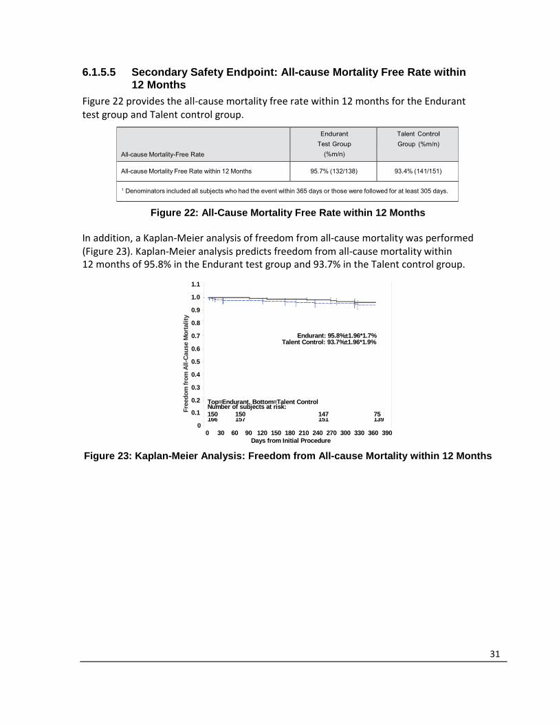
6.1.5.5 Secondary Safety Endpoint: All-cause Mortality Free Rate within 12 Months
Figure 22 provides the all‐cause mortality free rate within 12 months for the Endurant test group and Talent control group.
All-cause Mortality-Free Rate
Endurant
Test Group
(%m/n)
Talent Control
Group (%m/n)
All-cause Mortality Free Rate within 12 Months 95.7% (132/138) 93.4% (141/151)
1 Denominators included all subjects who had the event within 365 days or those were followed for at least 305 days.
Figure 22: All-Cause Mortality Free Rate within 12 Months In addition, a Kaplan‐Meier analysis of freedom from all‐cause mortality was performed (Figure 23). Kaplan‐Meier analysis predicts freedom from all‐cause mortality within 12 months of 95.8% in the Endurant test group and 93.7% in the Talent control group.
1.1
1.0
0.9
0.8
0.7
0.6
Endurant: 95.8%±1.96*1.7% Talent Control: 93.7%±1.96*1.9%
0.5
0.4
0.3
0.2
Top=Endurant, Bottom=Talent Control Number of subjects at risk:
0.1 150 150 147 75166 157 151 139
0 0 30 60 90 120 150 180 210 240 270 300 330 360 390
Days from Initial Procedure
Figure 23: Kaplan-Meier Analysis: Freedom from All-cause Mortality within 12 Months

32
Endurant Test Group Talent Control Group
Treatment to 30 days
31 to 182 days
183 to 365 days
Treatment to 30 days
31 to 182 days
183 to 365 days
No. at Risk1 150 150 147 166 157 151
No. of Events 0 2 4 3 3 4
No. Censored2 0 1 68 6 3 8
Kaplan-Meier Estimate3
1.000
0.987 0.958 0.982 0.963
0.937
Standard Error 0.000 0.009 0.017 0.010 0.015 0.019
1 Number of subjects at risk at the beginning of interval. 2 Subjects are censored because their last follow-up has not reached the end of the time interval or because they are lost to follow-up. 3 Estimate made at end of time interval.
Figure 24: Kaplan-Meier Estimates of Freedom from All-Cause Mortality through 12 Months

33
6.1.5.6 Acute Procedural Data
Figure 25 compares the clinical utility measures of the Endurant test group to the Talent control group. Acute procedural outcomes for the Endurant test group and the Talent control group with respect to procedure duration, blood loss, blood transfusion, time in the Intensive Care Unit (ICU), and length of stay in the hospital are presented below.
Acute Procedural Data Statistics
Endurant Test Group
Talent Control Group
Duration of procedure (min) N 150 166
Mean ± SD 101.5 ± 46.2 167.3 ± 53.2
Median 91.0 155.0
Min, Max 34, 318 85, 417
Subjects receiving general
anesthesia
% (m/n) 83.3% (125/150) 40.4% (67/166)
Estimated blood loss (cc) N 149 165
Mean ± SD 184.9 ± 167.9 335 ± 282.4
Median 150.0 250.0
Min, Max 0, 1450 25, 1750
Subjects requiring blood
transfusion
% (m/n) 0.7% (1/150) 18.2% (30/165)
Time in ICU (hours) N 150 166
Mean ± SD 6.2 ± 19.4 19.3 ± 73.9
Median 0.0 0.0
Min, Max 0, 135 0, 864
Overall hospital stay (days) N 150 166
Mean ± SD 2.1 ± 2.3 3.6 ± 6.4
Median 1.0 2.0
Min, Max 1, 17 1, 79
Figure 25: Acute Procedural Data 6.1.6 Study Results: Effectiveness Endpoints
6.1.6.1 Primary Effectiveness Endpoint: Successful Aneurysm Treatment
Successful aneurysm treatment rate through 12 months in the Endurant test group was 97.5% and 87.1% in the Talent control group (Figure 26).
Successful aneurysm treatment was an endpoint that included delivery and deployment of the graft and surrogate markers that represented treatment success. This included aneurysm growth, endoleak, occlusion, conversion to surgery, rupture, and migration. The information on these endpoints is presented in the sections below.
There were 3 subjects in the Endurant test group that were considered treatment failures. In addition to the technical failure noted above, 1 subject experienced an aneurysm rupture at

34
the index procedure and the other had a stent graft occlusion necessitating a femoral to femoral bypass.
Endurant Test Group
% (m/n)1
Talent Control Group
% (m/n)1
Successful Aneurysm Treatment % (m/n)1
97.5% (118/121) 87.1% (108/124)
1 Denominator is the number of subjects evaluable for this endpoint
Figure 26: Successful Aneurysm Treatment
6.1.6.2 Technical Success
During the index procedure, 99.3% subjects in the Endurant test group were recorded as having successful delivery and deployment of the Endurant bifurcated stent graft compared to 97.6% of the Talent control group. One subject in the Endurant test group had the main bifurcated body implanted but the physician was not able to cannulate the contralateral gate due to a pre‐existing challenging anatomy. The subject was ultimately converted to aorto‐ uni‐iliac in‐situ and a femoral to femoral bypass was performed.
Endurant Test Group
Talent Control Group
Technical Success1
99.3%
(149/150) 97.6%
(162/166)
1 Defined as the successful delivery and deployment of the stent graft.
Figure 27: Technical success
6.1.6.3 Secondary Effectiveness Endpoint: Endoleak
Core Lab identified all types of endoleak at 1 month, 6 months, and 12 months for Endurant test and Talent control groups (Figure 28). There were no Type I or III endoleaks at 1 month, 6 months, or 12 months for the Endurant test group.

35
1 Month % (m/n1) 6 Months % (m/n1) 12 Months % (m/n1)
Endoleaks
Endurant
Test Group
% (m/n1)
Talent Control Group
% (m/n1)
Endurant Test
Group
% (m/n1)
Talent Control Group
% (m/n1)
Endurant Test
Group % (m/n1)
Talent
Control Group
% (m/n1)
Type I
0.0%
(0/143)
9.3%
(14/151)
0.0%
(0/129)
4.2%
(5/118)
0.0%
(0/123)
2.5%
(3/122)
Type II
16.1%
(23/143)
8.6%
(13/151)
11.6%
(15/129)
8.5%
(10/118)
8.9%
(11/123)
6.6%
(8/122)
Type III
0.0%
(0/143)
0.0%
(0/151)
0.0%
(0/129)
0.0%
(0/118)
0.0%
(0/123)
0.0%
(0/122)
Type IV
0.0%
(0/143)
0.0%
(0/151)
0.0%
(0/129)
0.0%
(0/118)
0.0%
(0/123)
0.0%
(0/122)
Indeterminate
0.0%
(0/143)
1.3%
(2/151)
0.0%
(0/129)
1.7%
(2/118)
0.8%
(1/123)
0.8%
(1/122)
Subjects had
endoleaks of any type2
16.1%
(23/143)
19.2%
(29/151)
11.6%
(15/129)
14.4%
(17/118)
9.8%
(12/123)
9.8%
(12/122)
1 Denominator is the number of subjects who had readable images at the time of assessment. 2 A subject may have more than 1 type of endoleaks; hence, number of subjects with any type may not be the sum of those in each type.
Figure 28: All Endoleaks at 1-Month, 6-Months and 12-Months (Core Lab)
6.1.6.4 Secondary Effectiveness Endpoint: Stent Graft Migration
Core Lab report no stent graft migration in the Endurant test group and 1 for the Talent control group through 12 months (Figure 29).
Endurant Test Group
% (m/n)
Talent Control Group
% (m/n)
Stent graft migration through 12 months1
0.0% (0/125) 0.8% (1/128)
1 Migration is defined as evidence of movement of the stent graft relative to fixed anatomic landmarks, which is not due to remodeling of the subject’s vasculature. Migration is observed when the stent graft covers a renal artery or movement is >10 mm
Figure 29: Stent Graft Migration through 12 Months
6.1.6.5 Secondary Effectiveness Endpoint: Stent Graft Patency
Through 12 months, 4 subjects in the Endurant test group experienced stent graft occlusion and 3 subjects in the Talent control group resulting in the stent graft patency rate of 96.8% and 97.5% respectively. All 4 subjects in the Endurant test group underwent secondary procedures and were all treated successfully with blood flow restored to the lower extremity. Multiple factors contributed to the occlusion of the stent grafts including significant calcification in the common iliac artery, significant oversizing of the limb in a tortuous portion of the iliac artery, compression of the stent graft limb by pre‐existing thrombus at the aortic bifurcation, and sharp iliac angulation with pre‐existing stenosis.

36
Additionally, 1 subject experienced graft limb stenosis and was treated successfully with angioplasty and stenting. However, the patient did not experience stent graft occlusion.
6.1.6.6 Secondary Effectiveness Endpoint: Secondary Endovascular
Procedures
Through 12 months, 7 subjects required secondary intervention in the Endurant test group. Four subjects experienced limb occlusions and were treated successfully. Two subjects were treated for Type II endoleaks; neither subjects experienced an increase in aneurysm diameter. One subject experienced limb graft stenosis and was treated successfully with angioplasty with stenting. This resulted in an overall secondary procedure rate of 5.1%. Sixteen subjects in the Talent control group had secondary procedures through 12 months, resulting in an overall secondary procedure rate of 11.1%.
No subjects required secondary procedures to treat Type I or Type III endoleak through 12 months in the Endurant test group.
Endurant Test Group
% (m/n)1
Talent Control Group
% (m/n)1
Secondary procedures through 12 months % (m/n)1
5.1% (7/136) 11.1% (16/144)
1 Denominator is the number of subjects evaluable for this endpoint. A subject is evaluable if it had an event within 365 days post-implant or was followed for at least 305 days.
Figure 30: Secondary Procedures through 12 Months
6.1.6.7 Secondary Effectiveness Endpoint: Technical Observations
Technical observation is defined as an observed defect or malfunction of the stent graft which is not related to any adverse events. Based on Core Lab assessment, the technical observation rate at 12 months in both the Endurant test group and Talent control group was 2.3%.
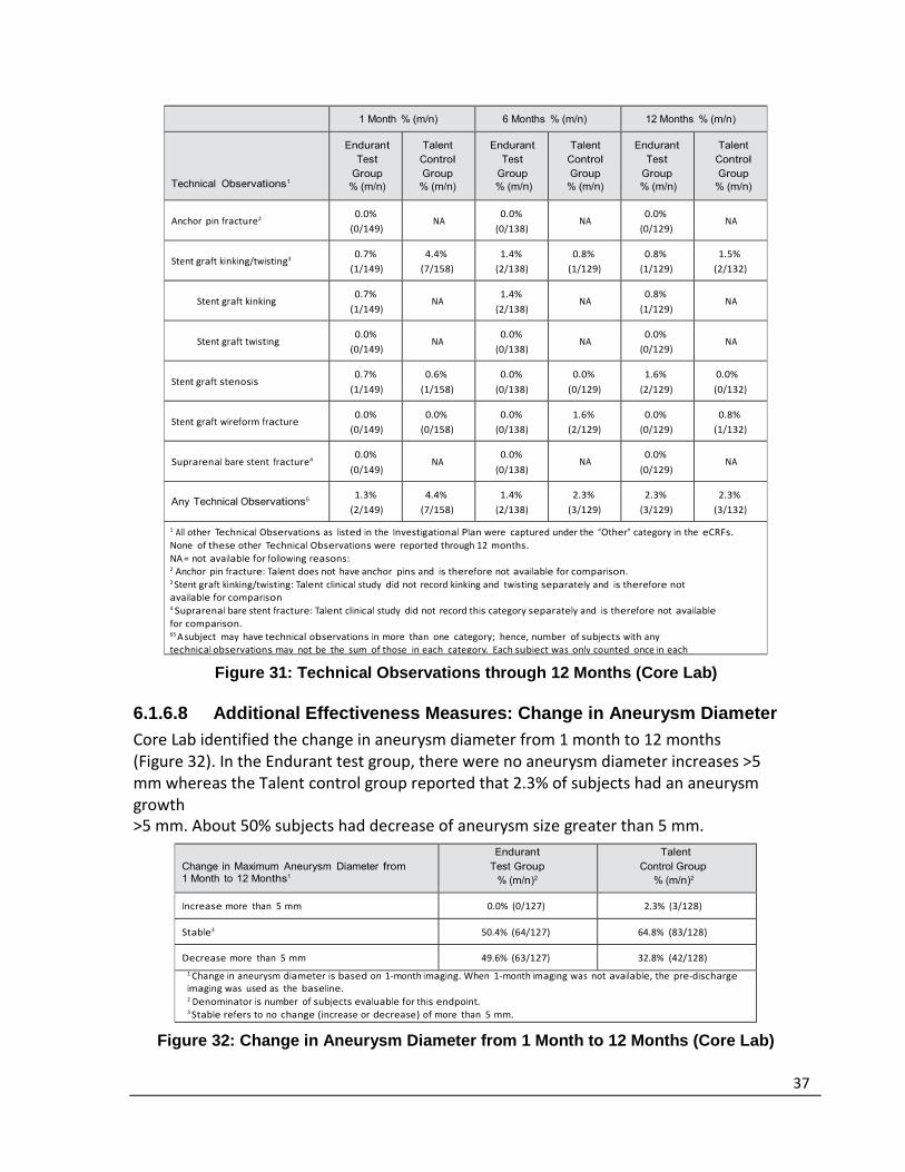
37
1 Month % (m/n) 6 Months % (m/n) 12 Months % (m/n)
Technical Observations1
Endurant Test
Group % (m/n)
Talent Control Group
% (m/n)
Endurant Test
Group % (m/n)
Talent Control Group
% (m/n)
Endurant Test
Group % (m/n)
Talent Control Group
% (m/n)
Anchor pin fracture2
0.0%
(0/149) NA
0.0%
(0/138) NA
0.0%
(0/129)
NA
Stent graft kinking/twisting3
0.7%
(1/149)
4.4%
(7/158)
1.4%
(2/138)
0.8%
(1/129)
0.8%
(1/129)
1.5%
(2/132)
Stent graft kinking
0.7%
(1/149) NA
1.4%
(2/138) NA
0.8%
(1/129)
NA
Stent graft twisting
0.0%
(0/149) NA
0.0%
(0/138) NA
0.0%
(0/129)
NA
Stent graft stenosis
0.7%
(1/149)
0.6%
(1/158)
0.0%
(0/138)
0.0%
(0/129)
1.6%
(2/129)
0.0%
(0/132)
Stent graft wireform fracture
0.0%
(0/149)
0.0%
(0/158)
0.0%
(0/138)
1.6%
(2/129)
0.0%
(0/129)
0.8%
(1/132)
Suprarenal bare stent fracture4
0.0%
(0/149) NA
0.0%
(0/138) NA
0.0%
(0/129)
NA
Any Technical Observations5
1.3%
(2/149)
4.4%
(7/158)
1.4%
(2/138)
2.3%
(3/129)
2.3%
(3/129)
2.3%
(3/132)
1 All other Technical Observations as listed in the Investigational Plan were captured under the “Other” category in the eCRFs.
None of these other Technical Observations were reported through 12 months.
NA = not available for following reasons: 2 Anchor pin fracture: Talent does not have anchor pins and is therefore not available for comparison. 3 Stent graft kinking/twisting: Talent clinical study did not record kinking and twisting separately and is therefore not available for comparison 4 Suprarenal bare stent fracture: Talent clinical study did not record this category separately and is therefore not available for comparison. 65 A subject may have technical observations in more than one category; hence, number of subjects with any technical observations may not be the sum of those in each category. Each subject was only counted once in each
Figure 31: Technical Observations through 12 Months (Core Lab)
6.1.6.8 Additional Effectiveness Measures: Change in Aneurysm Diameter
Core Lab identified the change in aneurysm diameter from 1 month to 12 months (Figure 32). In the Endurant test group, there were no aneurysm diameter increases >5 mm whereas the Talent control group reported that 2.3% of subjects had an aneurysm growth >5 mm. About 50% subjects had decrease of aneurysm size greater than 5 mm.
Change in Maximum Aneurysm Diameter from 1 Month to 12 Months1
Endurant Test Group
% (m/n)2
Talent Control Group
% (m/n)2
Increase more than 5 mm 0.0% (0/127) 2.3% (3/128)
Stable3 50.4% (64/127) 64.8% (83/128)
Decrease more than 5 mm 49.6% (63/127) 32.8% (42/128)
1 Change in aneurysm diameter is based on 1‐month imaging. When 1‐month imaging was not available, the pre‐dischargeimaging was used as the baseline. 2 Denominator is number of subjects evaluable for this endpoint. 3 Stable refers to no change (increase or decrease) of more than 5 mm.
Figure 32: Change in Aneurysm Diameter from 1 Month to 12 Months (Core Lab)
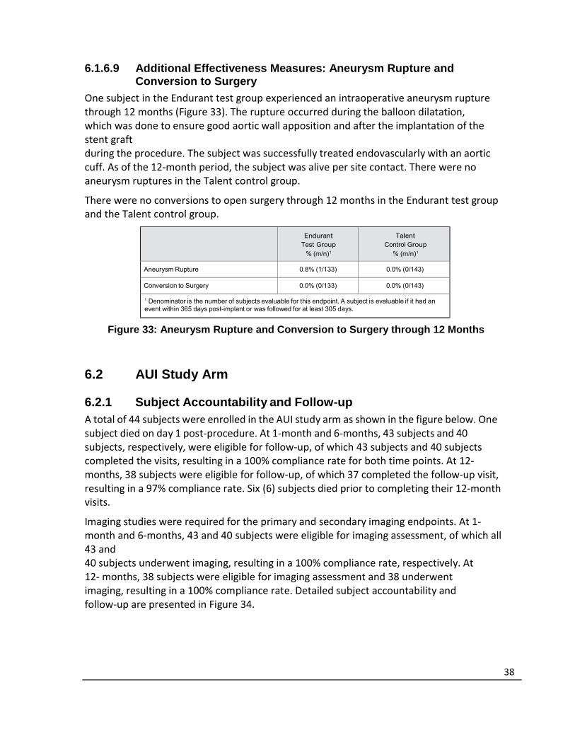
38
6.1.6.9 Additional Effectiveness Measures: Aneurysm Rupture and Conversion to Surgery
One subject in the Endurant test group experienced an intraoperative aneurysm rupture through 12 months (Figure 33). The rupture occurred during the balloon dilatation, which was done to ensure good aortic wall apposition and after the implantation of the stent graft during the procedure. The subject was successfully treated endovascularly with an aortic cuff. As of the 12‐month period, the subject was alive per site contact. There were no aneurysm ruptures in the Talent control group.
There were no conversions to open surgery through 12 months in the Endurant test group and the Talent control group.
Endurant Test Group
% (m/n)1
Talent Control Group
% (m/n)1
Aneurysm Rupture 0.8% (1/133) 0.0% (0/143)
Conversion to Surgery 0.0% (0/133) 0.0% (0/143)
1 Denominator is the number of subjects evaluable for this endpoint. A subject is evaluable if it had an event within 365 days post-implant or was followed for at least 305 days.
Figure 33: Aneurysm Rupture and Conversion to Surgery through 12 Months
6.2 AUI Study Arm 6.2.1 Subject Accountability and Follow-up A total of 44 subjects were enrolled in the AUI study arm as shown in the figure below. One subject died on day 1 post‐procedure. At 1‐month and 6‐months, 43 subjects and 40 subjects, respectively, were eligible for follow‐up, of which 43 subjects and 40 subjects completed the visits, resulting in a 100% compliance rate for both time points. At 12‐months, 38 subjects were eligible for follow‐up, of which 37 completed the follow‐up visit, resulting in a 97% compliance rate. Six (6) subjects died prior to completing their 12‐month visits.
Imaging studies were required for the primary and secondary imaging endpoints. At 1‐month and 6‐months, 43 and 40 subjects were eligible for imaging assessment, of which all 43 and 40 subjects underwent imaging, resulting in a 100% compliance rate, respectively. At 12‐ months, 38 subjects were eligible for imaging assessment and 38 underwent imaging, resulting in a 100% compliance rate. Detailed subject accountability and follow‐up are presented in Figure 34.

39
44 0
0 1 0 0 0
43
43 43 43 40 2 42 42
(100%) (100%) (100%) (93%) (5%) (98%) (98%)
0 3 0 0 0
40
40 40 40 39 2 40 39 40 40
(100%) (100%) (100%) (98%) (5%) (100%) (98%) (100%) (100%)
0 2 0 0 0
38
37 38 36 33 7 38 38 36 37 (97%)
(100%) (95%) (87%) (18%) (100%) (100%) (95%) (97%)
Interval
(Analysis
Window)
Subject follow‐up
Subjects with imaging
performed at time
interval
(Core Lab)
Subjects with adequate
imaging to assess the
parameter (Core Lab)
Subject events occurring before next visit
Elig
ible
Clin
ical Follow‐up
Imaging Follo
w‐up
CT/MRA Im
aging
KUB Im
aging
Other Imaging1
Aneu
rysm
size
increase
Endoleak
M
igration
Te
chnical O
bse
rvation2
No Implant
Conve
rsion to Surgery
Death
Withdrawal/
Early Te
rmination
Lost to Follo
w‐up
Not Due for Next Visit
Originally Enrolled
Events after
implant but
before a 1 Month
1 Month
(Day 1‐90)
Events after 1 Month visit but
before a 6
Month visit
6 Month
(Day 91‐304)
Events after 6 Month visit but before a 12 Month visit
12 Month
(≥ Day 3053)
Note: Data analysis sample size varies for each of the timepoints above and in the following tables. This
variability is due to subject availability for follow‐up, as well as, quantity and quality of images available
from specific timepoints for evaluation. For example, the number and quality of images available for
evaluation of endoleak at 6 months is different than the number and quality of images available at
12 months due to variation in the number of image exams performed, the number of images provided from the clinical site to the Core Lab, and/or the number of images with acceptable evaluation quality.
1 Other imaging includes angiogram, ultrasound, and other imaging of stent graft region.
2 Technical observations assessed by imaging include stent‐graft kinking, stent‐graft twisting,
stent‐ graft wireform fracture, suprarenal bare stent fracture, anchoring pin fracture, and stent‐
graft stenosis.
3 In cases where 12‐month imaging follow‐up data were not available, subsequent imaging follow‐up data were used.
Figure 34: Subject and Imaging Accountability — Endurant AUI 6.2.2 Study Demographics and Baseline Medical History The mean subject age was 73.8 ± 7.6 years and 43.2% were above the age of 75. Of the enrolled Endurant AUI subjects, 68.2% were male and 95.5% of the subjects were white. Among the female subjects, the mean age was 74.5 ± 8.0 years.

40
Parameter Statistics/Category Endurant AUI SVS Open Surgical Group
Age (years)
Total n 44 243
Mean ± SD 73.8 ± 7.6 70.1 ± 7.49
Median 73.5 70.0
Min, max 59, 89 46, 86
<65 6.8% (3/44) 22.2% (54/243)
65–75 50.0% (22/44) 51.4% (125/243)
>75 43.2% (19/44) 26.3% (64/243)
Male n 30 198
Mean ± SD 73.5 ± 7.5 69.7 ± 7.30
Median 74.0 70.0
Min, max 59, 89 49, 85
Female n 14 45
Mean ± SD 74.5 ± 8.0 72.2 ± 8.06
Median 72.0 72.0
Min, max 65, 87 46, 86
Gender % (m/n)
Male 68.2% (30/44) 81.5% (198/243)
Race % (m/n)
White 95.5% (42/44) 94.9% (168/177)
Non-white 4.5% (2/44) 5.1% (9/177)
Figure 35: Subject Demographics Cardiovascular risk factors were reported in high rates in this population with hyperlipidemia (88.6%), hypertension (86.4%), and tobacco use in the past 10 years (65.9%) being most prevalent. Cardiovascular diseases such as peripheral vascular disease (75.0%), coronary artery disease (59.1%), other vascular disease (50.0%), carotid artery disease (40.9%), arrhythmia (34.1%), valvular heart disease (31.8%), myocardial infarction (31.8%), and other cardiac disease (29.5%) were also reported in this population.
In addition to the cardiovascular risk factors and diseases noted above, Endurant AUI subjects also had a high rate of chronic obstructive pulmonary disease (59.1%), abnormal renal function (31.8%), other cerebrovascular/neurological disease (25.0%), and diabetes (25.0%).

41
Body System / Condition Endurant AUI
% (m/n)
SVS Open Surgical Group % (m/n)
Cardiac 1
Angina 25.0% (11/44) 17.4% (23/132)
Arrhythmia 34.1% (15/44) 11.5% (28/243)
Congestive heart failure 15.9% (7/44) 4.9% (12/243)
Coronary artery bypass graft 2 27.3% (12/44)
Coronary artery disease 59.1% (26/44) 61.3% (149/243)
Myocardial infarction 31.8% (14/44) 34.2% (83/243)
Percutaneous transluminal coronary angioplasty 2 29.5% (13/44)
Valvular heart disease (VHD) 31.8% (14/44)
Cardiac revascularization 2 46.1% (112/243)
Other cardiac 29.5% (13/44)
Pulmonary
Chronic obstructive pulmonary disease 59.1% (26/44) 30.0% (73/243)
Renal 3
Abnormal renal function 31.8% (14/44)
Renal failure 4.1% (10/243)
Renal insufficiency 15.9% (7/44)
Cerebrovascular/Neurological 4
Cerebral vascular accident 9.1% (4/44)
Paraparesis 0.0% (0/44)
Paraplegia 0.0% (0/44)
Transient ischemic attack (TIA) 6.8% (3/44)
Cerebrovascular disease 12.8% (31/243)
Other Cerebrovascular/Neurological 25.0% (11/44)
Vascular 5
Abdominal aortic aneurysm 0.0% (0/44)
Any thoracic aneurysm 0.0% (0/44)
Peripheral vascular disease 75.0% (33/44)
Thromboembolic event 4.5% (2/44)
Other vascular 50.0% (22/44)
Other conditions 6
Alcoholism 15.9% (7/44)
Bleeding disorder 0.0% (0/44)
Cancer 18.2% (8/44)
Carotid artery disease 40.9% (18/44)
Diabetes 25.0% (11/44) 11.9% (29/243)
Family history of aneurysm 11.4% (5/44)
GI complications 72.7% (32/44)
Hyperlipidemia 88.6% (39/44)
Hypertension 86.4% (38/44) 66.7% (162/243)
Liver disease 6.8% (3/44)
Tobacco use in the last 10 years 65.9% (29/44) 85.6% (208/243)
Other 90.9% (40/44)
1 “Valvular heart disease,” and “Other cardiac diseases,” were reported for the Endurant AUI group but not for the SVS Control group. 2 SVS Control group did not report “Coronary artery bypass graft” or “Percutaneous transluminal coronary angioplasty” as a separate category. “Cardiac Revascularization” includes Coronary Artery Bypass Grafting (CABG) or PTCA for the SVS Control group.
3 SVS Control group reported “Renal failure” but Endurant AUI group reported “Abnormal renal function” and “Renal insufficiency.” These categories are not comparable.
4 SVS Control group reported “Cerebrovascular disease” but Endurant AUI group reported “Cerebral vascular accident,” “Paraparesis,” “Paraplegia,” “Transient ischemic attack,” and “Other cerebrovascular/neurological.” These categories are not comparable.
5 SVS Control group did not report Vascular category for medical history but Endurant AUI group reported “Abdominal aortic aneurysm,” “Any thoracic aneurysm,” “Peripheral vascular disease,” “Thromboembolic event,” and “Other vascular.” These categories are not comparable.
6 SVS Control group reported “Diabetes,” “Hypertension,” and “Tobacco use in the last 10 years” but Endurant AUI group reported “Alcoholism,” “Bleeding disorder,” “Cancer,” “Carotid artery disease,” “Family
history of aneurysm,” “GI complications,” “Hyperlipidemia,” “Liver disease,” and “Other.” These categories are not comparable.
Figure 36: Medical History

42
In the Endurant AUI study arm, 34.1% of subjects were classified as SVS 3, 50.0% as SVS 2, 15.9% as SVS 1 and 0% as SVS 0. Thirty‐seven (37) of the 44 subjects (84.1%) were classified as having an SVS score of 2 or 3, demonstrating the higher severity of comorbidities in this subject population. SVS classification was not available for the SVS Open Surgical Group and, thus, is not shown below.
SVS Classification
Endurant AUI
% (m/n)
SV S 0 0.0% (0/44)
SV S 1 15.9% (7/44)
SV S 2 50.0% (22/44)
SV S 3 34.1% (15/44)
Figure 37: Baseline SVS Classification 6.2.3 Baseline Aneurysm Characteristics Figure 38 and Figure 39 provide the baseline aneurysm and anatomical measurements of the Endurant AUI study arm.

43
Endurant AUI
Dimension Statistics
Core Lab Reported
Maximum aneurysm diameter (mm) n1 44
Mean ± SD 50.6 ± 9.1
Median 50.4
Min, Max 35, 77
Proximal neck diameter (mm) n1 44
Mean ± SD 22.2 ± 3.0
Median 22.0
Min, Max 17, 32
Ipsilateral iliac diameter (mm) n1 44
Mean ± SD 12.7 ± 3.4
Median 12.0
Min, Max 9, 25
Contralateral iliac diameter (mm) n1 35
Mean ± SD 13.4 ± 5.7
Median 12.5
Min, Max 8, 36
Ipsilateral iliac stenosis (%)2 n1 44
Mean ± SD 24.0 ± 26.0
Median 17.5
Min, Max 0, 1003
Contralateral iliac stenosis (%)2 n1 44
Mean ± SD 57.0 ± 40.1
Median 60.0
Min, Max 0, 100
Distal diameter of the aorta or aneurysm
above the aortic bifurcation (mm) n1
44
Mean ± SD 17.8 ± 5.3
Median 16.5
Min, Max 9, 32
Proximal neck length (mm) n1 44
Mean ± SD 24.9 ± 13.5
Median 22.5
Min, Max 4, 48
Infrarenal angle (°) n1 44
Mean ± SD 35.2 ± 18.2
Median 33.2
Min, Max 9, 844
Suprarenal angle (°) n1 44
Mean ± SD 23.7 ± 16.3
Median 18.6
Min, Max 5, 875
1 n = number of subjects with readable scans. 2 Ipsilateral and contralateral iliac stenosis are site‐reported values as core lab did not assess these
data points at time of data collection.
3 One (1) subject had an ipsilateral iliac artery stenosis of 100% at baseline. The device was implanted in this
subject using an iliac conduit which was placed during the index procedure.
4 One (1) subject was measured by the core lab to have an infrarenal angle measurement > 60 degrees. This subject was assessed by the investigational site and the Physician Screening Committee (PSC) and noted to havean infrarenal angulation measurement which met the inclusion criteria and was deemed eligible by the PSC for enrollment. The PSC determined whether or not a subject was eligible for enrollment based on their baseline imaging and concluded that this subject met all inclusion criteria and approved this subject for enrollment. 5 One (1) subject was measured by the core lab to have a suprarenal angle measurement > 45 degrees. The investigational site and the Physician Screening Committee (PSC) reviewed the imaging and both noted that the suprarenal angulation measurement was < 45 degrees. The PSC determined whether or not a subject was li ibl f ll d l d d h hi bj ll i l i i i d d hi bj f
Figure 38: Baseline Aneurysm Characteristics (Core Lab reported)

44
Statistics/Category Endurant AUI
Maximum Aneurysm Diameter (mm) n1 44
Mean ± SD 50.6 ± 9.1
Median 50.4
Min, max 35, 77
Maximum Aneurysm Diameter % (m/n)1 < 30 mm 0.0% (0/44)
30 mm–< 40 mm 13.6% (6/44)
40 mm–< 50 mm 29.5% (13/44)
50 mm–< 60 mm 45.5% (20/44)
60 mm–< 70 mm 9.1% (4/44)
70 mm–< 80 mm 2.3% (1/44)
≥ 80 mm 0.0% (0/44)
Maximum Aneurysm Diameter % (m/n)1 < 50 mm 43.2% (19/44)
≥ 50 mm 56.8% (25/44)
1 n = number of subjects with readable scans.
Figure 39: Baseline Maximum Aneurysm Diameter (Core Lab reported) 6.2.4 Devices Implanted Figure 40 provides the number of Endurant stent graft devices (which includes the AUI stent graft as well as aortic and iliac extensions) implanted at the index procedure per subject.
Number of Endurant Devices Implanted
Endurant AUI
(% m/n)1
1 2.3% (1/44)
2 56.8% (25/44)
3 36.4% (16/44)
4 4.5% (2/44)
≥ 5 0.0% (0/44)
1 Denominator includes all subjects who were implanted with the test device.
Figure 40: Total Number of Devices Implanted at Index Procedure

45
Implanted Device Sizes
Figure 41 shows the distribution of sizes of the AUI stent graft used in the Endurant US Clinical Study.
Stent Graft Proximal Diameter (Aortic)
Endurant AUI
% (m/n)1
23 18.2% (8/44)
25 31.8% (14/44)
28 29.5% (13/44)
32 15.9% (7/44)
36 4.5% (2/44)
1 Denominator includes all subjects who were implanted with at least one (1) test device.
Figure 41: Devices Implanted by Size at Index Procedure Figure 42 shows the breakdown of device types implanted at the index procedure for the 44 enrolled AUI subjects.
Device Type
Endurant AUI
% (m/n)1
Aorto ‐Uni‐Iliac 100.0% (44/44)
Iliac Extension2 97.7% (43/44)
Extension‐ any type 6.8% (3/44)
Extension‐ iliac2 2.3% (1/44)
Extension‐ aorta 4.5% (2/44)
Talent occluder3 22.7% (10/44)
1 Denominator includes all subjects who received the test device. A subject may receive multiple device types
but is counted only once for each device type. 2 “Iliac Extension” refers to the Endurant Contralateral Limb component as the Endurant AUI stent graft device
does not have a contralateral iliac limb. “Extension – iliac” refers to the Endurant Iliac Extension component. 3 Data on the Talent occluder is presented. Physicians were instructed that they could use any commercially available method, including the Talent occluder, to occlude the contralateral iliac.
Figure 42: Device Type Implanted at Index Procedure
Accessory Devices Implanted
Accessory devices including stents and coils were implanted in six (6) Endurant AUI subjects. Four (4) subjects were implanted with stent(s), one (1) subject was implanted with coils, and one (1) subject was implanted with stents and coils.
Of the four (4) subjects implanted with a stent, the stent placement location in each subject is provided below:
• Proximal to the main body of the stent graft (first stent) and the right renal artery (second stent)
• Left renal artery

46
• Right external iliac artery
• Unknown location
One (1) subject was implanted with coils in the lumbar artery. The subject who received the stents and coils was implanted with a stent in the left internal iliac artery, a stent in the left proximal external iliac artery, and coils in an unknown location.
Additional Procedures
Femoral‐femoral artery bypasses are typically performed in order to perfuse the contralateral lower extremity. One (1) subject had a prior femoral‐femoral artery bypass which was unrelated to the Endurant AUI index procedure. Thirty‐one (31) subjects had the femoral‐ femoral artery bypass performed during the index procedure but prior to the insertion of the Endurant AUI stent graft device. For the remaining twelve (12) subjects, the timing and method of restoring blood flow to the contralateral extremity is unknown.
If the contralateral iliac artery was not occluded at the time of the index procedure, then an occlusion procedure was performed to prevent retrograde blood flow into the contralateral iliac artery. For the 44 AUI subjects, the occlusion method is provided below:
• Coils (3 subjects)
• Talent Occluder (10 subjects)
• Occlusion plugs (3 subjects in contralateral iliac arteries; 10 subjects in unknown locations)
• No occlusion device was required since the contralateral iliac arteries were occluded prior to the index procedure (14 subjects)
• Unknown occlusion method (4 subjects) 6.2.5 Study Results: Safety Endpoints
6.2.5.1 Primary Safety Endpoint: Major Adverse Events (MAEs) Free Rate
within 30 Days
The primary safety endpoint in the Endurant AUI arm of the clinical study was freedom from major adverse events (MAEs) within 30‐days of the index procedure. The MAE composite included all‐cause mortality, bowel ischemia, myocardial infarction, paraplegia, procedural blood loss ≥1000 cc, renal failure, respiratory failure, and stroke. All the MAEs were adjudicated by the Clinical Events Committee (CEC), with the exception of procedural blood loss. Figure 43 and Figure 44 provide an analysis of the MAEs within 30 days.
Freedom from Major Adverse Events (MAE)
within 30 Days
Endurant AUI
N = 44
% (m/n)
Freedom from MAEs within 30 Days 88.6% (39/44)

47
Figure 43: Freedom from Major Adverse Events within 30 days

48
Major Adverse Events (MAE) within 30 Days1,2
Endurant AUI
(% m/n)
MAEs within 30 days 11.4% (5/44)
All‐cause Mortality 2.3% (1/44)
Myocardial Infarction 6.8% (3/44)
Renal Failure 2.3% (1/44)
Respiratory Failure 4.5% (2/44)
Paraplegia 0.0% (0/44)
Stroke 2.3% (1/44)
Bowel Ischemia 0.0% (0/44)
Procedural Blood Loss ≥ 1000 cc 4.5% (2/44)
MAE w/o Blood Loss at 30 Days 11.4% (5/44)
1 A subject may report multiple MAEs; hence, number of subjects with any MAE may not be the sum of those in each MAE category. 2 The % MAE rate at 30 days is 100% ‐ % Freedom from MAEs within 30 days.
Figure 44: Major Adverse Events within 30 days
6.2.5.2 Secondary Safety Endpoint: Major Adverse Events (MAEs) Free Rate within 12 Months
Figure 45 and Figure 46 provide an analysis of the MAEs within 12 months for the Endurant AUI study arm.
Secondary Endpoint
Endurant AUI
(% m/n)1
Freedom f rom MAEs through 365 Days 72.7% (32/44)
1 Denominators included all subjects who had MAE(s) within 365 days or those were followed for at least 305 days.
Figure 45: Freedom from Major Adverse Events through 12 months
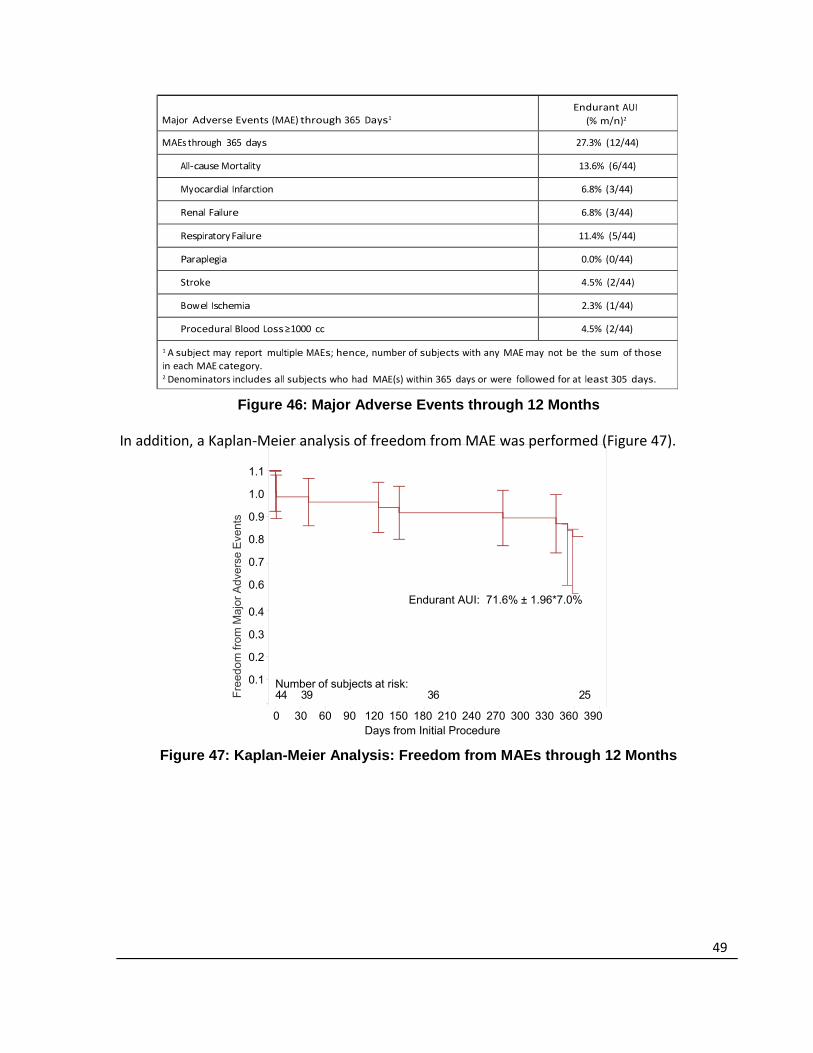
49
Fre
edom
from
Maj
or A
dver
se E
vent
s
Major Adverse Events (MAE) through 365 Days1
Endurant AUI
(% m/n)2
MAEs through 365 days 27.3% (12/44)
All‐cause Mortality 13.6% (6/44)
Myocardial Infarction 6.8% (3/44)
Renal Failure 6.8% (3/44)
Respiratory Failure 11.4% (5/44)
Paraplegia 0.0% (0/44)
Stroke 4.5% (2/44)
Bowel Ischemia 2.3% (1/44)
Procedural Blood Loss ≥ 1000 cc 4.5% (2/44)
1 A subject may report multiple MAEs; hence, number of subjects with any MAE may not be the sum of those in each MAE category. 2 Denominators includes all subjects who had MAE(s) within 365 days or were followed for at least 305 days.
Figure 46: Major Adverse Events through 12 Months In addition, a Kaplan‐Meier analysis of freedom from MAE was performed (Figure 47).
1.1
1.0
0.9
0.8
0.7
0.6
0.4
Endurant AUI: 71.6% ± 1.96*7.0%
0.3
0.2
0.1
Number of subjects at risk:44 39 36 25
0 30 60 90 120 150 180 210 240 270 300 330 360 390 Days from Initial Procedure
Figure 47: Kaplan-Meier Analysis: Freedom from MAEs through 12 Months

50
Endurant AUI
Treatment
to 30 days
31 to 182 days
183 to 365
days
No. at Risk1 44 39 36
No. of Events 5 3 4
No. Censored2 0 0 7
Kaplan-Meier Estimate3 0.886 0.818 0.716
Standard Error 0.048 0.058 0.070
1 Number of subjects at risk at the beginning of interval. 2 Subjects are censored because their last follow-up has not reached the end of the time interval or because they are lost to follow-up. 3 Estimate made at end of time interval.
Figure 48: Kaplan-Meier Estimates of Freedom from MAEs through 12 Months
6.2.5.3 Secondary Safety Endpoint: Aneurysm-Related Mortality (ARM) Free- Rate within 12 Months
The ARM free rate within 12 months was 97.5% in the Endurant AUI study arm (Figure 49). One (1) death occurred on day 1 due to a myocardial infarction, which the CEC adjudicated as not related to the aneurysm or the device, but related to the procedure. The CEC arrived at this conclusion based on the autopsy report, which noted “severe atherosclerosis with unremarkable aneurysm repair and fem‐fem bypass” and “no signs of thrombus or external hemorrhage in the endovascular stent graft or bypass.”
Endurant AUI
Secondary Endpoint % (m/n)1
95% Confidence
Interval2
Freedom from Aneurysm-Related Mortality within 365 Days3
97.5% (39/40) [84.1%, 100.0%]
1 Denominators included all subjects who had the event within 365 days or those were followed for at least 305 days. 2 Two-sided 95% confidence interval based on a binomial distribution and only calculated for 3 secondary endpoints along with Stent graft migration and secondary procedures. Adjustment of multiplicity was made using the Bonferroni method. 3 One (1) death occurred on day 1 due to a myocardial infarction, which the CEC adjudicated as not related to the aneurysm or the device, but related to the procedure. The CEC arrived at this conclusion based on the autopsy report, which noted “severe atherosclerosis with unremarkable aneurysm repair and fem-fem bypass” and “no signs of thrombus or external hemorrhage in the endovascular stent graft or bypass.”
Figure 49: Freedom from Aneurysm-Related Mortality through 12 Months In addition, a Kaplan‐Meier analysis of freedom from ARM was performed (Figure 50). Kaplan‐Meier analysis predicts freedom from ARM rate within 12 months of 97.7% in the Endurant AUI study arm. The data used in the Kaplan‐Meier analysis is presented below.
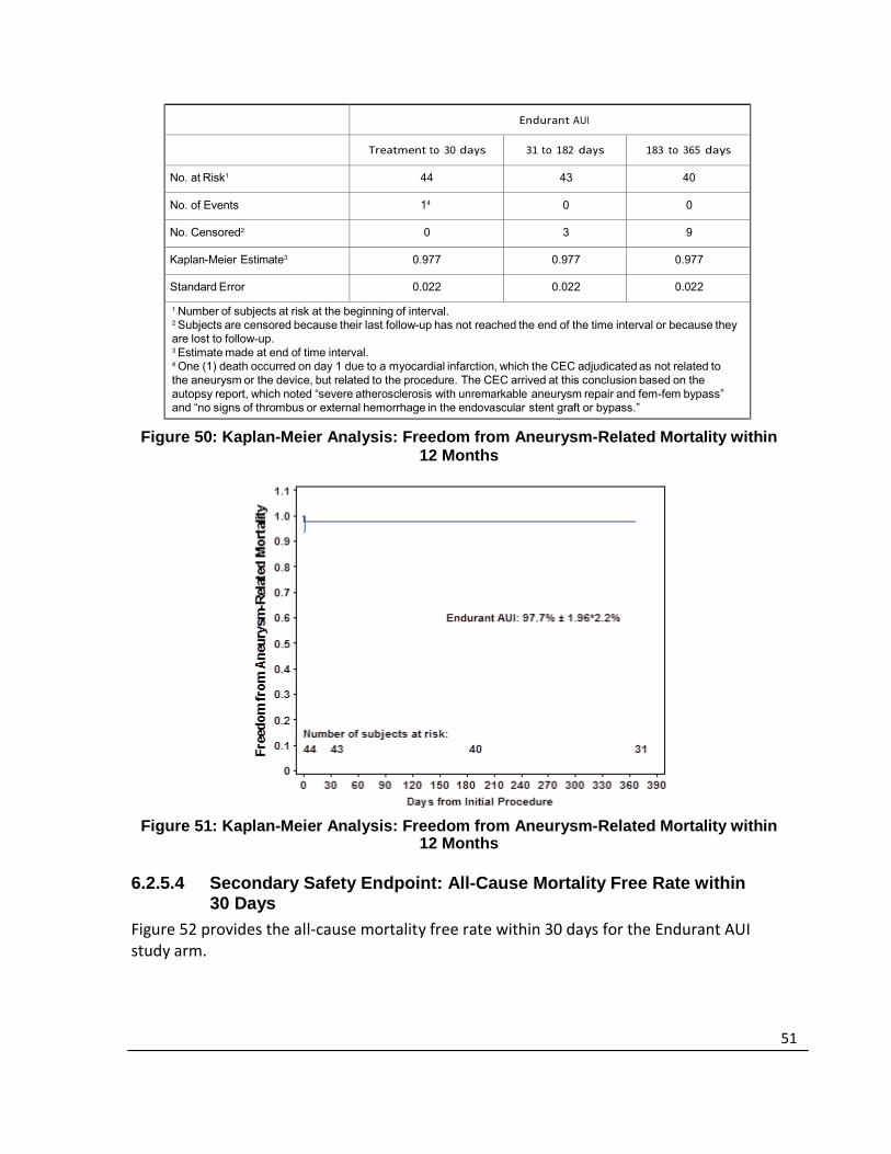
51
Endurant AUI
Treatment to 30 days 31 to 182 days
183 to 365 days
No. at Risk1 44 43 40
No. of Events 14 0 0
No. Censored2 0 3 9
Kaplan-Meier Estimate3 0.977 0.977 0.977
Standard Error 0.022 0.022 0.022
1 Number of subjects at risk at the beginning of interval. 2 Subjects are censored because their last follow-up has not reached the end of the time interval or because they are lost to follow-up. 3 Estimate made at end of time interval. 4 One (1) death occurred on day 1 due to a myocardial infarction, which the CEC adjudicated as not related to the aneurysm or the device, but related to the procedure. The CEC arrived at this conclusion based on the autopsy report, which noted “severe atherosclerosis with unremarkable aneurysm repair and fem-fem bypass” and “no signs of thrombus or external hemorrhage in the endovascular stent graft or bypass.”
Figure 50: Kaplan-Meier Analysis: Freedom from Aneurysm-Related Mortality within 12 Months
Figure 51: Kaplan-Meier Analysis: Freedom from Aneurysm-Related Mortality within 12 Months
6.2.5.4 Secondary Safety Endpoint: All-Cause Mortality Free Rate within
30 Days
Figure 52 provides the all‐cause mortality free rate within 30 days for the Endurant AUI study arm.

52
Fre
edom
from
All-
Cau
se M
orta
lity
Secondary Endpoint
Endurant AUI
(% m/n)1
Freedom from All-Cause Mortality within 30 Days 97.7% (43/44)
Figure 52: Freedom from All-Cause Mortality through 30 Days
6.2.5.5 Secondary Safety Endpoint: All-Cause Mortality Free Rate within 12 Months
Figure 53 provides the all‐cause mortality free rate within 12 months for the Endurant AUI study arm.
Secondary Endpoint
Endurant AUI
(% m/n)1
Freedom from All-Cause Mortality within 365 Days 86.4% (38/44)
1 Denominators included all subjects who had the event within 365 days or those were followed for at least 305 days.
Figure 53: Freedom from All-Cause Mortality through 12 Months In addition, a Kaplan‐Meier analysis of freedom from all‐cause mortality was performed (Figure 54)
1.1
1.0
0.9
0.8
0.7
0.6
0.4
Endurant AUI: 86.0% ± 1.96*5.3%
0.3
0.2
0.1
Number of subjects at risk:44 43 40 31
0 30 60 90 120 150 180 210 240 270 300 330 360 390 Days from Initial Procedure
Figure 54: Kaplan-Meier Analysis: Freedom from All-Cause Mortality within 12 Months

53
Endurant AUI
Treatmen
t to 30
31 to 182
days
183 to 365
days
No. at Risk1 44 43 40
No. of Events 1 3 2
No. Censored2 0 0 7
Kaplan-Meier Estimate3 0.977 0.909 0.860
Standard Error 0.022 0.043 0.053
1 Number of subjects at risk at the beginning of interval. 2 Subjects are censored because their last follow-up has not reached the end of the time interval or because they are lost to follow-up. 3 Estimate made at end of time interval.
Figure 55: Kaplan-Meier Estimates of Freedom from All-Cause Mortality through 12 Months
6.2.5.6 Acute Procedural Data
Figure 56 provides the clinical utility measures of the Endurant AUI study arm. Acute procedural outcomes for the Endurant AUI study arm with respect to procedure duration, blood loss, blood transfusion, time in the Intensive Care Unit (ICU), and length of stay in the hospital are presented below.

54
Acute Procedural Data Statistics Endurant AUI
Duration of procedure (min) n 44
Mean ± SD 117.7 ± 66.6
Median 91.5
Min, Max 33, 295
Subjects receiving general anesthesia % (m/n) 79.5% (35/44)
Estimated blood loss (cc) n 44
Mean ± SD 264.2 ± 221.2
Median 200.0
Min, Max 5, 1200
Subjects requiring blood transfusion % (m/n) 6.8% (3/44)
Time in ICU (hours) n 44
Mean ± SD 16.6 ± 39.3
Median 0.0
Min, Max 0, 186
Overall hospital stay (days) n 44
Mean ± SD 3.2 ± 2.7
Median 2.5
Min, Max 1, 13
Figure 56: Acute Procedural Data 6.2.6 Study Results: Effectiveness Endpoints
6.2.6.1 Primary Effectiveness Endpoint: Successful Aneurysm Treatment
Successful aneurysm treatment rate through 12 months in the Endurant AUI study arm group was 97.2% (Figure 57).
Successful aneurysm treatment was an endpoint that included delivery and deployment of the stent graft and surrogate markers that represented treatment success. This included aneurysm growth, endoleak, occlusion, conversion to surgery, rupture, and migration. The information on these endpoints is presented in the sections below.
Primary Effectiveness Endpoint
Endurant AUI % (m/n)1 95% Confidence Interval2
Successful Aneurysm Treatment 97.2% (35/36)3 [ 85.5%, 99.9%]
1 Denominator is number of subjects evaluable for this endpoint. 2 Two‐sided 95% confidence interval was based on a binomial distribution. 3 One (1) subject in the Endurant AUI study arm was considered a treatment failure. This subject experienced
a technical failure during the index procedure as the investigator had difficulty recapturing the tip of the
delivery system which engaged the suprarenal stents. While the investigator attempted to disengage the tip
from the suprarenal stent, the stent graft moved proximally, causing the left renal artery to be 50% obstructed
by the stent graft fabric. A left renal artery stent was placed to treat the unintentional coverage.
Figure 57: Successful Aneurysm Treatment

55
6.2.6.2 Technical Success
During the index procedure, 99.7% subjects (43/44) in the Endurant AUI study arm were recorded as having successful delivery and deployment of the Endurant AUI stent graft. One subject experienced a technical failure as the investigator had difficulty recapturing the tip of the delivery system which engaged the suprarenal stents. While the investigator attempted to disengage the tip from the suprarenal stent, the stent graft moved proximally, causing the left renal artery to be 50% obstructed by the stent graft fabric. A left renal artery stent was placed to treat the unintentional coverage.
6.2.6.3 Secondary Effectiveness Endpoint: Endoleak
Core Lab reported types of endoleak at 1‐month, 6‐months, and 12‐months for the Endurant AUI study arm (Figure 58). There were no Type I, III, IV, or V endoleaks at 1 month, 6 months, or 12 months.
Endurant AUI
Endoleaks
1 Month
% (m/n)1
6 Month
% (m/n)1
12 Month
% (m/n)1
Type I 0.0% (0/42) 0.0% (0/39) 0.0% (0/38)
Type II 14.3% (6/42) 12.8% (5/39) 10.5% (4/38)
Type III 0.0% (0/42) 0.0% (0/39) 0.0% (0/38)
Type I V 0.0% (0/42) 0.0% (0/39) 0.0% (0/38)
Type V 0.0% (0/42) 0.0% (0/39) 0.0% (0/38)
Indeterminate 0.0% (0/42) 0.0% (0/39) 0.0% (0/38)
Subjects had endoleaks of any type2 14.3% (6/42) 12.8% (5/39) 10.5% (4/38)
1 Denominator is the number of subjects who had readable images at the time of assessment. 2 A subject may have more than one type of endoleaks; hence, number of subjects with any type may not be
the sum of those in each type.
Figure 58: All Endoleaks at 1-Month, 6-Months and 12-Months (Core Lab)
6.2.6.4 Secondary Effectiveness Endpoint: Stent Graft Migration
As reported by the core lab, there was no stent graft migration through 12 months in the Endurant AUI study arm.
6.2.6.5 Secondary Effectiveness Endpoint: Stent Graft Patency
Through 12 months, no subjects in the Endurant AUI study arm experienced stent graft occlusion, and, therefore, the stent graft patency rate was 100%.
6.2.6.6 Secondary Effectiveness Endpoint: Secondary Endovascular
Procedures
One (1) Endurant AUI subject required a secondary endovascular procedure through 12 months. The investigational site identified a stenosis of the stent graft, and the subject was taken to the operating room for placement of a covered stent graft to treat the stent graft

56
stenosis. Patency was restored to the left lower extremity and into the left to right femoral‐ femoral bypass graft as a result of this procedure.
No secondary endovascular procedures were performed as a result of an event related to the femoral‐femoral bypass.
Subject
Days from
Implant
Reason for Secondary
Endovascular Procedures
Procedure Performed Outcome
008-010 280 Stent graft stenosis Stenting Resolved
Figure 59: Secondary Procedures through 12 Months
6.2.6.7 Secondary Effectiveness Endpoint: Technical Observations
Figure 60 outlines the technical observation rate through 12 months, as assessed by Core Lab. Four (4) subjects were reported as having a technical observation. All four (4) subjects were observed to have stent graft stenosis. At 6 months, three (3) subjects (7.5%) were reported to have stent graft stenosis. At 12 months, four (4) subjects (10.8%) were reported to have stent graft stenosis, three of which were observed and carried over from the 6‐month time point. None of the subjects with stent graft stenosis observed by core lab have had adverse events associated with the stent graft stenosis observations or required re‐ intervention. In addition to the core lab reported occurrences of stent graft stenosis previously stated, one (1) subject, as shown above in Figure 59, was identified as having stent graft stenosis by the investigational site and required a secondary endovascular procedure to restore patency to the left lower extremity and the left to right femoral‐femoral bypass graft.
Endurant AUI
Technical Observations
1 Month % (m/n1)
6 Months % (m/n1)
12 Months % (m/n1)
Anchoring pin fracture 0.0% (0/42) 0.0% (0/40) 0.0% (0/37)
Stent graft kinking/twisting 0.0% (0/42) 0.0% (0/40) 0.0% (0/37)
Stent graft kinking 0.0% (0/42) 0.0% (0/40) 0.0% (0/37)
Stent graft twisting 0.0% (0/42) 0.0% (0/40) 0.0% (0/37)
Stent graft stenosis 0.0% (0/42) 7.5% (3/40) 10.8% (4/37)
Stent graft wiref orm fracture 0.0% (0/42) 0.0% (0/40) 0.0% (0/37)
Suprarenal bare stent fracture 0.0% (0/42) 0.0% (0/40) 0.0% (0/37)
Any Technical Observations2 0.0% (0/42) 7.5% (3/40) 10.8% (4/37)
1 Denominator is number of subjects evaluable at the time of assessment. 2 A subject may have technical observations in more than one category; hence, number of subjects with any technical observations may not be the sum of those in each category. Each subject was only counted once in each category.
Figure 60: Technical Observations through 12 Months (Core Lab)
6.2.6.8 Additional Effectiveness Measures: Change in Aneurysm Diameter
Core Lab identified the change in aneurysm diameter from 1 month to 12 months (Figure 61). In the Endurant AUI study arm, there were no aneurysm diameter increases >5 mm. 36.8% (14/38) subjects had decrease of aneurysm size greater than 5 mm.

56
Change in Maximum Aneurysm Diameter from 1 Month to 12 Months1
Endurant AUI
% (m/n)2
Increase more than 5 mm 0.0% (0/38)
Stable3 63.2% (24/38)
Decrease more than 5 mm 36.8% (14/38)
1 Change in aneurysm diameter is based on 1‐month imaging. When 1‐month imaging was not available, the pre‐discharge imaging was used as the baseline. 2 Denominator is number of subjects evaluable for this endpoint. 3Stable refers to no change (increase or decrease) of more than 5 mm.
Figure 61: Change in Aneurysm Diameter from 1 Month to 12 Months (Core Lab)
6.2.6.9 Additional Effectiveness Measures: Aneurysm Rupture and Conversion to Surgery
There were no reports of aneurysm rupture or conversion to surgery in the Endurant AUI study arm through 12 months.
6.2.6.10 Data Post 12 Months
Sixteen (16) subjects have been followed through 2 years and one (1) subject has been followed through 3 years of follow‐up. Twenty‐one (21) subjects had not reached their 2 year visit. One (1) subject died on day 474 post‐procedure due to pulmonary failure. The death was adjudicated by the CEC as unrelated to the device, procedure, or aneurysm. No aneurysm ruptures, conversions to surgical repair, stent graft occlusion, stent graft migration, secondary procedures, endoleaks, MAEs, or device‐related adverse events have been reported beyond 12 months.

57
6.3 ANCHOR Registry Short Neck Cohort
6.3.1 Subject Accountability and Follow-up
The ANCHOR Registry protocol collected data from standard of care follow-up visits and imaging. Visit windows were not prospectively defined. For the purposes of the Short Neck Cohort evaluation, statistical analysis windows were defined broadly in order to include as many subjects as possible and present more complete follow-up information.
Clinical follow up of the Short Neck cohort was greater than 90% at both the 1-month and 12-month time points. 88% (58/66) of subjects had 12-month imaging follow-up, while 54/66 (82%), 53/66 (80%), and 41/66 (62%) subjects had adequate imaging to assess aneurysm size increase, Type Ia endoleaks, and migration, respectively.
Figure 62: Subject and Imaging Accountability
Interval (Analysis Window)
Subject follow‐up Subjects with
imaging performed
Subjects with adequate imaging
to assess the parameter1
Subject events occurring before
next visit
Eligible
2
Clinical Follow‐up
Imaging Follow‐up
Subjects with
follow‐up
pen
ding3
CT Im
aging4
KUB Im
aging
Duplex Ultrasound
Aneu
rysm
size
increase
Endoleak
Migration
No Im
plant
Death
Withdrawal/Early
Term
ination
Lost to Follow‐up
Originally Enrolled
70 0 0 0
1 Month (Day 1‐183)
70 64
(91%) 62
(89%) 0
(0%) 60
(86%)12
(17%)19
(27%)
59 (84%)
4 0 0
12 Months (Day 184‐
913)
66 61
(92%) 58
(88%) 0
(0%) 43
(65%)13
(20%)32
(48%) 54
(82%)53
(80%)41
(62%) 8 3 1
Based on number of all enrolled subjects within the Short Neck Cohort with available data. 1 Not the number of subjects with these reported events, but rather, the number with adequate imaging, such as a paired size data to evaluate aneurysm growth 2 Eligible for follow‐up = eligible for follow‐up from the previous interval – (death + withdrawn + lost to follow‐up) from the previous interval. All subjects that had an endovascular graft implanted are eligible for follow‐up for the operative row. 3 Subjects still within the follow‐up window, but data not yet available 4 CT count includes CT and MRA

58
6.3.2 Study Demographics and Baseline Medical History
The medical history and the risk factors were noted to be similar across the Short Neck Cohort, the Very Short Neck Cohort and the On-Label Neck Cohort. Cardiovascular risk factors, including hypertension, past or current tobacco use and hyperlipidemia, occurred frequently in the populations. A large percentage of subjects were diagnosed with cardiac disease. The baseline ASA risk classifications were also similar, with well over 80% of subjects in ASA Class III or IV across all three study groups.
Figures 63 and 64 provide the demographics, baseline medical history and ASA risk classification of the Short Neck, Very Short Neck and On-Label Neck Cohorts.
ANCHOR Short
Neck
(N = 70)
% (m/n)1
ANCHOR
supportive cohort:
Very Short Neck
< 4 mm
(all stent grafts)
(N = 32)
% (m/n)1
ANCHOR supportive cohort:On‐Label Neck
≥ 10 mm
(Endurant)
(N = 100)
% (m/n)1
Age (year)
n 70 32 100
Mean ± SD 71.31 ± 8.13 75.66 ± 8.37 71.95 ± 8.41
Gender %(m/n)
Male 72.9% (51/70) 53.1% (17/32) 79.0% (79/100)
Female 27.1% (19/70) 46.9% (15/32) 21.0% (21/100)
Tobacco Use (past or current) 80.0% (56/70) 87.5% (28/32) 78.0% (78/100)
Hypertension 84.3% (59/70) 87.5% (28/32) 78.0% (78/100)
Hyperlipidemia 74.3% (52/70) 68.8% (22/32) 71.0% (71/100)
Diabetes 22.9% (16/70) 12.5% (4/32) 19.0% (19/100)
Cardiac disease
Coronary Artery Disease 8.6% (6/70) 12.5% (4/32) 8.0% (8/100)
CHF 7.1% (5/70) 15.6% (5/32) 4.0% (4/100)
Prior MI 34.3% (24/70) 28.1% (9/32) 14.0% (14/100)
Chronic Obstructive
Pulmonary Disease (COPD)
44.3% (31/70) 43.8% (14/32) 27.0% (27/100)
Renal Disease
Renal Insufficiency 11.4% (8/70) 15.6% (5/32) 14.0% (14/100)
Dialysis‐Dependent Renal
Failure (ESRD)
0.0% (0/70) 0.0% (0/32) 0.0% (0/100)
Stroke/Cerebral Vascular
Accident
10.0% (7/70) 6.3% (2/32) 9.0% (9/100)
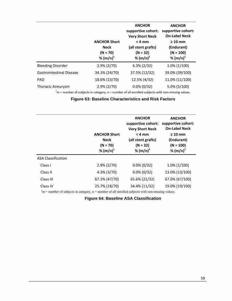
59
ANCHOR Short
Neck
(N = 70)
% (m/n)1
ANCHOR
supportive cohort:
Very Short Neck
< 4 mm
(all stent grafts)
(N = 32)
% (m/n)1
ANCHOR supportive cohort:On‐Label Neck
≥ 10 mm
(Endurant)
(N = 100)
% (m/n)1
Bleeding Disorder 2.9% (2/70) 6.3% (2/32) 1.0% (1/100)
Gastrointestinal Disease 34.3% (24/70) 37.5% (12/32) 39.0% (39/100)
PAD 18.6% (13/70) 12.5% (4/32) 11.0% (11/100)
Thoracic Aneurysm 2.9% (2/70) 0.0% (0/32) 5.0% (5/100) 1m = number of subjects in category, n = number of all enrolled subjects with non‐missing values.
Figure 63: Baseline Characteristics and Risk Factors
ANCHOR Short
Neck
(N = 70)
% (m/n)1
ANCHOR
supportive cohort:
Very Short Neck
< 4 mm
(all stent grafts)
(N = 32)
% (m/n)1
ANCHOR supportive cohort:On‐Label Neck
≥ 10 mm
(Endurant)
(N = 100)
% (m/n)1
ASA Classification
Class I 2.9% (2/70) 0.0% (0/32) 1.0% (1/100)
Class II 4.3% (3/70) 0.0% (0/32) 13.0% (13/100)
Class III 67.1% (47/70) 65.6% (21/32) 67.0% (67/100)
Class IV 25.7% (18/70) 34.4% (11/32) 19.0% (19/100) 1m = number of subjects in category, n = number of all enrolled subjects with non-missing values.
Figure 64: Baseline ASA Classification

60
6.3.3 Baseline Aneurysm Characteristics
Figure 65 provides the baseline aneurysm and anatomical measurements of the three cohorts. The mean maximum aneurysm diameter reported as per core lab was 57.70 ± 12.74 mm (range: 34.1 mm to 112.0 mm). The mean core lab-reported neck length (defined as that length over which the aortic diameter remains within 10% of the infrarenal diameter) was 6.86 ± 1.59 mm (range: 4.1 mm to 10.0 mm) for the Short Neck Cohort. Mean site-reported neck length (typically measured from the lowest main renal artery to where the neck visually dilates) was 12.07 ± 5.58 mm (range: 4.0 mm to 33.0 mm). The core lab methodology should be applied when determining whether EndoAnchors should be used with the endograft.
Measurement
ANCHOR Short
Neck
(Core laboratory
reported)
(N = 70)
ANCHOR
supportive cohort:
Very Short Neck
< 4 mm
(all stent grafts)
(Core laboratory
reported)
(N = 32)
ANCHOR supportive cohort:On‐Label Neck
≥ 10 mm
(Endurant)
(Core laboratory
reported)
(N = 100)
Proximal neck diameter at
renals
n 70 32 100
Mean ± SD 25.74 ± 4.04 25.55 ± 5.20 25.69 ± 4.13
Median 25.90 24.85 24.75
Min, Max 19.0, 36.5 16.9, 35.7 17.4, 39.8
Proximal Neck Length
n 70 32 100
Mean ± SD 6.86 ± 1.59 2.95 ± 0.82 22.37 ± 11.82
Median 6.69 3.03 18.20
Min, Max 4.1, 10.0 1.1, 4.0 10.0, 62.0
Distal Aortic Diameter
n 70 32 100
Mean ± SD 28.80 ± 4.79 28.34 ± 5.82 27.84 ± 4.53
Median 28.40 26.35 27.50
Min, Max 19.3, 40.5 19.1, 49.3 18.4, 43.2
Maximum aortic diameter
n 69 32 100
Mean ± SD 57.70 ± 12.74 56.46 ± 10.33 55.91 ± 11.06
Median 55.00 53.70 53.05
Min, Max 34.1, 112.0 41.7, 92.5 28.5, 101.0

61
Measurement
ANCHOR Short
Neck
(Core laboratory
reported)
(N = 70)
ANCHOR
supportive cohort:
Very Short Neck
< 4 mm
(all stent grafts)
(Core laboratory
reported)
(N = 32)
ANCHOR supportive cohort:On‐Label Neck
≥ 10 mm
(Endurant)
(Core laboratory
reported)
(N = 100)
Suprarenal Angulation
n 70 32 100
Mean ± SD 14.07 ± 8.28 13.31 ± 8.71 15.70 ± 10.34
Median 12.00 13.50 14.00
Min, Max 1.0, 38.0 2.0, 39.0 2.0, 49.0
Infrarenal Angulation
n 69 32 100
Mean ± SD 20.59 ± 14.44 23.16 ± 15.98 26.14 ± 18.55
Median 18.00 18.50 22.00
Min, Max 2.0, 69.0 1.0, 68.0 1.0, 80.9
Neck Thrombus Average
Thickness
n 64 29 94
Mean ± SD 0.85 ± 1.06 1.23 ± 1.42 0.89 ± 1.48
Median 0.00 1.30 0.00
Min, Max 0.0, 3.5 0.0, 5.3 0.0, 8.9
Neck Thrombus
Circumference > 1 mm
n 64 29 94
Mean ± SD 61.41 ± 86.33 62.94 ± 80.45 46.63 ± 72.63
Median 0.00 19.60 0.00
Min, Max 0.0, 320.0 0.0, 298.0 0.0, 320.0
Neck Calcium Average
Thickness
n 70 32 100
Mean ± SD 1.31 ± 1.23 1.77 ± 1.28 1.03 ± 1.11
Median 1.30 1.94 1.00
Min, Max 0.0, 4.0 0.0, 4.3 0.0, 4.6
Neck Calcium Circumference
> 1 mm

62
Measurement
ANCHOR Short
Neck
(Core laboratory
reported)
(N = 70)
ANCHOR
supportive cohort:
Very Short Neck
< 4 mm
(all stent grafts)
(Core laboratory
reported)
(N = 32)
ANCHOR supportive cohort:On‐Label Neck
≥ 10 mm
(Endurant)
(Core laboratory
reported)
(N = 100)
n 70 32 100
Mean ± SD 23.39 ± 29.83 38.50 ± 43.62 18.26 ± 24.91
Median 16.25 20.40 10.00
Min, Max 0.0, 155.0 0.0, 180.0 0.0, 114.0
Figure 65: Anatomical and Other Measurements (Core Laboratory-Reported)
6.3.4 Devices Implanted
Endurant II/Endurant IIs Stent Graft System: Endurant stent grafts were utilized in 47.1% (33/70) of subjects in the Short Neck Cohort and Endurant II/Endurant IIs stent grafts were utilized in 52.9% (37/70) of the subjects. The most frequently used proximal stent graft size in the Short Neck Cohort was 36 mm, which was used in 34.3% (24/70) of subjects. Most subjects were implanted with an Endurant or Endurant II/Endurant IIs stent graft with a proximal diameter of 28 mm or larger.
Heli-FX EndoAnchor implants: The average number of EndoAnchor implants per subject was 5.49 ± 2.08, with a median of 5.00, minimum of 2.0 and maximum of 12.0 (n=70).
Endurant/Endurant II/Endurant IIs Stent Graft System and Heli-FX EndoAnchor System: Figure 66 shows the proximal diameter of the endografts used in the Short Neck Cohort, the recommended number of EndoAnchors (as per the Heli-FX EndoAnchor IFU) and the actual number of EndoAnchors used, by percentage of subjects.

63
Proximal Size of the Endurant Stent Graft
23 mm (m/n)%
25 mm (m/n)%
28 mm (m/n)%
32 mm (m/n)%
36 mm (m/n)%
7.1% (5/70) 8.6% (6/70) 25.7% (18/70) 24.3% (17/70) 34.3% (24/70)
Recommended Number of EndoAnchor Implants1
4 4 4 4 6
Number of EndoAnchor Implants Used
2 0.0% (0/5) 0.0% (0/6) 5.6% (1/18) 5.9% (1/17) 0.0% (0/24)
3 20.0% (1/5) 16.7% (1/6) 0.0% (0/18) 0.0% (0/17) 8.3% (2/24)
4 40.0% (2/5) 16.7% (1/6) 44.4% (8/18) 41.2% (7/17) 33.3% (8/24)
5 20.0% (1/5) 16.7% (1/6) 11.1% (2/18) 5.9% (1/17) 0.0% (0/24)
6 20.0% (1/5) 33.3% (2/6) 27.8% (5/18) 17.6% (3/17) 29.2% (7/24)
> 6 0.0% (0/5) 16.7% (1/6) 11.1% (2/18) 29.4% (5/17) 29.2% (7/24)
Based on number of all enrolled subjects within the Short Neck Cohort with available data. m = number of subjects in category, n = number of all enrolled subjects within the Short Neck Cohort with non-missing values. 1The Heli-FX EndoAnchor System IFU provides a minimum recommended number of EndoAnchor implants based on aortic neck diameter, not device size. The recommended number of EndoAnchor implants used in an aortic neck of ≤ 29 mm is 4, and a minimum of 6 EndoAnchor implants are recommended in an aortic neck >29. The numbers provided in this table are based on the most likely aortic neck diameter for a given device size, based on the Endurant II/Endurant IIs Stent Graft System IFU recommended device sizing relative to the aortic vessel inner diameter.
Figure 66: Endurant Stent Graft Sizing (Proximal Diameter) and Heli‐FX EndoAnchor System
Heli-FX EndoAnchor System: Figure 67 provides information regarding the appliers and guides utilized for the Short Neck Cohort.
Short Neck Cohort
(N = 70)
Number of Heli-FX Appliers Used
n 49
Mean ± SD 1.02 ± 0.14
Median 1.00
Min, Max 1.0, 2.0
Size of Heli-FX Applier(s) Used (m/n)%
SA-85 Heli-FX Applier 98.0% (48/49)
HA-18-114 Heli-FX Applier 2.0% (1/49)
Number of Heli-FX Guides Used
n 49
Mean ± SD 1.04 ± 0.20
Median 1.00

64
Short Neck Cohort
(N = 70)
Min, Max 1.0, 2.0
Size of Heli-FX Guide(s) Used (m/n)%
Heli-FX Guide 22 mm – SG-64 73.5% (36/49)
Heli-FX Guide 22 mm – HG-18-90-22 0.0% (0/49)
Heli-FX Guide 42 mm – HG-18-90-42 0.0% (0/49)
Heli-FX Guide 28 mm – HG-16-62-28 26.5% (13/49)
Heli-FX Guide 32 mm – HG-18-90-32 2.0% (1/49)
Min, Max 2.0, 12.0 Based on number of all enrolled subjects within the Short Neck Cohort with available data. m = number of subjects in category, n = number of all enrolled subjects within the Short Neck Cohort with non-missing values.
Figure 67: Heli-FX EndoAnchor System Usage
Accessory Device Usage: Twenty-five of 70 subjects in the Short Neck Cohort received at least one accessory device during the index procedure. The type of accessory device used and number of subjects is as follows: Stent (11 subjects), balloon (10 subjects), coil (4 subjects), cuff (3 subjects), vascular plug (2 subjects), and vascular graft (1 subject). Note that subjects may have received more than one accessory device.
6.3.5 Acute Procedural Data
Acute procedural data for the ANCHOR Short Neck Cohort are shown in Figure 68. The mean duration of the procedure was 148.00 ± 80.03 minutes, with average time of EndoAnchor implant being 17.12 ± 11.54 minutes. The mean overall intensive care unit (ICU) stay was 0.84 ± 1.63 days, and the mean overall hospital stay was 3.73 ± 4.31 days.
Measurement Short Neck Cohort
(N = 70)
Type of Procedure (m/n)%
Elective 69.4% (34/49)
Urgent (investigator assessed) 22.4% (11/49)
Emergent (investigator assessed) 8.2% (4/49)
Procedure Entry Site1 (m/n)%
Left Femoral Artery 82.9% (58/70)
Right Femoral Artery 91.4% (64/70)
Left Iliac Artery 0.0% (0/70)
Right Iliac Artery 0.0% (0/70)
Left: Other - Brachial percutaneous 1.4% (1/70)
Right: Other - Brachial 1.4% (1/70)
Type of Access (m/n)%
Open 62.9% (44/70)

65
Measurement Short Neck Cohort
(N = 70)
Percutaneous 37.1% (26/70)
Duration of implant procedure (min)
N 70
Mean ± SD 148.00 ± 80.03
Median 127.50
Min, Max 38.0, 423.0
Type of anesthesia used (m/n)%
General 84.3% (59/70)
Spinal 2.9% (2/70)
Epidural 1.4% (1/70)
Local 11.4% (8/70)
Volume of contrast (cc)
N 26
Mean ± SD 126.12 ± 80.76
Median 102.50
Min, Max 30.0, 399.0
Total fluoroscopic time (mins)
N 44
Mean ± SD 35.34 ± 21.98
Median 30.00
Min, Max 7.0, 123.0
Subjects Receiving EndoAnchor implants (m/n)% 100.0% (70/70)
Time to Implant EndoAnchor implants (mins)5
N 68
Mean ± SD 17.12 ± 11.54
Median 13.50
Min, Max 4.0, 60.0
Hospital stay (Days)2
N 70
Mean ± SD 3.73 ± 4.31
Median 2.00
Min, Max 1.0, 24.0
Procedure stay (Days)3
N 70
Mean ± SD 3.03 ± 3.17
Median 2.00

66
Measurement Short Neck Cohort
(N = 70)
Min, Max 1.0, 23.0
Duration of ICU stay (Days)4
N 69
Mean ± SD 0.84 ± 1.63
Median 0.00
Min, Max 0.0, 11.0 Based on number of all enrolled subjects within the Short Neck Cohort with available data.
m = number of subjects in category, n = number of all enrolled subjects within the Short Neck Cohort with non-missing values. 1Note that more than one procedure entry site per subject may be reported if multiple entry sites were used. 2Overall hospital stay (days) = Date of Hospital Discharge – Date of Hospital Admission. In the case where Date of Hospital Discharge = Date of Hospital Admission, Overall hospital stay will be considered to be 0.5 day3Procedural hospital stay (days) = Date of Hospital Discharge – Date of Initial Procedure. In the case where Date of Hospital Discharge = Date of Initial Procedure, procedural hospital stay will be considered to be 0.5 day 4ICU stay: days collected as 0, 1, 2…10 days or 10+days. 10+days set to 11 days for summary statistics 5 EndoAnchor total implant time includes all EndoAnchor implants for one subject
Figure 68: Initial Procedural Data – Short Neck Cohort
6.3.6 Study Results: Safety Outcomes
Safety outcomes are reported in terms of the prospectively identified safety-related measures shown in Figure 69, with further detail on ARM in Figure 70 and other safety outcomes included in Figures 71 to 76.
Aneurysm-related mortality is defined as any death within 30 days of the index procedure or secondary procedure to address aneurysm, or death from any rupture. Four of 70 subjects (5.7%) died within 30 days of the index procedure. It is important to note that the adverse events leading to death were assessed by the investigator, and no events were noted as related to the aneurysm.
No subjects experienced aneurysm rupture through one year through one year. 15.7% (11/70) of Short Neck Cohort subjects experienced at least one major adverse event through one month. One subject experienced renal insufficiency within 30 days of the index procedure. No renal failure was reported by sites through one month; however, one subject with a history of renal insufficiency died on Day 13, and the investigator-determined cause of death included renal failure.
Safety-related measures ANCHOR Short Neck
(N = 70)
ANCHOR supportive cohort: Very Short Neck
< 4 mm (all stent grafts)
(N = 32)
ANCHOR supportive cohort:On‐Label Neck
≥ 10 mm (Endurant) (N = 100)
Aneurysm-related mortality through 30 days
5.7% (4/70) 6.3% (2/32) 0.0% (0/100)
Aneurysm-related mortality through 12 months
5.9% (4/68) 7.1% (2/28) 0.0% (0/91)

67
Safety-related measures ANCHOR Short Neck
(N = 70)
ANCHOR supportive cohort: Very Short Neck
< 4 mm (all stent grafts)
(N = 32)
ANCHOR supportive cohort:On‐Label Neck
≥ 10 mm (Endurant) (N = 100)
Aneurysm rupture through 30 days
0.0% (0/70) 0.0% (0/32) 0.0% (0/100)
Aneurysm rupture through 12 months
0.0% (0/64) 0.0% (0/26) 0.0% (0/91)
Major adverse events through 30 days
15.7% (11/70) 9.4% (3/32) 4.0% (4/100)
Renal insufficiency through 30 days1
0.0% (0/70)4 6.3% (2/32) 0.0% (0/100)
Renal failure through 30 days2 0.0% (0/70)3 0.0% (0/32) 0.0% (0/100)
m = number of subjects in category, n = number of subjects with available imaging assessments for aneurysm expansion and migration and endoleak at 12 months, and number of subjects who had an event in question or reached the lower time window of the time period for site-reported events. 1A rise in creatinine greater than 50% above the pre-procedure level resulting in a creatinine level above the upper limit of normal (site reported) 2Defined as when the need for dialysis is required, an increase in serum creatinine of 2x the baseline value or new need for hemodialysis (site reported) 3Subject 109-031 died on Day 13 post-index procedure and the investigator-determined cause of death included acute renal failure. Acute renal failure was captured under multi-system organ failure in Figure 73. 4Subject 138-012 experienced renal insufficiency through 30 days from the index procedure. The site entered the start date of this event as a partial date of March 2014 in the database. Due to the partial date entry, the days from index for this event was imputed per the statistical analysis plan. The imputation of this event resulted in the event populating at -2 days from index procedure. Although the exact start date is unknown, it is known that the event occurred within the same month of the index procedure. The renal insufficiency category in this table captures events between Day 0-30. Due to this, the renal insufficiency event for this subject at -2 days is not populating in this table.
Figure 69: Summary of Safety-Related Measures

68
Subject Number
Days to Death
Death within 30 days of the
initial procedure
Death within 30 days of a
re‐intervention
Cause of Death
(Investigator Determined)
172‐014 6 Yes No Cardiac arrest, congestive heart failure
109‐009 9 Yes No Cardiac and Respiratory Arrest
172‐026 5 Yes No Cardiac Arrest
109‐031 13 Yes No Acute Alcoholic Hepatitis, Acute Renal Failure, Acute Pancreatitis
Based on number of all enrolled subjects within the Short Neck Cohort with available data.
Figure 70: Summary of Aneurysm-related Deaths through 12 Months
6.3.6.1 Additional Safety Outcomes
Key additional safety outcomes of all-cause mortality, major adverse events through 12 months and serious adverse events through 12 months are shown in Figures 71 to 76.
Of the 70 subjects in the Short Neck Cohort, five died within the first 12 months of the index procedure. No subjects died on the day of the index procedure. Four of 70 subjects (5.7%) died within the 30 days of the index procedure. One additional subject died on Day 353 post-index procedure due to septic shock secondary to pneumonia/ septicemia. This event was not determined to be related to the aneurysm, device, or index procedure.
Kaplan-Meier survival estimates for all-cause mortality (ACM) were made through 365 days post-implant as shown in Figures 71 and 72. The Kaplan-Meier one-year survival estimate for ACM was 92.6%.
Day 0 Day 30 Day 182 Day 365
No. at Risk1 70 66 61 53
No. of Events 0 4 4 5
No. Censored2 0 0 5 12
Kaplan‐Meier Estimate3 1.000 0.943 0.943 0.926
Peto Standard Error 0.000 0.028 0.028 0.034 1Number of subjects at risk at each timepoint. Based on number of all enrolled subjects within the Short Neck Cohort with available data. 2Subjects are censored because no event was observed by the time point, including those not yet reached the correspondent time point or lost to follow-up. 3Estimate made at each timepoint. Note: All numbers except standard errors are cumulative.
Figure 71: Kaplan-Meier Estimates for All-Cause Mortality – Short Neck Cohort Subjects

69
Figure 12: Freedom from All-Cause Mortality
A total of 18 MAEs were reported among 15 subjects between 0 and 365 days. Among those, 13 MAEs were reported in 11 subjects between 0 and 30 days, and five MAEs were reported in five subjects between 31 and 365 days. Three subjects had more than one MAE within the first year.
There were no reports of paraplegia, or renal failure through 12 months. However, as mentioned previously, one subject with a history of renal insufficiency died on Day 13 post-index procedure and the investigator-determined cause of death included acute renal failure.
Event 0-30 Days % (m/n)
31-365 Days
% (m/n) 0-365 Days% (m/n)
One or more major adverse events (MAE)
15.7% (11/70) 7.6% (5/66) 21.4% (15/70)
Total Number of MAEs 13 5 18
All-Cause Mortality 5.7% (4/70) 1.5% (1/66) 7.1% (5/70)
Bowel ischemia 1.4% (1/70) 0.0% (0/66) 1.4% (1/70)
Myocardial infarction 2.9% (2/70) 1.5% (1/66) 2.9% (2/70)
Paraplegia 0.0% (0/70) 0.0% (0/66) 0.0% (0/70)
Procedural blood loss1 7.1% (5/70) 0.0% (0/66) 7.1% (5/70)

70
Event 0-30 Days % (m/n)
31-365 Days
% (m/n) 0-365 Days% (m/n)
Renal failure 0.0% (0/70)2 0.0% (0/66) 0.0% (0/70)2
Respiratory failure 1.4% (1/70) 3.0% (2/66) 4.3% (3/70)
Stroke 0.0% (0/70) 1.5% (1/66) 1.4% (1/70) Based on number of all enrolled subjects within the Short Neck Cohort with available data. m = number of subjects in category, n = number of subjects are at risk at the beginning of the time period. 1Volume of blood loss was not collected, therefore, procedural blood loss/hemorrhage reported as an SAE at the time of the index procedure or secondary procedure has been reported as an MAE 2 Subject 109-031 died on Day 13 post-index procedure and the investigator-determined cause of death included acute renal failure. Acute renal failure was captured under multi-system organ failure in Figure 74.
Figure 73: Major Adverse Events (MAEs) through 12 months
Thirty of 70 subjects (42.9%) experienced one or more serious adverse event between 0 and 365 days. Of those, 16 of 70 subjects (22.9%) experienced one or more serious adverse event between 0 and 30 days, and 19 out of 66 subjects (28.8%) experienced one or more serious adverse event between 31 and 365 days.
The most common type of SAEs were cardiac disorders, reported in 9 out of 70 subjects (12.9%). The second most common type of SAEs were vascular disorders and gastrointestinal disorders, reported in 7 out of 70 subjects (10.0%).
Category 0 to 30 Days
% (m/n)
31 to 365 Days
% (m/n) 0 to 365 Days
% (m/n) Number of Subjects Eligible for each follow-up window
70 66 70
Subjects Experiencing One or More SAEs1
22.9% (16/70) 28.8% (19/66) 42.9% (30/70)
Blood and lymphatic system disorders
2.9% (2/70) 1.5% (1/66) 4.3% (3/70)
Anemia 1.4% (1/70) 0.0% (0/66) 1.4% (1/70) Anemia postoperative 1.4% (1/70) 0.0% (0/66) 1.4% (1/70) Neutropenic fever 0.0% (0/70) 1.5% (1/66) 1.4% (1/70) Cardiac disorders 7.1% (5/70) 7.6% (5/66) 12.9% (9/70) Arrhythmia 1.4% (1/70) 0.0% (0/66) 1.4% (1/70) Cardiac arrest 2.9% (2/70) 0.0% (0/66) 2.9% (2/70) Congestive cardiac failure aggravated
1.4% (1/70) 0.0% (0/66) 1.4% (1/70)
Congestive heart failure 0.0% (0/70) 4.5% (3/66) 4.3% (3/70) Coronary artery disease aggravated 0.0% (0/70) 1.5% (1/66) 1.4% (1/70) Myocardial infarction 2.9% (2/70) 1.5% (1/66) 2.9% (2/70) Ventricular tachycardia 0.0% (0/70) 1.5% (1/66) 1.4% (1/70) Gastrointestinal disorders 2.9% (2/70) 7.6% (5/66) 10.0% (7/70)

71
Category 0 to 30 Days
% (m/n)
31 to 365 Days
% (m/n) 0 to 365 Days
% (m/n) Diverticulitis 0.0% (0/70) 1.5% (1/66) 1.4% (1/70) GI bleed 1.4% (1/70) 0.0% (0/66) 1.4% (1/70) Hernia inguinal 0.0% (0/70) 1.5% (1/66) 1.4% (1/70) Ischemic colitis Pancreatic cancer
1.4% (1/70) 0.0% (0/70)
0.0% (0/66) 1.5% (1/66)
1.4% (1/70) 1.4% (1/70)
Retroperitoneal hematoma 0.0% (0/70) 1.5% (1/66) 1.4% (1/70) Retroperitoneal mass 0.0% (0/70) 1.5% (1/66) 1.4% (1/70) General disorders and administration site conditions
4.3% (3/70) 0.0% (0/66) 4.3% (3/70)
Device occlusion 1.4% (1/70) 0.0% (0/66) 1.4% (1/70) Multi organ failure 1.4% (1/70) 0.0% (0/66) 1.4% (1/70) Stent-graft endoleak 1.4% (1/70) 0.0% (0/66) 1.4% (1/70) Infections and infestations 1.4% (1/70) 6.1% (4/66) 7.1% (5/70) Bronchitis 0.0% (0/70) 1.5% (1/66) 1.4% (1/70) Pneumonia 0.0% (0/70) 3.0% (2/66) 2.9% (2/70) Sepsis 1.4% (1/70) 0.0% (0/66) 1.4% (1/70) Septicaemia 0.0% (0/70) 1.5% (1/66) 1.4% (1/70) Urinary tract infection 0.0% (0/70) 1.5% (1/66) 1.4% (1/70) Injury, poisoning and procedural complications
4.3% (3/70) 1.5% (1/66) 5.7% (4/70)
Femoral artery injury 1.4% (1/70) 0.0% (0/66) 1.4% (1/70) Post procedural bleeding 2.9% (2/70) 0.0% (0/66) 2.9% (2/70) Procedural bleeding 2.9% (2/70) 0.0% (0/66) 2.9% (2/70) Scalp laceration 0.0% (0/70) 1.5% (1/66) 1.4% (1/70) Vascular access site bleeding 1.4% (1/70) 0.0% (0/66) 1.4% (1/70) Metabolism and nutrition disorders
1.4% (1/70) 3.0% (2/66) 4.3% (3/70)
Respiratory failure 1.4% (1/70) 3.0% (2/66) 4.3% (3/70) Neoplasms benign, malignant and unspecified (including cysts and polyps)
0.0% (0/70) 3.0% (2/66) 2.9% (2/70)
Bladder cancer recurrent 0.0% (0/70) 1.5% (1/66) 1.4% (1/70) Hepatic cancer 0.0% (0/70) 1.5% (1/66) 1.4% (1/70) Nervous system disorders 0.0% (0/70) 1.5% (1/66) 1.4% (1/70) Subarachnoid hemorrhage 0.0% (0/70) 1.5% (1/66) 1.4% (1/70) Psychiatric disorders 1.4% (1/70) 0.0% (0/66) 1.4% (1/70) Mental status changes 1.4% (1/70) 0.0% (0/66) 1.4% (1/70) Renal and urinary disorders 0.0% (0/70) 1.5% (1/66) 1.4% (1/70) Renal insufficiency 0.0% (0/70) 1.5% (1/66) 1.4% (1/70) Respiratory, thoracic and mediastinal disorders
0.0% (0/70) 1.5% (1/66) 1.4% (1/70)

72
Category 0 to 30 Days
% (m/n)
31 to 365 Days
% (m/n) 0 to 365 Days
% (m/n) COPD exacerbation 0.0% (0/70) 1.5% (1/66) 1.4% (1/70) Skin and subcutaneous tissue disorders
0.0% (0/70) 1.5% (1/66) 1.4% (1/70)
Lower extremities ulcers of 0.0% (0/70) 1.5% (1/66) 1.4% (1/70) Vascular disorders 5.7% (4/70) 6.1% (4/66) 10.0% (7/70) Aneurysm 1.4% (1/70) 1.5% (1/66) 2.9% (2/70) Deep vein thrombosis 1.4% (1/70) 0.0% (0/66) 1.4% (1/70) Hypertension 0.0% (0/70) 1.5% (1/66) 1.4% (1/70) Peripheral artery dissection 0.0% (0/70) 1.5% (1/66) 1.4% (1/70) Peripheral ischemia 1.4% (1/70) 1.5% (1/66) 2.9% (2/70) Thrombosis 1.4% (1/70) 1.5% (1/66) 2.9% (2/70) Based on number of all enrolled subjects within the Short Neck Cohort with available data. m = number of subjects experiencing one or more serious adverse events in a category, n = number of subjects at risk at the beginning of the time period. 1A subject may report multiple adverse events and in different subcategories; hence, number of subjects in each category may not be the sum of those in each subcategory. Each subject was only counted once in each subcategory and category.
Figure 74: Serious Adverse Events (SAEs) through 12 months
Investigators were asked to assess relatedness of reported SAEs to device, index or re-intervention procedure, or AAA disease, results of which are shown in Figure 75. All events determined to be related to device, procedure, or AAA occurred within 30 days of the index procedure, many of which occurred on the day of the index procedure. There were no re-intervention-related SAEs or unanticipated adverse device effects (UADEs) reported through 365 days. There were no device-related SAEs related to EndoAnchor implants.
Two out of 70 subjects (2.9%) were reported to have an AAA-related SAE between 0 and 30 days, both occurred on Day 0. One subject experienced a femoral artery injury and bleeding at the vascular access site. The other subject experienced post-procedural bleeding.
One out of 70 subjects was reported to have a device-related SAE between 0 and 30 days, which was an endograft limb occlusion. An additional subject had a device-related event at 59 days, which was peripheral ischemia. There were no device-related SAEs related to EndoAnchor implants.
0‐30 Days % (m/n)
31‐365 Days % (m/n)
0‐365 Days % (m/n)
Index procedure related SAEs 12.9% (9/70) 0.0% (0/66) 12.9% (9/70)
Re‐intervention related SAEs 0.0% (0/70) 0.0% (0/66) 0.0% (0/70)
AAA‐related SAEs 2.9% (2/70) 0.0% (0/66) 2.9% (2/70)
Device related SAE 1.4% (1/70) 0.0% (0/66) 1.4% (1/70)
Related to Endograft 1.4% (1/70) 0.0% (0/66) 1.4% (1/70)
Related to Heli‐FX 0.0% (0/70) 0.0% (0/66) 0.0% (0/70)
Related to both 0.0% (0/70) 0.0% (0/66) 0.0% (0/70)
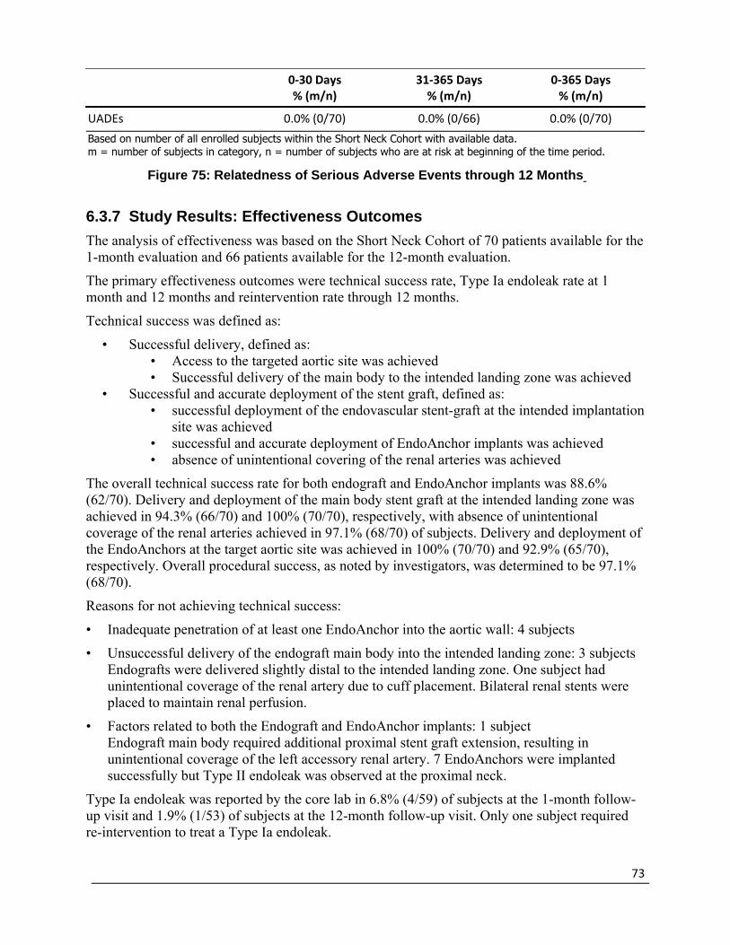
73
0‐30 Days % (m/n)
31‐365 Days % (m/n)
0‐365 Days % (m/n)
UADEs 0.0% (0/70) 0.0% (0/66) 0.0% (0/70)
Based on number of all enrolled subjects within the Short Neck Cohort with available data. m = number of subjects in category, n = number of subjects who are at risk at beginning of the time period.
Figure 75: Relatedness of Serious Adverse Events through 12 Months
6.3.7 Study Results: Effectiveness Outcomes
The analysis of effectiveness was based on the Short Neck Cohort of 70 patients available for the 1-month evaluation and 66 patients available for the 12-month evaluation.
The primary effectiveness outcomes were technical success rate, Type Ia endoleak rate at 1 month and 12 months and reintervention rate through 12 months.
Technical success was defined as:
• Successful delivery, defined as: • Access to the targeted aortic site was achieved • Successful delivery of the main body to the intended landing zone was achieved
• Successful and accurate deployment of the stent graft, defined as: • successful deployment of the endovascular stent-graft at the intended implantation site was achieved • successful and accurate deployment of EndoAnchor implants was achieved • absence of unintentional covering of the renal arteries was achieved
The overall technical success rate for both endograft and EndoAnchor implants was 88.6% (62/70). Delivery and deployment of the main body stent graft at the intended landing zone was achieved in 94.3% (66/70) and 100% (70/70), respectively, with absence of unintentional coverage of the renal arteries achieved in 97.1% (68/70) of subjects. Delivery and deployment of the EndoAnchors at the target aortic site was achieved in 100% (70/70) and 92.9% (65/70), respectively. Overall procedural success, as noted by investigators, was determined to be 97.1% (68/70).
Reasons for not achieving technical success:
• Inadequate penetration of at least one EndoAnchor into the aortic wall: 4 subjects
• Unsuccessful delivery of the endograft main body into the intended landing zone: 3 subjects Endografts were delivered slightly distal to the intended landing zone. One subject had unintentional coverage of the renal artery due to cuff placement. Bilateral renal stents were placed to maintain renal perfusion.
• Factors related to both the Endograft and EndoAnchor implants: 1 subject Endograft main body required additional proximal stent graft extension, resulting in unintentional coverage of the left accessory renal artery. 7 EndoAnchors were implanted successfully but Type II endoleak was observed at the proximal neck.
Type Ia endoleak was reported by the core lab in 6.8% (4/59) of subjects at the 1-month follow-up visit and 1.9% (1/53) of subjects at the 12-month follow-up visit. Only one subject required re-intervention to treat a Type Ia endoleak.

74
Three of 64 subjects (4.7%) had one or more re-interventions through 12 months. Thrombosis, occlusion, and Type Ia endoleak were the reasons for reintervention. There were no conversions to open surgical repair through 12 months.
A summary of primary outcomes for the Short Neck Cohort, Very Short Neck Cohort and On-Label Neck Cohort is provided in Figure 76, below. This is followed by a discussion of the impact of the number of EndoAnchors used on primary outcomes.

75
Figure 76: Summary of Primary Effectiveness Outcomes
Event
ANCHOR Short Neck
(N = 70) 95% Confidence
Interval
ANCHOR supportive cohort: Very Short Neck
< 4 mm (all stent grafts)
(N = 32)
ANCHOR supportive cohort: On‐Label Neck
≥ 10 mm (Endurant) (N = 100)
Type Ia Endoleak at 1-month1 6.8% (4/59) [1.9%, 16.5%] 16.0% (4/25) 0.0% (0/91)
Type Ia Endoleak at 1-year1 1.9% (1/53) [0.0%, 10.1%] 0.0% (0/20) 0.0% (0/73)
Secondary Procedures through 1 year 4.7% (3/64) [1.0%, 13.1%] 7.7% (2/26) 2.2% (2/91)
Secondary Endovascular Procedures through 1 year 4.7% (3/64) [1.0%, 13.1%] 7.7% (2/26) 2.2% (2/91)
Conversion to Open Surgical Repair through 1 year 0.0% (0/64) [0.0%, 5.6%] 0.0% (0/26) 0.0% (0/91)
Other Secondary Open Surgical Procedures through 1 year 1.6% (1/64) [0.0%, 8.4%] 0.0% (0/26) 0.0% (0/91)
Technical Success Rate at Index Procedure2: 88.6% (62/70) [78.7%, 94.9%] 84.4% (27/32) 94.9% (94/99)
Successful delivery:
Access to the targeted aortic site was achieved by the EndoAnchor system
100.0% (70/70) [94.9%, 100.0%] 100.0% (32/32) 100.0% (100/100)
Successful delivery of the main body to the intended landing zone
94.3% (66/70) [86.0%, 98.4%] 96.9% (31/32) 99.0% (99/100)
Successful and accurate deployment of the Endurant II/Endurant IIs stent graft and the Aptus Heli-FX EndoAnchor System
Successful deployment of the endovascular stent graft at the intended implantation site3
100.0% (70/70) [94.9%, 100.0%] 100.0% (32/32) 98.0% (97/99)
Successful and accurate deployment of EndoAnchor implants was acheived4
92.9% (65/70) [84.1%, 97.6%] 87.5% (28/32) 96.0% (96/100)
Absence of unintentional coverage of the renal arteries
97.1% (68/70) [90.1%, 99.7%] 96.9% (31/32) 100.0% (100/100)

76
Based on number of all enrolled subjects within the Short Neck Cohort with available data. m = number of subjects in category, n = number of subjects with available values. For secondary procedures through 1 year, the denominator includes subjects who had an event or had been followed for at least 184 days. 1Core laboratory reported 2Technical success was defined as: successful delivery and deployment of the stent graft, without unintentional coverage of the renal arteries, and successful implantation of the EndoAnchor implants at the target aortic site. 3Absence of misdeployment of the main body proximal to the intended landing zone 4Each EndoAnchor adequately penetrated the aortic wall (investigator’s assessment) and, in the investigator’s opinion, the implantation of the EndoAnchor implants was successful.

77
18 subjects did not have the recommended number of EndoAnchor devices implanted per the Heli-FX EndoAnchor System IFU. 15 of these achieved technical success, that is, successful delivery and deployment of the endograft and EndoAnchors without unintentional coverage of the renal arteries. Three subjects did not achieve technical success due to inadequate penetration of at least one EndoAnchor into the aortic wall. Three of the four subjects who reported Type Ia endoleak at 1-month follow-up had less than the recommended number of EndoAnchors implanted. Two of the three Short Neck Cohort subjects who underwent secondary procedures through 12 months were implanted with less than the recommended number of EndoAnchor implants, however, one of the two subjects had an intervention that was entirely unrelated to the quantity of EndoAnchor implants.
Subject Number
Recommended Number of
EndoAnchor Implants1
Number ofEndoAnchor
Implants Used
TechnicalSuccess
(Yes/No/UNK)
Type Ia Endoleak at
30 days2 (Yes/No/UNK)
Type Ia Endoleak at 12 months2 (Yes/No/
UNK)
Re-interventionthrough 365
days (Yes/No/UNK)
100-052 4 3 No No UNK UNK
109-041 6 2 Yes No No No
109-002 6 4 Yes Yes Yes Yes
109-048 6 3 Yes Yes No Yes
109-057 6 4 Yes No No No
109-016 6 4 Yes No No No
109-042 4 2 Yes No No No
109-039 6 4 Yes No UNK No
135-004 6 4 Yes No No No
135-006 6 4 No No No No
155-002 6 4 Yes UNK UNK No
168-010 6 4 Yes No No No
172-005 6 4 Yes No No No
172-024 6 4 Yes No No No
178-009 6 4 Yes No No No
178-011 6 4 No Yes UNK No
178-005 6 4 Yes No No No
189-003 6 3 Yes No No No Based on number of all enrolled subjects within the Short Neck Cohort with available data. 1Based on the IFU, the minimum recommended number of EndoAnchor implants used in an aortic neck of ≤ 29 mm is 4, and a minimum of 6 EndoAnchor implants are recommended in an aortic neck > 29 mm 2Core laboratory reported
Figure 77: Outcomes in Subjects where the Recommended Number of EndoAnchor Implants were Not Used

78
6.3.7.1 Supportive Effectiveness Outcomes
Supportive effectiveness outcomes included initial implantation outcomes, device-related measures (loss of endograft or EndoAnchor device integrity) and maintenance of EndoAnchor implants in the aortic wall.
Investigators reported an overall procedure success of 97.1% (68/70); unsuccessful procedures were reported in one subject due to a Type II endoleak observed at the proximal neck and in a second subject due to a persistent Type Ia endoleak.
Event
Short Neck Cohort (N = 70) (m/n)%
Overall Procedure was successful (Investigator assessed)
97.1% (68/70)
Endograft Components Misdeployed Proximally to Intended Landing Zone
Stent Graft Main Body1 0.0% (0/70)
Left Iliac Extension 0.0% (0/70)
Right Iliac Extension 0.0% (0/70)
Aortic Cuff 1.4% (1/70)
Endoleak present at end of procedure
Type Ia 12.9% (9/70)
Type Ib 1.4% (1/70)
Type II 20.0% (14/70)
Type III 0.0% (0/70)
Type IV 7.1% (5/70) Based on number of all enrolled subjects within the Short Neck Cohort with available data. m = number of subjects in category, n = number of subjects with available values 1Misdeployment of the main body component proximally to the intended landing zone is a component of technical success
Figure 78: Initial Implantation Outcomes
There was no loss of endograft or EndoAnchor device integrity reported within the 1-month or 12-month follow-up window for the Short Neck Cohort.
EndoAnchor implants maintained adequate penetration through the aortic wall in 98% (50/51) of the subjects at 30 days and in 94.9% (37/39) of subjects at 12 months. See Figure 79.

79
% (m/n)
Did EndoAnchor implants maintain adequate penetration into aortic wall
30 days 98.0% (50/51)
12 months 94.9% (37/39) Based on number of all enrolled subjects within the Short Neck Cohort with available data. m = number of subjects in category, n = number of subjects with available values
Figure 79: Maintenance of Adequate Penetration by EndoAnchor Implants
6.3.7.2 Other Effectiveness-Related Measures
Other effectiveness-related measures of interest are summarized in Figure 80. Error! Reference source not found.Three of 70 Short Neck Cohort subjects underwent secondary procedures through 1 month. No subjects underwent conversion to open surgical repair through 12 months. Type III endoleaks were reported by core lab in 1.7% (1/59) of subjects at 1-month follow-up and 1.9% (1/53) of subjects at 12-month follow-up. No subjects experienced endograft migration at one year.
Effectiveness-related measures
ANCHOR Short Neck(N = 70)
ANCHOR supportive cohort: Very Short Neck
< 4 mm (all stent grafts)
(N = 32)
ANCHOR supportive cohort:On Label Neck
≥ 10 mm (Endurant) (N = 100)
Aneurysm expansion at 12 months1,2
0.0% (0/54) 5.3% (1/19) 0.0% (0/73)
Migration at 12 months1,3 0.0% (0/41) 0.0% (0/16) 0.0% (0/58) Secondary procedures through 30 days
4.3% (3/70) 0.0% (0/32) 1.0% (1/100)
Conversion to open surgical repair through 12 months
0.0% (0/64) -- --
Type III endoleaks at 30 days1 1.7% (1/59) -- -- Type III endoleaks at 12 months1 1.9% (1/53) -- -- m = number of subjects in category, n = number of subjects with available imaging assessments for aneurysm expansion and migration and endoleak at 12 months, and number of subjects who had an event in question or reached the lower time window of the time period for site-reported events. 1Based on core laboratory related data. 2Aneurysm expansion (alternatively noted as AAA diameter increase) is defined as a > 5 mm increase in maximum diameter as compared to 1 month post-implantation measurement. 3Migration is defined as a > 10mm movement of the aortic endograft from its position at the 1 month post-implantation measurement.
Figure 80: Summary of Effectiveness-Related Measures

80

81
6.3.8 Data Post 12 Months
As of the data cut-off for this summary, one core lab-reported Type Ia endoleak was identified in one subject on Day 1271. The subject did not undergo re-intervention to treat it. One subject underwent a secondary endovascular procedure and conversion to open surgical repair on Day 900 to treat a Type II endoleak and aneurysm enlargement. One subject underwent a secondary endovascular procedure on Day 758 to treat a Type Ib endoleak and aneurysm enlargement. No aneurysm ruptures had occurred beyond the 12-month time point as of the data cut-off. One subject experienced renal failure on Day 888, which was assessed by the investigator as not related to the aneurysm, procedure or device and the event has been reported as resolved. Seven subjects experienced MAEs beyond the 12-month time point. A total of nine deaths occurred after Day 365, none of these deaths were determined to be aneurysm-related.
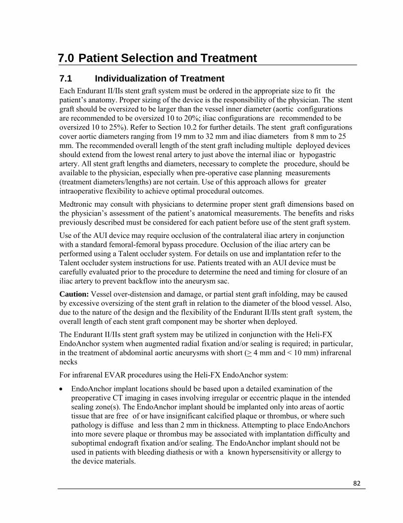
82
7.0 Patient Selection and Treatment
7.1 Individualization of Treatment Each Endurant II/IIs stent graft system must be ordered in the appropriate size to fit the patient’s anatomy. Proper sizing of the device is the responsibility of the physician. The stent graft should be oversized to be larger than the vessel inner diameter (aortic configurations are recommended to be oversized 10 to 20%; iliac configurations are recommended to be oversized 10 to 25%). Refer to Section 10.2 for further details. The stent graft configurations cover aortic diameters ranging from 19 mm to 32 mm and iliac diameters from 8 mm to 25 mm. The recommended overall length of the stent graft including multiple deployed devices should extend from the lowest renal artery to just above the internal iliac or hypogastric artery. All stent graft lengths and diameters, necessary to complete the procedure, should be available to the physician, especially when pre-operative case planning measurements (treatment diameters/lengths) are not certain. Use of this approach allows for greater intraoperative flexibility to achieve optimal procedural outcomes.
Medtronic may consult with physicians to determine proper stent graft dimensions based on the physician’s assessment of the patient’s anatomical measurements. The benefits and risks previously described must be considered for each patient before use of the stent graft system.
Use of the AUI device may require occlusion of the contralateral iliac artery in conjunction with a standard femoral-femoral bypass procedure. Occlusion of the iliac artery can be performed using a Talent occluder system. For details on use and implantation refer to the Talent occluder system instructions for use. Patients treated with an AUI device must be carefully evaluated prior to the procedure to determine the need and timing for closure of an iliac artery to prevent backflow into the aneurysm sac.
Caution: Vessel over-distension and damage, or partial stent graft infolding, may be caused by excessive oversizing of the stent graft in relation to the diameter of the blood vessel. Also, due to the nature of the design and the flexibility of the Endurant II/IIs stent graft system, the overall length of each stent graft component may be shorter when deployed.
The Endurant II/IIs stent graft system may be utilized in conjunction with the Heli-FX EndoAnchor system when augmented radial fixation and/or sealing is required; in particular, in the treatment of abdominal aortic aneurysms with short (> 4 mm and < 10 mm) infrarenal necks
For infrarenal EVAR procedures using the Heli-FX EndoAnchor system:
EndoAnchor implant locations should be based upon a detailed examination of the preoperative CT imaging in cases involving irregular or eccentric plaque in the intended sealing zone(s). The EndoAnchor implant should be implanted only into areas of aortic tissue that are free of or have insignificant calcified plaque or thrombus, or where such pathology is diffuse and less than 2 mm in thickness. Attempting to place EndoAnchors into more severe plaque or thrombus may be associated with implantation difficulty and suboptimal endograft fixation and/or sealing. The EndoAnchor implant should not be used in patients with bleeding diathesis or with a known hypersensitivity or allergy to the device materials.

83
See Section 11.2.17 for information regarding the recommended number of EndoAnchor implants.
Refer to the Heli-FX EndoAnchor system instructions for use for additional information.

84
8.0 Patient Counseling Information
The physician should review the following risks and benefits when counseling the patient about this endovascular device and procedure:
patient age and life expectancy
risks and benefits related to open surgical repair
risks and benefits related to endovascular repair
risks related to noninterventional treatment or medical management
risks of aneurysm rupture compared to endovascular repair
possibility that subsequent endovascular or open surgical repair of the aneurysm may be required
the long-term safety and effectiveness of the Endurant II/IIs stent graft system or the Endurant II/IIs stent graft system used in conjunction with the Heli-FX EndoAnchor system has not been established
long-term, regular follow-up is needed to assess patient health status and stent graft performance
patients with specific clinical findings (eg, endoleaks, enlarging aneurysms) should be monitored closely
symptoms of aneurysm rupture
risks and benefits related to femoral-femoral artery bypass, if applicable
Medtronic recommends that the physician disclose to the patient, in written form, all risks associated with treatment using the Endurant II/IIs stent graft system. Details regarding risks occurring during and after implantation of the device are provided in Adverse Events (Section 5.0). Additional counseling information can be found in the Patient Information Booklet.

85
9.0 How Supplied
For information on the supply of the Heli-FX EndoAnchor system, please see the instructions for use supplied with the device.
9.1 Sterility Each stent graft configuration (bifurcated, AUI, limb, aortic extension, and iliac extension) is individually contained within a delivery system. It is sterilized using electron beam and is supplied sterile for single use only.
Do not reuse or attempt to resterilize.
If the device is damaged or the integrity of the sterile barrier has been compromised, do not use the product and contact a Medtronic representative for return information.
9.2 Contents one Endurant II/IIs stent graft system
one set of patient tracking materials 9.3 Storage Store the system at room temperature in a dark, dry place.

86
10.0 Clinical Use Information
10.1 Physician Training Requirements All physicians should complete in-service training prior to using the Endurant II/IIs stent graft system.
Note: Endurant and Endurant II/IIs stent graft systems are considered technically and clinically equivalent. Physicians who have completed in-service training for Endurant do not need additional training for Endurant II/IIs.
Caution: The Endurant II/IIs stent graft system should only be used by physicians and teams trained in vascular interventional techniques, and in the use of this device. The following are the knowledge and skill requirements for physicians using the Endurant II/IIs stent graft system:
natural history of abdominal aortic aneurysms (AAA), aortoiliac aneurysms, and comorbidities associated with AAA repair
radiographic, fluoroscopic, and angiographic image interpretation
appropriate use of radiographic contrast material
arterial cutdown, arteriotomy, and repair or percutaneous access and closure techniques
nonselective and selective guidewire and catheter techniques
embolization
angioplasty
endovascular stent placement
snare techniques
techniques to minimize radiation exposure
device selection and sizing
Caution: The Heli-FX EndoAnchor system should only be used by physicians and teams trained in the use of this device. For physician training recommendations for the Heli-FX EndoAnchor system, please see the instructions for use provided with the device.
10.2 Recommended Device Sizing
10.2.1 Endurant II/IIs stent graft system The Endurant II/IIs stent graft systems are available in the sizes described in Table 2 through Table 7. For questions about device sizing, refer to contact information in the back of this instructions for use.

87
Table 2: Sizing Chart — Endurant II Bifurcated Configuration
OD (Fr) Proximal x Distal Diameter
(mm x mm)
Covered Length
(mm)
Vessel Inner Diameter
(mm)
20
36x20 145, 166
29–32 36x16
32x20
124, 145, 166
26–28 32x16
18
28x20 23-25 28x16 28x13 25x16 21–22 25x13 23x16 19–20 23x13
Table 3: Sizing Chart – Endurant IIs Bifurcated Configuration
OD (Fr) Proximal x Distal Diameter
(mm x mm)
Covered Length
(mm)
Vessel Inner Diameter
(mm)
20
36x14
103
29–32 32x14 26–28
18
28x14 23–25 25x14 21–22 23x14 19–20
Table 4: Sizing Chart — Aorto-Uni-Iliac (AUI) Configuration
OD (Fr) Proximal x Distal Diameter
(mm x mm)
Covered Length
(mm)
Vessel Inner Diameter
(mm)
20
36x14
102
29–32 32x14 26–28
18
28x14 23–25 25x14 21–22 23x14 19–20

88
Table 5: Sizing Chart — Limb Configuration
OD (Fr) Proximal x Distal Diameter
(mm x mm)
Covered Length
(mm)
Vessel Inner Diameter
(mm)
16
16x28 82, 93, 124, 156,
199
23–25 16x24 19–22 16x20 15–18 16x16
156, 199 12–14
16x13 10–11 16x10 8–9
14
16x16
82, 93, 124 12–14
16x13 10–11 16x10 8–9
Table 6: Sizing Chart — Iliac Extension Configuration
OD (Fr) Proximal x Distal Diameter
(mm x mm)
Covered Length
(mm)
Vessel Inner Diameter
(mm)
18 28x28
82
23–25
16 24x24 19–22 20x20 15–18
14 13x13 10–11 10x10 8–9
Table 7: Sizing Chart — Aortic Extension Configuration
OD (Fr) Proximal x Distal Diameter
(mm x mm)
Covered Length
(mm)
Vessel Inner Diameter
(mm)
20 36x36
49, 70
29–32 32x32 26–28
18 28x28 23–25 25x25 21–22 23x23 19–20
Caution: Proper sizing of the Endurant II/IIs stent graft is the responsibility of the physician. This stent graft sizing incorporates the recommended device oversizing for anatomical dimensions and was based on in-vitro test data.
10.2.2 Heli-FX EndoAnchor system For information on pre-implant planning for the Heli-FX EndoAnchor system, including the recommended number of EndoAnchors to be used, refer to the Heli-FX EndoAnchor system instructions for use. See also Section 11.2.17, below.
10.3 Device Inspection Inspect the device and packaging to verify that no damage or defects exist. If the “Use by”

89
date has elapsed, the device is damaged, or the sterile barrier has been compromised, do not use the device and contact a Medtronic representative for return or replacement information.
10.4 Additional Required Equipment Additional Endurant II/IIs stent graft systems of various lengths and diameters
For repair of short proximal neck aneurysms, additional Heli-FX EndoAnchor systems and compatible introducer sheaths.
Fluoroscope with digital angiographic capabilities (C-arm or fixed unit). Fluoroscopic imaging and the ability to record and recall all imaging.
Assorted guidewires of adequate length
Heparinized saline solution 10.5 Additional Recommended Equipment Introducer sheaths
Power injector
Radiopaque ruler with centimeter increments
Assorted balloon catheters
Compliant balloon catheters
Radiopaque contrast media
Sterile silicone lubricant or sterile mineral oil
Interventional snare devices
Endovascular coils and vascular plugs 10.6 MRI Information Nonclinical testing has demonstrated that the Endurant II/IIs stent graft is MR Conditional. A patient with this device can be safely scanned in an MR system meeting the following conditions:
Static magnetic field of 1.5 or 3.0 tesla only
Maximum spatial gradient magnetic field of 2500 gauss/cm or less
Maximum MR system reported, whole body averaged specific absorption rate (SAR) of 4 W/kg (First Level Controlled Mode)
Under the scan conditions defined above, the Endurant II/IIs stent graft is expected to produce:
a maximum temperature rise of 1.00ºC after 15 minutes of continuous scanning in a 1.5 tesla scanner
and a maximum temperature rise of 3.27ºC after 15 minutes of a continuous scanning in a

90
3.0 tesla scanner
The image artifact extends approximately 5 mm and 8 mm from the device, both inside and outside the device lumen when scanned in nonclinical testing using the sequence: spin echo and gradient echo, respectively, in a 3.0 tesla Siemens TrioTim (VB 13 Software) MR system with a whole body coil.
MRI safety and compatibility information for the Heli-FX EndoAnchor system can be found in the instructions for use provided with the device.

91
11.0 Implant Instructions
11.1 Vascular Access and Device Preparation Correct sizing of the aorta and iliac vessels must be determined before implantation of the aortic and iliac stent graft configurations using contrast-enhanced computer-aided tomography (CT), as well as angiograms of both the iliac arteries and aorta. 3D imaging may also be beneficial. Refer to Recommended Device Sizing (Section 10.2). These images should be available for review during the procedure. Vascular instruments and other surgical supplies necessary to gain access to the artery should also be available.
To reduce the risk of thromboembolism, it is recommended that the patient be heparinized for the duration of the procedure.
Caution: Do not retract the graft cover of the delivery system until it is accurately placed within the vasculature and ready for deployment.
Caution: Never advance or retract equipment from the vasculature without the use of fluoroscopy.
11.1.1 Vascular Access 1. Following aseptic procedure, perform a vascular access at the femoral arteries.
2. Place a guidewire in the ipsilateral femoral artery and advance it above the renal arteries.
3. When implanting a bifurcated stent graft, place a second guidewire via the contralateral side femoral artery and direct to the abdominal aorta.
4. Over this guidewire, place an angiography catheter above the renal arteries.
5. Take an angiogram.
Note: An additional incision might be necessary to access the common iliac artery. 11.1.2 Device Preparation 1. Prior to insertion, view the delivery system under fluoroscopy to visualize the radiopaque
markers on the stent graft. The radiopaque markers indicate the position of the proximal and distal edges of the graft material.
2. Turn the graft cover to align the radiopaque gate marker on the short leg of the bifurcated configuration with the patient’s contralateral iliac artery.
3. Flush the guidewire lumen with heparinized saline solution.
4. Prior to insertion into the vessel, activate the hydrophilic coating by wiping the outer surface of the graft cover with a sterile gauze, saturated in saline, until the graft cover is slippery to touch.
11.2 Delivery Procedure Medtronic recommends using an appropriate caliber introducer sheath to perform diagnostic tests. A sheath is not necessary for the introduction of the delivery system or deployment of the stent graft. For infrarenal EVAR procedures using the Heli-FX EndoAnchor system, the

92
access vessel diameter and morphology should be compatible for use with the device and should accommodate a 16 Fr introducer sheath.
Caution: Do not remove the guidewire while the delivery system is in the patient.
Warning: To prevent thrombotic problems, a second bolus of IV heparin is recommended before inserting the device.
Note: Section 11.2.1 is applicable to the Endurant II bifurcated, Endurant IIs bifurcated, and Endurant II AUI configurations. Sections pertinent to only individual main body stent graft configurations will be noted in parentheses.
11.2.1 Introduction of Main Body Configuration Warning: Do not advance the delivery system without using a guidewire.
1. Slowly insert the delivery system.
2. Advance over the guidewire so that the proximal most stents and the radiopaque markers are visualized in the target proximal aortic neck (Figure 80).
3. Inject contrast media through an angiographic (pigtail) catheter into the abdominal aorta and mark the position of the target location, either on the imaging screen or on the patient’s body.
4. Adjust the position of the stent graft so that the top edge of the graft fabric is just below the lowest renal artery. (Note: The edge of the graft fabric is 0.5 to 1.0 mm above the top edge proximal radiopaque markers.)
Note: If the top edge of the graft fabric is to be placed very close to the renal arteries, contrast media may be injected to identify the location of the lower renal artery and verify the position before full deployment.
Caution: Once proximal position has been identified, do not move the patient or imaging equipment, as it may compromise accuracy of stent graft placement.
Caution: The angiographic catheter can be removed prior to deployment. However, if the angiographic catheter is not removed until after deployment, ensure that the tip is straightened (such as with a pigtail catheter) with a guidewire before removal so that the stent graft is not pulled down.
Caution: When aligning the position of stent graft, be sure the fluoroscope is angled perpendicular to the center line of the infrarenal aorta to avoid parallax or other sources of visualization error. Some cranial caudal angulation of the image intensifier (I-I) tube may be necessary, especially if there is anterior angulation of the aneurysm neck.

93
Figure 80: Introduction of Aortic Delivery System 11.2.2 Confirm Position (Endurant II and Endurant IIs Bifurcated
Configurations Only) 1. Ensure that the distal portion of the contralateral stub leg is above the aortic bifurcation
and within the aneurysmal sac, and not within the iliac vessel.
2. Rotate the handle until the radiopaque marker on the distal stent of the contralateral stub leg is aligned with the contralateral iliac artery.
Note: When attempting to rotate the system, if the tip does not rotate with the handle, pull back the system and reposition until the intended position is achieved.
11.2.3 Deploy Proximal End of the Stent Graft Configuration 1. With 1 hand on the front grip, hold the delivery system stationary.
2. With the other hand, slowly withdraw the graft cover by rotating the external slider counterclockwise (in the direction of the arrow), until the constrained suprarenal stent is exposed and 2 to 3 of the covered stents have been fully deployed (Figure 81).
3. Use angiography to verify position of the stent graft in relation to the renal arteries.
4. If needed, gently push the entire delivery system proximally or pull distally until the proximal end of the graft material is even with the distal edge of the lowest renal artery.
Note: In the unlikely event of delivery system failure that results in partial stent graft deployment due to graft cover severance, the “handle disassembly” technique may permit successful deployment of the stent graft. Refer to Bail-Out Techniques (Section 12.0).
Caution: Do not rotate the graft cover during deployment as this may torque the device and cause it to rotate during deployment.
Caution: If the graft cover is accidentally withdrawn, the stent graft will prematurely deploy and may be incorrectly positioned.

94
Warning: Failure to properly align the radiopaque markers may result in improper deployment of the stent graft.
Figure 81: Deploying Proximal End of the Stent Graft Configuration 11.2.4 Deploy Contralateral Leg (Endurant II and Endurant IIs Bifurcated
Configurations Only) Continue holding the front grip of the delivery system stationary and then rotate the slider handle counterclockwise, stopping immediately after the contralateral leg is released from the graft cover or delivery sheath (Figure 82).
Figure 82: Deploy the Contralateral Leg (Endurant II and Endurant IIs Bifurcated Configurations Only)
11.2.5 Deploy Covered Portion (AUI Configuration Only) Continue holding the front grip of the delivery system stationary. Either continue to rotate the slider handle counterclockwise or use thumb to pull the trigger on the external slider and pull it back until the covered portion of the AUI configuration is completely released from the graft cover or delivery sheath.
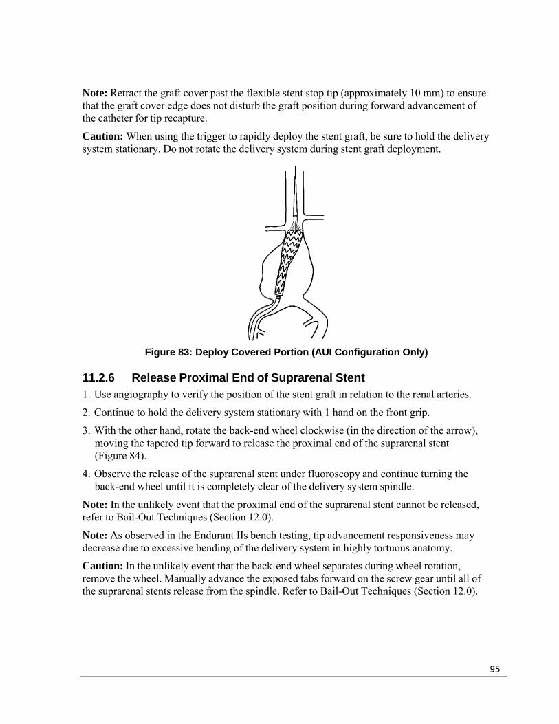
95
Note: Retract the graft cover past the flexible stent stop tip (approximately 10 mm) to ensure that the graft cover edge does not disturb the graft position during forward advancement of the catheter for tip recapture.
Caution: When using the trigger to rapidly deploy the stent graft, be sure to hold the delivery system stationary. Do not rotate the delivery system during stent graft deployment.
Figure 83: Deploy Covered Portion (AUI Configuration Only) 11.2.6 Release Proximal End of Suprarenal Stent 1. Use angiography to verify the position of the stent graft in relation to the renal arteries.
2. Continue to hold the delivery system stationary with 1 hand on the front grip.
3. With the other hand, rotate the back-end wheel clockwise (in the direction of the arrow), moving the tapered tip forward to release the proximal end of the suprarenal stent (Figure 84).
4. Observe the release of the suprarenal stent under fluoroscopy and continue turning the back-end wheel until it is completely clear of the delivery system spindle.
Note: In the unlikely event that the proximal end of the suprarenal stent cannot be released, refer to Bail-Out Techniques (Section 12.0).
Note: As observed in the Endurant IIs bench testing, tip advancement responsiveness may decrease due to excessive bending of the delivery system in highly tortuous anatomy.
Caution: In the unlikely event that the back-end wheel separates during wheel rotation, remove the wheel. Manually advance the exposed tabs forward on the screw gear until all of the suprarenal stents release from the spindle. Refer to Bail-Out Techniques (Section 12.0).

96
Figure 84: Release Proximal End of Suprarenal Stent
1. Endurant II bifurcated configuration or Endurant IIs bifurcated configuration
2. Endurant II AUI configuration 11.2.7 Deploy Distal End of the Stent Graft Configuration (Endurant II
and Endurant IIs Bifurcated Configurations Only) Either continue to rotate the external slider counterclockwise or while holding the front grip of the delivery system stationary, use thumb to pull the trigger on the external slider and pull it back until the bifurcated stent graft is completely deployed.
Note: Retract the graft cover past the flexible stent stop tip (approximately 10 mm) to ensure that the graft cover edge does not disturb the graft position during forward advancement of the catheter for tip recapture.
Caution: When using the trigger to rapidly deploy the stent graft, be sure to hold the delivery system stationary. Do not rotate the delivery system during stent graft deployment.
Figure 85: Deploy Distal End of the Bifurcated Configuration
1. Endurant II bifurcated configuration
2. Endurant IIs bifurcated configuration

97
11.2.8 Recapture Spindle in Tapered Tip (Endurant II Bifurcated and AUI
Configurations Only) Note: For the Endurant IIs bifurcated configuration, leave the delivery system in situ while deploying the limb stent graft into the contralateral leg. Skip to Deploy Limb Stent Graft into Contralateral Leg (Section 11.2.10).
1. Continue to hold the delivery system stationary with 1 hand on the front grip.
2. Confirm the spindle has fully separated from the suprarenal stent; gently torque the delivery system if it has not fully separated.
3. Gently rotate the delivery system while pushing the entire delivery system approximately 3 cm proximally so that the tapered tip and spindle are completely clear of the suprarenal stent.
4. With the other hand, rotate the back-end wheel counterclockwise to recapture the spindle in the tapered tip (Figure 86).
5. Observe the recapture of the spindle within the sleeve of the tapered tip under fluoroscopy.
6. Continue turning the back-end wheel counterclockwise until the spindle has been completely recaptured and the back-end wheel is at the bottom (Figure 86).
Note: When pushing the delivery system forward, be careful not to displace the distal end of the ipsilateral limb.
Note: Ensure that the suprarenal stent is fully disengaged from the spindle before pushing the delivery system forward.
Note: If the spindle catches on the suprarenal stent during advancement, completely advance the back-end wheel clockwise. Using a gentle in-and-out motion with the delivery system, rotate the delivery system until the spindle slips past the suprarenal stent. Then continue with the withdrawal process.
Caution: Stop rotating the back-end wheel when the bottom of the back-end screw gear is reached.
Warning: Failure to adequately advance the delivery system to recapture the spindle can result in the trapping of a suprarenal apex within the tapered tip sleeve. This will alter the proximal landing zone during delivery system withdrawal.

98
Figure 86: Recapture Spindle in Tapered Tip 11.2.9 Remove Delivery System (Endurant II Bifurcated and AUI
Configurations Only) 1. Continue to hold the delivery system stationary with 1 hand on the front grip and the other
hand on the external slider.
2. Gently torque and withdraw the delivery system until the spindle is retracted into the fabric portion of the stent graft.
3. Pull back the external slider trigger and hold it stationary while bringing the front grip to the external slider (Figure 87).
4. Use continual fluoroscopy and watch the top of the stent graft while slowly pulling back the tapered tip into the graft cover of the delivery system.
5. Gently remove the delivery system. Use fluoroscopy to ensure that the stent graft does not move during withdrawal.
Note: Maintain vessel access and wire placement until all stent graft configurations are in place.
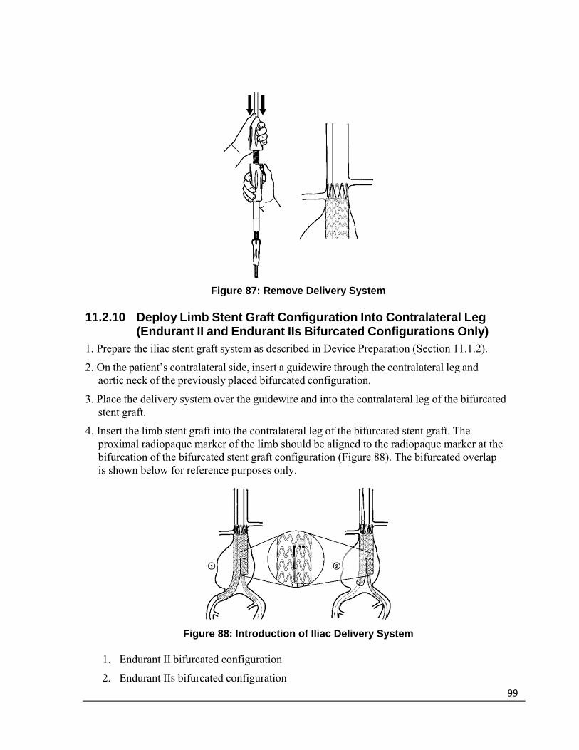
99
Figure 87: Remove Delivery System 11.2.10 Deploy Limb Stent Graft Configuration Into Contralateral Leg
(Endurant II and Endurant IIs Bifurcated Configurations Only) 1. Prepare the iliac stent graft system as described in Device Preparation (Section 11.1.2).
2. On the patient’s contralateral side, insert a guidewire through the contralateral leg and aortic neck of the previously placed bifurcated configuration.
3. Place the delivery system over the guidewire and into the contralateral leg of the bifurcated stent graft.
4. Insert the limb stent graft into the contralateral leg of the bifurcated stent graft. The proximal radiopaque marker of the limb should be aligned to the radiopaque marker at the bifurcation of the bifurcated stent graft configuration (Figure 88). The bifurcated overlap is shown below for reference purposes only.
Figure 88: Introduction of Iliac Delivery System
1. Endurant II bifurcated configuration
2. Endurant IIs bifurcated configuration
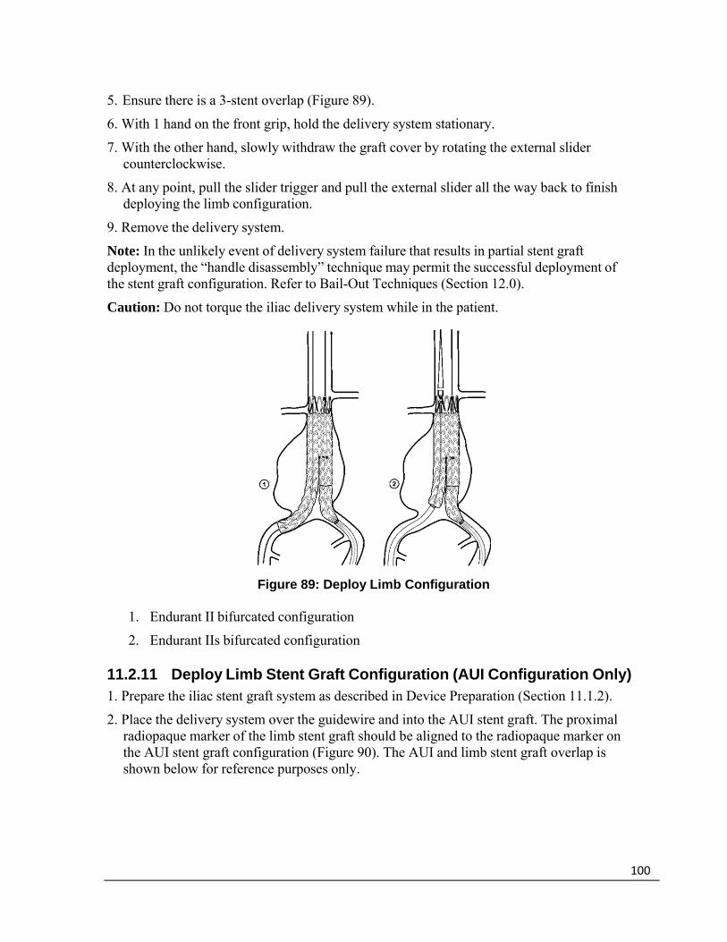
100
5. Ensure there is a 3-stent overlap (Figure 89).
6. With 1 hand on the front grip, hold the delivery system stationary.
7. With the other hand, slowly withdraw the graft cover by rotating the external slider counterclockwise.
8. At any point, pull the slider trigger and pull the external slider all the way back to finish deploying the limb configuration.
9. Remove the delivery system.
Note: In the unlikely event of delivery system failure that results in partial stent graft deployment, the “handle disassembly” technique may permit the successful deployment of the stent graft configuration. Refer to Bail-Out Techniques (Section 12.0).
Caution: Do not torque the iliac delivery system while in the patient.
Figure 89: Deploy Limb Configuration
1. Endurant II bifurcated configuration
2. Endurant IIs bifurcated configuration 11.2.11 Deploy Limb Stent Graft Configuration (AUI Configuration Only) 1. Prepare the iliac stent graft system as described in Device Preparation (Section 11.1.2).
2. Place the delivery system over the guidewire and into the AUI stent graft. The proximal radiopaque marker of the limb stent graft should be aligned to the radiopaque marker on the AUI stent graft configuration (Figure 90). The AUI and limb stent graft overlap is shown below for reference purposes only.

101
Figure 90: Introduction of Iliac Delivery System
3. Ensure there is a 3-stent overlap (Figure 91).
4. With 1 hand on the front grip, hold the delivery system stationary.
5. With the other hand, slowly withdraw the graft cover by rotating the external slider counterclockwise.
6. At any point, pull the slider trigger and pull the external slider all the way back to finish deploying the limb configuration.
7. Remove the delivery system.
Note: In the unlikely event of delivery system failure that results in partial stent graft deployment, the “handle disassembly” technique may permit the successful deployment of the stent graft configuration. Refer to Bail-Out Techniques (Section 12.0).
Caution: Do not torque the iliac delivery system while in the patient.
Figure 91: Deploy Limb Configuration

102
11.2.12 Recapture Spindle in Tapered Tip for Delivery System in the Ipsilateral Leg (Endurant IIs Bifurcated Configuration Only)
1. Hold the delivery system stationary with 1 hand on the front grip.
2. Confirm the spindle has fully separated from the suprarenal stent; gently torque the delivery system if it has not fully separated.
3. Gently rotate the delivery system while pushing the entire delivery system approximately 3 cm proximally so that the tapered tip and spindle are completely clear of the suprarenal stent.
4. With the other hand, rotate the back-end wheel counterclockwise to recapture the spindle in the tapered tip (Figure 92).
5. Observe the recapture of the spindle within the sleeve of the tapered tip under fluoroscopy.
6. Continue turning the back-end wheel counterclockwise until the spindle has been completely recaptured and the back-end wheel is at the bottom (Figure 92).
Note: When pushing the delivery system forward, be careful not to displace the distal end of the ipsilateral limb.
Note: Ensure that the suprarenal stent is fully disengaged from the spindle before pushing the delivery system forward.
Note: If the spindle catches on the suprarenal stent during advancement, completely advance the backend wheel clockwise. Using a gentle in-and-out motion with the delivery system, rotate the delivery system until the spindle slips past the suprarenal stent. Then continue with the withdrawal process.
Caution: Stop rotating the back-end wheel when the bottom of the back-end screw gear is reached.
Warning: Failure to adequately advance the delivery system to recapture the spindle can result in the trapping of a suprarenal apex within the tapered tip sleeve. This will alter the proximal landing zone during delivery system withdrawal.

103
Figure 92: Recapture Spindle in Tapered Tip 11.2.13 Remove Delivery System (Endurant IIs Bifurcated Configuration
Only) 1. Continue to hold the delivery system stationary with 1 hand on the front grip and the other
hand on the external slider.
2. Gently torque and withdraw the delivery system until the spindle is retracted into the fabric portion of the stent graft.
3. Pull back the external slider trigger and hold it stationary while bringing the front grip to the slider (Figure 93).
4. Use continual fluoroscopy and watch the top of the bifurcated stent graft while slowly pulling back the tapered tip into the graft cover of the delivery system.
5. Gently remove the delivery system. Use fluoroscopy to ensure that the bifurcated configuration does not move during withdrawal.
Note: Maintain vessel access and wire placement until all stent graft configurations are in place.

104
Figure 93: Remove Delivery System 11.2.14 Deploy Limb Stent Graft Into Ipsilateral Leg (Endurant IIs
Bifurcated Configuration Only) 1. Prepare the iliac stent graft system as described in Device Preparation (Section 11.1.2).
2. On the patient’s ipsilateral side, track the delivery system over the guidewire and into the ipsilateral leg of the previously placed Endurant IIs bifurcated configuration. The limb stent graft has 2 markers on its proximal edge, 2 markers on its distal edge, and 1 overlap marker approximately 25 mm distal of the proximal markers.
3. Position the device. Overlap criteria between the limb stent graft and the ipsilateral leg of the bifurcated stent graft is dependent on limb selection. See Table 8 for recommended device overlap.

105
Table 8: Recommended Device Overlap — Limb Stent Graft and Ipsilateral Leg of Endurant IIs Bifurcated Stent Graft
Proximal diameter
Distal diameter Length Overlap Deployment
reference
16
10 82
3 stents only
Refer to step a
93
13 82 93
20 82 93
24 82 93
28 82 93
16
10
124
3 to 5 stents
Refer to step b
156 199
13
124 156 199
16
82 93 124 156 199
20
124 156 199
24
124 156 199
28
124 156 199
a. 3 stent overlap. Align the overlap marker on the limb stent graft with the 2 markers on the distal end of the ipsilateral leg of the Endurant IIs bifurcated stent graft (Figure 94). Warning: For the limb stent graft overlap criteria with Endurant IIs ipsilateral leg only, please refer to Table 8. As noted in Table 8, for the limb stent graft configurations that have an overlap criteria of 3 stents only, do not overlap more than 3 stents.

106
Figure 94: Introduction of the Iliac Delivery System — 3 Stent Overlap
b. 3 to 5 stent overlap. For the limb stent grafts that can have an overlap between 3 to 5 stents, minimum 3 stent overlap can be achieved by following instruction in step a (Figure 94) or the maximum 5 stent overlap can be achieved by aligning the proximal edge markers on the limb stent graft to the bifurcation marker on the Endurant IIs bifurcated stent graft (Figure 95).
Figure 95: Introduction of the Iliac Delivery System — 5 Stent Overlap
4. With 1 hand on the front grip, hold the delivery system stationary.
5. With the other hand, slowly withdraw the graft cover by rotating the external slide counterclockwise.
6. At any point, pull the slider trigger and pull the external slider all the way back to finish deploying the limb configuration.

107
7. Remove the delivery system (Section 11.2.13).
Note: In the unlikely event of delivery system failure that results in partial stent graft deployment, the “handle disassembly” technique may permit the successful deployment of the stent graft configuration. Refer to Section 12.0, Bail-Out Techniques.
Caution: Do not torque the iliac delivery system while in the patient. 11.2.15 Iliac or Aortic Extension Stent Graft Configurations 1. If an aortic extension stent graft configuration is needed, ensure that there is a minimum 3-
stent overlap between the aortic extension stent graft and the main body stent graft configuration.
2. Follow the main body stent graft deployment process, except rotate the handle to open the extension component entirely before releasing the proximal end of the suprarenal stent of the aortic configuration.
3. If an iliac extension stent graft configuration is needed, ensure that there is a minimum 3- stent overlap between the iliac extension and the configuration it is being mated with. This is achieved by aligning the limb overlap marker with the most distal marker on the configuration with which the extension is being mated.
4. Follow the limb stent graft configuration deployment process (Section 11.2.10). 11.2.16 Smoothing Stent Graft Fabric and Modeling Stent Graft The Reliant™ stent graft balloon catheter (packaged separately) can be used to assist in stent graft implantation by modeling the covered portion of the stent graft and removing wrinkles and folds from the graft material, as needed. Use the balloon catheter to model the proximal and distal seal zones as well as any overlapping connection (or junction) areas between the stent graft components. Sub-optimal expansion of the self-expanding stent graft components may also be improved by use of the balloon catheter. Refer to the Reliant stent graft balloon catheter IFU for specific instructions.
Note: The Reliant stent graft balloon catheter is recommended for use with the Endurant II/IIs stent graft system. No data is available for the use of other balloon catheters in remodeling stent grafts.
Note: When using the Heli-FX EndoAnchor system, Medtronic recommends that the EndoAnchor implantation be done after the aortic endograft has been placed and any balloon remodeling of the infrarenal seal zone of the stent graft system has been completed. Exercise care in balloon remodeling of the stent graft system to avoid moving the main body endograft from its intended implant location.
Note: Care should be taken when inflating the balloon, especially with calcified, tortuous, stenotic, or otherwise diseased vessels. Inflate slowly. It is recommended that a backup balloon be available.
Caution: Expansion of the balloon outside of the graft material can result in rupture of the aorta, vessel dissection, or graft tears.
108
Warning: When expanding a vascular prosthesis, there is an increased risk of vessel injury or rupture, and possible patient death, if the balloon’s proximal and distal radiopaque markers are not completely within the covered (graft fabric) portion of the prosthesis.
Warning: Do not use the Reliant stent graft balloon catheter in the treatment of dissections. 11.2.17 Additional fixation and sealing with the Heli-FX EndoAnchor
system (infrarenal neck > 4 mm and < 10 mm in length) The Heli-FX EndoAnchor system is required when treating short infrarenal necks (> 4 mm and < 10 mm in length). For bifurcated endografts, the recommended minimum number of EndoAnchor implants is based on native vessel diameter and is independent of the amount of endograft oversizing. For aortic neck diameters < 29 mm, the recommended minimum number of EndoAnchors is 4. For aortic neck diameters 30 – 32 mm, the recommended minimum number of EndoAnchors is 6.
Medtronic recommends that the EndoAnchor implantation be done after the aortic endograft has been placed and any balloon remodeling of the infrarenal seal zone of the stent graft system has been completed.
1. Prepare the system, per the Heli-FX EndoAnchor system instructions for use.
Note: The access vessel diameter and morphology should be compatible for use with the Heli-FX EndoAnchor system.
2. Implant the EndoAnchors, per the Heli-FX EndoAnchor system instructions for use.
Note: Stability of the stent graft in short (> 4 mm and < 10 mm) infrarenal necks is augmented by the EndoAnchor implants. Ensure successful deployment of the recommended minimum number of EndoAnchor implants. Where the number of successfully deployed EndoAnchor implants is below the minimum recommended, there may be greater risk of postoperative Type 1a endoleak or migration.
Note: Always use fluoroscopy for guidance, delivery, and observation of any Heli-FX system components within the vasculature.
Note: The EndoAnchor implant should be implanted only into areas of aortic tissue that are free of or have insignificant calcified plaque or thrombus, or where such pathology is diffuse and less than 2 mm in thickness. It should not be used in patients with bleeding diathesis or with a known hypersensitivity or allergy to the device materials.
3. Remove the system, per the Heli-FX EndoAnchor system instructions for use. 11.2.18 Verify Placement and Seal 1. At the completion of the procedure, perform angiography to assess the stent graft for
proximal and distal endoleaks, to verify position of the implanted stent graft in relation to the aneurysm and renal arteries, to evaluate EndoAnchor implantation (if EndoAnchors are used) and, if applicable, to assess patency of femoral-femoral bypass graft.
2. Leaks at the attachment or connection sites should be treated using the balloon to remodel the stent graft against the vessel wall.
Note: For an infrarenal EVAR procedure using the Heli-FX EndoAnchor system, additional

109
EndoAnchor implants may be used to augment the seal.
3. Major leaks that cannot be corrected by reballooning may be treated by adding aortic or iliac extension components to the previously placed stent graft components.
Caution: Any leak left untreated during the implantation procedure must be carefully monitored after implantation.
11.2.19 Seal Entry Sites 1. Remove ancillary devices prior to repairing the entry site.
2. Repair the entry site with standard closure technique.

110
12.0 Bail-Out Techniques
In the unlikely event of Endurant II or Endurant IIs delivery system failure, the following bail-out techniques may be used. For information on Heli-FX EndoAnchor system failure, see the instructions for use provided with the device.
12.1 Screw Gear Handle Disassembly If partial stent graft deployment due to graft cover severance occurs, a screw gear handle disassembly technique may permit successful deployment of the stent graft.
1. Pull back the trigger and fully retract the slider.
2. Stabilize the delivery system.
3. Insert the tips of a pair of hemostats into each of the screw gear handle disassembly ports on the front grip.
4. Disengage the front grip from the screw gear by pressing the tips of the hemostats into the handle disassembly ports simultaneously advancing the front grip away from the screw gear.
5. Advance the front grip until it fully clears the screw gear.
6. Separate the screw gear halves in order to identify the location of graft cover severance.
7. Manually retract the graft cover with your fingers or with hemostats until the stent graft is fully deployed.
8. Follow the instructions for tip capture deployment and delivery system removal. 12.2 Ballooning If the captured proximal tip of the suprarenal stent cannot be deployed and the back-end wheel section still works, a ballooning technique may permit successful deployment of the suprarenal stent.
1. Use a compliant or semi-compliant balloon (Reliant balloon recommended).
2. Insert the balloon and move it to the bifurcated configuration’s aortic section.
3. Inflate the balloon inside the stent graft to vessel size to stabilize stent graft.
4. Follow the instructions for tip capture deployment and delivery system removal. 12.3 Back-End Handle Disassembly If no or partial deployment of the proximal end of the suprarenal stent occurs due to back-end wheel failure, a back-end handle disassembly technique may permit the successful deployment of the suprarenal stent.
1. Use hemostats to depress the exposed tabs to disassemble the back-end wheel.
2. Insert the tips of hemostats into each of the rear handle disassembly ports. 3. Disengage the rear handle by pressing the tips of the hemostats into the handle
disassembly ports simultaneously retracting the rear handle from the delivery system.

111
4. Stabilize the delivery system.
5. Manually push the exposed tabs of the back-end T-tube to release the suprarenal stent from the spindle.
6. Manually pull back the exposed tabs of the back-end T-tube to recapture the tapered tip after deployment.
7. Follow the instructions for delivery system removal.
8. Hold the exposed tabs of the back-end T-tube so that it remains retracted and the tapered tip recaptured during delivery system removal.
12.4 Snare the Tapered Tip If the back-end handle disassembly technique is unsuccessful due to an excessively high deployment force, a snare the tapered tip technique may permit successful deployment of the suprarenal stent.
1. Use a snare device.
2. Advance the snare device to the delivery system tapered tip section through upper torso access (ie, brachial).
3. Utilize fluoroscopy to snare the edge of the delivery system tapered tip.
4. Stabilize the delivery system, especially the back-end section.
5. Pull the snare device to separate the suprarenal stent from the tip capture.
6. Manually pull back the back-end T-tube to recapture the tapered tip after deployment.
7. Follow the instructions for delivery system removal.
8. Ensure that the back-end T-tube remains retracted and the tapered tip recaptured during delivery system removal.

112
13.0 Follow-up Imaging Recommendations
13.1 General Current imaging of stent graft patients includes abdominal X-ray and CT, with and without contrast medium. Alternative imaging modalities such as magnetic resonance imaging should be used in patients with impaired renal function or intolerance to contrast media. Determination of imaging techniques should be based on the physician’s clinical assessment of the patient and stent graft implant, including any adjunct procedures that may have been performed in conjunction with the endovascular stent graft procedure.
After endovascular graft placement, patients should be regularly monitored for perigraft flow, aneurysm growth or changes in the structure or position of the endovascular graft. Annual imaging is recommended, including 1) abdominal radiographs to examine device integrity (stent fracture, separation between bifurcated device and proximal cuffs or limb extensions, if applicable), and 2) contrast and non-contrast CT to examine aneurysm changes, perigraft flow, patency, tortuosity and progressive disease. If renal complications or other factors preclude the use of image contrast media, abdominal radiographs and duplex ultrasound may provide similar information.
Medtronic recommends consideration of an enhanced follow-up schedule for patients with short infrarenal necks (> 4 mm and < 10 mm in length), where the minimum recommended number of EndoAnchor implants were not used.
Additional imaging guidelines for Heli-FX EndoAnchors can be found in the instructions for use provided with the device.
13.2 X-ray Abdominal X-rays should be used to assess the presence of stent graft fracture. Four-view kidney, ureter, bladder (KUB) X-rays should be taken. Posterior/anterior (PA) and lateral images are recommended for visualization of the stent graft. Ensure the entire device is captured on images for assessment.
13.3 CT with Contrast Contrast-enhanced CT should be used to assess stent graft fixation, deformation, apposition to the vessel wall at proximal and distal fixation sites, stent graft migration, stent graft patency, AAA size, occlusion of branch vessels, and endoleak (including source and type if present).
A pre-contrast scan of 5 mm thick slices is suggested to determine if there are calcifications or areas where metal artifacts may be misinterpreted as endoleak. An arterial phase with <3 mm slice thickness and overlapping images with coverage from the celiac artery to the external iliac is recommended. In aneurysms that are not shrinking and have no apparent endoleak or fixation problems, a late venous phase scan may be performed. The venous phase scan may also be performed with thicker collimation (5 mm). It is recommended that the source data set be archived in case specialized evaluation is needed later (volume measurements, 3-dimensional reconstruction, or computer-aided measurement software). If the aneurysm is not shrinking by more than 5 mm within the first year, volume measurements

113
may be obtained as a more sensitive indicator of AAA size using 3-dimensional software. The physician will determine the requirement pre-operative care for patients with allergies to contrast.
13.4 Noncontrast CT For patients with impaired renal function or those who are allergic to contrast medium, a spiral CT without contrast may be considered to assess stent graft fixation, deformation, apposition to the vessel wall at proximal and distal fixation sites, stent graft migration, occlusion of vessels, and size of the AAA diameter and volume measurements.
13.5 Duplex Ultrasound For patients with impaired renal function or those who are allergic to contrast medium, a color-duplex ultrasound may be considered to assess size of the AAA diameter, endoleaks, and stent graft occlusion and stenosis. Use duplex ultrasound to assess the patency of any femoral-femoral artery bypass graft used in conjunction with the AUI device.
13.6 MRI or MRA Patients with impaired renal function, ie, renal insufficiency, may also be considered for magnetic resonance imaging or angiography (MRI, MRA) in facilities that have expertise in this area. Artifact may occur related to the stent, and care should be used to insure adequate imaging of the outer aneurysm wall to assess AAA size. Volume measurement may be helpful if the aneurysm is not clearly shrinking. If there are concerns regarding imaging of calcified areas, fixation sites, or the outer wall of the aneurysm sac, adjunctive CT without contrast may be needed.
13.7 Imaging Tests Refer to the table below for the recommended follow-up imaging schedule after stent graft implant.
Table 9: Imaging Recommendations
Interval Angiogram CT1 2[Contrast & Noncontrast]
Abdominal Radiographs
Preprocedure X
Procedural X
1 Month X
12 Months (annually thereafter) X X3
1 CT evaluation may include “3-phase technique,” volume studies, 3-D reconstruction, or computer-aided measurements. 2 If the patient is unable to tolerate a CT with contrast, then a CT without contrast and/or a duplex ultrasound may be considered to assess the aneurysm and stent graft integrity. 3 Based on the quality of the imaging obtained, an abdominal radiograph may be performed in addition if the imaging was unable to assess stent graft integrity.
114
13.8 Supplemental Imaging Note: Additional radiological imaging may be necessary to further evaluate the stent graft in situ based on findings revealed by one of the surveillance programs. The following recommendations may be considered. If there is evidence of poor or irregular position of the stent graft, severe angulation,
kinking or migration of the stent graft on abdominal X-rays, a spiral CT should be performed to assess aneurysm size and the presence or absence of an endoleak.
If a new endoleak or increase in AAA size is observed by spiral CT, adjunctive studies such as 3-D reconstruction or angiographic assessment of the stent graft and native vasculature may be helpful in further evaluating any changes of the stent graft or aneurysm.
Spiral CT without contrast, MRI or MRA may be considered in select patients who cannot tolerate contrast media or who have renal function impairment. For centers with appropriate expertise, gadolinium or CO2 angiography may be considered in patients with renal function impairment requiring angiographic assessment.

115
14.0 Additional Surveillance and Treatment
Additional endovascular repair or open surgical aneurysm repair should be considered for patients with evidence of suboptimal stent graft fixation, proximal endoleak, distal endoleak, junction endoleak, unknown origin of persistent perigraft flow, or increase in AAA size >5 mm.

116
15.0 Device Registration
The Endurant II/IIs stent graft system is packaged with additional specific information which includes:
Temporary Device Identification Card that includes both patient and stent graft information. Physicians should complete this card and instruct the patient to keep it in their possession at all times. The patients should refer to this card anytime they visit additional health practitioners, particularly for additional diagnostic procedures (eg, MRI). This temporary identification card should only be discarded when the permanent identification card is received.
Device Tracking Form to be completed by the hospital staff and forwarded to Medtronic for the purposes of tracking all patients who received an Endurant II/IIs stent graft (as required by Federal Regulation). The hospital’s submission of the device tracking form to Medtronic is also required for a patient to receive the permanent identification card. Upon receipt of the device tracking form, Medtronic will mail the patient a permanent device identification card. This card includes important information regarding the implanted stent graft. Patients should refer to this card anytime they visit health practitioners, particularly for any diagnostic procedures (eg, MRI). Patients should carry this card with them at all times. If a patient does not receive their permanent device identification card, or requires changes to the card, call 1-800-551- 5544. In addition a patient information booklet (PIB) will be provided to the physicians during training and additional copies will be available upon request. The PIB will also be available online on the Medtronic website (www.medtronic.com). This booklet provides patients with basic information on abdominal aortic aneurysms and endovascular repair therapy.

117
16.0 Disclaimer of Warranty
ALTHOUGH THE MEDTRONIC VASCULAR ENDURANT II/ENDURANT IIs STENT GRAFT AND DELIVERY SYSTEM, HEREAFTER REFERRED TO AS THE ‘PRODUCT’, HAS BEEN MANUFACTURED UNDER CAREFULLY CONTROLLED CONDITIONS, MEDTRONIC, INC., MEDTRONIC VASCULAR, INC. AND THEIR RESPECTIVE AFFILIATES, (COLLECTIVELY “MEDTRONIC”) HAVE NO CONTROL OVER THE CONDITIONS UNDER WHICH THIS PRODUCT IS USED. THE WARNINGS CONTAINED IN THE PRODUCT LABELING PROVIDE MORE DETAILED INFORMATION AND ARE CONSIDERED AN INTEGRAL PART OF THIS DISCLAIMER OF WARRANTY. MEDTRONIC, THEREFORE, DISCLAIMS ALL WARRANTIES, BOTH EXPRESSED AND IMPLIED, WITH RESPECT TO THE PRODUCT, INCLUDING, BUT NOT LIMITED TO, ANY IMPLIED WARRANTY OF MERCHANTABILITY OR FITNESS FOR A PARTICULAR PURPOSE. MEDTRONIC SHALL NOT BE LIABLE TO ANY PERSON OR ENTITY FOR ANY MEDICAL EXPENSES OR ANY DIRECT, INCIDENTAL OR CONSEQUENTIAL DAMAGES CAUSED BY ANY USE, DEFECT, FAILURE OR MALFUNCTION OF THE PRODUCT, WHETHER A CLAIM FOR SUCH DAMAGES IS BASED UPON WARRANTY, CONTRACT, TORT OR OTHERWISE. NO PERSON HAS ANY AUTHORITY TO BIND MEDTRONIC TO ANY REPRESENTATION OR WARRANTY WITH RESPECT TO THE PRODUCT.
The exclusions and limitations set out above are not intended to, and should not be construed so as to contravene mandatory provisions of applicable law. If any part or term of this Disclaimer of Warranty is held to be illegal, unenforceable or in conflict with applicable law by a court of competent jurisdiction, the validity of the remaining portions of this Disclaimer of Warranty shall not be affected, and all rights and obligations shall be construed and enforced as if this Disclaimer of Warranty did not contain the particular part or term held to be invalid.

118
Medtronic, Inc. 710 Medtronic Parkway Minneapolis, MN 55432 USA www.medtronic.com Tel: (+1-763) 514-4000 Fax: (+1-763) 514-4879
US Customer Service/Product Inquiries Tel: (+1-800) 961-9055 Fax: (+1-800) 929-2133
Protected by one or more of the following United States patents: 6,306,141; 6,911,039; 7,105,016; 7,264,632, and 7,655,034.
© 2017 Medtronic
M052195T001 Rev. 1D