Embed Size (px)
Citation preview
Evaluation of Thoracic Aortic Stent-Graft Devices for MR-Guided Real-Time Stent Placement
H. Eggebrecht1, H. Kuehl2, H. H. Quick3 1Department of Cardiology and Department of Diagnostic and Interventional Radiology, Essen, North-Rhine Westfalia, Germany, 2Department of Diagnostic and
Interventional Radiology, Essen, North Rhine Westfalia, Germany, 3Department of Diagnostic and Interventional Radiology, Essen, North-Rhine Westfalia, Germany
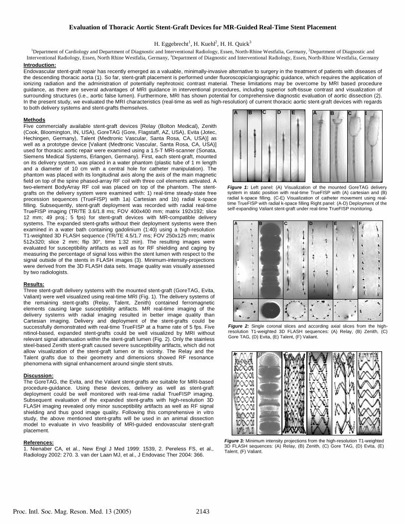
Introduction: Endovascular stent-graft repair has recently emerged as a valuable, minimally-invasive alternative to surgery in the treatment of patients with diseases of the descending thoracic aorta (1). So far, stent-graft placement is performed under fluoroscopic/angiographic guidance, which requires the application of ionizing radiation and the administration of potentially nephrotoxic contrast material. These limitations may be overcome by MRI based procedure guidance, as there are several advantages of MRI guidance in interventional procedures, including superior soft-tissue contrast and visualization of surrounding structures (i.e., aortic false lumen). Furthermore, MRI has shown potential for comprehensive diagnostic evaluation of aortic dissection (2). In the present study, we evaluated the MRI characteristics (real-time as well as high-resolution) of current thoracic aortic stent-graft devices with regards to both delivery systems and stent-grafts themselves. Methods Five commercially available stent-graft devices [Relay (Bolton Medical), Zenith (Cook, Bloomington, IN, USA), GoreTAG (Gore, Flagstaff, AZ, USA), Evita (Jotec, Hechingen, Germany), Talent (Medtronic Vascular, Santa Rosa, CA, USA)] as well as a prototype device [Valiant (Medtronic Vascular, Santa Rosa, CA, USA)] used for thoracic aortic repair were examined using a 1.5-T MRI-scanner (Sonata, Siemens Medical Systems, Erlangen, Germany). First, each stent-graft, mounted on its delivery system, was placed in a water phantom (plastic tube of 1 m length and a diameter of 10 cm with a central hole for catheter manipulation). The phantom was placed with its longitudinal axis along the axis of the main magnetic field on top of the spine phased-array RF coil with three coil elements activated. A two-element BodyArray RF coil was placed on top of the phantom. The stent-grafts on the delivery system were examined with: 1) real-time steady-state free precession sequences (TrueFISP) with 1a) Cartesian and 1b) radial k-space filling. Subsequently, stent-graft deployment was recorded with radial real-time TrueFISP imaging (TR/TE 3.6/1.8 ms; FOV 400x400 mm; matrix 192x192; slice 12 mm; 49 proj.; 5 fps) for stent-graft devices with MR-compatible delivery systems. The expanded stent-grafts without their deployment systems were then examined in a water bath containing gadolinium (1:40) using a high-resolution T1-weighted 3D FLASH sequence (TR/TE 4.5/1.7 ms; FOV 250x125 mm; matrix 512x320; slice 2 mm; flip 30°, time 1:32 min). The resulting images were evaluated for susceptibility artifacts as well as for RF shielding and caging by measuring the percentage of signal loss within the stent lumen with respect to the signal outside of the stents in FLASH images (3). Minimum-intensity-projections were derived from the 3D FLASH data sets. Image quality was visually assessed by two radiologists. Results: Three stent-graft delivery systems with the mounted stent-graft (GoreTAG, Evita, Valiant) were well visualized using real-time MRI (Fig. 1). The delivery systems of the remaining stent-grafts (Relay, Talent, Zenith) contained ferromagnetic elements causing large susceptibility artifacts. MR real-time imaging of the delivery systems with radial imaging resulted in better image quality than Cartesian imaging. Delivery and deployment of the stent-grafts could be successfully demonstrated with real-time TrueFISP at a frame rate of 5 fps. Five nitinol-based, expanded stent-grafts could be well visualized by MRI without relevant signal attenuation within the stent-graft lumen (Fig. 2). Only the stainless steel-based Zenith stent-graft caused severe susceptibility artifacts, which did not allow visualization of the stent-graft lumen or its vicinity. The Relay and the Talent grafts due to their geometry and dimensions showed RF resonance phenomena with signal enhancement around single stent struts. Discussion: The GoreTAG, the Evita, and the Valiant stent-grafts are suitable for MRI-based procedure-guidance. Using these devices, delivery as well as stent-graft deployment could be well monitored with real-time radial TrueFISP imaging. Subsequent evaluation of the expanded stent-grafts with high-resolution 3D FLASH imaging revealed only minor susceptibility artifacts as well as RF signal shielding and thus good image quality. Following this comprehensive in vitro study, the above mentioned stent-grafts will be used in an animal dissection model to evaluate in vivo feasibility of MRI-guided endovascular stent-graft placement. References: 1. Nienaber CA, et al., New Engl J Med 1999: 1539, 2. Pereless FS, et al., Radiology 2002: 270. 3. van der Laan MJ, et al., J Endovasc Ther 2004: 366.
Figure 2: Single coronal slices and according axial slices from the high-resolution T1-weighted 3D FLASH sequences: (A) Relay, (B) Zenith, (C) Gore TAG, (D) Evita, (E) Talent, (F) Valiant.
A B C E F D
Figure 1: Left panel: (A) Visualization of the mounted GoreTAG delivery system in static position with real-time TrueFISP with (A) cartesian and (B) radial k-space filling. (C-E) Visualization of catheter movement using real-time TrueFISP with radial k-space filling Right panel: (A-D) Deployment of the self-expanding Valiant stent-graft under real-time TrueFISP monitoring.
A B C D E A B C D
Figure 3: Minimum intensity projections from the high-resolution T1-weighted 3D FLASH sequences: (A) Relay, (B) Zenith, (C) Gore TAG, (D) Evita, (E) Talent, (F) Valiant.
A BB C EE FFDDA BB C EE FFDD
Proc. Intl. Soc. Mag. Reson. Med. 13 (2005) 2143