Embed Size (px)
Citation preview

Mestrado Integrado em Medicina Dentária
Leukocyte and platelet-rich fibrin in the regeneration
of periodontal infrabony defects
Ana Monteiro
Supervisor: Prof. Dr. Sérgio Matos
Co-supervisor: Dr. Tony Rolo
Coimbra
2017


Mestrado Integrado em Medicina Dentária
Leukocyte and platelet-rich fibrin in the regeneration
of periodontal infrabony defects
Monteiro A.*, Rolo T.**, Matos S.***
* Dentistry Student at the Faculty of Medicine, University of Coimbra. Email:
** Invited Assistant of Periodontology, Dentistry Department, Faculty of Medicine, University of
Coimbra
*** Assistant Professor of Periodontology, Dentistry Department, Faculty of Medicine, University of
Coimbra
Dentistry Department of the Faculty of Medicine of the University of Coimbra,
Avenida Bissaya Barreto, Bloco de Celas, 3000-075 Coimbra
Tel.: +351 239484183
Fax: +351 239402910
Coimbra, Portugal
Coimbra
2017

IV
Summary
Resumo
Abstract
List of abbreviations and acronyms
1. Introduction
1.1. Chronic periodontitis
1.2. Therapeutic approaches
1.2.1. Regenerative therapy
1.2.1.1. Autologous platelet concentrates (APC)
2. Systematic Review
2.1. Materials and Methods
2.2. Results
3. Cases series
3.1. Materials and Methods
3.2. Results
3.2.1. Clinical case 1
3.2.2. Clinical case 2
3.2.3. Clinical case 3
4. Discussion
5. Conclusions
Bibliography
Appendix
Acknowledgements
Index of figures and tables
Index

V
Resumo
Introdução: O crescente conhecimento na área da Periodontologia levou ao
desenvolvimento de várias técnicas regenerativas, mas também ao reavivar do uso de
concentrados plaquetários autólogos (APCs) no tratamento de defeitos periodontais infraósseos.
Objetivos: Este trabalho pretende verificar a aplicabilidade e eficácia dos concentrados
plaquetários (particularmente da fibrina rica em plaquetas e leucócitos (L-PRF)) na regeneração
dos defeitos periodontais infraósseos, através de uma revisão sistematizada. Adicionalmente,
pretende-se demonstrar o método de aplicação do L-PRF na regeneração recorrendo a uma
série de casos clínicos. Metodologia: Foi definida uma questão PICOT: "Em pacientes com
defeitos periodontais profundos, qual a eficácia da aplicação de L-PRF, isolado ou em
combinação com outros biomateriais, na regeneração de defeitos periodontais,
comparativamente à cirurgia de acesso e a outros APCs, após um período de cicatrização
mínimo de 6 meses?", seguida de uma pesquisa eletrónica nas bases de dados primárias
(PubMed e Cochrane) com as palavras-chave: “platelet rich plasma”, “platelet rich fibrin”, “plasma
rich growth factors”, “periodontal defect”, “infrabony”, “intrabony”, “bone regeneration” e
“periodontal regeneration”; com os conectores boleanos “AND” e “OR”. Sempre que possível,
foram usados termos MeSH. Os critérios de pesquisa incluíram Meta-Análises, Revisões
Sistemáticas e Ensaios Clínicos Randomizados, com o resumo disponível, em humanos,
publicados nos últimos 10 anos, em inglês ou português. Efectuou-se uma série de casos clínicos
para demonstrar a aplicabilidade do L-PRF na regeneração minimamente invasiva de defeitos
periodontais infraósseos profundos. Resultados: Sete revisões sistemáticas foram incluídas
nesta revisão. Os parâmetros avaliados foram a redução da profundidade de sondagem, o ganho
de inserção clínica e o preenchimento ósseo do defeito, bem como os parâmetros centrados no
paciente. A série de casos clínicos permitiu verificar a ausência de complicações pós-operatórias.
Discussão: Relativamente aos parâmetros clínicos (redução da profundidade de sondagem e
ganho de inserção clínica), o PRF parece apresentar melhores resultados que o plasma rico em
plaquetas (PRP). No que respeita aos parâmetros radiológicos (preenchimento ósseo), apesar
da limitada informação disponível, o PRF parece fornecer melhores resultados que o PRP. Os
parâmetros centrados no paciente são escassamente abordados na literatura revista, mas o PRF
aparente proporcionar um melhor pós-operatório. Conclusão: Considerando os limites desta
revisão, é possível concluir que a evidência científica disponível demonstra uma melhoria dos
parâmetros clínicos com a aplicação de L-PRF comparativamente à cirurgia de acesso e outros
APCs. Contudo, são necessários mais ensaios clínicos randomizados multicêntricos com
qualidade e validade científica para provar a dimensão da eficácia desta técnica.
Palavras-chave: PRF, L-PRF, concentrados plaquetares, defeitos infraósseos,
regeneração periodontal

VI
Abstract
Introduction: The increasing knowledge in the field of Periodontology lead to the
development of several new regenerative techniques, but also to the revival of the use of
autologous platelet concentrates (APCs) in the regeneration of infrabony defects. Objective:
This study aims to verify the applicability and effectiveness of the use of platelet concentrates (in
particular L-PRF) in the regeneration of periodontal infrabony defects, through a systematic
review. Additionally, it pretends to exemplify the usage of L-PRF in regenerative periodontal
surgery, through a case series. Methodology: A PICOT question was established: “In patients
with periodontal infrabony defects, what is the efficacy of L-PRF alone or in combination with other
biomaterials in periodontal regeneration, comparatively to open flap debridement and other
AOCs, after at least 6 months of healing?”. An electronic literature search was performed in
PubMed database and Cochrane Library with the keywords: “platelet rich plasma”, “platelet rich
fibrin”, “plasma rich growth factors”, “periodontal defect”, “infrabony”, “intrabony”, “bone
regeneration” and “periodontal regeneration”; and the boolean connectors “AND” and “OR”. The
MeSH Terms were applied when possible. Filter criteria were: the type of articles, including Meta-
Analysis, Systematic Reviews and Randomized Controlled Trial, with abstract text availability, in
humans, published in the last 10 years, in English or Portuguese language. A cases series is
presented to demonstrate the applicability of L-PRF in the minimally invasive regeneration of deep
infrabony defects. Results: Seven systematic reviews were included in this review. The analyzed
variables were probing depth reduction, clinical attachment level gain, bone fill and patient
centered outcomes. No post-operative complications were observed in the case series.
Discussion: Regarding clinical parameters (pocket depth reduction and clinical attachment level
gain), PRF seems to provide better outcomes than platelet-rich plasma (PRP). Concerning
radiologic parameters (bone fill), although with limited information, PRF seems to give better
outcomes than PRP. The patient center outcomes were scarcely assessed across the studies
included, but apparently PRF provides a better post-operatory recovery. Conclusions: Within the
limits of this study, it can be concluded that the literature demonstrated a statistically significant
clinical improvement when L-PRF is applied, in comparison to open flap debridment and other
APCs. Nonetheless, more multicentered randomized clinical trials with quality and scientific
validity are needed to prove the dimension of the effectiveness of this technique.
Keywords: PRF, L-PRF, platelet concentrates, infrabony defects, periodontal
regeneration
VII
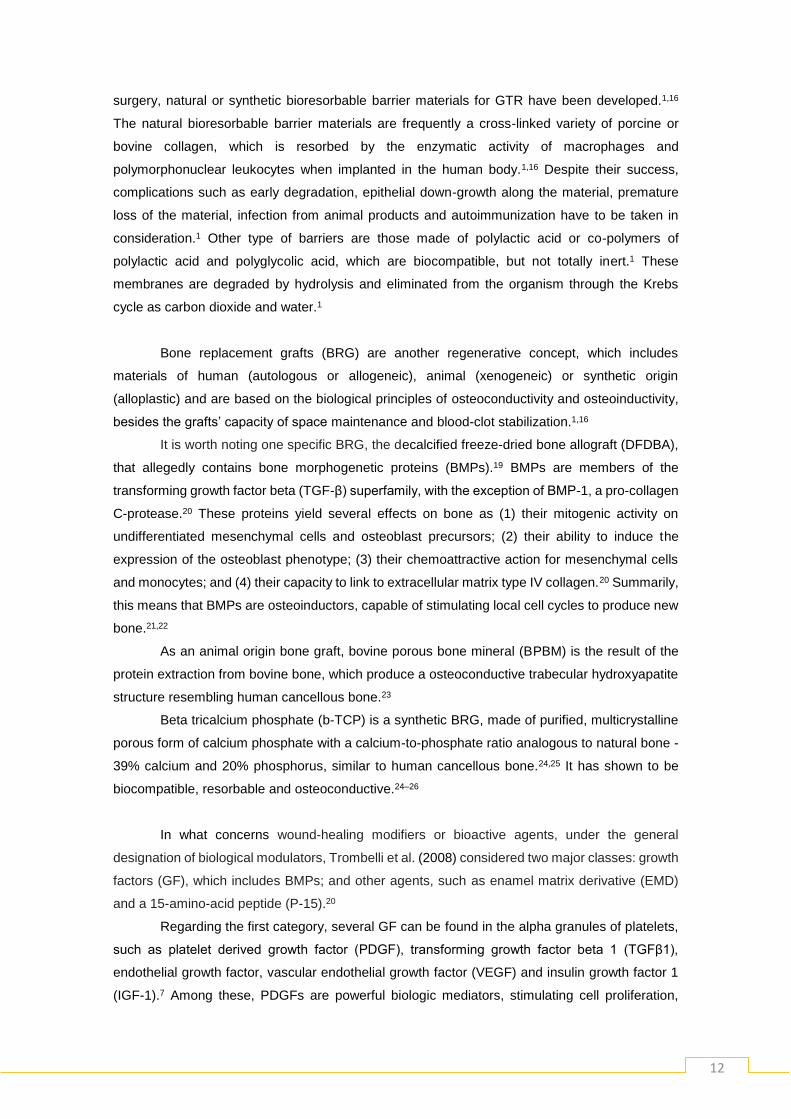
List of abbreviations and acronyms
BRG Bone replacement grafts
CAL Clinical attachment level
GTR Guided tissue regeneration
IFB Infrabony defect
L-PRF Leukocyte and platelet-rich fibrin
Mm Millimeters
NS Non-statistically significant
OFD Open flap debridement
% Percentage
PRGF Plasma rich in growth factors
PRP Platelet-rich plasma
PPD Probing pocket depth
SRP Scaling and root planning
SS Statistically significant

8
1. Introduction
1.1. Chronic periodontitis
The periodontium is a dynamic structure comprising four main tissues: gingiva,
periodontal ligament, root cementum and alveolar bone.1 The disturbance of the balance
between the host tissues and the resident microbiota results in disease of the periodontal tissues.1
Among the different periodontal diseases, chronic periodontitis is the most common form
in adults and it is clinically defined by the following signs and symptoms: (1) color, texture and
volume alterations of the marginal gingiva; (2) bleeding on probing from the gingival pocket area;
(3) diminished resistance to probing of the soft marginal tissues; (4) loss of probing attachment
level; (5) recession of the gingival margin; (6) loss of alveolar bone; (7) root furcation exposure;
(8) increased tooth mobility and (9) drifting and eventually exfoliation of teeth.1 Thereby,
periodontitis can be defined as an inflammatory disease that causes an irreversible loss of
attachment to the connective tissue and supporting alveolar bone.1–3
Bone resorption in periodontitis can produce three types of defects: suprabony (or
horizontal) defects, infrabony (or vertical) defects and interradicular (or furcation) defects.1,4 When
the base of the pocket is located coronal to the alveolar crest, it is considered a suprabony
defect.1,4 If the base of the pocket has an apical location regarding the alveolar crest, it is classified
as an infrabony defect.1,4 The interradicular defects develop when there is pathological bone
resorption of the furcation area due to periodontitis.5
Regarding infrabony defects, one of its main classifications is made according to the
number of residual alveolar bone walls (Figure 1), namely, (1) one-wall defects, with half of a
septum remaining between teeth (hemiseptal defects) or with one buccal/lingual wall; (2) two-wall
defects, with one proximal wall and one buccal/lingual wall or one buccal wall and one lingual wall
(which is specifically nominated as “crater”); and (3) three-wall defects, which consist of three
bony walls and the radicular surface and that may be additionally classified as intrabony defect
or circumferential osseous defect.1,5
The increasing knowledge about the importance of the number of walls in the
regenerative potential of bone defects, highlighted the need to define and standardize their
classification. In fact, the classical classification by Goldman & Cohen (1958), considers that two
types infrabony defects can be recognized: intrabony defects and craters.4 Intrabony defects are
bony defects whose infrabony component affects primarily one tooth, while in craters the defect
affects two adjacent root surfaces to a similar extent.4 But, according to Weinberg & Eskow
(2000), the expression “infrabony” may be applied to all vertical defects, although the term
“intrabony” should be used specifically when referring to a three-walled defect adjacent to a
radicular surface, with high regenerative potential.5,6
Furthermore, a circumferential bone defect extends to buccal or lingual surface of the
root, unlike intrabony defects.6

9
The morphology of infrabony defects can assume a more complex anatomy, when
different components are seen coronally and apically, for example, when the defect has a two-
wall component in its coronal portion and a three-wall component in its most apical portion.1,5,6 In
fact, these combined osseous defects represent the majority of infrabony defects, which reveals
the enormous variety and anatomic complexity of those.5
Other factors, as the width of the defect (or radiographic angle) or the topographic
extension around the tooth, can be also used to classify bone defects (figure 1).1
1.2. Therapeutic approaches
The primary goal of periodontal treatment is to promote the maintenance of the natural
dentition in optimum health and function.7,8 It is, therefore, desirable to not only prevent
periodontal disease progression, but also to regenerate all the tissues of the periodontium.9,10
Several non-surgical and surgical approaches have been studied and applied in
Periodontology, specifically in infrabony defects with deep pockets (figure 2). Non-surgical
treatments aim to disrupt the microbial biofilm and to suppress the inflammation, using pocket/root
instrumentation combined with supragingival plaque control measures. Scaling and root planning
(SRP), even when associated with proper oral hygiene program, does not predictably remove
plaque and subgingival calculus and therefore, other approaches may be necessary, mainly in
deep periodontal pockets.1,5
Classification of periodontal bone
defects
Suprabony defects
Infrabony defects
Width of the defect
Topographic extension around
the tooth
Number of residual walls
One-wall defects
Hemiseptal defects
One buccal/lingual wall
Two-wall defects
One proximal wall and one
buccal/lingual wall
Crater
Three-wall defects
Intrabony defects
Circumferential defects
Combined defects
Inter-radicular defects
Figure 1. Classification of periodontal bone defects

10
Despite their specific indications, surgical approaches must be seen as adjunctive
therapies to non-surgical techniques.1 The main goals of surgical approaches are to diminish the
gingival inflammation and to benefit the patient’s home care, both decisive factors for the long-
term prognosis.1 In order to achieve these goals, besides the access surgery therapy, a hard
tissue intervention may be needed. The hard tissues’ interventions, associated to an open flap
techniques, may include eliminating totally or partially the osseous defect, by osteoplasty and/or
ostectomy; instrumentation of the root surface; promoting healing through regenerative
procedures; or, ultimately, extracting of the involved tooth.1
Focusing on the treatment of infrabony defects, which are usually associated with deep
pockets (periodontal probing depth ≥ 6 mm), some approaches can be highlighted.1,5
Regarding a conservative approach, the main difference is the flap position at the end of
the surgical intervention – if it is apically positioned at the level of the bone crest (original Widman
flap, Neumann flap and apically repositioned flap) or maintained in a coronal position (Kirkland
flap, modified Widman flap, and papilla preservation flap).1 This conventional open flap
debridement intervention is particularly advantageous at pockets that extend beyond the
mucogingival border and/or where is necessary to treat bony or furcation defects, but fails to
predictably regenerate the periodontal tissues.1,7
In a more invasive approach of soft tissues, gingivectomy is mainly an tissue-
eliminating/resective technique, whose purpose is the complete elimination of the periodontal
pocket.1 Beside the aesthetic aspect, which can be problematic due to the gingival recessions,
the potential residual pockets left can be inaccessible to proper patient-performed tooth cleaning
during post‐treatment maintenance.1
Figure 2. Periodontal treatment of infrabony defects with deep pockets
Periodontal treatment of infrabony defects with deep pockets
Non-surgical therapy
Debridment, scaling and root planing
Surgical therapy
Conservative approach
Access flap
techniques
Modified Widman flap
Modified Kirkland flap
Papilla preservation flap
Apical position
techniques
Widman flap
Neumann flap
Agressive approach
Ressective techniques
Gingivectomy
Osteotomy
Regenerative approach

11
As for hard tissues, the goal is to convert an infrabony defect into a suprabony defect,
using apical repositioning of the soft tissue flap(s) and osseous recontouring techniques.1 These
bone contouring procedures may lead to poor aesthetic outcomes, since they often result in
recession of the gingival margin after healing.11
Although it is fair to say that conventional surgical approaches, that resect or eliminate
tissue, can be beneficial in early or shallow infrabony defects (<3 mm), it is important to
understand that the improvement in periodontal clinical parameters, when a non-regenerative
therapy is applied, are mainly due to the formation of a long junctional epithelium.1,12 The only
approach that allows a predictable and long-term sustainable reconstitution of functional
attachment apparatus is the regenerative therapy.12,13
1.2.1. Regenerative therapy
By definition, regeneration is the reproduction or reconstruction of a lost or injured part,
in such a way that the architecture and function of the lost or injured tissues are completely
restored, whereas repair refers to healing of a wound by tissue that does not fully restore the
original architecture or function.14
Although it is established that the regeneration of the periodontium includes the formation
of new cementum with inserting collagen fibers on the root surfaces and the regrowth of the
alveolar bone, the understanding of the regeneration mechanism of an architecturally complex
organ, such as periodontium, remained a challenge for many years.1,15
In 1976, Melcher developed a theory based on a “compartmentalization” concept,
according to which the type of cell that repopulates the root surface after periodontal surgery
determines the nature of the attachment that will be formed.1,15 Several experimental studies have
been conducted and it is known today, that the cells with the potential to produce a new connective
tissue attachment reside in the periodontal ligament.1,5
Three different regenerative concepts have been employed – barrier membranes, grafts
and biological modulators, plus other combinations between those.16
The barrier membranes behave as mechanical barriers that enable not only selective cell
growth, but also provide space and stability to the blood clot.16,17 The initial efforts on guided
tissue regeneration (GTR) started around the 80’s with a cellulose acetate bacterial filter
(Millipore® filter, type GS; Millipore SA,67 Molsheim, France), which was an occlusive membrane
– functional, but not clinically ideal.1,16 Later on, nonresorbable membranes of expanded-
polytetrafluoroethylene (Gore-tex®, W. L. Gore & Ass. Inc., Flagstaff, Arizona, USA) were
developed.1,16,18 These membranes of e‐PTFE are inert and biocompatible, but they persist after
healing and it is necessary a second intervention for their removal.1,16 In order to avoid this second
12
surgery, natural or synthetic bioresorbable barrier materials for GTR have been developed.1,16
The natural bioresorbable barrier materials are frequently a cross-linked variety of porcine or
bovine collagen, which is resorbed by the enzymatic activity of macrophages and
polymorphonuclear leukocytes when implanted in the human body.1,16 Despite their success,
complications such as early degradation, epithelial down-growth along the material, premature
loss of the material, infection from animal products and autoimmunization have to be taken in
consideration.1 Other type of barriers are those made of polylactic acid or co-polymers of
polylactic acid and polyglycolic acid, which are biocompatible, but not totally inert.1 These
membranes are degraded by hydrolysis and eliminated from the organism through the Krebs
cycle as carbon dioxide and water.1
Bone replacement grafts (BRG) are another regenerative concept, which includes
materials of human (autologous or allogeneic), animal (xenogeneic) or synthetic origin
(alloplastic) and are based on the biological principles of osteoconductivity and osteoinductivity,
besides the grafts’ capacity of space maintenance and blood-clot stabilization.1,16
It is worth noting one specific BRG, the decalcified freeze-dried bone allograft (DFDBA),
that allegedly contains bone morphogenetic proteins (BMPs).19 BMPs are members of the
transforming growth factor beta (TGF-β) superfamily, with the exception of BMP-1, a pro-collagen
C-protease.20 These proteins yield several effects on bone as (1) their mitogenic activity on
undifferentiated mesenchymal cells and osteoblast precursors; (2) their ability to induce the
expression of the osteoblast phenotype; (3) their chemoattractive action for mesenchymal cells
and monocytes; and (4) their capacity to link to extracellular matrix type IV collagen.20 Summarily,
this means that BMPs are osteoinductors, capable of stimulating local cell cycles to produce new
bone.21,22
As an animal origin bone graft, bovine porous bone mineral (BPBM) is the result of the
protein extraction from bovine bone, which produce a osteoconductive trabecular hydroxyapatite
structure resembling human cancellous bone.23
Beta tricalcium phosphate (b-TCP) is a synthetic BRG, made of purified, multicrystalline
porous form of calcium phosphate with a calcium-to-phosphate ratio analogous to natural bone -
39% calcium and 20% phosphorus, similar to human cancellous bone.24,25 It has shown to be
biocompatible, resorbable and osteoconductive.24–26
In what concerns wound-healing modifiers or bioactive agents, under the general
designation of biological modulators, Trombelli et al. (2008) considered two major classes: growth
factors (GF), which includes BMPs; and other agents, such as enamel matrix derivative (EMD)
and a 15-amino-acid peptide (P-15).20
Regarding the first category, several GF can be found in the alpha granules of platelets,
such as platelet derived growth factor (PDGF), transforming growth factor beta 1 (TGFβ1),
endothelial growth factor, vascular endothelial growth factor (VEGF) and insulin growth factor 1
(IGF-1).7 Among these, PDGFs are powerful biologic mediators, stimulating cell proliferation,

13
differentiation, angiogenesis and chemotaxis.9,19 The action of PDGF alone, with or without a
progression factor to induce mitosis, provoked the proliferation of both osteoblasts and isolated
periodontal ligament (PDL) cells.22 Another GF worth noting is TGF-β, capable of diverse
functions such as increasing the differentiated function of osteoblasts, osteoblasts precursors and
extracellular matrix formation/remodeling or stimulating the proliferation of gingival fibroblastic
cells and formation of blood vessels.22 Other GF related to periodontal renegeneration have been
mentioned in the literature, such as fibroblast growth factor (FGF) and recombinant human
platelet-derived growth factor-BB (rhPDGF-BB). Nevertheless, the majority of them still lack
scientific evidence for efficacy and safety for clinical application.16,20,27
Enamel matrix derivative (EMD) is another bioactive agent. EMD is mainly an amelogin-
compound (about 90%), plus porcine origin proteins – albumin, amelin and enamelin.20 According
to Hammarström et al. (1997), the initiating factor for cementum formation is the expressed
amelogenin at the apical end of the forming root of human teeth.28 The formation of cementum is
associated with the development of the periodontal ligament and the alveolar bone.28 The
combination of EMD with other regenerative therapies is based on an hypothetic synergistic
effect.10 Although it is not proven, the current literature available is very promising regarding
pocket-depth reduction, clinical attachment level gain and radiographic bone level.16
Another bioactive agent analyzed in some studies is the 15-amino-acid peptide (P-15), a
synthetic cell-binding peptide that is equal to part of the sequence of the α1 chain of type I
collagen.20 The P-15 has been shown to increase the rate and the extent of attachment and
migration of periodontal cells to root or biomaterial surfaces.20,29 Similarly to EMD, P-15 have
been combined with other products, such as anorganic bovine-derived matrix (ABM).5,13,29
1.2.1.1. Autologous platelet concentrates (APC)
Since 1970, autologous platelet concentrates have acquired more attention from medical
community, but their introduction in oral and maxillofacial surgery occurred only in the 90s .30
The first generation of platelet concentrates (APC) comprises platelet-rich plasma (PRP)
and plasma rich in growth factors (PRGF).30
PRP has in its constitution four to fivefold-increased platelet concentration above
baseline, thus being enriched with several growth factors, such as PDGF, transforming growth
factor-1 (TGF-1), transforming growth factor-2 (TGF-2), insulin growth factor-1 (IGF-1), insulin
growth factor-2 (IGF-2), basic fibroblast growth (bFGF), vascular endothelial growth factor
(VEGF), epithelial growth factor (EGF); and blood proteins related to osteoconduction (fibrin,
fibronectin and vitronectin).2,9,31,32 These growth factors reach their maximum release in the first
day, yet it continues for seven days.9
The PRP preparation requires the following three components: (1) anticoagulants at the
moment of blood collection, (2) bovine thrombin and (3) calcium chloride.30,33 There are several

14
PRP preparation protocols, but all of them require two centifrugations (with time and number of
rotations per minute variable according to the different authors).25,31
The different clinical applications of PRP, such as sinus floor elevation or treatment of
periodontal defects, are based on the basic premise that its high concentrations of platelets will
promote a local concentration of secreted growth factors, which will increase the initial bone repair
mechanisms and its effects will remain even when PRP fade away.32 Other properties attributed
to PRP are not only related to an angiogenetic, proliferative and differentiative effect on
osteoblasts, mainly due to PDGF and TGF-β; but also to its capacity of inducing clot formation
when reacting with thrombin, thanks to its fibrinogen content, and its potential haemostatic activity,
which provides blood-clot stability.21,31,33
It is worth noting that PRP is considered a safe autologous preparation, since it is
prepared with the patient’s own blood; is biologically acceptable; and it is economically viable.
Nevertheless, the required use of bovine thrombin, which is not an autologous material, still
remains a significant disadvantage, even if no disease transmission or immunogenic reactions
have been reported to this date.31 Additionally, Castro et al. (2017) emphasizes that the PRP fibrin
network is thin and non-condensed and it has a low tensile strength, which makes it less helpful
to use as space maintainer.30
The second generation of PC was introduced in 2001 by Choukroun and co-workers.34
The platelet-rich fibrin (PRF) preparation consists exclusively in the blood centrifugation at high
spin, without any additives, producing three layers: red blood corpuscles at the bottom of the tube,
platelet-poor plasma (PPP) on the top and a “buffy coat” as intermediate layer, where most
leucocytes and platelets are concentrated.30 This intermediate layer can be carefully compressed
and transformed into a membrane of, approximately, 1 mm in thickness.30 The PRF membrane is
a biocompatible, bioresorbable and three-dimensional polymerized fibrin matrix, capable of slowly
releasing growth factors over a period of 7-14 days, but also delivering platelet, leukocytes,
cytokines and matrix glycoproteins.19,30 Several authors emphasize the strong fibrin network of
PRF, due to the physiological concentration of thrombin during its preparation, which enhances
its mechanical properties.9,23,30,35
Briefly, when comparing PRP with PRF, the second has some advantages, such as the
less chair side time required (approximately 12 min. for preparation), no need of addition of bovine
thrombin or anticoagulants, the longer-term effect of growth factors and the fact that it is easier to
use as a membrane, similar to guided tissue regeneration (GTR) membrane.8,36
Conventionally, four main categories of APC can be distinguished: (1) pure PRP, with no
leucocytes; (2) leucocytes rich PRPs (L-PRP); (3) pure PRF, without leucocytes; and (4) PRF with
leucocytes (L-PRF).35 However, other APC can be mentioned, such as advanced PRF (A-PRF),
injectable PRF (i-PRF) or lyophilized PRF (Ly-PRF).
The standard protocol for PRF and L-PRF requires one step of centrifugation for 12
minutes at 2700 rpm.37–39 The advanced PRF (A-PRF), also developed by Choukroun, has a
slower centrifugation for a longer time (1500 rpm for 14 minutes).37,38 This protocol modification
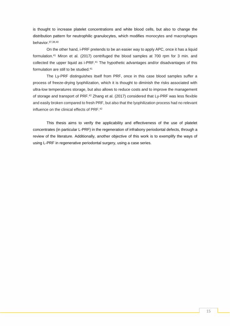
15
is thought to increase platelet concentrations and white blood cells, but also to change the
distribution pattern for neutrophilic granulocytes, which modifies monocytes and macrophages
behavior.37,38,40
On the other hand, i-PRF pretends to be an easier way to apply APC, once it has a liquid
formulation.41 Miron et al. (2017) centrifuged the blood samples at 700 rpm for 3 min. and
collected the upper liquid as i-PRF.41 The hypothetic advantages and/or disadvantages of this
formulation are still to be studied.41
The Ly-PRF distinguishes itself from PRF, once in this case blood samples suffer a
process of freeze-drying lyophilization, which it is thought to diminish the risks associated with
ultra-low temperatures storage, but also allows to reduce costs and to improve the management
of storage and transport of PRF.42 Zhang et al. (2017) considered that Ly-PRF was less flexible
and easily broken compared to fresh PRF, but also that the lyophilization process had no relevant
influence on the clinical effects of PRF.42
This thesis aims to verify the applicability and effectiveness of the use of platelet
concentrates (in particular L-PRF) in the regeneration of infrabony periodontal defects, through a
review of the literature. Additionally, another objective of this work is to exemplify the ways of
using L-PRF in regenerative periodontal surgery, using a case series.

16
2. Systematic Review
2.1. Materials and Methods
In order to establish an appropriated search protocol for this systematic review, a focused
question was made. Taking into account PRISMA (Preferred Reporting Items for Systematic
Reviews and Meta-Analyses) guidelines43, the following PICOT based question was elaborated:
“In patients with periodontal infrabony defects, what is the efficacy of L-PRF alone or in
combination with other biomaterials in periodontal regeneration, comparatively to open flap
debridment and other APCs, after at least 6 months of healing?”
(P) Population: patients with periodontal infrabony defects systemically healthy (ASA
I) or with mild systemic disease (ASA II);
(I) Intervention: application of L-PRF alone or in combination other biomaterials in
periodontal regenerative surgery;
C) Comparison: open flap debridement (OFD) and other APCs;
(O) Outcomes: probing pocket depth reduction, clinical attachment gain, radiographic
bone fill, patient centered outcomes;
(T) Time: post-operative follow-up of at least 6 months.
The study selection respected the following inclusion criteria:
• Systematic reviews, meta-analysis or randomized clinical trials (RCTs) evaluating
the effect of L-PRF in the regeneration of periodontal infrabony defects, alone or in combination
with other biomaterials;
• Publications with human histological, radiograph or clinical outcome parameters
assessing soft tissue and/or bone healing results after application of platelet concentrates;
• Human studies published in English and/or Portuguese.
As exclusion criteria:
• Narrative reviews, case series or case reports;
• Animal or in vitro studies;
• Non-randomized controlled trials or with an inadequate control group;
For this systematic review, an electronic literature search was performed in PubMed
database with the following keywords: “platelet rich plasma”, “platelet rich fibrin”, “plasma rich
growth factors”, “periodontal defect”, “infrabony”, “intrabony”, “bone regeneration” and
“periodontal regeneration”; and the Boolean connectors “AND” and “OR”. MeSH Terms were
applied when possible. Activated filter criteria were: the type of articles including Meta-Analysis,

17
Systematic Reviews and Randomized Controlled Trial; with abstract text availability; humans;
published in the last 10 years; in English or Portuguese language. The last search was performed
at 15th May, 2017 (figure 3).
Another online search was made in Cochrane Library, using the terms: “platelet-rich
plasma” (MeSH descriptor), “platelet rich fibrin”, “plasma rich growth factors”, “alveolar bone loss”
(MeSH descriptor), “infrabony”, “intrabony”, “bone regeneration” (MeSH descriptor) and
“periodontal regeneration”. The word variations for non-MeSH terms have been searched. The
publications timeframe used was 2007 to 2017. The last update was made at 15th May, 2017
(figure 4).
As a complementary search, cross-references and hand search were also taken into
account in the Journal of Clinical Periodontology and the Journal of Periodontology.
Cochrane Library
2007-2017
*(Word variations have been searched)
“platelet-rich plasma” (MeSH descriptor) OR “platelet rich fibrin”* OR “plasma rich
growth factors”* AND “alveolar bone loss” (MeSH descriptor) OR “infrabony”*
OR “intrabony”* AND “periodontal regeneration”* OR “bone regeneration”
(MeSH descriptor)
PubMed
Meta-Analysis, Randomized Controlled Trial, Systematic Reviews
Abstract
10 years
Humans
English or Portuguese
(((((((platelet rich plasma[MeSH Terms]) OR platelet rich fibrin) OR plasma rich
growth factors) AND periodontal defect) AND infrabony) OR intrabony) AND bone
regeneration[MeSH Terms]) OR periodontal regeneration
Figure 3. PubMed search
Figure 4. Cochrane Library search
18
2.2. Results
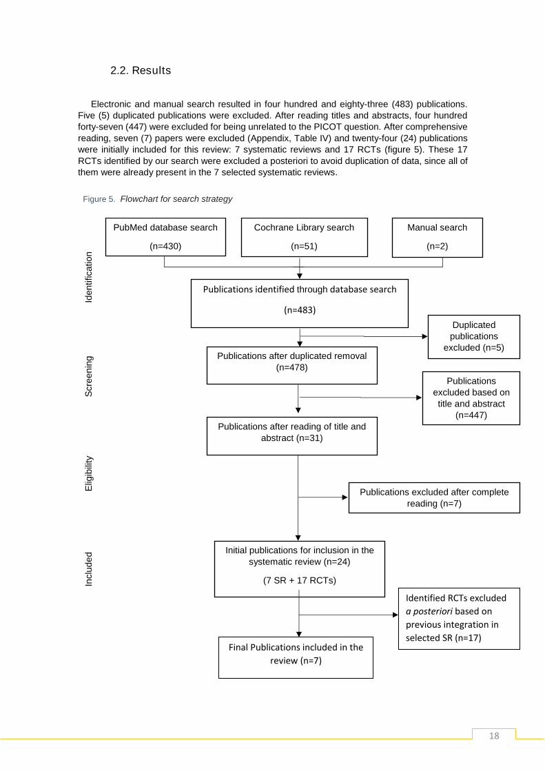
Electronic and manual search resulted in four hundred and eighty-three (483) publications.
Five (5) duplicated publications were excluded. After reading titles and abstracts, four hundred
forty-seven (447) were excluded for being unrelated to the PICOT question. After comprehensive
reading, seven (7) papers were excluded (Appendix, Table IV) and twenty-four (24) publications
were initially included for this review: 7 systematic reviews and 17 RCTs (figure 5). These 17
RCTs identified by our search were excluded a posteriori to avoid duplication of data, since all of
them were already present in the 7 selected systematic reviews.
Figure 5. Flowchart for search strategy
Identification
Scre
en
ing
Elig
ibili
ty
Inclu
ded
PubMed database search
(n=430)
Cochrane Library search
(n=51)
Manual search
(n=2)
Publications identified through database search
(n=483)
Publications after duplicated removal
(n=478)
Publications after reading of title and
abstract (n=31)
Initial publications for inclusion in the
systematic review (n=24)
(7 SR + 17 RCTs)
Duplicated
publications
excluded (n=5)
Publications excluded based on
title and abstract
(n=447)
Publications excluded after complete
reading (n=7)
Final Publications included in the
review (n=7)
Identified RCTs excluded
a posteriori based on
previous integration in
selected SR (n=17)

19
Table I. Systematic Reviews and Meta-analyses: general information and principal outcomes
Author, year
Meta-analysis
Number and type of studies included in the
MA
Platelet concentrated
Treatment evaluated
Follow-up (months)
Outcomes (PPD reduction (mm), CAL gain (mm), Radiographic parameters – Bone fill (mm or %))
Statistical significance
Castro et al., 2017
Yes
6 RCTs (in 13) Sharma & Pradeep 2011, Thorat et al. 2011, Pradeep m 2012a*, 2015, Rosamma et al. 2012, Ajwani et al. 2015
L-PRF
L-PRF+OFD vs OFD
PRF/PRP+OFD
Vs OFD*
9-12 months
PPD red: SMD: 1.10mm; 95% CI: 0.6-1.6 in favor of L-PRF. Cal gain: SMD: 1.20mm; 95% CI: 0.5-1.9 in favor of L-PRF. Bone fill (mm): SMD: 1.70mm; 95% CI: 1.0-2.3 in favor of L-PRF. Bone fill (%): SMD: 46.0%, 95% CI: 33.2–58.7 in favor of L-PRF.
SS P<0.001
Del Fabbro et al., 2011
Yes
10 RCTs (in 16) Hanna et al. 2004, Okuda et al. 2005, Ouyang & Qiao 2006 Christgau et al. 2006 Döri et al. 2007a Döri et al. 2007b Demir et al. 2007 Piermontese et al. 2008 Döri et al. 2008a Döri et al. 2008b
PRP
PRP + Bone graft Vs Bone
graft PRP + Bone
graft + GTR Vs Bone graft +
GTR PRP + Bone
graft + EMD Vs Bone graft +
EMD
6-12 months
10 RCT: SMD: 0.50 mm (95% CI: 0.12 to 0.88 mm) in favor of PRP. 4 RCT with GTR: SMD: 0.04mm(95%CI: -0.33 to 0.41mm). 6 RCT without GTR: SMD: 0.84mm(95% CI: 0.27 to 1.42mm). A significant positive effect of the adjunct of PRP was found for intrabony defects. Such an effect was magnified in studies in which GTR was not used, whereas in studies using GTR, the use of PRP had no adjunctive effect.
SS for CAL gain
NS for
PRP+GTR
Hou et al., 2016
Yes
12 RCTs (in 15) Okuda K et al. 2005 Hanna R et al. 2004 Döri et al. 2009 Demir et al. 2007 Agarwal et al. 2014 Piemontese et al. 2008 Özdemir et al. 2012 Kaushick et al. 2007 Christgau et al. 2006 Döri, Huszar et al. 2007 Döri et al. 2007 Döri et al. 2008
PRP
PRP + Bone graft Vs Bone
graft PRP + Bone
graft + GTR Vs Bone graft +
GTR
9-12 months
Clinically and significantly greater CAL gains and PPD reductions observed in subjects who received PRP as an adjunct to periodontal intrabony defect therapy: CAL: WMD 0.76 mm, 95 % CI = 0.34 to 1.18 mm, P = 0.0004; PPD: WMD 0.53 mm, 95 % CI = 0.21 to 0.85 mm, P = 0.001. Meta-analysis of patients who underwent GTR demonstrated that this approach did not significantly affect treatment outcomes (CAL: WMD 0.08 mm, 95 % CI = −0.30 to 0.46 mm, P = 0.67), as indicated by a comparison with patients who did not undergo GTR (CAL: WMD 1.22 mm, 95 % CI = 0.88 to 1.57 mm, P < 0.00001).
SS for CAL gain and PPD
NS for
PRP+GTR

20
Table I. Systematic Reviews and Meta-analyses: general information and principal outcomes (continuation)
Author, year Meta-
analysis
Number and type of studies included in the
MA
Platelet concentrated
Evaluated treatment
Follow-up (months)
Outcomes (PPD reduction (mm), CAL gain (mm), Radiographic parameters – Bone fill (mm or %))
Statistical significance
Panda et al., 2014
Yes
4 RCTs Thorat et al. 2011, Sharma & Pradeep 2011b***, Pradeep et al. 2012a*, Pradeep et al. 2012b**
PRF
PRF+OFD vs OFD
PRF/PRP+OFD
Vs OFD*
PRF + HA Vs OFD**
9 months
PRF has a significant additive effect when used along with OFD. Statistically significant PPD reduction and CAL gain at the end of the follow-up in both test and control group in all four studies. Radiologically, significantly greater bone fill for PRF+OFD, as compared to OFD alone, in all four studies. Unreadable data from Forrest Plots.
Unreadable data
Yes
7 RCTs Demir et al. 2007~ Döri et al. 2009 Hassan et al. 2012 Okuda et al. 2005 Parimala & Mehta 2010 Piemontese et al. 2008 Saini et al. 2011
PRP PRP + Bone graft Vs Bone
graft
9-12 months
Significant improvement in the CAL in the group using platelet concentrates in combination with graft materials over the group using graft materials alone. Consistent positive effect on radiological bone fill, when used along with bone substitutes. Unreadable data from Forrest Plots.
Unreadable data
Yes
4 RCTs Döri et al. 2008 Christgau et al. 2006 Döri et al. 2007a Döri et al. 2007b
PRP
PRP + Bone graft + GTR Vs
Bone graft + GTR
9-12 months
PRP showed no additive beneficial effect when combined with bone graft and GTR membrane for the treatment of intrabony defects. Meta-analysis of PPD reduction and clinical attachment gain in the experimental group over the control group was not significant. Unreadable data from Forrest Plots.
Unreadable data

21
Table I. Systematic Reviews and Meta-analyses: general information and principal outcomes (continuation)
Author, year
Meta-analysis
Number and type of studies included in the
MA
Platelet concentrated
Evaluated treatment
Follow-up (months)
Outcomes (PPD reduction (mm), CAL gain (mm), Radiographic parameters – Bone fill (mm or %))
Statistical significance
Plachokova et al., 2015
No
3 RCTs Hanna et al. 2004 Okuda et al. 2005 Sammartino et al. 2005
PRP Graft+PRP vs.
Graft 3-12
months
No meta-analysis due to heterogeneity. Differences in treatment effects for periodontal defects in terms of clinical attachment level (CAL) were significant, the mean differences ranging from 0.8 to 3.2mm.
-
Roselló-Camps et al., 2015
Yes
14 RCTS (in 21) Lekovic et al. 2002 Camargo et al. 2002 Okuda K et al. 2005 Ouyang & Qiao 2006 Demir et al. 2007 Döri et al. 2007b Döri et al. 2007a Döri et al. 2008 Piemontese et al. 2008 Camargo et al. 2009 Özdemir et al. 2012 Pradeep et al. 2012 Baja et al. 2013
PRP
PRP + Bone graft Vs Bone
graft PRP + Bone
graft + GTR Vs Bone graft +
GTR
9-12 months
14 RCTs for PPD reduction: WMD 0.55 mm, with a 95% CI= -0.09 mm to 1.20 mm (p= 0.09) in favor of PRP. 2 RCTs for bone level (BL) in mm: WMD was 0.76 mm (95% CI= 0.21 mm to 1.31 mm, p=0.007) in favor of PRP. 2 RCTs for bone level (BL) in %: WMD 47.41% (95% CI= 32.48% to 62.33%, p< 0.0001) in favor of PRP. 12 RCTs for CAL Gain: WMD 0.58 mm, with a 95% CI= 0.24 mm to 0.91 mm (p= 0.0008) in favor of PRP. High heterogeneity among studies. PRP might offer some beneficial effects on clinical and radiographic outcomes for regeneration of periodontal intrabony defects.
NS for PDD reduction
SS for bone level (in mm and %) and
CAL
Shah et al.,
2014 Yes
5 RCTs Thorat et al. 2011, Sharma & Pradeep 2011a, Pradeep et al. 2012a, Pradeep et al. 2012b**, Rosamma et al. 2012,
PRF
PRF+OFD vs OFD
PRF/PRP+OFD
Vs OFD*
PRF + HA Vs OFD**
9-12 months
PPD reduction: SMD: 1.10mm; 95% CI: 0.56-1.64 in favor of PRF (no significant heterogeneity). CAL gain: SMD: 0.95mm; 95% CI: 0.20-1.71 in favor of PRF (no significant heterogeneity). Bony defect reduction (mm): SMD: 2.33mm; 95% CI: 1.43-3.23 (no significant heterogeneity).
SS for PDD reduction and
IBD P<0.001
NS for CAL
gain P=0.006

22
3. Case series
3.1. Materials and Methods
In order to demonstrate the L-PRF protocol for the treatment of periodontal infrabony
defects, a case series is presented.
The clinical records of periodontal patients followed in a university clinical centre
(Dentistry Department of the Faculty of Medicine – University of Coimbra) were analyzed and the
patient selection occurred according to the following inclusion criteria:
• Systemically healthy humans (ASA I) or patients with mild systemic disease (ASA
II);
• Infrabony defects with PPD ≥ 6 mm, confirmed on a standard periapical
radiograph, and with a two or three walls morphology;
• Bleeding on probe (BOP) and a full‐mouth plaque score ≤ 20%;
• Non-smokers.
As exclusion criteria:
• Patients with uncontrolled systemic diseases or adverse conditions for
periodontal surgery;
• Infrabony defects with PPD ≤ 5 mm and/or with a one wall bony defect
morphology;
• BOP and plaque score ≥ 20%;
• Smokers.
After the screening and eligibility process, four patients fulfilled the inclusion criteria, but
one patient failed the pre-operatory appointments and was excluded. An informed consent was
given to the remaining three patients.
During pre-surgical therapy, each patient was given careful instructions regarding proper
oral hygiene measures. A full mouth supragingival and subgingival SRP procedure was
performed (when indicated), under local anesthesia, using ultrasonic and hand instrumentation.
Six to eight weeks after this preliminary treatment, a periodontal re-evaluation was performed to
confirm the suitability of the sites for periodontal surgery.
The L-PRF clinical protocol used is in accordance with the guidelines of the 1st European
Meeting on Enhanced Natural Healing in Dentistry (Leuven, Belgium; 2016)39, using the
IntraSpin™ centrifuge (Intra-Lock, Boca Raton, FL, USA), and includes the following steps:
23
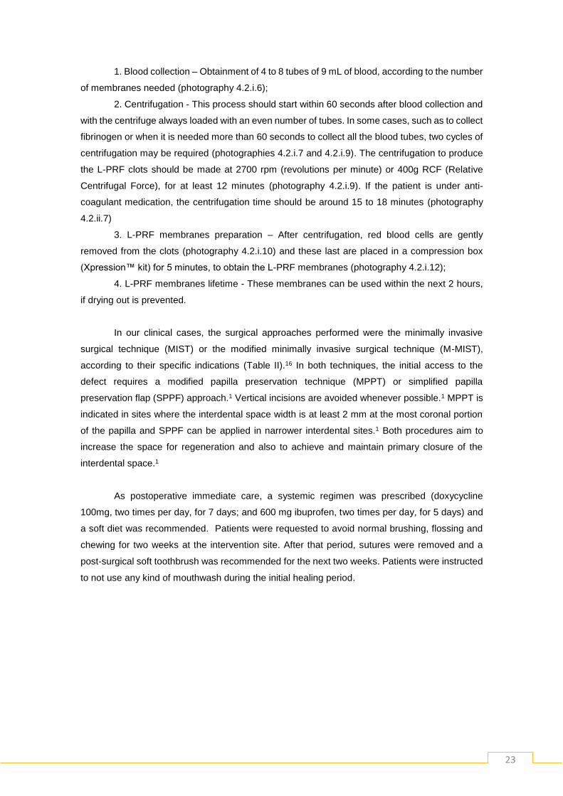
1. Blood collection – Obtainment of 4 to 8 tubes of 9 mL of blood, according to the number
of membranes needed (photography 4.2.i.6);
2. Centrifugation - This process should start within 60 seconds after blood collection and
with the centrifuge always loaded with an even number of tubes. In some cases, such as to collect
fibrinogen or when it is needed more than 60 seconds to collect all the blood tubes, two cycles of
centrifugation may be required (photographies 4.2.i.7 and 4.2.i.9). The centrifugation to produce
the L-PRF clots should be made at 2700 rpm (revolutions per minute) or 400g RCF (Relative
Centrifugal Force), for at least 12 minutes (photography 4.2.i.9). If the patient is under anti-
coagulant medication, the centrifugation time should be around 15 to 18 minutes (photography
4.2.ii.7)
3. L-PRF membranes preparation – After centrifugation, red blood cells are gently
removed from the clots (photography 4.2.i.10) and these last are placed in a compression box
(Xpression™ kit) for 5 minutes, to obtain the L-PRF membranes (photography 4.2.i.12);
4. L-PRF membranes lifetime - These membranes can be used within the next 2 hours,
if drying out is prevented.
In our clinical cases, the surgical approaches performed were the minimally invasive
surgical technique (MIST) or the modified minimally invasive surgical technique (M-MIST),
according to their specific indications (Table II).16 In both techniques, the initial access to the
defect requires a modified papilla preservation technique (MPPT) or simplified papilla
preservation flap (SPPF) approach.1 Vertical incisions are avoided whenever possible.1 MPPT is
indicated in sites where the interdental space width is at least 2 mm at the most coronal portion
of the papilla and SPPF can be applied in narrower interdental sites.1 Both procedures aim to
increase the space for regeneration and also to achieve and maintain primary closure of the
interdental space.1
As postoperative immediate care, a systemic regimen was prescribed (doxycycline
100mg, two times per day, for 7 days; and 600 mg ibuprofen, two times per day, for 5 days) and
a soft diet was recommended. Patients were requested to avoid normal brushing, flossing and
chewing for two weeks at the intervention site. After that period, sutures were removed and a
post‐surgical soft toothbrush was recommended for the next two weeks. Patients were instructed
to not use any kind of mouthwash during the initial healing period.

24
Table II. Case series methodology
Patient (female/male)
Age (years)
Defect morphology
(teeth: number of
walls)
Intervention
L-PRF preparation
Surgical access
technique Suture technique and thread
Hardware
Blood collected (number of tubes,
mL)
Centrifugation process
(number of cycles, force /
time)
B.C. (F) 27 16: 2w
OFD + Xenograft + Fibrinogen + L-PRF fragments + L-PRF
membrane
IntraSpin™ centrifuge
(Intra-Lock, Boca Raton,
FL, USA)
1 tube
8mL
4 tubes
9 mL
2 cycles
400 g / 3 min
400 g / 12 min
M-MIST Modified internal mattress suture with a 5-0 monofilament thread
G.I. (F) 66 36: 2w OFD + L-PRF fragments +
L-PRF membrane
4 tubes
9 mL
1 cycle
400 g / 15 min MIST
Modified internal mattress suture with a 6-0 monofilament thread
R.F. (F) 66
1) 44: 2w 2) 42: 2w 3) 32: 3w
(apically) + 2p (coronally)
OFD + L-PRF fragments + L-PRF membrane
6 tubes
9 mL
1 cycle
400 g / 12 min
1) M-MIST 2) M-MIST
3) MIST
Modified internal mattress sutures with a 6-0 monofilament
threads

25
Pre-operative data and follow-up of the case series are presented in Table III.
Table III. Pre-operative data
Patient (sex)
Age (years)
Defect(s) morphology
(teeth: number of
walls)
Intervention
Initial pocket depth (mm)
Initial PPD (mm)
Adverse effects
reported
Follow-up
(months)
B.C. (F)
27 16: 2w
OFD + Xenograft + L-PRF fragments
+ L-PRF membrane
7 8 No 6
G.I. (F) 66 36: 2w OFD + L-PRF fragments + L-
PRF membrane 5 6 No 3
R.F. (F)
66
1) 44: 2w 2) 42: 2w 3) 32: 3w
(apically) + 2p (coronally)
OFD + L-PRF fragments + L-
PRF membrane
1) 4 2) 4 3) 6
1) 5 2) 5 3) 7
No 1
F = Female; M = Male; W = Walls
At follow-up appointments, the soft tissues exhibited an apparently healthy healing and
no significant morbidity was mentioned for the patients.
26
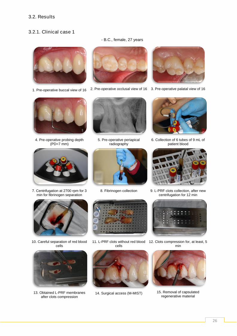
3.2. Results
3.2.1. Clinical case 1
- B.C., female, 27 years
1. Pre-operative buccal view of 16
2. Pre-operative occlusal view of 16 3. Pre-operative palatal view of 16
4. Pre-operative probing depth (PD=7 mm)
5. Pre-operative periapical
radiography 6. Collection of 6 tubes of 9 mL of
patient blood
7. Centrifugation at 2700 rpm for 3 min for fibrinogen separation
8. Fibrinogen collection 9. L-PRF clots collection, after new
centrifugation for 12 min
10. Careful separation of red blood cells
11. L-PRF clots without red blood
cells
12. Clots compression for, at least, 5
min
13. Obtained L-PRF membranes
after clots compression
14. Surgical access (M-MIST)
15. Removal of capsulated
regenerative material

27
16. Presence of residual calculus on a radicular groove
17. Cleaned infrabony defect (8mm deep)
18. Xenograft (Bio-Oss® - Geistlich Pharma AG, Switzerland)
19. Collection of the membrane
compression surplus fluid
20. Hydration of the xenograft with
the collected fluid 21. Mixture of hydrated xenograft with L-PRF membrane fragments
22. Addition of fibrinogen to obtain the bone block
23. Insertion of the bone block into the defect
24. Bone block compaction
25. Conformation of an L-PRF membrane to cover the bone block
26. Placement of L-PRF membrane over the bone block
27. Modified internal mattress suture with a 5-0 monofilament thread
28. Post-operative view at 5 days 29. PD at 6 months (5mm) 30. Post-operative periapical radiography at 6 months
Main conclusions: The combined approach of xenograft + L-PRF + fibrinogen induced a favorable healing of
soft tissues. No post-operative complication was reported. At 6 months, 2mm of CAL gain was obtained, along with a
good radiographic filling of the defect.

28
3.2.2. Clinical case 2
G.I., female, 66 years
1. Pre-operative buccal view of 36
2. Pre-operative occlusal view of 36 3. Pre-operative palatal view of 36
4.Pre-operative periapical radiography 5. Tissue Regeneration Kit (Intra-
lock®, Boca Raton, FL, USA)
6. Collection of patient blood
7. Centrifugation at 2700 rpm for 15 min (according to anticoagulated
patients’ guidelines)
8. Collection of 4 tubes of 9 mL of patient blood
9. Collection of L-PRF clots
10. Careful separation of red blood cells
11. L-PRF clots without red blood cells
12. Clots compression
29
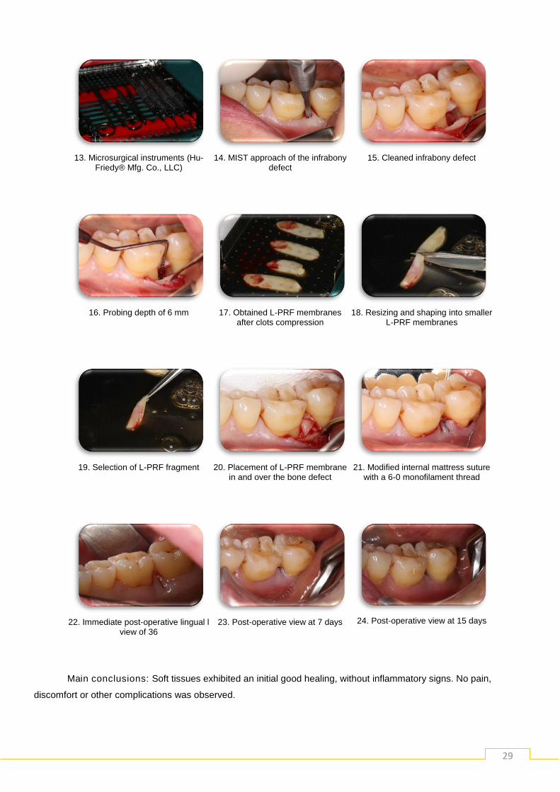
13. Microsurgical instruments (Hu-Friedy® Mfg. Co., LLC)
14. MIST approach of the infrabony defect
15. Cleaned infrabony defect
16. Probing depth of 6 mm 17. Obtained L-PRF membranes
after clots compression
18. Resizing and shaping into smaller
L-PRF membranes
19. Selection of L-PRF fragment
20. Placement of L-PRF membrane
in and over the bone defect
21. Modified internal mattress suture with a 6-0 monofilament thread
22. Immediate post-operative lingual l
view of 36
23. Post-operative view at 7 days
24. Post-operative view at 15 days
Main conclusions: Soft tissues exhibited an initial good healing, without inflammatory signs. No pain,
discomfort or other complications was observed.
30
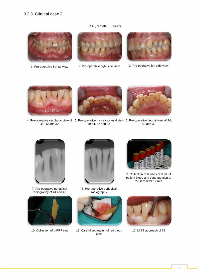
3.2.3. Clinical case 3
R.F., female, 66 years
1. Pre-operative frontal view
2. Pre-operative right side view
3. Pre-operative left side view
4. Pre-operative vestibular view of
44, 42 and 32
5. Pre-operative incisal/occlusal view
of 44, 42 and 32
6. Pre-operative lingual view of 44,
42 and 32
7. Pre-operative periapical radiography of 44 and 42
8. Pre-operative periapical
radiography
9. Collection of 6 tubes of 9 mL of patient blood and centrifugation at
2700 rpm for 12 min
10. Collection of L-PRF clot
11. Careful separation of red blood
cells
12. MIST approach of 32

31
13. Probing depth (32) of 7 mm
14. Cleaned infrabony defect
15. Obtained L-PRF membranes after
clots compression
16. Conformation of L-PRF
membrane
17. Selection of L-PRF membrane
fragment
18. Initial placement of L-PRF membrane in the bone defect
19. Conformation of L-PRF
membrane in the defect
20. Portion of the membrane
purposely left exposed for root coverage
21. Modified internal mattress
suture with a 6-0 monofilament thread
22. Close-up of pre-operative vestibular view of 42 and 44
23. M-MIST approach of 42 and 44 with protection of the interproximal
papillae
24. Pocket depth (44) of 5 mm

32
25. Pocket depth (tooth 42) of 5
mm
26.Transforming L-PRF membranes into smaller
membranes
27. Insertion of L-PRF membrane
fragments into the defect
28. Conformation of L-PRF
membrane in the infrabony defect of tooth 42
29. Conformation of L-PRF
membrane in the infrabony defect of tooth 44
30. Initial stabilization of a L-PRF
membrane over the infrabony defects
31. Conformation of the L-PRF
membrane over the defects
32. Modified internal mattress
suture with a 6-0 monofilament thread
33. Collection of the remaining
exudate from L-PRF membrane compression
34. Injection of exudate in the
infrabony defects area
35. Post-operative view at 7 days
36. Post-operative view at 15 days
Main conclusions: Even with a more extensive intervention (three defects treated simultaneously), no post-
operative discomfort was reported and soft tissues presented an initial good healing without signs of inflammation.

33
4. Discussion
Quality assessment of the systematic reviews
Most of the selected systematic reviews used the Cochrane Collaboration’s tool for
assessing risk of bias of the RCTs included. The vast majority of the evaluated studies have a
moderate risk of bias, with a split-mouth design, which has to be taken in consideration to adjust
the reported conclusions. Additionally, the systematic review and meta-analysis by Panda et al.
(2014) revealed itself as a publication with several flaws, namely: absence of the values of the
weighted mean differences for the three meta-analyses; poor graphical quality of the forest plots
which hampered data collection from it and possible error in the bibliography, where the reference
of the study by Sharma & Pradeep (2011) included in the meta-analysis is of a study in furcation
defects and not in infrabony defects. Additionally, Castro et al. (2017) and Shah et al. (2014),
considered the study by Rosamma et al. (2012) a RCT. In fact, this study is a controlled clinical
trial with a split model design and with unclear information about the allocation process of the
treatment delivered to each site, and according to our understanding, it shouldn´t be classified as
a RCT. This fact may limit the conclusions reported by Castro et al. (2017) and Shah et al. (2014).
Probing pocket depth (PPD) reduction
Besides studies heterogeneity, four meta-analyses reported data about probing depth
reduction. For PRF, Castro et al. (2017) and Shah et al. (2014) reported a statistically significant
mean difference of 1,10mm (95% CI: 0.6-1.6mm) and 1.10mm (95% CI: 0.56-1.64) in favor of
PRF, comparatively to OFD, respectively. For PRP, Hou et al. (2016) presented a statistically
significant mean difference of 0,53mm (95 % CI = 0.21 - 0.85 mm), but Roselló-Camps et al.
(2015) a non-significant mean difference of 0,55mm (95% CI= -0.09 -1.20 mm) in favor to
PRP+bone grafts vs bone grafts in monotherapy. Regarding this clinical outcome for intrabony
defects regenerative therapy, it can be seen that PRF almost duplicated the results obtained with
PRP; but these results must be interpreted carefully due to the high heterogeneity among the
studies and it is important to highlight that none of the meta-analyses identified any study
comparing PRP with OFD.
Clinical attachment level (CAL) gain
CAL gain was one of the main outcome evaluated by all the selected systematic reviews.
For PRF, Castro et al. (2017) obtained a statistically significant mean difference in CAL gain of
1.20mm (95% CI: 0.5-1.9mm) in favor of L-PRF. Panda et al. (2014) also refers a statistically
significant CAL gain in favor of PRF, but without indicating its quantitative value and presenting
unreadable Forrest Plots in their paper, thus preventing any data extraction. On the other hand,

34
Shah et al. (2014) reported a non-statistically significant mean difference of 0.95mm (95% CI:
0.20-1.71mm) in favor of PRF.
For PRP combined with bone grafts, there is a consensual statistically positive effect in
favor of this combination among all the meta-analyses, with mean differences of 0.50 mm (95%
CI: 0.12 to 0.88 mm) in Del Fabbro et al. (2001); 0.58 mm (95% CI= 0.24 to 0.91 mm) in Roselló-
Camps et al. (2015); 0.76 mm (95 % CI = 0.34 to 1.18 mm) in Hou et al. (2016).
When combined with bone grafts and GTR membrane, PRP showed no additive
beneficial effect for the treatment of intrabony defects, with several meta-analyses presenting non
statistically significant mean differences in CAL gain: 0.04mm (95%CI: -0.33 to 0.41mm) in Del
Fabbro et al. (2011); 0.08 mm (95 % CI = −0.30 to 0.46 mm) in Hou et al. (2016). Again, Panda
et al. (2014) refers a similar conclusion, but without indicating its quantitative value and presenting
unreadable Forrest Plots in their paper, thus preventing any data extraction.
Taking into account the limits of these conclusions due to the great heterogeneity in the
RCTs evaluated, PRF showed better clinical results than PRP. But, as refered earlier there are
no studies comparing PRP vs OFD, which difficults a direct comparison between PRF and PRP,
because PPR was always used in association with bone grafts or membranes and never alone.
Bone fill and pocket depth
Limited information has been published regarding bone fill and pocket depth (or reduction
of the infrabony component of the osseous defect), normally evaluated on calibrated periapical
radiographs. For PRF, Castro et al. (2017) presented a statistically significant mean difference in
bone fill of 1.70mm (95% CI: 1.0-2.3mm) or 46.0% (CI: 33.2–58.7 %) in favor of L-PRF (based on
6 RCTs). Shah et al. (2014) reported a statistically significant mean difference in bony defect
reduction of 2.33mm (95% CI: 1.43-3.23mm), but with significant heterogeneity. Panda et al.
(2014) reported a significantly greater bone fill for PRF+OFD, as compared to OFD alone, in all
four studies submitted to meta-analysis, but again with unreadable data on the corresponding
forrest plot.
Regarding PRP + bone grafts, Panda et al. (2014) refers, without any quantitative data,
a consistent positive effect on radiological bone fill based on its meta-analysis of 7 RCTs. 2 RCTs
for bone level (BL) in mm: WMD was 0.76 mm (95% CI= 0.21 mm to 1.31 mm, p=0.007) in favor
of PRP.
Roselló-Camps et al. (2015), based on 2 RCTs (Pradeep et al. (2012) and Bajaj et al.
(2013)), presented a significant mean difference for bone level (BL) in % of 47.41% (95% CI=
32.48% to 62.33%) in favor of PRP; and based in other 2 RCTs (Okuda K et al. (2005) and
Piemontese et al. (2008)) a significant mean difference for bone level (BL) in mm of 0.76 mm
(95% CI= 0.21 mm to 1.31 mm) also in favor of PRP.
Regarding radiographic bone fill, PRF seems to give better results than PRP.

35
Biological rational for clinical outcomes
L-PRF and PRP contain different cell concentrations, release different amount of growth
factors, and have different mechanical properties although both come from a blood sample.44
Platelet rich fibrin has shown better outcomes when compared to different types of platelet
concentrates, namely with PRP.7 Part of this results may be due to Its strong fibrin architecture
and its superior mechanical properties distinguish it from other kinds of APCs45, along with its
capability of a slow continuous release of growth factors over a period of 7–14 days (which
contributes to stimulates the local environment for differentiation and proliferation of stem and
progenitor cells).46 PRP, for example, has a thin and non-condensed fibrin network with a low
tensile strength so that it is less useful as a space maintainer.47 The strong fibrin network in L-
PRF is explained by the physiological concentrations of thrombin during its preparation. Rowe et
al. (2007) concluded that a high thrombin concentration resulted in a high-interconnected fibre
mesh with a fine fibre structure.48 However, as thrombin concentration decreased, fibre size
increased as well as the mechanical properties.30
One of the drawbacks reported by literature is the L-PRF short shelf-life.19 Plus, although
PRF is a denser and firmer agent than other biological preparations, it still has not the ideal ability
to space-maintaining.23 This leads to the importance of the configuration of the selected infrabony
defects, which may have influenced the outcomes presented, because 3 and 2 wall defects have
better regenerative potential. Another factor to take into consideration is the surgical access
technique. Apparently, none of the RCTs evaluated used a minimally invasive surgical technique.
Nowadays it is known that microsurgical access surgery, like MIST or M-MIST, can potentiate the
results of the principal periodontal clinical outcomes16, because these techniques have the
capacity to maintain space for regeneration and may overcome the limitation of a less firmer
regenerative material like PRF or other APCs.
Patient center outcomes
Patient-based variables such as esthetics and postoperative discomfort (i.e., pain,
swelling, infection, and abscess) are not properly assessed across the studies included in the
present review. According to Shah et al (2014), Rosamma et al. (2014) is the only study that used
visual analog score to compare the patient's response to PRF and OFD treatment and the results
showed that PRF resulted in slightly better results in experimental group for pain and healing.
Roselló-Camps et al. (2015) suggest a more rapid healing and less post-operative pain in PRP-
treated sites compared to controls and an uneventful post-operative healing when PRP was used
in conjunction with grafting materials.

36
Case series considerations
The main goal of our case series was to demonstrate the L-PRF preparation protocol, but
also the healing potential of L-PRF membranes. It is worth noting that the inherent specificity of
the defined inclusion/exclusion criteria narrowed the number of suitable patients, plus the patient’s
database was restricted to a university clinical centre (Dentistry Department of the Faculty of
Medicine – University of Coimbra).Nevertheless and contrary to the published evidence, we also
selected ASA II patients because there is a need of information for patients similar to those treated
in a everyday clinical practice; and all RCTs used only ASA I patients.
The immediate outcomes of our clinical cases suggest an improvement in a patient’s
quality of life, since no post-operative complications (such as pain and swelling) were reported.
In one case, a 2mm CAL gain was obtained with a good radiographic bone of the treated defect.
Implications for future research
Although the existing scientific evidence regarding the applicability of L-PRF membranes
in the regeneration of periodontal infrabony defects has a certain degree of reliability, a few
aspects should be improved in the future.
Firstly, it is important to diminish the heterogeneity in L-PRF preparation protocols, for
example, concerning the centrifuge used, the centrifugation time, the number of clots used and
the amount of blood drawn.
It would be very interestingly to analyze each type of infrabony defect, two and three-wall,
separately to understand the effect of L-PRF according to the defect morphology.
Another aspect to consider, it is the follow-up time. A longer follow-up time would allow to
conclude about the stability of L-PRF effects on periodontium regeneration.
Further studies should consider histologic analysis, since it is the only way to clarify if the
clinical attachment gain is a true histologic gain, and also incorporate more patient centered
outcomes.

37
5. Conclusions
The present systematic review and the case series report allowed for the following
conclusions:
- Platelet rich fibrin has shown better outcomes when compared with other kinds of APCs.
Part of this may be due to its strong fibrin architecture, superior mechanical properties and a slow
continuous release of growth factors. Further pre-clinical histological analysis should complement
this data.
- Platelet rich fibrin improved significantly clinical periodontal parameters, such as probing
depth reduction, clinical attachment level and radiographic parameters (bone fill) compared to
OFD.
- Patient-centered outcomes such as esthetics and post-operative complications were not
properly assessed across the studies included in the present review. The scarce information
available suggests that PRF resulted in slightly better results for pain and healing compared to
other regenerative treatments.
- The literature available had a moderate risk of bias, with detectable flaws, such as
absence of weighted mean differences, heterogeneity between studies and insufficient data
information.
- The immediate outcomes of the case series are according with the available literature
regarding excellent immediate post-operative healing and potential application in periodontal
regeneration.
- Despite the moderate level of evidence regarding the applicability of L-PRF in
periodontal regeneration, further studies should improve methodological issues and consider
specific infrabony defects morphological analysis. Longer follow-up studies and pragmatic clinical
trials are needed to improve future conclusions regarding L-PRF and other autologous platelet
concentrates.
,

XXXVIII
Bibliography
1. Lang NP, Lindhe J. Clinical Periodontology and Implant Dentistry [Internet]. 6th
ed. Lindhe, Jan; Lang NP, editor. Wiley; 2015. (Clinical Periodontology and Implant Dentistry).
Available from: https://books.google.pt/books?id=Kx3PBwAAQBAJ
2. Hou X, Yuan J, Aisaiti A, Liu Y, Zhao J. The effect of platelet-rich plasma on
clinical outcomes of the surgical treatment of periodontal intrabony defects: A systematic review
and meta-analysis. BMC Oral Health. 2016 Aug;16(1):71.
3. Chen F-M, Gao L-N, Tian B-M, Zhang X-Y, Zhang Y-J, Dong G-Y, et al.
Treatment of periodontal intrabony defects using autologous periodontal ligament stem cells: a
randomized clinical trial. Stem Cell Res Ther. 2016 Feb;7:33.
4. Goldman HM, Cohen DW. The Infrabony Pocket: Classification and Treatment. J
Periodontol [Internet]. 1958;29(4):272–91. Available from:
http://www.joponline.org/doi/10.1902/jop.1958.29.4.272
5. Matos SMA de. Aplicação de matrizes enriquecidas com moduladores biológicos
na regeneração de tecidos periodontais e tecidos ósseos. Universidade de Coimbra; 2008.
6. Weinberg MA, Eskow RN. Guest Editorial Osseous Defects : Proper Terminology
Revisited. J Periodontol. 2000;71(12):1928.
7. Shah M, Deshpande N, Bharwani A, Nadig P, Doshi V, Dave D. Effectiveness of
autologous platelet-rich fibrin in the treatment of intra-bony bony defects: A systematic review and
meta-analysis. J Indian Soc Periodontol. 2014;18(6):698–704.
8. Thorat M, Pradeep AR, Pallavi B. Clinical effect of autologous platelet-rich fibrin
in the treatment of intra-bony defects: A controlled clinical trial. J Clin Periodontol.
2011;38(10):925–32.
9. Panda S, Doraiswamy J, Malaiappan S, Varghese SS, Del Fabbro M. Additive
effect of autologous platelet concentrates in treatment of intrabony defects: a systematic review
and meta-analysis. J Investig Clin Dent [Internet]. 2014;1–14. Available from:
http://www.ncbi.nlm.nih.gov/pubmed/25048153
10. Aspriello SD, Ferrante L, Rubini C, Piemontese M. Comparative study of DFDBA
in combination with enamel matrix derivative versus DFDBA alone for treatment of periodontal
intrabony defects at 12 months post-surgery. Clin Oral Investig. 2011 Apr;15(2):225–32.
11. Isidor F, Karring T, Attström R. The effect of root planing as compared to that of
surgical treatment. J Clin Periodontol [Internet]. 1984;11(10):669–81. Available from:
http://www.ncbi.nlm.nih.gov/pubmed/6389611
12. Reynolds MA, Kao RT, Nares S, Camargo PM, Caton JG, Clem DS, et al.
Periodontal Regeneration — Intrabony Defects: Practical Applications From the AAP
Regeneration Workshop. Clin Adv Periodontics [Internet]. 2015;5(1):21–9. Available from:
http://www.joponline.org/doi/10.1902/cap.2015.140062
13. Matos SM, Guerra FA, Krauser J, Marques F, Ermida JM, Sanz M. Clinical
Evaluation of the Combination of Anorganic Bovine-Derived Hydroxyapatite Matrix/Cell-Binding
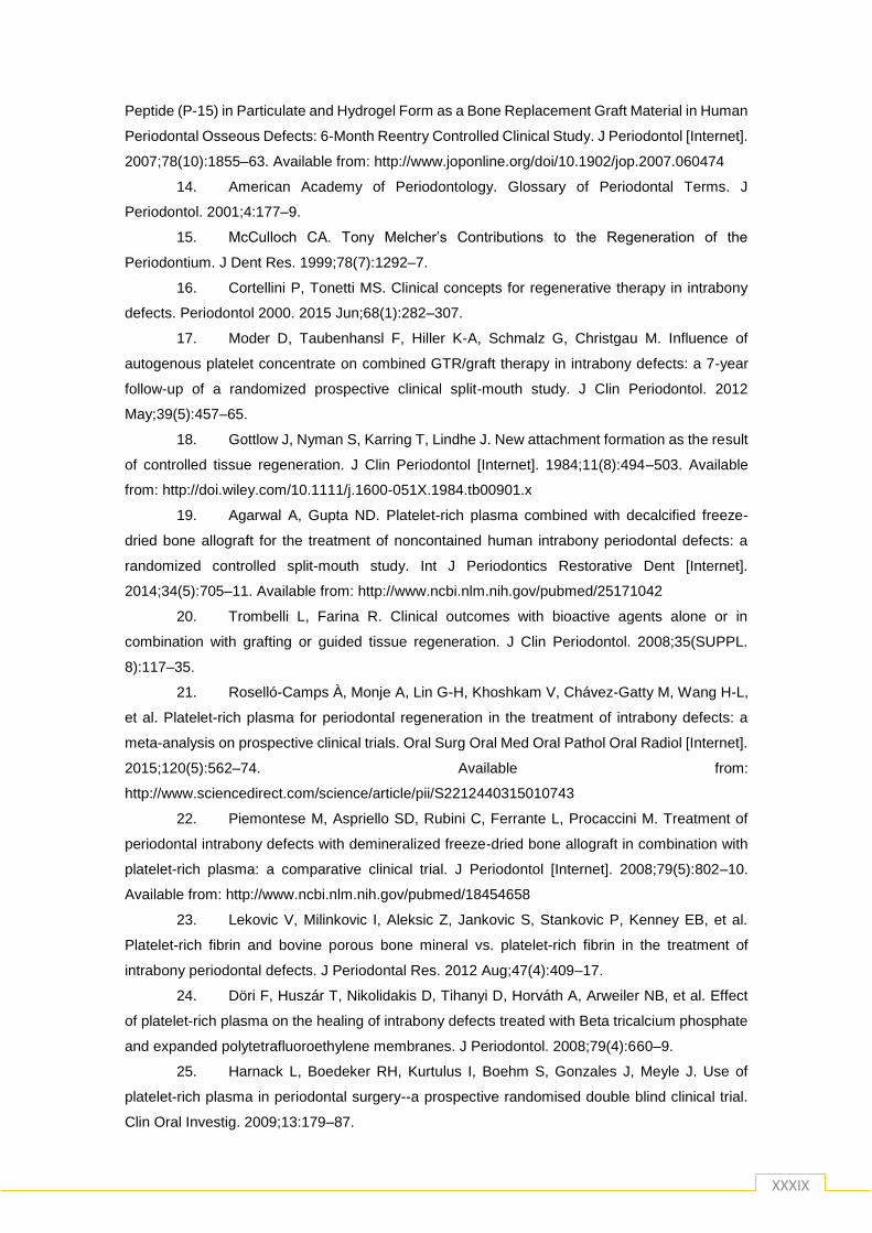
XXXIX
Peptide (P-15) in Particulate and Hydrogel Form as a Bone Replacement Graft Material in Human
Periodontal Osseous Defects: 6-Month Reentry Controlled Clinical Study. J Periodontol [Internet].
2007;78(10):1855–63. Available from: http://www.joponline.org/doi/10.1902/jop.2007.060474
14. American Academy of Periodontology. Glossary of Periodontal Terms. J
Periodontol. 2001;4:177–9.
15. McCulloch CA. Tony Melcher’s Contributions to the Regeneration of the
Periodontium. J Dent Res. 1999;78(7):1292–7.
16. Cortellini P, Tonetti MS. Clinical concepts for regenerative therapy in intrabony
defects. Periodontol 2000. 2015 Jun;68(1):282–307.
17. Moder D, Taubenhansl F, Hiller K-A, Schmalz G, Christgau M. Influence of
autogenous platelet concentrate on combined GTR/graft therapy in intrabony defects: a 7-year
follow-up of a randomized prospective clinical split-mouth study. J Clin Periodontol. 2012
May;39(5):457–65.
18. Gottlow J, Nyman S, Karring T, Lindhe J. New attachment formation as the result
of controlled tissue regeneration. J Clin Periodontol [Internet]. 1984;11(8):494–503. Available
from: http://doi.wiley.com/10.1111/j.1600-051X.1984.tb00901.x
19. Agarwal A, Gupta ND. Platelet-rich plasma combined with decalcified freeze-
dried bone allograft for the treatment of noncontained human intrabony periodontal defects: a
randomized controlled split-mouth study. Int J Periodontics Restorative Dent [Internet].
2014;34(5):705–11. Available from: http://www.ncbi.nlm.nih.gov/pubmed/25171042
20. Trombelli L, Farina R. Clinical outcomes with bioactive agents alone or in
combination with grafting or guided tissue regeneration. J Clin Periodontol. 2008;35(SUPPL.
8):117–35.
21. Roselló-Camps À, Monje A, Lin G-H, Khoshkam V, Chávez-Gatty M, Wang H-L,
et al. Platelet-rich plasma for periodontal regeneration in the treatment of intrabony defects: a
meta-analysis on prospective clinical trials. Oral Surg Oral Med Oral Pathol Oral Radiol [Internet].
2015;120(5):562–74. Available from:
http://www.sciencedirect.com/science/article/pii/S2212440315010743
22. Piemontese M, Aspriello SD, Rubini C, Ferrante L, Procaccini M. Treatment of
periodontal intrabony defects with demineralized freeze-dried bone allograft in combination with
platelet-rich plasma: a comparative clinical trial. J Periodontol [Internet]. 2008;79(5):802–10.
Available from: http://www.ncbi.nlm.nih.gov/pubmed/18454658
23. Lekovic V, Milinkovic I, Aleksic Z, Jankovic S, Stankovic P, Kenney EB, et al.
Platelet-rich fibrin and bovine porous bone mineral vs. platelet-rich fibrin in the treatment of
intrabony periodontal defects. J Periodontal Res. 2012 Aug;47(4):409–17.
24. Döri F, Huszár T, Nikolidakis D, Tihanyi D, Horváth A, Arweiler NB, et al. Effect
of platelet-rich plasma on the healing of intrabony defects treated with Beta tricalcium phosphate
and expanded polytetrafluoroethylene membranes. J Periodontol. 2008;79(4):660–9.
25. Harnack L, Boedeker RH, Kurtulus I, Boehm S, Gonzales J, Meyle J. Use of
platelet-rich plasma in periodontal surgery--a prospective randomised double blind clinical trial.
Clin Oral Investig. 2009;13:179–87.

XL
26. Yassibag-Berkman Z, Tuncer O, Subasioglu T, Kantarci A. Combined use of
platelet-rich plasma and bone grafting with or without guided tissue regeneration in the treatment
of anterior interproximal defects. J Periodontol [Internet]. 2007;78(5):801–9. Available from:
http://www.ncbi.nlm.nih.gov/pubmed/17470012
27. Nevins M, Kao RT, McGuire MK, McClain PK, Hinrichs JE, McAllister BS, et al.
PDGF Promotes Periodontal Regeneration in Localized Osseous Defects: 36 Month Extension
Results from a Randomized, Controlled, Double-masked Clinical Trial. J Periodontol.
2012;84(4):1–10.
28. Hammarstrom L. Enamel matrix, cementum development and regeneration. J
Clin Periodontol [Internet]. 1997;24(9):658–68. Available from:
http://doi.wiley.com/10.1111/j.1600-051X.1997.tb00247.x
29. Pradeep a R, Shetty SK, Garg G, Pai S. Clinical effectiveness of autologous
platelet-rich plasma and Peptide-enhanced bone graft in the treatment of intrabony defects. J
Periodontol. 2009;80(1):62–71.
30. Castro AB, Meschi N, Temmerman A, Pinto N, Lambrechts P, Teughels W, et al.
Regenerative potential of leucocyte- and platelet-rich fibrin. Part A: intra-bony defects, furcation
defects and periodontal plastic surgery. A systematic review and meta-analysis. J Clin Periodontol
[Internet]. 2016; Available from: http://doi.wiley.com/10.1111/jcpe.12643
31. Döri F, Huszár T, Nikolidakis D, Arweiler NB, Gera I, Sculean A. Effect of platelet-
rich plasma on the healing of intra-bony defects treated with a natural bone mineral and a collagen
membrane. J Clin Periodontol. 2007;34(3):254–61.
32. Plachokova AS, Nikolidakis D, Mulder J, Jansen JA, Creugers NHJ. Effect of
platelet-rich plasma on bone regeneration in dentistry: a systematic review. Clin Oral Implants
Res. 2008 Jun;19(6):539–45.
33. Kaushick BT, Jayakumar ND, Padmalatha O, Varghese S. Treatment of human
periodontal infrabony defects with hydroxyapatite + beta tricalcium phosphate bone graft alone
and in combination with platelet rich plasma: a randomized clinical trial. Indian J Dent Res.
2011;22(4):505–10.
34. Choukroun J, Adda F, Schoeffler C, Vervelle A. Une opportunité en paro-
implantologie: le PRF. Implantodontie. 2001;42(55):e62.
35. Sharma A, Pradeep a. R. Treatment of 3-Wall Intrabony Defects in Patients With
Chronic Periodontitis With Autologous Platelet-Rich Fibrin: A Randomized Controlled Clinical
Trial. J Periodontol. 2011;82(12):1705–12.
36. Gamal AY, Abdel Ghaffar KA, Alghezwy OA. Crevicular Fluid Growth Factors
Release Profile Following the Use of Platelets Rich Fibrin (PRF) and Plasma Rich Growth Factors
(PRGF) in Treating Periodontal Intrabony Defects (Randomized Clinical Trial). J Periodontol
[Internet]. 2016;1–13. Available from: http://www.ncbi.nlm.nih.gov/pubmed/26876351
37. Masuki H, Okudera T, Watanebe T, Suzuki M, Nishiyama K, Okudera H, et al.
Growth factor and pro-inflammatory cytokine contents in platelet-rich plasma (PRP), plasma rich
in growth factors (PRGF), advanced platelet-rich fibrin (A-PRF), and concentrated growth factors
(CGF). Int J Implant Dent [Internet]. 2016;2(1):19. Available from:

XLI
http://journalimplantdent.springeropen.com/articles/10.1186/s40729-016-0052-4
38. Kobayashi E, Fl??ckiger L, Fujioka-Kobayashi M, Sawada K, Sculean A, Schaller
B, et al. Comparative release of growth factors from PRP, PRF, and advanced-PRF. Clin Oral
Investig. 2016;20(9):2353–60.
39. Castro AB, Cortellini S, Quirynen M. Consensus Guidelines on the Use of L-PRF
from the 1st European Meeting on Enhanced Natural Healing in Dentistry . Consensus Guidelines
on the Use of L-PRF. 2016;(October).
40. Ghanaati S, Booms P, Orlowska A, Kubesch A, Lorenz J, Rutkowski J, et al.
Advanced Platelet-Rich Fibrin: A New Concept for Cell-Based Tissue Engineering by Means of
Inflammatory Cells. J Oral Implantol. 2014;40(6):679–89.
41. Miron RJ, Fujioka-Kobayashi M, Hernandez M, Kandalam U, Zhang Y, Ghanaati
S, et al. Injectable platelet rich fibrin (i-PRF): opportunities in regenerative dentistry? Clin Oral
Investig [Internet]. 2017; Available from:
http://www.ncbi.nlm.nih.gov/pubmed/28154995%0Ahttp://link.springer.com/10.1007/s00784-
017-2063-9
42. Zhang J, Qi X, Luo X, Li D, Wang H, Li T. Clinical and immunohistochemical
performance of lyophilized platelet-rich fibrin (Ly-PRF) on tissue regeneration. Clin Implant Dent
Relat Res [Internet]. 2017;(October 2016):1–12. Available from:
http://www.ncbi.nlm.nih.gov/pubmed/28192870%0Ahttp://doi.wiley.com/10.1111/cid.12473
43. Moher D, Liberati A, Tetzlaff J, Altman DG, Group TP. Preferred Reporting Items
for Systematic Reviews and Meta-Analyses : The PRISMA Statement. 2009;6(7).
44. Dohan DM, Choukroun J, Diss A, Dohan SL, Dohan AJJ, Mouhyi J, et al. Platelet-
rich fibrin ( PRF ): A second-generation platelet concentrate . Part I : Technological concepts and
evolution. 2006;
45. Khorshidi H, Raoofi S, Bagheri R, Banihashemi H. Comparison of the Mechanical
Properties of Early Leukocyte- and Platelet-Rich Fibrin versus PRGF / Endoret Membranes.
2016;2016.
46. Dohan DM, Choukroun J, Diss A, Dohan SL, Dohan AJJ, Mouhyi J, et al. Platelet-
rich fibrin ( PRF ): A second-generation platelet concentrate . Part II : Platelet-related biologic
features. 2006;
47. Burnouf T, Alphonse H, Chen T, Ou K, El-ekiaby M, Radosevic M. Blood Reviews
Blood-derived biomaterials and platelet growth factors in regenerative medicine. YBLRE
[Internet]. 2013;27(2):77–89. Available from: http://dx.doi.org/10.1016/j.blre.2013.02.001
48. Rowe SL, Lee S, Stegemann JP. Influence of thrombin concentration on the
mechanical and morphological properties of cell-seeded fibrin hydrogels. 2007;3:59–67.

XLII
Appendix
Table IV. Publications excluded
Author, year Type of study
Reason of exclusion
Aspriello et al., 2010 RCT Unrelated to PICOT question
Gamal et al., 2016 RCT Unrelated to PICOT question
Moder et al., 2012 RCT Undefined APC
Nevins et al., 2013 RCT Unrelated to PICOT question
Pradeep et al., 2009 RCT PRP used in all experimental groups
Rodrigues et al., 2011 RCT PRP used in all experimental groups
Trombelli et al., 2008 Review Not systematic review

XLIII
Acknowledgements
Ao meu orientador, prof. Dr. Sérgio Matos, que prontamente aceitou o desafio de tornar
este projeto possível. Quando for grande, irei conseguir saber tanto como o senhor professor.
Ao meu co-orientador, Dr. Tony Rolo, que com a sua paciência inesgotável respondeu a
todas as minhas perguntas (e se elas foram muitas!). Obrigada por ter estado sempre disponível,
tanto neste projeto como na clínica. Tornou-se um exemplo não só como profissional, mas
também como pessoa.
Ao meu pai, que esteve irredutivelmente sempre comigo em todas as lutas dos últimos
cinco anos. Espero que te orgulhes da Médica Dentista que irei ser.
À minha mãe, que foi essencial para terminar esta etapa.
Aos meus tios, cuja generosidade e amor não têm descrição.
Aos meus amigos, que nunca deixaram de me apoiar… mesmo aqueles que foram
trocados por épocas de exames!
A todos os professores, que me ensinaram tudo o que sei de Medicina Dentária.
A todos os funcionários desta casa, que me receberam de braços abertos.
À FMUC.
A Coimbra… Até sempre!

XLIV
Index of figures and tables
Figure 1. Classification of periodontal bone defects 9
Figure 2. Periodontal treatment of infrabony defects with deep pockets 10
Figure 3. PubMed search 17
Figure 4. Cochrane Library search 17
Figure 6 v Flowchart for search strategy 18
Table I. Systematic Reviews and Meta-analyses: general information
and principal outcomes
19
Table II. Case series methodology 24
Table III. Pre-operative data 25
Table IV. Publications excluded of this systematic review XLII

XLV
Index
Summary IV
Resumo V
Abstract VI
List of abbreviations and acronyms VII
1. Introduction 8
1.1. Chronic periodontitis 8
1.2. Therapeutic approaches 9
1.2.1. Regenerative therapy 11
1.2.1.1. Autologous platelet concentrates (APC) 13
2. Systematic Review 16
2.1. Materials and Methods 16
2.2. Results 18
3. Case series 22
3.1. Materials and Method 22
3.2. Results 26
3.2.1. Clinical case 1 26
3.2.1. Clinical case 2 28
3.2.2. Clinical case 3 30
4. Discussion 33
5. Conclusions 37
Bibliography XXXVIII
Appendices XLII
Acknowledgements XLIII
Index of figures and tables XLIV
Index XLV

XLVI

![The effect of autologous leukocyte platelet rich fibrin on ... · these two platelet concentrates. [15,16] Although they are both clinically effective in accelerating the healing](https://img.pdfslide.us/doc/110x75/5f74cde1d357e407be22081f/the-effect-of-autologous-leukocyte-platelet-rich-fibrin-on-these-two-platelet.jpg)