Embed Size (px)
Citation preview

Stem Cell Therapies in Pulmonary
hypertension
Olga Tura Ceide
BRN Seminars
27/09/2018

Pulmonary Hypertension
Serious progressive disorder
Characterised by increased pulmonary arterial pressure
and vascular remodeling
Right heart failure
death

Pulmonary Hypertension
Endothelial dysfunction
Dysregulation of smooth muscle cell and endothelial cell funtion
Increased proliferation, perivascular inflammation and decreased
vessel luminal diameter
Endothelial dysfunction
Dysregulation of smooth muscle cell and endothelial cell funtion
Increased proliferation, perivascular inflammation and decreased
vessel luminal diameter

Despite advances in the treatment:
1) Prostacyclin and analogs
2) Endothelin receptor antagonists
3) Phosphodiesterase type 5 inhibitors
Modest improvements measured by:
1) Exercise capacity
2) Quality of life measurements
Pulmonary Hypertension

Despite advances in the treatment:
1) Prostacyclin and analogs
2) Endothelin receptor antagonists
3) Phosphodiesterase type 5 inhibitors
Modest improvements measured by:
1) Exercise capacity
2) Quality of life measurements
➢ PH remains a fatal disease
➢ Patients refractory to pharmacologic therapy-- lung trasplantation
➢ Limited availability of organs, postoperative infection, graft rejection--limited patient
survival.
Pulmonary Hypertension

Facing obstacles
➢ Stem cell therapy have been reported to have a beneficial effects in
experimental models of PH.
➢ Encouraging results (feasibility, safety and efficency of cell therapy).

However:
➢ Different types of stem cells
➢ Different types of PH
➢ Different administration routes
Facing obstacles
Stem cell therapy have been reported to have a beneficial effects in experimental models of
PH.
Encouraging results (feasibility, safety and efficency of cell therapy).
➢ Difficult to compare results as the exact nature of delivered cells remains undetermined.
➢ Urgent need to move to a standardized stem cell identification before use.
➢ Few studies in vitro designed to elucidate potential their potential mechanisms of action.

Different types of stem cells
➢Mononuclear stem cells (MNCs)
➢Mesenchymal stromal cells (MSCs)
➢Endothelial progenitor cells (EPCs)
➢Adipose-derived stem cells (ADSc)
➢iPS derived cells (iPS)
➢Cardiac progenitor cells (CPCs)

Different types of stem cells
➢Mononuclear stem cells (MNCs)
➢ Umbilical cord blood PBMNCs
Advantatges
➢ Pro-angiogenic
➢ Readely available
➢ Easy to obtain
Disadvantatges
➢ comprises different cell types
➢ Allogenic source (most of the times)
Different types of stem cells
Using a pulmonary artery banding athymic murine model (PAB), UCB-MNCs
injections led to improvement in:
➢ RV function, structure
➢Reduction in RV fibrosis
➢Increase in angiogenic biomarkers.
Oommen et al. Stem Cell Research and Therapy (2015)

Mononuclear stem cells_Cord blood
UCB-MNCs transplantation is safe (0.4-0.8 million cells/heart) without altering QT or ST-
segments or electrocardiograph waves morphology
Oommen et al. Stem Cell Research and Therapy (2015)
Intramyocardial injections to the right ventricle (5 injections 2,5 ul each)

Mononuclear stem cells_Cord blood
Myocardial delivery of UCB-MNCs improves RV
Oommen et al. Stem Cell Research and Therapy (2015)
For weeks after CB-MNCs injection, MRI revealed a recovery of right ventricular chamber size and volume

Mononuclear stem cells_Cord blood
Oommen et al. Stem Cell Research and Therapy (2015)
CB-MNCs transplantation augments myocardial cell proliferation and attenuates
myocardial fibrosis.

Different types of stem cells
➢Mononuclear stem cells (MNCs)
➢Mesenchymal stromal cells (MSCs)
➢Endothelial progenitor cells (EPCs)
➢Adipose-derived stem cells (ADSc)
➢iPS derived cells (iPS)
➢Cardiac progenitor cells (CPCs)

Mesenchymal stromal cells (MSCs)
➢ Isolated from: Bone marrow, wharton´s jelly, placenta, adipose tissue and muscle
➢ Multipotent stem cells
➢ Proangiogenic
➢ Cytoprotective
➢ Immune tolerance
➢ Easy to isolate
➢ Rapidly amplified

Mesenchymal stromal cells (MSCs)
➢ They concluded that MSCs reduced RV pressure overload, RV dysfunction and
lung pathology.
➢ Autologous MSCs may alleviate cardiac and pulmonary symptoms in PH
patients.
Umar et al. Am.J. Physiol. Heart Circ. Physiol 297 (2009)

Mesenchymal stromal cells (MSCs)
Intravenous injection of BM and UCB-derived MSCs improved RV hypertrophy
and RV ejection fraction of MCT-induced PAH rats.
Umar et al. Am.J. Physiol. Heart Circ. Physiol 297 (2009)

Mesenchymal stromal cells (MSCs)
Umar et al. Am.J. Physiol. Heart Circ. Physiol 297 (2009)
MSC therapy drecreased the arterioral thickness and medial hypertrophy

Mesenchymal stromal cells (MSCs)
Intravenously administered gene-transduced MSC overexpressing
eNOS in MCT-induced PAH rats.
Increase endothelial cells
Secretion of NO
Constriction of pulmonar arteries
Resistance
Pulmonary hypertension
Kanki-Horimoto et al. Circulation. 2006;114

Mesenchymal stromal cells (MSCs)
➢Both MSC and MSC/eNOS groups significantly inhibited RVSP progression and RV/body
weight ratio compared to nontreated MCT-PH rats.
➢MSC/eNOS group significantly prolonged survival time compared to non-treated PH group
➢MSC overexpresing eNOS offers therapeutic effects for PH-induced RV impairment.
Kanki-Horimoto et al. Circulation. 2006;114

Mesenchymal stromal cells (MSCs)
➢ Heme oxygenase-1 isoform (HO-1) activity restores homostasis by exerting
antiinflammatory, antiapoptotic and antiproliferative effects on diverse cell
types.
➢ It has been shown that upregulation of endogenous HO-1 can prevent
hypoxia-induced PAH in rat.
Liang et al. STEM CELLS 2011;29:99–107
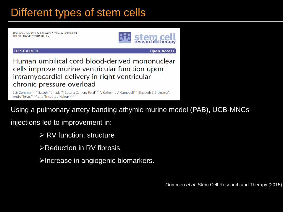
MSC transplantation reverses established PAH
Mesenchymal stromal cells (MSCs)
Intravenous injection of MSCs expressing HO-1 in a chronic hypoxia induced PAH model
resulted in nomal RVSP and a significant reduction of RVH.
MSC transplantation reverses lung vascular remodelling

Mesenchymal stromal cells (MSCs)
MSC-CM inhibits pulmonary artery smooth
muscle cell proliferation
MSC transplantation modulates the hypoxia-
induced lung inflammation

Different types of stem cells
➢Mononuclear stem cells (MNCs)
➢Mesenchymal stromal cells (MSCs)
➢Endothelial progenitor cells (EPCs)
➢Adipose-derived stem cells (ADSc)
➢iPS derived cells (iPS)
➢Cardiac progenitor cells (CPCs)
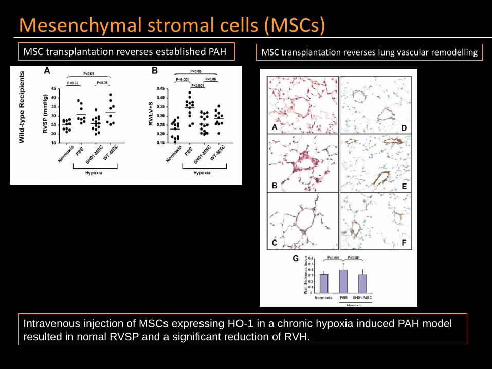
ANGIOGENESIS; PARADIGM
ENDOTHELIUM
ENDOTHELIAL INJURY
•Migration and proliferation of mature endothelial cells
It was believed that the vasculature was only repaired locally by
outgrowth from existing vessels (angiogenesis).

TAKAYUKI ASAHARA- DISCOVERY OF ENDOTHELIAL PROGENITOR CELLS IN THE ADULT.
MONONUCLEAR CELLSMAGNETIC SEPARATION
CD34+ & KDR+FIBRONECTIN PLATES
Asahara et al, Science; 275:964-7
Endothelial progenitor cells isolated from the bone marrow could differentiate
into mature endothelial cells and form new blood vessels in vitro.
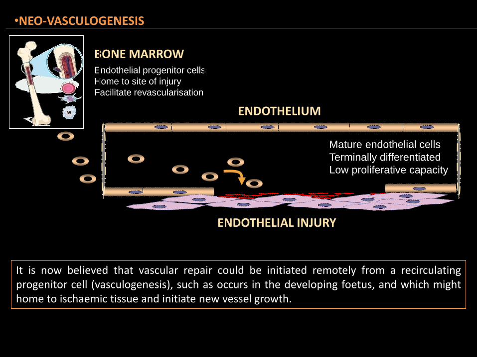
•NEO-VASCULOGENESIS
ENDOTHELIUM
ENDOTHELIAL INJURY
BONE MARROW
Mature endothelial cells
Terminally differentiated
Low proliferative capacity
Endothelial progenitor cells
Home to site of injury
Facilitate revascularisation
It is now believed that vascular repair could be initiated remotely from a recirculatingprogenitor cell (vasculogenesis), such as occurs in the developing foetus, and which mighthome to ischaemic tissue and initiate new vessel growth.

The discovery of circulating endothelial progenitor cells has stimulatedclinical interest in the potential of these cells to promote vascularregeneration in patients with ischaemic heart disease or peripheral vasculardisease.
CLINICAL INTEREST

Patients with bilateral leg ischaemia wererandomly injected with bone marrowmononuclear cells in one leg, and saline in theother as a control.
Laser Doppler blood perfusion. Right leg was implanted with bone marrow-mononuclear cells (red to white colour shows enriched perfusion). Left leg injected with saline. Right digit IV was amputated.
CLINICAL INTEREST Eriko Tateishi-Yuyama, et al THE LANCET • Vol 360 • August 10, 2002

Over the years, there have been many studies involving administration ofautologous EPC as a treatment for PH
CLINICAL INTEREST-PH

Endothelial progenitor cells
-Cyclooxygenase isoform 1-prostacyclin synthase–expressing ELPCs reversed MCT-induced
PAH.
-A single jugular vein injection offered survival benefits for at least 4 weeks and may provide a
promising option for PAH patients.
Zhou, et al. Circulation. 2013;128:982-994.

Engineered ELPCs attenuated monocrotaline
(MCT)-induced pulmonary vessel wall thickening
Zhou, et al. Circulation. 2013;128:982-994.
Endothelial progenitor cells
Both engineered ELPCs and control ELPCs provided
a survival benefit that was accompanied by
a decrease in right ventricular hypertrophy.

WHAT IS AN EPC?

What is an EPC?
There is as yet no DEFINITIVE description of what is an EPC.
Different putative EPC assays reflect different cell populations.
EPCs cannot be reliably quantified or enriched by any agreed standard
This controversy has HINDERED the development of effective cellular
therapies for vascular regeneration

EPCs CAN BE defined by 3 methods:
Phenotypic analysis by
Flow Cytometry
(Original description)
Functional analysis by colony forming assay
(CFU-EPC)
1
1997
Asahara et al, Science
2
2003
Hill et al, N Eng J Med2007
Yoder et al, Blood
3
Functional analysis by colony forming assay
(Late outgrowth endothelial cells, EOC)
Tura et al 2007 Tura et al in preparation
CD 34
CD 133
KDR

•Early studies characterised EPCs based on the
expression of:
• CD34 and/or CD133 (haematopoietic stem cell
markers)
•VEGFR2 (Vascular endothelium growth factor
receptor 2)
VEGFR-2
EPC defined by phenotypic analysis (original description, Asahara et al,.)1
Endothelial Progenitor cell
Mature endothelial cells
CD31 VE-cadherinTie-2
vWf
•Differentiation
CD34
CD133
VEGFR2
•In the original description authors described a
population of adult human circulating
CD34+VEGFR2+ cells that could differentiate into
cells with endothelial-like characteristics in vitro
(Asahara et al., 1997 Science; 275:964-7).

EPC defined by phenotypic analysis (original description, Asahara et al,.)1
•20%• CD34VEGFR2+
SS
C-H
FCS-H
SS
C-H
CD34-APC
VE
GF
R2
-PE
CD34-APC
•0.34%CD34+
•19.58% •CD34+VEGFR2+
•VIABLE CELLS
Tura O, et al, Journal of Translational Medicine 2007.
**
Cord blood G-CSFMobilised blood
Bone marrow • normal peripheral blood
0.0
2.5
5.0
7.5
10.0
Num
ber
of
CD
34+
VEG
FR2+
ce
lls/m
l (1
03)
Sangre periférica movilizada es la fuente más rica de CPE tal como se define por las células que expresan número de células CD34 + en combinación con VEGFR2.

• Phenotypic analysis by
• Flow Cytometry
• (Original description)
Colony assay (CFU-EPC) Late outgrowth endothelial cells (EOC)
EPC CAN BE DEFINED BY 3 METHODS:
•CD 34
•CD 133
•VEGFR2
1997Asahara et al.,
2003Hill et al.,
Hill et al.,2003 N Engl J Med;348:593-600
2007Yoder et al.,
Asahara et al, Science; 275:964-7 Yoder et al.,2007 Blood;109:1801-9
2 31

ENDOTHELIAL COLONY ASSAY (CFU-EPC) (early EPC) Hill et al2

CBm
PBBM
nPB
0
10
20
30
40
50
EP
C c
olo
nie
s p
er
10
6 n
on
-ad
he
ren
t ce
lls p
late
d
•***
Normal peripheral blood (nPB) had the greatest number of CFU-EPC (CFU-Hill),while mobilised PB (mPB) which contained the most CD34+VEGFR2+ arevirtually devoid of CFU-EPC.
Tura O, et al, Journal of Translational Medicine 2007.
ENDOTHELIAL COLONY ASSAY (CFU-EPC) (early EPC) Hill et al2

Tura O, et al, Journal of Translational Medicine 2007.Mills and Tura et al , Heart. 2009;95(24):2003-8.
0.0 0.1 0.2 0.3 0.4 0.5 0.6 0.7 0.8
0.00
0.02
0.04
0.06
0.08
0.10
Number of CFU (x104)
CD
34+
VE
GF
R2+
cell
s (
% o
f
MN
Cs)
There is no correlation between the CFU-EPC ‘functional’ definition and the phenotypic
definition of EPCs.
Cells identified using current phenotype EPC definitions (CD34+ and/or CD133+ jointly
expressing VEGFR2) do not appear to be the population of cells defined in the CFU-EPC
assay.
ENDOTHELIAL COLONY ASSAY (CFU-EPC) (early EPC) Hill et al2
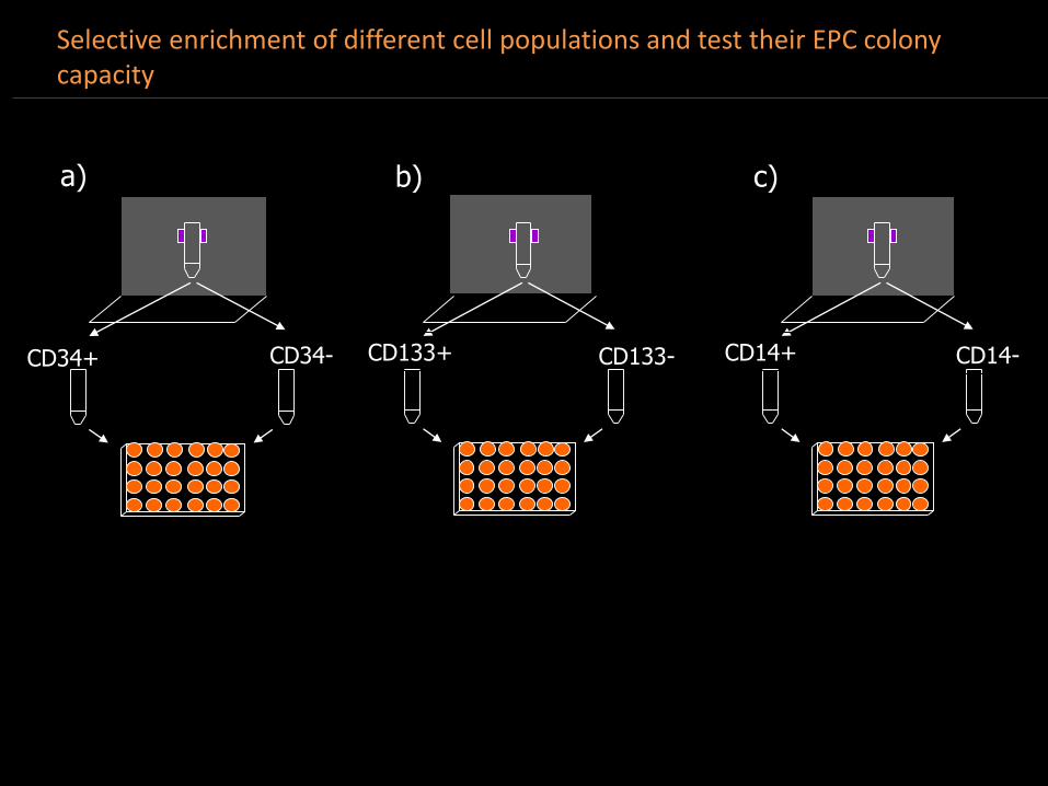
Selective enrichment of different cell populations and test their EPC colony capacity
CD34+ CD34-
a)
CD133-
b)
CD133+
c)
CD14+ CD14-

CD34
-enric
hed(>
90%
puri
ty)
CD34
-dep
lete
d
CD13
3-en
rich
ed (>
90%
puri
ty)
CD13
3-dep
lete
d
2h a
dhrence
cel
ls (8
0% C
D14
+)
2h n
on-adher
ent c
ells
CD14
-enri
ched
(98%
CD14
+)
CD14
-dep
lete
d
Unse
parat
ed M
NCs
0
10
20
30
40
50
Nu
mb
er
of
CF
U-E
PC
pe
r 1
06 c
ell
s p
late
dCD34-enriched
cells
CD133-enriched
cells
CD14-enriched
cells
Selective enrichment of different cell populations and test their EPC colony capacity

CD34
-enric
hed(>
90%
puri
ty)
CD34
-dep
lete
d
CD13
3-en
rich
ed (>
90%
puri
ty)
CD13
3-dep
lete
d
2h a
dhrence
cel
ls (8
0% C
D14
+)
2h n
on-adher
ent c
ells
CD14
-enri
ched
(98%
CD14
+)
CD14
-dep
lete
d
Unse
parat
ed M
NCs
0
10
20
30
40
50
Nu
mb
er
of
CF
U-E
PC
pe
r 1
06 c
ell
s p
late
dCD34-enriched
cells
CD133-enriched
cells
CD14-enriched
cells
Selective enrichment of different cell populations and test their EPC colony capacity

EPCs CAN BE defined by 3 methods:
Phenotypic analysis by
Flow Cytometry
(Original description)
Functional analysis by colony forming assay
(CFU-EPC)
1
1997
Asahara et al, Science
2
2003
Hill et al, N Eng J Med2007
Yoder et al, Blood
3
Functional analysis by colony forming assay
(Late outgrowth endothelial cells, EOC)
Tura et al 2007 Tura et al in preparation
CD 34
CD 133
KDR

They form tube-like structures in MatrigelThese cells resemble mature endothelial cells, and express the same surface marker/receptor profiles by immunophenotyping (CD31 (red), CD146 (green)).
EPC defined by late outgrowth Endothelial cells (EOC) or (ECFU)

Human endothelial outgrowth cells form blood vessels
•3EPC defined by late outgrowth Endothelial cells (EOC) or (ECFU)

EPCs CAN BE defined by 3 methods:
Phenotypic analysis by
Flow Cytometry
(Original description)
Functional analysis by colony forming assay
(CFU-EPC)
1
1997
Asahara et al, Science
2
2003
Hill et al, N Eng J Med2007
Yoder et al, Blood
3
Functional analysis by colony forming assay
(Late outgrowth endothelial cells, EOC)

Results-Endothelial cell proliferative capacity
0 1 2 3 4 5 6 7 8 9 1 0 1 1 1 2 1 3 1 4 1 5
0 .0
0 .5
1 .0
1 .5
2 .0
2 .5P O P U L A T IO N D O U B L IN G T IM E S
P a s s a g e N u m b e r
Fo
ld e
xp
an
sio
n/d
ay
H P A E
T E P -6
T E P -2
T E P -1 3
T E P -1 4
T E P -1 5
They showed an hyperproliferative phenotype when compared with control
endothelial cell lines (HPAE), Fold expansion/days of culture (p<0.002 at P4)
Fold expansion/day= (No of final cells/No of seeded cells)/days of culture.

WHY?

WHY?
Endothelial dysfunctionality
Endothelial metabolism?

Endothelial metabolism
100000X100000X
HPAE CPTEH-EC
TEM pictures show that in CTEPH mitochondrial inner membranes and crestae
are strange. CTEPH-EC cells have more multivesicular bodies than HPAE and an
inflamed and disorganised ER.

-MSCs have beneficial effects on endothelial progenitor cells their co-injection could
allow the formation of a more developed vascular network than with either MSC or EPC
alone.
-As a result it might be intersting to administrate MSC in combination with EPC in a PAH
to achive greater benefits.
MSCs have beneficial effects on EPC

Different types of stem cells
➢Mononuclear stem cells (MNCs)
➢Mesenchymal stromal cells (MSCs)
➢Endothelial progenitor cells (EPCs)
➢Adipose-derived stem cells (ADSc)
➢iPS derived cells (iPS)
➢Cardiac progenitor cells (CPCs)

Different types of stem cells
➢Mononuclear stem cells (MNCs)
➢Mesenchymal stromal cells (MSCs)
➢Endothelial progenitor cells (EPCs)
➢Adipose-derived stem cells (ADSc)
➢iPS derived cells (iPS)
➢Cardiac progenitor cells (CPCs)

Adipose-derived stem cells (ADSc)
➢ Can be readely isolated from white adipose tissue by liposuction
➢ They are suitable for autotransplantation
➢ Pro-angiogenic and anti-apoptotic
➢ Multipotential
➢ plentiful
➢ Proven to be safe and feasible (phase I trial in ischemic cardiomyopathy (Precise trial)
➢ Appear to be a good choice of stem cells to treat PAH

Adipose-derived stem cells (ADSc)
Luo, et al. Clin Exp Hyperten. 2015;37 (3):241-248.
ADSC colonize the pulmonary arteries,
attenuate pulmonary arterial hypertension
and ameliorate pulmonary arterial
remodeling.

➢iPS derived cells (iPS)
➢ iPS might provide a useful source of patient specific cells,as they can be isolated
from their own dermal fibroblasts.
➢ Big numbers
➢ Tumorigenic

➢iPS derived cells (iPS)
iPSC-based therapy could resotre the hemodynamic function of the RV with benefits
for preveniting the ongoing inflammation in the lungs of MCT-induced PAH rats.

➢ Right ventricular function is the main prognostic risk factor in PH.
➢ Stem cell therapies targeting the left ventricle (LV) are proving to be promising
therapies in animal models.
➢ In contrast to left heart disease, stem cell therapy applied to the RV has not
been studied much, despite indications that it may be a viable therapeutic option.
➢ Future new therapeutics in the next decade that specifically target the RV are
required.
-Improvements in the RV are they due to direct effect of stem cell
therapy or due to indirect reduction of pulmonary resistance?

Different types of stem cells
➢Mononuclear stem cells (MNCs)
➢Mesenchymal stromal cells (MSCs)
➢Endothelial progenitor cells (EPCs)
➢Adipose-derived stem cells (ADSc)
➢iPS derived cells (iPS)
➢Cardiac progenitor cells (CPCs)

➢Cardiac progenitor cells (CPCs)
➢ Heterogenous group of cells in the adult heart (c-kit+, Sca-1+..) similar problem
than EPC.
➢ Clear identification and characterization of the cells used are crucial to discuss
the results of studies.
➢ The therapeutic potential of CPCs has been documented for myocardial
infarction.
➢ Thought to stimulate cardiomyocytes and vascular cell trandifferentiation as
well secreting paracrine factors that promote neovasularizarion and activation
of endogenous CPCs.
➢ These properties could be beneficial for the RV in PAH by causing an increase
in capillary density and by activating the production of efficient new
cardiomyocytes.
➢ Is an invasive procedure isolated from the heart tissue

➢Cardiosphere derived cells (CDC)
➢ Can be obtain from human biopsies.
➢ CDC were tested clinically ina CADUCEUS trial (cardiosphere-derived autologous
stem cells to reverse ventricular dysfunction), which examined safety and efficacy
of intracoronary autologous administration of CDC in patients with LV dysfunction
after MI.

➢Conclusions
➢ Stem cell therapies in pulmonay hypertension seem very promising
➢ Clear identification and characterization of the cells used are crucial
➢ Ethical, embryo-derived cells (CPC), immunological issues
➢ Reprogramming may induce genetic changes –tumor formation
➢ For practical reasons, cells should be readely available at the right clinical
dose at the time of injection. Isolated cells previously criopreserved.
➢ Autologous--- PAH-EPC numbers are reduced compared to controls.
➢ Route of administration, scaffolds..
Choose the best cells and the best way to administer them

Thank you