Embed Size (px)
Citation preview

u n i ve r s i t y o f co pe n h ag e n
Københavns Universitet
Identification of Fungal Plasma Membrane H+-ATPase Inhibitors in Lecaniodiscuscupanioides by HPLC-HRMS-SPE-NMRStraadt, Ida Krestine; Kongstad, Kenneth Thermann; Stærk, Dan
Publication date:2015
Document VersionEarly version, also known as pre-print
Citation for published version (APA):Straadt, I. K., Kongstad, K. T., & Stærk, D. (2015). Identification of Fungal Plasma Membrane H+-ATPaseInhibitors in Lecaniodiscus cupanioides by HPLC-HRMS-SPE-NMR. Poster session presented at 36th DanishNMR Meeting, Lund, Sweden.
Download date: 07. aug.. 2019
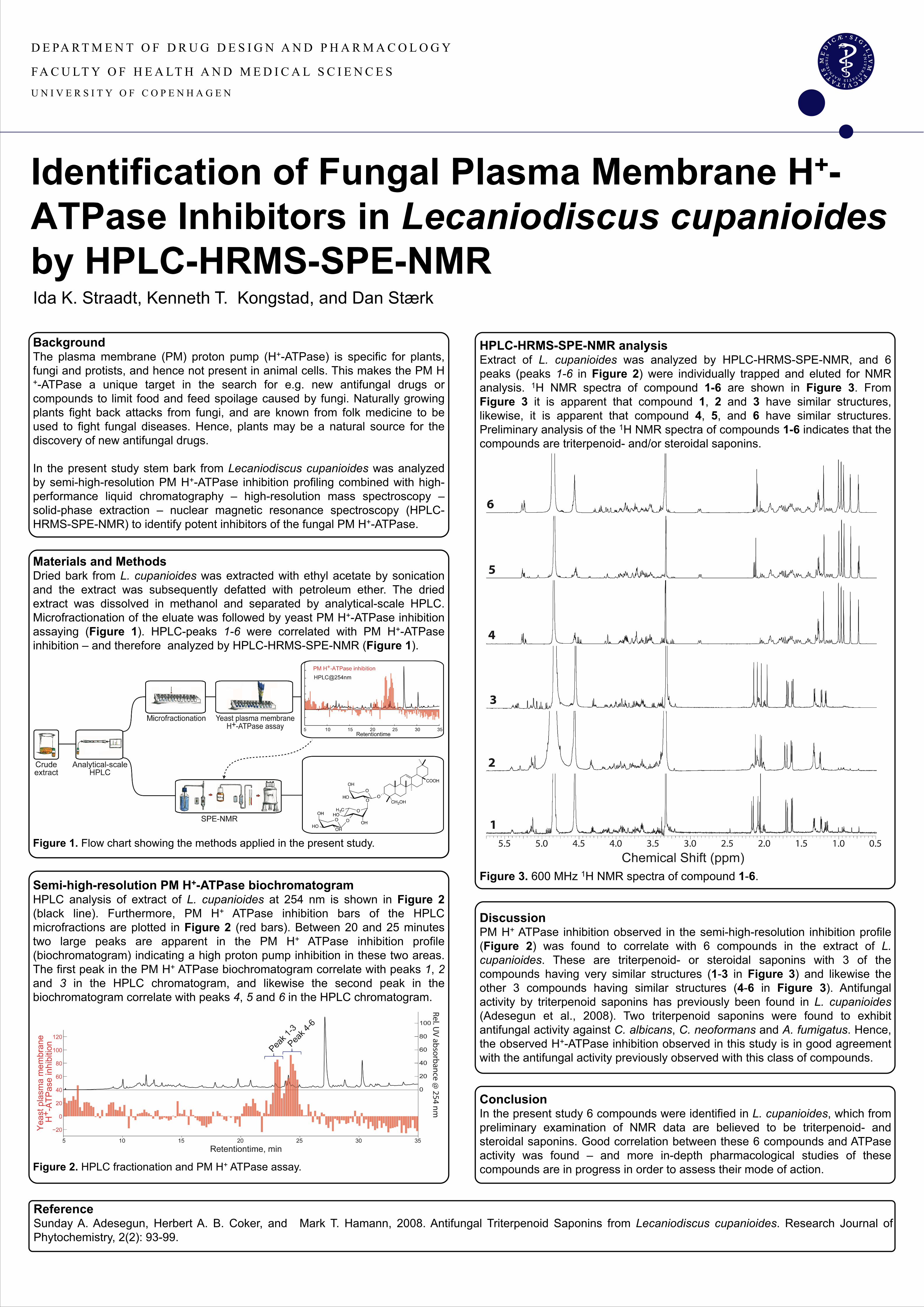
HPLC-HRMS-SPE-NMR analysis Extract of L. cupanioides was analyzed by HPLC-HRMS-SPE-NMR, and 6 peaks (peaks 1-6 in Figure 2) were individually trapped and eluted for NMR analysis. 1H NMR spectra of compound 1-6 are shown in Figure 3. From Figure 3 it is apparent that compound 1, 2 and 3 have similar structures, likewise, it is apparent that compound 4, 5, and 6 have similar structures. Preliminary analysis of the 1H NMR spectra of compounds 1-6 indicates that the compounds are triterpenoid- and/or steroidal saponins. Figure 3. 600 MHz 1H NMR spectra of compound 1-6. Discussion PM H+ ATPase inhibition observed in the semi-high-resolution inhibition profile (Figure 2) was found to correlate with 6 compounds in the extract of L. cupanioides. These are triterpenoid- or steroidal saponins with 3 of the compounds having very similar structures (1-3 in Figure 3) and likewise the other 3 compounds having similar structures (4-6 in Figure 3). Antifungal activity by triterpenoid saponins has previously been found in L. cupanioides (Adesegun et al., 2008). Two triterpenoid saponins were found to exhibit antifungal activity against C. albicans, C. neoformans and A. fumigatus. Hence, the observed H+-ATPase inhibition observed in this study is in good agreement with the antifungal activity previously observed with this class of compounds. Conclusion In the present study 6 compounds were identified in L. cupanioides, which from preliminary examination of NMR data are believed to be triterpenoid- and steroidal saponins. Good correlation between these 6 compounds and ATPase activity was found – and more in-depth pharmacological studies of these compounds are in progress in order to assess their mode of action.
Identification of Fungal Plasma Membrane H+-ATPase Inhibitors in Lecaniodiscus cupanioides by HPLC-HRMS-SPE-NMR
Background The plasma membrane (PM) proton pump (H+-ATPase) is specific for plants, fungi and protists, and hence not present in animal cells. This makes the PM H+-ATPase a unique target in the search for e.g. new antifungal drugs or compounds to limit food and feed spoilage caused by fungi. Naturally growing plants fight back attacks from fungi, and are known from folk medicine to be used to fight fungal diseases. Hence, plants may be a natural source for the discovery of new antifungal drugs. In the present study stem bark from Lecaniodiscus cupanioides was analyzed by semi-high-resolution PM H+-ATPase inhibition profiling combined with high-performance liquid chromatography – high-resolution mass spectroscopy – solid-phase extraction – nuclear magnetic resonance spectroscopy (HPLC-HRMS-SPE-NMR) to identify potent inhibitors of the fungal PM H+-ATPase.
Materials and Methods Dried bark from L. cupanioides was extracted with ethyl acetate by sonication and the extract was subsequently defatted with petroleum ether. The dried extract was dissolved in methanol and separated by analytical-scale HPLC. Microfractionation of the eluate was followed by yeast PM H+-ATPase inhibition assaying (Figure 1). HPLC-peaks 1-6 were correlated with PM H+-ATPase inhibition – and therefore analyzed by HPLC-HRMS-SPE-NMR (Figure 1). Figure 1. Flow chart showing the methods applied in the present study.
Semi-high-resolution PM H+-ATPase biochromatogram HPLC analysis of extract of L. cupanioides at 254 nm is shown in Figure 2 (black line). Furthermore, PM H+ ATPase inhibition bars of the HPLC microfractions are plotted in Figure 2 (red bars). Between 20 and 25 minutes two large peaks are apparent in the PM H+ ATPase inhibition profile (biochromatogram) indicating a high proton pump inhibition in these two areas. The first peak in the PM H+ ATPase biochromatogram correlate with peaks 1, 2 and 3 in the HPLC chromatogram, and likewise the second peak in the biochromatogram correlate with peaks 4, 5 and 6 in the HPLC chromatogram. Figure 2. HPLC fractionation and PM H+ ATPase assay.
Ida K. Straadt, Kenneth T. Kongstad, and Dan Stærk
D E PA R T M E N T O F D R U G D E S I G N A N D P H A R M A C O L O G Y
FA C U LT Y O F H E A LT H A N D M E D I C A L S C I E N C E S
U N I V E R S I T Y O F C O P E N H A G E N
Reference Sunday A. Adesegun, Herbert A. B. Coker, and Mark T. Hamann, 2008. Antifungal Triterpenoid Saponins from Lecaniodiscus cupanioides. Research Journal of Phytochemistry, 2(2): 93-99.