Embed Size (px)
Citation preview
1
Localized tufts of fibrils on Staphylococcus epidermidis NCTC 11047 are 1
comprised of the Accumulation Associated Protein (Aap) 2
Running Title: Fibrils of Staphylococcus epidermidis NCTC 11047 3
Pathogens and pathogenicity 4
Miriam A. Banner,1 John G. Cunniffe,
2 Robin L. Macintosh,
1 Timothy J. Foster,
3 5
Holger Rohde,4 Dietrich Mack,
5 Emmy Hoyes,
6 Jeremy Derrick,
7 Mathew Upton,
8 6
and Pauline S. Handley1*
7
1 Faculty of Life Sciences, 1.800 Stopford Building, The University of Manchester, 8
Oxford Road, M13 9PT, UK 9
2
Wirral Hospital NHS Trust, Arrowe Park Hospital, Arrowe Park Road, Upton, 10
Wirral, Merseyside, LH49 5PE, UK 11
3 Dept. of Microbiology, Moyne Institute of Preventative Medicine, Trinity College, 12
Dublin 2, Ireland. 13
4 Institut für Medizinische Mikrobiologie, Virologie und Hygiene, Zentrum für 14
Klinische Pathologie, Universitätsklinikum Hamburg-Eppendorf, Martinistrasse 52, 15
20246 Hamburg, Germany 16
5 Medical Microbiology and Infectious Diseases, The School of Medicine, University 17
of Wales Swansea, Singleton Park, Swansea SA2 8PP, UK 18
6 NeuTec Pharma PLC, Clinical Sciences Building 1, Manchester Royal Infirmary, 19
Oxford Road, Manchester, M13 9WL, UK 20
7 Faculty of Life Sciences, MIB Building, North Campus, The University of 21
Manchester.
22
8 Division of Laboratory and Regenerative Medicine, School of Medicine, The 23
University of Manchester, Clinical Sciences Building 1, Manchester Royal Infirmary, 24
Oxford Road, Manchester, M13 9WL, UK 25
26
27
ACCEPTED
Copyright © 2007, American Society for Microbiology and/or the Listed Authors/Institutions. All Rights Reserved.J. Bacteriol. doi:10.1128/JB.00952-06 JB Accepts, published online ahead of print on 2 February 2007
on February 2, 2020 by guest
http://jb.asm.org/
Dow
nloaded from
2
* Corresponding author:- 28
Pauline. S. Handley, 29
phone:- 0161 275 5265 31
fax:- 0161 275 5656 32
33
Abbreviations: Fib+, tufted sub-population; Fib-, non-tufted sub-population; Aap, 34
accumulation associated protein; CSPs, cell surface proteins; EPS, extracellular 35
polysaccharide; CWA, cell wall anchored. 36
37
SUMMARY 38
Staphylococcus epidermidis is both a human skin commensal and an opportunistic 39
pathogen, causing infections linked to implanted medical devices. This paper 40
describes localized tufts of fibrillar appendages on a sub-population (25%) of wild 41
type (WT) S. epidermidis NCTC 11047 cells. The fibrils (122.2 +/- 10.8 nm long) are 42
usually in a lateral position on the cells. Fibrillar (Fib+) and non-fibrillar (Fib-) sub-43
populations were separated (enriched) by 34 sequential partitions of wild type (WT) 44
cells between a buffer phase and a hexadecane phase. Following enrichment, 45
hydrophobic cells from the hexadecane phase comprised of 70 % Fib+ cells and the 46
less hydrophobic cells from the buffer phase comprised entirely of Fib- cells. The 47
Fib+ and Fib- sub-populations did not revert on subculture (x 34) on solid medium. 48
SDS PAGE of cell surface proteins from WT, Fib+ and Fib- cells revealed two high 49
molecular mass proteins (280 kDa and 230 kDa) on the WT and Fib+ cells that were 50
absent from the Fib- cells. Amino acid sequencing revealed that fragments of both the 51
280 and 230 kDa proteins had 100 % identity to the accumulation associated protein 52
(Aap). Aap is known to cause biofilm formation if it is truncated by loss of the 53
terminal A domain. Immunogold staining with anti-Aap antibodies labelled tuft fibrils 54
of the WT and Fib+ cells, but not the cell surface of Fib- cells. The tufts labelled with 55
N-terminal directed antibodies (anti-A domain) showing that the fibrillar Aap was not 56
truncated on the cell surface. Thus the presence of full length Aap correlated with the 57
low biofilm forming ability of both WT and Fib+ S. epidermidis NCTC 11047 58
populations. RT-PCR showed that aap was transcribed in both Fib+ and Fib- cells. 59
We therefore propose that full length Aap is expressed on cells of S. epidermidis 60
ACCEPTED
on February 2, 2020 by guest
http://jb.asm.org/
Dow
nloaded from
3
NCTC 11047 as tufts of short fibrils and that fibril expression is regulated at a 61
posttranscriptional level. 62
63
INTRODUCTION 64
Staphylococcus epidermidis is an opportunistic pathogen causing serious nosocomial 65
infections associated with implanted medical devices such as intravascular catheters, 66
prosthetic heart valves, artificial hips and knees, continuous ambulatory peritoneal 67
dialysis catheters and cardiac pacemakers(9, 32). S. epidermidis cells form biofilms 68
on the implant materials resulting in infections and multilayered clusters of cells are 69
known to be recalcitrant to antibiotics (13) so surgical removal of the device is the 70
only means of combating the infection (25). Biofilm formation is considered to be the 71
main virulence factor of S. epidermidis (43) and can be divided into two main stages: 72
adhesion and accumulation (41). Very early adhesion events to uncoated device 73
materials mainly involve non-specific physicochemical forces such as hydrophobic 74
interactions and van der Waal’s forces (24) and may be mediated by the capsular 75
polysaccharide adhesin (PS/A) (35, 38), the autolysin AtlE (20) or by two 76
staphylococcal surface proteins SSP-1 and SSP-2 (55). However, once inserted into 77
the body the device materials are very quickly covered by a film of extracellular 78
matrix proteins, adhesion to which is mediated mainly by specific protein adhesins. 79
These include the vitronectin binding autolysin AtlE (20), the fibrinogen, fibronectin 80
and vitronectin binding autolysin Aae (22), the fibrinogen binding protein Fbe (19, 81
39) and the extracellular matrix binding protein (Embp) which binds fibronectin as 82
well as other extracellular matrix proteins (56). In addition cell wall teichoic acid can 83
augment adhesion of S. epidermidis to fibronectin (26). 84
The accumulation phase completes biofilm formation, and involves cell 85
multiplication and the formation of multilayered cell clusters (33). Accumulation 86
requires intercellular adhesion, mediated by a polysaccharide intercellular adhesin 87
(PIA)(21, 34) synthesised by the products of the ica operon. In addition, a 140 kDa 88
cell surface protein known as the accumulation-associated protein (Aap) is involved in 89
vitro in accumulation of strain RP62A on polymer surfaces (27) and has been linked 90
to virulence of S. epidermidis (54). Rohde et al., (47) proposed that a 140 kDa Aap 91
species found in S. epidermidis 5179 is the truncated isoform of the larger 220 kDa 92
Aap, and the smaller protein is functional in biofilm accumulation independent of 93
PIA. This truncation was shown to be necessary for biofilm formation as the Aap 94
ACCEPTED
on February 2, 2020 by guest
http://jb.asm.org/
Dow
nloaded from
4
negative, biofilm negative strain S. epidermidis 1585 was able to form a biofilm when 95
it expressed the truncated form of Aap, but remained biofilm negative when full 96
length Aap was expressed (47). Aap is prevalent in clinical isolates of S. epidermidis 97
(46). The structure and location of Aap on the cell surface is so far unknown. 98
Aap is a cell wall anchored (CWA) protein with an N-terminal signal sequence 99
and a C-terminal sorting signal including an LPXTG motif followed by a hydrophobic 100
transmembrane region and a positively charged cytoplasmic tail(3, 45). It has a mosaic 101
structure with an N-terminal region of short, 16 amino acid repeats, an all-β A region, 102
a B repeat region consisting of 12 repeats of 128 a.a. (in S. epidermidis RP62A) and 103
19 repeats of 6 a.a., with a sortase-recognition sequence of LPDTG, used for linking 104
the polypeptide to peptidoglycan, at the C terminus (3). Aap is also named as SesF (S. 105
epidermidis surface protein) in the RP62A amino acid sequence posted by TIGR 106
(SERP2398) (14). 107
This paper reports on the presence of localised tufts of novel fibrillar appendages 108
on a sub-population of cells of S. epidermidis NCTC 11047, and that the fibrils are 109
comprised of Aap. 110
111
METHODS 112
Bacterial strains and culturing conditions
113
Bacterial strains used in this study were the nasal isolate S. epidermidis NCTC 11047 114
(ATCC 14990), S. epidermidis RP62A (ATCC 35984) a clinical, biofilm-forming 115
strain (5) the genome sequence of which has recently been determined (14) and S. 116
epidermidis ATCC 12228, a biofilm negative strain (57). Bacteria were grown on 117
tryptone soy agar (TSA) (Oxoid) or in tryptone soy broth (TSB) (Oxoid) for 18 h 118
under static conditions at 37 oC unless otherwise stated. Colony morphology on 119
Congo red agar was used as a surrogate marker of PIA production according to the 120
method of Ziebuhr et al.,(58). 121
E. coli DH5α and M15 pREP4 (Qiagen) were used for expression of 122
recombinant hexahistidine (His6)-tagged proteins. Both strains were grown in LB 123
broth, media was supplemented with ampicillin (100 µg ml-1
), kanamycin (25 µg ml-1
) 124
or IPTG (1mM) as appropriate. 125
Transmission electron microscopy 126
Cell surface structures of S. epidermidis NCTC 11047 were visualised by negative 127
staining in the transmission electron microscope (TEM) using published methods 128
ACCEPTED
on February 2, 2020 by guest
http://jb.asm.org/
Dow
nloaded from
5
(17). Formvar coated copper grids (600 mesh: Agar Scientific) were carbon-coated 129
(Bio-Rad E6200 Carbon Coater) and plasma-glowed (Plasma Barrel Etcher PT7150, 130
Fisons) prior to use to render the surface hydrophilic. Cells from 18h stationary phase 131
cultures were harvested by centrifugation (16 000 g 1 min), washed three times and 132
re-suspended in dH2O. A drop of cell suspension was applied to the grids and the 133
bacteria stained with 2 % (w/v) methylamine tungstate (pH 6.5) (Agar Scientific). 134
The lengths of fibrils were measured from the base to the tips of the longest fibrils on 135
about 20-30 cells from a batch of cells. To investigate the effects of protease and 136
lysostaphin on the fibrils, cells were incubated in phosphate buffered saline (PBS) 137
(140 mM NaCl, 270 µM KCl, 430 µM Na2HPO4 and 147 µM KH2PO4, pH 7.4) 138
containing either 0.5 mg ml-1
protease XIV (Sigma) for 1 h at 37 oC or 100 µg ml
-1 139
lysostaphin (Sigma) and 1 mM PMSF (Sigma) for 4 h at 37 oC prior to being washed 140
in water and applied to the grids for negative staining. No stabilisation of protoplasts 141
was needed as neither enzyme lysed the cells in the conditions used. 142
Immunogold negative staining was carried out using a modification of the 143
method of McNab et al.,(37). Cells from 18h stationary phase cultures were 144
harvested by centrifugation (16,000 g 1 min), washed three times and re-suspended in 145
dH2O. Formvar-coated copper grids (400 mesh, Agar scientific) were carbon coated 146
(Bio-Rad E6200 Carbon Coater), plasma-glowed (Plasma Barrel Etcher PT7150, 147
Fisons), and then placed for 5 min onto drops of cell suspension that had been washed 148
three times in water. The grids were then inverted onto drops of Anti-Aap antisera 149
raised against the recombinant A domain (Anti-Aap53-608) or the recombinant B 150
domain (Anti-Aap609-752) of Aap. The antisera were diluted in buffer (0.05 M Tris (pH 151
8.0) containing 1 % w/v ovalbumin, 0.1 % w/v gelatin and 0.05 % v/v Tween 20) and 152
incubated at room temperature for 30 min. The grid was then washed five times in 153
drops of dH2O and incubated for 30 min with 10 nm-diameter gold-anti-rabbit IgG at 154
room temperature. Grids were washed five times in drops of dH2O again and then 155
stained with 2 % (w/v) methylamine tungstate (pH 6.5) (Agar Scientific). Control 156
grids were prepared using either pre-immune serum instead of anti-Aap antisera or 157
with no primary antibody, to test for non-specific binding of the gold particles. 158
All cells were photographed using a FEI Tecnai 12 electron microscope (FEI 159
Company, Eindhoven, The Netherlands) at 100 kV. 160
Hexadecane enrichment assay 161
ACCEPTED
on February 2, 2020 by guest
http://jb.asm.org/
Dow
nloaded from
6
Strain NCTC 11047 was separated into sub-populations that either carried (Fib+) or 162
lacked (Fib-) fibrillar surface appendages using a hexadecane enrichment procedure 163
(18, 49). Cells from a stationary phase culture of S. epidermidis NCTC 11047 were 164
harvested by centrifugation, washed in Sorenson’s phosphate buffer (SPB; 66.6 mM 165
Na2HPO4, 66.6 mM KH2PO4, pH 7.2) and suspended to an OD440 of 0.5. A 3 ml 166
aliquot of cell suspension was partitioned with 200 µl hexadecane (Sigma). Vortex 167
and partition times varied depending on whether hydrophobic or hydrophilic cells 168
were being enriched. For enrichment of hydrophobic cells, the tube was vortexed for 169
10 s, after a 1 min partition time a 20 µl aliquot was removed from the hexadecane 170
and inoculated into 20 ml TSB. This culture was grown for 18 h at 37 oC and was 171
then used for the next hydrophobic enrichment. For enrichment of hydrophilic cells 172
the tube was vortexed for 1 min and partitioned for 15 min, after which a 20 µl aliquot 173
was removed from the buffer phase and inoculated into 20 ml TSB, then grown 18 h 174
at 37 oC. This culture was then used for the next hydrophilic enrichment. 175
Hydrophilic and hydrophobic enrichments of cells were repeated and the cells were 176
examined by negative staining in the TEM at intervals during the 34 enrichments for 177
the presence of fibrils. 178
Cell surface hydrophobicity assays. 179
A comparison of the cell surface hydrophobicity of the S. epidermidis subpopulations 180
was measured by two procedures. Firstly, the same hexadecane affinity partition assay 181
previously used for the enrichment process (49) was employed. Briefly, stationary 182
phase cells (18 h cultures) were harvested by washing cells three times and re-183
suspending them to an OD440 of 0.5 in SPB, dH2O or PBS. A 3 ml aliquot of 184
bacterial-cell suspension was vortexed for 1 min with 200 µl n-hexadecane (Sigma). 185
The suspension was allowed to stand for 15 min for partitioning, after which 1 ml was 186
removed from the aqueous layer. Cell surface hydrophobicity was calculated as the 187
percentage decrease in optical density due to cells removed from the aqueous phase 188
after vortexing with hexadecane. 189
Secondly, hydrophobic interaction chromatography (HIC) was used to further 190
quantify the relative differences in cell surface hydrophobicity of the subpopulations. 191
In this method affinity for the hydrophobic octyl Sepharose in columns was measured 192
using a modification of the technique developed by Smyth et al., (50). Columns were 193
prepared from short glass pipettes (internal diameter 5mm; length 150mm) (Poulten & 194
Graf Ltd., Barking, UK), with the necks plugged with a small amount of glass wool 195
ACCEPTED
on February 2, 2020 by guest
http://jb.asm.org/
Dow
nloaded from
7
and with clamped Teflon tubing attached to control flow. Columns were packed with 196
0.7 ml octyl Sepharose CL-4B or Sepharose CL-4B (both from Sigma, UK), as a 197
control to test for retention by the non-hydrophobic agarose gel. The gel columns had 198
a bed height of ~15mm and were washed through with 5 ml of 1M NaCl prior to the 199
experiment. An overnight culture of cells was washed 3 times in 1M NaCl and set to 200
an OD470 of 0.2 and 2 ml of cells (~2.6 x 10⁷
c.f.u. ml-1
) were applied to the columns. 201
The cells were washed through with 5ml of 1M NaCl and the eluates collected. The 202
viable counts of the cell suspension applied to the top of the column and of the cells 203
eluted from the bottom of the column were quantified by the Miles & Misra 204
technique. The percentage retention of cells on the columns was calculated as follows: 205
- % retention = {(numbers of cells applied to the column – numbers of cells washed 206
through)/numbers of cells applied)} x 100. Three separate batches of cells were 207
tested, each in triplicate. 208
Quantitative determination of biofilm formation 209
Biofilm formation of wild type S. epidermidis NCTC 11047 and its two sub-210
populations were compared using the crystal violet biofilm assay (58) who also 211
quantified biofilm formation by S. epidermidis strains. The assay was first described 212
by Christensen et al. (6) and has been subsequently used extensively to quantify 213
biofilm formation by many genera (8). Cells were grown to stationary phase (18 h) in 214
TSB, each culture was adjusted to an OD490 of 1.0, and the culture was then diluted 215
1:100 in fresh TSB. Individual wells of a sterile 96 well, flat bottomed tissue culture 216
plate (Corning Inc.) were inoculated with 200 µl aliquots of diluted culture. Every 217
other row was not inoculated and contained 200 µl TSB as a negative control. Plates 218
were incubated at 37 oC, 24 h. After incubation the OD490 of each well was measured 219
to ensure that each culture had reached the same density, and then each well was 220
washed three times with PBS (pH 7.4). Biofilms were stained with 200 µl of 0.4 % 221
(v/v) crystal violet (Sigma) for 10 min. Plates were rinsed with PBS and air-dried, a 222
200 µl aliquot of 100 % ethanol was added to each well to redissolve the crystal 223
violet. Plates were incubated at 4 oC for 4 h, after which the OD490 was read in a 224
microtitre plate reader (Synergy HT, BIO-TEK), using the mean of the negative 225
control wells as a blank. S. epidermidis RP62A (ATCC 35984) and S. epidermidis 226
ACTC 12228 were included as positive and negative controls respectively. 227
Quantitative determination of initial adhesion to polystyrene 228
ACCEPTED
on February 2, 2020 by guest
http://jb.asm.org/
Dow
nloaded from
8
Initial adhesion of S. epidermidis NCTC 11047 and the Fib- and Fib+ sub-populations 229
to polystyrene was quantified by quantifying adhesion in a 96 well plate. Overnight 230
cultures were re-suspended in fresh TSB to an OD490 of 1.0; 200 µl of culture was 231
then added to each well and the plates incubated at 37 oC for 2 h. Plates were then 232
washed three times with PBS and each well stained with 200 µl 4 % (v/v) crystal 233
violet for 10 min. Cells were then washed five times with PBS and dried. Aliquots 234
(200 µl) of 100 % ethanol were then added to each well and the plate incubated for 4 235
h at 4 oC and the OD490 of each well was then read. The assay was repeated three 236
times 237
Preparation of cell wall proteins and SDS PAGE analysis 238
Cell surface polypeptides were prepared according to the method of Timmerman et al. 239
(52) with slight modifications. Stationary phase cells (20 ml) were adjusted to an 240
OD450 of 1.3. Cells were washed in PBS and harvested by centrifugation at 3500 g for 241
10 min. The resulting pellet was re-suspended in 150 µl of PBS containing 100 µg 242
ml-1
lysostaphin (Sigma) and 1 mM PMSF (Sigma). The suspension was then 243
incubated with shaking at 37 oC for 4 h after which the supernatant, containing cell 244
surface proteins (CSPs), was collected by centrifugation for 30 min at 16 000 g. To 245
investigate the effects of protease on CSPs, the supernatant containing CSPs extracted 246
with lysostaphin was incubated with 0.5 mg ml-1
protease XIV (Sigma) for 1 h at 37 247
oC. 248
Aliquots (10µl) of the supernatant were dissolved in an equal amount of 2 x 249
SDS loading buffer [4 % w/v SDS, 200 mM DTT, 20 % w/v glycerol, 0.2 % 250
bromophenol blue in 100 mM Tris-HCl buffer, pH 6.8] and the solution boiled for 5 251
min. Samples were subjected to electrophoresis through 10 % (w/v) acrylamide 252
resolving gel and 5 % (w/v) acrylamide stacking gel according to the method of 253
Laemmli & Favre (29). Gels were stained with Coomassie Brilliant Blue R250 254
(Fluka). Molecular masses of proteins were estimated by comparing distance 255
migrated to a plot of migration distances against log molecular mass of marker 256
proteins (Bio-Rad pre-stained SDS-PAGE standards, broad range). 257
In order to detect glycoproteins, the cell surface proteins were run on a 12% 258
SDS PAGE gel and stained using a glycoprotein Detection Kit (Sigma), containing 259
periodic-schiff reagents. Horseradish peroxidase was included as a positive control 260
and any bands containing glycoprotein stained pink. 261
ACCEPTED
on February 2, 2020 by guest
http://jb.asm.org/
Dow
nloaded from
9
Amino acid sequence determination by MS/MS analysis of protein bands on SDS 262
PAGE gels 263
Protein fragments from polypeptide bands of interest were analysed using nano-264
electrospray on a quadrupole time of flight mass spectrometer (Q-Tof, Waters, 265
Manchester). Tandem mass spectrometry was used to fragment the tryptic peptides to 266
be able to elucidate the amino acid sequence. The amino acid sequence was then used 267
for database searching using the blast function on NCBI database 268
(www.ncbi.nlm.nih.gov) to identify the protein. 269
Isolation of genomic DNA 270
Genomic DNA was prepared from S. epidermidis NCTC 11047 WT, Fib+ and Fib- 271
sub-populations and S. epidermidis RP62A using a QIAmp DNA Mini Kit (Qiagen) 272
as directed by the manufacturer; with the modification that cells were incubated at 273
37oC for 1 h with 200 µg ml
-1 lysostaphin prior to the lysis step, after 1 h 20 µl 274
proteinase K was added to the cells and the culture was incubated at 56 oC for 30 min 275
then 95 oC for 15 min in order to lyse the cells. 276
Construction of recombinant His6-tagged fusion proteins 277
A single B-repeat unit of aap of S. epidermidis NCTC 11047 was amplified using 278
primer 9 and primer 10 (Table 1). PCR was carried out using Expand High Fidelity 279
PCR system according to the manufacturer’s instructions (Table 1). Products were 280
run on an agarose gel and a band at ~ 400 bp (corresponding to a single 128 a.a. 281
repeat) was excised from the gel and purified using a gel extraction kit (Qiagen). The 282
purified DNA fragment was cleaved with the restriction endonucleases Bam HI 283
(Roche) and Hind III (Roche) and ligated into pQE30 Xa vector (Qiagen). Ligation 284
products were then transformed into E. coli DH5α. Plasmids with the correct insert 285
(pQE30 Xa:: rAap609-752) were transformed into E. coli M15 pREP4 for over-286
expression. 287
Over-Expression, Purification and Concentration of Recombinant Proteins 288
Cell pellets from 2 L of over-expressed E. coli M15 pREP4 cultures with either 289
pQE30:: rAap53-608 (44) or pQE30 Xa:: rAap609-752 were re-suspended in 50 ml of 50 290
mM sodium phosphate, 0.3 M NaCl (pH 7). The cell suspension was then sonicated 291
and centrifuged at 10,000 rpm, for 30 min (Sorvall RC – 5B Refrigerated Superspeed 292
Centrifuge, DuPont Instruments). The polyhistidine tagged protein was then purified 293
from the cell lysate using a TALON Histidine-tag purification resin (BD Biosciences). 294
ACCEPTED
on February 2, 2020 by guest
http://jb.asm.org/
Dow
nloaded from
10
Purified proteins were concentrated using a stirred ultrafiltration cell (Amicon). 295
Aliquots of each protein were analysed by SDS PAGE to check purity 296
Preparation of anti-Aap antibodies 297
Antibodies to rAap53-608 (A-region) and rAap609-752 (B-region) were raised by 298
Eurogentec (Seraing, Belgium) in New Zealand White rabbits according to the 299
standard immunization protocol of the company. 300
PCR of aap and the ica operon. 301
PCR amplification of aap was carried out using Expand Long Template PCR System 302
(Roche) using primers 1 and 2 (Table 1). PCR was also carried out to detect the 303
presence of the ica operon, which encodes polypeptides involved in the synthesis of 304
PS/A and PIA using Expand High Fidelity PCR System (Roche). Primers used are 305
detailed in Table 1. All reactions were carried out according to the manufacturer’s 306
instructions in a BIO-RAD i-cycler. A positive control (genomic DNA of S. 307
epidermidis RP62A) and a negative control (reaction mixture without DNA) were 308
included in each PCR run. 309
Detection of the aap transcript by RT PCR 310
RNA was extracted from S. epidermidis NCTC 11047 WT, Fib- and Fib+ cells and S. 311
epidermidis RP62A. A 500 µl aliquot of an overnight culture was inoculated into 5 ml 312
of fresh TSB broth and incubated at 37 oC, 2 h (OD 450 0.6). Following incubation 313
400 µl of the culture was treated with RNAprotect Bacteria Reagent (Qiagen) and 314
RNA extraction was then carried out using an RNeasy Mini Kit (Qiagen). The S. 315
epidermidis cells were lysed with TE buffer (10 mM Tris, 1 mM EDTA, pH 8.0) 316
containing 200 µg ml-1
lysostaphin (Sigma), 400 U ml-1
mutanolysin (Sigma) and 40 317
µg ml-1
proteinase K (Roche). Contaminating DNA was eliminated by treating the 318
RNA with DNase (Qiagen). The amount of RNA contained in each sample was 319
measured using an ND-100 Spectrophotometer (Nanodrop) and a volume 320
corresponding to 100 ng of RNA was used for 1st Strand Synthesis. Reverse 321
transcription of the RNA was carried out using a Reverse IT 1st Strand Synthesis Kit 322
(ABgene) according to the manufacturer’s instructions using the random decamers 323
supplied. Following 1st Strand Synthesis an amplification step was carried out. Two 324
different fragments of aap were amplified using two primer pairs which amplified 325
nucleotides 1357 to 1756 (primers 3 and 4) and 6657 to 7121 (primers 5 (r.c.) and 2) 326
(Table 1), using 5 µl of the 1st strand reaction mix or 5 µl of RNA (negative control). 327
Negative controls using RNA, without the first strand synthesis stage, were included 328
ACCEPTED
on February 2, 2020 by guest
http://jb.asm.org/
Dow
nloaded from
11
to ensure that positive results were not due to contaminating DNA. A negative control 329
with dH2O was also included as well as a positive control with genomic DNA. 330
Aliquots of the amplified products were separated on a 1% agarose gel and visualised 331
following staining with ethidium bromide. 332
Analysis of the Aap promoter regions in NCTC 11047 sub-populations 333
Amplification of the aap promoter regions of strains, NCTC 11047 WT, Fib+ and 334
Fib- sub-populations and RP62A was carried out using primer 11 (Table 1), targeting 335
a conserved upstream hypothetical protein (SE2400 in RP62A genome sequence, 336
GenBank Accession number NC_002976), and primer 6 (r.c) (Table 1), located just 337
after the N-terminal signal sequence of aap (Table 1). PCR was carried out using 338
Expand High Fidelity PCR system (Roche) according to the manufacturer’s 339
instructions. 340
The products generated by PCR were sequenced on both strands using Big 341
Dye Terminator mix, Version 3.1 (Applied Biosystems) with primers 11 and 6 (r.c.). 342
Reaction products were analysed using an ABI Prism 3100 Genetic Analyser. The 343
resulting chromatograms were analysed using Chromas Pro version 1.33 and a 344
consensus sequence was derived from trace files using VectorNTI and aligned with 345
corresponding regions from strains RP62A and 12228. 346
Statistical analysis 347
Statistical analysis was carried out using SPSS (version 11.5), significant differences 348
were tested with 1-way analysis of variance (ANOVA) with tukey post hoc tests and 349
the mean difference was significant at the 0.05 level. For the percent hydrophobicity 350
values the ANOVA was performed on arcsine square root transformed data. 351
352
RESULTS 353
Negative staining of S. epidermidis NCTC 11047 sub-populations 354
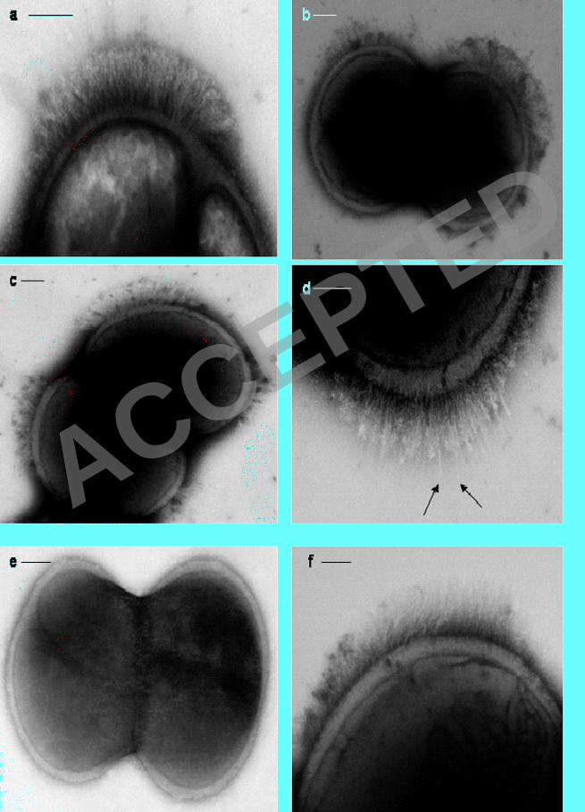
Stationary phase cells of S. epidermidis NCTC 11047 carried short fibrillar 355
appendages, localised on the cell surface in a tuft of fibrils projecting away from the 356
cell wall (Fib+ cells) (Fig. 1a-d). Counts on different batches of 18 h stationary phase 357
cells always detected tufts on approximately 25 % of cells and tufts were also present 358
on a sub-population of cells during logarithmic phase. The mean length of the fibrils 359
in the tufts was 122.2 +/- 10.8 nm as measured from the fibril base to the fibril tip on 360
three batches of cells (20-30 cells per batch). The position of tufts in relation to the 361
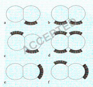
septum varied from cell to cell (represented diagrammatically in Fig. 2a-f). Tufts 362
ACCEPTED
on February 2, 2020 by guest
http://jb.asm.org/
Dow
nloaded from
12
never extended over more than a quarter of the cell circumference and they were 363
usually in a lateral position but could be located in a polar position. Typically a 364
dividing Fib+ cell had one visible tuft in a lateral position on one daughter cell only 365
(Fig. 1a & 2a), but on a very small number of cells two lateral tufts were present on 366
one cell as it divided (Fig. 1b & 2b). Very rarely, four tufts were detected on a 367
dividing cell (Fig. 1c & 2d). Counts of cells with multiple tufts were not possible due 368
to the very low numbers of such cells detected. The tufts comprised of densely packed 369
fibrils and separate fibrils could be seen only rarely (Fig. 1d). The majority of 370
stationary phase cells (75 %) showed no detectable fibrils over the cell circumference 371
(Fig. 1e). Treatment of wild type cells of S. epidermidis NCTC 11047 with protease (1 372
mg ml -1
2 h, 37 ºC) and lysostaphin (100 µg ml-1
) completely removed all tuft fibrils 373
as detected by negative staining (data not shown). Also, cells were still intact after 374
both enzyme treatments. Loss of the fibrils after protease treatment indicated that tuft 375
fibrils were either comprised of protein or were attached to the cell surface by a 376
protein component. 377
Only very small numbers of cells of S. epidermidis RP262A carried detectable 378
fibrils and extensive screening of many batches of cells stained by methylamine 379
tungstate showed that <1% of the cells carried visible tufts of fibrils (Fig 1f). The 380
fibrils in the RP62A tufts were 159 +/- 35nm long and projected out laterally from 381
one side of a cell. The RP62A tufts were indistinguishable from those on NCTC 382
11047 cells and there was no significant difference in the lengths of the tuft fibrils on 383
the two strains. Since there was a much higher number of tufted cells detected in the 384
WT population of NCTC 11047 compared with RP62A S. epidermidis NCTC 11047 385
was selected for the subsequent work on fibril characterization. 386
387
Enrichment of the tufted and non-tufted sub-populations of Staphylococcus 388
epidermidis NCTC 11047 389
Since tufts of fibrils on species of oral streptococci have hydrophobic properties (16, 390
18) it was predicted that the NCTC 11047 fibrils would also have hydrophobic 391
properties. Therefore partitioning between the inert hydrocarbon hexadecane and a 392
buffer (49) was used to enrich for the S. epidermidis NCTC 11047 cells carrying the 393
tufts of fibrils. We had previously used the hexadecane assay to successfully separate 394
tufted and non-tufted cells of the oral bacterium Streptococcus sanguis (4) and it was 395
ACCEPTED
on February 2, 2020 by guest
http://jb.asm.org/
Dow
nloaded from
13
used in preference to any other hydrophobic partitioning method due to the speed and 396
simplicity with which each enrichment step could be carried out. Also it is already 397
known that contact of S. epidermidis NCTC 11047 cells with hexadecane did not 398
significantly reduce their viability (40). 399
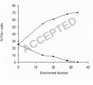
When the NCTC 11047 WT cells were partitioned, the more hydrophobic cells 400
were recovered from the hexadecane phase and the more hydrophilic cells were 401
recovered from the buffer phase. At intervals during the enrichment process both the 402
putative ‘hydrophobic’ and ‘hydrophilic’ cultures were grown to stationary phase (18 403
h) and the relative numbers of ‘tufted’ (Fib+) and ‘non-tufted’ (Fib-) cells in a 404
population were counted using negative staining (Fig. 3). Fib+ cells were successfully 405
enriched using this approach and the percentage of tufted cells in the culture enriched 406
from the hexadecane phase began to plateau at a maximum of 70 % after 34 407
enrichment steps. Conversely the percentage of Fib+ cells in the sub-population 408
enriched from the buffer phase reduced to zero after 34 enrichment steps (Fig. 3). The 409
mean fibril length on the enriched tufted sub-population (121.8 +/- 10.5 nm) was not 410
significantly different (p > 0.05) to the mean length of fibrils on the wild type (122.2 411
+/- 10.8 nm). Each measurement was taken from three batches of stationary phase 412
cells. Both Fib- and Fib+ strains have the same colony morphology so they could only 413
be distinguished from each other by negative staining. 414
Quantitative assessment of cell surface hydrophobicity 415
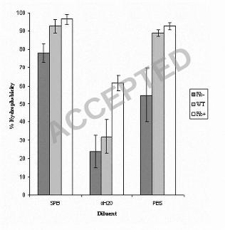
Wild type NCTC 11047 cells and the Fib+ and Fib- enriched sub-populations were 416
tested in the hexadecane assay (49) to give an estimate of their relative cell surface 417
hydrophobicity. Stationary phase cells of the three strains were partitioned from SPB, 418
PBS and dH2O into hexadecane (Fig. 4). For all diluents the Fib+ cells always showed 419
a higher affinity for hexadecane than the WT, but the difference was only significant 420
when cells were partitioned from water (p < 0.05). The Fib- cells showed a reduced 421
affinity for the hexadecane compared to the WT cells but the difference was only 422
significant for SPB and PBS (p < 0.05). However, for all diluents the Fib+ cells 423
always adhered in significantly greater numbers to the hexadecane droplets than did 424
the Fib- cells (Fig 4) (p < 0.05), indicating that the presence of fibrils correlated with 425
increased cell surface hydrophobicity. 426
HIC with octyl-sepharose confirmed that cells of the tufted, Fib+ 427
subpopulation were more hydrophobic than the non-fibrillar Fib- subpopulation as 428
there was an 89 +/- 1% retention for the Fib+ cells and an 80 +/- 2 % retention for the 429
ACCEPTED
on February 2, 2020 by guest
http://jb.asm.org/
Dow
nloaded from
14
Fib- cells on the octyl sepharose. This difference was statistically significant (p < 430
0.05). The overall retention of both strains by the non-hydrophobic Sepharose was not 431
significantly different and Fib+ and Fib- cells showed an overall mean retention of 20 432
+/- 2 %. This value is a measure of the entrapment by the agarose gel itself (50). Thus 433
two separate methods for quantifying cell surface hydrophobicity showed that the 434
cells with tufts of fibrils are more hydrophobic than cells without any tufts. These 435
results suggest that the fibrils contribute to cell surface hydrophobicity and therefore 436
are likely to have hydrophobic properties. 437
Assessment of biofilm forming ability 438
Biofilm formation by the S. epidermidis NCTC 11047 strains was compared and 439
found to be low in all three strains. Biofilm formation by the Fib+ and WT strains was 440
not significantly different to each other with crystal violet OD490 values of 0.205 ± 441
0.009 and 0.195 ± 0.003 respectively. However biofilm formation by the Fib- strain 442
gave an OD 490 of 0.168 ± 0.003, which was only slightly less biofilm than that 443
formed by the WT & Fib+ strains, although due to very low standard error there was a 444
very small but significant difference (p < 0.05) in the OD490 values. The biofilm 445
negative strain S. epidermidis ATCC 12228 was included in the study and gave an OD 446
value of 0.135 ± 0.005, which is comparable to the values of the NCTC 11047 strains. 447
In comparison, the biofilm positive strain S. epidermidis RP62A gave an OD 490 448
reading of 0.942 ± 0.047, which is a factor of 4.6 greater than that formed by the Fib+ 449
sub-population of NCTC11047. These data suggest that the S. epidermidis NCTC 450
11047 strains are all poor biofilm formers and can be described as biofilm negative 451
strains. Thus, the increase in the presence of fibril tufts on the Fib+ sub-population to 452
70 % compared to 25 % on the WT cells does not significantly enhance biofilm 453
formation. 454
Assessment of adhesion to polystyrene 455
The ability of S. epidermidis NCTC 11047 WT, Fib- and Fib+ strains to adhere to 456
polystyrene for 2h was compared using the 96 well plate assay and Fib+ cells attached 457
in much higher numbers than the Fib- cells. After 2h incubation on polystyrene, Fib+ 458
cells gave a final OD490 of 0.126 ± 0.002 which was slightly, but significantly (p < 459
0.05), higher than that of the WT population (0.112 ± 0.002. However, adhesion of 460
the non-tufted Fib- cells to polystyrene (0.062 ± 0.001) was significantly lower (by 50 461
%) in comparison with adhesion of the tufted Fib+ cells (P<0.05). These results show 462
ACCEPTED
on February 2, 2020 by guest
http://jb.asm.org/
Dow
nloaded from
15
that a higher number of fibrillar tufts is correlated with higher adhesion to 463
polystyrene, indicating that the fibrils may play a role in adhesion to polystyrene for 464
S. epidermidis NCTC 11047. 465
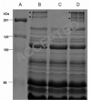
Analysis of cell surface proteins on S. epidermidis by SDS PAGE 466
To identify protein components of the fibrils, lysostaphin was used to solubilise the 467
cell surface polypeptides (51). SDS PAGE profiles of the protein extracts revealed 468
two polypeptides with estimated molecular masses of 280 and 230 kDa, which were 469
present on the WT and from the Fib+ populations (Fig. 5, Lanes B & D) but absent on 470
the Fib- population (Fig. 5 Lane C). As far as it was possible to discern all other 471
polypeptide bands were similar for the three NCTC 11047 strains, although a very 472
faint band (~ 200kDa) could be sometimes be detected in the Fib+ protein extract 473
(Fig. 5 Lane D). 474
Fibrils on Streptococcus gordonii DL1 are comprised of the high molecular 475
mass protein CshA (259 kDa) (37), so in order to identify putative fibril polypeptides 476
on S. epidermidis, the two high molecular mass bands from S. epidermidis NCTC 477
11047 WT and Fib+ were excised and identified by electrospray-tandem mass 478
spectrometry. The faint 200 ~kDa protein on some batches of Fib+ cells could not be 479
sequenced. Five fragments from each of the 280 & 230 kDa proteins from NCTC 480
11047 shared 100 % identity with Aap of RP62A (Accession number NC_002976). 481
The fully sequenced strain RP62A also showed two high molecular mass bands (280 482
& 230 kDa) on SDS PAGE gels (data not presented) which were sequenced and found 483
to be Aap. Therefore both NCTC 11047 and RP62A both expressed two high 484
molecular mass bands comprising of Aap that correlated with the presence of tufts of 485
fibrils. 486
The two Aap bands on NCTC 11047 (WT and Fib+ strains) and RP62A did not 487
stain with the periodic-schiff reagent (data not presented) indicating the lack of 488
glycosylation of Aap. No fibrils could be detected by negative staining on Fib + cells 489
of NCTC 11047 treated with lysostaphin. Protease treatment of collected cell surface 490
proteins after lysostaphin extraction resulted in the loss of the 230 kDa and 280 kDa 491
proteins from the gel (data not shown). Protease treatment also removed fibrils from 492
the cell surface of Fib+ cells. These data indicate that the presence of fibrils correlates 493
with the expression of Aap, detected as two high molecular mass polypeptides. 494
Stability of Fib+ and Fib- populations 495
ACCEPTED
on February 2, 2020 by guest
http://jb.asm.org/
Dow
nloaded from
16
In order to test whether the presence or absence of the tufts were stable traits, the Fib+ 496
and Fib- populations were serially subcultured to test for reversion by sequentially 497
plating cells on TSA 34 times. The percentage of cells with tufts was recorded, after 498
negative staining, in the TEM. The percentage of Fib+ and Fib- cells in both 499
populations showed very little change after 34 subcultures. No tufts were detected on 500
the Fib- population and the ‘tuft count’ only reduced from 75 % to 60 % on the Fib+ 501
population. 502
Counting the numbers of Fib+ cells is extremely difficult and it was not 503
possible to count enough cells to do a statistical comparison on the presence of tufts 504
before and after subculture of the Fib+ strain. However we can conclude that no 505
major reduction in fibril expression has occurred after extensive subculture of the 506
Fib+ sub-population. In addition SDS PAGE gels of surface proteins from the serially 507
subcultured Fib- cells showed no reappearance of the Aap bands (280 & 230 kDa) 508
(data not presented). These data indicate that the presence or absence of the tuft on the 509
two sub-populations is an apparently stable property and that reversion to the wild 510
type ratio of 75 % Fib- to 25 % Fib+ does not easily occur on subculture. In addition 511
when the Fib+ and Fib- strains were grown as biofilms on cellulose filters overnight 512
they did not respectively loose or regain the two high molecular weight bands or the 513
fibrils (data not presented). 514
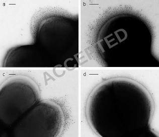
Immunogold negative staining with anti-Aap antibodies 515
In order to determine whether the localised fibrillar tufts of S. epidermidis NCTC 516
11047 were comprised of Aap, immunogold negative staining was carried out. Cells 517
were incubated with antisera specific to either the A-region of Aap or to the B-repeat 518
of Aap. With both antibodies, gold particles were found to label the fibrillar tufts of 519
Fib+ cells (Fig. 6a & b) and the WT cells (Fig. 6 c). There was no apparent difference 520
in the distribution of the gold on the tufts for either antibody. In order to show the gold 521
particles, cells have been selected with a low level of negative staining (Fig. 6 a-c) and 522
fibrils are not heavily stained. However, on more darkly stained cells the fibrils could 523
always be seen underneath the gold (data not presented). The outline of gold particles 524
always followed the outline of the shape of the fibrillar tuft. No label (above 525
background) was detected with either antiserum on Fib- cells (Fig. 6 d) proving a 526
complete lack of the Aap protein on these cells. It was possible to find cells that had 527
gold label on one daughter cell (on the tuft) but there was no tuft and no label on the 528
ACCEPTED
on February 2, 2020 by guest
http://jb.asm.org/
Dow
nloaded from
17
other daughter cell. Thus is possible to detect the cells switching expression of the 529
fibrillar tuft as the cells divide. 530
PCR amplification of aap and genes of the ica operon 531
Genomic DNA was isolated from S. epidermidis NCTC 11047 WT, Fib- and Fib+ 532
cells and S. epidermidis RP62A. PCR was carried out in order to amplify the whole 533
aap gene of these populations. All three NCTC 11047 populations gave an amplicon 534
of approximately 7 kb, which corresponded to the size of the aap amplicon of RP62A, 535
the positive control. Fib- cells gave the same size amplicon as the WT and Fib+ 536
populations implying that there is no insertion or deletion in this gene that prevents 537
Aap from being expressed on the cell surface of Fib- cells. 538
S. epidermidis NCTC 11047 was screened for the ica operon, which encodes 539
polypeptides involved in the synthesis of PIA (21, 34). All three 11047 strains were 540
found to be negative for the ica operon with all six primer pairs used, whereas the 541
control strain RP62A was positive for all. Consistently, synthesis of PIA could not be 542
detected on Congo red agar. The NCTC 11047 strains all produced large, smooth, red 543
colonies indicating a lack of PIA, whereas RP62A produced dry, black, highly 544
crenated colonies due to the presence of PIA (58). 545
Detection of aap transcription products by RT-PCR 546
RT-PCR was carried out in order to determine whether the aap gene was being 547
transcribed by the Fib- cells as well as the Fib+ cells. All NCTC 11047 strains 548
produced mRNA corresponding to nucleotide positions 1357-1756 and 6657-7121 of 549
aap. As a 400 bp fragment of the A-region of aap and a 500 bp region at the C-550
terminus of aap were both amplified it can be deduced that full length aap is 551
transcribed in all four strains tested. Equal amounts of RNA were used in each 552
reaction and amplified PCR products from cDNA of all four strains (S. epidermidis 553
NCTC 11047 WT, Fib-, Fib+ and S. epidermidis RP62A) showed the same band 554
intensity on agarose gels, indicating that the level of aap transcription was equal in 555
each strain. 556
DNA sequence analysis of the Aap promoter region 557
To investigate any possible variation in the aap promoter region for the different 558
NCTC 11047 sub-populations, the region was sequenced and compared with the aap 559
promoter region of strain RP62A. The aligned regions of DNA sequence upstream of 560
aap in NCTC 11047 WT and the Fib+ and Fib- sub-populations shared 100% identity 561
(data not shown). In addition, a putative ribosome binding site (GGGAG) and -10 and 562
ACCEPTED
on February 2, 2020 by guest
http://jb.asm.org/
Dow
nloaded from
18
-35 regions were identified. This proof of integrity of the promoter regions for all 563
three NCTC 11047 strains is consistent with the results of RT-PCR analyses which 564
together suggest that aap is transcribed in Fib- cells as well as in the Fib+ cells. 565
566
DISCUSSION 567
Fibril tufts correlate with high cell surface hydrophobicity. 568
This study has shown that a sub-population of cells of S. epidermidis NCTC 11047 569
carries fibrillar appendages that have not been previously reported on any strain of S. 570
epidermidis. It is proposed that the fibrils have hydrophobic properties as serial 571
enrichment in a hydrophobic partition assay using hexadecane enabled separation of 572
the more hydrophobic Fib+ cells from the less hydrophobic Fib- cells. This enrichment 573
using hexadecane was previously successful as a method for enriching for tufted cells 574
of Streptococcus cristatus NCTC 12479 (formerly Streptococcus sanguis CR311) (4, 575
18) and the more hydrophobic streptococci with long fibrils in the tufts also 576
preferentially partitioned into the hexadecane layer. However, attachment of bacteria 577
to hexadecane droplets results from a combination of both hydrophobic and 578
electrostatic interactions (12). Therefore HIC using octyl sepharose was used as an 579
independent method (40, 50) to more accurately compare the relative hydrophobicity 580
of the two subpopulations. Since the Fib+ cells attached in higher numbers than the 581
Fib- cells to octyl sepharose, the results are consistent with the hypothesis that the 582
fibrils have hydrophobic properties. Because the fibrils are localized they are predicted 583
to contribute to micro-scale heterogeneity of cell surface hydrophobicity. Thus the cell 584
surface is likely to be comprised of a smaller, more hydrophobic, fibril tuft zone 585
surrounded by a more extensive, less hydrophobic, non-fibrillar cell surface area. 586
However, since adhesion of the Fib- cells to octyl sepharose is still fairly high (80 % 587
for Fib-cells compared to 89 % for Fib+ cells) there are clearly many other cell surface 588
molecules on the non-fibrillar part of the cell surface that have hydrophobic properties. 589
It was not possible to increase the relative numbers of tufted S. epidermidis 590
NCTC 11047 cells to above 70 %, even after 34 enrichment steps through hexadecane, 591
although there is no apparent reason why 100 % Fib+ cells could not be achieved by 592
enrichment. Counting the numbers of ‘tufted’ cells in the TEM is likely to be an 593
underestimate. Tuft fibrils are difficult to detect, and if the tuft was underneath or 594
above the visible circumference, it would have been not have been counted as the cell 595
would appear to be non-tufted. In addition the fibrils often do not show a pronounced 596
ACCEPTED
on February 2, 2020 by guest
http://jb.asm.org/
Dow
nloaded from
19
contrast in the negative stain. For both these reasons is possible that 100 % of the Fib+ 597
cells carry fibrils, but 30 % of Fib+ cells were not detectable. It is therefore possible 598
that the parent strain of S. epidermidis NCTC 11047 contains approximately 50 % 599
Fib+ cells rather than the 25 % detected. The Fib+ and Fib- sub-populations are clonal 600
descendants of the NCTC 11047 WT as it is highly unlikely that the enrichment 601
procedure led to the selection of a sub-population of contaminants for a number of 602
reasons. Firstly, multiplication of cells only occurred in the non-selective medium 603
TSB, secondly the mean lengths of tuft fibrils from WT NCTC 11047 and the Fib+ 604
cells were not significantly different and thirdly, the SDS PAGE profiles were 605
identical apart from differences in the presence of the 280 & 230 kDa proteins (see 606
later). 607
Organization of fibrils into localised tufts 608
This is the first description of localised fibrillar appendages on a staphylococcus, but 609
tufts of fibrils are common on oral streptococci and have been found on Streptococcus 610
oralis (28), Streptococcus mitis (formerly Streptococcus sanguis biotype II) (17)and S 611
cristatus (15, 18). The functions of these prominent structures are not well understood, 612
although S. cristatus cells adhere specifically via the tuft of fibrils to other plaque 613
bacteria (30) in coaggregation interactions between different genera in mature dental 614
plaque. 615
Negative staining of the enriched Fib+ sub-population revealed that tufts could 616
sometimes be present at a number of locations simultaneously on the cell surface (Fig. 617
1b-d; Fig. 2b-d & f). The majority of tufts were in the lateral position but some were 618
polar. This could mean that the fibrils can migrate around the cell surface (from lateral 619
to polar), or it may be a result of the septum always occurring at right angles to the 620
previous septum resulting in the lateral surface becoming the apparent pole after two 621
cell division rounds. Localised protein export is necessary to produce these patches of 622
localised fibrils and recently a laterally positioned microdomain for protein secretion 623
via the general secretory (Sec) pathway has been discovered in Streptococcus 624
pyogenes (48). A similar localised export process may operate for fibril proteins in S. 625
epidermidis NCTC 11047. 626
The fibrils detected on NCTC 11047 are clearly distinct from the much longer, 627
very thin ‘fimbria-like’ appendages found on S. epidermidis 354 that mediate in 628
adhesion to polystyrene (55). These appendages are comprised of the proteins SSP1 629
and SSP2 and both have a molecular mass in excess of 200 kDa. Since the proteins 630
ACCEPTED
on February 2, 2020 by guest
http://jb.asm.org/
Dow
nloaded from
20
have not been sequenced and the genes encoding these proteins have not been 631
identified it is not possible to relate the two morphologically distinct structures on the 632
two strains. 633
Since only a sub-population of cells of the wild type S. epidermidis NCTC 634
11047 carries tufts of fibrils, it is possible that fibril expression is subject to phase 635
variation. Phase variation of fimbriae (pili) on Gram negative bacteria is a very 636
common phenomenon (2, 11, 42, 53) and phase variation mechanisms exist that 637
alternate between expression (phase ON) and non-expression (phase OFF) of fimbriae 638
(23). Phase variation in Gram positives is less well studied; although several surface 639
proteins linked with virulence of Streptococcus pyogenes are known to phase vary (7). 640
Currently we have no experimental evidence to support the hypothesis that 641
fibrils are phase variable, as in this study no reversion of either the Fib+ or the Fib- 642
phenotype was detected. Even after 34 subcultures the Fib- cells did not regain 643
fibrillar tufts or the high molecular weight protein bands of Aap (see later), and the 644
Fib+ cells did not apparently loose them. This lack of ability to revert back to the WT 645
ratio of 3:1, Fib- and Fib+ cannot currently be explained. However, the possibility of 646
a point mutation having occurred in the Fib- sub-population, leading to some as yet 647
unknown effect on translation, cannot be ruled out. However, the Fib- sub-population 648
within the WT population clearly has a mechanism which prevents fibril expression 649
on the cell surface, whereas the Fib+ cells can express the fibrils. In addition, S. 650
epidermidis NCTC 11047 WT cells often have Fib+ and Fib- daughter cells attached 651
to each other as they divide (Fig. 2a) implying that the mechanism controlling fibril 652
expression is switched OFF in one cell while it remains switched ON in the other cell 653
as the DNA replicates 654
All three NCTC 11047 strains carry the aap gene and PCR products of the 655
whole aap of each population are the same length, indicating that there is no major 656
insertion or deletion in the aap gene of Fib- cells that is inhibiting expression of Aap 657
on the cell surface. In addition, using RT-PCR, we have shown that WT, Fib- and Fib+ 658
cells all transcribe aap, indicating that expression of Aap is regulated at the post-659
transcriptional level, although as yet it is not clear what this mechanism of regulation 660
is. If translation also occurs, it is possible that the fibrils could be retained in the cell 661
or that cell wall anchoring is prevented by some unknown mechanism. These 662
possibilities are currently under investigation. 663
Correlation between fibrils, high molecular mass proteins and Aap 664
ACCEPTED
on February 2, 2020 by guest
http://jb.asm.org/
Dow
nloaded from
21
The 280 & 230 kDa polypeptides present in CSP extracts of Fib+ cells and WT 665
cells were the only high molecular mass proteins absent from Fib- cells. Both bands 666
were identified as Aap by mass spectrometric analysis as fragments from them share 667
100 % identity with Aap from the already sequenced S. epidermidis strains RP62A and 668
ATCC 12228 (14, 57). Antibodies raised against the N terminal A-region of Aap and 669
the single B-repeat unit (128 a.a repeat) both labelled the fibrillar tufts, conclusively 670
showing that Aap is expressed as a full-length molecule on the cell surface of S. 671
epidermidis NCTC 11047. Therefore it is proposed that the 280 kDa molecule of Aap 672
is the full length protein represented on the cell surface as 122 nm fibrillar appendages 673
localized in a tuft structure. 674
Thus each fibril in the tuft of NCTC 11047 is proposed to be a molecule of 675
Aap. This is similar in principle to the 60 nm peritrichous fibrils carried by 676
Streptococcus gordonii DL1, the dental plaque primary coloniser. Each fibril is one 677
molecule of the 259 kDa cell-wall-anchored (CWA) protein CshA (37). CshA also has 678
a domain structure and is anchored via a LPXTG motif to peptidoglycan. It is a 679
multifunctional fibrillar adhesive protein involved in coaggregation to other oral 680
bacteria and in adhesion to fibronectin (36). Aap has not been reported to be 681
glycosylated and the 280 and 230 kDa Aap bands of NCTC 11047 do not stain with 682
periodic-schiff reagent confirming the lack of glycosylation. 683
Biofilm formation by S. epidermidis 5179 depends on proteolytic cleavage of 684
the full length (220 kDa) Aap to a 140 kDa truncated form, which consists of the B-685
domain of Aap without the A-region (47). Strain 5179 also expresses a 180 kDa Aap 686
thought to be a partially truncated form of the 220 kDa Aap, although its function has 687
not yet been determined. Like S. epidermidis 5179, strain NCTC 11047 expresses two 688
high molecular mass Aap proteins (280 & 230 kDa), although it is not yet clear why 689
these two forms are expressed and what the significance is in the difference between 690
these two Aap bands. As both anti-A and anti-B antibodies labelled the tufts of fibrils 691
it is clear that Aap is expressed as a full length molecule on the cell surface of S. 692
epidermidis NCTC 11047 and so according to the mechanism of biofilm formation 693
proposed by Rohde et al., (47) this strain would be biofilm negative. In support of this 694
there is only a very small difference in biofilm-forming ability between the Fib+ and 695
Fib- sub-populations, as biofilms formed by NCTC 11047 and its Fib+ and Fib- sub-696
populations are very similar in thickness to the non-biofilm forming strain S. 697
epidermidis ATCC 12228. Additional support for the proposal that NCTC 11047 has 698
ACCEPTED
on February 2, 2020 by guest
http://jb.asm.org/
Dow
nloaded from
22
a biofilm negative phenotype is provided by the fact that it lacks the ica operon which 699
is responsible for PIA biosynthesis, the polymer responsible for the biofilm positive 700
phenotype of many strains, including RP62A (31). 701
The crystal violet (CV) assay for biofilm formation was used in this study as 702
since it was developed by Christensen et al., (6), it has been used consistently in the 703
majority of biofilm quantification studies for a very wide range of Gram positive and 704
Gram negative bacteria. Christensen et al., (6), proved that the numbers of cells (by 705
weight) in a biofilm correlated with the optical density of the crystal violet taken up by 706
the cells in the biofilm. 707
The presence of tufts of fibrils on a very small number of cells on RP62A also 708
correlated with the presence of the aap gene in this strain, but its ability to form a 709
copious biofilm is due to the production of the PIA polymer as already discussed (31). 710
Aap of strain RP62A is not truncated as SDS PAGE gels show the same two high 711
molecular weight Aap bands shown by NCTC 11047. S. epidermidis 12228 carries the 712
aap gene (57) but we do not yet know whether it carries tufts of fibrils. On the basis of 713
the biofilm formation mechanism proposed by Rohde et al., (47) it is predicted that 714
proteolytic cleavage of the Aap protein on NCTC 11047 would result in an increase in 715
the ability of this strain to form biofilms, although this hypothesis is yet to be tested. A 716
bigger study to correlate the presence of the aap gene with the expression of fibrils on 717
a large number of S. epidermidis strains is currently underway. 718
Although there is very little difference in the amount of biofilm formed by 719
Fib+ and Fib- cells of strain NCTC 11047 after 24h, the presence of the hydrophobic 720
fibrillar tufts does enhance initial adhesion of the Fib+ cells to polystyrene after 2h. At 721
this early stage in adhesion, the level of Fib+ adhesion is twice as high in comparison 722
to the Fib- cells. It is possible that the fibrillar tufts mediate adhesion to polystyrene by 723
a specific mechanism in a similar process to that mediated by the high molecular mass 724
SSP1 and SSP2 proteins present on S. epidermidis 354 (51, 55). By the time the 24 h 725
biofilm of strain NCTC 11047 is formed, the differences in initial adhesion of the Fib+ 726
and Fib- cells have been almost lost as the numbers of cells of these two 727
subpopulations build up in the biofilms to a similar level in the accumulation phase. 728
The reasons for this are not clear as both strains have a very similar doubling time and 729
reach the same final OD in the wells (data not presented). 730
This study has demonstrated an unequivocal correlation between the presence of 731
localised tufts of fibrils and the presence of Aap on the cell surface. It also predicts 732
ACCEPTED
on February 2, 2020 by guest
http://jb.asm.org/
Dow
nloaded from
23
that the localised fibrils of Aap contribute to the overall cell surface hydrophobicity 733
for strain S. epidermidis NCTC 11047. Revealing the localisation and molecular 734
architecture of Aap on the cell surface will allow a more detailed understanding of its 735
function in processes relating to colonisation and infection. 736
737
ACKNOWLEDGEMENTS 738
This study was supported by a Biotechnology and Biological Sciences Research 739
Council (BBSRC) CASE award supported by NeuTec Pharma PLC. We thank Lee 740
Cosgrove for Figure 2 and Wiesia Woodyatt for measuring the fibril lengths of S. 741
epidermidis NCTC 11047. The authors thank the staff in the EM Facility in the 742
Faculty of Life Sciences (University of Manchester) for their assistance and the 743
Wellcome Trust for equipment grant support to the EM Facility. 744
745
REFERENCES 746
1. Arciola, C.R., S. Gamberoni, D. Capoccia, L.Visai, P. Speziale, L. 747 Baldassarri, and L. Montanaro. 2005. A multiplex PCR method for the 748
detection of all five individual genes of ica locus in Staphylococcus 749
epidermidis. A survey on 400 clinical isolates from prosthesis-associated 750
infections. J. Biomed. Mater. Res. A. 75: 408-413 751
752 2. Bayliss, C. D., W. A. Sweetman, and E. R. Moxon. 2004. Mutations in 753
Haemophilus influenzae mismatch repair genes increase mutation rates of 754
dinucleotide repeat tracts but not dinucleotide repeat-driven pilin phase 755
variation rates. J. Bacteriol. 186: 2928-2935. 756
757
3. Bowden, M. G., W. Chen, J. Singvall, Y. Xu, S. J. Peacock, V. Valtulina, 758 P. Speziale, and M. Hook. 2005. Identification and preliminary 759
characterization of cell-wall-anchored proteins of Staphylococcus epidermidis. 760
Microbiology. 151; 1453-1464. 761
762
4. Busscher, H. J., P. S. Handley, P. G. Rouxhet, L. M. Hesketh, and H. C. 763 van der Mei. 1991. The relationship between structural and physicochemical 764
surface properties of tufted Streptococcus sanguis strains. In Microbial cell 765
surface analysis, structural and physicochemical methods, pp. 317-338. Edited 766
by P. G. Rouxhet: VCH Publishers Inc. 767
768
5. Christensen, G. D., A. L. Bisno, J. T. Parisi, B. McLaughlin, M. G. Hester, 769 and R. W. Luther.1982.Nosocomial septicaemia due to multiply antibiotic-770
resistant Staphylococcus epidermidis. Ann. Intern. Med. 96: 1-10. 771
772
6. Christensen, G. D., W. A. Simpson, J. J. Younger, L. M. Baddour, F. F. 773 Barrett, D. M. Melton, and E. H. Beachey. 1985. Adherence of coagulase-774
negative staphylococci to plastic tissue culture plates: a quantitative model for 775
ACCEPTED
on February 2, 2020 by guest
http://jb.asm.org/
Dow
nloaded from
24
the adherence of staphylococci to medical devices. J. Clin. Microbiol. 22: 996-776
1006. 777
778
7. Cleary, P. P., l. McLandsborough, L. Ikeda, D. Cue, J. Krawczak, and H. 779 Lam. 1998. High frequency intracellular infection and erythrogenic toxin A 780
expression undergo phase variation in M1 group A streptococci. . Mol. 781
Microbiol. 28: 157-167. 782
783
8. Deighton, M.A., J. Capstick, E. Domalewski, and T. van Nguyen. 2001. 784
Methods for studying biofilms produced by Staphylococcus epidermidis. 785
Methods Enzymol. 336: 177-195 786
787
9. Finch, R. G., P. Hill, and P. Williams. 1995. Staphylococci - the emerging 788
threat. Chem. Ind. 6: 225-228. 789
790
10. Frebourg, N. B., S. Lefebvre, S. Baert, and J.-F. Lemeland. 2000. PCR-791
Based Assay for Discrimination between Invasive and Contaminating 792
Staphylococcus epidermidis Strains. J. Clin. Microbiol. 38: 877-880. 793
794
11. Gally, D. L., J. Leathart, and I. C. Blomfield. 1996. Interaction of FimB and 795
FimE with the fim switch that controls the phase variation of type 1 fimbriae 796
in Escherichia coli K-12. Mol. Microbiol. 21: 725-738. 797
798
12. Geertsema-Doornbusch, G. I., H. C. van der Mei, and H.J. Busscher. 799 1993. Microbial cell surface hydrophobicity: The involvement of electrostatic 800
interactions in microbial adhesion to hydrocarbons (MATH). J. Micro. 801
Methods. 18: 61-68. 802
803
13. Gilbert, P., T. Maira-Litran, A. J. McBain, A. H. Rickard, and F. W. 804 Whyte. 2002. The physiology and collective recalcitrance of microbial biofilm 805
communities. Adv. Microb. Physiol. 46: 202-256. 806
807
14. Gill, S. R., D. E. Fouts, G. L. Archer, E. F. Mongodin, R. T. Deboy, J. 808
Ravel, I. T. Paulsen, J. F. Kolonay, L. Brinkac, M. Beanan, R. J. Dodson, 809
S. C. Daugherty, R. Madpu, S. V. Angiuoli, A. S. Durkin, D. H. Haft, 810
J.Vamathevan, H. Khouri, T. Utterback, C. Lee, G. Dimitrov, L. Jiang, H. 811
Qin, J. Weidman, K. Tran, K. Kang, I. R. Hance, K. E. Nelson, and C. M. 812 Fraser. 2005. Insights on evolution of virulence and resistance from the 813
complete genome analysis of an early methicillin-resistant Staphylococcus 814
aureus strain and a biofilm-producing methicillin-resistant Staphylococcus 815
epidermidis strain. J. Bacteriol. 187: 2426-2438. 816
817
15. Handley, P., A. Coykendall, D. Beighton, J. M. Hardie, and R. A.Whiley. 818 1991a. Streptococcus crista sp. nov., a viridans streptococcus with tufted 819
fibrils, isolated from the human oral cavity and throat. Int. J. Syst. Bacteriol. 820
41: 543-547. 821
822
16. Handley, P. S., L. M. Hesketh, and R. A. Moumena. 1991b. Charged and 823
hydrophobic groups are localized in the short and long tuft fibrils of 824
Streptococcus sanguis strains. Biofouling 4: 105-111. 825
ACCEPTED
on February 2, 2020 by guest
http://jb.asm.org/
Dow
nloaded from
25
826
17. Handley, P. S., P. L. Carter, J. E. Wyatt, and L. M. Hesketh. 1985. Surface 827
structures (peritrichous fibrils and tufts of fibrils) found on Streptococcus 828
sanguis strains may be related to their ability to coaggregate with other oral 829
genera. Infect. Immun. 47: 217-227. 830
831
18. Handley, P. S., F. F. Correia, K. Russell, B. Rosan, and J. M. DiRienzo. 832 2005. Association of a novel high molecular weight, serine-rich protein (SrpA) 833
with fibril-mediated adhesion of the oral biofilm bacterium Streptococcus 834
cristatus. Oral Microbiol. Immunol. 20: 131-140. 835
836
19. Hartford, O., L. O'Brien, K. Schofield, J. Wells, and T. J. Foster. 2001. 837
The Fbe (SdrG) protein of Staphylococcus epidermidis HB promotes bacterial 838
adherence to fibrinogen. Microbiology. 147: 2545-2552. 839
840
20. Heilmann, C., M. Hussain, G. Peters, and F. Gotz. 1997. Evidence for 841
autolysin-mediated primary attachment of Staphylococcus epidermidis to a 842
polystyrene surface. Mol. Microbiol. 24: 1013-1024. 843
844
21. Heilmann, C., O. Schweitzer, C. Gerke, N. Vanittanakom, D. Mack, and 845 F.Gotz. 1996. Molecular basis of intercellular adhesion in the biofilm-forming 846
Staphylococcus epidermidis. Mol. Microbiol. 20: 1083-1091. 847
848
22. Heilmann, C., G. Thumm, G. S. Chhatwal, J. Hartleib, A.Uekotter, and 849 G. Peters. 2003. Identification and characterization of a novel autolysin (Aae) 850
with adhesive properties from Staphylococcus epidermidis. Microbiology. 851
149: 2769-2778. 852
853
23. Hernday, A., M. Krabbe, B. Braaten, and D. Low, 2002. Self-perpetuating 854
epigenetic pili switches in bacteria. Proc. Natl. Acad. Sci. USA. 99: Suppl 4, 855
16470-16476. 856
857
24. Hogt, A. H., J. Dankert, and J. Feijen. 1983. Encapsulation, slime 858
production and surface hydrophobicity of coagulase-negative staphylococci. 859
FEMS Microbiol. Lett. 18: 211-215. 860
861
25. Hoyle, B. D. and J. W. Costerton. 1991. Bacterial resistance to antibiotics: 862
the role of biofilms. Prog. Drug Res. 37: 91-105. 863
864
26. Hussain, M., C. Heilmann, G. Peters, and M. Herrmann. 2001. Teichoic 865
acid enhances adhesion of Staphylococcus epidermidis to immobilized 866
fibronectin. Microb. Pathog. 31: 261-270. 867
868
27. Hussain, M., M. Herrmann, C. von Eiff, F. Perdreau-Remington, and G. 869 Peters. 1997. A 140-kilodalton extracellular protein is essential for the 870
accumulation of Staphylococcus epidermidis strains on surfaces. Infect. 871
Immun. 65: 519-524. 872
873
ACCEPTED
on February 2, 2020 by guest
http://jb.asm.org/
Dow
nloaded from
26
28. Jameson, M. W., H. F. Jenkinson, K. Parnell, and P. S. Handley. 1995. 874
Polypeptides associated with tufts of cell-surface fibrils in an oral 875
Streptococcus. Microbiology. 141: 2729-2738. 876
877
29. Laemmli, U. K. and M. Favre. 1973. Maturation of the head of 878
bacteriophage T4. I. DNA packaging events. J. Mol. Biol. 80: 575-599. 879
880
30. Lancy, P., J. M. Dirienzo Jr., B. Appelbaum, B. Rosan, and S. C. Holt. 881 1983. Corncob formation between Fusobacterium nucleatum and 882
Streptococcus sanguis. Infect. Immun. 40: 303-309. 883
884
31. Mack, D., M. Haeder, N. Siemssen, and R. Laufs. 1996a. Association of 885
biofilm production of coagulase-negative staphylococci with expression of a 886
specific polysaccharide intercellular adhesin. J. Infect. Dis. 174: 881-884. 887
888
32. Mack, D., M. A. Horstkotte, H. Rohde, and J. K.-M. Knobloch. 2006a. 889
Coagulase-negative staphylococci. In Biofilms, Infection, and Antibiotic 890
Therapy, pp. 109-153. Edited by R. G. Finch: CRC Press, Taylor and Francis 891
Group, Boca Raton. 892
893
33. Mack, D., H. Rohde, L. G. Harris, A. P. Davies, M. A. Horstkotte, and J. 894 K.-M. Knobloch. 2006b. Biofilm formation in medical device-related 895
infection. Int. J. Artif. Organs 29: 343-359. 896
897
34. Mack, D., W. Fischer, A. Krokotsch, K. Leopold, R. Hartmann, H. Egge, 898 and R. Laufs. 1996b. The intercellular adhesin involved in biofilm 899
accumulation of Staphylococcus epidermidis is a linear beta-1,6-linked 900
glucosaminoglycan: purification and structural analysis. J. Bacteriol. 178: 175-901
183. 902
903
35. McKenney, D., J. Hubner, E. Muller, Y. Wang, D. A. Goldmann, and G. 904 B. Pier. 1998. The ica locus of Staphylococcus epidermidis encodes 905
production of the capsular polysaccharide/adhesin. Infect. Immun. 66: 4711-906
4720. 907
908
36. McNab, R., A. Holmes, J. Clarke, G. Tannock, and H. Jenkinson. 1996. 909
Cell surface polypeptide CshA mediates binding of Streptococcus gordonii to 910
other oral bacteria and to immobilized fibronectin. Infect. Immun. 64: 4204-911
4210. 912
913
37. McNab, R., H. Forbes, P. S. Handley, D. M. Loach, G. W. Tannock, and 914 H. F. Jenkinson. 1999. Cell wall anchored CshA polypeptide (259 915
kilodaltons) in Streptococcus gordonii forms surface fibrils that confer 916
hydrophobic and adhesive properties. J. Bacteriol. 181: 3087-3095. 917
918
38. Muller, E., S. Takeda, H. Shiro, D. Goldmann, and G. B. Pier. 1993. 919
Occurrence of capsular polysaccharide/adhesin among clinical isolates of 920
coagulase-negative staphylococci. J. Infect Dis. 168: 1211-1218. 921
922
ACCEPTED
on February 2, 2020 by guest
http://jb.asm.org/
Dow
nloaded from
27
39. Nilsson, M., L. Frykberg, J. I. Flock, L. Pei, M. Lindberg, and B. Guss. 923 1998. A fibrinogen-binding protein of Staphylococcus epidermidis. Infect. 924
Immun. 66: 2666-2673. 925
926
40. Pembrey, R.S., K. C. Marshall, and R.P. Schneider. 1999. Cell surface 927
analysis techniques: what do cell preparation protocols do to cell surface 928
properties? Appl. Env. Microbiol. 65: 2877-2894. 929
930
41. Peters, G., R. Locci, and G. Pulverer. 1982. Adherence and growth of 931
coagulase-negative staphylococci on surfaces of intravenous catheters. J. 932
Infect Dis. 146: 479-482. 933
934
42. Power, P. M., L. F. Roddam, K. Rutter, S. Z. Fitzpatrick, Y. N. Srikhanta, 935 and M. P. Jennings. 2003. Genetic characterization of pilin glycosylation and 936
phase variation in Neisseria meningitidis. Mol. Microbiol. 49: 833-847. 937
938
43. Raad, I., A. Alrahwan, and K. Rolston.1998. Staphylococcus epidermidis: 939
emerging resistance and need for alternative agents. Clin. Infect. Dis. 26: 940
1182-1187. 941
942
44. Roche, F. M., M. Meehan, and T. J. Foster. 2003a. The Staphylococcus 943
aureus surface protein SasG and its homologues promote bacterial adherence 944
to human desquamated nasal epithelial cells. Microbiology. 149: 2759-2767. 945
946
45. Roche, F. M., R. Massey, S. J. Peacock, N. P. Day, L. Visai, P. Speziale, A. 947 Lam, M. Pallen, and T. J. Foster. 2003b. Characterization of novel LPXTG-948
containing proteins of Staphylococcus aureus identified from genome 949
sequences. Microbiology. 149: 643-654. 950
951
46. Rohde, H., M. Kalitzky, N. Kroger, S. Scherpe, M. A. Horstkotte, J. K.-M. 952 Knobloch, A. R. Zander, and D. Mack. 2004. Detection of virulence-953
associated genes not useful for discriminating between invasive and 954
commensal Staphylococcus epidermidis strains from a bone marrow transplant 955
unit. J. Clin. Microbiol. 42: 5614-5619. 956
957
47. Rohde, H., C. Burdelski, K. Bartscht, M. Hussain, F. Buck, M. A. 958
Horstkotte, J. K. M. Knobloch, C. Heilmann, M. Herrmann, and D. 959 Mack. 2005. Induction of Staphylococcus epidermidis biofilm formation via 960
proteolytic processing of the accumulation-associated protein by 961
staphylococcal and host proteases. Mol. Microbiol. 55: 1883-1895. 962
963
48. Rosch, J. and M. Caparon. 2004. A microdomain for protein secretion in 964
Gram-positive bacteria. Science 304: 1513-1515. 965
966
49. Rosenburg, M., D. Gutnick, and E. Rosenburg. 1980. Adherence of bacteria 967
to hydrocarbons: a simple method for measuring cell surface hydrophobicity. 968
FEMS Microb. Lett. 9: 29-33. 969
970
50. Smyth,C.J., P. Jonsson, E. Olsson, O. Soderlind, J. Rosengren, S. Hjerten, 971 and T. Wadstrom. 1978. Differences in hydrophobic surface characteristics 972
ACCEPTED
on February 2, 2020 by guest
http://jb.asm.org/
Dow
nloaded from
28
of porcine enteropathogenic Escherichia coli with or without K88 antigen as 973
revealed by hydrophobic interaction chromatography. Infect. Immun. 22: 462-974
472. 975
976
51. Timmerman, C. P., A. Fleer, J. M. Besnier, L. De Graaf, F. Cremers, and 977 J. Verhoef. 1991a. Characterisation of a proteinaceous adhesin of 978
Staphylococcus epidermidis which mediates attachment to polystyrene. Infect. 979
Immun. 59: 4187-4192. 980
981
52. Timmerman, C. P., J. M. Besnier, L. De Graaf, R. Torensma, A. 982 J.Verkley, A. Fleer, and J.Verhoef.1991b. Characterisation and functional 983
aspects of monoclonal antibodies specific for surface proteins of coagulase-984
negative staphylococci. J. Med. Microbiol. 35: 65-71. 985
986
53. Van der Woude, M. W., and A. J. Baumler. 2004. Phase and antigenic 987
variation in bacteria. Clin. Microbiol. Rev. 17: 581-611. 988
989
54. Vandecasteele, S. J., W. E. Peetermans, R. Merckx, and J. V. Eldere. 990 2003. Expression of biofilm-associated genes in Staphylococcus epidermidis 991
during in vitro and in vivo foreign body infections. J. Infect. Dis. 188: 730-992
737. 993
994
55. Veenstra, G. J. C., F. F. M. Cremers, H. van Dijk, and A. Fleer. 1996. 995
Ultrastructural organization and regulation of a biomaterial adhesin of 996
Staphylococcus epidermidis. J. Bacteriol. 178: 537-541. 997
998
56. Williams, R. J., B. Henderson, L. J. Sharp, and S. P. Nair. 2002. 999
Identification of a fibronectin-binding protein from Staphylococcus 1000
epidermidis. Infect. Immun. 70: 6805-6810. 1001
1002
57. Zhang, Y.-Q., S.-X. Ren, G. Fu, J. Yang, Z.-Q. Qin, Y.-G. Miao, W.-Y. 1003
Wang, R.-S. Chen, Y. Shen, Z. Chen, Z.-H. Yuan, G.-P. Zhao, D. Qu, A. 1004 Danchin, and Y.-M. Wen. 2003. Genome based analysis of virulence genes 1005
in a non-biofilm- forming Staphylococcus epidermidis strain (ATCC 12228). 1006
Mol. Microbiol. 49: 1577-1593. 1007
1008
58. Ziebuhr, W., C. Heilmann, F. Gotz, P. Meyer, K. Wilms, E. Straube, and 1009 J. Hacker. 1997. Detection of the intercellular adhesion gene cluster (ica) and 1010
phase variation in Staphylococcus epidermidis blood culture strains and 1011
mucosal isolates. Infect.Immun. 65: 890-896. 1012
1013
1014
1015
List of Figure legends 1016
1017
Fig. 1. Micrographs of S. epidermidis NCTC 11047 cells (a-e) and S.epidermidis 1018
RP62A (f) negatively stained with 2 % (w/v) methylamine tungstate. (a) One 1019
ACCEPTED
on February 2, 2020 by guest
http://jb.asm.org/
Dow
nloaded from
29
lateral tuft of fibrils on one side of the septum. No fibrils were detected on the 1020
remaining cell surface, not shown in the image (b) Two lateral tufts of fibrils, 1021
one either side of the septum. (c) Four lateral tufts symmetrically positioned 1022
either side of the septum. (d) Tuft of fibrils showing a few longer individual 1023
fibrils (arrowed) projecting through the mass of shorter fibrils. (e) Dividing 1024
cell without any tufts of fibrils. (f) Cell of strain RP62A carrying a tuft of 1025
fibrils. Bar markers all represent 100 nm. 1026
Fig. 2. Schematic diagram of the different positions occupied by fibrillar tufts on 25 1027
% of S. epidermidis NCTC 11047 cells in relation to the septum of the 1028
dividing cell. (a-d) The majority of tufts were in the lateral position. (e-f) A 1029
minority of tufts were in a polar position. 1030
Fig. 3. Change in percentage of Fib+ and Fib- cells of S. epidermidis NCTC 11047 1031
sub-populations after repeated hydrophobic and hydrophilic enrichments in the 1032
hexadecane partition assay. 1033
Fig. 4. Comparison of cell surface hydrophobicity of S. epidermidis NCTC 11047 1034
WT, Fib+ and Fib- sub-populations after partitioning between hexadecane and 1035
SPB, dH2O or PBS. Error bars are +/- SE of the mean. 1036
Fig. 5. SDS PAGE analysis of cell surface polypeptides of S. epidermidis strains, 1037
extracted using lysostaphin (100 µg ml-1, 4 h, 37 oC), run on a 10% SDS 1038
PAGE gel stained with Coomassie Brilliant Blue. (A) Standard molecular 1039
weight markers (kDa). (B) S. epidermidis NCTC 11047 WT, (C) S. 1040
epidermidis NCTC 11047 Fib-, (D) S. epidermidis NCTC 11047 Fib+. Both 1041
NCTC 11047 WT and Fib+ sub-populations express two high molecular 1042
weight (280 and 230 kDa) polypeptides (indicated by arrows), which are 1043
absent from the Fib- sub-population. 1044
ACCEPTED
on February 2, 2020 by guest
http://jb.asm.org/
Dow
nloaded from
30
Fig. 6. Electron micrographs of S. epidermidis NCTC 11047 cells labelled with anti-1045
Aap antibodies and 10nm colloidal gold conjugated secondary antibody 1046
negatively stained with 2 % (w/v) methylamine tungstate. (a) Fib+ cell 1047
labelled on the tuft fibrils with anti-Aap A region antibody (top cell) attached 1048
to a Fib- cell with no tuft and no gold labelling. (b) Fib+ cell labelled with 1049
anti-Aap B region antibody revealing a very extensive fibrillar tuft (c) WT 1050
cells labelled with anti-A antibody, showing a labelled cell with a tuft (Fib+) 1051
attached to a cell with no labelling and no tuft (Fib-) (d) Fib- cell showing no 1052
specific gold attached after labelling with anti-A antibody. 1053
ACCEPTED
on February 2, 2020 by guest
http://jb.asm.org/
Dow
nloaded from

31
Primer
number
Primer name
*
Sequence (5' - 3')
Reference
1 aap 1 F TAC ATG GGA GGT ATA ATA TGG This paper
2 aap 2 R CCA AAT ATG AAC AAT GAT CCG This paper
3 rAAP1357-1756 F ATA CAA CTG GTG CAG ATG GTT G 54
4 rAAP1357-1756 R GTA GCC GTC CAA GTT TTA CCA G 54
5 aap rev 2 TGG TTC TGC TTT TGT TGG ACC ATA C 47
6 rAAP53-608 F CCG GGA TCC GCA GAA GAA AAA CAA GTT GAT C 44
7 rAAP53-608 R
CGG AAG CTT TTA TGC TTT AGG AGT GTA TGT CAA
TG 44
8 aap repeat_end GGA CCA TAT TTT STC ACA TC This paper
9 aap repeat 1_Bam HI CCG GGA TCC GTC GAT GGA GAT CCA ATT ATA TC This paper
10 aap repeats_end_Hind III CGG AAG CTT GGA CCA TAT TTT STC ACA TC This paper
11 aap US 1 F AGC ACA AGC AGA AGA TGG This paper
12 icaR F TTA TCA ATG CCG CAG TTG TC 1
13 icaR R GTT TAA CGC GAG TGC GCT AT 1
14 icaA F TAA TCC CGA ATT TTT GTG AA 1
15 icaA R AAC GCA ATA ACC TTA TTT TCC 1
16 icaD F ACA GTC GCT ACG AAA AGA AA 1
17 icaD R GGA AAT GCC ATA ATG ACA AC 1
18 icaB F ATG GTC AAG CCC AGA CAG AG 1
19 icaB R CGT GTT TTC AAC ATT TAA TGC AA 1
20 icaC F CTG ATC AAG AAT TTA AAT CAC AAA 1
21 icaC R AAA GTC CCA TAA GCC TGT TT 1
22 icaAB-F TAA CTT TAG GCG CAT ATG TTT T 11
23 icaAB-R TTC CAG TTA GGC TGG TAT TG 11
Table 1. Oligonucleotide primers used for PCR and RT-PCR reactions.
*F = forward primer, R = reverse primer
ACCEPTED
on February 2, 2020 by guest
http://jb.asm.org/
Dow
nloaded from