Embed Size (px)
Citation preview
Staphylococcal Superantigens Stimulate Epithelial Cellsthrough CD40 To Produce Chemokines
Patrick M. Schlievert,a Michael P. Cahill,a Bruce S. Hostager,a Amanda J. Brosnahan,c Aloysius J. Klingelhutz,a
Francoise A. Gourronc,a Gail A. Bishop,a,b Donald Y. M. Leungd
aDepartment of Microbiology and Immunology, Carver College of Medicine, University of Iowa, Iowa City, Iowa, USAbVAMC, Iowa City, Iowa, USAcDepartment of Microbiology and Immunology, School of Medicine, University of Minnesota, Minneapolis, Minnesota, USAdDepartment of Pediatrics, National Jewish Health, Denver, Colorado, USA
ABSTRACT Mucosal and skin tissues form barriers to infection by most bacterialpathogens. Staphylococcus aureus causes diseases across these barriers in part de-pendent on the proinflammatory properties of superantigens. We showed, throughuse of a CRISPR-Cas9 CD40 knockout, that the superantigens toxic shock syndrometoxin 1 (TSST-1) and staphylococcal enterotoxins (SEs) B and C stimulated chemokineproduction from human vaginal epithelial cells (HVECs) through human CD40. Thisresponse was enhanced by addition of antibodies against CD40 through an un-known mechanism. TSST-1 was better able to stimulate chemokine (IL-8 and MIP-3�)production by HVECs than SEB and SEC, suggesting this is the reason for TSST-1’sexclusive association with menstrual TSS. A mutant of TSST-1, K121A, caused TSS ina rabbit model when administered vaginally but not intravenously, emphasizing theimportance of the local vaginal environment. Collectively, our data suggested thatsuperantigens facilitate infections by disruption of mucosal barriers through theirbinding to CD40, with subsequent expression of chemokines. The chemokines facili-tate TSS and possibly other epithelial conditions after attraction of the adaptive im-mune system to the local environment.
IMPORTANCE Menstrual toxic shock syndrome (TSS) is a serious infectious diseaseassociated with vaginal colonization by Staphylococcus aureus producing the exo-toxin TSS toxin 1 (TSST-1). We show that menstrual TSS occurs after TSST-1 interac-tion with an immune costimulatory molecule called CD40 on the surface of vaginalepithelial cells. Other related toxins, where the entire family is called the superanti-gen family, bind to CD40, but not with a high-enough apparent affinity to causeTSS; thus, TSST-1 is the only exotoxin superantigen associated. Once the epithelialcells become activated by TSST-1, they produce soluble molecules referred to aschemokines, which in turn facilitate TSST-1 activation of T lymphocytes and macro-phages to cause the symptoms of TSS. Identification of small-molecule inhibitors ofthe interaction of TSST-1 with CD40 may be useful so that they may serve as addi-tives to medical devices, such as tampons and menstrual cups, to reduce the inci-dence of menstrual TSS.
KEYWORDS CD40, Staphylococcus aureus, chemokines, superantigens, toxic shocksyndrome toxin
Superantigens (SAgs) are a large family of proteins produced by staphylococci,primarily Staphylococcus aureus, and beta-hemolytic streptococci (1–3). There are at
least 22 S. aureus SAgs, designated staphylococcal enterotoxins (SEs) A to E and G,staphylococcal enterotoxin-like (SEl) H to X, and toxic shock syndrome toxin 1 (TSST-1).The subfamilies within the staphylococcal SAgs have unique activities (for example, the
Citation Schlievert PM, Cahill MP, Hostager BS,Brosnahan AJ, Klingelhutz AJ, Gourronc FA,Bishop GA, Leung DYM. 2019. Staphylococcalsuperantigens stimulate epithelial cellsthrough CD40 to produce chemokines. mBio10:e00214-19. https://doi.org/10.1128/mBio.00214-19.
Editor Eric A. Johnson, University of Wisconsin-Madison
Copyright © 2019 Schlievert et al. This is anopen-access article distributed under the termsof the Creative Commons Attribution 4.0International license.
Address correspondence to Patrick M.Schlievert, [email protected].
This article is a direct contribution from aFellow of the American Academy ofMicrobiology. Solicited external reviewers:Carolyn J. Bohach, University of Idaho; KeunSeok Seo, Mississippi State University.
Received 25 January 2019Accepted 4 February 2019Published 19 March 2019
RESEARCH ARTICLEHost-Microbe Biology
crossm
March/April 2019 Volume 10 Issue 2 e00214-19 ® mbio.asm.org 1
on July 1, 2020 by guesthttp://m
bio.asm.org/
Dow
nloaded from
SEs cause food poisoning and emesis) (1–3). However, all SAgs share the properties ofbinding to the variable component of the �-chain of T cell receptors (V�-TCRs) and the�- and/or �-chains of major histocompatibility complex (MHC) II molecules (1, 2, 4). Theresult of these interactions is massive T lymphocyte activation and proliferation plusactivation of macrophages to produce proinflammatory cytokines, including but notlimited to tumor necrosis factors � and �, interleukin-1�, interleukin-2, and gammainterferon (1, 2, 5).
While the interactions of SAgs with V�-TCRs and MHC II have been extensivelystudied, other receptors for SAgs have been proposed but remain relatively unstudied.For example, there is a dodecapeptide region, relatively highly conserved in SAgs, thathas been proposed to bind to alternative receptors on immune cells (CD28), epithelialcells (possibly CD40), keratinocytes, and adipocytes (gp130) (6–13). Minor changes inthe amino acid sequence of the dodecapeptide region may explain the greater mucosalpenetration of TSST-1 than of other SAgs (1, 11).
The majority of S. aureus infections are initiated from mucosal surfaces, whereepithelial cells dominate those barriers. It is well known that TSST-1 causes 100% ofmenstrual TSS, and in the vast majority of instances the causative USA200 strain of S.aureus remains on the mucosal surfaces (1, 14, 15). Nonmenstrual staphylococcal TSSprimarily is caused by TSST-1 (50%) and SEs B and C (50%) (1–3); SEB and SEC are 75%identical and have nearly identical contact amino acid residues with V�-TCRs and MHCII molecules (1, 2, 16, 17). Our prior studies suggest that epithelial cells may beimportant in initiation of harmful inflammatory responses to disrupt the mucosalbarrier and facilitate onset of TSS (18–21).
We previously showed that highly purified TSST-1 interacts with isolated humanvaginal epithelial cells (HVECs) to upregulate over 500 genes (18). These cells haveimportant typical characteristics, including that they lack Toll-like receptor-4 (TLR4) andthus are not responsive to lipopolysaccharide (LPS); the cells do have TLR2/6 on theirsurfaces. As expected, the HVEC line also forms partial tight junctions, with barrierfunction dependent on layers of cells and lipid vesicles (18, 22, 23). HVECs alsocharacteristically lack MHC II molecules, but they can be stimulated with gammainterferon to upregulate MHC II expression. Although constitutively present on thesurface of HVECs, when treated with TSST-1, one of the upregulated genes in HVECsencodes CD40 (18). CD40 can be expressed by a variety of cell types, but it is particularlyimportant for the normal activation of the antigen-presenting cells of the immunesystem, such as B cells, macrophages, and dendritic cells. CD40 exists as either mono-mers or preformed trimers that assemble to form a complex that interacts with theCD40 ligand, CD154, found on activated CD4 T cells and platelets (24). However, therole of CD40 on HVECs, if any, in inflammation or adaptive immune responses to SAgsremains unknown.
We have recently shown that humans with diabetes mellitus type II have largenumbers of S. aureus organisms on their surfaces (9). These strains produce significantamounts of the SAgs TSST-1, SEB, and SEC (9). We have used the amounts of SAgs foundon human surfaces to produce diabetes mellitus II in a rabbit model in which the SAgis applied in subcutaneously implanted miniosmotic pumps (9). We hypothesized thatthe SAgs may be interacting with multiple human cells in a way similar to HVECs tofacilitate chronic inflammation with consequent development of diabetes mellitus II.
The goal of this study was to characterize more fully the role of S. aureus SAgs instimulating HVECs as representative of mucosal epithelial cells, focusing on the role ofCD40 in this interaction. We hypothesized that SAg interaction with CD40 on such cellsis beneficial to the pathogen through signaling by induced chemokines to disrupt themechanical barriers and facilitating inflammatory responses and TSS.
RESULTS
Prior research suggests that many pathogens cause human diseases across mucosalsurfaces at least in part through induction of chemokine (for example, IL-8 and MIP-3�)production by epithelial cells, such as human vaginal epithelial cells (HVECs) (20, 25, 26).
Schlievert et al. ®
March/April 2019 Volume 10 Issue 2 e00214-19 mbio.asm.org 2
on July 1, 2020 by guesthttp://m
bio.asm.org/
Dow
nloaded from
TSST-1 induces chemokine production at these barriers, thereby recruiting innate andadaptive immune cells, some of which participate in barrier disruption through inflam-mation (18, 20, 22, 23). Subsequently, there is massive activation of CD4 T cells andmacrophages to cause a cytokine storm that we see as TSS (1). We initiated studies toassess the local vaginal mucosal environment in production of TSS and possibly otherconditions.
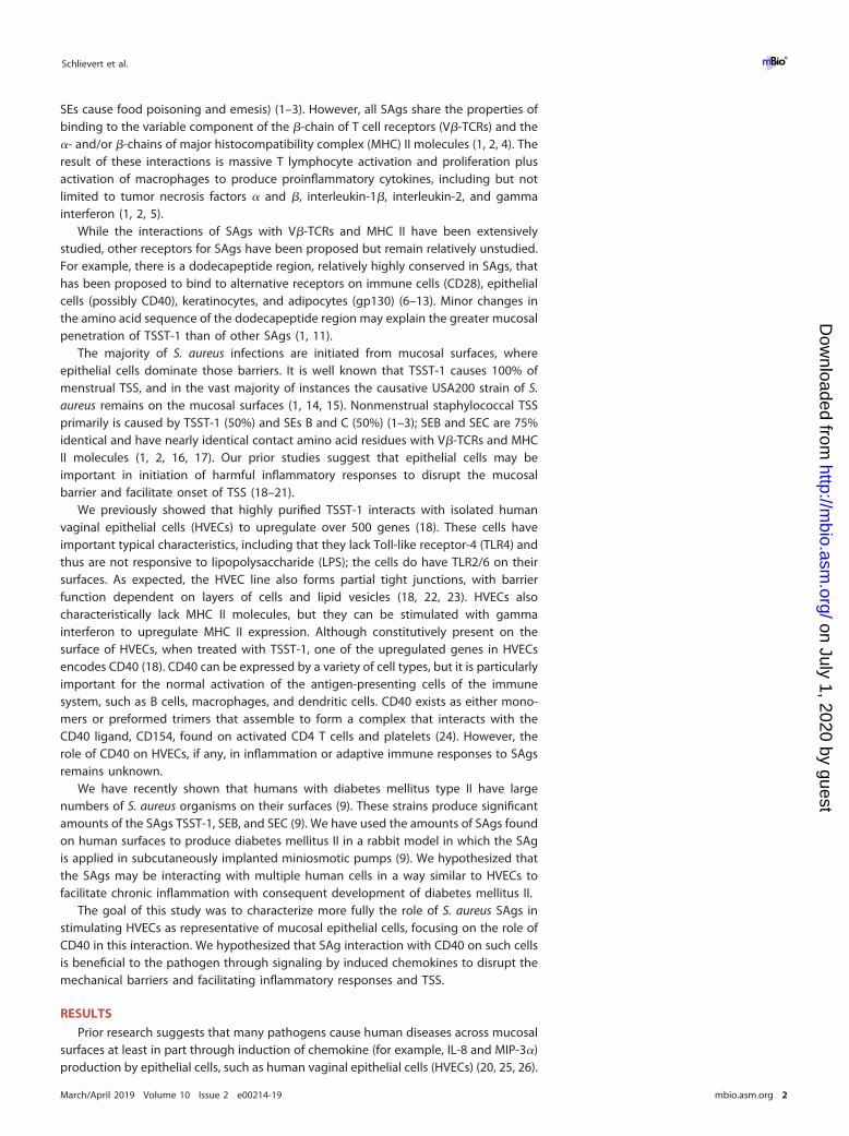
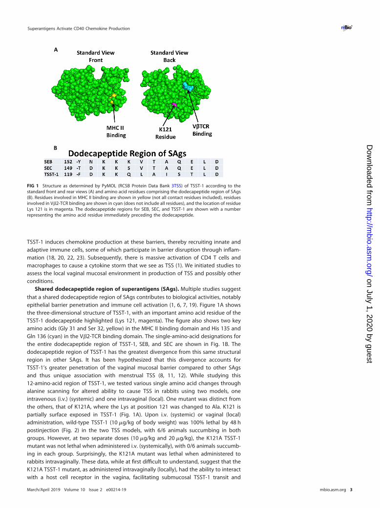
Shared dodecapeptide region of superantigens (SAgs). Multiple studies suggestthat a shared dodecapeptide region of SAgs contributes to biological activities, notablyepithelial barrier penetration and immune cell activation (1, 6, 7, 19). Figure 1A showsthe three-dimensional structure of TSST-1, with an important amino acid residue of theTSST-1 dodecapeptide highlighted (Lys 121, magenta). The figure also shows two keyamino acids (Gly 31 and Ser 32, yellow) in the MHC II binding domain and His 135 andGln 136 (cyan) in the V�2-TCR binding domain. The single-amino-acid designations forthe entire dodecapeptide region of TSST-1, SEB, and SEC are shown in Fig. 1B. Thedodecapeptide region of TSST-1 has the greatest divergence from this same structuralregion in other SAgs. It has been hypothesized that this divergence accounts forTSST-1’s greater penetration of the vaginal mucosal barrier compared to other SAgsand thus unique association with menstrual TSS (8, 11, 12). While studying this12-amino-acid region of TSST-1, we tested various single amino acid changes throughalanine scanning for altered ability to cause TSS in rabbits using two models, oneintravenous (i.v.) (systemic) and one intravaginal (local). One mutant was distinct fromthe others, that of K121A, where the Lys at position 121 was changed to Ala. K121 ispartially surface exposed in TSST-1 (Fig. 1A). Upon i.v. (systemic) or vaginal (local)administration, wild-type TSST-1 (10 �g/kg of body weight) was 100% lethal by 48 hpostinjection (Fig. 2) in the two TSS models, with 6/6 animals succumbing in bothgroups. However, at two separate doses (10 �g/kg and 20 �g/kg), the K121A TSST-1mutant was not lethal when administered i.v. (systemically), with 0/6 animals succumb-ing in each group. Surprisingly, the K121A mutant was lethal when administered torabbits intravaginally. These data, while at first difficult to understand, suggest that theK121A TSST-1 mutant, as administered intravaginally (locally), had the ability to interactwith a host cell receptor in the vagina, facilitating submucosal TSST-1 transit and
FIG 1 Structure as determined by PyMOL (RCSB Protein Data Bank 3TSS) of TSST-1 according to thestandard front and rear views (A) and amino acid residues comprising the dodecapeptide region of SAgs(B). Residues involved in MHC II binding are shown in yellow (not all contact residues included), residuesinvolved in V�2-TCR binding are shown in cyan (does not include all residues), and the location of residueLys 121 is in magenta. The dodecapeptide regions for SEB, SEC, and TSST-1 are shown with a numberrepresenting the amino acid residue immediately preceding the dodecapeptide.
Superantigens Activate CD40 Chemokine Production ®
March/April 2019 Volume 10 Issue 2 e00214-19 mbio.asm.org 3
on July 1, 2020 by guesthttp://m
bio.asm.org/
Dow
nloaded from
activation of locally resident CD4 T cells and macrophages, to result in a cytokine stormmanifested as TSS.
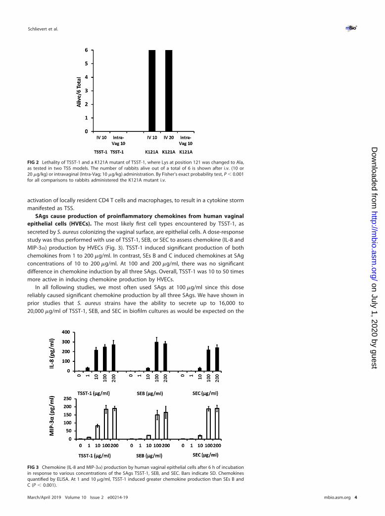
SAgs cause production of proinflammatory chemokines from human vaginalepithelial cells (HVECs). The most likely first cell types encountered by TSST-1, assecreted by S. aureus colonizing the vaginal surface, are epithelial cells. A dose-responsestudy was thus performed with use of TSST-1, SEB, or SEC to assess chemokine (IL-8 andMIP-3�) production by HVECs (Fig. 3). TSST-1 induced significant production of bothchemokines from 1 to 200 �g/ml. In contrast, SEs B and C induced chemokines at SAgconcentrations of 10 to 200 �g/ml. At 100 and 200 �g/ml, there was no significantdifference in chemokine induction by all three SAgs. Overall, TSST-1 was 10 to 50 timesmore active in inducing chemokine production by HVECs.
In all following studies, we most often used SAgs at 100 �g/ml since this dosereliably caused significant chemokine production by all three SAgs. We have shown inprior studies that S. aureus strains have the ability to secrete up to 16,000 to20,000 �g/ml of TSST-1, SEB, and SEC in biofilm cultures as would be expected on the
FIG 2 Lethality of TSST-1 and a K121A mutant of TSST-1, where Lys at position 121 was changed to Ala,as tested in two TSS models. The number of rabbits alive out of a total of 6 is shown after i.v. (10 or20 �g/kg) or intravaginal (Intra-Vag; 10 �g/kg) administration. By Fisher’s exact probability test, P � 0.001for all comparisons to rabbits administered the K121A mutant i.v.
FIG 3 Chemokine (IL-8 and MIP-3�) production by human vaginal epithelial cells after 6 h of incubationin response to various concentrations of the SAgs TSST-1, SEB, and SEC. Bars indicate SD. Chemokinesquantified by ELISA. At 1 and 10 �g/ml, TSST-1 induced greater chemokine production than SEs B andC (P � 0.001).
Schlievert et al. ®
March/April 2019 Volume 10 Issue 2 e00214-19 mbio.asm.org 4
on July 1, 2020 by guesthttp://m
bio.asm.org/
Dow
nloaded from

vaginal mucosa with tampon use (27). Thus, the 100-�g/ml dose chosen for furtherstudies was physiologic. Additionally, for the majority of studies we assumed that SEBand SEC would function similarly because of their highly shared structures; we thusused TSST-1 and SEB for subsequent assays.
SAgs plus antibodies against CD40 show enhanced chemokine production. Ourprior human microarray studies demonstrate that, among other molecules, expressionof CD40 is upregulated in response to TSST-1 challenge. TSST-1 binds to CD40 with aKd of 2.7 � 10�6 M (28). We were previously unable to demonstrate SEB or SEC bindingto CD40, possibly because of low or no affinity of these two SAgs for the molecule. Ourstudies showed that nonstimulated HVECs have CD40 on their surfaces (Fig. 4A).
We thus tested whether or not antibodies against CD40 would block SAg stimula-tion of HVECs to produce chemokines. Surprisingly, stimulation of HVECs individuallywith the two tested SAgs (TSST-1 and SEB) caused enhanced chemokine production inthe presence of the antibodies (Fig. 5 and 6) compared to SAg alone. At matched dosesof 10 �g of TSST-1 and SEB (Fig. 5), TSST-1 led to greater IL-8 production by HVECs than
FIG 4 Flow cytometry of (A) human vaginal epithelial cells (HVECs) stained with antibodies to CD40 (phycoerythrin [PE] labeled) or anisotype-matched irrelevant antibody, (B) CD40-deficient HVECs through CRISPR-Cas9 knockout stained with FITC-labeled antibodies to CD40, and(C) deficient HVECs reconstituted with CD40 on a plasmid as stained with FITC-labeled antibodies or isotype-matched irrelevant antibodies.
FIG 5 Chemokine (IL-8) production by human vaginal epithelial cells after 6-h exposure to TSST-1(10 �g), TSST-1 (10 �g plus 20 �l of antibodies [Ab] against CD40), SEB (10 �g), SEB (10 �g plus 20 �l ofantibodies against CD40), 20 �l of antibodies alone, or cells alone in keratinocyte serum-free medium(KSFM). Bars indicate SD. All responses in the presence of SAg with or without antibodies are significantlygreater than HVECs in KSFM alone or KSFM plus antibodies (P � 0.001). Additionally, the responses ofSAg alone compared to SAg plus antibodies were significantly different (P � 0.001). Finally, responses toTSST-1 with or without antibodies were greater than SEB with or without antibodies (P � 0.001).
Superantigens Activate CD40 Chemokine Production ®
March/April 2019 Volume 10 Issue 2 e00214-19 mbio.asm.org 5
on July 1, 2020 by guesthttp://m
bio.asm.org/
Dow
nloaded from
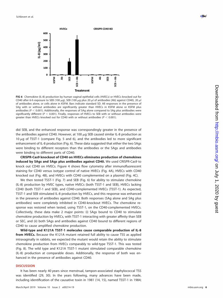
did SEB, and the enhanced response was correspondingly greater in the presence ofthe antibodies against CD40. However, at 100 �g SEB caused similar IL-8 production as10 �g of TSST-1 (compare Fig. 5 and 6), and the antibodies led to more significantenhancement of IL-8 production (Fig. 6). These data suggested that either the two SAgswere binding to different receptors than the antibodies or the SAgs and antibodieswere binding to different parts of CD40.
CRISPR-Cas9 knockout of CD40 on HVECs eliminates production of chemokinesinduced by SAgs and SAgs plus antibodies against CD40. We used CRISPR-Cas9 toknock out CD40 on HVECs. Figure 4 shows flow cytometry after immunofluorescentstaining for CD40 versus isotype control of native HVECs (Fig. 4A), HVECs with CD40knocked out (Fig. 4B), and HVECs with CD40 complemented on a plasmid (Fig. 4C).
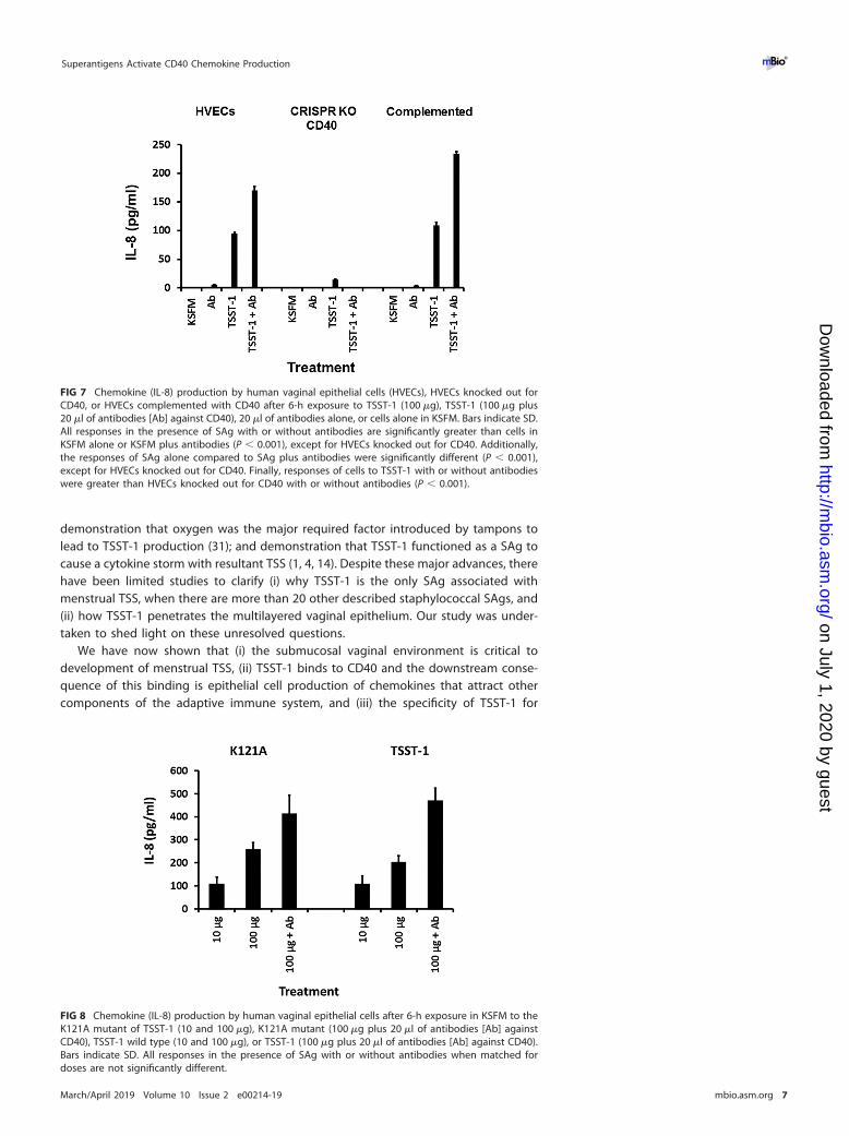
We then tested TSST-1 (Fig. 7) and SEB (Fig. 6) for ability to stimulate chemokine(IL-8) production by HVEC types, native HVECs (both TSST-1 and SEB), HVECs lackingCD40 (both TSST-1 and SEB), and CD40-complemented HVECs (TSST-1). As expected,TSST-1 and SEB stimulated IL-8 production by HVECs, and this response was enhancedin the presence of antibodies against CD40. Both responses (SAg alone and SAg plusantibodies) were completely inhibited in CD40-knockout HVECs. The chemokine re-sponse was restored when tested, using TSST-1, on the CD40-complemented HVECs.Collectively, these data make 2 major points: (i) SAgs bound to CD40 to stimulatechemokine production by HVECs, with TSST-1 interacting with greater affinity than SEBor SEC, and (ii) both SAgs and antibodies against CD40 bound to different regions ofCD40 to cause amplified chemokine production.
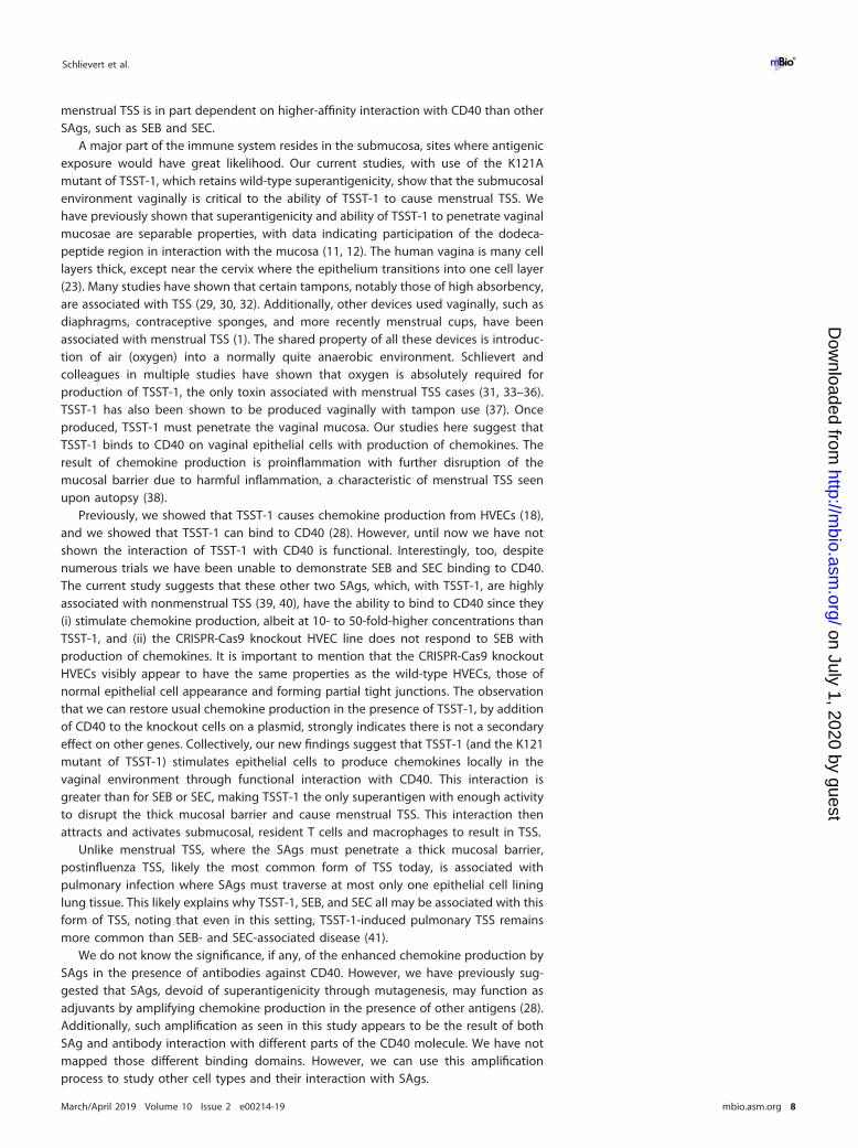
Wild-type and K121A TSST-1 molecules cause comparable production of IL-8from HVECs. Because the K121A mutant retained full ability to cause TSS as appliedintravaginally in rabbits, we expected the mutant would retain the ability to stimulatechemokine production from HVECs comparably to wild-type TSST-1. This was tested(Fig. 8). The wild type and K121A TSST-1 mutant stimulated comparable chemokine(IL-8) production at comparable doses. Additionally, the response of both was en-hanced in the presence of antibodies against CD40.
DISCUSSION
It has been nearly 40 years since menstrual, tampon-associated staphylococcal TSSwas identified (29, 30). In the years following, many advances have been made,including identification of the causative toxin in 1981 (14, 15), named TSST-1 in 1984;
FIG 6 Chemokine (IL-8) production by human vaginal epithelial cells (HVECs) or HVECs knocked out forCD40 after 6-h exposure to SEB (100 �g), SEB (100 �g plus 20 �l of antibodies [Ab] against CD40), 20 �lof antibodies alone, or cells alone in KSFM. Bars indicate standard SD. All responses in the presence ofSAg with or without antibodies are significantly greater than HVECs in KSFM alone or KSFM plusantibodies (P � 0.001). Additionally, the responses of SAg alone compared to SAg plus antibodies weresignificantly different (P � 0.001). Finally, responses of HVECs to SEB with or without antibodies weregreater than HVECs knocked out for CD40 with or without antibodies (P � 0.001).
Schlievert et al. ®
March/April 2019 Volume 10 Issue 2 e00214-19 mbio.asm.org 6
on July 1, 2020 by guesthttp://m
bio.asm.org/
Dow
nloaded from
demonstration that oxygen was the major required factor introduced by tampons tolead to TSST-1 production (31); and demonstration that TSST-1 functioned as a SAg tocause a cytokine storm with resultant TSS (1, 4, 14). Despite these major advances, therehave been limited studies to clarify (i) why TSST-1 is the only SAg associated withmenstrual TSS, when there are more than 20 other described staphylococcal SAgs, and(ii) how TSST-1 penetrates the multilayered vaginal epithelium. Our study was under-taken to shed light on these unresolved questions.
We have now shown that (i) the submucosal vaginal environment is critical todevelopment of menstrual TSS, (ii) TSST-1 binds to CD40 and the downstream conse-quence of this binding is epithelial cell production of chemokines that attract othercomponents of the adaptive immune system, and (iii) the specificity of TSST-1 for
FIG 7 Chemokine (IL-8) production by human vaginal epithelial cells (HVECs), HVECs knocked out forCD40, or HVECs complemented with CD40 after 6-h exposure to TSST-1 (100 �g), TSST-1 (100 �g plus20 �l of antibodies [Ab] against CD40), 20 �l of antibodies alone, or cells alone in KSFM. Bars indicate SD.All responses in the presence of SAg with or without antibodies are significantly greater than cells inKSFM alone or KSFM plus antibodies (P � 0.001), except for HVECs knocked out for CD40. Additionally,the responses of SAg alone compared to SAg plus antibodies were significantly different (P � 0.001),except for HVECs knocked out for CD40. Finally, responses of cells to TSST-1 with or without antibodieswere greater than HVECs knocked out for CD40 with or without antibodies (P � 0.001).
FIG 8 Chemokine (IL-8) production by human vaginal epithelial cells after 6-h exposure in KSFM to theK121A mutant of TSST-1 (10 and 100 �g), K121A mutant (100 �g plus 20 �l of antibodies [Ab] againstCD40), TSST-1 wild type (10 and 100 �g), or TSST-1 (100 �g plus 20 �l of antibodies [Ab] against CD40).Bars indicate SD. All responses in the presence of SAg with or without antibodies when matched fordoses are not significantly different.
Superantigens Activate CD40 Chemokine Production ®
March/April 2019 Volume 10 Issue 2 e00214-19 mbio.asm.org 7
on July 1, 2020 by guesthttp://m
bio.asm.org/
Dow
nloaded from
menstrual TSS is in part dependent on higher-affinity interaction with CD40 than otherSAgs, such as SEB and SEC.
A major part of the immune system resides in the submucosa, sites where antigenicexposure would have great likelihood. Our current studies, with use of the K121Amutant of TSST-1, which retains wild-type superantigenicity, show that the submucosalenvironment vaginally is critical to the ability of TSST-1 to cause menstrual TSS. Wehave previously shown that superantigenicity and ability of TSST-1 to penetrate vaginalmucosae are separable properties, with data indicating participation of the dodeca-peptide region in interaction with the mucosa (11, 12). The human vagina is many celllayers thick, except near the cervix where the epithelium transitions into one cell layer(23). Many studies have shown that certain tampons, notably those of high absorbency,are associated with TSS (29, 30, 32). Additionally, other devices used vaginally, such asdiaphragms, contraceptive sponges, and more recently menstrual cups, have beenassociated with menstrual TSS (1). The shared property of all these devices is introduc-tion of air (oxygen) into a normally quite anaerobic environment. Schlievert andcolleagues in multiple studies have shown that oxygen is absolutely required forproduction of TSST-1, the only toxin associated with menstrual TSS cases (31, 33–36).TSST-1 has also been shown to be produced vaginally with tampon use (37). Onceproduced, TSST-1 must penetrate the vaginal mucosa. Our studies here suggest thatTSST-1 binds to CD40 on vaginal epithelial cells with production of chemokines. Theresult of chemokine production is proinflammation with further disruption of themucosal barrier due to harmful inflammation, a characteristic of menstrual TSS seenupon autopsy (38).
Previously, we showed that TSST-1 causes chemokine production from HVECs (18),and we showed that TSST-1 can bind to CD40 (28). However, until now we have notshown the interaction of TSST-1 with CD40 is functional. Interestingly, too, despitenumerous trials we have been unable to demonstrate SEB and SEC binding to CD40.The current study suggests that these other two SAgs, which, with TSST-1, are highlyassociated with nonmenstrual TSS (39, 40), have the ability to bind to CD40 since they(i) stimulate chemokine production, albeit at 10- to 50-fold-higher concentrations thanTSST-1, and (ii) the CRISPR-Cas9 knockout HVEC line does not respond to SEB withproduction of chemokines. It is important to mention that the CRISPR-Cas9 knockoutHVECs visibly appear to have the same properties as the wild-type HVECs, those ofnormal epithelial cell appearance and forming partial tight junctions. The observationthat we can restore usual chemokine production in the presence of TSST-1, by additionof CD40 to the knockout cells on a plasmid, strongly indicates there is not a secondaryeffect on other genes. Collectively, our new findings suggest that TSST-1 (and the K121mutant of TSST-1) stimulates epithelial cells to produce chemokines locally in thevaginal environment through functional interaction with CD40. This interaction isgreater than for SEB or SEC, making TSST-1 the only superantigen with enough activityto disrupt the thick mucosal barrier and cause menstrual TSS. This interaction thenattracts and activates submucosal, resident T cells and macrophages to result in TSS.
Unlike menstrual TSS, where the SAgs must penetrate a thick mucosal barrier,postinfluenza TSS, likely the most common form of TSS today, is associated withpulmonary infection where SAgs must traverse at most only one epithelial cell lininglung tissue. This likely explains why TSST-1, SEB, and SEC all may be associated with thisform of TSS, noting that even in this setting, TSST-1-induced pulmonary TSS remainsmore common than SEB- and SEC-associated disease (41).
We do not know the significance, if any, of the enhanced chemokine production bySAgs in the presence of antibodies against CD40. However, we have previously sug-gested that SAgs, devoid of superantigenicity through mutagenesis, may function asadjuvants by amplifying chemokine production in the presence of other antigens (28).Additionally, such amplification as seen in this study appears to be the result of bothSAg and antibody interaction with different parts of the CD40 molecule. We have notmapped those different binding domains. However, we can use this amplificationprocess to study other cell types and their interaction with SAgs.
Schlievert et al. ®
March/April 2019 Volume 10 Issue 2 e00214-19 mbio.asm.org 8
on July 1, 2020 by guesthttp://m
bio.asm.org/
Dow
nloaded from
MATERIALS AND METHODSSuperantigens. TSST-1 was purified from a clone after culture in RN6390 in the presence of
erythromycin. RN6390 does not produce detectable SAgs; the strain has the gene only for SE-like X butdoes not produce detectable protein. SEB was purified from S. aureus strain MNHO, and SEC was purifiedfrom strain MNDON. We could not use clones for the SAg production because we are not select agentlaboratories with permission to have plasmid clones of wild-type SEs. All SAgs were purified after cultureby 80% ethanol precipitation and thin-layer isoelectric focusing. Proteins thus purified were free ofcontaminants as established by SDS-PAGE and by assays for known exotoxins (proteases, lipase, andcytotoxins) and peptidoglycan and lipopolysaccharide. SAgs were stored lyophilized (TSST-1) or frozen(SEs B and C); the SEs were stored frozen to reduce their biohazards as powders.
Antibodies and ELISA. Monoclonal (catalog number MAB6321) and polyclonal anti-human CD40(catalog number AFG32 affinity-purified goat IgG) antibodies were purchased from R&D Systems andcould be used interchangeably. These antibodies were used undiluted at 20 �l/200-�l well of volume.Kits (IL-8 and MIP-3� Quantikine) for chemokine determination were purchased from R&D Systems,Minneapolis, MN, and used exactly as described by the manufacturer.
Cell lines. HVECs from a premenopausal woman were described previously (18). HVECs have beenobserved to function comparably to primary cell lines in expression of surface molecules and in functionin tissue culture plates (18). HVECs were cultured in keratinocyte serum-free medium (KSFM) withantibiotics until 24 h before use. At that time, the cells were changed to KSFM without antibiotics.Experiments were performed for 6 h in KSFM without antibiotics. Most experiments were performed byat least two individuals to ensure reproducibility.
CRISPR-Cas9 knockout cells. CD40 guide RNA sequences were designed using the CRISPR designtool (crispr.mit.edu) (42) maintained by Feng Zhang (MIT, Cambridge, MA). Two double-stranded guidesequences were generated using the following pairs of oligonucleotides (IDT, Coralville, IA): hCD40 ex1A,CAC CGA GGC AGA CGA ACC ATA GCG and AAA CCG CTA TGG TTC GTC TGC CTC; hCD40 ex1B, CAC CGCCTC TGC AGT GCG TCC TCT and AAA CAG AGG ACG CAC TGC AGA GGC.
The hCD40 ex1A and ex1B oligonucleotides were phosphorylated with T4 polynucleotide kinase andannealed as described previously (43). Phosphorylated double-stranded oligonucleotides were ligated(Quick ligase; NEB) into pX330 Addgene plasmid ID 42230 (44) cut with BbsI and treated with calfintestinal phosphatase. The ligated DNA was used to transform DH5� Escherichia coli (Invitrogen).Plasmids were purified from individual bacterial colonies and sequenced (Iowa Institute of HumanGenetics, Genomics Division) to verify proper insertion of the guide sequences. HVECs (4 � 106) werewashed and resuspended in 400 �l Opti-MEM (Invitrogen) containing 2.5 �g hCD40 ex1A plasmid, 2.5 �ghCD40 ex1B plasmid, 0.5 �g pEGFP-C1 (Clontech), and 5 �g synthetic double-stranded oligonucleotide(62 bp, random sequence). Cells were transferred to 4-mm cuvettes for electroporation (200 V, 30 ms; BTXelectroporator). Five days after electroporation, cells were stained with anti-human CD40 antibody(G28-5) and an appropriate IPTG-labeled secondary antibody. CD40-negative, GFP-expressing cells weresorted into 96-well plates (1 to 5 cells per well) with a FACSAria Fusion flow cytometer (Becton,Dickinson). Clones were expanded and evaluated for CD40 expression by flow cytometry. As a control forvarious experiments, one of the CD40-deficient HVEC clones was stably transfected (see electroporationconditions, above) with a plasmid encoding hCD40 (45). The plasmid was modified to contain apuromycin resistance cassette in place of the original neomycin cassette. Transfectants were selectedwith puromycin (2 �g/ml) in bulk culture. Flow cytometry was used to verify CD40 expression by theresulting line.
Rabbit assays. TSST-1 and the K121A mutant of TSST-1 were tested in vivo using two rabbit models.These models test the ability of SAgs to synergize with lipopolysaccharide (LPS) up to 106-fold, throughacceleration of cytokine release; the animals succumb within 48 h whether the superantigen is admin-istered intravenously (i.v.; systemic) or intravaginally (locally) (12, 46). SAgs, dissolved in phosphate-buffered saline (0.005 M sodium phosphate, pH 7.2, 0.15 M NaCl), were given to young adult AmericanDutch-belted rabbits (1.0 to 2.0 kg) i.v. (10 to 20 �g/ml) or intravaginally (10 �g/kg). Toxins given i.v. wereadministered in the marginal ear veins. Intravaginal administrations of SAgs were made throughcatheters threaded into the vaginas of rabbits after anesthesia with ketamine (25 mg/kg; PhoenixPharmaceuticals, Inc., St. Joseph, MO) and xylazine (20 mg/kg; Phoenix Pharmaceuticals, Inc.). Toxinswere delivered intravaginally in 0.1-ml volumes. For all conditions, LPS (5 �g/kg) isolated from Salmonellaenterica serovar Typhimurium was administered i.v. in the marginal ear veins 4 h after the initialsuperantigen dose, and the rabbits were monitored for 48 h. In agreement with the University ofMinnesota IACUC, rabbits that failed to exhibit escape behavior and could not remain upright wereconsidered to have lethal TSS and were euthanized with Beuthanasia D (1 ml/kg; Schering-Plough AnimalHealth Corp., Union, NJ). We followed an approved University of Minnesota IACUC protocol, in agreementwith these requirements.
Statistics. Data are reported as means � standard deviations. For comparison of animal survival,Fisher’s exact probability test was used. For other studies, Student’s t test analysis of unpaired evenlydistributed data was used to evaluate differences in means. In all studied data, P � 0.05 was consideredsignificant.
ACKNOWLEDGMENTSSome data presented here were obtained at the Flow Cytometry Facility, which is a
Carver College of Medicine/Holden Comprehensive Cancer Center core research facilityat the University of Iowa. The Facility is funded through user fees and the generous
Superantigens Activate CD40 Chemokine Production ®
March/April 2019 Volume 10 Issue 2 e00214-19 mbio.asm.org 9
on July 1, 2020 by guesthttp://m
bio.asm.org/
Dow
nloaded from
financial support of the Carver College of Medicine, Holden Comprehensive CancerCenter, and Iowa City Veteran’s Administration Medical Center, and by the NationalCenter for Research Resources of the National Institutes of Health under award number1 S10 OD016199-01A. This research was funded by HHS National Institutes of HealthU19 AI117673 (NIAID), NIAMS AR41256, AI123107, and AI119163. This research alsoreceived support by facilities and equipment provided by the Department of VeteransAffairs, Veterans Health Administration, Office of Research and Development; G.A.B. isa Senior Research Career Scientist of the VAMC.
REFERENCES1. Spaulding AR, Salgado-Pabón W, Kohler PL, Horswill AR, Leung DY,
Schlievert PM. 2013. Staphylococcal and streptococcal superantigenexotoxins. Clin Microbiol Rev 26:422– 447. https://doi.org/10.1128/CMR.00104-12.
2. McCormick JK, Yarwood JM, Schlievert PM. 2001. Toxic shock syndromeand bacterial superantigens: an update. Annu Rev Microbiol 55:77–104.https://doi.org/10.1146/annurev.micro.55.1.77.
3. Dinges MM, Orwin PM, Schlievert PM. 2000. Exotoxins of Staphylococcusaureus. Clin Microbiol Rev 13:16 –34. https://doi.org/10.1128/CMR.13.1.16.
4. Marrack P, Kappler J. 1990. The staphylococcal enterotoxins and theirrelatives. Science 248:705–711. https://doi.org/10.1126/science.2185544.
5. Kotzin BL, Leung DY, Kappler J, Marrack P. 1993. Superantigens and theirpotential role in human disease. Adv Immunol 54:99 –166. https://doi.org/10.1016/S0065-2776(08)60534-9.
6. Arad G, Levy R, Hillman D, Kaempfer R. 2000. Superantigen antagonistprotects against lethal shock and defines a new domain for T-cellactivation. Nat Med 6:414 – 421. https://doi.org/10.1038/74672.
7. Arad G, Levy R, Nasie I, Hillman D, Rotfogel Z, Barash U, Supper E, ShpilkaT, Minis A, Kaempfer R. 2011. Binding of superantigen toxins into theCD28 homodimer interface is essential for induction of cytokine genesthat mediate lethal shock. PLoS Biol 9:e1001149. https://doi.org/10.1371/journal.pbio.1001149.
8. Hamad AR, Marrack P, Kappler JW. 1997. Transcytosis of staphylococcalsuperantigen toxins. J Exp Med 185:1447–1454. https://doi.org/10.1084/jem.185.8.1447.
9. Vu BG, Stach CS, Kulhankova K, Salgado-Pabón W, Klingelhutz AJ,Schlievert PM. 2015. Chronic superantigen exposure induces systemicinflammation, elevated bloodstream endotoxin, and abnormal glucosetolerance in rabbits: possible role in diabetes. mBio 6:e02554-14. https://doi.org/10.1128/mBio.02554-14.
10. Vu BG, Gourronc FA, Bernlohr DA, Schlievert PM, Klingelhutz AJ. 2013.Staphylococcal superantigens stimulate immortalized human adi-pocytes to produce chemokines. PLoS One 8:e77988. https://doi.org/10.1371/journal.pone.0077988.
11. Shupp JW, Jett M, Pontzer CH. 2002. Identification of a transcytosisepitope on staphylococcal enterotoxins. Infect Immun 70:2178 –2186.https://doi.org/10.1128/IAI.70.4.2178-2186.2002.
12. Schlievert PM, Jablonski LM, Roggiani M, Sadler I, Callantine S, MitchellDT, Ohlendorf DH, Bohach GA. 2000. Pyrogenic toxin superantigen sitespecificity in toxic shock syndrome and food poisoning in animals. InfectImmun 68:3630 –3634. https://doi.org/10.1128/IAI.68.6.3630-3634.2000.
13. Kushnaryov VM, MacDonald HS, Reiser R, Bergdoll MS. 1984. Staphylo-coccal toxic shock toxin specifically binds to cultured human epithelialcells and is rapidly internalized. Infect Immun 45:566 –571.
14. Schlievert PM, Shands KN, Dan BB, Schmid GP, Nishimura RD. 1981.Identification and characterization of an exotoxin from Staphylococcusaureus associated with toxic-shock syndrome. J Infect Dis 143:509 –516.https://doi.org/10.1093/infdis/143.4.509.
15. Bergdoll MS, Crass BA, Reiser RF, Robbins RN, Davis JP. 1981. A newstaphylococcal enterotoxin, enterotoxin F, associated with toxic-shock-syndrome Staphylococcus aureus isolates. Lancet i:1017–1021. https://doi.org/10.1016/S0140-6736(81)92186-3.
16. Li H, Llera A, Tsuchiya D, Leder L, Ysern X, Schlievert PM, Karjalainen K,Mariuzza RA. 1998. Three-dimensional structure of the complex betweena T cell receptor beta chain and the superantigen staphylococcal en-terotoxin B. Immunity 9:807– 816. https://doi.org/10.1016/S1074-7613(00)80646-9.
17. Fields BA, Malchiodi EL, Li H, Ysern X, Stauffacher CV, Schlievert PM,
Karjalainen K, Mariuzza RA. 1996. Crystal structure of a T-cell receptorbeta-chain complexed with a superantigen. Nature 384:188 –192.https://doi.org/10.1038/384188a0.
18. Peterson ML, Ault K, Kremer MJ, Klingelhutz AJ, Davis CC, Squier CA,Schlievert PM. 2005. The innate immune system is activated by stimu-lation of vaginal epithelial cells with Staphylococcus aureus and toxicshock syndrome toxin 1. Infect Immun 73:2164 –2174. https://doi.org/10.1128/IAI.73.4.2164-2174.2005.
19. Brosnahan AJ, Schaefers MM, Amundson WH, Mantz MJ, Squier CA,Peterson ML, Schlievert PM. 2008. Novel toxic shock syndrome toxin-1amino acids required for biological activity. Biochemistry 47:12995–13003. https://doi.org/10.1021/bi801468w.
20. Brosnahan AJ, Schlievert PM. 2011. Gram-positive bacterial superantigenoutside-in signaling causes toxic shock syndrome. FEBS J 278:4649 – 4667. https://doi.org/10.1111/j.1742-4658.2011.08151.x.
21. Brosnahan AJ, Mantz MJ, Squier CA, Peterson ML, Schlievert PM. 2009.Cytolysins augment superantigen penetration of stratified mucosa. JImmunol 182:2364 –2373. https://doi.org/10.4049/jimmunol.0803283.
22. Davis CC, Kremer MJ, Schlievert PM, Squier CA. 2003. Penetration oftoxic shock syndrome toxin-1 across porcine vaginal mucosa ex vivo:permeability characteristics, toxin distribution, and tissue damage.Am J Obstet Gynecol 189:1785–1791. https://doi.org/10.1016/S0002-9378(03)00873-1.
23. Squier CA, Mantz MJ, Schlievert PM, Davis CC. 2008. Porcine vagina exvivo as a model for studying permeability and pathogenesis in mucosa.J Pharm Sci 97:9 –21. https://doi.org/10.1002/jps.21077.
24. Anand SX, Viles-Gonzalez JF, Badimon JJ, Cavusoglu E, Marmur JD. 2003.Membrane-associated CD40L and sCD40L in atherothrombotic disease.Thromb Haemost 90:377–384. https://doi.org/10.1160/TH03-05-0268.
25. Li Q, Estes JD, Schlievert PM, Duan L, Brosnahan AJ, Southern PJ, ReillyCS, Peterson ML, Schultz-Darken N, Brunner KG, Nephew KR, Pambuc-cian S, Lifson JD, Carlis JV, Haase AT. 2009. Glycerol monolaurate pre-vents mucosal SIV transmission. Nature 458:1034 –1038. https://doi.org/10.1038/nature07831.
26. Haase AT, Rakasz E, Schultz-Darken N, Nephew K, Weisgrau KL, Reilly CS,Li Q, Southern PJ, Rothenberger M, Peterson ML, Schlievert PM. 2015.Glycerol monolaurate microbicide protection against repeat high-doseSIV vaginal challenge. PLoS One 10:e0129465. https://doi.org/10.1371/journal.pone.0129465.
27. Schlievert PM, Peterson ML. 2012. Glycerol monolaurate antibacterialactivity in broth and biofilm cultures. PLoS One 7:e40350. https://doi.org/10.1371/journal.pone.0040350.
28. Spaulding AR, Lin YC, Merriman JA, Brosnahan AJ, Peterson ML, Schliev-ert PM. 2012. Immunity to Staphylococcus aureus secreted proteinsprotects rabbits from serious illnesses. Vaccine 30:5099 –5109. https://doi.org/10.1016/j.vaccine.2012.05.067.
29. Shands KN, Schmid GP, Dan BB, Blum D, Guidotti RJ, Hargrett NT,Anderson RL, Hill DL, Broome CV, Band JD, Fraser DW. 1980. Toxic-shocksyndrome in menstruating women: association with tampon use. N EnglJ Med 303:1436 –1442. https://doi.org/10.1056/NEJM198012183032502.
30. Davis JP, Chesney PJ, Wand PJ, LaVenture M. 1980. Toxic-shocksyndrome: epidemiologic features, recurrence, risk factors, and preven-tion. N Engl J Med 303:1429 –1435. https://doi.org/10.1056/NEJM198012183032501.
31. Schlievert PM, Blomster DA. 1983. Production of staphylococcal pyro-genic exotoxin type C: influence of physical and chemical factors. J InfectDis 147:236 –242. https://doi.org/10.1093/infdis/147.2.236.
32. Osterholm MT, Davis JP, Gibson RW, Mandel JS, Wintermeyer LA, HelmsCM, Forfang JC, Rondeau J, Vergeront JM. 1982. Tri-state toxic-state
Schlievert et al. ®
March/April 2019 Volume 10 Issue 2 e00214-19 mbio.asm.org 10
on July 1, 2020 by guesthttp://m
bio.asm.org/
Dow
nloaded from
syndrome study. I. Epidemiologic findings. J Infect Dis 145:431– 440.https://doi.org/10.1093/infdis/145.4.431.
33. Pragman AA, Yarwood JM, Tripp TJ, Schlievert PM. 2004. Characteriza-tion of virulence factor regulation by SrrAB, a two-component system inStaphylococcus aureus. J Bacteriol 186:2430 –2438. https://doi.org/10.1128/JB.186.8.2430-2438.2004.
34. Yarwood JM, McCormick JK, Schlievert PM. 2001. Identification of a noveltwo-component regulatory system that acts in global regulation ofvirulence factors of Staphylococcus aureus. J Bacteriol 183:1113–1123.https://doi.org/10.1128/JB.183.4.1113-1123.2001.
35. Yarwood JM, Schlievert PM. 2000. Oxygen and carbon dioxide regulationof toxic shock syndrome toxin 1 production by Staphylococcus aureusMN8. J Clin Microbiol 38:1797–1803.
36. Hill DR, Brunner ME, Schmitz DC, Davis CC, Flood JA, Schlievert PM,Wang-Weigand SZ, Osborn TW. 2005. In vivo assessment of humanvaginal oxygen and carbon dioxide levels during and post menses. JAppl Physiol 99:1582–1591. https://doi.org/10.1152/japplphysiol.01422.2004.
37. Schlievert PM, Nemeth KA, Davis CC, Peterson ML, Jones BE. 2010.Staphylococcus aureus exotoxins are present in vivo in tampons. ClinVaccine Immunol 17:722–727. https://doi.org/10.1128/CVI.00483-09.
38. Larkin SM, Williams DN, Osterholm MT, Tofte RW, Posalaky Z. 1982. Toxicshock syndrome: clinical, laboratory, and pathologic findings in ninefatal cases. Ann Intern Med 96:858 – 864. https://doi.org/10.7326/0003-4819-96-6-858.
39. Schlievert PM, Tripp TJ, Peterson ML. 2004. Reemergence of staphylo-coccal toxic shock syndrome in Minneapolis-St. Paul, Minnesota, during
the 2000 –2003 surveillance period. J Clin Microbiol 42:2875–2876.https://doi.org/10.1128/JCM.42.6.2875-2876.2004.
40. Schlievert PM, Kim MH. 1991. Reporting of toxic shock syndrome Staph-ylococcus aureus in 1982 to 1990. J Infect Dis 164:1245–1246. https://doi.org/10.1093/infdis/164.6.1245.
41. MacDonald KL, Osterholm MT, Hedberg CW, Schrock CG, PetersonGF, Jentzen JM, Leonard SA, Schlievert PM. 1987. Toxic shock syn-drome. A newly recognized complication of influenza and influen-zalike illness. JAMA 257:1053–1058. https://doi.org/10.1001/jama.1987.03390080043027.
42. Hsu PD, Scott DA, Weinstein JA, Ran FA, Konermann S, Agarwala V, Li Y,Fine EJ, Wu X, Shalem O, Cradick TJ, Marraffini LA, Bao G, Zhang F. 2013.DNA targeting specificity of RNA-guided Cas9 nucleases. Nat Biotechnol31:827– 832. https://doi.org/10.1038/nbt.2647.
43. Bauer DE, Canver MC, Orkin SH. 2015. Generation of genomic deletionsin mammalian cell lines via CRISPR/Cas9. J Vis Exp (95):e52118. https://doi.org/10.3791/52118.
44. Ran FA, Hsu PD, Wright J, Agarwala V, Scott DA, Zhang F. 2013. Genomeengineering using the CRISPR-Cas9 system. Nat Protoc 8:2281–2308.https://doi.org/10.1038/nprot.2013.143.
45. Hostager BS, Hsing Y, Harms DE, Bishop GA. 1996. Different CD40-mediated signaling events require distinct CD40 structural features. JImmunol 157:1047–1053.
46. Schlievert PM. 1982. Enhancement of host susceptibility to lethal endo-toxin shock by staphylococcal pyrogenic exotoxin type C. Infect Immun36:123–128.
Superantigens Activate CD40 Chemokine Production ®
March/April 2019 Volume 10 Issue 2 e00214-19 mbio.asm.org 11
on July 1, 2020 by guesthttp://m
bio.asm.org/
Dow
nloaded from