Embed Size (px)
Citation preview
INFECTION AND IMMUNITY, Mar. 1992, P. 922-9270019-9567/92/030922-06$02.00/0Copyright C) 1992, American Society for Microbiology
Staphylococcal Exopolysaccharides Inhibit LymphocyteProliferative Responses by Activation of Monocyte
Prostaglandin ProductionROBERT D. STOUT,* KAETHE P. FERGUSON, YI-NING LI, AND DWIGHT W. LAMBE, JR.
Department of Microbiology, James H. Quillen College of Medicine at East TennesseeState University, P.O. Box 70579, Johnson City, Tennessee 37614-0579
Received 16 October 1991/Accepted 13 December 1991
The glycocalyx (exopolysaccharides) of Staphylococcus epidermidis has been reported to inhibit a variety ofhost defense mechanisms. We have examined the inhibitory effects of glycocalyx on the proliferation of humanperipheral blood mononuclear cells (PBMC) and the mechanism of this inhibition. Glycocalyx isolated andpartially purified under endotoxin-free conditions from defined liquid medium cultures of S. epidermidis andStaphylococcus lugdunensis inhibited the proliferative response of PBMC when added to cultures at 10 to 100,ug/ml. Glycocalyx-mediated inhibition of phytohemagglutinin-stimulated proliferation of PBMC required thepresence of plastic-adherent peripheral blood monocytes. Culture supernatants of monocytes stimulated withglycocalyx contained a soluble factor that inhibited the proliferation of monocyte-depleted PBMC. This solubleinhibitory factor was not produced in the absence of glycocalyx or in the presence of both glycocalyx andindomethacin. Analysis of the supernatants of cultures of adherent monocytes revealed that glycocalyx from S.epidermidis and from S. lugdunensis could activate monocyte production of prostaglandin E2 (PGE2), humaninterleukin-1, and tumor necrosis factor alpha. The addition of purified PGE2, at the same levels of PGE2( 10-9 M) generated in the monocyte cultures, to PBMC cultures resulted in a similar inhibition ofproliferative responses. It is concluded that, contrary to previous suggestions, the bacterial glycocalyx does nothave a direct inhibitory effect on T lymphocytes. However, it does appear that glycocalyx from coagulase-negative staphylococci can activate monocyte PGE2 production and that it is this activity that in turncontributes to the inhibition of T-cell proliferation.
The bacterial glycocalyx may be found attached to thebacterial cell, forming the capsule, or detached from the cell,forming the bacterial slime (14, 20, 31, 36). Bacteria adhereto surfaces and form biofilms composed of bacterial cells andglycocalyx (11, 17, 31). This microcolonial form of growthhas been observed in bacterial diseases including cysticfibrosis (26), prosthesis-related colonization (11, 23, 24) andinfection (27), experimental animal osteomyelitis (19, 20, 28)and endocarditis (2), and human osteomyelitis (20, 25). It hasbeen suggested by several research groups that the produc-tion of glycocalyx may be a major factor in the pathogenesisof foreign body infections (6, 15, 18, 25, 30). Glycocalyxproduction can protect bacteria from surfactants (12), anti-bodies (3), antibiotics (5), and phagocytosis (10, 16, 34, 39).Glycocalyx has been reported to inhibit normal phagocytefunctions, such as movement along chemotactic gradientsand oxidative burst, as well as particle engulfment (16, 30,34, 39).
Recently, another attribute of bacterial glycocalyx thatmay be relevant to its role in pathogenesis of infectiousdiseases has been suggested by the observation that theglycocalyx from coagulase-negative staphylococci can in-hibit the proliferation of human peripheral blood mononu-
clear cells (PBMC) stimulated with the T-cell mitogen phy-tohemagglutinin (PHA) (13, 30). In the present study we
examined the mechanism of this inhibition to determinewhether the inhibitory effect was a result of the direct actionof the glycocalyx on T lymphocytes or whether the inhibition
* Corresponding author.
922
was mediated through another cell type, such as peripheralblood monocytes.
MATERIALS AND METHODS
Reagents. Prostaglandin E2 (PGE2) and a kit for radioim-munoassay for PGE2 were obtained from Advanced Magnet-ics, Inc. (Cambridge, Mass.). E-Toxate Limulus assay kitsfor the detection of bacterial endotoxin were obtained fromSigma Chemical Co. (St. Louis, Mo.). Enzyme-linked im-munosorbent assay (ELISA) kits for the detection of humaninterleukin-1 (IL-113) and tumor necrosis factor alpha(TNF-ox) were obtained from Cistron Biotechnology (PineBrook, N.J.) and Endogen, Inc. (Boston, Mass.), respec-tively. Lipopolysaccharide (LPS; from Escherichia coli O11:B4-W) and indomethacin were obtained from Sigma. A 10-2M solution of indomethacin in ethanol was prepared on theday of use and was diluted to the desired concentration inculture medium. The M1/70 hybridoma-producing anti-CD11a antibody was obtained from the American TypeCulture Collection (Rockville, Md.). Anti-CD2 (fluoresceinisothiocyanate conjugate) was obtained from Coulter Immu-nology (Hialeah, Fla.).
Preparation of bacterial glycocalyx. The Staphylococcusstrains used in this study included S. epidermidis G-19-85,isolated from a cesarian section wound (East TennesseeState University, Johnson City), and S. lugdunensis G-6-87(strain N850412 from the blood of a patient with septicemia)and G2-89 (strain CRS 307 from the blood of a patient withinfective endocarditis), isolated by J. Fleurette (Laboratoirede Bacteriologie-Virologie, Facult6 de Medicine Alexis Car-rel, Lyon, France). The glycocalyx was isolated from bac-
Vol. 60, No. 3
Dow
nloa
ded
from
http
s://j
ourn
als.
asm
.org
/jour
nal/i
ai o
n 28
Dec
embe
r 20
21 b
y 21
8.39
.95.
103.
STAPHYLOCOCCAL GLYCOCALYX ACTIVATES MONOCYTES 923
TABLE 1. Characterization of bacterial glycocalyx preparations
Amt (per 100 p.g of glycocalyx) of:
Glycocalyx prep Organism Endo- Carbo- Protein Phosphatetoxin hydrate G g) (nmol)(ng) (p.g)
G-19-85 S. epidermidis <0.1 18 64 94G-6-87 S. lugdunensis <0.1 8.3 55 67G-2-89 S. lugdunensis <0.1 5.0 66 151G-2-89 (Westphal S. Iugdunensis <0.2 9.5 14 200
extracted)
teria grown in an endotoxin-free chemically defined medium.The use of defined medium is the method of choice overpreviously published methods (12), since it yields a more
consistent product without the impurities that may be de-rived from nutrient agar (14). A chemically defined mediumdeveloped previously for glycocalyx production in Bacteroi-des spp. (17) was modified for growth of staphylococci as
follows. NaHCO3 and hemin were omitted. Individual aminoacids were used at the following final amounts per liter:glutamic acid, phenylalanine, leucine, isoleucine, valine,proline, aspartic acid, glycine, lysine, arginine, asparagine,and glutamine, 1.0 mg; histidine and serine, 0.5 mg; alanineand methionine, 0.25 mg; threonine, 2.0 mg; and cysteine,5.0 mg. These concentrations were used by van de Rijn andKessler (38) and for the isolation of S. epidermidis glycoca-lyx (31).
Endotoxin-free glass-distilled water and glassware were
used throughout the preparation and isolation procedures.Bacteria were grown for 18 h at 37°C in chemically definedmedium and then centrifuged at 33,000 x g for 10 min.Supernatant fluid was centrifuged at 3,000 x g and reducedto one-third volume by using a rotary evaporator. Theglycocalyx was then dialyzed (cellulose acetate dialysistubing; molecular weight cutoff, 12,000 to 14,000) againstdistilled water for 5 days, centrifuged at 3,000 x g for 20 min,and filtered through a 0.22-pum-pore-size filter. The resultingpreparation was frozen and lyophilized.The results of the biochemical analysis of the glycocalyx
preparations are displayed in Table 1. The total carbohy-drate content was determined by the phenol-sulfuric acidassay of Dubois et al. (9). Total protein was estimated by themethod of Lowry et al. with bovine serum albumin as thestandard (22). Total phosphate was measured by the methodof Ames with KH2PO4 as the standard (1). Endotoxin was
estimated by using E-Toxate Limulus kits. Depletion ofprotein was accomplished by hot phenol extraction (41).Briefly, an equal volume of hot (70°C) phenol was added toglycocalyx suspended in distilled water at an approximateconcentration of 4 mg/ml. The mixture was incubated withstirring for 60 min at 70°C, and the phenol and aqueous
layers were separated by centrifugation at 20,000 x g for 20min. An equal volume of water was added to the phenollayer, and incubation and centrifugation were repeated. Theaqueous layers were combined, and residual phenol was
removed by exhaustive dialysis against distilled water. Theresulting preparation was frozen and lyophilized. A proce-
dure control was prepared by dissolving starch in distilledwater and subjecting it to the same dialysis and hot phenolextraction procedures.
Cell preparation and culture. Peripheral blood from normaldonors (aged 26 to 45 years) was aseptically collected fromthe antecubital vein in a heparinized syringe (A. H. Robins,
Richmond, Va.). The PBMC were isolated by density gradi-ent centrifugation on Lymphoprep (Accurate Chemical andScientific Co., Westbury, N.Y.) and then washed once withcold physiological saline and three times in cold Dulbeccophosphate-buffered saline (0.15 M, pH 7.3) supplementedwith 2% (vol/vol) heat-inactivated (56°C for 60 min) fetalbovine serum (GIBCO, Grand Island, N.Y.) that had beenprescreened for an endotoxin level of <50 pg/mI. Themononuclear cells were then resuspended in RPMI 1640(GIBCO) supplemented with 5% (vol/vol) heat-inactivatedfetal bovine serum, 10 mM N-2-hydroxyethylpiperazine-N'-2-ethanesulfonic acid (GIBCO), 1 mM pyruvate, and 50 ,ugof gentamicin (Sigma) per ml (henceforth referred to ascomplete medium) and dispensed in 0.1-ml aliquots intoflat-bottom Micro-Test III plates (Falcon Plastics, Oxnard,Calif.) to a final concentration of 5 x 10⁵ cells/ml. PHA-M(Sigma) was then added at 1 ,ug/ml in the presence orabsence of bacterial glycocalyx. After incubation for 5 daysat 37°C in a humidified atmosphere of 5% CO2 and 95% air,the cultures were pulsed for 4 h with [3H]thymidine (36) andharvested on a MASH-I1 harvester (M. A. Bioproducts,Walkersville, Md.). The filters were washed with distilledwater, air dried, immersed in a scintillation fluid, andcounted on a Beckman LS7000 scintillation spectrometer.Monocyte depletion of PBMC. PBMC were depleted of
monocytes by dispensing a 10-ml aliquot of PBMC at 2 x 10⁶cells per ml of complete medium to a 60-mm2 culture dish(Falcon). After 2 to 4 h of incubation at 37°C, the nonadher-ent cells were gently agitated and collected. This procedurereduced the monocyte content by 5- to 10-fold from the 16 to22% monocyte content of unseparated PBMC to 2 to 5%.
Generation of monocyte supernatants. Peripheral bloodmonocytes were enriched in 96-well culture plates by incu-bating PBMC at 2 x 10⁶ cells per ml for 2 to 4 h and thenwashed three to five times to remove nonadherent cells. Theadherent cells were >90% CD11a+ and <2% CD2+ asdetermined by fluorescence microscopy after labeling withthe appropriate fluorescein isothiocyanate-conjugated mono-clonal antibody. The adherent cells were then cultured for 1or 2 days in the absence or presence of glycocalyx and/orindomethacin. Supernatants were collected, and the pres-ence of a soluble inhibitory factor was determined by addingthese supernatants (50%, vol/vol) to cultures of PHA-stimu-lated, nonadherent PBMC. The proliferation of these cul-tures was determined by measuring [3H]thymidine incorpo-ration as described above. Samples of these supernatantswere also subjected to the radioimmunoassay for PGE2 andto the ELISA for IL-11 and TNF-a. Bioassays for IL-1 (21)and TNF-a- (8) were run to corroborate the ELISA results.Briefly, IL-1-dependent D10.G4 cells were cultured at 2 x10⁴ cells in 200 ,ul with 2 jig of concanavalin A per ml andthreefold serial dilutions of the test supernatant. Prolifera-tion was assessed by measuring [3H]thymidine incorporationafter 3 days of culture (21). One unit of IL-1 was defined asthe amount supporting half-maximal proliferation of D10.G4in these cultures (21). For the TNF-a bioassay, L929 fibro-blasts were cultured overnight as previously described (8)with supernatants diluted 5- to 100-fold. The plates werewashed, stained with crystal violet, and read on a VMAXautomated plate reader at 558 nm (Molecular Devices,Menlo Park, Calif.). A standard curve generated by titrationof 0.05 to 10 U of recombinant TNF-a (Genzyme Corp.,Cambridge, Mass.) per ml was run in parallel for calculationof the activity in the supernatants. Anti-TNF-ot antibody(Genzyme) was added to replicates of the supernatants toconfirm that cytotoxicity was caused by TNF-ot.
VOL. 60, 1992
Dow
nloa
ded
from
http
s://j
ourn
als.
asm
.org
/jour
nal/i
ai o
n 28
Dec
embe
r 20
21 b
y 21
8.39
.95.
103.
924 STOUT ET AL.
Z _ G6-870 G2-89
M G2(Westphal)F 601 ControlLLJ
0~IL. 40-0z0
M 20-
z
010 Atg/m1 30 A.g/ml 100 p0g/Ml
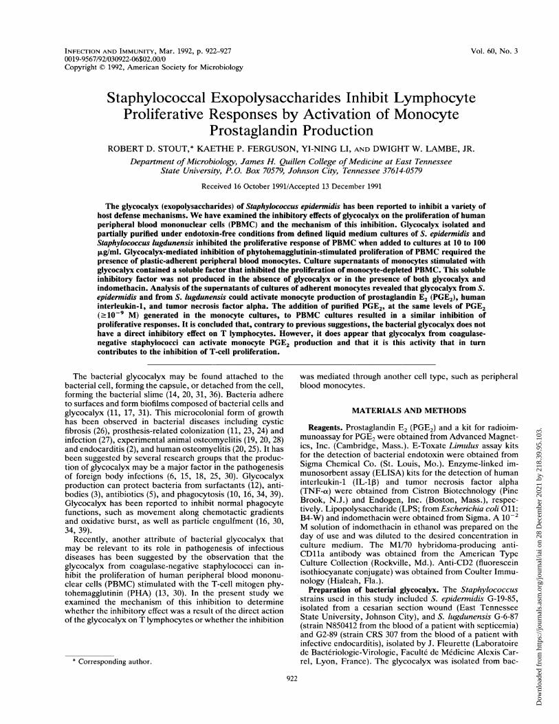
FIG. 1. Inhibition of lymphocyte proliferative response by gly-cocalyx. PBMC were stimulated with PHA (1 ,ug/ml) in the absenceor in the presence of 10 to 100 ,ug of the indicated glycocalyxpreparation per ml. The control is a solution of starch subjected tothe same hot phenol extraction and processing as G2 (Westphal).Proliferation was determined by the uptake of tritiated thymidineafter 5 days of culture. The percent inhibition is based on theproliferative response of PBMC cultured with PHA in the absence ofglycocalyx (49,745 ± 3,160 cpm). The background proliferation ofPBMC cultured without stimulus was 816 ± 168 cpm. The averageresults of triplicate cultures (± standard deviations) in a repre-sentative experiment are displayed. Similar results were obtained in5 to 10 trials with each preparation.
RESULTS
The addition of Staphylococcus glycocalyx to cultures ofPBMC inhibited the lymphocyte proliferative response toPHA (Fig. 1). In agreement with the data reported by Grayet al. (13), the inhibition was apparent 3 days after cultureinitiation and increased to 50 to 70% by the peak of theproliferative response (day 5). Proliferation of glycocalyx-inhibited PBMC cultures had decreased to background lev-els (<2,000 cpm) 7 days after culture initiation. This inhibi-tory effect was observed in >20 trials on PBMC collectedfrom six different donors. This effect was observed withthree or more different preparations of glycocalyx from eachof three different staphylococcal isolates (three are displayedin Fig. 1). None of the preparations effected an inhibition ofof the proliferative response that was greater than 70%. Hotphenol (Westphal) extraction of the G2-89 glycocalyx re-sulted in a fivefold reduction in protein content (Table 1) butdid not reduce the inhibitory activity of the preparation (Fig.1).PBMC were depleted of adherent cells by incubation on
plastic culture dishes as a first step in lymphocyte purifica-tion to address the question of whether the inhibitory activ-ity of glycocalyx resulted from a direct effect on the Tlymphocytes. However, upon testing for responsiveness toPHA and glycocalyx, it was found that these plastic-nonad-herent cells were not sensitive to the inhibitory effects ofglycocalyx (Table 2). The plastic-adherent monocy.cs,which were >90% CD11a+ and <2% CD2+, were thereforetested for their responsiveness to glycocalyx. Supernatantsof monocytes cultured in medium alone, with 1 ,ug of LPSper ml, or with 100 jg of the glycocalyx preparations per mlwere collected and assayed for PGE2 by the radioimmuno-assay and for IL1-,B and TNF-ao by the ELISA. All three
TABLE 2. Inability of glycocalyx to inhibit proliferation ofmonocyte-depleted PBMC"
[33Hjthymidine incorporation by PBMCGlycocalyx (cpm ± SD)
Unseparated Nonadherent
None 15,488 + 1,922 10,524 + 1,339G-19-85 9,871 - 1,139 12,767 + 2,523G-2-89 6,063 + 1,202 12,503 ± 999G-6-87 4,923 ± 197 11,351 + 1,787a PBMC with a normal monocyte component (unseparated PBMC) or
PBMC depleted of plastic adherent monocytes (nonadherent PBMC) werecultured with PHA plus 100 p.g of the indicated glycocalyx preparation per mlfor 5 days. Averages of thymidine incorporation of triplicate cultures aredisplayed. Values for background incorporation by PBMC cultured withoutPHA were 887 ± 122 and 662 + 112 cpm for unseparated PBMC andnonadherent PBMC, respectively.
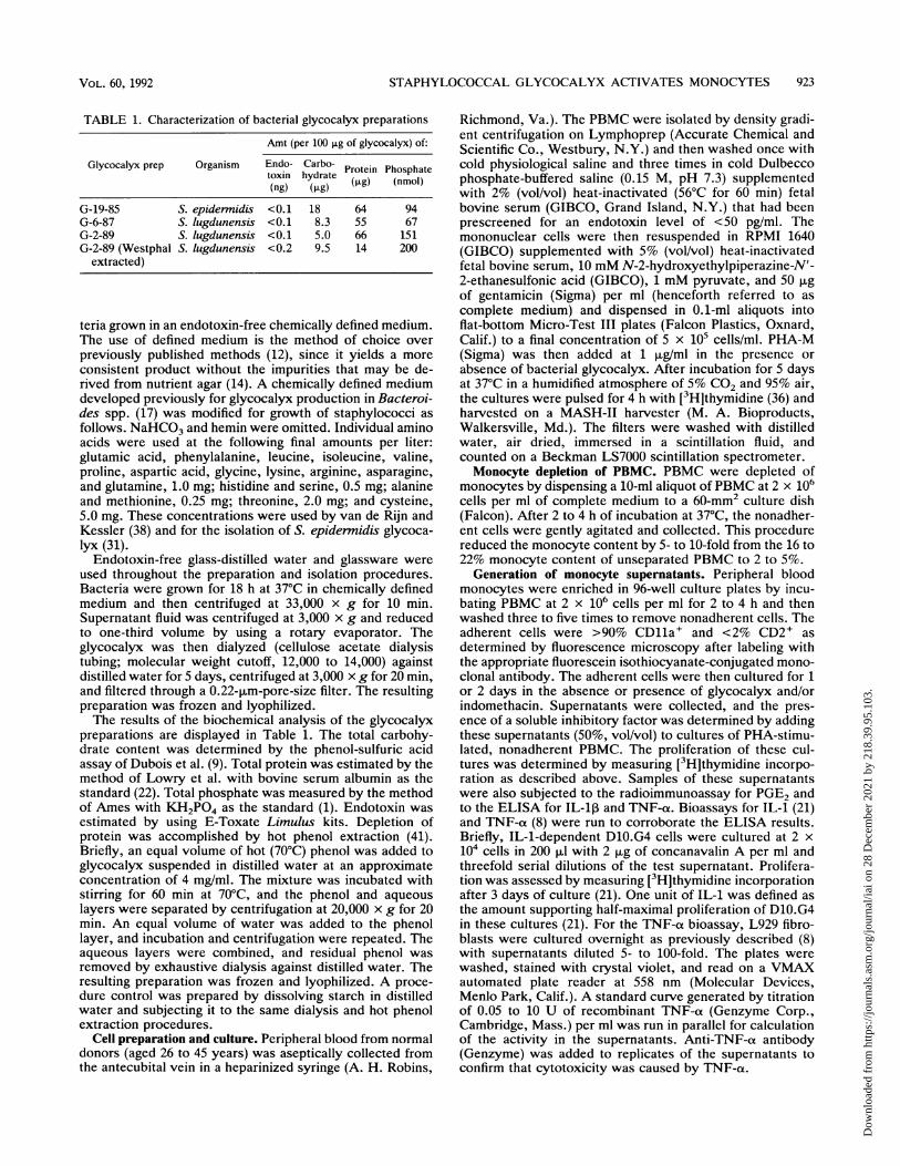
glycocalyx preparations activated monocytes to producesignificant (P < 0.01) amounts of PGE2, IL-1p, and TNF-ot,although the response was 5- to 10-fold lower than themonocyte response to E. coli LPS (Fig. 2). Similar resultswere obtained with the D10.G4 proliferation bioassay forIL-1 (21) and the L929 cytotoxicity assay for TNF-al (8). LPSstimulated >10-fold more IL-1 (116 U/ml) and TNF-at (15U/ml) than did G-19-85, G-6-87, or G-2-89.To determine whether glycocalyx stimulation of the ad-
herent monocytes could result in secretion of a factor(s) thatinhibited the proliferative response of the nonadherentfraction of PBMC, monocytes were incubated for 2 dayswith 100 ,ug of glycocalyx per ml, and the supernatant fluidwas collected and tested for inhibitory activity on plastic-nonadherent PBMC. Although glycocalyx alone did notinhibit the proliferative response of nonadherent PBMC, thesupernatants of cultures of glycocalyx-stimulated adherentmonocytes did inhibit proliferation (Table 3). One of themechanisms by which adherent monocytes could inhibitlymphocyte proliferation is by the elaboration of prostaglan-dins. The addition of 10-6 to 10-9 M PGE2 to cultures ofPBMC did inhibit the proliferative response to PHA, with a
Medium
LPS
G19-85
G2-89
<100 pM
G6-87
102 103100
pM PGE2
102 1b3 104
pg/ml IL-1l# pg/ml TNF-a
FIG. 2. Activation of adherent monocyte cytokine production byglycocalyx. Adherent monocytes from 4 x 10⁵ PBMC were incu-bated overnight in 200 ,ul of complete medium without stimulus(medium), with the designated glycocalyx preparation (100 ,ug/ml),or with LPS (1 ,ug/ml). The culture supernatant was collected andassayed for PGE, IL-1l, and TNF-cx as described in Materials andMethods. The results of all stimulated groups are significantly (P <
0.01) higher than those of the medium control. The results of allglycocalyx-stimulated groups are significantly (P < 0.01) lower thanthose of the LPS-stimulated group.
INFECT. IMMUN.
Dow
nloa
ded
from
http
s://j
ourn
als.
asm
.org
/jour
nal/i
ai o
n 28
Dec
embe
r 20
21 b
y 21
8.39
.95.
103.
STAPHYLOCOCCAL GLYCOCALYX ACTIVATES MONOCYTES 925
TABLE 3. Secretion of an inhibitor of lymphocyte proliferationby glycocalyx-stimulated monocytes
Inhibitor added to Stimulus [3HJthymidinenonadherent PBMC added incorporated(cpm ± SD)
None None 1,308 ± 292None PHA 23,671 + 2,604Glycocalyx PHA 25,211 + 1,917CM (monocytes alone) PHA 20,830 ± 2,050CM (monocytes + glycocalyx) PHA 12,462 + 909CM (monocytes + glycocalyx + PHA 31,482 ± 4,187
indomethacin)
a The conditioned medium (CM) was collected from cultures of plasticadherent monocytes after culture for 2 days without stimulus (monocytesalone), with 100 ,ug of glycocalyx per ml, or with 100 ,ug of glycocalyx per mlplus 1 ,uM indomethacin. Plastic nonadherent PBMC were then stimulatedwith PHA in the presence of 100 ,ug of glycocalyx per ml or in the presence ofa 1:1 dilution of the conditioned medium. The average results of triplicatecultures are presented.
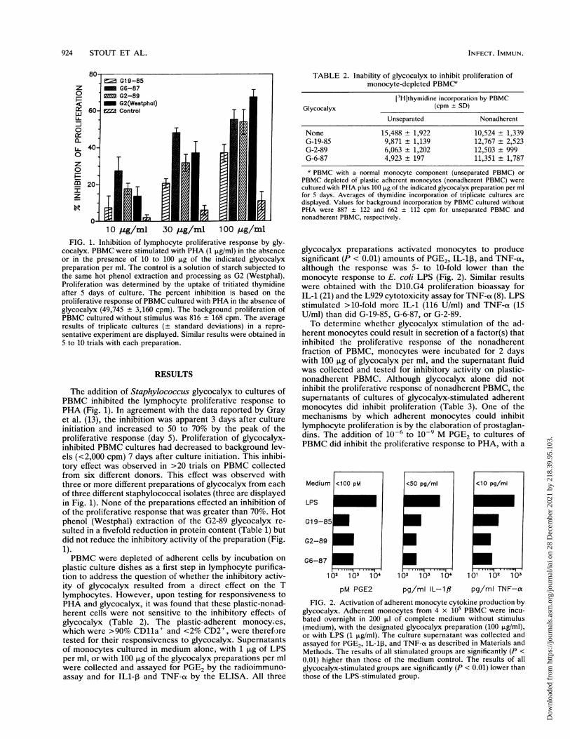
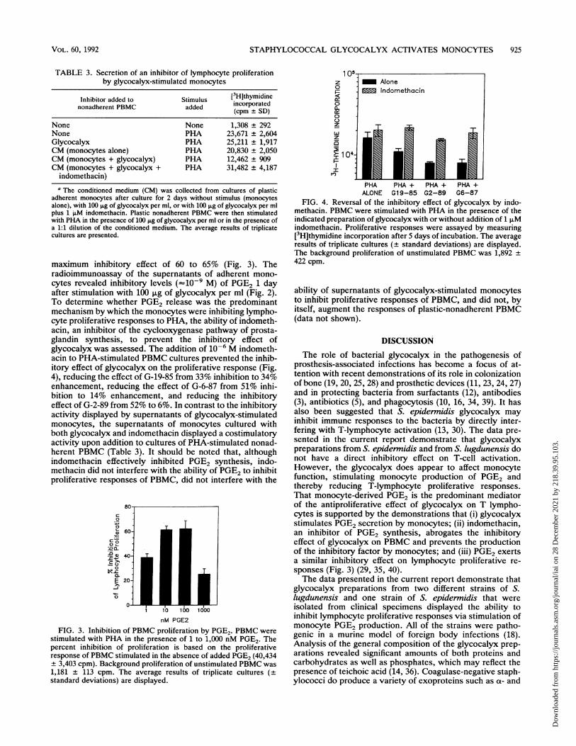
maximum inhibitory effect of 60 to 65% (Fig. 3). Theradioimmunoassay of the supernatants of adherent mono-cytes revealed inhibitory levels (=10-9 M) of PGE2 1 dayafter stimulation with 100 ,ug of glycocalyx per ml (Fig. 2).To determine whether PGE2 release was the predominantmechanism by which the monocytes were inhibiting lympho-cyte proliferative responses to PHA, the ability of indometh-acin, an inhibitor of the cyclooxygenase pathway of prosta-glandin synthesis, to prevent the inhibitory effect ofglycocalyx was assessed. The addition of 10-6 M indometh-acin to PHA-stimulated PBMC cultures prevented the inhib-itory effect of glycocalyx on the proliferative response (Fig.4), reducing the effect of G-19-85 from 33% inhibition to 34%enhancement, reducing the effect of G-6-87 from 51% inhi-bition to 14% enhancement, and reducing the inhibitoryeffect of G-2-89 from 52% to 6%. In contrast to the inhibitoryactivity displayed by supernatants of glycocalyx-stimulatedmonocytes, the supernatants of monocytes cultured withboth glycocalyx and indomethacin displayed a costimulatoryactivity upon addition to cultures of PHA-stimulated nonad-herent PBMC (Table 3). It should be noted that, althoughindomethacin effectively inhibited PGE2 synthesis, indo-methacin did not interfere with the ability of PGE2 to inhibitproliferative responses of PBMC, did not interfere with the
80
c
00
Cp-a40-
0
E 20-
-J
0
0-
1 10 100 1000
nM PGE2
FIG. 3. Inhibition of PBMC proliferation by PGE2. PBMC werestimulated with PHA in the presence of 1 to 1,000 nM PGE2. Thepercent inhibition of proliferation is based on the proliferativeresponse of PBMC stimulated in the absence of added PGE2 (40,434+ 3,403 cpm). Background proliferation of unstimulated PBMC was1,181 + 113 cpm. The average results of triplicate cultures (±
standard deviations) are displayed.
z
0
0
a.
0
105{AloneIndomethacin
z
z
>_ 10441 0 l |
PHA PHA + PHA + PHA +ALONE G19-85 G2-89 G6-87
FIG. 4. Reversal of the inhibitory effect of glycocalyx by indo-methacin. PBMC were stimulated with PHA in the presence of theindicated preparation of glycocalyx with or without addition of 1 ,uMindomethacin. Proliferative responses were assayed by measuring[3H]thymidine incorporation after 5 days of incubation. The averageresults of triplicate cultures (+ standard deviations) are displayed.The background proliferation of unstimulated PBMC was 1,892 ±422 cpm.
ability of supernatants of glycocalyx-stimulated monocytesto inhibit proliferative responses of PBMC, and did not, byitself, augment the responses of plastic-nonadherent PBMC(data not shown).
DISCUSSION
The role of bacterial glycocalyx in the pathogenesis ofprosthesis-associated infections has become a focus of at-tention with recent demonstrations of its role in colonizationof bone (19, 20, 25, 28) and prosthetic devices (11, 23, 24, 27)and in protecting bacteria from surfactants (12), antibodies(3), antibiotics (5), and phagocytosis (10, 16, 34, 39). It hasalso been suggested that S. epidermidis glycocalyx mayinhibit immune responses to the bacteria by directly inter-fering with T-lymphocyte activation (13, 30). The data pre-sented in the current report demonstrate that glycocalyxpreparations from S. epidermidis and from S. lugdunensis donot have a direct inhibitory effect on T-cell activation.However, the glycocalyx does appear to affect monocytefunction, stimulating monocyte production of PGE2 andthereby reducing T-lymphocyte proliferative responses.That monocyte-derived PGE2 is the predominant mediatorof the antiproliferative effect of glycocalyx on T lympho-cytes is supported by the demonstrations that (i) glycocalyxstimulates PGE2 secretion by monocytes; (ii) indomethacin,an inhibitor of PGE2 synthesis, abrogates the inhibitoryeffect of glycocalyx on PBMC and prevents the productionof the inhibitory factor by monocytes; and (iii) PGE2 exertsa similar inhibitory effect on lymphocyte proliferative re-sponses (Fig. 3) (29, 35, 40).The data presented in the current report demonstrate that
glycocalyx preparations from two different strains of S.lugdunensis and one strain of S. epidermidis that wereisolated from clinical specimens displayed the ability toinhibit lymphocyte proliferative responses via stimulation ofmonocyte PGE2 production. All of the strains were patho-genic in a murine model of foreign body infections (18).Analysis of the general composition of the glycocalyx prep-arations revealed significant amounts of both proteins andcarbohydrates as well as phosphates, which may reflect thepresence of teichoic acid (14, 36). Coagulase-negative staph-ylococci do produce a variety of exoproteins such as ao- and
VOL. 60, 1992
Dow
nloa
ded
from
http
s://j
ourn
als.
asm
.org
/jour
nal/i
ai o
n 28
Dec
embe
r 20
21 b
y 21
8.39
.95.
103.
926 STOUT ET AL.
b-hemolysins, DNase, and proteases. These do not appear toplay a dominant role in the inhibitory activity of the glyco-calyx, since S. lugdunensis 6-87 and 2-89 do not elaboratedetectable amounts of these exoproteins (18), yet glycocalyxpreparations from these strains were effective inducers ofmonocyte PGE2 production. In addition, depletion of proteinby hot phenol extraction did not reduce the ability of theglycocalyx preparations to induce monocyte PGE2 produc-tion and to inhibit proliferation of PHA-stimulated PBMC.These observations suggest that the activity may be due topolysaccharide or teichoic acid components of the glycoca-lyx. Attempts to identify and purify the active component(s)are currently in progress.The endotoxin of gram-negative bacteria is known to
stimulate monocytes. However, endotoxin contamination ofthe glycocalyx preparations is not likely to be responsible forthe observed effects, because (i) control preparations ofstarch, generated simultaneously with glycocalyx prepara-tions under the same endotoxin-free conditions, were inac-tive; (ii) glycocalyx preparations contained negligible levels(<0.1 ng/100 ,ug) of endotoxin according to the Limulusassay; and (iii) the addition of endotoxin (E. coli 011:B4-W)to PBMC cultures at levels 10- to 100-fold higher (1 ng/ml)than the maximum possible endotoxin contamination ofglycocalyx did not result in detectable inhibition of theproliferative response of the PBMC to PHA (data notshown).
Several recent studies have indicated that microorganismsproduce several different bioactive factors that stimulatemacrophages (32, 33, 37). These factors share with glyco-calyx the characteristic of being implicated in macro-phage-mediated immunosuppression. Oligosaccharides andpolysaccharides from bacteria and yeast cells, free of theclassical gram-negative endotoxins, have been reported toreduce immunoproliferative responsiveness of T lympho-cytes by stimulating suppressive activity in macrophages(32, 33). Immunosuppression during treponeme infectionshas been shown to result from prostaglandin production bytreponeme-stimulated macrophages (37). It has been re-ported that a component or components of bacterial glyco-calyx may directly inhibit some granulocyte functions suchas the response to chemotactic gradients, oxidative burst,and engulfment of particles (10, 16, 30, 34, 39). The currentstudy indicates that the glycocalyx of coagulase-negativestaphylococci does contain a component(s) that is capable ofactivating monocytes. This activation results not only inPGE2 production but also in IL-1 and TNF-a production andsecretion. IL-1 and TNF-ox, like PGE2, play significant rolesin acute inflammatory responses (4, 7). How glycocalyxcould be of such apparent benefit to coagulase-negativestaphylococci in the establishment of foreign body infectionswhile stimulating monocytes to secrete inflammatory cyto-kines represents a fascinating paradox. Molecular identifica-tion and characterization of the components of glycocalyxthat interact with macrophages and neutrophils may lead toa resolution of this paradox.
ACKNOWLEDGMENTSWe appreciate W. R. Mayberry for assistance in the biochemical
characterization of the glycocalyx, J. Keplinger for assistance inpreparation of the glycocalyx, and B. Stokes and J. Taylor forsecretarial assistance.
REFERENCES1. Ames, B. 1966. Assay of inorganic phosphorus, total phospho-
rus, and phosphates. Methods Enzymol. 8:115-118.
2. Baddour, L. M., G. D. Christensen, M. G. Hester, and A. L.Bisno. 1984. Production of experimental endocarditis by coagu-lase-negative staphylococci: variability in species virulence. J.Infect. Dis. 150:721-727.
3. Baltimore, R. S., and M. Mitchell. 1980. Immunologic investi-gation of mucoid strains of Pseudomonas aeruginosa. Compar-ison of susceptibility to opsonic antibody in mucoid and non-mucoid strains. J. Infect. Dis. 141:238-247.
4. Beutler, B., and A. Cerami. 1989. The biology of cachectin/TNFot-a primary mediator of the host response. Annu. Rev.Immunol. 7:625-656.
5. Costerton, J. W. 1977. Cell envelope as a barrier to antibiotics,p. 151-157. In D. Schlessinger (ed.), Microbiology-1977.American Society for Microbiology, Washington, D.C.
6. Costerton, J. W., R. T. Irvin, and K. J. Cheng. 1981. The role ofbacterial cell surface components in pathogenesis. Crit. Rev.Microbiol. 8:303-338.
7. Dinarello, C. A. 1989. Interleukin 1 and its biologically relatedcytokines. Adv. Immunol. 44:153-206.
8. Drysdale, B.-E., C. M. Zacharchuk, and H. S. Shin. 1983.Mechanism of macrophage-mediated cytotoxicity: productionof a soluble cytotoxic factor. J. Immunol. 131:2362-2367.
9. Dubois, M., K. A. Gilles, J. K. Hamilton, P. A. Rabers, and F.Smith. 1956. Colorimetric method for determination of sugarsand related substances. Anal. Chem. 28:350-356.
10. Falcieri, E., P. Vaudaux, E. Huggler, D. Lew, and F. Waldvogel.1987. Role of bacterial exopolymers and host factors on adher-ence and phagocytosis of Staphylococcus aureus in foreignbody infection. J. Infect. Dis. 155:524-531.
11. Franson, T. R., N. K. Sheth, H. D. Rose, and P. G. Sohnle. 1984.Scanning electron microscopy of bacteria adherent to intravas-cular catheters. J. Clin. Microbiol. 20:500-505.
12. Govan, J. R. W. 1975. Mucoid strains of Pseudomonas aerug-inosa: the influence of culture medium on the stability of mucusproduction. J. Med. Microbiol. 8:513-522.
13. Gray, E. D., G. Peters, M. Verstegen, and W. E. Regelmann.1984. Effect of extracellular slime substance from Staphylococ-cus epidermidis on the human cellular immune response. Lanceti:365-367.
14. Hussain, M., J. G. M. Hastings, and P. J. White. 1991. Isolationand composition of the extracellular slime made by coagulase-negative staphylococci in a chemically defined medium. J.Infect. Dis. 613:534-541.
15. Ishak, M. A., D. M. Groschel, G. L. Mandell, and R. P. Wenzel.1985. Association of slime with pathogenicity of coagulase-negative staphylococci causing nosocomial septicemia. J. Clin.Microbiol. 22:1025-1029.
16. Johnson, G. M., D. A. Lee, W. E. Regelmann, E. D. Gray, G.Peters, and P. G. Quie. 1986. Interference with granulocytefunction by Staphylococcus epidermidis slime. Infect. Immun.54:13-20.
17. Lambe, D. W., Jr., K. P. Ferguson, and D. A. Ferguson. 1988.The Bacteroides glycocalyx as visualized by differential inter-ference contrast microscopy. Can. J. Microbiol. 34:1189-1195.
18. Lambe, D. W., Jr., K. P. Ferguson, J. L. Keplinger, C. G.Gemmell, and J. H. Kalbfileisch. 1990. Pathogenicity of Staphy-lococcus lugdunensis, Staphylococcus schleiferi, and threeother coagulase-negative staphylococci in a mouse model andpossible virulence factors. Can. J. Microbiol. 36:455-463.
19. Lambe, D. W., Jr., K. P. Ferguson, K. J. Mayberry-Carson,B. K. Tober-Meyer, and J. W. Costerton. 1991. Foreign-body-associated experimental osteomyelitis induced with Bacteroidesfragilis and Staphylococcus epidermidis in rabbits. Clin. Or-thop. 266:285-294.
20. Lambe, D. W., Jr., K. J. Mayberry-Carson, W. R. Mayberry,B. K. Tober-Meyer, and J. W. Costerton. 1987. The effect ofsubinhibitory concentrations of clindamycin on the adherenceand glycocalyx of Staphylococcus aureus and Bacteroides spe-cies in vitro and in vivo, p. 35-49. In A. Szentivanyi, H.Friedman, and G. Gillissen (ed.), Antibiosis and host immunity.Plenum Publishing Corp., New York.
21. Lichtman, A. H., E. A. Kurt-Jones, and A. K. Abbas. 1987. Bcell stimulatory factor 1 and not interleukin 2 is the autocrine
IN FECTr. IMMUN .
Dow
nloa
ded
from
http
s://j
ourn
als.
asm
.org
/jour
nal/i
ai o
n 28
Dec
embe
r 20
21 b
y 21
8.39
.95.
103.
STAPHYLOCOCCAL GLYCOCALYX ACTIVATES MONOCYTES 927
growth factor for some helper T lymphocytes. Proc. Natl. Acad.Sci. USA 84:824.
22. Lowry, 0. H., N. J. Rosenbrough, A. L. Farr, and R. J. Randall.1951. Protein measurement with the Folin phenol reagent. J.Biol. Chem. 193:265-275.
23. Marrie, T. J., and J. W. Costerton. 1983. A scanning andtransmission electron microscopic study of the surface of intra-uterine contraceptive devices. Am. J. Obstet. Gynecol. 146:384-394.
24. Marrie, T. J., and J. W. Costerton. 1983. Scanning electronmicroscopic study of uropathogen adherence to a plastic sur-face. Appl. Environ. Microbiol. 45:1018-1024.
25. Marrie, T. J., and J. W. Costerton. 1985. Mode of growth ofbacterial pathogens in chronic polymicrobial human osteomy-elitis. J. Clin. Microbiol. 22:924-933.
26. Marrie, T. J., G. K. M. Harding, A. R. Ronald, J. Dikkema, J.Lam, S. Hoban, and J. W. Costerton. 1979. Influence of anti-body coating of Pseudomonas aeruginosa. J. Infect. Dis. 19:357-361.
27. Marrie, T. J., J. Nelligan, and J. W. Costerton. 1982. A scanningand transmission electron microscopic study of an infectedendocardial pacemaker lead. Circulation 66:1339-1341.
28. Mayberry-Carson, K. J., B. K. Tober-Meyer, J. K. Smith, D. W.Lambe, Jr., and J. W. Costerton. 1984. Bacterial adherence andglycocalyx formation in osteomyelitis experimentally inducedwith Staphylococcus aureus. Infect. Immun. 43:825-833.
29. Metzger, Z., J. T. Hoffeld, and J. J. Oppenheim. 1980. Macro-phage mediated suppression. I. Evidence for participation ofboth hydrogen peroxide and prostaglandins in suppression ofmurine lymphocyte proliferation. J. Immunol. 124:983.
30. Peters, G., E. D. Gray, and G. M. Johnson. 1989. Immunomod-ulating properties of extracellular slime substance, p. 61-74. InA. L. Bisno and F. A. Waldvogel (ed.), Infections associatedwith indwelling medical devices. American Society for Micro-biology, Washington, D.C.
31. Peters, G., F. Schumacher-Perdreau, B. Jansen, M. Bey, and G.Pulverer. 1987. Biology of S. epidernidis extracellular slime, p.15-32. In G. Pulverer, P. G. Quie, and G. Peters (ed.), Patho-genicity and clinical significance of coagulase negative staphylo-
cocci. Gustav Fischer, New York.32. Podzorski, R. P., G. R. Gray, and R. D. Nelson. 1990. Different
effects of native Candida albicans mannan and mannan-derivedoligosaccharides on antigen-stimulated lymphoproliferation invitro. J. Immunol. 144:707-716.
33. Regan, D. R., P. L. Cohen, W. J. Cromartie, and J. H. Schwab.1988. Immunosuppressive macrophages induced by arthro-pathic peptidoglycan-polysaccharide polymers from bacterialcell walls. Clin. Exp. Immunol. 74:365-370.
34. Schwarzmann, S., and J. R. Boring III. 1971. Antiphagocyticeffect of slime from a mucoid strain of Pseudomonas aerugi-nosa. Infect. Immun. 3:762-767.
35. Stobo, J. D., M. S. Kennedy, and M. E. Goldyne. 1979. Prosta-glandin E modulation of the mitogenic response of human Tcells. Differential response of T cell subpopulations. J. Clin.Invest. 64:1188.
36. Tojo, M., N. Yamashita, D. A. Goldmann, and G. B. Pier. 1988.Isolation and characterization of a capsular polysaccharideadhesin from Staphylococcus epidennidis. J. Infect. Dis. 157:713-722.
37. Tomai, M. A., B. J. Elmquist, S. M. Warmka, and T. J.Fitzgerald. 1989. Macrophage mediated suppression of conA-induced IL2 production in spleen cells from syphilitic rabbits.J. Immunol. 143:309-314.
38. Van de Rijn, I., and R. E. Kessler. 1980. Growth characteristicsof group A streptococci in a new chemically defined medium.Infect. Immun. 27:444 448.
39. Veringa, E. M., D. A. Ferguson, D. W. Lambe, Jr., and J.Verhoef. 1989. Trospectomycin enhances surface phagocytosisof Bacteroides and Staphylococcus by altering the bacterialglycocalyx. Zentralbl. Bakteriol. Parasitenkd. Infektionskr.Hyg. Abt. 1 Orig. 271:311-320.
40. Walker, C., F. Kristensen, F. Bettens, and A. L. DeWeck. 1983.Lymphokine regulation of activated lymphocytes. I. Prostaglan-din E2 induced inhibition of interleukin 2 production. J. Immu-nol. 130:1770.
41. Westphal, O., and K. Jann. 1965. Bacterial lipopolysaccharides.Extraction with phenol-water and further applications of theprocedure. Methods Carbohydr. Chem. 5:83-91.
VOL. 60, 1992
Dow
nloa
ded
from
http
s://j
ourn
als.
asm
.org
/jour
nal/i
ai o
n 28
Dec
embe
r 20
21 b
y 21
8.39
.95.
103.


![An epidemiological model for proliferative kidney disease ... · An epidemiological model for proliferative ... [18, 35]. Overt infec-tion ... An epidemiological model for proliferative](https://img.pdfslide.us/doc/110x75/5c00b25409d3f225538b84ad/an-epidemiological-model-for-proliferative-kidney-disease-an-epidemiological.jpg)