Embed Size (px)
Citation preview
Standing Committee of Analysts
The Microbiology of Recreational and Environmental Waters
(2015) – Part 5 – Methods for the isolation and enumeration of
sulphite-reducing clostridia and Clostridium perfringens
Methods for the Examination of Waters and Associated Materials

2

3
The Microbiology of Recreational and Environmental Waters (2015) – Part 5 – Methods for the isolation and enumeration of sulphite-reducing clostridia and Clostridium perfringens
Methods for the Examination of Waters and Associated Materials This booklet contains methods for the isolation and enumeration of sulphite-reducing clostridia and Clostridium perfringens. A Enumeration of sulphite-reducing clostridia by a membrane filtration technique B Enumeration of Clostridium perfringens by membrane filtration techniques C Enumeration of Clostridium perfringens by a multiple tube most probable number technique This bluebook replaces and updates section 7.5 of the earlier version of The Microbiology of Recreational and Environmental Waters published in 2000. Whilst specific commercial products may be referred to in this document, this does not constitute an endorsement of these products. They serve only as illustrative examples of the types of products available. Equivalent products may be available and it should be understood that the performance of the method might differ when other materials are used and all should be confirmed by validation of the method.
4
Contents About this series 6 Warning to users 6
A Enumeration of sulphite-reducing clostridia by a membrane filtration technique 7 A1 Introduction 7 A2 Scope 7 A3 Definitions 7 A4 Principle 7 A5 Limitations 7 A6 Health and safety 8 A7 Apparatus 8 A8 Media and reagents 8 A9 Analytical procedure 9 A10 Calculations 11 A11 Expression of results 11 A12 Quality assurance 11 A13 References 11 B Enumeration of Clostridium perfringens by membrane filtration techniques 12 B1 Introduction 12 B2 Scope 12 B3 Definitions 12 B4 Principle 12 B5 Limitations 13 B6 Health and safety 13 B7 Apparatus 13 B8 Media and reagents 14 B9 Analytical procedure 17 B10 Calculations 24 B11 Expression of results 24 B12 Quality assurance 24 B13 References 24 C Enumeration of Clostridium perfringens by a multiple tube most probable number technique 26 C1 Introduction 26 C2 Scope 26 C3 Definitions 26 C4 Principle 26 C5 Limitations 27 C6 Health and safety 27 C7 Apparatus 27 C8 Media and reagents 27 C9 Analytical procedure 31 C10 Calculations 38 C11 Expression of results 38 C12 Quality assurance 39

5
C13 References 39 Appendix C1 Tables of most probable numbers 40 Appendix 1 Verification of the acid phosphatase test for the confirmation of
Clostridium perfringens isolated from various waters 45
Address for correspondence 52 Members assisting with these methods 52

6
About this series Introduction This booklet is part of a series intended to provide authoritative guidance on recommended methods of sampling and analysis for determining the quality of drinking water, ground water, river water and sea water, waste water and effluents as well as sewage sludges, sediments, soils (including contaminated land) and biota. In addition, short reviews of the most important analytical techniques of interest to the water and sewage industries are included. Performance of methods Ideally, all methods should be fully evaluated with results from performance tests. These methods should be capable of establishing, within specified or pre-determined and acceptable limits of deviation and detection, whether or not any sample contains concentrations of parameters above those of interest. For a method to be considered fully evaluated, individual results from at least three laboratories should be reported. The specifications of performance generally relate to maximum tolerable values for total error (random and systematic errors) systematic error (bias) total standard deviation and limit of detection. Often, full evaluation is not possible and only limited performance data may be available. In addition, good laboratory practice and analytical quality control are essential if satisfactory results are to be achieved. Standing Committee of Analysts The preparation of booklets within the series “Methods for the Examination of Waters and Associated Materials” and their continuing
revision is the responsibility of the Standing Committee of Analysts (established 1972 by the Department of the Environment). At present, there are seven working groups, each responsible for one section or aspect of water quality analysis. They are 1 General principles of sampling and accuracy of results 2 Microbiological methods 3 Empirical, Inorganic and physical methods, Metals and metalloids 4 Solid substances 5 Organic impurities 6 Biological, biodegradability and inhibition methods 7 Radiochemical methods The actual methods and reviews are produced by smaller panels of experts in the appropriate field, in co-operation with the working group and strategic committee. The names of those members principally associated with these methods are listed at the back of this booklet. Publication of new or revised methods will be notified to the technical press. If users wish to receive copies or advanced notice of forthcoming publications or obtain details of the index of methods then contact the Secretary on the Agency’s web-page (http://standingcommitteeofanalysts.co.uk/) or by post. Every effort is made to avoid errors appearing in the published text. If, however, any are found, please notify the Secretary. Users should ensure they are aware of the most recent version they seek. Robert Carter Secretary June 2015
_______________________________________________________________________ Warning to users The analytical procedures described in this booklet should only be carried out under the proper supervision of competent, trained analysts in properly equipped laboratories. All possible safety precautions should be followed and appropriate regulatory requirements complied with. This should include compliance with the Health and Safety at Work etc Act 1974 and all regulations made under the Act, and the Control of Substances Hazardous to Health Regulations 2002 (SI 2002/2677). Where particular or exceptional hazards exist in carrying out the procedures described in this booklet, then specific attention is noted. Numerous publications are available giving practical details on first aid and laboratory safety.
These should be consulted and be readily accessible to all analysts. Amongst such resources are; HSE website HSE: Information about health and safety at work ; RSC website http://www.rsc.org/learn-chemistry/collections/health-and-safety “Safe Practices in Chemical Laboratories” and “Hazards in the Chemical Laboratory”, 1992, produced by the Royal Society of Chemistry; “Guidelines for Microbiological Safety”, 1986, Portland Press, Colchester, produced by Member Societies of the Microbiological Consultative Committee; and “Biological Agents: Managing the Risks in Laboratories and Healthcare Premises”, 2005 and “The Approved List of Biological Agents” 2013, produced by the Advisory Committee on Dangerous Pathogens of the Health and Safety Executive (HSE).
7
A Enumeration of sulphite-reducing clostridia by a membrane filtration technique A1 Introduction Tests for sulphite-reducing clostridia play only a subsidiary role in water examination. The organisms occur widely in soils and sediments and form spores which are environmentally resistant. Their numbers may, therefore, be indicative of environmental loading. Clostridium perfringens is a sulphite-reducing species and is generally associated with faecal contamination. The significance of sulphite-reducing clostridia and Clostridium perfringens in recreational and other waters is described elsewhere(1) in this series. A2 Scope The method is suitable for the examination of freshwater, surface waters and saline waters, swimming pools, spa pools and hydrotherapy pools and primary and secondary wastewater effluents. Water samples with higher turbidities should be analysed using an appropriate multiple tube most probable number (MPN) method (see Method C).
Users wishing to employ this method should verify its performance under their own laboratory conditions(2). A3 Definitions Sulphite-reducing clostridia are Gram-positive anaerobic spore-forming rod-shaped bacteria which, in the context of this method, reduce sulphite to sulphide at 37°C within 24 hours. Some sulphite-reducing clostridia may grow as colourless colonies on the medium used in this method and, therefore, would not be included under this definition. A4 Principle A volume of sample, or diluted sample, is filtered and the membrane filter placed on the surface of an agar medium containing sulphite, iron(III) and D-cycloserine (which inhibits other bacteria and reduces the size of colonies that develop). The agar medium is then incubated under anaerobic conditions at 37°C. Sulphite-reducing clostridia usually produce black or grey colonies as a result of the reduction of sulphite to sulphide, which then reacts with the iron(III) salt. If only a spore count is required then the sample is heat treated at 60°C prior to filtration in order to kill vegetative bacteria. A5 Limitations The method is suitable for most types of aqueous samples except those with high turbidities which tend to block the membrane filter. This will limit the volume of sample that can be filtered. Accumulated deposit on the membrane filter may mask or inhibit the growth of indicator organisms. Where high numbers of organisms may be expected (for example, untreated wastewater) serial ten-fold dilutions should be made to obtain a countable number of colonies. The maximum number of colonies that should be counted from a single membrane filter is approximately 100. Counts can be obtained from membranes containing more than 100 colonies providing that isolated colonies are present and that a hand lens or similar magnifying aid is used. Counts obtained in this way should be reported as estimated counts. Some clostridia may produce spreading colonies. In

8
these circumstances the potential maximum count may be restricted to dilutions giving distinct, unmerged, colonies. A6 Health and safety Media, reagents and bacteria used in this method are covered by the Control of Substances Hazardous to Health Regulations(3) and appropriate risk assessments should be made before adopting this method. Standard laboratory microbiology safety procedures should be followed and guidance is given elsewhere(2) in this series. A7 Apparatus Standard laboratory equipment should be used which conforms to the performance criteria outlined elsewhere (2) in this series. Principally appropriate membrane filtration apparatus and incubators (fan assisted, static temperature) are required. Other items include: A7.1 Sterile sample bottles of appropriate volume, made of suitable material, should be used. For swimming pools, spa pools and hydrotherapy pools the containers should not be made of glass and should contain sufficient sodium thiosulphate pentahydrate to give a final concentration in the sample of not less than 18 mg/l. For example, 0.1 ml of a 1.8 % m/v solution of sodium thiosulphate pentahydrate (Na2S2O3.5H2O) per 100 ml of sample, or equivalent may be suitable. A7.2 Incubator capable of maintaining a temperature of 37.0 ± 1.0°C. A7.3 Waterbath capable of holding bottles of sample at 60 ± 2°C. A7.4 Anaerobic jars, or similar equipment, and anaerobic gas-generating system (for generating anaerobic atmospheres with approximately 9 - 13 % carbon dioxide). A7.5 Filtration apparatus, sterile filter funnels (or sterile disposable funnels) and source of vacuum. A7.6 Sterile membrane filters, for example white 47 mm diameter cellulose-based, 0.45 µm nominal pore size. A7.7 Smooth-tipped forceps. A8 Media and reagents Commercial formulations of these media and reagents may be available, but may possess minor variations to their formulation. The performance of all media and reagents should be verified prior to their use in this method(2). Variations in the preparation and storage of media should also be verified. Water should be distilled, deionised or of similar grade quality. Unless otherwise stated chemical constituents should be added as the anhydrous salts. If the pH of media are not within the stated range, then, before heating, they should be adjusted accordingly.
A8.1 Tryptose sulphite cycloserine agar without egg yolk (TSCA)(4, 5) Yeast extract 5 g Tryptose 15 g

9
Soya peptone 5 g Sodium metabisulphite 1 g Iron (III) ammonium citrate 1 g D-cycloserine 400 mg Agar 14 g Water 1 litre Suspend the ingredients, except the D-cycloserine, in the water and dissolve by heating and stirring the mixture. Sterilise the solution by autoclaving at 121°C for 15 minutes. Allow the medium to cool to 46 ± 2°C. Add 4 ml of a filter-sterilised solution of D-cycloserine in water at a concentration of 100 mg/ml. Mix the solution thoroughly, and dispense into Petri dishes. The final pH of the medium should be 7.6 ± 0.2. The Petri dishes should be the vented type to ensure anaerobic conditions for the medium during storage and incubation. Performance of the medium deteriorates during storage due to exposure to oxygen. Prepared media may be stored in a refrigerator under anaerobic conditions at a temperature in the range of 5 ± 3 °C for up to one week. However, some anaerobic generating systems may not work satisfactorily at this temperature. When fresh medium is used, the colony characteristics that are observed tend to be more defined. Medium, once removed from the refrigerator and left on the bench for more than an hour, should be discarded if not used. A8.2 Other media Standard and commercial formulations of other media and reagents used in this method include quarter-strength Ringer’s solution and maximum recovery diluent. A9 Analytical procedure A9.1 Sample preparation The volumes and dilutions of samples should be chosen so that the number of colonies to be counted on the membrane filter lies, if possible, between 20 and 80. With some waters, it may be advantageous to filter a selection of different volumes or dilutions of sample, so that the number of colonies on one of the membrane filters is likely to fall within this range. For swimming pool, spa and hydrotherapy pool waters, filter 100 ml of the sample. For polluted waters and wastewater either filter smaller volumes, or dilutions of the sample made with quarter-strength Ringer’s solution or maximum recovery diluent. If it is the intention to count only the spores of sulphite reducing clostridia then the volume of sample should be heated to 60 ± 2°C (for example in a water bath) and maintained at this temperature for 15 ± 1 minutes. The temperature may be monitored by placing an appropriate thermometer in a similar bottle containing a volume of water similar to the volume of sample being treated. A9.2 Sample processing Place the sterile filtration apparatus in position and connect to a source of vacuum, with the stopcock turned off. Remove the funnel and, holding the edge of the membrane filter with sterile smooth-tipped forceps, place a sterile membrane filter, grid-side upwards, on the porous disc of the filter base. Replace the sterile funnel securely on the filter base. Pour or pipette the required volume of sample, or diluted sample, into the funnel. When the

10
volume of sample to be filtered is less than 10 ml, add 10 - 20 ml of sterile diluent (for example, quarter-strength Ringer’s solution or maximum recovery diluent) to the funnel before the addition of the sample. This aids dispersion of the bacteria over the entire surface of the membrane filter during filtration. Open the stopcock and apply a vacuum not exceeding 65 kPa (500 mm of mercury) and filter the sample slowly through the membrane filter. Close the stopcock as soon as the sample has been filtered. Remove the funnel and transfer the membrane filter carefully to a Petri dish of well-dried TSCA. Ensure that no air bubbles are trapped between the membrane filter and the medium. ‘Rolling’ the membrane filter onto the medium minimises the likelihood of air bubbles becoming trapped. As the spores of sulphite reducing clostridia are very resilient, funnels that have been used once should be sterilised by autoclaving before being used again. Placing funnels in a water bath at this stage may not be sufficient to kill spores. If different volumes of the same sample are to be examined, the funnel may be re-used without sterilising the funnel provided that the smallest volume, or highest dilution, of sample is filtered first. For different samples, take a fresh pre-sterilised funnel and repeat the filtration process. During the filtration of a series of samples, the filter base need not be sterilised unless it becomes, or is suspected of being, contaminated or a membrane filter becomes damaged. When funnels are not in use they should be covered with a sterile lid or a sterile Petri dish lid. Alternatively, commercially available, sterile, disposable funnels may be used. The time between the end of the filtration step and the beginning of the incubation stage should be as short as possible, and no longer than 2 hours. Incubate the Petri dishes at 37°C in an anaerobic jar or similar system containing an indicator of anaerobiosis (e.g. resazurin indicator strips) and an atmosphere containing 9 - 13 % carbon dioxide. Examine the dishes after 21 ± 3 hours incubation. A9.3 Reading of results After incubation, count all black or grey colonies (see Figure A1). Figure A1 Typical colonies of sulphite-reducing clostridia on tryptose sulphite
cycloserine agar
11
A9.4 Confirmation tests The specificity of TSCA, together with the incubation conditions, is such that confirmation of isolates is not usually required. A10 Calculations A10.1 Confirmed sulphite-reducing clostridia The number of confirmed sulphite-reducing clostridia colonies is generally quoted as the number of colonies per 100 ml. Calculate the confirmed count as follows: Confirmed count per 100 ml = Number of colonies counted on membrane filter x 100 x DF Volume of sample filtered (ml) Where DF is dilution factor if appropriate. A11 Expression of results Counts for sulphite-reducing clostridia are expressed in colony forming units per volume of sample. For most samples the volume is typically 100 ml. A12 Quality assurance New batches of media and reagents should be tested with appropriate reference strains of target bacteria (for example Clostridium perfringens) and non-target bacteria (for example Bacillus species). Petri dishes should be incubated for 21 ± 3 hours at 37°C under anaerobic conditions. Further details are given elsewhere(2) in this series. A13 References 1. Standing Committee of Analysts, The Microbiology of Recreational and Environmental Waters (2014) - Part 1 - Water quality, epidemiology and public health. Methods for the Examination of Waters and Associated Materials, in this series, Environment Agency. 2. Standing Committee of Analysts, The Microbiology of Drinking Water (2002) - Part 3 - Practices and procedures for laboratories. Methods for the Examination of Waters and Associated Materials, Environment Agency. 3. The Control of Substances Hazardous to Health Regulations 2002, Statutory Instrument 2002 No. 2677, The Stationery Office. 4. Enumeration of food-borne Clostridium perfringens in egg yolk free tryptose-sulphite-cycloserine agar, Applied Microbiology, A W H Hauschild and R Hillsheimer, 1974, 27, pp521-526. 5. Membrane filtration enumeration of faecal clostridia and Clostridium perfringens in water, Water Research, D Sartory, 1986, 20, pp1255-1260.
12
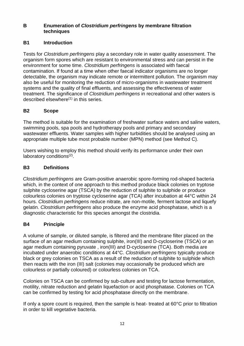
B Enumeration of Clostridium perfringens by membrane filtration techniques B1 Introduction Tests for Clostridium perfringens play a secondary role in water quality assessment. The organism form spores which are resistant to environmental stress and can persist in the environment for some time. Clostridium perfringens is associated with faecal contamination. If found at a time when other faecal indicator organisms are no longer detectable, the organism may indicate remote or intermittent pollution. The organism may also be useful for monitoring the reduction of micro-organisms in wastewater treatment systems and the quality of final effluents, and assessing the effectiveness of water treatment. The significance of Clostridium perfringens in recreational and other waters is described elsewhere(1) in this series. B2 Scope The method is suitable for the examination of freshwater surface waters and saline waters, swimming pools, spa pools and hydrotherapy pools and primary and secondary wastewater effluents. Water samples with higher turbidities should be analysed using an appropriate multiple tube most probable number (MPN) method (see Method C). Users wishing to employ this method should verify its performance under their own laboratory conditions(2). B3 Definitions Clostridium perfringens are Gram-positive anaerobic spore-forming rod-shaped bacteria which, in the context of one approach to this method produce black colonies on tryptose sulphite cycloserine agar (TSCA) by the reduction of sulphite to sulphide or produce colourless colonies on tryptose cycloserine agar (TCA) after incubation at 44°C within 24 hours. Clostridium perfringens reduce nitrate, are non-motile, ferment lactose and liquefy gelatin. Clostridium perfringens also produce the enzyme acid phosphatase, which is a diagnostic characteristic for this species amongst the clostridia. B4 Principle A volume of sample, or diluted sample, is filtered and the membrane filter placed on the surface of an agar medium containing sulphite, iron(III) and D-cycloserine (TSCA) or an agar medium containing pyruvate , iron(III) and D-cycloserine (TCA). Both media are incubated under anaerobic conditions at 44°C. Clostridium perfringens typically produce black or grey colonies on TSCA as a result of the reduction of sulphite to sulphide which then reacts with the iron (III) salt (colonies may occasionally be produced which are colourless or partially coloured) or colourless colonies on TCA. Colonies on TSCA can be confirmed by sub-culture and testing for lactose fermentation, motility, nitrate reduction and gelatin liquefaction or acid phosphatase. Colonies on TCA can be confirmed by testing for acid phosphatase directly on the membrane. If only a spore count is required, then the sample is heat- treated at 60°C prior to filtration in order to kill vegetative bacteria.

13
B5 Limitations The method is suitable for most types of aqueous samples except those with high turbidities which tend to block the membrane filter. This will limit the volume of sample that can be filtered. Accumulated deposit on the membrane filter may mask or inhibit the growth of indicator organisms. Where high numbers of organisms may be expected (for example, untreated wastewater) serial ten-fold dilutions should be made to obtain a countable number of colonies. Ideally the number of colonies counted from a single membrane filter should fall within the range of 20 – 80 and the maximum number is approximately 100. Counts can be obtained from membranes containing more than 100 colonies providing that isolated colonies are present and that a hand lens or similar magnifying aid is used. Counts obtained in this way should be reported as estimated counts. B6 Health and safety Media, reagents and bacteria used in this method are covered by the Control of Substances Hazardous to Health Regulations(3) and appropriate risk assessments should be made before adopting this method. Standard laboratory microbiology safety procedures should be followed and guidance is given elsewhere(2) in this series. B7 Apparatus Standard laboratory equipment should be used which conforms to the performance criteria outlined elsewhere(2) in this series. Principally appropriate membrane filtration apparatus and incubators (fan assisted, static temperature) are required. Other items include: B7.1 Sterile sample bottles of appropriate volume, made of suitable material, should be used. For swimming pools, spa pools and hydrotherapy pools the containers should not be made of glass and should contain sufficient sodium thiosulphate pentahydrate to give a final concentration in the sample of not less than 18 mg/l. For example, 0.1 ml of a 1.8 % m/v solution of sodium thiosulphate pentahydrate (Na2S2O3.5H2O) per 100 ml of sample, or equivalent may be suitable. B7.2 Incubators capable of maintaining temperatures of 37.0 ± 1.0°C and 44.0 ± 0.5°C. B7.3 Waterbath capable of holding bottles of sample at 60 ± 2°C. B7.4 Anaerobic jars, or similar equipment, and anaerobic gas-generating system (for generating anaerobic atmospheres containing approximately 9 - 13 % carbon dioxide). B7.5 Filtration apparatus, sterile filter funnels (or sterile disposable funnels) and source of vacuum. B7.6 Sterile gridded membrane filters, for example white, 47 mm diameter cellulose-based, 0.45 µm nominal pore size. B7.7 Smooth-tipped forceps.

14
B8 Media and reagents Commercial formulations of these media and reagents may be available, but may possess minor variations to their formulation. The performance of all media and reagents should be verified prior to their use in this method(2). Variations in the preparation and storage of media should also be verified. Water should be distilled, deionised or of similar grade quality. Unless otherwise stated chemical constituents should be added as the anhydrous salts. If the pH of media are not within the stated range, then, before heating, they should be adjusted accordingly.
B8.1 Tryptose sulphite cycloserine agar without egg yolk (TSCA)(4, 5)
Yeast extract 5 g Tryptose 15 g Soya peptone 5 g Sodium metabisulphite 1 g
Iron (III) ammonium citrate 1 g D-cycloserine 400 mg
Agar 14 g Water 1 litre
Suspend the ingredients, except the D-cycloserine, in the water and dissolve by heating and stirring. Sterilise the solution by autoclaving at 121°C for 15 minutes. Allow the medium to cool to 46 ± 2°C. Add 4 ml of a filter-sterilised solution of D-cycloserine in distilled water at a concentration of 100 mg/ml. Mix thoroughly, and dispense into Petri dishes. The final pH of the medium should be 7.6 ± 0.2. The Petri dishes should be the vented type to ensure anaerobic conditions for the medium during storage and incubation. Performance of the medium deteriorates during storage due to exposure to oxygen. Prepared media may be stored in a refrigerator under anaerobic conditions at a temperature in the range of 5 ± 3 °C for up to one week. However, some anaerobic generating systems may not work satisfactorily at this temperature. When fresh medium is used, the colony characteristics that are observed tend to be more defined. Medium, once removed from the refrigerator and left on the bench for more than an hour, should be discarded if not used. B8.2 Tryptose cycloserine agar with pyruvate (TCA)(6)
Yeast extract 5 g Tryptose 15 g Soya peptone 5 g Sodium pyruvate 0.5 g
Iron (III) ammonium citrate 1 g D-cycloserine 400 mg
Agar 14 g Water 1 litre
Suspend the ingredients in the water, except the D-cycloserine, and dissolve by heating and stirring. Sterilise the solution by autoclaving at 121 °C for 15 minutes. Allow the medium to cool to 46 ± 2 °C. Add 4 ml of a filter-sterilised solution of D-cycloserine in distilled water at a concentration of 100 mg/ml. Mix thoroughly, and dispense into Petri
15
dishes. The final pH of the medium should be 7.6 ± 0.2. The Petri dishes should be the vented type to ensure anaerobic conditions for the medium during storage and incubation. Performance of the medium deteriorates during storage due to exposure to oxygen. Prepared media should be stored in a refrigerator under anaerobic conditions at a temperature between 5 ± 3 °C for up to one week. However, some anaerobic generating systems may not work satisfactorily at this temperature. When fresh medium is used, the colony characteristics that are observed tend to be more defined. Medium, once removed from the refrigerator and left on the bench for more than an hour, should be discarded if not used. B8.3 Buffered nitrate-motility medium(7)
Beef extract 3 g Peptone 5 g Potassium nitrate 5 g D-Galactose 5 g Glycerol 5 g Disodium hydrogen phosphate 2.5 g Agar 3 g Water 1 litre
Dissolve the solid ingredients in 950 ml water by heating to boiling point whilst stirring continuously. Dissolve the glycerol in 50 ml water in a separate container and add this to the base medium and mix thoroughly. Dispense the resulting solution, typically in 10 ml aliquots, in appropriately sized tubes. Cap the tubes. Sterilise the medium by autoclaving at 121°C for 15 minutes. The final pH of the medium should be 7.3 ± 0.2. Prepared tubes should be stored at a temperature in the range 5 ± 3°C for up to one month if protected against dehydration. Before use stored media should be heated for 10 - 15 minutes in a boiling water bath, ensuring that the contents have melted to eliminate any absorbed oxygen. The tubes should then be allowed to cool and the medium to solidify ready for use. B8.4 Nitrate reduction test reagents(8)
Reagent A Sulphanilic acid 0.8 g Glacial acetic acid 30 ml Water 100 ml
Warm gently to aid dissolution.
Reagent B N, N-dimethyl-1-naphthylamine 0.6 ml Glacial acetic acid 30 ml Water 100 ml
Dissolve the amine in the acetic acid solution. To aid dissolution, warm gently (for example, by placing in a water bath at 40 - 60°C).

16
The reagents may be stored at a temperature at 5 ± 3°C for up to six months. For the combined reagent, mix equal volumes of reagents A and B immediately prior to use. Prepare in small volumes sufficient for the tests to be performed. The combined reagent can be stored at a temperature of 5 ± 3°C, protected from direct light, and should be used within 24 hours. B8.5 Lactose-gelatin medium(7)
Tryptose 15 g Yeast extract 10 g Disodium hydrogen phosphate 5 g Gelatin 120 g Lactose 10 g Phenol red (0.4 % m/v solution) 12.5 ml Water 1 litre
Dissolve the ingredients, except the gelatin, lactose and phenol red, in the water. Add the gelatin gradually whilst stirring the mixture continuously and warming gently to aid dissolution. Adjust the pH to 7.5 ± 0.2. Add the lactose and dissolve and add the phenol red solution. Dispense the resulting solution, typically in 10 ml aliquots in appropriately sized tubes. Cap the tubes. Sterilise the medium at 121°C for 15 minutes. The final pH should be 7.5 ± 0.2. Prepared media may be stored at a temperature in the range 5 ± 3°C for up to one month, if protected against dehydration. Before use stored media should be heated for 10 - 15 minutes in a boiling water bath to ensure that the contents have melted to eliminate any absorbed oxygen. The tubes should then be allowed to cool and the medium to solidify ready for use. B8.6 Columbia blood agar base
Special peptone 23 g Starch 1g Sodium chloride 5 g Agar 15 g Water 1 litre
Suspend the ingredients in the water and dissolve by heating and stirring the mixture. Sterilise the solution by autoclaving at 121°C for 15 minutes. Cool and dispense into Petri dishes. The Petri dishes should be the vented type to ensure anaerobic conditions for the medium during incubation. The final pH of the medium should be 7.3 ± 0.2. Sterile media may be stored at a temperature of 5 ± 3°C for up to one month, if protected against dehydration. B8.7 Acid phosphatase reagent(9) Acetate buffer Glacial acetic acid 0.3 ml Sodium acetate 0.4 g Water to 100 ml

17
Thoroughly mix the ingredients. The final pH value should be 4.6 ± 0.2. The buffer may be stored at room temperature for up to six months. Complete reagent 1-naphthyl phosphate monosodium salt 0.4 g o-dianisidine tetrazotized zinc chloride complex 0.8 g (Fast Blue B) Acetate buffer 20 ml Add the ingredients to the acetate buffer and shake well to dissolve. Store the reagent at 5 ± 3°C for one hour. Filter the solution to remove any precipitate(8). The reagent may be stored at 5 ± 3°C for up to two weeks. B8.8 Other media Standard and commercial formulations of other media and reagents used in this method include zinc powder, quarter-strength Ringer’s solution and maximum recovery diluent. B9 Analytical procedure B9.1 Sample preparation The volumes and dilutions of samples should be chosen so that the number of colonies to be counted on the membrane filter lies, if possible, between 20 and 80. With some waters, it may be advantageous to filter a selection of different volumes or dilutions of sample, so that the number of colonies on one of the membrane filters is likely to fall within this range. For swimming pool, spa and hydrotherapy pool waters, filter 100 ml of the sample. For polluted waters and wastewater either filter smaller volumes, or dilutions of the sample made with quarter-strength Ringer’s solution or maximum recovery diluent. If it is the intention to count only the spores of Clostridium perfringens then the volume of sample should be heated to 60 ± 2°C (for example in a water bath) and maintained at this temperature for 15 ± 1 minutes. The temperature may be monitored by placing an appropriate thermometer in a similar bottle containing a volume of water similar to the volume of sample being treated. B9.2 Sample processing Place the sterile filtration apparatus in position and connect to a source of vacuum, with the stopcock turned off. Remove the funnel and, holding the edge of the membrane filter with sterile smooth-tipped forceps, place a sterile membrane filter, grid-side upwards, on the porous disc of the filter base. Replace the sterile funnel securely on the filter base. Pour or pipette the required volume of sample, or diluted sample, into the funnel. When the volume of sample to be filtered is less than 10 ml, add 10 - 20 ml of sterile diluent (for example, quarter-strength Ringer’s solution or maximum recovery diluent) to the funnel before the addition of the sample. This aids dispersion of the bacteria over the entire surface of the membrane filter during filtration. Open the stopcock and apply a vacuum not exceeding 65 kPa (500 mm of mercury) and filter the sample slowly through the membrane filter. Close the stopcock as soon as the sample has been filtered.

18
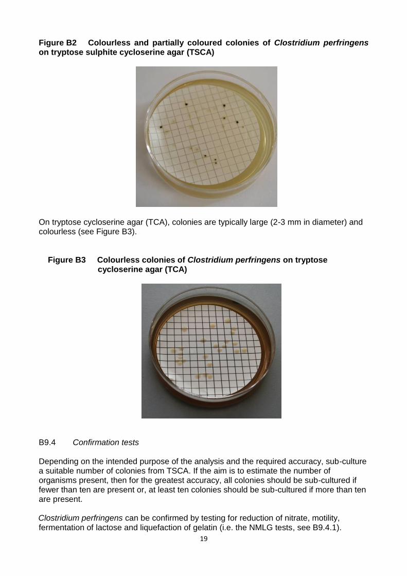
Remove the funnel and transfer the membrane filter carefully to a Petri dish of well-dried tryptose sulphite cycloserine agar (TSCA) or tryptose cycloserine agar (TCA). Ensure that no air bubbles are trapped between the membrane filter and the medium. ‘Rolling’ the membrane filter onto the medium minimises the likelihood of air bubbles becoming trapped. As the spores of Clostridium perfringens are very resilient, funnels that have been used once should be sterilised by autoclaving before being used again. Placing funnels in a water bath at this stage may not be sufficient to kill spores. If different volumes of the same sample are to be examined, the funnel may be re-used without sterilising the funnel provided that the smallest volume, or highest dilution of sample, is filtered first. For different samples, take a fresh pre-sterilised funnel and repeat the filtration process. During the filtration of a series of samples, the filter base need not be sterilised unless it becomes, or is suspected of being, contaminated or a membrane filter becomes damaged. When funnels are not in use they should be covered with a sterile lid or a sterile Petri dish lid. Alternatively, commercially available, sterile, disposable funnels may be used. The time between the end of the filtration step and the beginning of the incubation stage should be as short as possible and no longer than 2 hours. Incubate the Petri dishes of TSCA or TCA at 44°C in an anaerobic jar or similar system containing an indicator of anaerobiosis (e.g. resazurin indicator strips) and an atmosphere containing 9 - 13 % carbon dioxide. Hydrogen-free anaerobiosis generation systems are available. Examine the dishes after 21 ± 3 hours incubation. B9.3 Reading of results Under anaerobic conditions at 44°C colonies of clostridia on TSCA are typically black or grey in colour (see Figure B1) and on TCA will be colourless. However, on occasion colourless colonies on TSCA may be encountered, see Figure B2. Thus, all colonies growing on TSCA and TCA at 44°C should be counted as presumptive Clostridium perfringens.
Figure B1 Colonies of Clostridium perfringens from wastewater on tryptose sulphite cycloserine agar (TSCA)
19
Figure B2 Colourless and partially coloured colonies of Clostridium perfringens on tryptose sulphite cycloserine agar (TSCA)
On tryptose cycloserine agar (TCA), colonies are typically large (2-3 mm in diameter) and colourless (see Figure B3).
Figure B3 Colourless colonies of Clostridium perfringens on tryptose
cycloserine agar (TCA)
B9.4 Confirmation tests Depending on the intended purpose of the analysis and the required accuracy, sub-culture a suitable number of colonies from TSCA. If the aim is to estimate the number of organisms present, then for the greatest accuracy, all colonies should be sub-cultured if fewer than ten are present or, at least ten colonies should be sub-cultured if more than ten are present. Clostridium perfringens can be confirmed by testing for reduction of nitrate, motility, fermentation of lactose and liquefaction of gelatin (i.e. the NMLG tests, see B9.4.1).
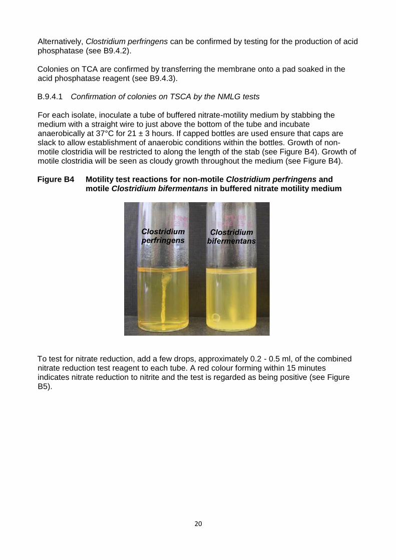
20
Alternatively, Clostridium perfringens can be confirmed by testing for the production of acid phosphatase (see B9.4.2). Colonies on TCA are confirmed by transferring the membrane onto a pad soaked in the acid phosphatase reagent (see B9.4.3). B.9.4.1 Confirmation of colonies on TSCA by the NMLG tests For each isolate, inoculate a tube of buffered nitrate-motility medium by stabbing the medium with a straight wire to just above the bottom of the tube and incubate anaerobically at 37°C for 21 ± 3 hours. If capped bottles are used ensure that caps are slack to allow establishment of anaerobic conditions within the bottles. Growth of non-motile clostridia will be restricted to along the length of the stab (see Figure B4). Growth of motile clostridia will be seen as cloudy growth throughout the medium (see Figure B4). Figure B4 Motility test reactions for non-motile Clostridium perfringens and
motile Clostridium bifermentans in buffered nitrate motility medium
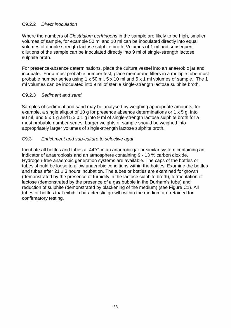
To test for nitrate reduction, add a few drops, approximately 0.2 - 0.5 ml, of the combined nitrate reduction test reagent to each tube. A red colour forming within 15 minutes indicates nitrate reduction to nitrite and the test is regarded as being positive (see Figure B5).

21
Figure B5 Nitrate reduction test reactions for Clostridium perfringens (positive) and Clostridium bifermentans (negative) in buffered
nitrate motility medium
If a red colour does not develop within 15 minutes, add a small amount of zinc powder and leave to stand for 10 minutes. If after this time there is still no red colour, this indicates that nitrate has been reduced to nitrite, which has been further reduced to nitrogen. This test is regarded as being positive. However, if a red colour subsequently develops after the addition of zinc powder, this indicates that nitrate has not been reduced and the test is regarded as being negative. In addition, inoculate a tube of lactose-gelatin medium by stabbing with a straight wire or inoculator and incubate anaerobically at 37°C for 44 ± 4 hours. After incubation the medium will be liquid, irrespective of whether gelatin liquefaction has occurred or not. In order to establish whether gelatin liquefaction has occurred, the tubes should be placed in a refrigerator for at least one hour. Gelatin liquefaction will have occurred in tubes where the medium remains liquid after refrigeration. If necessary, the tubes may be examined after incubating at 37°C for 21 ± 3 hours and refrigerated (for example, for about one hour) and if gelatin liquefaction occurs, i.e. the test is regarded as positive, the result is recorded. If negative, i.e. the medium remains solid after refrigeration, the tubes should be returned to the incubator. Incubation should be continued until the total incubation period of 44 ± 4 hours has been achieved. The tubes are then re-examined. Lactose fermentation is indicated by the colour of the medium turning from red to orange/yellow. A set of tubes inoculated with appropriate positive (Clostridium perfringens) and negative (Bacillus species) strains should be incubated and tested and included with each batch of tests. Clostridium perfringens are confirmed by the following reactions: (i) Non-motile - growth along the line of the stab and not spread through the buffered nitrate motility medium (ii) Nitrate reduction - red colour after addition of combined nitrate reduction test reagent to buffered nitrate-motility medium, or remaining colourless after addition of zinc powder.

22
(iii) Lactose fermentation - orange/yellow colouration of lactose-gelatin medium. (iv) Gelatin liquefaction - contents of the lactose-gelatin medium tube become liquefied. Further identification may be carried out by means of appropriate biochemical and other tests. Suitable commercial identification kits may be used following appropriate performance verification at the laboratory. B9.4.2 Confirmation of colonies on TSCA by the acid phosphatase test Clostridium perfringens can be confirmed by demonstration of acid phosphatase. Data on the verification of the performance of the acid phosphatase confirmation procedure are given in Appendix 1. Sub-culture presumptive positive colonies on TSCA onto Columbia blood agar base and incubate anaerobically at 37°C for 21 ± 3 hours. Place two or three drops of acid phosphatase reagent onto the growth. Development of a purplish or dark brown colour within three minutes is considered positive (see Figure B6). Figure B6 Positive (Clostridium perfringens) and negative (Clostridium
bifermentans) acid phosphatase reactions by dropping of acid phosphatase reagent on colonies on Columbia blood agar base
Left side of the Petri dishes show Clostridium perfringens (growth stained dark brown) and right side of the Petri dishes show Clostridium bifermentans (growth not dark brown) Alternatively, soak a filter paper with acid phosphatase reagent, transfer some of the colonies on the Columbia blood agar base and smear them onto the pre-soaked filter paper. The development of a purplish colour within three minutes is considered positive (see Figure B7).

23
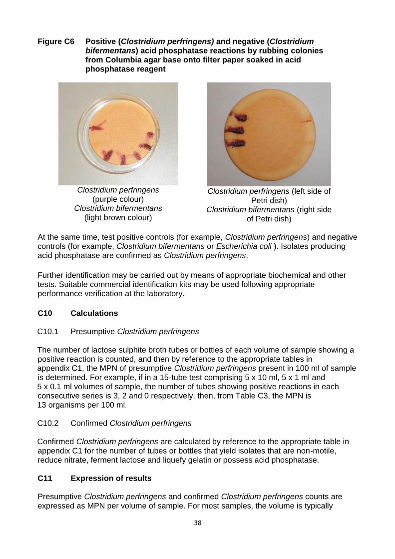
Figure B7 Positive (Clostridium perfringens) and negative (Clostridium bifermentans) acid phosphatase reactions by rubbing colonies
from Columbia agar base onto filter paper soaked in acid phosphatase reagent
Clostridium perfringens
(purple colour) Clostridium bifermentans
(light brown colour)
Clostridium perfringens (left side of
Petri dish) Clostridium bifermentans (right side
of Petri dish)
At the same time, test positive controls (for example, Clostridium perfringens) and negative controls (for example, Clostridium bifermentans or Escherichia coli). Isolates producing acid phosphatase are confirmed as Clostridium perfringens.
B9.4.3 Confirmation of colonies on TCA by the acid phosphatase test using membrane transfer Count all colourless colonies on TCA as presumptive Clostridium perfringens. Transfer the membrane to a pad soaked in acid phosphatase reagent and incubate at room temperature for 10 minutes. Count all dark or pale purple colonies as confirmed Clostridium perfringens (see Figure B8). Figure B8 TCA acid phosphatase positive colonies of Clostridium perfringens
24
At the same time, test positive controls (for example, Clostridium perfringens) and negative controls (for example, Clostridium bifermentans or Escherichia coli). Isolates producing acid phosphatase are confirmed as Clostridium perfringens. Further identification may be carried out by means of appropriate biochemical and other tests. Suitable commercial identification kits may be used following appropriate performance verification at the laboratory.
B10 Calculations B10.1 Presumptive Clostridium perfringens The number of presumptive Clostridium perfringens colonies is generally quoted as the number of colonies per 100 ml. Calculate the presumptive count as follows: Presumptive count per 100 ml = Number of colonies counted x 100 x DF Volume of sample filtered (ml) Where DF is dilution factor if appropriate. B10.2 Confirmed Clostridium perfringens The number of confirmed Clostridium perfringens colonies on TSCA is calculated by multiplying the number of presumptive Clostridium perfringens by the proportion of the isolates that are either non-motile, reduce nitrate, ferment lactose and liquefy gelatin or produce acid phosphatase. On TCA the number of confirmed Clostridium perfringens is the count of acid phosphatase positive colonies obtained from the membrane transfer technique. B11 Expression of results The number of presumptive and confirmed Clostridium perfringens is expressed in colony forming units per volume of sample. For most samples the volume is typically 100 ml. B12 Quality assurance New batches of isolation medium (TSCA or TCA) should be tested with appropriate reference strains of target bacteria (Clostridium perfringens) and non-target bacteria (for example Bacillus species). Petri dishes should be incubated for 21 ± 3 hours at 44°C. New batches of confirmatory media and reagents should be tested at 37°C with appropriate reference strains of bacteria chosen to verify positive and negative reactions in each case. Petri dishes should be incubated for 21 ± 3 hours at 37°C or 44°C as appropriate under anaerobic conditions. Further details of media and analytical quality control are given elsewhere(2) in this series. B13 References 1. Standing Committee of Analysts, The Microbiology of Recreational and Environmental Waters (2015) - Part 1 - Water quality, epidemiology and public health. Methods for the Examination of Waters and Associated Materials, in this series, Environment Agency.

25
2. Standing Committee of Analysts, The Microbiology of Drinking Water (2002) - Part 3 - Practices and procedures for laboratories. Methods for the Examination of Waters and Associated Materials, Environment Agency. 3. The Control of Substances Hazardous to Health Regulations 2002, Statutory Instrument 2002 No. 2677, The Stationery Office. 4. Enumeration of food-borne Clostridium perfringens in egg yolk free tryptose-sulphite-cycloserine agar, Applied Microbiology, A H W Hauschild and R Hillsheimer, 1974, 27, pp521-526. 5. Membrane filtration enumeration of faecal clostridia and Clostridium perfringens in water, Water Research, D P Sartory, 1986, 20, pp1255-1260. 6. The evaluation of a membrane filtration method for the rapid enumeration of confirmed Clostridium perfringens from water, Letters in Applied Microbiology, J Watkins and D P Sartory, 2015, in press. 7. Media for confirming Clostridium perfringens from food and feces, Journal of Food Protection, S M Harmon, and D A Kautter, 1978, 41, pp626-630. 8. Medical Microbiology, Volume Two: The Practice of Medical Microbiology, Twelfth Edition. Edited by R Cruikshank, J P Duguid, B P Marmion and R H Swain, Edinburgh, Churchill Livingstone,1975. 9. A study of rapid and simple confirmatory tests for Clostridium perfringens, Journal of Applied Bacteriology, G C Mead, L Paez de Leon, and B W Adams, 1981, 51, pp355-361.
26
C Enumeration of Clostridium perfringens by a multiple tube most probable number technique This method has not been subjected to widespread use nor verification of performance. Users of this method are encouraged to contact the Secretary of the Standing Committee of Analysts at the address given at the end of this booklet with their experiences and any relevant data on its performance. Information on the routine use of this method and similar methods, would be welcomed to assess their full capabilities.
C1 Introduction Tests for Clostridium perfringens play a secondary role in water quality assessment. The organism form spores which are resistant to environmental stress and can persist in the environment for some time. Clostridium perfringens are associated with faecal contamination. If found at a time when other faecal indicator organisms are no longer detectable, the organism may indicate remote or intermittent pollution. The organism may also be useful for monitoring the reduction of micro-organisms in wastewater treatment systems and the quality of final effluents, and assessing the effectiveness of water treatment. The significance of Clostridium perfringens in recreational and other waters is described elsewhere(1) in this series. C2 Scope The method is suitable for the examination of freshwater surface waters and saline waters, swimming pools, spa pools and hydrotherapy pools and primary and secondary wastewater effluents that contain high levels of sediments, or are highly turbid. Users wishing to employ this method should verify its performance under their own laboratory conditions(2). C3 Definitions Clostridium perfringens are Gram-positive anaerobic spore-forming rod-shaped bacteria which, in the context of this method, ferment lactose and reduce sulphite to sulphide at 44°C within 24 hours. Clostridium perfringens are also non-motile, reduce nitrate and liquefy gelatin. Clostridium perfringens also produce the enzyme acid phosphatase, which is a diagnostic characteristic for this species amongst the clostridia. C4 Principle Aliquots of sample are inoculated in a liquid medium containing lactose (with a Durham tube for collection of gas from fermentation of lactose), a source of sulphite and an iron(III) salt for the detection of reduction of sulphite. If only a spore count is required, then the sample is heat treated at 60 °C prior to inoculation in order to kill vegetative bacteria. In this method, measured volumes of sample, or dilution of sample, or membrane filtration filtered dilutions, are added to a series of tubes or bottles containing a liquid differential medium. If, following incubation, some of the tubes or bottles within the series exhibit no characteristic growth in the medium and other tubes or bottles exhibit some characteristic growth, then the most probable number of organisms in 100 ml of sample can be estimated from appropriate probability tables, see Appendix C1. Confirmation that positive

27
reactions (i.e. those tubes or bottles showing characteristic growth) are due to Clostridium perfringens can be obtained by sub-culture to obtain pure isolates followed by the inoculation of confirmation media. C5 Limitations This method is labour intensive and may require the preparation of large numbers of tubes or bottles of media and appropriate sub-cultures. C6 Health and safety Media, reagents and bacteria used in this method are covered by the Control of Substances Hazardous to Health Regulations(3) and appropriate risk assessments should be made before adopting this method. Standard laboratory microbiology safety procedures should be followed and guidance is given elsewhere(2) in this series. C7 Apparatus Standard laboratory equipment should be used which conforms to the performance criteria outlined elsewhere(2) in this series. Principally appropriate membrane filtration apparatus and incubators (fan assisted, static temperature) are required. Other items include: C7.1 Sterile sample bottles of appropriate volume, made of suitable material, should be used. For swimming pools, spa and hydrotherapy pools the containers should not be made of glass and should contain sufficient sodium thiosulphate pentahydrate to give a final concentration in the sample of not less than 18 mg/l. For example, 0.1 ml of a 1.8 % m/v solution of sodium thiosulphate pentahydrate (Na2S2O3.5H2O) per 100 ml of sample, or equivalent may be suitable. C7.2 Incubators capable of maintaining temperatures of 37.0 ± 1.0°C and 44.0 ± 0.5°C. C7.3 Waterbath capable of holding bottles of sample at 60 ± 2°C. C7.4 Anaerobic jars, or similar equipment, and anaerobic gas-generating system (for generating anaerobic atmospheres containing approximately 9 - 13 % carbon dioxide). C7.5 Suitable bottle or test tube racks. C8 Media and reagents Commercial formulations of these media and reagents may be available, but may possess minor variations to their formulation. The performance of all media and reagents should be verified prior to their use in this method(2). Variations in the preparation and storage of media should also be verified. Water should be distilled, deionised or of similar grade quality. Unless otherwise stated chemical constituents should be added as the anhydrous salts. If the pH of media are not within the stated range, then before heating, they should be adjusted accordingly. C8.1 Single-strength lactose sulphite broth (LSB)(4) Yeast extract 2.5 g
28
Tryptone 5 g Lactose 10 g Sodium chloride 2.5 g Cysteine hydrochloride 0.3 g Sodium metabisulphite 0.7 g Iron (III) ammonium citrate 0.6 g Water 1 litre
Gently heat the ingredients in the water to dissolve and then allow to cool. Adjust the pH of the solution to 7.1 ± 0.2. Distribute the solution into suitable tubes or bottles containing an inverted fermentation (Durham) tube in 10 ml and 50 ml volumes. Cap the containers and sterilise at 115°C for 20 minutes. This single-strength formulation medium may be stored at a temperature in the range 5 ± 3°C for not more than two weeks. Prepare double-strength LSB medium by gently heating twice the amount of ingredients in 1000 ml of water to dissolve and then allow to cool. Adjust the pH of the solution to 7.1 ± 0.2. Distribute in 5 ml volumes in tubes containing an inverted fermentation (Durham) tube. Sterilise at 115°C for 20 minutes. This double-strength formulation medium may be stored at a temperature in the range 5 ± 3°C for not more than two weeks. C8.2 Buffered nitrate-motility medium(5)
Beef extract 3 g Peptone 5 g Potassium nitrate 5 g D-Galactose 5 g Glycerol 5 g Disodium hydrogen phosphate 2.5 g Agar 3 g Water 1 litre
Dissolve the solid ingredients in 950 ml water by heating to boiling point whilst stirring continuously. Dissolve the glycerol in 50 ml water in a separate container and add this solution to the base medium and mix thoroughly. Dispense the resulting solution, typically in 10 ml aliquots, in appropriately sized tubes. Cap the tubes. Sterilise the medium by autoclaving at 121°C for 15 minutes. The final pH of the medium should be 7.3 ± 0.2. Prepared tubes may be stored at a temperature in the range 5 ± 3°C for up to one month, if protected against dehydration. Before use, stored tubes should be heated for 10 - 15 minutes in a boiling water bath, to ensure that the contents have melted and to eliminate any absorbed oxygen. The tubes should then be allowed to cool and the media to solidify ready for use. C8.3 Nitrate reduction test reagents(6)
Reagent A Sulphanilic acid 0.8 g Glacial acetic acid 30 ml Water to 100 ml
Warm gently to aid dissolution.

29
Reagent B N, N-dimethyl-1-naphthylamine 0.6 ml Glacial acetic acid 30 ml Water to 100 ml
Dissolve the amine in the acetic acid solution. To aid dissolution, warm gently (for example, by placing in a water bath at 40 - 60°C).
The reagents may be stored at a temperature in the range 5 ± 3°C for up to six months. For the combined reagent, mix equal volumes of reagents A and B immediately prior to use. Prepare in small volumes sufficient for the tests to be performed. The combined reagent may be stored at a temperature of 5 ± 3°C, protected from direct light, and should be used within 24 hours. C8.4 Lactose-gelatin medium(5)
Tryptose 15 g Yeast extract 10 g Disodium hydrogen phosphate 5 g Gelatin 120 g Lactose 10 g Phenol red (0.4 % m/v solution) 12.5 ml Water 1 litre
Dissolve the ingredients, except the gelatin, lactose and phenol red, in the water. Add the gelatin gradually whilst stirring the mixture continuously and warming gently to aid dissolution. Adjust the pH to 7.5 ± 0.2. Add the lactose and dissolve and add the phenol red solution. Dispense the resulting solution, typically in 10 ml aliquots in appropriately sized tubes. Cap the tubes. Sterilise the medium at 121°C for 15 minutes. The final pH should be 7.5 ± 0.2. Prepared media may be stored at a temperature in the range 5 ± 3°C for up to one month if protected against dehydration. Before use stored media should be heated for 10 - 15 minutes in a boiling water bath, to ensure that the contents have melted and to eliminate any absorbed oxygen. The tubes should then be allowed to cool and the media to solidify ready for use. C8.5 Columbia blood agar base
Special peptone 23 g Starch 1 g Sodium chloride 5 g Agar 15 g Water 1 litre
Suspend the ingredients in the water and dissolve by heating and stirring the mixture. Sterilise the solution by autoclaving at 121°C for 15 minutes. Cool and dispense into Petri dishes. The Petri dishes should be the vented type to ensure anaerobic conditions for the medium during incubation. The final pH of the medium should be 7.3 ± 0.2. Sterile media

30
may be stored at a temperature of 5 ± 3°C for up to one month, if protected against dehydration. C8.6 Columbia blood agar Special peptone 23 g Starch 1 g Sodium chloride 5 g Agar 15 g Horse blood 50 ml D-cysloserine 400mg Water 1 litre Suspend the ingredients, except the horse blood and D-cycloserine, in the water and dissolve by heating and stirring the mixture. Sterilise the solution by autoclaving at 121°C for 15 minutes. Cool the medium, add 5% (v/v) horse blood and 4 ml of a filter-sterilised solution of D-cycloserine in distilled water at a concentration of 100 mg/ml. Mix well and dispense into Petri dishes. The Petri dishes should be the vented type to ensure anaerobic conditions for the medium during incubation. The final pH of the medium should be 7.3 ± 0.2. Sterile media may be stored at a temperature of 5 ± 3°C for up to one week, if protected against dehydration. C8.7 Acid phosphatase reagent(7) Acetate buffer Glacial acetic acid 0.3 ml Sodium acetate 0.4 g Water 100 ml Thoroughly mix the ingredients. The final pH value should be 4.6 ± 0.2. The buffer may be stored at room temperature for up to six months. Complete reagent 1-naphthyl phosphate monosodium salt 0.4 g o-dianisidine tetrazotized zinc chloride complex 0.8 g (Fast Blue B) Acetate buffer 20 ml Add the ingredients to the acetate buffer and shake well to dissolve. Store the reagent at 5 ± 3°C for one hour. Filter the solution to remove any precipitate. The reagent may be stored at 5 ± 3°C for up to two weeks.
C8.8 Filter-aid(8)
Diatomaceous earth 1 g (approximately) Water 15 ml
Weigh out appropriate amounts of filter-aid into suitable bottles, add the water and cap. Sterilise by autoclaving at 121°C for 15 minutes. The sterilised filter-aid may be stored in the dark at room temperature for up to 12 months.

31
C8.9 Other media Standard and commercial formulations of other media and reagents used in this method include zinc powder, quarter-strength Ringer’s solution and maximum recovery diluent. C9 Analytical procedure C9.1 Sample preparation C9.1.1 Surface waters and sea water Due to the likelihood that, if present, the numbers of Clostridium perfringens in some surface waters and sea water are likely to be low, for presence-absence determinations, a sample volume of at least 1000 ml should be examined. For the membrane filtration multiple tube technique, typically, an 11-tube series can be used, i.e. the membrane filtration of 1 x 500 ml, 5 x 100 ml and 5 x 10 ml of sample. Alternatively, volumes of 1 x 500 ml and 5 x 100 ml can be filtered and the 10 ml volumes can be added directly to 10 ml volumes of double-strength lactose sulphite broth. For a different series, smaller volumes of sample, for example 1 ml, may be appropriate and these can be added directly to 9 ml of single-strength lactose sulphite broth. Turbid waters, unsuitable for direct membrane filtration, may be filtered using filter aid (C8.8). C9.1.2 Treated wastewater Treated wastewater may be analysed as described in C9.1.1 although several membrane filters may be required for presence-absence determinations. A sample volume of at least 100 ml may need to be examined. The volumes may be reduced and volumes of 1 x 50 ml, 5 x 10 ml and 5 x 1 ml be used. The 1 x 50 ml and 5 x 10 ml volumes can be membrane filtered or added to equal volumes of double-strength lactose sulphite broth. To represent smaller volumes of samples, a 1:10 dilution of the sample, for example 1 ml of sample diluted with quarter strength Ringer’s solution or maximum recovery diluent, may be appropriate. Typically, 1 ml of these diluted samples can be added directly to 9 ml of single-strength lactose sulphite broth. C9.1.3 Untreated wastewater For presence-absence determinations, 100 ml of untreated wastewater sample may be required, as it may not be possible (owing to turbidity) to process larger volumes by membrane filtration. For an 11-tube most probable number series, the volumes of untreated wastewater are usually 1 x 50 ml, 5 x 10 ml and 5 x 1 ml. The 1 x 50 ml and 5 x 10 ml volumes can be filtered or added to equal volumes of double-strength lactose sulphite broth. The 1 ml volumes can be added to 9 ml of single-strength lactose sulphite broth. To represent smaller volumes, for example 0.1 ml and 0.01 ml volumes of sample, a 1:10 and 1:100 dilution of the sample, may be appropriate. Typically, 1 ml of these diluted samples can be added directly to 9 ml of single-strength lactose sulphite broth. C9.1.4 Sediment and sand Solid material can be dispensed as a single weight for presence-absence determinations
32
by weighing, for example 10 g of sample into an appropriate volume (typically 100 ml) of single-strength lactose sulphite broth. For the multiple tube technique, weigh 1 x 50 g, 5 x 10 g and 5 x 1 g quantities of sample into appropriate volumes (typically 450 ml, 5 x 100 ml and 5 x 10 ml respectively) of single-strength lactose sulphite broth. For smaller quantities, for example 100 mg, these may be added directly to 10 ml of single-strength lactose sulphite broth. C9.2 Sample processing C9.2.1 Membrane presence-absence or filtration multiple tube technique Appropriate volumes of sample are filtered through membrane filters.
Place the sterile or disinfected filtration apparatus in position and connect to a source of vacuum, with the stopcock turned off. Remove the funnel and, holding the edge of the membrane filter with sterile smooth-tipped forceps, place a sterile membrane filter onto the porous disc of the filter base. If a gridded membrane filter is used, place grid-side upwards. Replace the sterile funnel securely on the filter base. Pour or pipette the required volume of sample into the funnel. Open the stopcock and apply a vacuum not exceeding 65 kPa (500 mm of mercury) and filter the water slowly through the membrane filter. Close the stopcock as soon as the sample has been filtered. Remove the funnel and carefully transfer the membrane filter to a tube or bottle containing, typically, 10 - 15 ml of single-strength lactose sulphite broth, ensuring that the membrane filter is fully submerged. Record the volume filtered. Other volumes of sample should be similarly treated until all the filters are transferred to the corresponding tubes or bottles of single-strength lactose sulphite broth. The largest single volume of sample may require more than one membrane filter and, if so, all filters used for this volume should be transferred to the bottle or tube of single-strength lactose sulphite broth. Ensure that all membrane filters are fully submerged. Where an MPN serried of different volumes of the same sample are to be examined, the same funnel may be re-used provided that the smallest volume of sample is filtered first. For different samples, a fresh pre-sterilised funnel should be used. As the spores of Clostridium perfringens are very resilient, funnels that have been used once should be sterilised by autoclaving before being used again. Placing funnels in a water bath at this stage may not be sufficient to kill spores. If different volumes of the same sample are to be examined, the funnel may be re-used without sterilising the funnel provided that the smallest volume, or highest dilution of sample, is filtered first. For different samples, take a fresh pre-sterilised funnel and repeat the filtration process. During the filtration of a series of samples, the filter base need not be sterilised unless it becomes contaminated or a membrane filter becomes damaged. Known polluted and non-polluted samples should be filtered using separate filtration equipment. Alternatively, polluted samples should only be processed after non-polluted samples. When disinfected funnels are not in use they should be covered with a sterile lid or a sterile Petri dish lid. Alternatively, commercially available, sterile, disposable funnels may be used. The time between the end of the filtration step and the beginning of the incubation stage should be as short as possible and no longer than 2 hours.
33
C9.2.2 Direct inoculation
Where the numbers of Clostridium perfringens in the sample are likely to be high, smaller volumes of sample, for example 50 ml and 10 ml can be inoculated directly into equal volumes of double strength lactose sulphite broth. Volumes of 1 ml and subsequent dilutions of the sample can be inoculated directly into 9 ml of single-strength lactose sulphite broth. For presence-absence determinations, place the culture vessel into an anaerobic jar and incubate. For a most probable number test, place membrane filters in a multiple tube most probable number series using 1 x 50 ml, 5 x 10 ml and 5 x 1 ml volumes of sample. The 1 ml volumes can be inoculated into 9 ml of sterile single-strength lactose sulphite broth. C9.2.3 Sediment and sand
Samples of sediment and sand may be analysed by weighing appropriate amounts, for example, a single aliquot of 10 g for presence absence determinations or 1 x 5 g, into 90 ml, and 5 x 1 g and 5 x 0.1 g into 9 ml of single-strength lactose sulphite broth for a most probable number series. Larger weights of sample should be weighed into appropriately larger volumes of single-strength lactose sulphite broth. C9.3 Enrichment and sub-culture to selective agar Incubate all bottles and tubes at 44°C in an anaerobic jar or similar system containing an indicator of anaerobiosis and an atmosphere containing 9 - 13 % carbon dioxide. Hydrogen-free anaerobic generation systems are available. The caps of the bottles or tubes should be loose to allow anaerobic conditions within the bottles. Examine the bottles and tubes after 21 ± 3 hours incubation. The tubes or bottles are examined for growth (demonstrated by the presence of turbidity in the lactose sulphite broth), fermentation of lactose (demonstrated by the presence of a gas bubble in the Durham’s tube) and reduction of sulphite (demonstrated by blackening of the medium) (see Figure C1). All tubes or bottles that exhibit characteristic growth within the medium are retained for confirmatory testing.

34
Figure C1 Characteristic growth in lactose sulphite broth
1 x 50 ml, plus equal
volume of double-
strength medium
5 x 10 ml, plus equal volumes of
double-strength medium
5 x 1 ml, plus 9 ml of single-strength
medium
Bottles that exhibit growth within the medium are indicated by black colouration, regard these as positive. Bottles that exhibit no growth within the medium are indicated by yellow colouration, regard these as negative. C9.4 Reading of results The number of tubes or bottles for the series of each sample volume is recorded where a positive reaction is given, as demonstrated by growth, lactose fermentation and sulphite reduction. After this, confirmation tests are carried out as required. When dilutions of sample have been used, a consecutive series of volumes is chosen whereby some of the tubes or bottles show a positive reaction and some show a negative reaction. From the results, the MPN of bacteria in the sample is determined from probability tables, see Appendix C1. C9.5 Confirmation tests For each tube or bottle showing growth within the medium, sub-culture to a Columbia blood agar (C8.6) Petri dish in a manner that enables or encourages single colonies to grow. Incubate the Petri dishes anaerobically at 37°C for 21 ± 3 hours. Presumptive Clostridium perfringens will grow as large convex colonies (generally smooth with a regular edge, but may be rough with an irregular edge and 2 - 4 mm in diameter), usually with a zone of β-haemolysis (see Figure C2). Colonies with diffuse spreading morphologies are considered motile and not subjected to confirmation testing and the corresponding tube or bottle is regarded as negative for confirmed Clostridium perfringens. If a pure culture is obtained on Columbia blood agar then perform the confirmation tests. Where a mixed

35
culture is obtained, sub-culture a typical colony to a fresh Columbia blood agar base (C8.5) Petri dish and incubate at 37 ± 1°C anaerobically for 21 ± 3 hours before testing. Figure C2 Colonies of Clostridium perfringens showing β-haemolysis on
Columbia blood agar supplemented with D-cycloserine
Clostridium perfringens can be confirmed by testing for reduction of nitrate, motility, fermentation of lactose and liquefaction of gelatin (i.e. the NMLG tests). Alternatively, Clostridium perfringens can be confirmed by testing for the production of acid phosphatase. C.9.5.1 Confirmation by the NMLG tests For each isolate, inoculate a tube of buffered nitrate-motility medium by stabbing the medium with a straight wire to just above the bottom of the tube and incubate anaerobically at 37°C for 21 ± 3 hours. If capped bottles are used ensure that caps are slack to allow establishment of anaerobic conditions within the bottles. Growth of non-motile clostridia will be restricted to along the length of the stab (see Figure C3). Growth of motile clostridia will be seen as cloudy growth throughout the medium (see Figure C3). Figure C3 Motility test reactions for non-motile Clostridium perfringens and
motile Clostridium bifermentans in buffered nitrate motility medium
36
To test for nitrate reduction, add a few drops, approximately 0.2 - 0.5 ml, of the combined nitrate reduction test reagent to each tube. A red colour forming within 15 minutes indicates nitrate reduction to nitrite and the test is regarded as being positive (see Figure C4). Figure C4 Nitrate reduction test reactions for Clostridium perfringens (positive) and Clostridium bifermentans (negative) in buffered nitrate motility
medium
If a red colour does not develop within 15 minutes, add a small amount of zinc powder and leave to stand for 10 minutes. If after this time there is still no red colour, this indicates that nitrate has been reduced to nitrite, which has been further reduced to nitrogen. This test is regarded as being positive. However, if a red colour subsequently develops after the addition of zinc powder, this indicates that nitrate has not been reduced and the test is regarded as being negative. In addition, inoculate a tube of lactose-gelatin medium by stabbing with a straight wire or inoculator and incubate anaerobically at 37°C for 44 ± 4 hours. After incubation the medium will be liquid, irrespective of whether gelatin liquefaction has occurred or not. In order to establish whether gelatin liquefaction has occurred, the tubes should be placed in a refrigerator for at least one hour. Gelatin liquefaction will have occurred in tubes where the medium remains liquid after refrigeration. If necessary, the tubes may be examined after incubating at 37°C for 21 ± 3 hours and refrigerated (for example, for about one hour) and if gelatin liquefaction occurs, i.e. the test is regarded as positive, the result is recorded. If negative, i.e. the medium remains solid after refrigeration, the tubes should be returned to the incubator. Incubation should be continued until the total incubation period of 44 ± 4 hours has been achieved. The tubes are then re-examined. Lactose fermentation is indicated by the colour of the medium turning from red to orange/yellow. A set of tubes inoculated with appropriate positive (Clostridium perfringens) and negative (Bacillus species) strains should be incubated and tested in parallel. Clostridium perfringens are confirmed by the following reactions:

37
(i) Non-motile - growth along the line of the stab and not spread through the buffered nitrate motility medium. (ii) Nitrate reduction - red colour after addition of combined nitrate reduction test reagent to buffered nitrate-motility medium, or remaining colourless after addition of zinc powder. (iii) Lactose fermentation - orange/yellow colouration of lactose-gelatin medium. (iv) Gelatin liquefaction - contents of the lactose-gelatin medium tube become liquefied. Further identification may be carried out by means of appropriate biochemical and other tests. Suitable commercial identification kits may be used following appropriate performance verification at the laboratory. C9.5.2 Confirmation by the acid phosphatase test Clostridium perfringens can be confirmed by demonstration of acid phosphatase. Data on the verification of the performance of the acid phosphatase confirmation procedure are given in Appendix 1. Place two or three drops of acid phosphatase reagent onto the growth. Development of a purplish or dark brown colour within three minutes is considered positive (see Figure C5). Figure C5 Positive (Clostridium perfringens) and negative (Clostridium
bifermentans) acid phosphatase reactions by dropping of acid phosphatase reagent on colonies on Columbia blood agar base
Left side of the Petri dishes show Clostridium perfringens (growth stained dark brown) and right side of the Petri dishes show Clostridium bifermentans (growth not dark brown). Alternatively, soak a filter paper with acid phosphatase reagent, transfer some of the colonies on the Columbia agar base and smear them onto the pre-soaked filter paper. The development of a purplish colour within three minutes is considered positive (see Figure C6).
38
Figure C6 Positive (Clostridium perfringens) and negative (Clostridium bifermentans) acid phosphatase reactions by rubbing colonies
from Columbia agar base onto filter paper soaked in acid phosphatase reagent
Clostridium perfringens
(purple colour) Clostridium bifermentans
(light brown colour)
Clostridium perfringens (left side of
Petri dish) Clostridium bifermentans (right side
of Petri dish) At the same time, test positive controls (for example, Clostridium perfringens) and negative controls (for example, Clostridium bifermentans or Escherichia coli ). Isolates producing acid phosphatase are confirmed as Clostridium perfringens.
Further identification may be carried out by means of appropriate biochemical and other tests. Suitable commercial identification kits may be used following appropriate performance verification at the laboratory.
C10 Calculations C10.1 Presumptive Clostridium perfringens The number of lactose sulphite broth tubes or bottles of each volume of sample showing a positive reaction is counted, and then by reference to the appropriate tables in appendix C1, the MPN of presumptive Clostridium perfringens present in 100 ml of sample is determined. For example, if in a 15-tube test comprising 5 x 10 ml, 5 x 1 ml and 5 x 0.1 ml volumes of sample, the number of tubes showing positive reactions in each consecutive series is 3, 2 and 0 respectively, then, from Table C3, the MPN is 13 organisms per 100 ml. C10.2 Confirmed Clostridium perfringens Confirmed Clostridium perfringens are calculated by reference to the appropriate table in appendix C1 for the number of tubes or bottles that yield isolates that are non-motile, reduce nitrate, ferment lactose and liquefy gelatin or possess acid phosphatase. C11 Expression of results Presumptive Clostridium perfringens and confirmed Clostridium perfringens counts are expressed as MPN per volume of sample. For most samples, the volume is typically

39
100 ml. For sediment and sand samples the counts are usually expressed as MPN per wet weight of sample, usually adjusted to MPN count per gram wet weight. For inter-laboratory comparison purposes, a dry weight basis may be more appropriate(9). C12 Quality assurance New batches of isolation medium should be tested with appropriate reference strains of target bacteria (for example Clostridium perfringens) and non-target bacteria (for example Bacillus species). New batches of confirmatory media and reagents should be tested with appropriate reference strains of bacteria chosen to verify a positive and a negative reaction in each case. Tubes should be incubated for 21 ± 3 hours at 37°C or 44°C under anaerobic conditions as appropriate. Further details are given elsewhere(2) in this series. C13 References 1. Standing Committee of Analysts, The Microbiology of Recreational and Environmental Waters (2015) - Part 1 - Water quality, epidemiology and public health. Methods for the Examination of Waters and Associated Materials, in this series, Environment Agency. 2. Standing Committee of Analysts, The Microbiology of Drinking Water (2002) - Part 3 - Practices and procedures for laboratories. Methods for the Examination of Waters and Associated Materials, Environment Agency. 3. The Control of Substances Hazardous to Health Regulations 2002, Statutory Instrument 2002 No. 2677, The Stationery Office. 4. A liquid medium for the enumeration of Clostridium perfringens in food and faeces. In Isolation and Identification Methods for Food Poisoning Organisms (Edited by J E L Corry, D Roberts and F A Skinner) London, Academic Press, H Beerens, C L Romond, C Lepage and J Criquelion, 1982, pp137-149. 5. Media for confirming Clostridium perfringens from food and faeces, Journal of Food Protection, S M Harmon and D A Kautter, 1978, 41, pp626-630. 6. Medical Microbiology, Volume Two: The Practice of Medical Microbiology, Twelfth Edition. Edited by R Cruikshank, J P Duguid, B P Marmion and R H A Swain, Edinburgh, Churchill Livingstone,1975. 7. A study of rapid and simple confirmatory tests for Clostridium perfringens, Journal of Applied Bacteriology, G C Mead, L Paez de Leon, and B W Adams, 1981, 51, pp355-361. 8. Concentration technique for demonstrating small amounts of bacteria in tap water, Acta Pathologica et Microbiologica Scandinavia, E Hammarstrom and V Ljutov, 1954, 35, 365-369. 9. Standing Committee of Analysts, The Microbiology of Sewage Sludge (2003) - Part 2- Practices and procedures for sampling and sample preparation, Methods for the Examination of Waters and Associated Materials, Environment Agency.
40
Appendix C1 Tables of most probable numbers From the various combinations of positive and negative reactions for the different volumes examined, the following tables indicate the MPN of bacteria in 100 ml of sample. It is important to realise that the MPN is only an estimate, based on statistical probabilities and that the actual number may lie within a range of values. Approximate 95 % confidence intervals, which demonstrate the range of possible numbers (the MPR) which could yield the number of positive reactions, have been published(1). A procedure for estimating these confidence intervals for other dilution series has also been published(2). These confidence intervals are seldom of practical use when reporting results because they apply to the accuracy of the method and not the likely variability of organisms at the sampling source(3). The MPR in tables C1 - C3 illustrates those situations where the method becomes relatively imprecise, particularly when nearly all the tubes show growth within the medium. In these situations, further dilutions should have been prepared and added to tubes of medium. Table C1 gives the MPN (and where applicable the MPR) for a 6-tube series containing 1 x 50 ml and 5 x 10 ml volumes of sample. Similarly table C2 gives the MPN (and where applicable the MPR) for an 11-tube series comprising 1 x 50 ml, 5 x 10 ml and 5 x 1 ml volumes of sample. Table C3 shows data for a 15-tube series of 5 x 10 ml, 5 x 1 ml and 5 x 0.1 ml volumes of samples but gives only those values of the more likely combinations of positive and negative reactions. For example, positive reactions in the 0.1 ml tubes would not be expected if all of the 10 ml and 1 ml tubes were negative. Hence, MPN and MPR values for a combination of results like for instance 0, 0, 2 etc are not tabulated. If these unlikely combinations are observed in practice with greater than expected frequencies, then this might indicate that the statistical assumptions underlying the MPN estimation are not correct(3, 4, 5). For example, the organisms may not have been uniformly distributed throughout the sample, or toxic substances may have been present. Calculation of MPN The number of positive reactions for each set of tubes is recorded and, from the relevant table, the MPN of organisms present in 100 ml of the sample is determined. Where a series of dilutions of the sample is used, then the following rules should be applied, as illustrated by the numbers in bold, underlined, italic type in table C4. (i) Use only three consecutive sets of dilutions for calculating the MPN. (ii) Wherever possible, select three consecutive dilutions where the results are neither all positive nor all negative. The most efficient statistical estimate will result when about half the tubes are positive (see examples (a), (b) and (c) in table C4). (iii) If less than three sets of dilutions give positive results, begin with the set containing the largest volume of sample (see example (d) in table C4). (iv) If only one set of tubes gives a positive reaction, use this dilution and the one higher and one lower (see example (e) in table C4).

41
Table C1 MPN and MPR per 100 ml of sample for a 6-tube series containing 1 x 50 ml and 5 x 10 ml volumes of sample
Number of tubes giving a positive reaction MPN per 100 ml MPR* per 100 ml
1 x 50 ml 5 x 10 ml
0 0 None found 0 1 1 0 2 2 0 3 3 0 4 4 4-5 0 5 6 1 0 1 1 1 2 1 2 5 4-5 1 3 9 8-10 1 4 15 13-18 1 5 >18**
* These numbers are at least 95 % as probable as the MPN. ** There is no discrimination when all tubes are positive; the theoretical
MPN is infinity. The true count is likely to exceed 18.
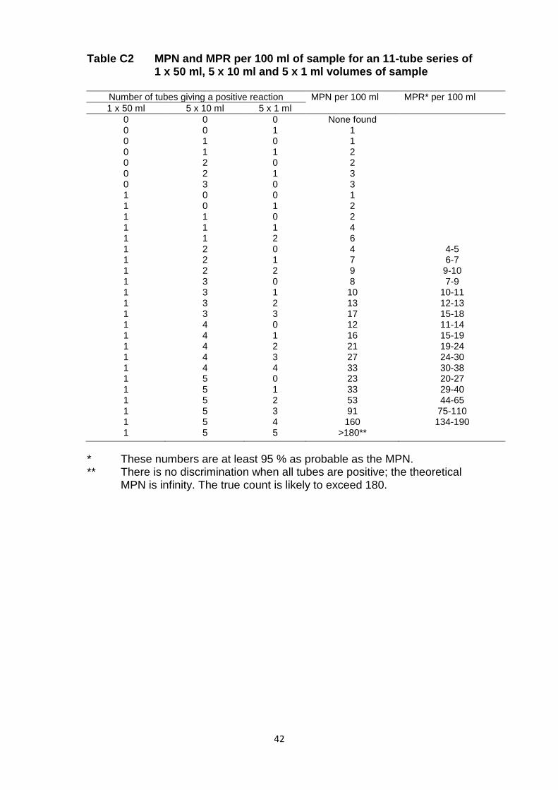
42
Table C2 MPN and MPR per 100 ml of sample for an 11-tube series of 1 x 50 ml, 5 x 10 ml and 5 x 1 ml volumes of sample
Number of tubes giving a positive reaction MPN per 100 ml MPR* per 100 ml
1 x 50 ml 5 x 10 ml 5 x 1 ml
0 0 0 None found 0 0 1 1 0 1 0 1 0 1 1 2 0 2 0 2 0 2 1 3 0 3 0 3 1 0 0 1 1 0 1 2 1 1 0 2 1 1 1 4 1 1 2 6 1 2 0 4 4-5 1 2 1 7 6-7 1 2 2 9 9-10 1 3 0 8 7-9 1 3 1 10 10-11 1 3 2 13 12-13 1 3 3 17 15-18 1 4 0 12 11-14 1 4 1 16 15-19 1 4 2 21 19-24 1 4 3 27 24-30 1 4 4 33 30-38 1 5 0 23 20-27 1 5 1 33 29-40 1 5 2 53 44-65 1 5 3 91 75-110 1 5 4 160 134-190 1 5 5 >180**
* These numbers are at least 95 % as probable as the MPN. ** There is no discrimination when all tubes are positive; the theoretical
MPN is infinity. The true count is likely to exceed 180.

43
Table C3 MPN and MPR per 100 ml of sample for a 15-tube series containing 5 x 10 ml, 5 x 1 ml and 5 x 0.1 ml volumes of sample
Number of tubes giving a positive reaction MPN per 100 ml MPR* per 100 ml
5 x 10 ml 5 x 1 ml 5 x 0.1 ml
0 0 0 None found 0 0 1 2 0 1 0 2 1 0 0 2 1 0 1 4 1 1 0 4 2 2 0 4 2 0 1 5 2 1 0 5 2 1 1 7 2 2 0 7 7-9 2 3 0 11 3 0 0 7 3 0 1 9 3 1 0 9 3 1 1 13 3 2 0 13 3 2 1 16 14-16 3 3 0 16 14-16 4 0 0 11 11-13 4 0 1 14 14-16 4 1 0 16 14-16 4 1 1 20 18-20 4 2 0 20 18-22 4 2 1 25 23-27 4 3 0 25 23-27 4 3 1 31 29-34 4 4 0 32 29-34 4 4 1 38 34-41 5 0 0 22 20-23 5 0 1 29 25-34 5 0 2 41 36-50 5 1 0 31 27-36 5 1 1 43 36-50 5 1 2 60 50-70 5 1 3 85 70-95 5 2 0 50 40-55 5 2 1 70 60-80 5 2 2 95 80-110 5 2 3 120 105-135 5 3 0 75 65-90 5 3 1 110 90-125 5 3 2 140 120-160 5 3 3 175 155-200 5 3 4 210 185-240 5 4 0 130 110-150 5 4 1 170 150-200 5 4 2 220 190-250 5 4 3 280 240-320 5 4 4 345 300-390 5 5 0 240 200-280 5 5 1 350 290-420 5 5 2 540 450-600 5 5 3 910 750-1100 5 5 4 1600 1350-1900 5 5 5 >1800**
* These numbers are at least 95 % as probable as the MPN. ** There is no discrimination when all tubes are positive; the theoretical
MPN is infinity. The true count is likely to exceed 1800.
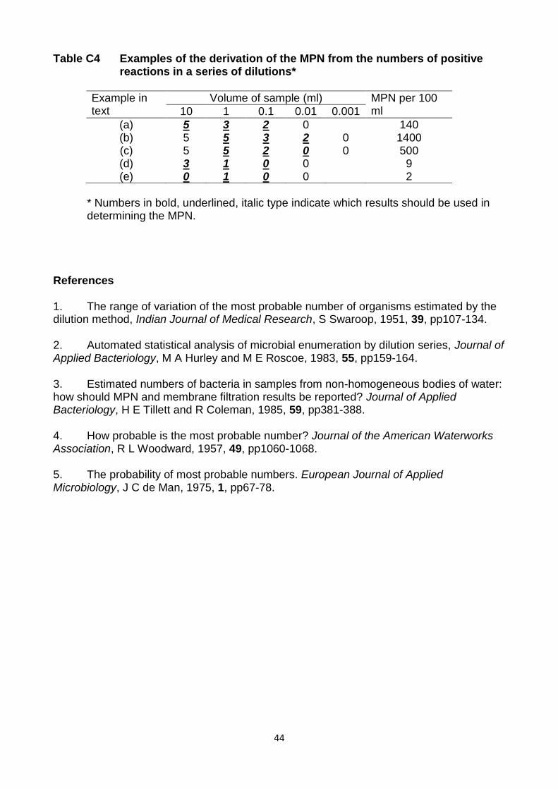
44
Table C4 Examples of the derivation of the MPN from the numbers of positive reactions in a series of dilutions*
Example in text
Volume of sample (ml) MPN per 100 ml 10 1 0.1 0.01 0.001
(a) 5 3 2 0 140 (b) 5 5 3 2 0 1400 (c) 5 5 2 0 0 500 (d) 3 1 0 0 9 (e) 0 1 0 0 2
* Numbers in bold, underlined, italic type indicate which results should be used in
determining the MPN. References 1. The range of variation of the most probable number of organisms estimated by the dilution method, Indian Journal of Medical Research, S Swaroop, 1951, 39, pp107-134. 2. Automated statistical analysis of microbial enumeration by dilution series, Journal of Applied Bacteriology, M A Hurley and M E Roscoe, 1983, 55, pp159-164. 3. Estimated numbers of bacteria in samples from non-homogeneous bodies of water: how should MPN and membrane filtration results be reported? Journal of Applied Bacteriology, H E Tillett and R Coleman, 1985, 59, pp381-388. 4. How probable is the most probable number? Journal of the American Waterworks Association, R L Woodward, 1957, 49, pp1060-1068. 5. The probability of most probable numbers. European Journal of Applied Microbiology, J C de Man, 1975, 1, pp67-78.

45
Appendix 1 Verification of the acid phosphatase test for the confirmation of
Clostridium perfringens isolated from various waters
1 Introduction In an earlier document(1) in the series for drinking water analysis procedures are described for confirming presumptive Clostridium perfringens from membrane filters incubated on tryptone sulphite cycloserine (TSC) agar involving sub-culture to buffered nitrate-motility medium and lactose-gelatin medium (i.e. the NMLG tests) to test for nitrate reduction, motility, lactose fermentation and gelatin liquefaction. Isolates that reduce nitrate, are non-motile, ferment lactose and liquefy gelatin are considered to be confirmed as Clostridium perfringens. An alternative method for confirming Clostridium perfringens based upon the demonstration of the production of acid phosphatase has been reported(2) where the acid phosphatase test was reportedly more specific for Clostridium perfringens than the NMLG tests. However, some strains of other species of Clostridium were also found to be acid phosphatase-positive. Of 114 environmental isolates of Clostridium perfringens, 108 (i.e. 94.7 %) were acid phosphatase-positive compared to 104 (i.e. 91.2 %) that produced typical reactions in the NMLG tests(2). Failure to reduce nitrate was the most common atypical result from the NMLG tests, which complements reports(3, 4) that about 10 % of strains of Clostridium perfringens are nitrate-negative A multi-laboratory study was therefore organised under the auspices of the Standing Committee of Analysts to assess the acid phosphate test for the confirmation of Clostridium perfringens and to demonstrate the equivalency of the method to the NMLG tests procedure(1). 2 Materials and Methods Samples from a range of environmental water types were analysed according to previously published procedures(1) using membrane filtration and enumeration on TSC agar. Following incubation, membrane filters exhibiting between 10 - 30 colonies were selected for confirmation, and colonies were counted and presumptive counts recorded.
Colonies of presumptive Clostridium perfringens were sub-cultured onto Columbia agar
base and incubated anaerobically at 37 1°C for 21 ± 3 hours. For each isolate, a tube of buffered nitrate-motility medium was inoculated by stabbing with a straight wire or inoculator to just above the bottom of the tube and incubated anaerobically at 37 °C for 21 ± 3 hours. Testing for nitrate reduction was achieved by adding a few drops, approximately 0.2 - 0.5 ml, of the combined nitrate test reagent to each tube. A red colour forming within 15 minutes indicates nitrate reduction to nitrite and the test was regarded as positive. If a red colour did not develop within this time, a small amount of zinc powder was added and the tube left to stand for 10 minutes. If, after this time there was still no red colour, this indicates that the nitrate has been reduced to nitrite, which had been further reduced, and the test was, therefore, deemed positive. However, if a red colour subsequently developed after the addition of zinc powder, this indicates that nitrate had not been reduced and the test was regarded as being negative. Motility was assessed as growth along the line of the stab spreading through buffered nitrate-motility medium.

46
In addition, a tube of lactose-gelatin medium was inoculated by stabbing with a straight wire or inoculator and incubated anaerobically at 37 °C for a minimum of 21 ± 3 hours and a maximum of 44 ± 4 hours. After incubation the tubes were placed in a refrigerator for at least one hour. A positive gelatin liquefaction reaction was recorded for tubes where the medium remained liquid after refrigeration. Tubes examined after 21 ± 3 hours incubation that were negative, i.e. did not exhibit gelatin liquefaction were returned for further incubation until the total incubation period of 44 ± 4 hours had been achieved. The tubes were then re-examined. Lactose fermentation was indicated by an orange/yellow colouration of the lactose-gelatin medium. The remaining growth on the Columbia agar base plate was used for the acid phosphatase test. Two or three drops of acid phosphatase reagent(5) were placed onto growth of each culture. The development of a purplish or brown colour within three minutes was considered as positive. Alternatively, a filter paper was soaked with the acid phosphatase reagent and some growth was smeared onto the filter paper. Development of a purplish colour within three minutes was considered as positive. At the same time positive (Clostridium perfringens) and negative (Clostridium bifermentans) controls were tested. The participating laboratories were also requested to test a selection of strains giving unusual results by sub-culturing to cooked meat medium (BioMerieux), staining using Gram stain (clostridia being Gram-positive) and identified, for example using API 20A miniaturised identification system (BioMerieux). The water types examined ranged from surface freshwaters (for example river, stream, canal and reservoir waters) groundwaters, raw sewage and sewage effluents and saline waters (marine and bathing beach). 3 Results and Discussion Fourteen laboratories participated, from which data from 13 laboratories were suitable for analysis (Table 1). Table 1 Numbers of samples of types of waters and isolates of presumptive
Clostridium perfringens analysed by 13 participating laboratories
Laboratory Fresh-waters
Ground-waters
Raw sewage
Sewage effluent
Saline waters
Number of samples
Number of isolates
1 8 12 6 4 30 376 2 30 30 297 3 30 30 426 4 28 2 30 491 5 26 2 28 120 6 26 26 645 7 32 32 514 8 31 31 385 9 13 2 15 204 10 10 2 12 187 11 30 30 300 12 8 4 12 120 13 2 17 19 81
Total 274 6 12 29 4 325 4146
Data were generated for 274 samples of surface freshwaters and similar waters (including

47
one drinking water sample and one unclassified sample) ,6 samples of groundwaters, 12 samples for raw sewage, 29 samples for sewage effluent and 4 samples for saline waters, i.e. a total of 325 samples. From these samples, 4146 isolates of presumptive Clostridium perfringens were tested by the NMLG confirmation tests and the acid phosphatase test, the data for which are summarised in a 2x2 matrix shown in Table 2. The data for each laboratory with respect to all possible combinations of the NMLG profiles and acid phosphatase reactions are summarised in Table 3. Of the 4146 isolates, 3499 (i.e. 84.4 %) were acid phosphatase-positive, of which 3270 (i.e. 78.9 %) were confirmed as Clostridium perfringens according to the NMLG tests (test profile = + − + + respectively) (see Table 2). Of the 647 (15.6 %) acid phosphatase-negative isolates, 462 (11.1 %) gave NMLG profiles other than that for Clostridium perfringens. Of the remaining isolates, 229 (5.5 %) were acid phosphatase-positive but did not confirm as Clostridium perfringens by the NMLG tests and 185 (4.5 %) were acid phosphatase-negative but confirmed as Clostridium perfringens by the NMLG tests. Thus, there is 90.0 % agreement between the NMLG tests and the acid phosphatase test for confirming Clostridium perfringens.
Table 2 Summary of comparative results from NMLG tests and acid phosphatase test for presumptive Clostridium perfringens isolated from various waters
NMLG profile
+ − + + other
Acid
phosphatase
reaction
+ 3270 (78.9 %) 229 (5.5 %) 3499 (84.4
%)
− 185 (4.5 %) 462 (11.1 %) 647 (15.6 %)
3455 (83.4%) 691 (16.6 %) 4146
Three laboratories (laboratories 8, 9 and 10) provided data on the identification of presumptive Clostridium perfringens isolates, principally targeting isolates that gave discrepant results between some of the NMLG tests and the acid phosphatase test. These are summarised in Table 4. For 67 isolates that were confirmed as Clostridium perfringens by the NMLG tests (test profile = + − + + respectively), 33 (i.e. 49. 3 %) were identified as Clostridium perfringens, of which 13 (19.4 %) were acid phosphatase-negative and 34 (50.7 %) were identified as species other than Clostridium perfringens. These were, principally Clostridium beijerinckii / Clostridium butyricum, i.e. 26 (38.8 %) of the 67 isolates.
Additionally, 29 acid phosphatase-positive isolates with non-perfringens NMLG profiles were identified, of which 6 (20. 7 %) were identified as Clostridium perfringens. Three of these isolates were nitrate-negative, one was gelatin-negative and one was negative for
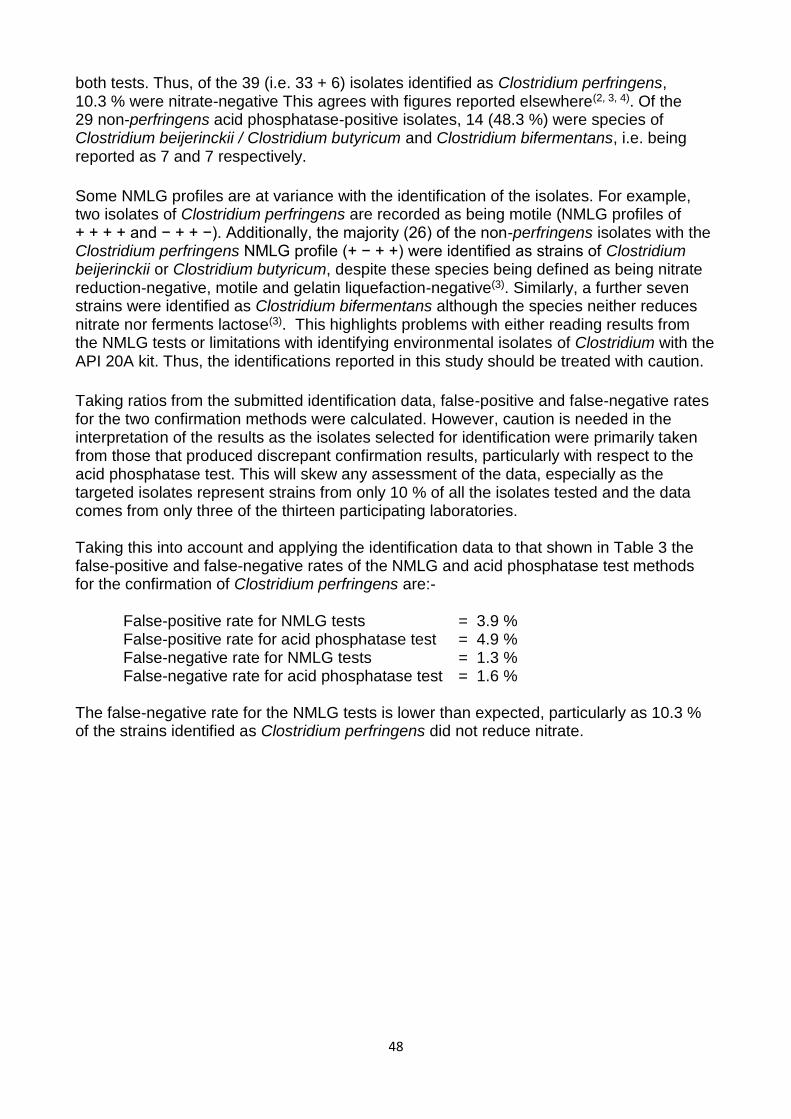
48
both tests. Thus, of the 39 (i.e. 33 + 6) isolates identified as Clostridium perfringens, 10.3 % were nitrate-negative This agrees with figures reported elsewhere(2, 3, 4). Of the 29 non-perfringens acid phosphatase-positive isolates, 14 (48.3 %) were species of Clostridium beijerinckii / Clostridium butyricum and Clostridium bifermentans, i.e. being reported as 7 and 7 respectively.
Some NMLG profiles are at variance with the identification of the isolates. For example, two isolates of Clostridium perfringens are recorded as being motile (NMLG profiles of + + + + and − + + −). Additionally, the majority (26) of the non-perfringens isolates with the Clostridium perfringens NMLG profile (+ − + +) were identified as strains of Clostridium beijerinckii or Clostridium butyricum, despite these species being defined as being nitrate reduction-negative, motile and gelatin liquefaction-negative(3). Similarly, a further seven strains were identified as Clostridium bifermentans although the species neither reduces nitrate nor ferments lactose(3). This highlights problems with either reading results from the NMLG tests or limitations with identifying environmental isolates of Clostridium with the API 20A kit. Thus, the identifications reported in this study should be treated with caution.
Taking ratios from the submitted identification data, false-positive and false-negative rates for the two confirmation methods were calculated. However, caution is needed in the interpretation of the results as the isolates selected for identification were primarily taken from those that produced discrepant confirmation results, particularly with respect to the acid phosphatase test. This will skew any assessment of the data, especially as the targeted isolates represent strains from only 10 % of all the isolates tested and the data comes from only three of the thirteen participating laboratories. Taking this into account and applying the identification data to that shown in Table 3 the false-positive and false-negative rates of the NMLG and acid phosphatase test methods for the confirmation of Clostridium perfringens are:- False-positive rate for NMLG tests = 3.9 % False-positive rate for acid phosphatase test = 4.9 % False-negative rate for NMLG tests = 1.3 % False-negative rate for acid phosphatase test = 1.6 % The false-negative rate for the NMLG tests is lower than expected, particularly as 10.3 % of the strains identified as Clostridium perfringens did not reduce nitrate.
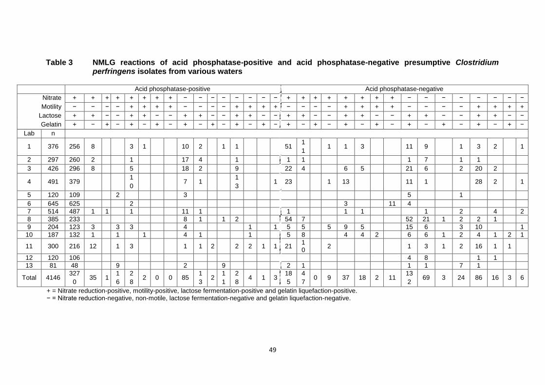
49
Table 3 NMLG reactions of acid phosphatase-positive and acid phosphatase-negative presumptive Clostridium perfringens isolates from various waters
Acid phosphatase-positive Acid phosphatase-negative
Nitrate + + + + + + + + − − − − − − − − + + + + + + + + − − − − − − − −
Motility − − − − + + + + − − − − + + + + − − − − + + + + − − − − + + + +
Lactose + + − − + + − − + + − − + + − − + + − − + + − − + + − − + + − −
Gelatin + − + − + − + − + − + − + − + − + − + − + − + − + − + − + − + −
Lab n
1 376 256 8 3 1 10 2 1 1 51 1
1 1 1 3 11 9 1 3 2 1
2 297 260 2 1 17 4 1 1 1 1 7 1 1
3 426 296 8 5 18 2 9 22 4 6 5 21 6 2 20 2
4 491 379 1
0 7 1
1
3 1 23 1 13 11 1 28 2 1
5 120 109 2 3 5 1
6 645 625 2 3 11 4
7 514 487 1 1 1 11 1 1 1 1 1 2 4 2
8 385 233 8 1 1 2 54 7 52 21 1 2 2 1
9 204 123 3 3 3 4 1 1 5 5 5 9 5 15 6 3 10 1
10 187 132 1 1 1 4 1 1 5 8 4 4 2 6 6 1 2 4 1 2 1
11 300 216 12 1 3 1 1 2 2 2 1 1 21 10
2 1 3 1 2 16 1 1
12 120 106 4 8 1 1
13 81 48 9 2 9 2 1 1 1 7 1
Total 4146 327
0 35 1
1
6
2
8 2 0 0 85
1
3 2
1
1
2
8 4 1 3
18
5
4
7 0 9 37 18 2 11
13
2 69 3 24 86 16 3 6
+ = Nitrate reduction-positive, motility-positive, lactose fermentation-positive and gelatin liquefaction-positive. − = Nitrate reduction-negative, non-motile, lactose fermentation-negative and gelatin liquefaction-negative.
50
Table 4 Identification of isolates of presumptive Clostridium perfringens
with respect to their NMLG profiles and acid phosphatase reactions
NMLG profile Clostridium Identification Acid
phosphatase-positive
Acid phosphatase-
negative
+ − + +
Clostridium perfringens Clostridium baratii / Clostridium paraputrificum Clostridium beijerinckii / Clostridium butyricum Clostridium bifermentans
20 2
13 1 24 7
+ − + −
Clostridium perfringens Clostridium baratii Clostridium baratii / Clostridium paraputrificum Clostridium beijerinckii / Clostridium butyricum Clostridium bifermentans Clostridium histolyticum Clostridium ramosum
1 1
2 1 3 3 1 1
+ − − − Clostridium clostridioforme Clostridium ramosum Clostridium tertium
1 1 1
+ + + + Clostridium perfringens Clostridium beijerinckii / Clostridium butyricum Clostridium innocuum
1 2
1 1
+ + + − Clostridium beijerinckii / Clostridium butyricum 1
− − + +
Clostridium perfringens Clostridium beijerinckii / Clostridium butyricum Clostridium bifermentans Clostridium bifermentans / Clostridium cadaveris Clostridium paraputrificum Clostridium ramosum
3 3 6 1 1 1
1 10 1
− − + −
Clostridium beijerinckii / Clostridium butyricum Clostridium bifermentans Clostridium innocuum Clostridium paraputrificum Clostridium histolyticum
1 1
3 1 1 1
− + + + Clostridium bifermentans 1 2
− + + − Clostridium perfringens Clostridium beijerinckii / Clostridium butyricum Clostridium septicum
1 1
1
− + − − Clostridium clostridioforme 1
51 80
4 Conclusions The results of this study indicate that the acid phosphatase test for the confirmation of Clostridium perfringens from water is at least as reliable as the current method(1) based upon the demonstration of reduction of nitrate, lack of motility, fermentation of lactose and liquefaction of gelatin. The two procedures show similar false-positive and false-negative rates, at a level expected from application to a large number of a wide range of environmental isolates. There is an agreement rate of 90.0 %. The false-positive rates for both procedures are less than 5 % and appear to be primarily due to species of Clostridium beijerinckii or Clostridium butyricum, and Clostridium bifermentans, although these identifications need to be treated with caution. The acid phosphatase test is considerably simpler to perform and is potentially more specific(2).
51
5 References
1. Standing Committee of Analysts, The Microbiology of Drinking Water (2010) - Part 6 - Methods for the isolation and enumeration of Sulphite-Reducing Clostridia and Clostridium perfringens by membrane filtration. Methods for the Examination of Waters and Associated Materials, in this series, Environment Agency. 2. Evaluation of acid phosphatase as a confirmation test for Clostridium perfringens isolated from water, Letters in Applied Microbiology, D P Sartory, R Waldock, C E Davies and A M Field, 2006, 42, 418-424. 3. Genus Clostridium Prazmowski 1880. In Bergey’s Manual of Systematic Bacteriology Volume 2 (Edited by P H A Sneath, N S Mair, M E Sharpe and J G Holt), E P Cato, W L George and S M Finegold, 1141-1200. Baltimore, Williams and Wilkins, 1986. 4. Cowan and Steel’s Manual for the Identification of Medical Bacteria, 3rd Edition. Edited by G I Barrow and R K A Feltham, Cambridge, Cambridge University Press, 1993.
5. A study of rapid and simplified confirmatory tests for Clostridium perfringens, Journal of Applied Bacteriology, G C Mead, L Paez de Leon and B W Adams, 1981, 51, 355-361.
6 Acknowledgements The Standing Committee of Analysts is indebted to the managers and analysts of the following laboratories that participated in this study: AES Laboratories (Newcastle-upon-Tyne) CREH Analytical (Leeds) Northern Ireland Water Services (Londonderry) Scottish Water (Dundee) Scottish Water (Edinburgh) Scottish Water (Turriff) Severn Trent Laboratories (Bridgend) Severn Trent Laboratories (Coventry) Severn Trent Water (Nottingham) Severn Trent Water (Shrewsbury) South West Water (Exeter) Southern Water (Winchester) United Utilities (Warrington) Wessex Water (Bath).

52
Address for correspondence However well procedures may be tested, there is always the possibility of discovering hitherto unknown problems. Analysts with such information are requested to contact the Secretary of the Standing Committee of Analysts at the address given below. In addition, if users wish to receive advance notice of forthcoming publications, please contact the Secretary. Secretary Standing Committee of Analysts Environment Agency (National Laboratory Service) NLS Nottingham Meadow Lane Nottingham NG2 3HN (http://www.gov.uk/environment-agency) Standing Committee of Analysts Members assisting with this method Without the good will and support given by these individuals and their respective organisations SCA would not be able to continue and produce the highly valued and respected blue book methods. Peter Boyd Formerly Public Health England Simon Cole Wessex Water Caroline Edwards South West Water David Gaskell United Utilities John V Lee Formerly Public Health England Malcom Morgan SCA Strategic Board Karen Murray Scottish Water Martin Reeve UKAS David Sartory SWM Consulting Helen Shapland Wessex Water
Rhys Stephens Welsh Water/Dŵr Cymru Martin Walters Environment Agency John Watkins CREH Analytical Limited John Watson South West Water David Westwood Formerly Environment Agency Grateful acknowledgement is made to David Gaskell (United Utilities) and John Watkins
(CREH Analytical) for providing colour photographs.

53

54


![[Micro] clostridia](https://img.pdfslide.us/doc/110x75/55d6fc72bb61ebfa2a8b47f2/micro-clostridia.jpg)