Embed Size (px)
Citation preview

Stage-specific Effects of cAMP Signaling during Distal LungEpithelial Development*
Received for publication, October 3, 2006 Published, JBC Papers in Press, October 3, 2006, DOI 10.1074/jbc.M609339200
Jingsong Xu1, Jun Tian, Sandra M. Grumelli, Kathleen J. Haley, and Steven D. ShapiroFrom the Division of Pulmonary and Critical Care Medicine, Brigham and Women’s Hospital at Harvard Medical School,Boston, Massachusetts 02115
cAMP signaling is postulated to play a role in distal lung epi-thelial differentiation based on several observations. First, itenhances fibroblast growth factor-induced transdifferentiationof early tracheal epithelium into respiratory epithelium. Sec-ond, there are cAMP-responsive elements in the heterologouspromoters of Sftpb and Sftpa genes. Third, cAMP augments theeffect of dexamethasone in maintaining differentiation ofhuman fetal type II pneumocyte culture. However, this concepthas not been thoroughly tested in vivo. In the current study, wemodulated cAMP signaling in developing distal lung epitheliumin vivo using an inducible transgenic system that expressed amutant form of G�s (G�sQ227L). We failed to demonstrate theability of cAMP to promote distal epithelial maturation duringembryonic stages. The results argue against its physiologicalrole in this process. In addition, induction of cAMP signaling atthe late pseudoglandular stage but not during the canalicular orsaccular stage surprisingly delayed distal differentiation by sup-pressing the expression of Sftpc, Sftpa, and Aquaporin5 as wellas the formation of lamellar bodies. This stage-specific inhibi-tory effect was observed in the absence of cellular toxicity orchanges in branching. Transgenic lungs did not show significantchanges in the known pathways that are important for distaldifferentiation. Therefore,we propose the existence of yet-to-beidentified cAMP-sensitive novel regulators of early distal lungepithelial differentiation. Although the delay of differentiationseemed to be reversible at later stages, it still led to pronouncedpermanent postnatal airspace enlargement due to impairedparacrine function of distal epithelium in regulating alveolarmyofibroblast development.
Development of the murine lung is initiated at embryonicday 9.5 (E9.5),2 followed by the pseudoglandular (E10.5–16.5),
canalicular (E16.5–17.5), saccular (E17.5–P5), and alveolarstages (P5–35) (1). Threemorphogenic events at the pseudoglan-dular stage are important for distal lung epithelial development.First, through branching morphogenesis, the lung primordiumevolves into anextensivenetworkof tubular epitheliumcomposedof eight generations of branches. This process requires a complexinteraction between several signaling molecules, such as FGF10(fibroblast growth factor 10), SHH (sonic hedgehog), and BMP4(bone morphogenetic protein 4) (2–6). Second, distal-proximalpolarity of the branching epithelial tree is established at the sametimethatbranching takesplace.AsearlyasE10.5–11.5, theexpres-sion of many genes is spatially restricted to the distal end of thebranches. Shh, Wnt7b, Id2, thrombospondin, transforminggrowth factor-�2, Etv5/Pea3, and Bmp4 are among the genesshowing this pattern of expression (7). Distal fate specificationrequires the BMP4, FGFs, and�-catenin pathways.Mice deficientineither theBMP4or�-cateninpathways showedcomplete trans-formation of distal epithelium into proximal epitheliumwith littlesign of type I or type II alveolar cell development (8, 9). Recentlypublished data suggest that �-catenin achieves this function byacting as a transcriptional factor downstream of Wnt signaling(10). FGF10andFGF7expressed in thedistalmesenchymearealsoproposed to regulate distal fate, because in a mesenchyme-freetracheal transdifferentiation assay, FGFs are the most potent dis-talizing signals (11).The third event in distal lung epithelial development is the
initiation of differentiation of epithelial cells at the end of thepseudoglandular stage. At E12.5, the distal epithelium starts toexpress differentiation-associated genes, such as Sftpc. At E13,glycogen accumulation (which is required for synthesis of phos-pholipids, a key component of surfactant) is evident in the distalepithelium (12). Expression of surfactant-associated protein A(SP-A), another differentiation marker of type II cells, is initi-ated distally 2 days later at E15.5. In addition, early events inlamellar body biosynthesis, a key feature of type II cell differen-tiation, are activated between E15.5 and E16.5. At E15.5, mul-tivesicle bodies (the precursors of lamellar bodies) can bevisualized under electron microscopy (EM). At E16–16.5,immature lamellar bodies are present in the distal cells (13).Very little is known about the genetic regulation of the initia-tion phase of distal epithelial differentiation, since most trans-genic models with distal differentiation defects manifestedtheir phenotype at the canalicular or early saccular stage. How-ever, one study did indicate roles for transcription factors Foxa1and Foxa2 in this early phase of differentiation (14).Differentiation of distal epithelium is accelerated at the
canalicular and saccular stages. During E17 and E18, Sftpc and
* This work was supported by NHLBI, National Institutes of Health, Grants P01HL29594 and R01 HL54853 (to S. D. S.), a Parker B. Francis fellowship (to J. X.),and a Scientist Development Grant from the American Heart Association (toJ. X.). The costs of publication of this article were defrayed in part by the pay-ment of page charges. This article must therefore be hereby marked “adver-tisement” in accordance with 18 U.S.C. Section 1734 solely to indicate this fact.
1 To whom correspondence should be addressed: Dept. of Medicine, HarvardMedical School, Brigham and Women’s Hospital, 75 Francis St., Boston, MA02115. Tel.: 617-278-0397; Fax: 617-232-4623; E-mail: [email protected].
2 The abbreviations used are: En, embryonic day n; Pn, postnatal day n; FGF,fibroblast growth factor; SP, surfactant-associated protein; EM, electronmicroscopy; RT, reverse transcription; PDGFR, platelet-derived growth fac-tor receptor; CREB, cAMP-response element-binding protein; BrdUrd,bromodeoxyuridine; TEM, transmission electron microscopy; IBMX,3-isobutyl-1-methylxanthine; �-SMA, smooth muscle �-actin.
THE JOURNAL OF BIOLOGICAL CHEMISTRY VOL. 281, NO. 50, pp. 38894 –38904, December 15, 2006© 2006 by The American Society for Biochemistry and Molecular Biology, Inc. Printed in the U.S.A.
38894 JOURNAL OF BIOLOGICAL CHEMISTRY VOLUME 281 • NUMBER 50 • DECEMBER 15, 2006
by guest on May 6, 2020
http://ww
w.jbc.org/
Dow
nloaded from
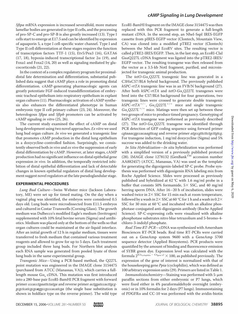
Sftpa mRNA expression is increased severalfold, more maturelamellar bodies are generated in type II cells, and the processingof pro-SP-C and pro-SP-B is also greatly increased (13). Type Icells start to emerge at E17.5 and can be identified by expressionof aquaporin 5, a type I cell-specific water channel. Type I andType II cell differentiation at these stages requires the functionof transcription factors TTF-1 (15), Etv5/Pea3 (16), GATA6(17, 18), hypoxia-induced transcriptional factor 2� (19), andFoxa1 and Foxa2 (14, 20) as well as signaling mediated by glu-cocorticoids (21, 22).In the context of a complex regulatory program for proximal-
distal fate determination and differentiation, substantial pub-lished data suggest that cAMP plays a role in promoting distaldifferentiation. cAMP-generating pharmacologic agents cangreatly potentiate FGF-induced transdifferentiation of embry-onic tracheal epithelium into respiratory epithelium in explantorgan cultures (11). Pharmacologic activation of cAMP synthe-sis also enhances the differentiated phenotype in humanembryonic type II cell primary culture (23, 24). Moreover, theheterologous Sftpa and Sftpb promoters can be activated bycAMP signaling in vitro (25, 26).The current study examined the effect of cAMP on distal
lung development using two novel approaches.Ex vitroweusedlung bud organ culture. In vivo we generated a transgenic linethat promotes cAMP production in the distal lung epitheliumin a doxycycline-controlled fashion. Surprisingly, we consis-tently observed both in vivo and ex vivo the suppression of earlydistal differentiation by cAMP. However, at later stages, cAMPproduction hadno significant influence ondistal epithelial geneexpression in vivo. In addition, the temporally restricted inhi-bition of distal epithelial differentiation and lack of detectablechanges in known epithelial regulators of distal lung develop-ment suggest novel regulators at the late pseudoglandular stage.
EXPERIMENTAL PROCEDURES
Lung Bud Culture—Swiss Webster mice (Jackson Labora-tory, ME) were set up for timed mating. On the day when avaginal plug was identified, the embryos were considered 0.5days old. Lung buds were microdissected from E11.5 embryosand cultured in Costar Transwells (CorningGlass). The growthmedium was Dulbecco’s modified Eagle’s medium (Invitrogen)supplemented with 10% fetal bovine serum (Sigma) and antibi-otics.Mediumwas placed only at the bottomof thewells so thatorgan cultures could be maintained at the air-liquid interface.After an initial growth of 12 h in regular medium, tissues weretransferred to fresh medium that contained various treatmentreagents and allowed to grow for up to 5 days. Each treatmentgroup included three lung buds. For Northern blot analysiseach RNA sample was generated from pooled lysate of threelung buds in the same experimental group.Transgenic Mice—Using a PCR-based method, the Q227L
point mutation was engineered in the IMAGE clone 3154475(purchased from ATCC (Manassas, VA)), which carries a full-length mouse G�s cDNA. This mutation was first introducedinto a 280-base pair EcoRI-BamHI PCR fragment with forwardprimer cccacctgaattctatga and reverse primer actggatccacttgcg-gcgctcatcgcgcaggccgcccacatcga (the single base substitution isshown in boldface type on the reverse primer). The wild type
EcoRI-BamHI fragment on the IMAGEclone 3154475was thenreplaced with this PCR fragment to generate a full-lengthmutant cDNA. In the second step, an NheI-SspI IRES-EGFPcassette from pIRES-EGFP vector (Clontech, Mountain View,CA) was cloned into a modified pTRE2 vector (Clontech)between the NheI and EcoRV sites. The resulting vector iscalled pTRE2-IRES/EGFP. Then, in the last step, an EcoRI-ClaIGs�Q227L cDNA fragment was ligated into the pTRE2-IRES/EGFP vector. The resulting transgene was then released fromthe vector as a 3.5-kb NotI fragment, purified, and microin-jected for transgenic animal production.The tetO-G�sQ227L transgenic line was generated in a
C3HxC57/BL6 hybrid background. The previously publishedhSPC-rtTA transgenic line was in an FVB/N background (27).After both hSPC-rtTA and tetO-G�sQ227L transgenes werebred into the C57/BL6 background for four generations, twotransgenic lines were crossed to generate double transgenichSPC-rtTA�/�, G�sQ227L�/� mice and single transgenicG�sQ227L�/� mice. Matings were then set up between thesetwo groups ofmice to produce timed pregnancy. Genotyping ofhSPC-rtTA trasngene was performed as previously described(27). The tetO-G�sQ227L transgene was identified throughPCR detection of GFP coding sequence using forward primercgtaaacggccacaagttcag and reverse primer atgccgttcttctgcttgtcg.For transgene induction, 1 mg/ml doxycycline (Sigma) and 5%sucrose was added to the drinking water.In Situ Hybridization—In situ hybridization was performed
with a simplified version of a previously published protocol(28). IMAGE clone 1278132 (GenBankTM accession numberAA882457) (ATCC, Manassas, VA) was used as the templatefor generating the digoxigenin-labeled SP-C probe. Probe syn-thesis was performed with digoxigenin RNA labeling mix fromRoche Applied Science. Slides were processed as previouslypublished and hybridized at 62 °C with 1.6 mg/ml probe in abuffer that consists 50% formamide, 5� SSC, and 40 mg/mlherring sperm DNA. After 16–20 h of incubation, slides werewashed twice in 2� SSC for 15 min each at room temperature,followed by awash in 2� SSC at 60 °C for 1 h and awash in 0.2�SSC for 30 min at 60 °C and incubated with an alkaline phos-photase-conjugated anti-digoxigenin antibody (Roche AppliedScience). SP-C-expressing cells were visualized with alkalinephosphotase substrates nitro blue tetrazolium and 5-bromo-4-chloro-3-indolyl phosphate.Real Time RT-PCR—cDNAwas synthesizedwithAmersham
Biosciences RT-PCR beads. Real time RT-PCRs were carriedout on a GeneAmp system 9600 with a GeneAmp 5700sequence detector (Applied Biosystems). PCR products werequantified by the amount of binding and fluorescence emissionof SYBR green dye. Expression level was calculated with theformula 2(Ctcyclophilin� Ctgene X) � 100, as published previously. Theexpression of the gene of interest is normalized with that ofthe housekeeping gene Ppia (cyclophilin), whichwas defined as100 arbitrary expressionunits (29). Primers are listed inTable 1.Immunohistochemistry—Staining was performed with 5-�m
paraffin sections from either embryonic or P7 lungs, whichwere fixed either in 4% paraformaldehyde overnight (embry-onic) or in 10% formalin for 2 days (P7 lungs). Immunostainingof PDGFR� and CC-10 was performed with the avidin-biotin
cAMP Signaling in Lung Development
DECEMBER 15, 2006 • VOLUME 281 • NUMBER 50 JOURNAL OF BIOLOGICAL CHEMISTRY 38895
by guest on May 6, 2020
http://ww
w.jbc.org/
Dow
nloaded from
horseradish peroxidase method (Vector Laboratories, Burl-ingame, CA), using 3,3�-diaminobenzidine as the chromogenicsubstrate. Immunostaining of phospho-CREB was performedwith the tyramide signal amplification biotin system(PerkinElmer Life Sciences) also using 3,3�-diaminobenzidineas the substrate. Antibody against phospho-CREB was pur-chased from Cell Signaling Technology (Danvers, MA). Anti-bodies against PDGFR� andCC-10were purchased from SantaCruz Biotechnology, Inc. (Santa Cruz, CA). Immunostaining of�-SMA was performed with an alkaline phosphatase-conju-gated monoclonal antibody (Sigma) using Vector Red as thesubstrate (Vector Laboratories). A terminal deoxynucleotidyl-transferase-mediated dUTP nick end-labeling assay was per-formed with an Apoptag�Plus peroxidase in situ apoptosisdetection kit (Chemicon International, Temecula, CA). Anti-BrdUrd immunohistochemistry was performed as describedpreviously (28). BrdUrd incorporation into embryonic lungswas achieved via intraperitoneal injection of 100mg/kg BrdUrdinto pregnant females 1 h 20 min before harvesting theembryos. In all of the above procedures, slides were counter-stained with methyl green.Western Blot Analysis—E15.5 lungs were homogenized in a
buffer that contained 25 mM Tris-HCl, pH 7.5, 50 mM NaCl, 1mM EDTA, 1 mM EGTA, 2.5 mM sodium pyrophosphate, 1 mM
�-glycerophosphate, and a complete proteinase inhibitor mix-ture (Roche Applied Science). For further protein extraction,the homogenates were supplemented with 1% Trition X-100and rocked at room temperature for 1 h. Insoluble fractionswere then removed by a 15-min centrifugation at 13,000 � g.The resulting protein lysates were resolved on an SDS-polyac-rylamide gel and transferred to ImmobilonTMmembrane (Mil-lipore Corp., Bedford, MA). After incubation of the membranewith horseradish peroxidase-conjugated secondary antibody,followed by chemiluminescent substrates (Amersham Bio-sciences), the target proteins were visualized by x-ray autora-diograph. Anti-phospho-CREB, anti-CREB, and anti-phospo-Smad1/5/8 antibodies were purchased from Cell SignalingTechnology. Anti-�-tubulin antibody was purchased fromSigma.Transmission Electron Microscopy—Accessory lobes from
E17 lungs were first fixed in Karnovsky’s solution and thenpostfixed in osmium tetroxide. Thin sections were stained withuranyl acetate and lead citrate. Lamellar bodiesweremonitoredunder JEOL 1200EX transmission EM at the Harvard Cell Biol-ogy Core facility for EM analysis.
Morphometry—Adult mouse lungs were inflated with 10%formalin through an intratracheal catheter under a pressure of25 cm H2O. After 48 h of fixation in 10% formalin at roomtemperature, lungswere rinsedwith phosphate-buffered saline,dehydrated in ethanol series, and embedded in paraffin. Mid-sagittal 5-�m lung sections stained with Gill’s hematoxylinwere used to calculate the cord length (CL) as previouslydescribed (30). 10 randomly selected �200 fields per slide werephotographed using MetaMorph image analysis software(Molecular Devices, Downingtown, PA). The images were ana-lyzed using the Scion Image software (Scion Corp.). Airway andvascular structures were excluded from the analysis. For alveo-lar septation analysis, postnatal day 7 lungs were inflated with100 �l of 10% formalin and then processed, embedded, andsectioned the same as described above. Midsagittal 5-�m lungsections stained with hematoxylin/eosin were photographed toobtain random �400 high power images. 15 such images fromeach lung were used to calculate the number of septa per field.For branching analysis, the number of distal epithelial tubuleswas quantified with �100 high power images collected fromhematoxylin/eosin-stained midsagittal sections from the leftlobes of E15.5 or E16.5 lungs; three sections were used for eachlung.
RESULTS
Lung BudCulture—We first examined the effect of cAMP onlung development using an in vitro lung bud organ culture sys-tem. Timed matings were set up as described under “Experi-mental Procedures.” When embryos were 11.5 days old, lungbuds were microdissected and cultured on microporous mem-branes at an air-liquid interface in Dulbecco’s modified Eagle’smedium supplemented with 10% fetal calf serum. The explantscontinued to branch for up to 5 days. They not only recapitu-lated branching morphogenesis in culture but also demon-strated time-dependent maturation of many features of distaldifferentiation. For example, SP-B was expressed only in eitherthe propeptide form or the partially processed form on days 1and 2 by Western blot analysis (data not shown). The com-pletely processed mature form was not evident until days 3 and4, and it became much more abundant at day 5 (data notshown). After the initial 12 h of culture in regularmedium, lungbuds were transferred to fresh medium supplemented witheither vehicle (Me2SO) or 5 �M forskolin and 5 �M 3-isobutyl-1-methylxanthine (IBMX). Forskolin directly activates adenylylcyclases, whereas IBMX inhibits all of the phosphodiesterasesthat can degrade cAMP.The combination of these two reagentshas been used routinely to achieve sustained intracellularcAMP signaling (31, 32).Foskolin and IBMX had at least four effects on lung bud cul-
ture. First, forskolin and IBMX exposure induced distal epithe-lial dilation (Fig. 1B, black arrows) compared with the vehiclecontrol group (Fig. 1A). Second, after 3 days of culture, forsko-lin and IBMXsignificantly decreased the number of branches inthe treatment groups (Fig. 1C), which was 43.7 � 3.19 (n � 3)compared with 58.7 � 3.1 (n � 3) in the control group (p �0.004). Third, these two agents induced thinning of the mesen-chyme, which was often evident on a whole mount image (Fig.1B, red arrows). Histological analysis of the midtransverse sec-
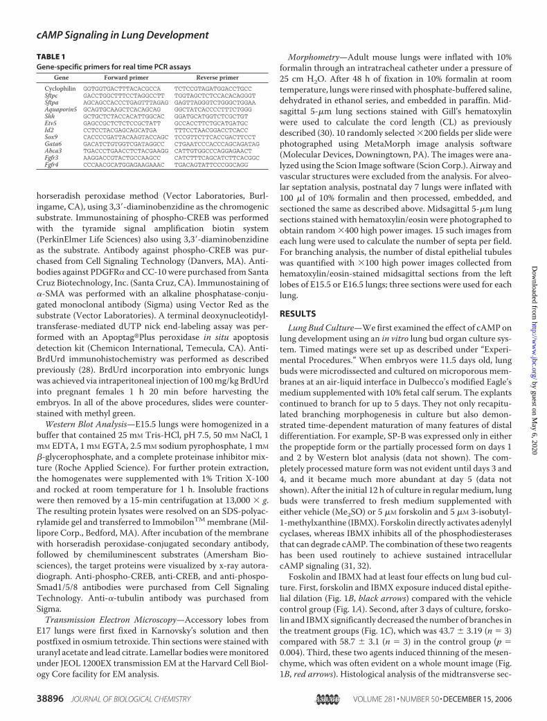
TABLE 1Gene-specific primers for real time PCR assays
Gene Forward primer Reverse primer
Cyclophilin GGTGGTGACTTTACACGCCA TCTCCGTAGATGGACCTGCCSftpc GACCTGGCTTTCCTAGGCCTT TGGTAGCTCTCCACACAGGGTSftpa AGCAGCCACCCTGAGTTTAGAG GAGTTAGGGTCTGGGCTGGAAAquaporin5 GCAGTGCAAGCTCACAGCAG GGCTATCACCCCTTTCTGGGShh GCTGCTCTACCACATTGGCAC GGATGCATGGTCTCGCTGTEtv5 GAGCCGCTCTCTCCGCTATT GCCACCTTCTGCATGATGCId2 CCTCCTACGAGCAGCATGA TTTCCTAACGGACCTCACCSox9 CACCCCGATTACAAGTACCAGC TCCGTTCTTCACCGACTTCCTGata6 GACATCTGTGGTCGATAGGCC CTGAATCCCACCCAGCAGATAGAbca3 TGACCCTGAACCTCTACGAAGG CATTGTGGCCCAGGAGAACTFgfr3 AAGGACCGTACTGCCAAGCC CATCTTTCAGCATCTTCACGGCFgfr4 CCCAACGCATGGAGAAGAAAC TGACAGTATTCCCGGCAGG
cAMP Signaling in Lung Development
38896 JOURNAL OF BIOLOGICAL CHEMISTRY VOLUME 281 • NUMBER 50 • DECEMBER 15, 2006
by guest on May 6, 2020
http://ww
w.jbc.org/
Dow
nloaded from

tions (Fig. 1,D and E) of these lung buds showed that these twoagents reduced the mesenchymal to epithelial cell ratio (Fig.1F), which was 1.32 � 0.08 in the treatment group (n � 3)compared with 2.08 � 0.05 in the control group (n � 3) (p �0.001), suggesting an effect of cAMP-elevating agents on mes-enchyme development. Last, Northern blot analysis of RNAsamples isolated from lung buds showed a dramatic decrease inSftpc expression in the forskolin- and IBMX-treated group after2 or 3 days in culture (Fig. 1C). However, we observed that thissuppression was gradually reversed after prolonged exposure(4–5 days), suggesting that this effect is stage-specific. Thedelayed onset of Sftpc expression was an unexpected observa-tion, challenging a positive role of cAMP in promoting distalepithelial differentiation during development. To address the invivo relevance of these findings, we designed a transgenicmodelto examine the role of cAMP signaling in lung epithelial devel-opment in vivo. Although we chose to focus on the effect ofcAMP signaling on epithelial differentiation in the rest of thisstudy, there are ongoing efforts investigating its effects onbranchingmorphogenesis and lungmesenchymedevelopment.Generation of g�sQ227L Transgenic Mice—To achieve
ectopic cAMP synthesis in vivo, we expressed a constitutively
active mutant form of G�s, whichcarries a glutamine-to- leucine sub-stitution (G�sQ227L) and was ini-tially identified as a somatic muta-tion in patients with pituitary orthyroid adenomas (33). Because thismutation disrupts GTPase activity,G�sQ227L stimulates adenylylcyclases constitutively in theabsence of receptor ligation (Fig.2A). To avoid unwanted negativeeffects of prolonged constitutiveexpression, we employed a bitrans-genic system that is pharmacologi-cally regulated by either tetracylcineor its analog doxycycline. Similartransgenic systems have been usedin several studies to modulate invivo gene function in the murinelung (9, 14, 27, 34). Fig. 2B schemat-ically illustrates this system. Inshort, the g�sQ227L transgene isunder the control of a cytomegalov-irus minimum promoter that har-bors seven tetO sites, serving ascis-binding elements for the heter-ologous transcription factor rtTA,which is expressed from a separateline of transgene that is driven bythe 3.7-kb human surfactant-asso-ciated protein C promoter, as pub-lished previously (27). Becausebinding of rtTA to tetO requirestetracycline or its analog doxycy-cline, the G�sQ227L transgene isexpected to be inactive until the ani-
mals are exposed to doxycycline.Initially, we tested the functionality of the tetO-g�sQ227L
transgene in an in vitro transfection assay. COS7 cells weretransfected with cytomegalovirus-rtTA (an expression vectorwith a viral promoter) driving a luciferase reporter that con-tains cAMP-responsive elements in its promoter and the tetO-g�sQ227L transgene. Exposure of transfectants to doxycyclineresulted in a 3-fold increase in luciferase activity (Fig. 2C). Sub-sequently, several transgenic lines in a C3HxC57BL/6 hybridbackground were established. Offspring of these lines were setup for timed mating with hSPC-rtTA mice. After pregnantfemales were exposed to doxycylcine from E13.5 to E16.5,embryonic lungs were harvested for RNA isolation. Northernblot analysis of one g�sQ227L line showed doxycycline-dependent expression of a transcript with the predicted size of2.9 kb at E16.5 (Fig. 2D). However, the transgene transcript isexpressed at a level much lower than the 2.1 and 3.1-kb endog-enous transcripts (Fig. 2D).We bred the g�sQ227L transgene tohomozygosity in order to enhance the level of expression.Mostof the analyses presented in this studywere performedusing themating scheme shown in Fig. 3A, in which 50% of the offspringare double transgenic for both hSPC-rtTA and tetO-g�sQ227L.
FIGURE 1. Effects of cAMP-elevating agents on in vitro lung bud organ culture. A, a representative vehicle(Me2SO; DMSO)-exposed lung bud after 3 days in culture. B, a representative forskolin and IBMX (F � I)-exposedlung bud after 3 days in culture showing dilated distal epithelial tubes (arrows) and thinning of the mesen-chyme (red arrowheads). C, lung buds treated with forskolin � IBMX (open bar, n � 3) had a significantly lowernumber of branches than lung buds exposed to Me2SO (filled bar, n � 3) after 3 days in culture. D andE, representative hematoxylin/eosin staining of transverse sections (red lines in A and B) of Me2SO-exposed (D)and forskolin � IBMX-exposed (E ) lung buds showing lower cellular density in the mesenchymal compartmentof the latter group. F, mesenchymal to epithelial cell ratio was reduced in the forskolin � IBMX-exposed group(n � 3) compared with the Me2SO-exposed control group (n � 3) after 3 days in culture. G, Northern blotanalysis of SP-C expression in lung buds exposed to either vehicle (�) or forskolin � IBMX (�) for indicatedperiods of time. Ethidium bromide staining shows similar loading of RNA samples.
cAMP Signaling in Lung Development
DECEMBER 15, 2006 • VOLUME 281 • NUMBER 50 JOURNAL OF BIOLOGICAL CHEMISTRY 38897
by guest on May 6, 2020
http://ww
w.jbc.org/
Dow
nloaded from

The other 50% are single-transgenic for tetO-g�sQ227L andserve as experimental controls for the double transgenic mice.All animals in this mating scheme, including both parents andtheir offsprings, carried the g�sQ227L transgene in a homozy-gous configuration. Because the transgene is only expected toexpress in a small fraction of cells in the lung, total tissue cAMPquantification is not a sensitive assay for transgene function.Instead, we monitored phosphorylation of CREB as a read-outfor cAMP signaling. Immunohistochemical staining using anantibody that binds specifically to the phosphorylated form ofCREB identified its activation in the bitransgenic distal epithe-lium (asterisks in Fig. 2G), whereas single transgenic controlsshowed little CREB phosphorylation in the distal epithelium(asterisks in Fig. 2F). It is interesting that control lungs showedstriking CREB phosphorylation in the proximal epithelium,suggesting a role for CREB in regulating proximal epithelialdevelopment. Ectopic CREB phosphorylation was not detectedin the vasculature or othermesenchymal cells; nor was there anobvious increase in endogenous CREB phosphorylation in theproximal epithelium.Western blot analysis of total lung lysatesisolated from E15.5 mice exposed to doxycycline for 4 days also
showed increased phosphorylationof CREB (Fig. 2E). In summary, thebitransgenic systemwas able to acti-vate cAMP signaling in a distal epi-thelium-specific fashion. This sys-tem was regulatable by doxycyclineand demonstrated high stringencywith almost no base-line leakage ofexpression of the mutant G�s in theabsence of doxycycline exposure(Figs. 3B and 6, A–C).Suppression of Distal Differentia-
tion at the Late PseudoglandularStage—The effect of ectopic cAMPsignaling on distal lung epithelialdifferentiation was first tested atthe late pseudoglandular stage.g�sQ227L transgene expression wasinduced by doxycycline from E14.5to E16.5. Using RNA samples iso-lated from E16.5 lungs, the expres-sion of the transgene and distal epi-thelial markers was quantified byreal time RT-PCR. To ensure that inutero doxycycline exposure was suf-ficient and the transgene wasinduced, every embryonic litterused in this study was first tested forthe level of transgene expressionprior to further phenotypic analysis.Since the transgene is transcribedinto a bicistronic mRNA that con-tains both G�sQ227L and GFP cod-ing sequences (Fig. 2B), detection ofGFP-encoding cDNA by real timePCR was used as a surrogate read-out of transgene expression. As
expected, in all cases, double transgenic lungs showed robustexpression of GFP-encoding cDNA (Figs. 3B and 6). We firstexamined three distal differentiation markers, Sftpc, Sftpa, andAquaporin5. The expression levels of these genes were normal-ized to the level of the housekeeping gene cyclophilin, in whichcyclophilin expression level was defined as 100 arbituary units,as published previously (29) (see “Experimental Procedures” forthe calculation formula). Double transgenic lungs showed 79%suppression of Sftpc expression (p � 0.0001) and a 94% sup-pression of Sftpa expression (p � 0.011) compared with theirlittermate controls (Fig. 3B). Although Aquaporin5 expressionwas not detectable at E16.5 by either in situ hybridization orimmunohistochemistry, it was detectable at a low level by realtime RT-PCR. Again, double transgenic lungs showed a 63%reduction of Aquaporin5 expression (p � 0.022) (Fig. 3B).Moreover, g�sQ227L expression from E14.5 to E17 also inhib-ited lamellar body biosynthesis according to TEM studies (Fig.3, F and G). Since E16.5 is early for detecting mature lamellarbodies, embryos were exposed to doxycycline for an additionalhalf day and harvested at E17. Three single transgenic and threedouble transgenic lungs were included in TEM analysis. From
FIGURE 2. Lung-specific, inducible, constitutively active G�s transgenic mice. A, schematic diagram illus-trates the constitutively active property of the mutant G�sQ227L subunit (see “Results” for details). B, aninducible transgenic system designed for regulatable expression of G�sQ227L. C, normalized luciferase activ-ities of cells transfected with CRE-luciferase, cytomegalovirus-rtTA, and TetO-G�sQ227L with (open bar) orwithout (black bar) doxycycline treatment. D, Northern blot detection of both the endogenous G�s transcriptsand the transgenic G�sQ227L transcript in total lung RNA from E16.5 embryos exposed to Dox for 3 days.E, Western blot detection of phosphorylated and total CREB proteins in the lung lysates from three singletransgenic and three double transgenic E15.5 lungs exposed to Dox for 4 days. Note the similar amount of totalCREB but increased p-CREB detection in double trangenic lungs compared with controls. F and G, immunohis-tochemical detection of phosphorylated CREB in E16.5 lungs exposed to Dox for 2 days. *, distal epithelium.
cAMP Signaling in Lung Development
38898 JOURNAL OF BIOLOGICAL CHEMISTRY VOLUME 281 • NUMBER 50 • DECEMBER 15, 2006
by guest on May 6, 2020
http://ww
w.jbc.org/
Dow
nloaded from
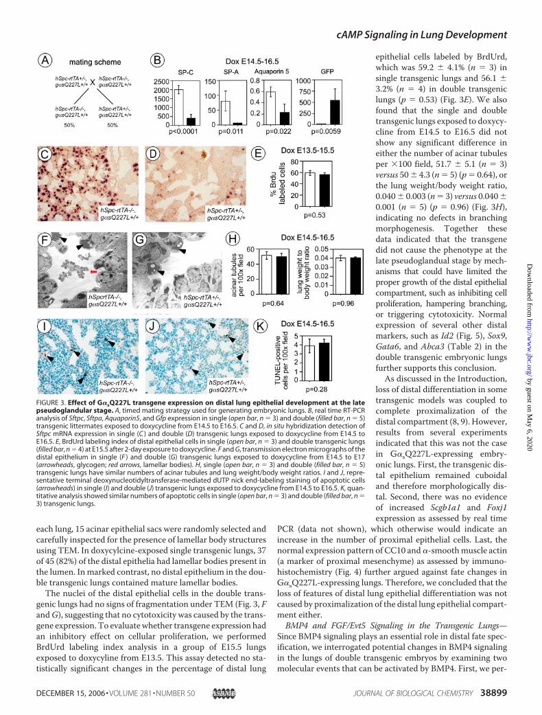
each lung, 15 acinar epithelial sacs were randomly selected andcarefully inspected for the presence of lamellar body structuresusing TEM. In doxycylcine-exposed single transgenic lungs, 37of 45 (82%) of the distal epithelia had lamellar bodies present inthe lumen. Inmarked contrast, no distal epithelium in the dou-ble transgenic lungs contained mature lamellar bodies.The nuclei of the distal epithelial cells in the double trans-
genic lungs had no signs of fragmentation under TEM (Fig. 3, FandG), suggesting that no cytotoxicity was caused by the trans-gene expression. To evaluatewhether transgene expression hadan inhibitory effect on cellular proliferation, we performedBrdUrd labeling index analysis in a group of E15.5 lungsexposed to doxycyline from E13.5. This assay detected no sta-tistically significant changes in the percentage of distal lung
epithelial cells labeled by BrdUrd,which was 59.2 � 4.1% (n � 3) insingle transgenic lungs and 56.1 �3.2% (n � 4) in double transgeniclungs (p � 0.53) (Fig. 3E). We alsofound that the single and doubletransgenic lungs exposed to doxycy-cline from E14.5 to E16.5 did notshow any significant difference ineither the number of acinar tubulesper �100 field, 51.7 � 5.1 (n � 3)versus 50� 4.3 (n� 5) (p� 0.64), orthe lung weight/body weight ratio,0.040� 0.003 (n� 3) versus 0.040�0.001 (n � 5) (p � 0.96) (Fig. 3H),indicating no defects in branchingmorphogenesis. Together thesedata indicated that the transgenedid not cause the phenotype at thelate pseudoglandual stage by mech-anisms that could have limited theproper growth of the distal epithelialcompartment, such as inhibiting cellproliferation, hampering branching,or triggering cytotoxicity. Normalexpression of several other distalmarkers, such as Id2 (Fig. 5), Sox9,Gata6, and Abca3 (Table 2) in thedouble transgenic embryonic lungsfurther supports this conclusion.As discussed in the Introduction,
loss of distal differentiation in sometransgenic models was coupled tocomplete proximalization of thedistal compartment (8, 9). However,results from several experimentsindicated that this was not the casein G�sQ227L-expressing embry-onic lungs. First, the transgenic dis-tal epithelium remained cuboidaland therefore morphologically dis-tal. Second, there was no evidenceof increased Scgb1a1 and Foxj1expression as assessed by real time
PCR (data not shown), which otherwise would indicate anincrease in the number of proximal epithelial cells. Last, thenormal expression pattern of CC10 and�-smoothmuscle actin(a marker of proximal mesenchyme) as assessed by immuno-histochemistry (Fig. 4) further argued against fate changes inG�sQ227L-expressing lungs. Therefore, we concluded that theloss of features of distal lung epithelial differentiation was notcaused by proximalization of the distal lung epithelial compart-ment either.BMP4 and FGF/Evt5 Signaling in the Transgenic Lungs—
Since BMP4 signaling plays an essential role in distal fate spec-ification, we interrogated potential changes in BMP4 signalingin the lungs of double transgenic embryos by examining twomolecular events that can be activated by BMP4. First, we per-
FIGURE 3. Effect of G�sQ227L transgene expression on distal lung epithelial development at the latepseudoglandular stage. A, timed mating strategy used for generating embryonic lungs. B, real time RT-PCRanalysis of Sftpc, Sftpa, Aquaporin5, and Gfp expression in single (open bar, n � 3) and double (filled bar, n � 5)transgenic littermates exposed to doxycycline from E14.5 to E16.5. C and D, in situ hybridization detection ofSftpc mRNA expression in single (C ) and double (D) transgenic lungs exposed to doxycycline from E14.5 toE16.5. E, BrdUrd labeling index of distal epithelial cells in single (open bar, n � 3) and double transgenic lungs(filled bar, n � 4) at E15.5 after 2-day exposure to doxycycline. F and G, transmission electron micrographs of thedistal epithelium in single (F ) and double (G) transgenic lungs exposed to doxycycline from E14.5 to E17(arrowheads, glycogen; red arrows, lamellar bodies). H, single (open bar, n � 3) and double (filled bar, n � 5)transgenic lungs have similar numbers of acinar tubules and lung weight/body weight ratios. I and J, repre-sentative terminal deoxynucleotidyltransferase-mediated dUTP nick end-labeling staining of apoptotic cells(arrowheads) in single (I) and double (J) transgenic lungs exposed to doxycycline from E14.5 to E16.5. K, quan-titative analysis showed similar numbers of apoptotic cells in single (open bar, n � 3) and double (filled bar, n �3) transgenic lungs.
cAMP Signaling in Lung Development
DECEMBER 15, 2006 • VOLUME 281 • NUMBER 50 JOURNAL OF BIOLOGICAL CHEMISTRY 38899
by guest on May 6, 2020
http://ww
w.jbc.org/
Dow
nloaded from
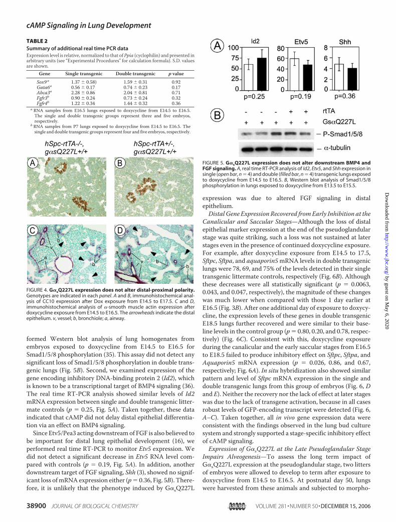
formed Western blot analysis of lung homogenates fromembryos exposed to doxycycline from E14.5 to E16.5 forSmad1/5/8 phosphorylation (35). This assay did not detect anysignificant loss of Smad1/5/8 phosphorylation in double trans-genic lungs (Fig. 5B). Second, we examined expression of thegene encoding inhibitory DNA-binding protein 2 (Id2), whichis known to be a transcriptional target of BMP4 signaling (36).The real time RT-PCR analysis showed similar levels of Id2mRNA expression between single and double transgenic litter-mate controls (p � 0.25, Fig. 5A). Taken together, these dataindicated that cAMP did not delay distal epithelial differentia-tion via an effect on BMP4 signaling.Since Etv5/Pea3 acting downstreamof FGF is also believed to
be important for distal lung epithelial development (16), weperformed real time RT-PCR to monitor Etv5 expression. Wedid not detect a significant decrease in Etv5 RNA level com-pared with controls (p � 0.19, Fig. 5A). In addition, anotherdownstream target of FGF signaling, Shh (3), showed no signif-icant loss ofmRNAexpression either (p� 0.36, Fig. 5B). There-fore, it is unlikely that the phenotype induced by G�sQ227L
expression was due to altered FGF signaling in distalepithelium.Distal Gene Expression Recovered fromEarly Inhibition at the
Canalicular and Saccular Stages—Although the loss of distalepithelial marker expression at the end of the pseudoglandularstage was quite striking, such a loss was not sustained at laterstages even in the presence of continued doxycycline exposure.For example, after doxycycline exposure from E14.5 to 17.5,Sftpc, Sftpa, and aquaporin5mRNA levels in double transgeniclungs were 78, 69, and 75% of the levels detected in their singletransgenic littermate controls, respectively (Fig. 6B). Althoughthese decreases were all statistically significant (p � 0.0063,0.043, and 0.047, respectively), the magnitude of these changeswas much lower when compared with those 1 day earlier atE16.5 (Fig. 3B). After one additional day of exposure to doxycy-cline, the expression levels of these genes in double transgenicE18.5 lungs further recovered and were similar to their base-line levels in the control group (p� 0.80, 0.20, and 0.78, respec-tively) (Fig. 6C). Consistent with this, doxycycline exposureduring the canalicular and the early saccular stages from E16.5to E18.5 failed to produce inhibitory effect on Sftpc, Sftpa, andAquaporin5 mRNA expression (p � 0.026, 0.86, and 0.67,respectively; Fig. 6A). In situ hybridization also showed similarpattern and level of Sftpc mRNA expression in the single anddouble transgenic lungs from this group of embryos (Fig. 6, Dand E). Neither the recovery nor the lack of effect at later stageswas due to the lack of transgene activation, because in all casesrobust levels of GFP-encoding transcript were detected (Fig. 6,A–C). Taken together, all in vivo gene expression data wereconsistent with the findings observed in the lung bud culturesystem and strongly supported a stage-specific inhibitory effectof cAMP signaling.Expression of G�sQ227L at the Late Pseudoglandular Stage
Impairs Alveogenesis—To assess the long term impact ofG�sQ227L expression at the pseudoglandular stage, two littersof embryos were allowed to develop to term after exposure todoxycycline from E14.5 to E16.5. At postnatal day 50, lungswere harvested from these animals and subjected to morpho-
TABLE 2Summary of additional real time PCR dataExpression level is relative, normalized to that of Ppia (cyclophilin) and presented inarbitrary units (see “Experimental Procedures” for calculation formula). S.D. valuesare shown.
Gene Single transgenic Double transgenic p valueSox9 a 1.37 � 0.58) 1.59 � 0.31 0.92Gata6a 0.56 � 0.17 0.74 � 0.23 0.17Abca3a 2.28 � 0.86 2.04 � 0.81 0.71Fgfr3b 0.90 � 0.24 0.73 � 0.24 0.32Fgfr4b 1.22 � 0.34 1.44 � 0.32 0.36
a RNA samples from E16.5 lungs exposed to doxycycline from E14.5 to E16.5.The single and double transgenic groups represent three and five embryos,respectively.
b RNA samples from P7 lungs exposed to doxycycline from E14.5 to E16.5. Thesingle and double transgenic groups represent four and five embryos, respectively.
FIGURE 4. G�sQ227L expression does not alter distal-proximal polarity.Genotypes are indicated in each panel. A and B, immunohistochemical anal-ysis of CC10 expression after Dox exposure from E14.5 to E17.5. C and D,immunohistochemical analysis of �-smooth muscle actin expression afterdoxycycline exposure from E14.5 to E16.5. The arrowheads indicate the distalepithelium. v, vessel; b, bronchiole; a, airway.
FIGURE 5. G�sQ227L expression does not alter downstream BMP4 andFGF signaling. A, real time RT-PCR analysis of Id2, Etv5, and Shh expression insingle (open bar, n � 4) and double (filled bar, n � 4) transgenic lungs exposedto doxycycline from E14.5 to E16.5. B, Western blot analysis of Smad1/5/8phosphorylation in lungs exposed to doxycycline from E13.5 to E15.5.
cAMP Signaling in Lung Development
38900 JOURNAL OF BIOLOGICAL CHEMISTRY VOLUME 281 • NUMBER 50 • DECEMBER 15, 2006
by guest on May 6, 2020
http://ww
w.jbc.org/
Dow
nloaded from

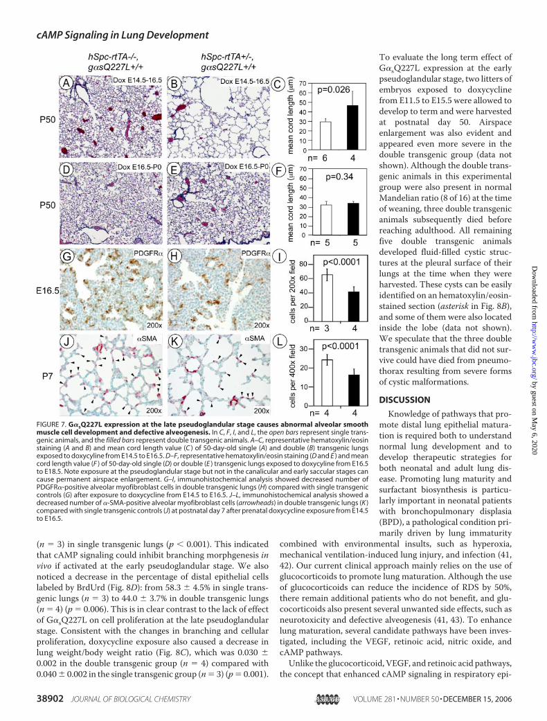
metric analysis. Double transgenic lungs in this experiment dis-played pronounced alveolar space enlargement (Fig. 7B).Despite some variation in the severity of parenchymal histolog-ical defect, the average mean CL of the double transgenic lungsin one of the two litters was 49.45� 15.22 (n� 4), representinga 4.7-fold increase in alveolar volume compared with the CL of29.40 � 3.38 (n � 6) derived from their single transgenic litter-mates (Fig. 7C) (p � 0.026). Despite severe airspace enlarge-ment, the double transgenic animals in these two litters werepresent in normalMandelian ratio (9 of 18 total) at the weaningage, and all survived until postnatal day 50, when they wereeuthanized for tissue harvesting. In contrast to the structuraldefects caused by early doxycycline exposure, double trans-genic lungs exposed to doxycycline at later stages (fromE16.5 toE18.5) exhibited normal parenchymal histology when analyzedat P50 (Fig. 7, compare E with D). In one of the two littersanalyzed, CL of the double transgenic lungs was 32.26 � 1.93
(n � 5), which was not statistically different from the CL value,30.46 � 3.46 (n � 5), derived from the single transgenic group(Fig. 7F) (p � 0.34). Again these data reiterated that theunknowndistal regulators inhibited by cAMP signaling seem tohave pseudoglandular stage-specific roles that have long termlung development effects.To understand the underlying mechanism of airspace
enlargement, we performedmorphometry analysis of postnatalday 7 lungs from a litter of animals that had been exposed todoxycycline from E14.5 to E16.5. The number of secondarysepta per high power field in the double transgenic group was14.6� 2.1 (n� 4), whichwas significantly lower comparedwiththe number of 21.2 � 2.3 (n � 4) in the single transgenic group(p � 0.0001) (Fig. 7, J and K). Therefore, G�sQ227L expressionat the late pseudoglandular stage caused defective postnatalalveogenesis.Expression of G�sQ227L Causes Defective AlveolarMyofibro-
blast Development—Defective alveolar septation could have anumber of underlying reasons. Both defects in fibroblastgrowth factor receptor 3 and 4 signaling and lack of elastogen-esis were shown to cause abnormal septation (37, 38). However,real time PCR analysis showed normal Fgfr3/4 expression indouble transgenic lungs at postnatal day 7 (Table 2), and elastinstaining also appeared to be normal (data not shown). Defectivealveogenesis may also occur as a result of improper alveolarmyofibroblast development (39). These �-SMA-expressingcells are located at the tip of septation during alveogenesis andare thought to play an essential role in the initiation of second-ary septa. We performed anti-�-SMA immunohistochemstryto quantify the number of alveolar myofibroblasts in postnatalday 7 lungs, which was significantly decreased from 24.2 � 2.9in single transgenic lungs (n � 4) to 16.2 � 3.2 in double trans-genic lungs (n � 4) (p � 0.0001) (Fig. 7, J–L). Consistent withthis finding, we also found that after doxycyline exposure fromE14.5 to E16.5, double transgenic lungs developed fewerPDGFR-positive cells (p� 0.0001) (Fig. 7,G–I) at E16.5, which,at this stage of development, are considered to be the progeni-tor cells for alveolar myofibroblasts (40). The number of cellsper �200 high power field was 65.3 � 8.5 in single transgeniclungs (n � 3) versus 41.4 � 7.3 (n � 4) in double transgeniclungs. Since short term transgene activation did not alter cellu-lar proliferation and branching morphogenesis (Fig. 3) andthere was no obvious evidence of an increase in inflammatorycells on the HE staining of either P7 (not shown) or P50 (Fig. 7)lungs, defective alveolar myofibroblast development was likelyto be the primary cause of failed alveogenesis.Expression of G�sQ227L at the Early Pseudoglandular Stage
Impairs Branching andCellular Proliferation—Wehave shownearlier that pharmacological activation of cAMPsignaling ham-pered branching in lung bud culture. However, in vivo activa-tion of cAMP signaling by G�sQ227l at the late pseudoglandu-lar stage did not show such an effect. We suspect that thisdifference was due to stage-specific sensitivity of branchingmorphogenesis to cAMP signaling. To test this, we exposedembryos to doxycycline fromE11.5 to E15.5. Double transgeniclungs at E15.5 showed a significant decrease in the number ofdistal tubules per �100 high power field (Fig. 8C), which was46.6 � 2.6 (n � 4) in double transgenic lungs versus 63.0 � 3.2
FIGURE 6. G�sQ227L transgene expression does not affect distal lung epi-thelial development at the canalicular and early saccular stages. A–C, realtime RT-PCR analysis of Sftpc, Sftpa, and Aquaporin5 expression in single (openbar) and double (filled bar) transgenic littermates exposed to doxycycline forvarious periods as indicated. Each data point represents four embryos, exceptfor the single transgenic group in B, which represents three embryos. D andE, in situ hybridization detection of Sftpc expression using a digoxigenin-la-beled probe in the E18.5 lungs exposed to doxycycline from E16.5 to E18.5.
cAMP Signaling in Lung Development
DECEMBER 15, 2006 • VOLUME 281 • NUMBER 50 JOURNAL OF BIOLOGICAL CHEMISTRY 38901
by guest on May 6, 2020
http://ww
w.jbc.org/
Dow
nloaded from
(n � 3) in single transgenic lungs (p � 0.001). This indicatedthat cAMP signaling could inhibit branching morphgenesis invivo if activated at the early pseudoglandular stage. We alsonoticed a decrease in the percentage of distal epithelial cellslabeled by BrdUrd (Fig. 8D): from 58.3 � 4.5% in single trans-genic lungs (n � 3) to 44.0 � 3.7% in double transgenic lungs(n � 4) (p � 0.006). This is in clear contrast to the lack of effectof G�sQ227L on cell proliferation at the late pseudoglandularstage. Consistent with the changes in branching and cellularproliferation, doxycycline exposure also caused a decrease inlung weight/body weight ratio (Fig. 8C), which was 0.030 �0.002 in the double transgenic group (n � 4) compared with0.040� 0.002 in the single transgenic group (n� 3) (p� 0.001).
To evaluate the long term effect ofG�sQ227L expression at the earlypseudoglandular stage, two litters ofembryos exposed to doxycyclinefrom E11.5 to E15.5 were allowed todevelop to term and were harvestedat postnatal day 50. Airspaceenlargement was also evident andappeared even more severe in thedouble transgenic group (data notshown). Although the double trans-genic animals in this experimentalgroup were also present in normalMandelian ratio (8 of 16) at the timeof weaning, three double transgenicanimals subsequently died beforereaching adulthood. All remainingfive double transgenic animalsdeveloped fluid-filled cystic struc-tures at the pleural surface of theirlungs at the time when they wereharvested. These cysts can be easilyidentified on an hematoxylin/eosin-stained section (asterisk in Fig. 8B),and some of them were also locatedinside the lobe (data not shown).We speculate that the three doubletransgenic animals that did not sur-vive could have died from pneumo-thorax resulting from severe formsof cystic malformations.
DISCUSSION
Knowledge of pathways that pro-mote distal lung epithelial matura-tion is required both to understandnormal lung development and todevelop therapeutic strategies forboth neonatal and adult lung dis-ease. Promoting lung maturity andsurfactant biosynthesis is particu-larly important in neonatal patientswith bronchopulmonary displasia(BPD), a pathological condition pri-marily driven by lung immaturity
combined with environmental insults, such as hyperoxia,mechanical ventilation-induced lung injury, and infection (41,42). Our current clinical approach mainly relies on the use ofglucocorticoids to promote lung maturation. Although the useof glucocorticoids can reduce the incidence of RDS by 50%,there remain additional patients who do not benefit, and glu-cocorticoids also present several unwanted side effects, such asneurotoxicity and defective alveogenesis (41, 43). To enhancelung maturation, several candidate pathways have been inves-tigated, including the VEGF, retinoic acid, nitric oxide, andcAMP pathways.Unlike the glucocorticoid, VEGF, and retinoic acid pathways,
the concept that enhanced cAMP signaling in respiratory epi-
FIGURE 7. G�sQ227L expression at the late pseudoglandular stage causes abnormal alveolar smoothmuscle cell development and defective alveogenesis. In C, F, I, and L, the open bars represent single trans-genic animals, and the filled bars represent double transgenic animals. A–C, representative hematoxylin/eosinstaining (A and B) and mean cord length value (C ) of 50-day-old single (A) and double (B) transgenic lungsexposed to doxycyline from E14.5 to E16.5. D–F, representative hematoxylin/eosin staining (D and E ) and meancord length value (F ) of 50-day-old single (D) or double (E ) transgenic lungs exposed to doxycyline from E16.5to E18.5. Note exposure at the pseudoglandular stage but not in the canalicular and early saccular stages cancause permanent airspace enlargement. G–I, immunohistochemical analysis showed decreased number ofPDGFR�-positive alveolar myofibroblast cells in double transgenic lungs (H) compared with single transgeniccontrols (G) after exposure to doxycycline from E14.5 to E16.5. J–L, immunohistochemical analysis showed adecreased number of �-SMA-positive alveolar myofibroblast cells (arrowheads) in double transgenic lungs (K )compared with single transgenic controls (J) at postnatal day 7 after prenatal doxycycline exposure from E14.5to E16.5.
cAMP Signaling in Lung Development
38902 JOURNAL OF BIOLOGICAL CHEMISTRY VOLUME 281 • NUMBER 50 • DECEMBER 15, 2006
by guest on May 6, 2020
http://ww
w.jbc.org/
Dow
nloaded from

thelium may promote lung maturation is primarily supportedby in vitro observations (11, 23–26). The transgenic system wedeveloped allowed us to investigate this concept in vivo. Sincethe current system allows activation of cAMP signaling in adistal epithelium-specific fashion, it is more specific than thepreviously used pharmacological methods for promotingcAMP signaling in vivo. The lack of substantial activation ofSftpc, Sftpa, Aquaporin5, and Abca3 expression at the canalic-ular and sacular stages in transgenic lungs with induced cAMPsignaling argues against a physiological role for cAMP in pro-moting distal epithelial maturation.Given these findings, how do we reconcile the difference
between observations in vivo versus in vitro? cAMP is known topromote cell adhesion and polarity (44, 45), two cellular fea-tures that are often compromised or lost under in vitro condi-tions. Therefore, one possible mechanism by which cAMPenhances differentiation in vitro is to promote cell adhesionand/or polarity. If cell adhesion and polarity are maintained invivo by pathways other than cAMP, ectopic cAMP is notrequired to influence distal differentiation in vivo. Despite thefact that we were unable to detect a positive contribution ofcAMP signaling in normal lung development, cAMP may helpto restore distal lung epithelial function in disease states, par-ticularly in those with cell injury that is associated with loss ofadhesion or polarity. The transgenic model developed in thecurrent study certainly presents a unique opportunity to testthe role of cAMP signaling in acute lung injury in futureinvestigations.The fact that enhanced cAMP signaling only suppresses dif-
ferentiation at the pseudoglandular stage, but not at the canal-
icular and early saccular stages, suggests that some regulators ofthe early phase of differentiation have pseudoglandular stage-specific roles. In other words, the transcriptional regulation ofdistal markers may employ different mechanisms at differentdevelopmental stages. We also provided evidence that thecAMP-sensitive early regulators are not in the known pathwaysfor distal differentiation, such as BMP4 and FGF signaling path-ways. Although we did not provide a molecular analysis of�-catenin or GATA6 signaling, the phenotype caused byG�sQ227L does not resemble those caused by deficit in either�-catenin or GATA6 signaling. For example, an epithelium-specific �-catenin deficit caused proximalization of the distalepithelial fate (9), which was not observed in G�sQ227L-ex-pressing lungs. Suppression of GATA6 function led to substan-tial loss of Aquaporin5 expression at E18.5 (18), whereas inG�sQ227L-expressing lungs Aquaporin5 expression wasrecovered by E18.5 following early suppression. N-myc isanother transcription factor known to be involved in distal epi-thelial development (46). Loss of N-myc function causes a dras-tic loss of Sox9 expression (46). Since Sox9 expression was nor-mal in G�sQ227L-expressing lungs (Table 2), the phenotype isunlikely linked to changes inN-myc function either. Therefore,based on molecular and phenotypic comparisons, we postulatethe existence of yet-to-be-identified cAMP-sensitive novelpathway of early distal epithelial development.Epithelium-specific G�sQ227L expression at the end of
pseudoglandular stage had an indirect inhibitory effect onmyo-fibroblast progenitor lineage development, causing a reducedproduction of alveolar myofibroblasts, consequently decreasedpostnatal secondary septation, and eventually airspace enlarge-ment. This observation suggests that differentiating distal lungepithelium at the end of the pseudoglandular stage provides aparacrine signal that promotes myofibroblast development.And production of this paracrine signal is sensitive to the inhi-bition by cAMP signaling.Findings in the current report also indicated that develop-
mental defects occurred during early lung development couldhave a long term structure consequence without causing mor-tality or morbidity by early adulthood. One may speculate thatif similar defects take place in human subjects, then theymay beunnoticed and undiagnosed until later in life, when injury ordiminished reserve capacity unmasks subtle abnormalities inlung development. Therefore, we believe that the potential eti-ologic link between subtle developmental defects and a subsetof adult pulmonary diseases is worth future investigation inhuman subjects.
Acknowledgments—We thank Ron McCarthy (Washington Univer-sity Transgenic Core Facility) for generating the transgenic lines, Lou-iseM. Trakimas (HarvardCell Biology Core Facility) for EManalysis,Yolanda Porrata for anti-phospho-CREB immunohistochemistry,and Ruiyang Tian for performing some of the real time PCR assays.We thank Caroline Owen for critical reading of the manuscript.
REFERENCES1. Ten Have-Opbroek, A. A. (1991) Exp. Lung Res. 17, 111–1302. Warburton, D., Schwarz, M., Tefft, D., Flores-Delgado, G., Anderson,
K. D., and Cardoso, W. V. (2000)Mech. Dev. 92, 55–81
FIGURE 8. G�sQ227L expression at the early pseudoglandular stageimpairs branching morphogenesis and causes cystic malformations inpostnatal lungs. A and B, representative hematoxylin/eosin staining of50-day-old single (A) or double (B) transgenic lungs exposed to doxycyclinefrom E11.5 to E15.5. Note cystic structures (*) in double transgenic lungs.C, after doxycycline exposure from E11.5 to E15.5, double transgenic lungs(filled bars, n � 4) showed a reduced number of acinar tubules and decreasedlung weight/body weight ratio compared with their single transgenic litter-mate controls (open bars, n � 3). D, immunohistochemistry showed thatdoxycycline exposure from E11.5 to E15.5 caused a significant decrease inBrdUrd-labeled cells in the distal epithelium of double transgenic lungs (filledbar, n � 4) compared with their single transgenic controls (open bar, n � 3).
cAMP Signaling in Lung Development
DECEMBER 15, 2006 • VOLUME 281 • NUMBER 50 JOURNAL OF BIOLOGICAL CHEMISTRY 38903
by guest on May 6, 2020
http://ww
w.jbc.org/
Dow
nloaded from
3. Lebeche, D., Malpel, S., and Cardoso, W. V. (1999) Mech. Dev. 86,125–136
4. Bellusci, S., Furuta, Y., Rush, M. G., Henderson, R., Winnier, G., andHogan, B. L. (1997) Development 124, 53–63
5. Bellusci, S., Grindley, J., Emoto, H., Itoh, N., and Hogan, B. L. (1997)Development 124, 4867–4878
6. Chuang, P. T., and McMahon, A. P. (2003) Trends Cell Biol. 13, 86–917. Liu, Y., and Hogan, B. L. (2002) Gene Expr. Patterns 2, 229–2338. Weaver, M., Yingling, J. M., Dunn, N. R., Bellusci, S., and Hogan, B. L.
(1999) Development 126, 4005–40159. Mucenski, M. L., Wert, S. E., Nation, J. M., Loudy, D. E., Huelsken, J.,
Birchmeier, W., Morrisey, E. E., and Whitsett, J. A. (2003) J. Biol. Chem.278, 40231–40238
10. Shu, W., Guttentag, S., Wang, Z., Andl, T., Ballard, P., Lu, M. M., Piccolo,S., Birchmeier, W., Whitsett, J. A., Millar, S. E., and Morrisey, E. E. (2005)Dev. Biol. 283, 226–239
11. Shannon, J. M., Gebb, S. A., and Nielsen, L. D. (1999) Development 126,1675–1688
12. Alescio, T., and Dani, A. M. (1972) J. Embryol. Exp. Morphol. 27, 155–16213. Stahlman, M. T., Gray, M. P., Falconieri, M. W., Whitsett, J. A., and
Weaver, T. E. (2000) Lab. Invest. 80, 395–40314. Wan, H., Dingle, S., Xu, Y., Besnard, V., Kaestner, K. H., Ang, S. L., Wert,
S., Stahlman, M. T., and Whitsett, J. A. (2005) J. Biol. Chem. 280,13809–13816
15. DeFelice, M., Silberschmidt, D., DiLauro, R., Xu, Y., Wert, S. E., Weaver,T. E., Bachurski, C. J., Clark, J. C., andWhitsett, J. A. (2003) J. Biol. Chem.278, 35574–35583
16. Liu, Y., Jiang, H., Crawford, H. C., and Hogan, B. L. (2003) Dev. Biol. 261,10–24
17. Liu, C., Morrisey, E. E., and Whitsett, J. A. (2002) Am. J. Physiol. 283,L468–L475
18. Yang, H., Lu, M. M., Zhang, L., Whitsett, J. A., and Morrisey, E. E. (2002)Development 129, 2233–2246
19. Compernolle, V., Brusselmans, K., Acker, T., Hoet, P., Tjwa, M., Beck, H.,Plaisance, S., Dor, Y., Keshet, E., Lupu, F., Nemery, B., Dewerchin,M., VanVeldhoven, P., Plate, K., Moons, L., Collen, D., and Carmeliet, P. (2002)Nat. Med. 8, 702–710
20. Besnard, V., Wert, S. E., Kaestner, K. H., and Whitsett, J. A. (2005) Am. J.Physiol. 289, L750–L759
21. Muglia, L. J., Bae, D. S., Brown, T. T., Vogt, S. K., Alvarez, J. G., Sunday,M. E., andMajzoub, J. A. (1999) Am. J. Respir. Cell Mol. Biol. 20, 181–188
22. Cole, T. J., Solomon,N.M., VanDriel, R.,Monk, J. A., Bird, D., Richardson,S. J., Dilley, R. J., and Hooper, S. B. (2004) Am. J. Respir. Cell Mol. Biol. 30,613–619
23. Alcorn, J. L., Smith,M. E., Smith, J. F.,Margraf, L. R., andMendelson, C. R.(1997) Am. J. Respir. Cell Mol. Biol. 17, 672–682
24. Bates, S. R., Gonzales, L. W., Tao, J. Q., Rueckert, P., Ballard, P. L., andFisher, A. B. (2002) Am. J. Physiol. 282, L267–L276
25. Li, J., Gao, E., and Mendelson, C. R. (1998) J. Biol. Chem. 273, 4592–460026. Yan, C., and Whitsett, J. A. (1997) J. Biol. Chem. 272, 17327–1733227. Tichelaar, J. W., Lu, W., and Whitsett, J. A. (2000) J. Biol. Chem. 275,
11858–1186428. Xu, J., Liu, Z., and Ornitz, D. M. (2000) Development 127, 1833–184329. Xu, J., Tian, J., and Shapiro, S. D. (2005) Am. J. Respir. Cell Mol. Biol. 32,
381–38730. Houghton, A. M., Quintero, P. A., Perkins, D. L., Kobayashi, D. K., Kelley,
D. G., Marconcini, L. A., Mecham, R. P., Senior, R. M., and Shapiro, S. D.(2006) J. Clin. Invest. 116, 753–759
31. Hoang, T., Fenne, I. S., Cook, C., Borud, B., Bakke, M., Lien, E. A., andMellgren, G. (2004) J. Biol. Chem. 279, 49120–49130
32. Pearson, G. W., Earnest, S., and Cobb, M. H. (2006) Mol. Cell. Biol. 26,3039–3047
33. Lania, A., Mantovani, G., and Spada, A. (2001) Eur. J. Endocrinol. 145,543–559
34. Ray, P., Tang, W., Wang, P., Homer, R., Kuhn, C., III, Flavell, R. A., andElias, J. A. (1997) J. Clin. Invest. 100, 2501–2511
35. Liu, F., Hata, A., Baker, J. C., Doody, J., Carcamo, J., Harland, R. M., andMassague, J. (1996) Nature 381, 620–623
36. Miyazono, K., and Miyazawa, K. (2002) Sci. STKE 2002, PE4037. Weinstein, M., Xu, X., Ohyama, K., and Deng, C. X. (1998) Development
125, 3615–362338. Yanagisawa, H., Davis, E. C., Starcher, B. C., Ouchi, T., Yanagisawa, M.,
Richardson, J. A., and Olson, E. N. (2002) Nature 415, 168–17139. Bostrom,H.,Willetts, K., Pekny,M., Leveen, P., Lindahl, P., Hedstrand, H.,
Pekna, M., Hellstrom, M., Gebre-Medhin, S., Schalling, M., Nilsson, M.,Kurland, S., Tornell, J., Heath, J. K., and Betsholtz, C. (1996) Cell 85,863–873
40. Lindahl, P., Karlsson, L., Hellstrom, M., Gebre-Medhin, S., Willetts, K.,Heath, J. K., and Betsholtz, C. (1997) Development 124, 3943–3953
41. Jobe, A. H., and Ikegami, M. (2000) Annu. Rev. Physiol. 62, 825–84642. Jobe, A. H., and Ikegami, M. (2001) Curr. Opin. Pediatr. 13, 124–12943. Grier, D. G., and Halliday, H. L. (2004) Treat. Respir. Med. 3, 295–30644. Porter, S. E., Dwyer-Nield, L. D., and Malkinson, A. M. (2001) Am. J.
Physiol. 280, L1282–L128945. Nishiyama, M., Hoshino, A., Tsai, L., Henley, J. R., Goshima, Y., Tessier-
Lavigne, M., Poo, M. M., and Hong, K. (2003) Nature 423, 990–99546. Okubo, T., Knoepfler, P. S., Eisenman, R. N., and Hogan, B. L. (2005)
Development 132, 1363–1374
cAMP Signaling in Lung Development
38904 JOURNAL OF BIOLOGICAL CHEMISTRY VOLUME 281 • NUMBER 50 • DECEMBER 15, 2006
by guest on May 6, 2020
http://ww
w.jbc.org/
Dow
nloaded from

Jingsong Xu, Jun Tian, Sandra M. Grumelli, Kathleen J. Haley and Steven D. ShapiroDevelopment
Stage-specific Effects of cAMP Signaling during Distal Lung Epithelial
doi: 10.1074/jbc.M609339200 originally published online October 3, 20062006, 281:38894-38904.J. Biol. Chem.
10.1074/jbc.M609339200Access the most updated version of this article at doi:
Alerts:
When a correction for this article is posted•
When this article is cited•
to choose from all of JBC's e-mail alertsClick here
http://www.jbc.org/content/281/50/38894.full.html#ref-list-1
This article cites 45 references, 18 of which can be accessed free at
by guest on May 6, 2020
http://ww
w.jbc.org/
Dow
nloaded from