Embed Size (px)
Citation preview
Stage- and Ribosome-Specific Alterations in Nascent Chain-Sec61p Interactions Accompany Translocation across the ER Membrane Chris topher V. Nicchit ta,* Edwin C. Murphy III ,* Robin Haynes,* and Gregory S. Shelness*
Department of Cell Biology, Duke University Medical Center, Durham, North Carolina 27705; and * Department of Comparative Medicine, Bowman Gray School of Medicine, Wake Forest University, Winston-Salem, North Carolina 27157
Abstract. Near-neighbor interactions between trans- locating nascent chains and Sec61p were investigated by chemical cross-linking. At stages of translocation before signal sequence cleavage, nascent chains could be cross-linked to Sec61p at high (60-80%) efficien- cies. Cross-linking occurred through the signal se- quence and the mature portion of wild-type and signal cleavage mutant nascent chains. At later stages of transloeation, as represented through truncated translo- cation intermediates, cross-linking to Sec61p was markedly reduced. Dissociation of the ribosome into its large and small subunits after assembly of the
precursor into the translocon, but before cross-linking, resulted in a dramatic reduction in subsequent cross- linking yield, indicating that at early stages of translo- cation, nascent chain-Sec61p interactions are in part mediated through interactions of the ribosomewith components of the ER membrane, such as Sec61p. Dissociation of the ribosome was, however, without effect on subsequent translocation. These results are discussed with respect to a model in which Sec61p performs a function essential for the initiation of pro- tein translocation.
I N eukaryotic cells, the trafficking of secretory proteins to the extracellular environment is initiated upon translo- cation across the ER membrane. In addition to provid-
ing the entry point into the secretory pathway, the process of protein translocation also functions in the topological assembly of integral membrane proteins (Blobel, 1980; reviewed in Nunnari and Walter, 1992; Sanders and Schek- man, 1992; Rapoport, 1992). Recent experimental efforts to elucidate the mechanism of translocation, through ge- netic, biochemical, biophysical, and electrophysiological ap- proaches, have yielded significant insights into this seem- ingly complex process.
Genetic selection strategies have allowed identification of three gene products, Sec61p, Sec62p, and Sec63p, which are essential for protein translocation in yeast ER (Deshaies and Schekman, 1987, 1989; Rothblatt et al., 1989; Stifling et al., 1992). The SEC61 gene encodes a 53-kD polytopic ER membrane protein that is necessary for both secretory and membrane protein translocation (Rothblatt et al., 1989; Stirling et al., 1992). Studies with chemical cross-linking re- agents have indicated that Sec61p resides in close physical proximity to translocating nascent chains (Miisch et al., 1992; Sanders et al., 1992). Sec62p and Sec63p are also in-
Address all correspondence to Christopher V. Nicchitta, Department of Cell Biology, Duke University Medical Center, Durham, NC 27705. Tel.: (919) 684-8948. Fax: (919) 684-5481.
tegral ER membrane proteins (Deshaies and Schekman, 1987, 1989). Screens for synthetic lethality as well as coim- munoprecipitation experiments indicate that Sec62p and Sec63p physically interact with one another, as well as with Sectlp (Rothblatt et al., 1989; Deshaies et al., 1991). Two additional yeast membrane proteins, Sec66p and Sec72p, have been implicated in protein translocation (Feldlieim et al., 1993; Kuriliara and Silver, 1993; Feldheim and Schek- man, 1994). Neither Sec66p nor Sec72p is essential for growth, although the null mutants display disruptions in translocation (Feldheim et al., 1993; Kurihara and Silver, 1993; Feldheim and Schekman, 1994).
Biochemical studies of protein translocation have pro- gressed on two, complementary fronts, chemical cross- linking and biochemical reconstitution. Photocross-linking studies have yielded the identification of two glycoprotein components of mammalian rough microsomes (RM)~ that reside in physical proximity to translocating nascent chains. One component, SSRo~ (now referred to as TRAPot; Hart- mann et al., 1993), was identified in cross-links to both secretory and membrane protein precursors (Wiedmann et al., 1987; Krieg et al., 1989; Thrift et al., 1991). The func-
1. Abbreviations used in this paper: MBS, m-maleimidobenzoyl-N-hydroxy- succinimide ester; PDI, protein disulphide isomerase; pPl, preprolactin; RM, rough microsome; SCM, signal sequence cleavage mutant; VSV-G, vesicular stomatitis virus G-protein; WT, wild type.
© The Rockefeller University Press, 0021-9525/95/05/957/14 $2.00 The Journal of Cell Biology, Volume 129, Number 4, May 1995 957-970 957
Dow
nloaded from http://rupress.org/jcb/article-pdf/129/4/957/1264472/957.pdf by guest on 24 M
arch 2022

tion, with respect to protein translocation, of TRAPot is not known, as vesicles reconstituted from detergent extracts lacking the TRAPc~ complex display unimpaired transloca- tion activity (Migliaccio et al., 1992). An integral mem- brane glycoprotein similar in molecular weight to TRAPot, termed TRAM, was also identified through photocross- linking (Gtrlich et al., 1992a). TRAM has been demon- strated to promote translocation into vesicles reconstituted from a glycoprotein-depleted detergent extract (Gtrlich et al., 1992a). Cross-linking studies have also been instrumen- tal in the identification of a nonglycoprotein component of mammalian RM as the predominant cross-linking partner of translocating nascent chains (Kellaris et al., 1991; Gtrlich et al., 1992b; Gtrlich and Rapoport, 1993; High et al., 1993a,b). This protein, recently identified as the mam- malian homologue of yeast Sec61p, is found in association with membrane-bound ribosomes and has been demonstrated to perform a central function in translocation (Gtrlich et al., 1992b; Gtrlich and Rapoport, 1993). Proteoliposomes con- taining the signal recognition particle receptor and Sec61p as the only protein components have been shown to translo- cate precursor proteins, thus indicating that Sec61p likely functions as the ER protein translocation channel (Gtrlich and Rapoport, 1993).
A prominent focus of current research in the field of pro- tein translocation has been the determination of the molecu- lar environment of the translocating nascent chain. Although originally proposed to occur through a proteinaceous, aque- ous channel (Blobel and Dobberstein, 1975), experimental evidence in support of this hypothesis has until recently been lacking. The identification of protein components residing in physical proximity to the nascent chain during translocation has added substantial, although indirect, experimental sup- port for this proposal. Johnson and colleagues have recently analyzed the physical environment of translocating nascent chains through use of environment-sensitive fluorescent probes, covalently incorporated into nascent chains (Crow- ley et al., 1993, 1994). By analysis of the fluorescence quenching parameters of such probes, Crowley et al. (1993, 1994) were able to determine that the ribosome is tightly as- sociated with the ER membrane and that translocating na- scent chains reside in an aqueous environment during trans- location. The conclusion that translocation proceeds through an aqueous environment has also received substantial ex- perimental support from electrophysiological studies (Si- mon and Blobel, 1991, 1992). In studies of the conductance properties of ER vesicles fused into planar bilayer mem- branes, Simon and Blobel (1991) demonstrated that the re- lease of translocating nascent chains from the ribosome, upon addition of puromycin, was accompanied by a dramatic increase in membrane conductance. The puromycin-sensi- tive increase in membrane conductance presumably reflects the movement of ions through the unoccupied, protein-con- ducting channel (Simon and Blobel, 1991).
The remarkable corroboration in the results generated by these differing experimental approaches has supported definitive proposals on the mechanism of protein transloca- tion in the ER. Sec61p is thought to mediate directly the transfer of the nascent chain across the membrane and in this regard function as the protein-conducting channel (Sanders et al., 1992; G/Srlich and Rapoport, 1993; Mothes et al.,
1994). In mammalian systems, translocation appears to pro- ceed from an aqueous tunnel in the ribosome to the channel defined by Sec61p. The direct coupling of the two channel domains is a consequence of a high affinity interaction be- tween the ribosome and Sec61p, which acts to seal the junc- tion from the cytoplasm (Gtrlich et al., 1992; Crowley et al., 1993, 1994; Kalies et al., 1994). Protein translocation across the ER membrane may thus require a single oligo- meric complex, the Sec6 lp complex (Gtrlich and Rapoport, 1993; Mothes et al., 1994). TRAM is postulated to promote the assembly of the signal sequence into a loop configuration into the membrane and to disengage from the translocation apparatus during translocation (Mothes et al., 1994). Other ER proteins, such as Sec62p, Sec63p, BiP (Kar2p), and lu- menal proteins, have also been proposed to function in pro- tein translocation (Deshaies and Schekman, 1989; Vogel et al., 1990; Sanders et al., 1992; Brodsky et al., 1993; Nic- chitta and Blobel, 1993). The precise function of these com- ponents has not yet been unequivocally established. It is likely, however, that these and perhaps other components are necessary to sustain protein translocation at in vivo rates and efficiencies.
In this report, we present data demonstrating that the near- neighbor interactions between Sec61p and translocating na- scent chains vary as a function of the stage of translocation as well as the state of association of the nascent chain with the ribosome. Short chain translocation intermediates (~90 amino acids) were found to cross-link to Sec61p at unusually high (60-80%) efficiency. Mapping of the cross-linking sites indicated that cross-links to short intermediates occur through both the signal sequence and the mature region of the protein. In evaluating the cross-linking patterns of trans- location intermediates of increasing length, as well as mu- tants unable to undergo signal peptide cleavage, it was ob- served that nascent chains reside in close physical proximity to Sec61p at early stages of translocation. This near- neighbor interaction is greatly potentiated by the intact ribo- some. When the large and small ribosomal subunits of na- scent chain-ribosome-membrane complexes are dissociated by EDTA treatment before cross-linking, the efficiency of na- scent chain-Sec61p cross-linking is dramatically reduced. Under these conditions, however, the capacity of the nascent chain to undergo translocation is unaltered.
Materials and Methods
Reagents Hemin, creatine phosphate, creatine phosphokinase, and CHAPS were ob- tained from Calbiochem Corp. (San Diego, CA). Staphylococcal nuclease, calf liver tRNA, puromycin, and proteinase K were obtained from Boeh- ringer Mannheim Biochemicais (Indianapolis, IN). Phenylhydrazine hy- drate was from Sigma Chemical Co. (St. Louis, MO). m-Maleimido- benzoyI-N-hydroxysuccinimide ester (MBS) was obtained from Pierce (Rockford, IL). Restriction enzymes, DNA modification enzymes, and PCR components were obtained from either New England Biolabs Inc. (Beverly, MA) or Promega Corp. (Madison, WI). [35S]Pro-Mix ([3SS]me- thionine plus cysteine) was obtained from Amersham Corp. (Arlington Heights, IL). Nucleotides were obtained from Pharmacia LKB Biotech- nology Inc. (Piscataway, NJ).
Generation of Signal Cleavage Mutants A signal cleavage mutant (SCM) of bovine preprolactin (pPl) containing a Ser to Tyr substitution at -1 and a Val to Phe substitution at +3 was pre-
The Journal of Cell Biology, Volume 129, 1995 958
Dow
nloaded from http://rupress.org/jcb/article-pdf/129/4/957/1264472/957.pdf by guest on 24 M
arch 2022

pared. These mutations were designed to abolish both the wild-type (WT) cleavage site and an additional potential cleavage site located 2 amino acid residues downstream of the WT site. PCR amplification was performed using the WT bovine pPl-containing plasmid pGEMBPl (Connolly and Gilmore, 1986) as template, an oligonucleotide hybridizing to the T7 pro- moter as the 5' sense strand primer, and a mutagenic oligonncleotide (5' GG CCC ATT GGG ACA GAA ~ GGT GTA GAC CAC ACC CTG GCA C 3') as the 3' antisanse strand primer. The resulting PCR product (PCR-1) was gel purified, and a second round of PCR was performed using pGEMBP1 as temph te, PCR-1 as the 5' sense strand "megaprimer" (Sarkar and Sommer, 1990) and an oligonncleotide hybridizing to the SP6 pro- moter as the 3' antisense-strand primer. The resulting PCR product was digested with HindIlI and EcoRI, gel purified, and ligated to HindIII- and EcoRI-digested pGEM-4Z, to yield pGEMBPl-1. This plasmid was se- quenced and found to encode the desired amino acid substitutions. Upon transcription of pGEMBPI-1 and translation in the presence of RM, it was established that the mutant precursor was processed by signal peptidase at an apparent upstream site that we speculated, based on criteria of yon Heijne (1986), to exist between Giy ( -4 ) and Val ( -3 ) (data not shown). An additional round of PCR mutagenesis was therefore performed to con- vert the Gly ( -4 ) to Asn while retaining the -1 mutation at the WT site. For this construct, PCR was performed using pGEMBPl-1 as template, an oligonucleotide hybridizing to the T7 promoter as the 5' sense strand primer, and a mutagenic oligonucleotide, (5' ~ GGT GTA GAC CAC ATT CTG GCA CAA GAG TAG 3') as the 3' antisense strand primer. The PCR product (PCR-A) was gel purified and used in a subsequent round of PCR amplification using the WT pGEMBP1 plasmid as template, PCR-A as the 5' sense strand primer, and an oligonncleotide hybridizing to the SP6 promoter as the 3' antisense strand primer. The final PCR product was digested with EcoRI and HindlH, gel purified, and ligated to the EcoRI and HindIII sites of pGEM-4Z to yield pGEMBP1-2. The identity of this mutant form ofpP1 was examined by dideoxy sequencing and was observed to con- tain, in addition to the two expected mutagenized residues, a PCR error resulting in conversion of Val (+3) to Ile. The final pGEMBP1-2 amino acid sequence therefore contains the following mutations: Gly ( -4 ) to Asn, Ser (-1) to Tyr, and Val (+3) to Ile. A signal cleavage form of the vesicular stomatitus virus G-protein (VSV-G) was prepared as follows. With pT7VSV-G as template, two rounds of PCR, giving rise to overlapping PCR products, were performed. For PCR-I, an oligonucleotide hybridizing to the T7 pro- moter was used as the 5' sense strand primer and a mutagenic oligonucleo- tide (5' GCA ATT CTC CCC AAT GAA 3') was used as the 3' antisense strand primer. For PCR-II, a mutagenic oligonucleotide (5' AT TGG GGA GAA TTG CAA G 31 was used as the 5' sense strand primer, and an oligo- nncleotide hybridizing to the SP6 promoter was used as the 3' antisense strand primer. Both PCR products (PCR-I and PCR-ID were gel purified and used as templates in PCR using the "1"7 and SP6 promoter oligonucleo- tides as primers. The final PCR product was gel purified and digested with HindIH, and the ends were filled in with Klenow fragment. The product was subsequently digested with XbaI and ligated to pGEMBPl that had been prepared as follows: pGEMBP1 was digested with EcoRI, and a fill-in reac- tion was performed with Klenow fragment. The plasmid was subsequently digested with XbaI. The vector was separated from the pP1 insert by gel electrophoresis, purified, and ligated to the final PCR product. The identity of the mutant form of VSV-G was established by dideoxy sequencing analysis.
Cell-free Transcription and Translation The plasmids pGEMBP1 (Connolly and Gilmore, 1986) and pGEMBPI-2, containing cDNA inserts for bovine pPl, were linearized within the coding region with PvulI or RsaI to prepare run-off transcripts encoding polype W tides of 86 and 131 amino acids, respectively. For transcripts encoding the pP1 169-met, pGEMBP1 and pGEMBP1-2 were digested with EcoRI and MscI, isolated after agarose gel electrophoresis, and religated with T4 ligase. Subsequent digestion with EcoRl and run-off transcription yielded mRNA encoding the pPl 169-mer. The plasmids pGEMVSV-G and pGEMVSV-SCM were digested with AvaII and NdeI and transcribed to yield mRNAs encoding truncated transcripts encoding polypeptides of 90 and 173 amino acids, respectively. Transcription reactions were performed according to the procedure of Weitzmann et al. (1990). Reactions were per- formed in a buffer containing 40 mM Tris-HCl, pH 8.0, 8 mM magnesium acetate, 25 mM NaCi, 2 mM spermidine, 10 mM DTT, 2.5 mM ATE CTP, UTP, and GTP, 5 U/mi yeast inorganic pyrophosphatase, and 1 U/#I T7 RNA polymerase. Cell-free translations were performed in a rabbit reticu-
locyte lysate system as described (Nicchitta and Blobel, 1989). Translations (20 #1) contained 8 #1 of nuclease-treated rabbit reticulocyte lysate, 25 #CI of [35S]Pro-Mix (methioulne plus cysteine), 0.05 U/#I RNasin, 1 mM DTT, 80 #M-methionine amino acid mix, and where indicated, I equivalent of RM, as defined by Walter and Blobel (1983). Reactions were adjusted to 110 mM potassium acetate, 2.5 mM magnesium acetate. Rabbit reticulo- cyte lysate was prepared by the method of Jackson and Hunt (1983), and canine pancreas RM were prepared by the method of Walter and Blobel (1983). Translations were performed for 30 min at 25°C. Where indicated, puromycin was present at 0.5 mM.
Membrane Association of Translation Products For analyses of the vesicle localization of translation products, translation reactions were chilled on ice and diluted 10-fold in a buffer composed of 50 mM Hepes, 50 mM CAPS, adjusted with KOH to pH 7.5, 9.5, 10.5, or 11.5. After incubation on ice for 30 min, samples were overlayed onto a cushion of 0.5 M sucrose, 25 mM K-Hepes, pH 7.4 (superuatant/cushion ratio of 3:1), and centrifuged for 10 min at 60,000 rpm in the TLA100 rotor (Beckman Instr., Fullerton, CA). The supernatant and cushion were har- vested and fractionated by ammonium sulfate precipitation at 66 % satura- tion. After a 20-rain incubation on ice, samples were centrifuged for 10 min at 15,000 g, the supernatants were discarded, and pellet fractions were washed in 10% TCA. Samples were processed for SDS-PAGE as indicated in Sample Analysis. In these experiments, paired incubations were per- formed in the absence of translation to compare the release of the lumenal proteins GRP94 and protein disulphide isomerase (PDI). For these sam- ples, supernatant/cushion fractions were concentrated by precipitation with 10% TCA and prepared for SDS-PAGE, and the protein composition of the supernatant and pellet fractions was determined by densitometric analysis of the Coomassie blue-stained gels using a ScanJet Plus (Hewlett-Packard Co., Palo Alto, CA) and NIH Image software.
Chemical Cross-Linking Chemical cross-linking of completed translation reactions was performed as follows. Reactions were chilled on ice and diluted 10-fold with a physio- logical salts buffer consisting of 110 mM potassium acetate, 25 mM K-Hepes, pH 7.4, and 2.5 mM magnesium acetate. Diluted reactions were overiayed onto a 1/3 volume cushion of 0.5 M sucrose, 25 mM K-Hepes, pH 7.4, and centrifuged for 10 rain at 60,000 rpm in the TLI00 rotor. Super- natant and cushion fractions were discarded, and the membrane pellet was resuspended to 2 equivalents per #1 in 0.25 M sucrose, 50 mM potassium acetate, 2.5 mM magnesium acetate. Cross-linking reactions (50 #1) were performed for 30 min at 25°C, or 60 rain at 4°C, as indicated, by addition of MBS to a final concentration of I mM from a 50 mM stock in dimethyl- formamide. Reactions were quenched by addition of 1 vol of PBS containing 50 mM DTT and 50 mM lysine and incubation on ice for 10 min. Cross- linking reactions were precipitated by addition of TCA to 10% and pro- cessed for SDS-PAGE. In experiments involving ribosome dissociation, iso- lated ribosome-nascent chain-membrane complexes were incubated on ice for 30 rain in the presence of 10 mM EDTA, and chemical cross-linking was performed on ice as described.
Protease Protection Protocol
Completed translation reactions were chilled on ice and diluted to 50 #1 in a physiological salts buffer containing 110 mM potassium acetate, 25 mM K-Hepes, and 2.5 mM magnesium acetate. Proteinase K, from a stock con- centration of 5 mg/ml, was added to a final concentration of 100 #g/ml, and digestions were performed for 30 rain on ice. Digestions were quenched by addition of PMSF to a final concentration of 4 rnM from a 150 mM stock in isopropanol. Reactions were fractionated by addition of ammonium sul- fate to a final concentration of 66% and processed as previously indicated.
Preparation of Antisera and Immunoprecipitation Anti-peptide antibodies directed against canine Sec61p and canine TRAM (G6rlich et al., 1992a,b) were prepared according to protocols described by Harlow and Lane (1988). Peptides representing amino acids 2-12 of the amino terminus of Sec61p (G6rlich et al., 1992b) and the 12 carboxy- terminal amino acids of TRAM (G6flich et al., 1992a) were synthesized with carboxy-terminal cysteine residues (Multiple Peptide Systems, San Diego, CA). Peptides were conjugated to keyhole limpet bemocyanin with MBS, as described by Harlow and Lane (1988). Conjugates were gel filtered
Nicchitta ¢t al. Regulation of Nascent Chain-Sec61p Interactions 959
Dow
nloaded from http://rupress.org/jcb/article-pdf/129/4/957/1264472/957.pdf by guest on 24 M
arch 2022

in sterile PBS and prepared for injection. Primary injections (1 nag) were performed in Freund's complete adjuvant, and secondary injections (0.25 rag) were in Freund's incomplete adjuvant. Injections were per fo rmed at days 0, 14, and 28 and at subsequent 28-d intervals. For affinity purification of IgG, 5 nag of each peptide was coupled to Sulfo-Link resin (Pierce) ac- cording to the manufacturer's instructions. 50 ml of crude serum was cycled over the peptide affinity columns overnight at 4°C. Columns were exten- sively washed with PBS, PBS plus 0.5 M NaCl, and water. Bound IgG was eluted by addition of 0.2 M glyeine, pH 2.3. Eluted IgG was immediately neutralized by addition of Tris base, pH 10.0, supplemented with sodium azide to 0.02%, and stored at 4"C. Immunoprecipitation of cross-linked complexes was performed according to the following protocol. Completed, quenched cross-linking reactions were overlayed onto a 0.5 M sucrose, 25 mM K-Hepes, pH 7.4, cushion (3:1, supernatanl/cnshion) and centrifuged for 10 rain at 60,000 rpm in the TL100 rotor. The supernstant and cushion fractions were removed, and the pellet was resnspended in PBS, 1% SDS for 15 min at 37"C. Samples were diluted fourfold with PBS, 1% Triton X- 100, 2 mM PMSF, 2.5 #g/ml leupeptin and supplemented with either crude sera (1:200 dilution) or affinity-purified IgG (10/~g/ml). Antibody binding was conducted overnight at 4°C. For indirect immunopreeipitations, 40 #1 of a 50% slurry of protein A-Sepharose was added, and samples were in- cubated for 60 min at room temperature. Protein A-Sepharose beads were collected by centrifugation and washed twice in PBS, 0.1% Triton X-100 and once in PBS. Bound antigen was eluted by addition of 50 ~1 of 0.5 M Tris, 5 % SDS and heating at 37°C for 20 min. Yields for Sec61p immunoprecipi- tations were highly variable (20-80%); preimmune and nonimmune con- trois, as well as peptide competition experiments, indicated that binding was specific. The source of the variability has not been identified.
Sample Analysis and Quantitation
To resolve the short chain translocation intermediates, translation products were separated on the Tris-Tricine gel system of Sch~igger and von Jagow (1987). Precipitated proteins were solubilized in 0.5 M Tris, 5% SDS, 50 mM DTT at 55°C for 20 min. In experiments in which cross-linking was not performed, samples were alkylated by addition of iodoacetamide to 125 mM final concentration and incubation at 37°C for 30 min. After SDS- PAGE using the Tris-Tricine buffer system, gels were fixed in 35 % metha- nol, 10% acetic acid and subsequently equilibrated in 5% glycerol. Quanti- tation of translation products in the dried gels was performed on a MacBAS1000 phosphorimaging system (Fuji Medical Systems, Inc., Stam- ford, CT) with version 1.0 software. All depicted translation product gels, with the exception of that shown in Fig. 8, are Fuji MacBAS1000 digital images. Image dimensions were adjusted in Photoshop, version 2.5 (Adobe Systems, Inc., Mountain View, CA). The gel shown in Fig. 8 is a digital image of an autoradiogram obtained by digital scanning of the exposed film on a Hewlett-Packard ScanJet Plus.
Results
Translocation Substrates
The process of protein translocation across the ER mem- brane can be biochemically dissected into at least three dis- crete stages; (i) targeting of the nascent chain to the ER membrane, (ii) assembly of the nascent chain into the trans- location apparatus, and (iii) transport across the ER mem- brane. We and others have previously observed that the as- sembly stage can be biochemically distinguished from the transport stage by chemical alkylation of the ER membrane by the sulphydryl directed alkylating agent N-ethylmaleimide (Nicchitta and Blobel, 1989; Zimmermann et al., 1990; Kel- laris et al., 1991).
In an effort to identify the membrane component(s) inter- acting with translocating nascent chains at the assembly/ transport transition, chemical cross-linking studies were performed with the heterobifunctional cross-linking agent MBS, which reacts with amine and sulphydryl groups. In these studies, well-characterized translocation intermediates ofpP1 and VSV-G were used. Fig. 1 depicts the relative loca- tion of the susceptible lysyl and cysteinyl residues of the
N H ~ O O e, • • ~ o tRNA pPL-86
o o • • • o o • 9 pPL-131 pPL N H ~ . . . . . . . . . tRNA
L oo • • • O O • o o o NH~ . . . . . . . . . . --- tRNA pPL-169
F VSV-G
/
o= = o o g • o¢,o =ao VSV-G-90 N H ~ . . . . . . . tRNA
NH~,Oe e , o o o • ooo coo o eo o • ea " tRNA VSV-G-172
1 25 50 75 100 125 150 175 200
Scale(a.a) ] I ] [ I I I I I $= Cysteine l = R i b o s o m e Protected Segment o= Lysine w........ .= Signal Sequence
Figure 1. Schematic illustration of translocation intermediates and potential cross-linking sites. Truncated pPl nascent chains of 86, 131, and 169 amino acids (pPl-86, pPl-131, and pPl-169) and VSV-G nascent chains of 90 and 172 amino acids (VSV-G-90 and VSV- G-172) are schematically depicted. The location of the lysyl and cysteinyl residues representing potential cross-linking sites with the heterobifunctional cross-linking agent MBS are illustrated as open and closed circles, respectively. Cross-hatched domains at the amino termini represent signal sequences; the stippled domains at the carboxyl termini/aminoacyl tRNA junctions represent those areas of the nascent chains that lie within the ribosome. The scale (in amino acids) is shown along the bottom of the illustration.
different intermediates, as well as those predicted to reside within the ribosome. For both precursors, artificially trun- cated nascent chains were synthesized after digestion of cDNAs with appropriate restriction enzymes and subsequent transcription. Nascent chains arising through translation of these transcripts lack termination codons and thus remain in association with the ribosome as peptidyl-tRNA adducts (Perara et al., 1986; Gilmore et al., 1991). The ribosome is known to shield the 30-40 carboxy-terminal amino acids of the nascent chain (Malkin and Rich, 1967; Blobel and Sabatini, 1970), and thus for the pPl 86-mer, the lysyl resi- dues at - 2 7 and - 2 2 and the cysteinyl residues at -6 , +4, and +11 are potential substrates for MBS-mediated cross- linking. For the pP1 131- and 169-mer, potential cross-link- ing residues would also include the cysteinyl residue at +58 and the lysyl residue at + 69 and lysyl residues +106 and +124, respectively. As is apparent from Fig. 1, abundant potential cross-linking sites exist in the two VSV-G con- structs studied, the VSV-G 90- and 172-mer.
Translocation Behavior of WT and SCM Precursors
At early stages of translocation, the nascent chain assumes a hairpin loop structure in which the amino terminus of the signal sequence is exposed to the cytoplasmic side oftho ER membrane (Shaw et al., 1988; Mothes et al., 1994). To aid in the study of these early translocation stages, signal cleav- age mutants of pP1 and VSV-G were prepared. It was ex- pected that such precursors would prove useful for studying the ER membrane components residing in physical prox- imity to the signal sequence.
The translocation behavior of the WT and SCM forms of the pP1 86-mer and the VSV-G 90-mer are shown in Fig.
The Journal of Cell Biology, Volume 129, 1995 960
Dow
nloaded from http://rupress.org/jcb/article-pdf/129/4/957/1264472/957.pdf by guest on 24 M
arch 2022
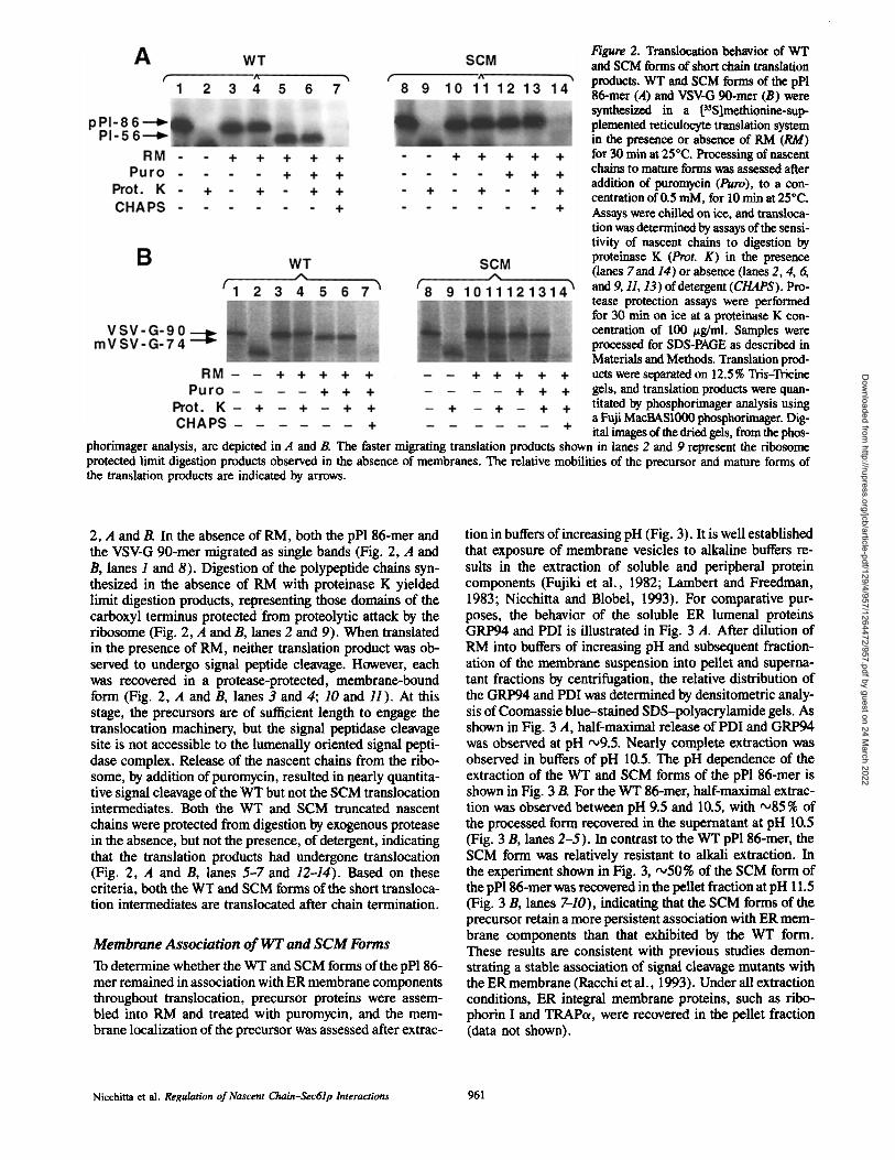
Figure 2. Translocation behavior of WT and SCM forms of short chain translation products. WT and SCM forms of the pPl 86-mer (A) and VSV-G 90-mer (B) were synthesized in a PSSlmethionine-sup- plemented reticulocyte translation system in the presence or absence of RM (RM) for 30 rain at 25°C. Processing of nascent chains to mature forms was assessed after addition of puromycin (Puro), to a con- centration of 0.5 mM, for 10 min at 25°C. Assays were chilled on ice, and transloca- tion was determined by assays of the sensi- tivity of nascent chains to digestion by proteinase K (Prot. K) in the presence (lanes 7 and 14) or absence Oanes 2, 4, 6, and 9, 11, 13) of detergent (CHAPS). Pro- tease protection assays were performed for 30 rain on ice at a proteinase K con- centration of 100 #g/ml. Samples were processed for SDS-PAGE as described in Materials and Methods. Translation prod- ucts were separated on 12.5 % Tris-Tricine gels, and translation products were quan- titated by phosphorimager analysis using a Fuji MacBAS1000 phosphorimager. Dig- ital images of the dried gels, from the phos-
phorimager analysis, are depicted in A and JR The faster migrating translation products shown in lanes 2 and 9 represent the ribosome protected limit digestion products observed in the absence of membranes. The relative mobilities of the precursor and mature forms of the translation products are indicated by arrows.
2, A and B. In the absence of RM, both the pPl 86-mer and the VSV-G 90-mer migrated as single bands (Fig. 2, A and B, lanes 1 and 8). Digestion of the polypeptide chains syn- thesized in the absence of RM with proteinase K yielded limit digestion products, representing those domains of the carboxyl terminus protected from proteolytic attack by the ribosome (Fig. 2, A and B, lanes 2 and 9). When translated in the presence of RM, neither translation product was ob- served to undergo signal peptide cleavage. However, each was recovered in a protease-protected, membrane-bound form (Fig. 2, A and B, lanes 3 and 4; 10 and H ). At this stage, the precursors are of sufficient length to engage the translocation machinery, but the signal peptidase cleavage site is not accessible to the lumenally oriented signal pepti- dase complex. Release of the nascent chains from the ribo- some, by addition of puromycin, resulted in nearly quantita- tive signal cleavage of the WT but not the SCM translocation intermediates. Both the WT and SCM truncated nascent chains were protected from digestion by exogenous protease in the absence, but not the presence, of detergent, indicating that the translation products had undergone translocation (Fig. 2, A and B, lanes 5-7 and 12-14). Based on these criteria, both the WT and SCM forms of the short transloca- tion intermediates are translocated after chain termination.
Membrane Association of W T and SCM Forms
To determine whether the WT and SCM forms of the pPl 86- mer remained in association with ER membrane components throughout translocation, precursor proteins were assem- bled into RM and treated with puromycin, and the mem- brane localization of the precursor was assessed after extrac-
tion in buffers of increasing pH (Fig. 3). It is well established that exposure of membrane vesicles to alkaline buffers re- sults in the extraction of soluble and peripheral protein components (Fujiki et al., 1982; Lambert and Freedman, 1983; Nicchitta and Blobel, 1993). For comparative pur- poses, the behavior of the soluble ER lumenal proteins GRP94 and PDI is illustrated in Fig. 3 A. After dilution of RM into buffers of increasing pH and subsequent fraction- ation of the membrane suspension into pellet and superna- tant fractions by centrifugation, the relative distribution of the GRP94 and PDI was determined by densitometric analy- sis of Coomassie blue-stained SDS-polyacrylamide gels. As shown in Fig. 3 A, half-maximal release of PDI and GRP94 was observed at pH ,,o9.5. Nearly complete extraction was observed in buffers of pH 10.5. The pH dependence of the extraction of the WT and SCM forms of the pP1 86-mer is shown in Fig. 3 B. For the WT 86-mer, half-maximal extrac- tion was observed between pH 9.5 and 10.5, with ",,85% of the processed form recovered in the supernatant at pH 10.5 (Fig. 3 B, lanes 2-5). In contrast to the WT pPl 86-mer, the SCM form was relatively resistant to alkali extraction. In the experiment shown in Fig. 3, '~,50 % of the SCM form of the pP186-mer was recovered in the pellet fraction at pH 11.5 (Fig. 3 B, lanes 7-10), indicating that the SCM forms of the precursor retain a more persistent association with ER mem- brane components than that exhibited by the WT form. These results are consistent with previous studies demon- strating a stable association of signal cleavage mutants with the ER membrane (Racchi et al., 1993). Under all extraction conditions, ER integral membrane proteins, such as ribo- phorin I and TRAPcx, were recovered in the pellet fraction (data not shown).
Nicchitta et al. Regulation of Nascent Chain-$ec61p Interactions 961
Dow
nloaded from http://rupress.org/jcb/article-pdf/129/4/957/1264472/957.pdf by guest on 24 M
arch 2022

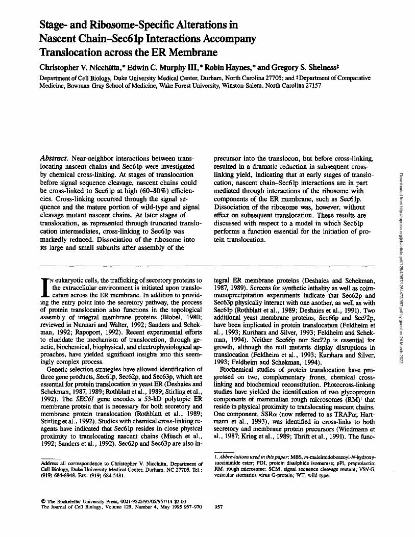
Figure 3. Membrane association of WT and SCM mutant forms of translocated pPl 56-mer and VSV-G 74-met: comparison with ER lumenal proteins. (,4) RM were diluted 10-fold in a 50 mM Hepes, 50 mM CAPS buffers adjusted to the indicated pH values with KOH and incubated on ice for 30 min. Membrane-associated (pellet) and released (supematant) fractions were prepared by centrifugation of the diluted membrane suspension through a 0.5 M sucrose cushion as described in Materials and Methods. Pellet fractions were directly solubilized in SDS-PAGE sample buffer. Supernatant frac- tions were concentrated by TCA precipitation. Fractions were sepa- rated on 10% SDS-PAGE gels, and the relative quantities of the lu- menal proteins GRP94 and PDI were determined by densitometric analysis of the Coomassie blue-stained gel. (B) WT and SCM forms of the pPL 86-mer mRNAs were translated in a reticulocyte lysate system for 30 rain at 25°C. Puromycin was subsequently added to a final concentration of 0.5 raM, and incubations were continued for 10 rain at 25°C. Reactions were chilled on ice and diluted 10-fold in a 50 mM Hepes, 50 mM CAPS buffer adjusted to the indicated pH values with KOH. Diluted'reactions were in- cubated on ice for 30 rain and overlaid onto a 0.5 M sucrose cush- ion. The pellet (P) and supernatant (S) fractions were separated as described in Materials and Methods. Supernatant fractions, comprising the supernatant and cushion, were collected, and the translation products were concentrated by ammonium sulfate frac- tionation. Pellet fractions were resuspended directly in SDS-PAGE sample buffer. Samples were analyzed as described in the legend to Fig. 2.
Figure 4. Chemical cross-linking of translocation intermediates of WT and SCM forms of the pPl 86-mer: cross-link localization. (A) WT and SCM pPl 86-mer mRNAs were translated in a reticulocyte lysate translation system in the presence of RM, as described in the legend to Fig. 1. After translation and, where indicated, puromycin treat- ment, reactions were diluted 10-fold in a physiological salt buffer (110 mM potassium acetate, 25 mM K-Hepes, pH 7.4, 2.5 mM magne- sium acetate) and collected by cen- trifugation through a 0.5 M sucrose cushion. Pelleted membrane frac- tions were resuspended in mem- brane buffer (0.25 M sucrose, 25 mM K-Hepes, 50 mM potassium acetate, 2.5 mM magnesium ace- tate) and MBS added to I mM. Cross-linking was performed for 30 min at 25°C, and reactions were quenched by addition of I vol of PBS, 50 mM DTT, 50 mM lysine and incubation on ice. Samples were TCA precipitated, and trans-
lation products were separated by SDS-PAGE using a Tris-Tricine buffer system. A MacBASI000 digital image is depicted. The migration of the pPl 86-mer and the processed, translocated 56-met is indicated by arrows. (B) Localization of MBS-mediated cross-linking was performed by comparison of the efliciencies of cross-linking between the SCM form of the pP186-met and a pPl mutant lacking the cysteinyl residues at positions +4 and +II (ACys). Cross-linking and sample analysis were performed as described in A. For both panels, the asterisk marks the location of the primary cross-linked complex.
The Journal of Cell Biology, Volume 129, 1995 962
Dow
nloaded from http://rupress.org/jcb/article-pdf/129/4/957/1264472/957.pdf by guest on 24 M
arch 2022
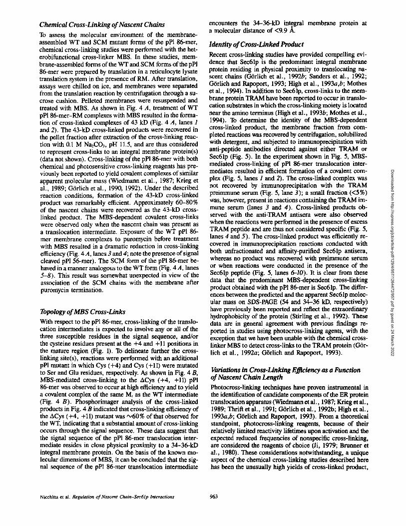
Chemical Cross-Linking of Nascent Chains To assess the molecular environment of the membrane- assembled WT and SCM mutant forms of the pPl 86-mer, chemical cross-linking studies were performed with the het- erobifunctional cross-linker MBS. In these studies, mem- brane-assembled forms of the WT and SCM forms of the pP1 86-mer were prepared by translation in a reticulocyte lysate translation system in the presence of RM. After translation, assays were chilled on ice, and membranes were separated from the translation reaction by centrifugation through a su- crose cushion. Pelleted membranes were resuspended and treated with MBS. As shown in Fig. 4 A, treatment of WT pPl 86-mer-RM complexes with MBS resulted in the forma- tion of cross-linked complexes of 43 kD (Fig. 4 A, lanes 1 and 2). The 43-kD cross-linked products were recovered in the pellet fraction after extraction of the cross-linking reac- tion with 0.1 M Na2CO3, pH 11.5, and are thus considered to represent cross-links to an integral membrane protein(s) (data not shown). Cross-linking of the pPl 86-mer with both chemical and photosensitive cross-linking reagents has pre- viously been reported to yield covalent complexes of similar apparent molecular mass (Wiedmann et al., 1987; Krieg et al., 1989; Gtrlich et al., 1990, 1992). Under the described reaction conditions, formation of the 43-kD cross-linked product was remarkably efficient. Approximately 60-80% of the nascent chains were recovered as the 43-kD cross- linked product. The MBS-dependent covalent cross-links were observed only when the nascent chain was present as a translocation intermediate. Exposure of the WT pPl 86- mer membrane complexes to puromycin before treatment with MBS resulted in a dramatic reduction in cross-linking efficiency (Fig. 4 A, lanes 3 and 4; note the presence of signal cleaved pP1 56-mer). The SCM form of the pPl 86-mer be- haved in a manner analogous to the WT form (Fig. 4 A, lanes 5-8). This result was somewhat unexpected in view of the association of the SCM chains with the membrane after puromycin termination.
Topology of MBS Cross-Links With respect to the pP1 86-mer, cross-linking of the translo- cation intermediates is expected to involve any or all of the three susceptible residues in the signal sequence, and/or the cysteine residues present at the +4 and +11 positions in the mature region (Fig. 1). To delineate further the cross- linking site(s), reactions were performed with an additional pPl mutant in which Cys (+4) and Cys (+11) were mutated to Ser and Glu residues, respectively. As shown in Fig. 4 B, MBS-mediated cross-linking to the ACys (+4, +11) pPl 86-mer was observed to occur at high efficiency and to yield a covalent complex of the same Mr as the WT intermediate (Fig. 4 B). Phosphorimager analysis of the cross-linked products in Fig. 4 B indicated that cross-linking efficiency of the ACys (+4, +11) mutant was '~60% of that observed for the WT, indicating that a substantial amount of cross-linking occurs through the signal sequence. These data suggest that the signal sequence of the pPl 86-mer translocation inter- mediate resides in close physical proximity to a 34-36-kD integral membrane protein. On the basis of the known mo- lecular dimensions of MBS, it can be concluded that the sig- nal sequence of the pP1 86-mer translocation intermediate
encounters the 34-36-kD integral membrane protein at a molecular distance of <9.9 A.
Identity of Cross-Linked Product Recent cross-linking studies have provided compelling evi- dence that Sec61p is the predominant integral membrane protein residing in physical proximity to translocating na- scent chains (G-trlich et al., 1992b; Sanders et al., 1992; G6rlich and Rapoport, 1993; High et al., 1993a,b; Mothes et al., 1994). In addition to Sec61p, cross-links to the mem- brane protein TRAM have been reported to occur in translo- cation substrates in which the cross-linking moiety is located near the amino terminus (High et al., 1993b; Mothes et al., 1994). To determine the identity of the MBS-dependent cross-linked product, the membrane fraction from com- pleted reactions was recovered by centrifugation, solubilized with detergent, and subjected to immunoprecipitation with anti-peptide antibodies directed against either TRAM or Sec61p (Fig. 5). In the experiment shown in Fig. 5, MBS- mediated cross-linking of pP1 86-mer translocation inter- mediates resulted in efficient formation of a covalent com- plex (Fig. 5, lanes 1 and 2). The cross-linked complex was not recovered by immunoprecipitation with the TRAM preimmune serum (Fig. 5, lane 3); a small fraction (<5%) was, however, present in reactions containing the TRAM im- mune serum (lanes 3 and 4). Cross-linked products ob- served with the anti-TRAM antisera were also observed when the reactions were performed in the presence of excess TRAM peptide and are thus not considered specific (Fig. 5, lanes 4 and 5). The cross-linked product was efficiently re- covered in immunoprecipitation reactions conducted with both unfractionated and affinity-purified Sec61p antisera, whereas no product was recovered with preimmune serum or when reactions were conducted in the presence of the Sec61p peptide (Fig. 5, lanes 6-10). It is clear from these data that the predominant MBS-dependent cross-linking product obtained with the pPl 86-mer is Sec61p. The differ- ences between the predicted and the apparent Sec61p molec- ular mass on SDS-PAGE (54 and 34-36 kD, respectively) have previously been reported and reflect the extraordinary hydrophobicity of the protein (Stirling et al., 1992). These data are in general agreement with previous findings re- ported in studies using photocross-linking agents, with the exception that we have been unable with the chemical cross- linker MBS to detect cross-links to the TRAM protein (Gtr- lich et al., 1992a; Gtrlich and Rapoport, 1993).
Variations in Cross-Linking E~ciency as a Function of Nascent Chain Length Photocross-linking techniques have proven instrumental in the identification of candidate components of the ER protein translocation apparatus (Wiedmann et al., 1987; Krieg et al., 1989; Thrift et al., 1991; Gtdich et al., 1992b; High et al., 1993a,b; G-trlich and Rapoport, 1993). From a theoretical standpoint, photocross-linking reagents, because of their relatively limited reactivity lifetimes upon activation and the expected reduced frequencies of nonspecific cross-linking, are considered the reagents of choice (Ji, 1979; Brurmer et al., 1980). These considerations notwithstanding, a unique aspect of the chemical cross-linking studies described here has been the unusually high yields of cross-linked product,
Nicchitta et al. Regulation of Nascent Chain-Sec61p Interactions 963
Dow
nloaded from http://rupress.org/jcb/article-pdf/129/4/957/1264472/957.pdf by guest on 24 M
arch 2022

Figure 5. Characterization of cross-linked com- plexes by immunoprecipitation, pP186-mer trans- location intermediates were synthesized in a re- ticulocyte lysate translation system and subjected to MBS-dependent cross-linking as described in the legend to Fig. 4. Quenched samples were cen- trifuged through a 0.5 M sucrose cushion, and the pellet fraction was processed for indirect immuno- precipitation as described in Materials and Meth- ods. Samples were subject to immunoprecipitation with preimmune (PI) sera or anti-peptide immune sera directed against a peptide representing the carboxy-terminal of TRAM or the amino-terminal of Sec61p. Peptide competition experiments were performed by preincubation of serum or IgG frac- tions with excess peptide.
relative to that obtained by photocross-linking approaches. Indeed, others have reported an increase in cross-linking efficiencies in chemical, versus photochemical, cross-link- ing studies (Gtrlich et al., 1990; Kellaris et al., 1991). We have observed that MBS-dependent cross-linking of the pPl 86-met and the VSV-G 90-mer to Sec61p can be per- formed under conditions in which extremely high yields (60-80%) of cross-linked products are obtained (Fig. 6). This observation has significant experimental ramifications. For example, in view of such high efficiencies, it is likely that the appearance of the cross-linked product represents a sam- pling of the physical environment of the majority of trans- locating nascent chains, rather than that of a small subpopu- lation. Furthermore, because the molecular environment of the majority of translocating chains is being sampled, addi- tional elements of the translocation process, including analy- ses of stage- and ribosome-specific effects on nascent chain- Sec61p interactions, can be studied.
Although the SCM form of the pPl 86-mer translocation intermediate can be cross-linked with high efficiency to Sec61p, little or no cross-linking was observed after puromy- cin treatment, even though the precursor remained in associ- ation with components of the ER membrane (Fig. 3). To ascertain whether the puromycin-dependent loss of cross- linking was simply a consequence of chain termination or, alternatively, due to a change in the topology of the precursor in the membrane, translocation intermediates of both the WT and SCM forms of the pP1 86-, 131-, and 169-mer, as well as the WT and SCM forms of the VSV-G 90- and 172- mer, were studied. As shown in Fig. 6, both the WT and SCM forms of relatively short translocation intermediates
(pPl 86-mer and VSV-G 90-mer) were cross-linknd to Sec61p at the expected efficiencies (Fig. 6, A and B, lanes--f-4). When both the WT and SCM forms of the pPl 131-mer were assayed, cross-links to Sec61p were observed at somewhat reduced efficiencies (Fig. 6 A, lanes 5-8). Note that the pPl 131-mer translocation intermediate is of sufficient length to undergo partial signal sequence processing and thus repre- sents a later stage of the translocation process than that as- sayed with the pP186-mer. When the size of the translocation intermediate was extended to either 169 (pPl) or 172 (VSV-G) amino acids, the yields of cross-linked product were dramat- ically reduced (Fig. 6, A and B, lanes 9-12 and 5-8, respec- tively). Quantitation, by phosphorimager analysis of anti- Sec61p immunoprecipitations, of the data depicted in Fig. 6 indicated that the yields of cross-linked product for the 169- and 172-met were '~5-10% of those obtained with the 86- and 90-mer, respectively. This result was unexpected, since we had observed that for short nascent chains a substantial portion of the cross-links occurred through the signal se- quence. Because the nascent chain is present in the mem- brane in a loop topology, we had assumed that SCM translo- cation intermediates would cross-link tO Sec61p through the signal sequence, regardless of nascent chain length. It ap- pears, therefore, that the topological relationship between the signal sequence of the translocating nascent chain and Sec61p varies through the course of translocation. In addi- tion, because the cross-linking behavior of the WT and SCM forms of the various translocation intermediates are identi- cal, it can be inferred that the topological relationship be- tween Sec61p and the mature portion of the nascent chain also varies through the course of translocation.
The Journal of Cell Biology, Volume 129. 1995 964
Dow
nloaded from http://rupress.org/jcb/article-pdf/129/4/957/1264472/957.pdf by guest on 24 M
arch 2022

Figure 6. Variations in MBS-mediated nascent chain-Sec61p complex formation as a function of nascent chain length. Translations of WT and SCM forms of the pP186-, 131-, and 169-mer (A) and the VSV-G 90- and 172-mer (B) were performed in a reticulocyte lysate translation system supplemented with RM. After assembly of the translocation intermediates, membrane-associated nascent chains were collected by centrifugation through a 0.5 M sucrose cushion (as described in Materials and Methods) resuspended in membrane buffer, and subjected to cross-linking as described in the legend to Fig. 4. Cross-linking reactions were quenched, and samples were precipitated with 10% TCA. Cross-linked complexes were resuspended in SDS-PAGE buffer and analyzed as described in the legend to Fig. 2. The relative migra- tion of the different translation products is indicated by arrows. Note that the pPl 131-mer is partially processed to the PI 101-mer and the pPl 169-mer is fully processed to the PI 139-met (A). For A, the stacking gel has been included to ascertain whether the apparent lack of MBS-dependent complex formation was due to the formation of high molecular weight aggregates that were unable to enter the separating gel.
Effects of Ribasome Disassembly on Nascent Chain-Sec61p Interactions
On the basis of the results shown in Fig. 6, it is apparent that near-neighbor interactions between Sec61p and the trans- locating nascent chain vary over the course of translocation. To gain further insight into this phenomenon, the role of the ribosome in the regulation of nascent chain-Sec61p interac- tions was examined. Recent experimental evidence indicates that Sec61p, in addition to its role as a potential protein- conducting channel, functions in the binding of ribosomes to the ER membrane (Gtrlich et ai., 1993; Kalies et al., 1994). To determine the contribution of the ribosome to na- scent chain-Sec61p near-neighbor interactions, transloca- tion intermediates of the WT and SCM forms of the pPl 86- mer were assembled and chilled on ice, which serves to block any further translocation (Nicchitta and Blobel, 1989). Subsequently, ribosomes were dissociated by treatment with EDTA (Gesteland, 1966). At the EDTA concentrations used in these assays (10 mM), the ribosome dissociates into its component subunits and both the large and the small ribo- somal subunits are released from the membrane (Sabatini et ai., 1966; Pryme, 1988; data not shown). The topology of the nascent chain was then assayed by protease digestion. The results of these experiments are shown in Fig. 7. For both the WT and SCM forms of the pPl 86-mer, digestion of the ribosome-associated nascent chains, in the absence of membranes, yielded a limit digestion product of 3--4 kD (Fig. 7 A, lanes I and 2; 7and 8). The limit digestion prod- uct represents that part of the nascent chain protected by the ribosome (Fig. 2) (Malkin and Rich, 1967; Blobel and Saba-
tini, 1970). Translocation intermediates of both the WT and SCM forms of the pP1 86-mer were resistant to proteolyric digestion (Fig. 7 A, lanes 3 and 4; 9 and 10), an observation consistent with previous reports (Connolly and Gilmore, 1986; Nicchitta and Blobel, 1989). More significantly, how- ever, dissociation of the ribosome under conditions (reduced temperature) in which translocation does not occur resulted in nearly complete accessibility of the precursor to digestion by exogenous protease (Fig. 7 A, lanes 5 and 6; 11 and 12).
The precise size of the domain of the translocarion inter- mediate exposed after ribosome dissociation has not been determined. This domain is expected to include the 3--4-kD domain that resides within the intact ribosome. Although the Tris-Tricine gel system used in these experiments allows resolution of fragments as small as 1.5-2 kD, we have been unable to identify limit digestion products of the transloca- tion intermediates after ribosome dissociation. It appears, therefore, that the size of the exposed domain may be as large as 6.6 kD.
To investigate the effects of ribosome dissociation on na- scent chain-Sec61p near-neighbor interactions, pPl 86-mer translocation intermediates were formed, chilled on ice, treated with EDTA, and subsequently cross-linked with MBS. As shown in Fig. 7 B, when pP1 86-mer translocation intermediates were treated with MBS, on ice, efficient for- marion of the 43-kD cross-linked product, representing a covalent complex of the nascent chain with Sec61p, was ob- served (lanes 1 and 2). When the ribosome-nascent chain-membrane complexes were treated with EDTA before cross-linking with MBS, the yield of the cross-linked prod- uct was greatly reduced. On average, EDTA pretreatment re-
Nicchitta et al. Regulation of Nascent Chain-Sec61p Interactions 965
Dow
nloaded from http://rupress.org/jcb/article-pdf/129/4/957/1264472/957.pdf by guest on 24 M
arch 2022
Figure 7. The effects of EDTA-mediated ribosome dissociation on pP186-mer processing, translocation, and near-neighbor interaction with Sec61p. (A) WT and SCM forms of the pPl 86-mer were translated in the presence or absence of RaM in a reticulocyte lysate translation system for 30 min at 25°C. After translation, reactions were chilled on ice and, where indicated, adjusted to 10 mM EDTA. After a 30-rain incubation on ice, proteinase K was added, and di- gestions were performed for 30 rain on ice. Protease digestion reactions were quenched and processed for SDS-PAGE as described in Materials and Methods. Samples were analyzed as described in the legend to Fig. 2. The faster migrating form shown in lanes 2 and 8 represents the ribosome protected fragment observed in the absence of RaM. (B) pPl 86-mer translocation intermediates were assembled as de- scribed in A. Reactions were chilled on ice and ad- justed to either 10 mM EDTA (lane 3), 10 mM EGTA (lane 4), 250 U/ml staphylococcal nuclease (lane 5), or 10 ram EDTA, 250 U/mi staphylococ- cal nuclease (lane 6). Incubations were performed for 30 rain on ice, and samples were subsequently cross-linked by addition of MBS to 1 raM. Cross- linking reactions were performed for 60 rain at 4°C. The primary cross-linked product is indicated by the asterisk.
duced the formation of the nascent chain-Sec61p product by 60-70% (n = 5). This effect was apparently specific for Mg 2+, as treatment with equivalent concentrations of EGTA, which exhibits preferential binding to Ca 2÷, was without effect (Fig. 7 B, lane 4). In addition, treatment with ribonuclease did not alter the yield of the 43-kD cross-linked product (Fig. 7 B, lane 2 versus lane 5). Pretreatment with a combination of EDTA and nuclease resulted in a further, although marginal, decrease in the yield of cross-linked product.
It is apparent that when a ribosome bearing the translo- cation intermediate is dissociated into its subunits, the efficiency of nascent chain-Sec61p cross-link formation is greatly reduced. These data indicate that efficient near-neigh- bor interactions between the translocating nascent chain and Sec61p may arise primarily through interactions between the ribosome and Sec61p, a conclusion consistent with the ob- servation that Sec61p is a ribosome-binding protein (Kalies et al., 1994). Because these interactions are to a great extent lost upon dissociation of the ribosome into its subunits, translocation intermediates of the pP1 86-mer were assayed to determine whether disruption of nascent chain-Sec61p in- teractions would affect translocation of the cytosolic domain of the nascent chain intermediate. The results of a represen-
tative experiment are depicted in Fig. 8. In this experiment, translocation intermediates of the pP1 86-mer were first as- sembled at 25°C, and the efficiency of translocation at 25°C was assessed after either dissociation of the ribosome (plus EDTA) or upon polypeptide termination (plus puromycin). Puromycin addition resulted in both efficient processing and translocation of pPl 86-mer nascent chains (Fig. 8, lanes 2 and 3). Similarly, dissociation of the ribosome by EDTA ad- dition yielded efficient processing and translocation of the pP1 86-mer (Fig. 8, lanes 5 and 6). Thus, under the de- scribed experimental conditions, disruption of the ribosome by EDTA addition had only marginal effects on the efficiency of translocation.
In experiments in which the effects of ribosome disso- ciation on near-neighbor, nascent chain-Sec61p interac- tions were determined (Fig. 7 B), the ribosome-nascent chain-membrane complexes were preincubated in the pres- ence of EDTA for 30 min at 4°C before chemical cross- linking. This extended incubation time may have allowed diffusion of the translocating nascent chain from Sec61p and thus the loss of MBS-dependent complex formation. To com- pare the effects of ribosome dissociation on translocation un- der conditions comparable to those used for cross-linking, translocation intermediates of the pP1 86-mer were pre-
The Journal of Cell Biology, Volume 129, 1995 966
Dow
nloaded from http://rupress.org/jcb/article-pdf/129/4/957/1264472/957.pdf by guest on 24 M
arch 2022

Figure 8. The effects of EDTA-mediated ribosome dissociation on translocation of the pPl 86-mer. Translocation intermediates of the pP1 86-mer were assembled by translation in a reticulocyte lysate system for 30 min at 25°C. After a 30-min translation period, sam- ples were adjusted, as indicated, to 0.5 mM puromycin (lanes 2 and 3) or 10 mM EDTA (lanes 5 and 6), and the reaction was continued for 10 min at 25°C. Paired samples were chilled on ice and subse- quently adjusted to either 10 mM EDTA or 0.5 mM puromycin (lanes 9 and 10 or 11 and 12, repectively). After a 30-min incuba- tion on ice, reactions were brought to 25°C for 15 rain. To deter- mine the efficiency of translocation, samples were subjected to pro- teinase K digestion for 30 min at 4°C with 100 #g/ml proteinase K and processed for SDS-PAGE as described in Materials and Methods. In lanes 1-6, translocation reactions were performed im- mediately after translation. In lanes 7-12, reactions were chilled on ice, supplemented as previously described, and subsequently re- warmed to 25°C.
pared, chilled on ice, and treated with either EDTA or puromycin for 30 min. Samples were subsequently warmed to 25°C for 10 rain, to allow translocation to continue. As shown in Fig. 8, lanes 9-12, prolonged dissociation of the ribosome, under conditions in which cross-linking of the na- scent chain to Sec61p was markedly reduced was without effect on translocation, as judged by protease protection. For both the EDTA- and puromycin-treated samples, signal pep- tide processing occurred at 50-70% efficiency, and of the processed chains, 60-80% were protected from proteolytic digestion. These data indicate that disruption of near- neighbor interactions between translocating nascent chains and Sec61p is not accompanied by a loss of translocation ac- tivity.
Discussion
Current models of protein translocation across the ER mem- brane invoke a central role for Sec61p in the translocation of secretory protein precursors across, and the assembly of in- tegral membrane proteins into, the ER membrane (Stirling et al., 1992; Sanders et al., 1992; Gfrlich et al., 1992b; G/Srlich and Rapoport, 1993; Mothes et al., 1994). Sec61p, the product of the SEC61 gene, was initially identified in genetic screens for components of the translocation appara- tus necessary for the insertion of secretory and membrane proteins into the yeast ER membrane (Deshaies and Schek- man, 1987; Stirling et al., 1992). Subsequently, Sec61p was observed, by photo- and chemical cross-linking, to reside in close physical proximity to translocating nascent chains in both yeast and mammalian ER (Sanders et al., 1992; Mtisch et al., 1992; Gfrlich et al., 1992b; High et al., 1993a,b).
In addition, recent biochemical reconstitution studies have identified Sec61p and the signal recognition particle receptor as the only protein components essential for translocation across the ER membrane, although other membrane pro- teins, such as TRAM, may further enable the translocation of specific precursors (G6rlich and Rapoport, 1993). On the basis of these results, Sec61p has been proposed to function either as a protein-conducting channel (G6rlich and Rapoport, 1993; Mothes et al., 1994) or as a component that transports the nascent chain across the ER membrane (Sanders et al., 1992).
To elaborate further a molecular description of the mecha- nism of protein translocation, we have used chemical cross- linking as a tool to identify ER proteins residing in physical proximity to translocating nascent chains at discrete stages of the translocation process. These studies were performed with WT and SCM forms of pP1 and VSV-G. The SCM precursors contained point mutations in the signal cleavage domain that blocked processing of the nascent chain. With relatively short nascent chains (~<90 amino acids), cross- links to Sec61p were observed to occur at remarkably high (60-80%) efficiency. The efficiency of Sec61p translocation intermediate cross-linking decreased, however, as a function of increasing chain length. These data suggest that near- neighbor interactions between translocating nascent chains and Sec61p predominate at early stages of translocation and are consistent with recent models in which precursor pro- teins are thought to assemble inititially into the translocation apparatus in contact with Sec61p (Sanders et al., 1992). Other investigators have previously identified cross-linked complexes of late stage translocation intermediates and Sec61p, and thus it is clear that such intermediates do en- counter, at least for time periods sufficient for cross-linking, the Sec61p complex (Wiedmann et al., 1989; Miisch et al., 1992; G6rlich et al., 1992b; Mothes et al., 1994). In these studies, however, cross-linking was performed with photo- cross-linking reagents, in which the yield of cross-linked product is by nature relatively low. With such low yields, it is difficult to ascertain directly whether the appearance of photocross-linked nascent chain-Sec61p complexes accu- rately reflects the membrane disposition of the majority pop- ulation of nascent chains.
To extend further the observation of stage-specific cross- linking, experiments were performed with a series of trun- cated intermediates containing mutations in the signal se- quence cleavage site that blocked processing. The rationale for these experiments was as follows. It is known that the na- scent chain assembles into the membrane in a loop confor- mation (Shaw et al., 1988; Mothes et al., 1994). Further- more, in studies of a SCM form of VSV-G, it was observed that the loop topology of the nascent chain was maintained throughout translocation and that the amino terminus of the signal sequence remained exposed on the cytoplasmic face of the ER membrane, even after the completion of translation (Shaw et al., 1988). In addition, recent cross-linking studies using a novel suppressor tRNA approach have demonstrated that the signal sequence is immediately adjacent to Sec61p (High et al., 1993b). Integrating these observations with cur- rent models of the mammalian translocation apparatus, we reasoned that the signal sequence of the SCM forms of trans- locating nascent chains would remain in association with components of the translocation machinery, such as Sec61p,
Nicchitta et al. Regulation of Nascent Chain-Sec61p Interactions 967
Dow
nloaded from http://rupress.org/jcb/article-pdf/129/4/957/1264472/957.pdf by guest on 24 M
arch 2022

throughout translocation. In fact, the SCM forms of the pPl 86-mer and the VSV-G 90-mer cross-link efficiently to Sec61p (Figs. 4 and 6). Site-directed mutagenesis of the potential cross-linking sites in the pP1 86-mer indicated that at least 50% of the cross-links occurred through residues present in the signal sequence, indicating a near-neighbor in- teraction between the signal sequence and Sec61p, in agree- ment with High et al. (1993b). Although the short chain SCM intermediates could be efficiently cross-linked to Sec61p, late stage SCM intermediate-Sec61p cross-links were ob- served at very low efficiencies. These data support the con- clusion that near-neighbor interactions between the translo- caring nascent chain and Sec61p predominate at early rather than late stages of the translocation process.
Several interpretations of these data, with respect to models in which Sec61p defines the immediate molecular en- vironment of the nascent chain, are possible. The decrease in cross-linking of the longer SCM intermediates to Sec61p could be reconciled as a simple steric phenomenon, in which an appropriate arrangement of amino acid side chains is lacking in the longer translocation intermediates. Although this explanation may be valid, suitable amino acids are pres- ent throughout the pPl intermediates and are especially abundant in the VSV-G intermediates (Fig. 1). Furthermore, a comparison of the relative locations of potential cross- linking sites in the amino termini of the pPl and VSV-G con- structs, as well as the carboxy-terminal domains extending from the ribosome protected region towards the amino ter- minus of the pP1 131-mer and VSV-G 172-mer, shows a nearly identical order of suitable amino acids. On the basis of these similarities, the cross-linking pattern of the pP1 131- mer and the VSV-G 172-mer should be similar. That they are not is likely a reflection of topological alterations in the na- scent chain-Sec61p interactions during translocation. Be- cause '~50% of the cross-linking of short chain intermedi- ates was observed to occur through the signal sequence, it is perhaps more likely that at an intermediate stage of trans- location, the topological relationship between the signal se- quence of the SCM intermediate and Sec61p, is altered. How might this occur? Because the signal sequence of a SCM form of VSV-G has previously been demonstrated to act as a signal anchor domain (Shaw et al., 1988), it is possible that during translocation, the signal sequence of the SCM inter- mediates diffuses from the protein-conducting channel and integrates into the lipid bilayer. This interpretation places significant constraints on the point at which such topological alterations might occur. For example, pP1 131-mer transloca- tion intermediates undergo partial amino-terminal process- ing and are therefore exposed to the ER lumen. Cross- linking between Sec61p and the WT and SCM pP1 131-mer is, however, efficient. Should the decrease in cross-linking reflect diffusion of the signal sequence from the channel, these results indicate that diffusion would be delayed until the translocation intermediate exceeds a length of ~131 amino acids.
The high efficiency of MBS-dependent cross-linking of short chain translocation intermediates to Sec61p strongly supports the conclusion that the observed cross-links are reflective of the behavior of the total population of nascent chains, rather than that of a discrete subpopulation. An in- trigning aspect of these studies is the strong dependence of high efficiency cross-link formation on the presence of the
intact ribosome. In this regard, it is particularly significant that recent studies have demonstrated that Sec61p can func- tion as a ribosome receptor (G6rllch et al., 1992b; Kalies et al., 1994). Experiments were therefore performed to charac- terize the role of the ribosome in the genesis of nascent chain-Sec61p cross-links. Ribosome-associated transloca- tion intermediates of either the WT or SCM forms of the pPl 86-mer were assembled and chilled on ice, and the ribo- somes were dissociated, by chelation of Mg 2+, into their large and small subunits (CJesteland, 1966). Dissociation of the ribosome results in exposure of the translocation inter- mediate to the cytoplasmic face of the membrane, as demon- strated by the dramatic increase in the sensitivity of the precursor to digestion by exogenous protease. When the physical interaction of the nascent chain with Sec61p was as- sayed under similar conditions by chemical cross-linking, the efficiency of cross-linking was reduced by 60-70 %. This decrease in near-neighbor interactions between the nascent chain and Sec61p was apparent even when cross-linking times were extended to 2 h, conditions in which random, collision-dependent cross-links would be expected to occur (data not shown) (Ji, 1979). The relative inability to identify translocation intermediate-Sec61p cross-links after dissoci- ation of the ribosome was not a simple consequence of re- duced temperature; cross-linking of ribosome-associated na- scent chains to Sec61p was as efficient at 4 as at 25"C. These data, in combination with those previously discussed, best support a model in which near-neighbor interactions be- tween the translocating nascent chain and Sec61p are greatly potentiated by physical interactions between the ribosome and Sec61p. In the absence of such interactions, the trans- locating nascent chain appears to sample, but not reside in, an environment physically adjacent to Sec61p. This scenario can be easily rationalized if the following criteria are sa- tisfied: (i) at early stages of translocation, the nascent chain exit site on the ribosome is in close proximity to Sec61p, thus providing a suitable environment for cross-linking of short nascent chains to Sec61p, and (ii) at late stages of transloca- tion, translation and translocation are not efficiently cou- pled, thereby removing the topological constraint necessary for efficient cross-linking to Sec61p. The predominance of studies demonstrating cross-linking of short chain transloca- tion intermediates to Sec61p clearly supports the first criterion (Wiedmann et al., 1986; G6rlich et al., 1992; High et al., 1993a,b). With regard to the second criterion, translo- cation intermediates are known to exist in at least two popu- lations, one of which is sensitive to digestion with exogenous proteases (Blobel and Sabatini, 1970). Given the topology of the translocation reaction, the appearance of protease sen- sitivity must reflect either an uncoupling of translation and translocation, which would allow the precursor to form an exposed loop between the ribosome and the membrane, or dissociation of the ribosome from the membrane during translocation. More recent studies with truncated pP1 and VSV-G translocation intermediates have clearly demon- strated that either dissociation of the ribosome from the ER membrane or formation of an exposed domain of the nascent chain occurs when precursors exceed a length of ,,o100 amino acids (ConnoUy et al., 1989).
By similar experimental protocols, assays were performed to determine whether disruptions in the near-neighbor na- scent chain-Sec61p interaction affected the process of trans-
The Journal of Cell Biology, Volume 129, 1995 968
Dow
nloaded from http://rupress.org/jcb/article-pdf/129/4/957/1264472/957.pdf by guest on 24 M
arch 2022

I A " lnact ive" Ribosome Binding "Act ive"
Figure 9. Conformational equilibrium model for Sec61p function. Sec61p is depicted as existing in two conformational states, I and A, representing, respectively, the inactive and active conformations of the protein. In this model, the interaction of the translationally active ribosome with Sec61p promotes conversion of Sec61p from the I to the A state. In the A state, Sec61p assists assembly of the signal sequence with components of the ER membrane at a site dis- tinct from, yet physically near, Sec61p.
location. It was observed that at 25°C, dissociation of the ribosome allowed translocation of the cytosol-disposed re- gion of truncated nascent chains. When translocation inter- mediates of the pP186-mer were chilled on ice, the ribosome dissociated, and when the membrane-nascent chain com- plexes were subsequently warmed to 25°C, translocation proceeded at identical efficiency. On the basis of those data, it should be considered that translocation across the ER membrane may not require a continuous, direct physical in- teraction of the nascent chain with Sec61p, but rather Soc61p would serve an essential function in the initiation of translo- cation, and perhaps other protein components would func- tion in the completion of translocation. Although it is pres- ently unclear what additional factors may assist translocation or provide an environment suitable for transport of the na- scent chain, that recent experimental evidence in Esche- richia coli indicates that translocation across the bacterial inner membrane is driven primarily by the SecA protein, bound to the membrane, in part through interactions with SecY, a bacterial homologue of Sec61p (Lill et al., 1990; Watanabe and Blobel, 1993; Economou and Wickner, 1994; Kim et al., 1994; Stirling et al., 1992; C~rlich et al., 1992b). By analogy to E. coli, perhaps Sec61p functions in the assembly of additional components to the translocation site, as has been proposed for SecY (Economou and Wick- ner, 1994; Kim et al., 1994). In this regard, it is interesting to note that in bacterial inverted vesicles, late stage proOmpA translocation intermediates can be chemically cross-linked to SecY (Joly and Wickner, 1993). In this sys-
tem, the late stage nor-neighbor nascent chain-SecY inter- actions may well be a consequence of a direct physical asso- ciation of SecA with SecY, rather than interactions between the nascent chain and SecY.
It is clear that at early stages of translocation, nascent chains are in close physical proximity to Sec61p. It appears equally as clear, however, that the apparent near-neighbor relationship between the translocating nascent chain and Sec61p is greatly influenced by the continued association of the nascent chain with the ribosome. Although such a model readily explains the near-neighbor interactions between translocating nascent chains and Sec61p, when such interac- tions are assayed cotranslationally, experimental observa- tions ofposttranslational (i.e., ribosome-independent) inter- actions between nascent chains and Sec61p are more difficult to rationalize (Sanders et al., 1992; Klappa et al., 1994). To integrate these apparently disparate observations, we pro- pose the following "conformational equilibrium" model, which is schematically illustrated in Fig. 9. In this model, Sec61p is depicted as existing in kinetic equilibrium between two distinct conformational states. In conformation state A, Sec61p defines the site at which translocation is initiated, whereas in conformation state I, Sec61p is inactive and translocation cannot occur. The equilibrium between the two conformational states is depicted as being influenced by the binding of the ribosome, which favors the formation of the A state. Dissociation of the bound ribosome into its compo- nent subunits is therefore predicted to result in a shift in Sec61p from the A to the I state and a subsequent alteration in the topological relationship between the nascent chain and Sec61p.
We wish to thank Pamela Wearsch and Teresa Anne Graham for critical comments and helpful discussions and Dr. John Rose (Yale University, New Haven, CT) for the generous gift of cDNA encoding VSV-G.
This work was supported by National Institutes of Health grants RO1 DK 47897 (to C. V. Nicchitta) and PO1 HL 49373 (to G. S. Sbelness). This work was done during the tenure of an Established Investigatorship from the American Heart Association (G. S. Sbelness). A summary of these findings has been published in abstract form (Mol. Biol. Cell 5:205a).
Received for publication 20 December 1994 and in revised form 2 February 1995.
References
Blobel, O. 1980. Intracellular protein topogenesis. Proc. Natl. Acad. Sci. USA. 77:1496-1500.
Blobel, G., and B. Dobberstein. 1975. Transfer of proteins across membranes. If. Reconstitution of functional rough microsemes from hetemlogous com- poneots. J. Cell Biol. 67:852-862.
Blob¢l, G., and D. Sabatini. 1970. Controlled proteolysis of nascent polypep- tides in rat liver cell fractions. I. Location of the polypeptides within ribo- somes. J. Cell Biol. 45:835-851.
Brodsky, J. L., S. Hamamoto, D. Feldheim, and R. Schekmnn. 1993. Reconsti- tution of protein translocation from selubilized yeast membranes reveals topologically distinct roles for BiP and cytosolic Hsc70. J. Cell Biol. 120:95-102.
Brunner, J., H. Seun, and F. M. Richards. 1980. 3-Trifluoromethyl-3- phenyldiazirine. A new carbene generating group for photolabcling reagents. J. Biol. Chem. 255:3313-3318.
Connolly, T., and R. Gilmore. 1986. Formation of a functional ribosome- membrane junction during tranalocation requires the participation of a OTP- binding protein. J. Cell Biol. 103:2253-2261.
Connoily, T., P. Collins, and R. Gilmore. 1989. Access of proteinase K te par- tially translocated nascent polypeptides in intact and detergent-selubilized membranes. J. Cell Biol. 108:299-307.
Crowley, K. S., G. D. Reinhart, and A. E. Johnson. 1993. The signal sequence moves through a ribosomal tunnel into a noncytoplesmic aqueous environ- ment at the ER membrane early in translocation. Cell. 73:1101-1115.
Crowley, K. S., S. Liao, V. E. Worroll, G. D. Reinhart, and A. E. Johnson. 1994. Secretory proteins move through the endbplasmic reticulum mem- brane via an aqueous, gated pore. Cell. 78:461--471.
Nicchitta et al. Regulation of Nascent Chain-Sec61p Interactions 969
Dow
nloaded from http://rupress.org/jcb/article-pdf/129/4/957/1264472/957.pdf by guest on 24 M
arch 2022

Deshaies, R. L, and R. Schekman. 1987. A yeast mutant defective at an early stage in import of secretory protein precursors into the endoplasmic reticu- lure. J. Cell Biol. 105:633-645.
Deshaies, R. J., and R. Schekman. 1989. SEC62 encodes a putative membrane protein required for protein translocation into the yeast endoplasmic reticu- lure. J. Cell Biol. 109:2653-2664.
Deshaies, R. J., S. L. Sanders, D. A. Feldheim, and R. Schelffnan. 1991. As- ' sembly of yeast Sec proteins involved in translocation into the endoplasmic reticulum into a membrane-bound multisubunit complex. Nature (Lond.). 349:806-808.
Economou, A., and W. Wickner. 1994. SecA promotes preprotein transloca- tion by undergoing ATP driven cycles of membrane insertion and deinser- tion. Cell. 78:835-843.
Feidheim, D., and R. Schekman. 1994. Sec72p contributes to the selective rec- ognition of signal peptides by the secretory polypeptide translocation com- plex. J. Cell Biol. 126:935-943.
Feldheim, D., K. Yoshimura, A. Admon, and R. Schekman. 1993. Structural and functional characterization of Sec66p, a new subunit of the polypeptide translocation apparatus in the yeast endoplasmic reticulum. Mol. Biol. Cell. 4:931-939.
Fujiki, Y., A. L. Hubbard, S. Fowler, and P. B. Lazarow. 1982. Isolation of intracellular membranes by means of sodium carbonate treatment: applica- tion to endoplasmic reticulum. J. Cell Biol. 93:97-102.
Gesteland, R. F. 1966. Unfolding of Escherichia coli ribosomes by removal of magnesium. J. Mol. Biol. 18:356-371.
Gilmore, R., P. Collins, J. Johnson, K. Kellaris, and P. Rapiejko. 1991. Tran- scription of full-length and truncated mRNA transcripts to study protein translocation across the endoplasmic reticulum. Methods Cell Biol. 34: 223-239.
Gtrlich, D., S. Prehn, E. Hartmann, J. Herz, A. Otto, R. Kraft, M. Wiedmaun, S. Knespel, B. Dobberstein, and T. A. Rapoport. 1990. The signal sequence receptor has a second subnnit and is part of a translocation complex in the endoplasmic reticulum as probed by bifunctional reagents. J. Cell Biol. 111:2283-2294.
G6rlich, D., and T. A. Rapoport. 1993. Protein translocation into proteolipo- somes reconstituted from purified components of the endoplasmic reticulum membrane. Cell. 75:615-630.
Gtriich, D., E. Hartmann, S. Prehn, and T. A. Rapoport. 1992a. A protein of the endoplasmic reticulum involved early in polypeptide translocation. Nature (Lond.). 357:47-52.
G0rlich, D., S. Prehn, E. Hartmann, K. U. Kalies, andT. A. Rapoport. 1992b. A mammalian homolog of Sec61p and SecYp is associated with ribosomes and nascent polypeptides during translocation. Cell. 71:489-503.
Harlow, E., and D. Lane. 1988. Antibodies: A Laboratory Manual. Cold Spring Harbor Laboratory Press, Cold Spring Harbor, NY.
Hartmann, E., D. G-trlich, S. Kostka, A. Otto, R. Kraft, S. Knespel, E. Burger, T. A. Rapoport, and S. Prehn. 1993. A tetrameric complex of membrane proteins in the endoplasmic reticulum. Fur. J. Biochem. 214:375-381.
High, S., S. S. Andersen, D. Gtrlich, E. Hartmann, S. Prehn, T. A. Rapoport, and B. Dobberstein. 1993a. Sec61p is adjacent to nascent type I and type II signal-anchor proteins during their insertion. J. Cell Biol. 121:743-750.
High, S., B. Martoglio, D. C-trlich, S. S. Andersen, A. J. Ashford, A. Giner, E. Hartmann, S. Prehn, T. A. Rapoport, B. Dobberstein, and J. Brunet. 1993b. Site-specific photocross-linking reveals that Sec61p and TRAM con- tact different regions of a membrane-inserted signal sequence. J. Biol. Chem. 268:26745-26751.
Jackson, R. J., and T. Hunt. 1983. Preparation and use of nuclease-treated rab- bit reticulocyte lysates for the translation of eukaryotic messenger RNA. Methods Enzymol. 96:50-73.
Ji, T. H. 1979. The application of chemical cross-linking for studies on cell sur- face membranes and the identification of surface reporters. Biochim. Bio- phys. Acta. 559:39-69.
Joly, J. C., and W. Wickner. 1993. The SecA and SecY subunits of translocase are the nearest neighbors of a translocating preprotein, shielding it from phospholipids. EMBO (Eur. Mol. Biol. Organ.) J. 12:255-263.
Kalies, K. U., D. G-trlich, and T. A. Rapopurt. 1994. Binding of ribosomes to the rough endoplasmic reticulum mediated by the Sec61p-complex. J. Cell Biol. 126:925-934.
Kellaris, K. V., S. Bowen, and R. Gilmore. 1991. ER translocation intermedi- ates are adjacent to a non-glycosylated 34-kD integral membrane protein. J. Cell Biol. 114:21-33.
Kim, Y. J. K., T. Rajapandi, and D. Oliver. 1994. SecA protein is exposed to the periplasmic surface of the E. coli inner membrane in its active state. Cell. 78:845-853.
Klappa, P., M. Zimmermann, and R. Zimmermann. 1994. The membrane pro- teins TRAMp and See6101 p may be involved in post-translational transport of presecretory proteins into mammalian microsomes. FEBS (Fed. Fur. Bio- chem. Soc.) Lett. 341:281-287.
Krieg, U. C., A. E. Johnson, and P. Walter. 1989. Protein translocation across the endoplasmic reticulum membrane: identification by photocross-linking ofa 39-kD integral membrane glycoprotein as part of a putative translocation tunnel. J. Cell Biol. 109:2033-2043.
Kurihara, T., and P. Silver. 1993. Suppression of a see63 mutation identifies a novel component of the yeast endoplasmic reticulum translocation appara- tus. Mol. Biol. Cell. 4:919-930.
Lambert, N., and R. B. Freedman. 1983. The latency of rat liver microsomal protein disulphide-isomerase. Biochem. J. 213:225-234.
Lill, R., W. Dowhan, and W. Wickner. 1990. The ATPase activity of SecA
is regulated by acidic phospholipids, SecY, and the leader and mature do- mains of precursor proteins. Cell. 60:271-280.
Malkin, L. I., and A. Rich. 1967. Partial resistance of nacent polypeptide chains to proteolytic digestion due to ribosomal shielding. J. Mol. Biol. 26: 329-346.
Migliaccio, G., C. V. Nicchitta, and G. Biobel. 1992. The signal sequence receptor, unlike the signal recognition particle receptor, is not essential for protein translocation. J. Cell Biol. 117:15-25.
Mothes, W., S. Prehn, and T. A. Rapoport. 1994. Systematic probing of the environment of a translocating secretory protein during translocation through the ER membrane. EMBO (Fur. Mol. Biol. Organ.) J. 13:3973-3982.
Miisch, A., M. Wiedmarm, and T. Rapoport. 1992. Yeast Sec proteins interact with polypeptides traversing the endoplasmic reticulum. Cell. 69:343-352.
Nicchitta, C. V., and G. Biobel. 1989. Nascent chain binding and translocation are distinct processes: differentiation by chemical alkylation. J. Cell Biol. 108:789-795.
Nicchitta, C. V., and G. Blobel. 1993. Lumenal proteins of the mammalian en- doplasmic reticulum are required to complete protein translocation. Cell. 73:989-998.
Nunnari, J., and P. Walter. 1992. Protein targeting to and translocation across the membrane of the endoplasmic reticulum. Curr. Opin. Cell Biol. 4: 573-580.
Perara, E., R. Rothman, and V. Linguppa. 1986. Uncoupling translocation from translation: implications for transport of proteins across membranes. Science (Wash. DC). 232:348-352.
Pryme, I. F. 1988. Differences in the association of ribosomes with heavy rough and light rough endoplasmic reticulum membranes of MPC-11 cells. Bio- chim. Biophys. Acta. 967:218-223.
Racchi, M., H. H. Watzke, K. A. High, and M. O. Lively. 1993. Human coagulation factor X deficiency caused by a mutant signal peptide that blocks cleavage by signal peptidase but not targeting and translocation to the en- doplasmic reticulum. J. Biol. Chem. 268:5735-5740.
Rapoport, T. 1992. Transport of proteins across the endoplasmic reticulum membrane. Science (Wash. DC). 258:931-936.
Rothblatt, J. A., R. J. Deshaies, S. L. Sanders, G. Daum, and R. Schekman. 1989. Multiple genes are required for proper insertion of secretory proteins into the endoplasmic reticolum in yeast. J. Cell Biol. 109:2641-2652.
Sabatini, D. D., Y. Tashiro, and G. E. Palade. 1966. On the attachment of ribo- somes to microsomal membranes. J. Mol. Biol. 19:503-524.
Sanders, S. L., and R. Schekman. 1992. Polypeptide translocation across the endoplasmic reticalum membrane. J. Biol. Chem. 267:13791-13794.
Sanders, S. L., K. M. Whitfield, I. P. Vogel, M. D. Rose, and R. W. Schek- man. 1992. Sec61p and BiP directly facilitate polypeptide translocation into the ER. Cell. 69:353-365.
Sarkar, G., and S. S. Sommer. 1990. The "megaprimer" method of site-directed mutagenesis. Biotechniques. 8:404-407.
Sch~igger, H., and G. yon Jaguw. 1987. Tricine-sodium dodecyl sulfate- polyacrylamide gel electrophoresis for the separation of proteins in the range from 1 to 100 kDa. Anal. Biochem. 166:368-379.
Shaw, A., P. J. M. Rottier, and L K. Rose. 1988. Evidence for the loop model of signal-sequence insertion into the endoplasmic reticulum. Proc. Natl. Acad. Sci. USA. 85:7592-7596.
Simon, S. M., and G. Blobel. 1991. A protein-conducting channel in the en- doplasmic reticulum. Cell. 65:371-380.
Simon, S. M., and G. Blobel. 1992. Signal peptides open protein-conducting channels in E. coli. Cell. 69:677-684.
Stiriing, C. J., J. Rothblatt, M. Hosobuchi, R. Deshaies, and R. Schekman. 1992. Protein translocation mutants defective in the insertion of integral membrane proteins into the endoplasmic reticulum. Mol. Biol. Cell. 3: 129-142.
Thrift, R. N., D. W. Andrews, P. Walter, and A. E. Johnson. 1991. A nascent membrane protein is located adjacent to ER membrane proteins throughout its integration and translation. J. Cell Biol. 112:809-821.
Vogel, J. P., L. M. Misra, and M. D. Rose. 1990. Loss of BiP/GRP78 function blocks translocation of secretory proteins in yeast. J. Cell Biol. 110: 1885-1895.
yon Heijne, G. 1986. A new method for predicting signal sequence cleavage sites. Nucleic Acids Res. 14:4683--4690.
Walter, P., and G. Blobel. 1983. Preparation of microsomal membranes for cotranslational protein translocation. Methods Enzymol. 96:84-93.
Watanabe, M., and G. Blobel. 1993. SecA protein is required for translocation of a model precursor protein into inverted vesicles of Escherichia col i plasma membrane. Proc. Natl. Acad. Sci. USA. 90:9011-9015.
Weitzmann, C. J., P. R. Cunningham, andJ. Ofengand. 1990. Cloning, in vitro transcription, and biological activity of Escherichia coli 23S ribosomal RNA. Nucleic Acids Res. 18:3515-3520.
Wiedmann, M., T. V. Kurzchalia, E. Hartmann, and T. A. Rapoport. 1987. A signal sequence receptor in the endoplasmic reticulum membrane. Nature (Lond.). 328:830-833.
Wiedmann, M., D. Goeriich, E. Hartmann, T. V. Kurzchalia, and T. A. Rapoport. 1989. Photocross-linking demonstrates proximity of a 34 kDa membrane protein to different portions of preprolactin during translocation through the endoplasmic reticulum. FEBS (Fed. Eur. Biochem. Soc.) Lett. 257:263-268.
Zimmermann, R., M. Sagstetter, and G. Schlenstedt. 1990. Ribonucleopar- ticle-independent import of proteins into mammalian microsomes involves a membrane protein which is sensitive to chemical alkylation. Biochimie. 72:95-101.
The Journal of Cell Biology, Volume 129, 1995 970
Dow
nloaded from http://rupress.org/jcb/article-pdf/129/4/957/1264472/957.pdf by guest on 24 M
arch 2022


![Ribosome Stoichiometry: From Form to Function · Ribosome abundance: A major model, also termed the ribosome concentration hypothesis [3], that explains how ribosomes could exert](https://img.pdfslide.us/doc/110x75/60de31e56d30fc4fb30719b8/ribosome-stoichiometry-from-form-to-function-ribosome-abundance-a-major-model.jpg)