Embed Size (px)
Citation preview
Human Movement Science 22 (2003) 297–320
www.elsevier.com/locate/humov
Stability of inter-joint coordination duringcircle drawing: Effects of shoulder-joint
articular properties
Jurjen Bosga a,*, Ruud G.J. Meulenbroek a,Stephan P. Swinnen b
a Nijmegen Institute for Cognition and Information, P.O. Box 9104, 6500 HE Nijmegen,
The Netherlandsb Laboratory of Motor Control, Department of Kinesiology, F.L.O.K., K.U.L., Tervuurse Vest 101,
3001 Heverlee, Belgium
Abstract
The present study addressed the effect of articular conformity of the shoulder joint on the
stability of inter-joint coordination during circular drawing movements. Twelve right-handed
participants performed clockwise and counter-clockwise circular drawing movements at nine
locations in the mid-sagittal plane. The task was paced acoustically at 1.0, 1.5 and 2.0 Hz and
performed without visual control. Displacements of seven infrared light emitting diodes that
were fixated at relevant joints were sampled at 100 Hz by means of a 3D-motion tracking sys-
tem (Optotrak 3020). From these data, shoulder, elbow and wrist angular excursions were de-
rived as well as the continuous relative phase of the proximal and distal joint pairs of the arm.
The results confirmed earlier observations that the shoulder and elbow are more strongly cou-
pled than the elbow and wrist in sagittal-plane movements. However, a typical characteristic
of the architecture of the shoulder joint, that is, its built-in mechanical ‘‘joint play’’, was shown
to induce a position-dependent variation in inter-joint coordination stability. We conclude
that besides polyarticular-muscle induced synergies and inertial coupling, articular conformity
of the shoulder joint constitutes an additional determinant of inter-joint coordination stability
that, to date, has been neglected.
� 2003 Elsevier B.V. All rights reserved.
PsycINFO classification: 2330
Keywords: Biomechanical properties; Articular conformity; Inter-joint coordination; Circle drawing
* Corresponding author. Present address: De Beaufortweg 18, 3941 PB Doorn, The Netherlands.
E-mail address: [email protected] (J. Bosga).
0167-9457/$ - see front matter � 2003 Elsevier B.V. All rights reserved.
doi:10.1016/S0167-9457(03)00045-9
298 J. Bosga et al. / Human Movement Science 22 (2003) 297–320
1. Introduction
In this study we investigate the stability of the coordination of shoulder, elbow
and wrist rotations during circular drawing movements performed in the mid-sagit-
tal plane. The aim of the study was to evaluate the extent to which articular confor-mity of the shoulder joint co-determines inter-joint coordination stability in cyclical
motor tasks. To date, various temporal and spatial variables have been identified as
control variables that modulate the stability of relative phase (as indexed by its vari-
ability), not only in single-limb multi-joint movements but also in inter-limb, multi-
limb and two-person coordination. In previous studies, coordination stability has
been shown to vary as a function of movement frequency (Buchanan, Kelso, &
Fuchs, 1996; Fink, Foo, Jirsa, & Kelso, 2000; Kelso, 1984), inertial loading (Jeka
& Kelso, 1995), movement direction (Swinnen et al., 1998), arc curvature (Buchanan,Kelso, & de Guzman, 1997), vision (Buchanan & Horak, 1999) and posture (Bu-
chanan & Kelso, 1993). The effects of articular conformity on coordination stability,
however, have been neglected. Based on specific joint-surface characteristics that will
be explained in the following, we expected that the strong coupling between shoulder
and elbow rotations would break down when the circular drawing task was per-
formed at certain locations in the mid-sagittal plane.
1.1. Articular conformity
Joints are unions of two or more bones and have two main functions: to permit
motion and to provide stability (Wilk, Arrigo, & Andrews, 1997). Whereas high male
joint mating surface curvature is related to joint mobility, high female joint mating
surface curvature is related to joint stability under loads of different orientation
(Hamrick, 1996). The geometry of joint rotations is quite complex. The axes around
which a joint can rotate are evolutive, that is, their positions and orientations change
during movement. Consequently, we should always refer to them as instantaneousrotation axes, implying that joints have what has been called a ‘‘built-in mechanical
play’’ (cf. Kapandji, 1974). This ‘‘built-in mechanical play’’ is generally dependent on
joint position and potentially provides joints with more (or less) mechanical degrees
of freedom than their primary axes of rotation would seem to suggest. These intra-
articular positional changes during movements are described as translations and are
part of normal joint kinematics. Optical stereophotogrammetry (SPG) studies (Bi-
gliani, Kelkar, Flatow, Pollock, & Mow, 1996; Kelkar et al., 2001) have shown that
the normal shoulder exhibits very small translations of the center of the humeralhead during elevation in the scapular plane, and that tightening of the anterior cap-
sular structures results in a posterior translation and a shift in glenoid contact when
compared with untightened shoulders (Soslowsky, Flatow, Bigliani, & Mow, 1992).
Furthermore, glenohumeral translations are more pronounced during active motions
in positions where articular conformity is low (Karduna, Williams, Williams, & Ian-
notti, 1997; Wuelker, Schmotzer, Thren, & Korell, 1994). In a study in which SPG
was used to investigate the functional relations between the articular surface geo-
metry, contact patterns, and kinematics of the glenohumeral joint, a larger-than-
J. Bosga et al. / Human Movement Science 22 (2003) 297–320 299
average incongruence in the shoulder joint was associated with larger anterio-
inferior translation of the humeral head and an anterio-inferior shift of contact on
the glenoid as a function of elevation angle (Kelkar et al., 2001). Translations of
the glenohumeral joint decreased in all dimensions as the elevation angle of the
shoulder increased from 0� to 90� and, conversely, translations increased as shoulderelevation increased from 90� to 180�. Congruence, a measure of the conformity be-
tween two surfaces, can be defined as the difference in the radii of curvature of the
humeral head and the glenoid. The closer this difference is to zero, the more congru-
ent is the joint. With the shoulder adducted there exists a slight glenohumeral mis-
match. However, the joint becomes more congruent and thus the contact area
of the humeral head on the glenoid increases as the shoulder is abducted (Warner
et al., 1998). In general, in the shoulder, as in all other diarthrodial joints, the artic-
ular cartilage surface geometry (representing the structure) influences the contactareas and kinematics (representing the function) of the joint.
In the circular drawing task used in the present experiment, we expected articular
conformity of the glenohumeral joint to be low at particular locations in the mid-sag-
ittal plane where the task had to be performed. At these locations, or ‘‘loose packed
positions’’ (LPPs), articular conformity is low and the laxity of the capsule is often
such that it allows a separation of the articular surfaces by an externally applied dis-
tractive force (Warwick & Williams, 1973). At which locations this was expected to
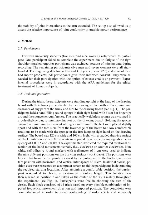
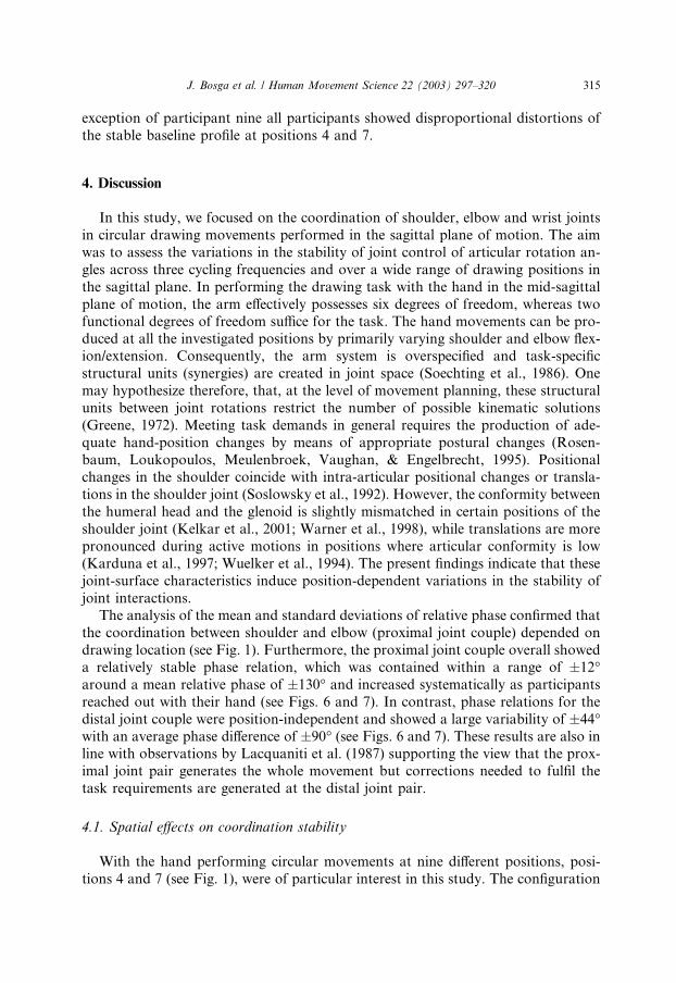
take place is discussed next.In the experiment (see Fig. 1), the nine positions on the drawing surface formed a
3� 3 matrix. Flexion/extension in both shoulder and elbow 1 were expected to be the
primary direction of joint motion of the arm in the present circular drawing task at
the nine positions in the sagittal plane with concomitant motions of the wrist joint
in radioulnar direction. Articular conformity in the glenohumeral joint is low and
the joint capsule is relatively lax when the arm is in a neutral flexion/extension posi-
tion combined with a slight adduction. This is like positioning the arm in such a way
that it allows the hand to rest comfortably in the lap when a person is seated, or whenthe arm is hung in a sling after a shoulder injury. Intra-articular translations are more
prominent when the shoulder joint rotates in the vicinity of the resting position due to
low glenohumeral conformity. These translations decrease in all dimensions as the el-
evation angle of the shoulder is increased from 0� to 90�. We reasoned that by choos-
ing a wide range of positions in the mid-sagittal plane in which the tasks were to be
carried out, certain task conditions would be performed in the vicinity of the resting
position of the shoulder. By constraining the task in the mid-sagittal plane of motion,
a slight degree of adduction of the shoulder joint in positions 4 and 7 (see Fig. 1) couldbe maintained, thereby allowing the shoulder to perform in a position associated with
low articular conformity. However, neutral flexion/extension of the shoulder joint is
best approximated with an arm posture in position 7. We therefore expected that
1 The shoulder mechanism consists of three synovial joints, i.e., the sternoclavicular joint, the
acromioclavicular joint and the glenohumeral joint. The glenohumeral joint is usually referred to as the
shoulder joint because it is the main contributor to joint rotations of the shoulder mechanism.
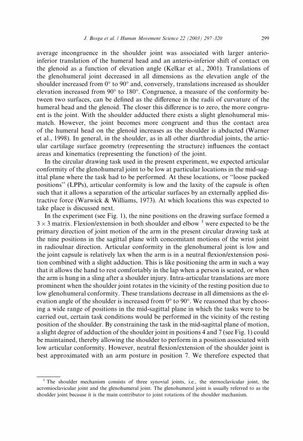
Fig. 1. Panel A: Schematic view of experimental setup. Participant is standing upright at the head of the
drawing board (shown partially) with his or her trunk perpendicular to the drawing surface with a 10-cm
minimum clearance of any part of the trunk and hips to the drawing board, while holding a hand-filling
round sponge. Nine white, self-adhesive round markers with a diameter of 1 cm (shown as numbers 1–9)
indicate the nine different positions on the drawing surface. The numbers corresponding to positions
(numbers 4 and 7) with low glenohumeral conformity are in bold-underscore. Seven infrared light emitting
diodes (IREDs shown as shaded circles and numbered from 1 to 7) were attached to the dorsal surface of
the hand and near joints of the wrist, elbow, shoulder, hip, knee and ankle on the right-hand side of the
participant�s body. OPTOTRAK system (not shown) is fixated at the ceiling at distance of 2 m right of the
participant and facing downward at an angle of 35� relative to ceiling. IRED positions are measured in a
coordinate system that has Z-axis pointing orthogonal to (toward) the projection screen, X -axis pointingparallel to the screen, and Y -axis upward. Origin of this coordinate system is lower left corner of drawing
board. Right-handed participant number 3 performs clockwise circular hand movements at position 5 in
an acoustically paced task at 1.0 Hz without visual control. Angular joint excursions were extracted off-
line from the calculated positions of the five IREDs. The consecutive plots depict the angular excursion-
time functions (panel B), the angular velocity-time functions (panel C) and the phase-time functions (panel
D) of the elbow and shoulder joint of the aforementioned 15-s trial. Panel E shows the continuous relative
phase-time function between the elbow and shoulder joints.
300 J. Bosga et al. / Human Movement Science 22 (2003) 297–320
J. Bosga et al. / Human Movement Science 22 (2003) 297–320 301
position 7 and, to a lesser extent, position 4 would most likely be influenced by intra-
articular translations that occur in the shoulder as a result of rotating the shoulder in
areas with a low articular conformity. We expected that these positions would be as-
sociated with a decreased stability of shoulder–elbow coordination, as reflected by an
increase in variability of the continuous relative phase.
1.2. Other determinants of inter-joint coordination stability
As stated above, the coupling of joints in the arm is partially effected by the sta-
bilizing role of poly-articular muscles (e.g., biceps brachii acting as wrist supinator,
elbow and shoulder flexor), by inertia (Lacquaniti & Soechting, 1982; van Bolhuis,
van Gielen, & van Ingen Schenau, 1998), and by movement frequency (Dounskaia,
Swinnen, Walter, Spaepen, & Verschueren, 1998; Kelso, Buchanan, & Wallace,1991). It is by virtue of their spring-like properties that the coupling between neigh-
boring joints is achieved by bi-articular muscles, thereby markedly influencing the
organization of limb synergies (Gielen, van Ingen Schenau, Tax, & Theeuwen,
1990). Due to the inertia of the limb segments, angular motion at the wrist joint
has an impact on elbow and shoulder motion. If a torque is exerted at the shoulder
or the elbow, it may result in an angular motion at the wrist and vice versa. However,
the inertia of the hand is considerably less than that of the upper arm and forearm.
Consequently, torques at the wrist will have less effect on the motion at more prox-imal joints (Soechting, 1984). Reaching out with the hand and thereby extending the
arm in the mid-sagittal plane of motion increases the moments of inertia at the shoul-
der and elbow joints, which in turn, may also cause higher muscle-activity levels.
Participants can anticipate these consequences and compensate for them during task
performance (cf. Flanagan & Lolley, 2001). Hence, we did not expect the coordina-
tion stability to be systematically reduced between the shoulder and elbow joint pairs
at more distant locations relative to the trunk.
Since earlier studies have shown that the elbow and wrist are loosely coupled(Lacquaniti, Ferrigno, Pedotti, Soechting, & Terzuolo, 1987), whereas the shoulder
and elbow are tightly coupled (Lacquaniti, Soechting, & Terzuolo, 1986; Lacquaniti
et al., 1987; Soechting, Lacquaniti, & Terzuolo, 1986), we decided to investigate the
effects of movement speed on a loosely and a tightly coupled joint pair within the
same effector system. To examine these effects, participants performed the circular
drawing task at three different movement frequencies. In general, increased move-
ment speed has been shown to destabilize certain coordination patterns of multi-
joint arm movements. More specifically, Kelso et al. (1991) studied rhythmicalunidirectional and bidirectional coordination of flexion and extension between the
elbow and wrist joints of the right arm that were performed in the sagittal plane
for two forearm positions: supine and prone. As cycling frequency increased, phase
relations between the elbow and wrist joints only destabilized for the bidirectional
coordination patterns. These observations support loss of stability as a central, self-
organizing process underlying coordinative change. Also Dounskaia et al. (1998)
have shown that increased frequency destabilizes certain coordination patterns of
the loosely coupled elbow–wrist joint pair. In this study, unidirectional, bidirectional
302 J. Bosga et al. / Human Movement Science 22 (2003) 297–320
and free-wrist flexion/extension movement patterns of the elbow–wrist joint pair
were analyzed across five cycling frequencies. Results provided evidence for two
types of interactive torques exerted at the wrist: inertial torques arising from elbow
motion and restraining torques arising from physical limits imposed on wrist rota-
tion. The interactive torques were the primary source of wrist motion, whereas it ap-peared from the findings that the main function of wrist-muscle activity was to
intervene with the interactive effects and to adjust the wrist movement to comply
with the required coordination pattern. Furthermore, the unidirectional pattern
was shown to be more in agreement with interactive effects than the bidirectional
pattern, thus causing their differential difficulty at moderate cycling frequencies.
However, when cycling frequency was further increased to 2.25 Hz, both the unidi-
rectional and bidirectional movements lost their individual features and acquired
features of the free-wrist pattern. These results suggest that multi-joint movementpatterns that are more in agreement with interaction effects can be maintained at
higher speed levels than patterns requiring substantial muscular interference with
the interactive torques.
In the present circular drawing task we expected overall relatively stable multi-
joint movement patterns of the arm to emerge because the participants were free
in choosing the size of the circle. On the other hand, we did expect higher movement
frequencies to differentially affect the coordination stability of the contributing joint
pairs. The rationale for this expectation was the following: (1) higher muscle-activitylevels are required at higher frequencies to cope with interactive torques, and (2) ac-
tive control requires timely processing of afferent feedback (Dounskaia et al., 1998).
Consequently, we assumed that combined increases in muscle-activity levels and
more rapid intervention of active control in movement because of increased fre-
quency would only affect relatively unstable inter-joint coordination patterns. We ex-
pected the strong prime movers of the already tightly coupled shoulder–elbow joint
pair to be able to comply with both requirements. Therefore, cycling frequency was
not expected to affect the coordination stability of the shoulder–elbow joint pair.However, it was expected that the coordination stability of the loosely coupled
elbow–wrist joint pair would decrease with increasing cycling frequency. Since coor-
dination patterns at positions 4 and 7 of the shoulder–elbow joint pair were expected
to be relatively unstable, we also expected coordination stability in positions 4 and 7
of the shoulder–elbow joint pair to decrease with increasing cycling frequency.
In sum, the rationale of the present study was as follows. We assumed that, in ad-
dition to the effects of bi-articular muscles in terms of joint coupling, a second impor-
tant biomechanical factor, joint conformity, would also influence the coordinationstability of both joint pairs differentially in circular drawing movements with the
arm rotating at nine different locations in the mid-sagittal plane. Position-dependent
variations of joint mobility were presumed whereby relatively unstable proximal
inter-joint coordination should occur if the shoulder was moved in positions with
low articular conformity (positions 4 and 7). Furthermore, we expected coordination
stability only to decrease at these positions with increasing cycling frequency. Coor-
dination stability for the loosely coupled distal joint pair was also expected to de-
crease at higher cycling frequencies. Inertia, however, was not expected to increase
J. Bosga et al. / Human Movement Science 22 (2003) 297–320 303
the stability of joint-interactions as the arm extended. The set-up also allowed us to
assess the relative importance of joint conformity in graphic motor performance.
2. Method
2.1. Participants
Fourteen university students (five men and nine women) volunteered to partici-
pate. One participant failed to complete the experiment due to fatigue of the right
shoulder muscles. Another participant was excluded because of missing data during
recording. The remaining participants (five men and seven women) were all right-
handed. Their age ranged between 17.6 and 41.9 years (mean: 22.6) and none of themhad motor problems. All participants gave their informed consent. They were re-
warded for their participation with the option of course credits or payment. Exper-
imental procedures were in accordance with the APA guidelines for the ethical
treatment of human subjects.
2.2. Task and procedure
During the trials, the participants were standing upright at the head of the drawingboard with their trunk perpendicular to the drawing surface with a 10-cm minimum
clearance of any part of the trunk and hips to the drawing board (see Fig. 1). The par-
ticipants held a hand-filling round sponge in their right hand, with his or her fingertips
around the sponge�s circumference. The practically weightless sponge was wrapped in
a polyethylene bag to minimize friction on the drawing board. Holding the sponge
ensured a minimum involvement of fingers and thumb. The feet were placed slightly
apart and with the toes 4 cm from the lower edge of the board to allow comfortable
rotations to be made with the sponge in the free hanging right hand on the drawingsurface. The board was 120 cm wide and 100 cm high, with a padded drawing surface
of black imitation leather. Movements were paced by acoustic pacing signals at a fre-
quency of 1.0, 1.5 and 2.0 Hz. The experimenter instructed the required rotational di-
rection of the hand movements verbally (i.e., clockwise or counter-clockwise). Nine
white, self-adhesive round markers with a diameter of 1 cm were used to indicate
the nine different positions on the drawing surface (workspace). The positions were
labeled 1–9 from the top position closest to the participant to the bottom, most dis-
tant position with horizontal and vertical inter-spaces of 10 cm. In all trial blocks, po-sition cues were presented on a computer screen to aid the participants in determining
the required starting locations. After assuming a comfortable position, the partici-
pant was asked to choose a location at shoulder height. This location was
then marked as position 5 and taken as the center of the 3� 3 matrix throughout
the experiment (see Fig. 1). Participants were free in choosing the size of the
circles. Each block consisted of 54 trials based on every possible combination of im-
posed frequency, movement direction and imposed position. The conditions were
counterbalanced in order to avoid confounding of order effects and separately
304 J. Bosga et al. / Human Movement Science 22 (2003) 297–320
randomized for each block. In a pilot study, the participants experienced visual hin-
drance by the drawing board that was positioned at close face-range in the mid-sag-
ittal plane. They showed a tendency to bend their head sideways and lean over to the
right. The tasks were therefore performed without visual control, that is, after having
positioned the hand at the starting location for a particular trial, the participant wasasked to close his or her eyes. We also carefully monitored the posture of the partic-
ipants to ensure that they maintained a proper upright standing posture during trial
execution. Per trial, the experimenter started a 15-s recording period as soon as the
participant had matched his or her movements to the imposed frequency.
2.3. Recording system
Movements were recorded at a rate of 100 Hz and with a spatial accuracy higherthan 0.2 mm in X , Y and Z direction by means of a 3D-motion tracking system (Op-
totrak 3020). Seven infrared light emitting diodes (IREDs) were attached to the
right-hand side of the participant�s body at the following consecutive locations:
the dorsal surface near the head of the fifth metacarpal bone (IRED-1), the styloid
process of the ulna (IRED-2), the lateral epicondyle of the humerus (IRED-3), the
acromion of the scapula (IRED-4), the anterior superior iliac spine (IRED-5), the
lateral epicondyle of the femur (IRED-6), and the lateral malleolus of the fibula
(IRED-7). IRED-1, mounted above the knuckle of the fifth metacarpal bone, wasused to record the end-effector position.
2.4. Data analysis
All position data were filtered with a second-order Butterworth, zero phase lag,
low-pass filter with a cut-off frequency of 8 Hz. For each 15-s trial the filtered
end-effector trajectory, angles and angular velocities of shoulder, elbow and wrist
joints were derived and visually inspected. To assess effects of practice and/or fatiguethe 15-s recordings were divided by means of a computer-search procedure into a
first, mid and last 5-s phase. The rotational directions (clockwise and counter-clock-
wise) were pooled in the data analyses since our main research question was focused
on variations in the stability of inter-joint coordination as a function of the position
and frequency task constraints.
2.4.1. Workspace kinematics
Frequency: Zero-crossings of the Y component of the end-effector position–timesignal were determined to identify individual cycles. The mean cycle duration for
the initial, middle and final 5-s phase of each trial was calculated and converted to
a frequency unit (Hz).
Starting position: The first cycle of the XY end-effector position–time signal was
isolated. The mean X -position and Y -position of this cycle was calculated separately
for all conditions for all participants (N ¼ 12). Next, the mean spatial variability
(root-mean-square error) around the subsequent target positions for the correspond-
ing X and Y starting positions was calculated.
J. Bosga et al. / Human Movement Science 22 (2003) 297–320 305
Circle size: The peak–peak distance of the X and Y component of the end-effector
position–time signal were obtained per cycle. From these data, the realized average
circle size per trial was determined.
Circularity: The X and Y component of the position–time signals of the hand
(IRED-1) were filtered with a second-order Butterworth, zero phase lag high-pass fil-ter with a cut-off frequency of 0.5 Hz to eliminate within-trial positional drift. As-
sessment of the circularity of hand movements was based on the standard
deviation (SD) of curvature in each cycle (see Verschueren, Swinnen, Cordo, &Douns-
kaia, 1999). The equation for the computation of curvature was
% i
%ou
fun
vect
vect
vect
vect
alfa
2 In
(With
SdK ¼ ðx0y00 � x00y0Þ=ðx02 þ y02Þ3=2 ð1Þ
where x and y are the current coordinates on the end-point trajectory; x0, y 0 and x00, y00
are the first and second time derivatives of x and y, respectively. In a circle, the
curvature is constant at all times and the SD of curvature is zero. Accordingly, in-
creases in the SD of curvature reflect distortions of circularity. The means of the SD
of circularity (SdK) were computed per trial, and extreme outliers in the data wereeliminated.
Plane-dependent angular displacements: The mean plane-dependent angular dis-
placements of the shoulder (Mpdh in �) were extracted off-line from the calculated
positions of the third, fourth and fifth IRED. The Mpdh expresses the plane-depen-
dent contribution of the shoulder angular displacements to arm movements in the
Cartesian coordinate system. Elevation was defined as the angle of the sagittal pro-
jection of the upper arm and a vector pointing forward through the trunk. Azimuth
was defined as the angle of the projection of the upper arm in a horizontal plane rel-ative to the forward direction and roll was defined as the angle of the projection in
the fronto-parallel plane relative to the forward direction.
2.4.2. Kinematics
Plane independent angular rotations were extracted off-line from the calculated
positions of the seven IREDs. The wrist, elbow, shoulder, hip and knee angles were
defined as the enclosed angle between two neighboring limb segments. 2
nput: positions ½x y z� of three joints (e.g., shoulder, elbow or wist).
tput: enclosed angle (alfa) of the second joint.
ction[alfa]¼ enclosed_angle(pos1,pos2,pos3);
or1¼ pos1)pos2;
or1¼ vector1/norm(vector1); % normalization to length 1
or2¼ pos3)pos2;
or2¼ vector2/norm(vector2); % normalization to length 2
¼ acos(dot(vector10,vector20)) � (180/pi)
this footnote we present the code of the user defined Matlab v5.3 function of the enclosed angle.
the permission of Mary Klein Bretler, 10-24-2001.)
306 J. Bosga et al. / Human Movement Science 22 (2003) 297–320
Decreasing angular rotations at the wrist joint indicate a radial abduction, 180� atthe elbow joint indicates a full elbow extension. Increasing angular rotations at the
shoulder and hip joints indicate a combined elevation/adduction/exorotation of the
shoulder and a combined extension/abduction/exorotation of the hip. An angle of
180� at the knee joint indicates full knee extension. Anatomical joint space wasdefined as the three-dimensional joint space consisting of the radial/ulnar abduction
of the wrist, the flexion/extension of the elbow, the combined elevation/adduction/
exorotation and retroflexion/abduction/endorotation of the shoulder, the combined
extension/abduction/exorotation and flexion/adduction/endorotation of the hip and
the flexion/extension of the knee.
Joint amplitudes: The mean realized plane-independent angular displacements
(MJh in �) for all joint rotations were obtained per cycle from the position-time sig-
nal of each joint.
2.4.3. Relative phase
Continuous relative-phase time functions were inspected for branch cut crossings
(phase wraps). No branch cut crossings were found. 3 The means (Mu) and standard
deviations (Sdu) of the continuous relative-phase signals of the joint angle functionsof the neighboring joints of the arm (wrist, elbow and shoulder) were calculated
using Batschelet�s (1981) procedure for circular statistics (see Meulenbroek, Thomas-
sen, van Lieshout, & Swinnen, 1998).
2.4.4. Statistical evaluation
When relevant in the context of the presently formulated predictions, the depen-
dent variables were evaluated by means of univariate analyses of variance (ANO-
VA). The within-subject factors were imposed frequency (1.0, 1.5 and 2.0 Hz),
imposed position (nine positions) and movement direction (clockwise and counter-
clockwise). The Scheff�ee method (a ¼ 0:05) was used for post-hoc comparisons of
means.Positions 4 and 7 (P47): To evaluate the effects of positions with low glenohu-
meral conformity, the means of the dependent variables at positions 4 and 7 were
contrasted with the means of the dependent variables at the remaining seven posi-
tions where the task had to be performed. The results of these analyses will be re-
ported under the factor P47.
3 We decided to use cross-correlation measures between time functions of various joints to check for
possible inconsistencies in our continuous relative phase assessments. Peak–peak detection and amplitude
normalization may, in the case of noisy, low-amplitude signals yield a continuous relative phase signal
with artificially high standard deviations. Such noisy signals should have resulted in unsystematic
variations in the cross-correlation measures as a function of the experimental variables. Since cross-
correlations proved to vary systematically as a function of the task variables and duplicated the results of
the continuous relative phase signals, continuous relative phase was considered to be a reliable and
representative measure of spatio-temporal inter-joint coordination.
J. Bosga et al. / Human Movement Science 22 (2003) 297–320 307
3. Results
3.1. Task performance
Before assessing the variations in the stability of joint control across the three im-posed frequencies and the nine imposed positions on the sagittal plane, we first ver-
ified whether the participants satisfied the imposed temporal and spatial constraints
of the experimental task. In addition, effects of fatigue and/or practice were exam-
ined.
3.1.1. Realized frequency and realized positions
Throughout the three phases of a trial, the participants accurately produced the
instructed movement frequencies (see Table 1). Consequently, practice and/or fatiguedid not affect the realized frequencies. The mean spatial variability around the in-
structed target positions was 9.75 mm in the X and 9.90 mm in the Y dimension.
Given the inter-target distance of neighboring targets of 100 mm, these results
show that the imposed-position constraint of the experimental task was satisfied.
3.1.2. Circle size
Circle size varied between 106 mm at position 6 to 120 mm at position 5 (Table 2).
A control analysis showed that the mean circle size pooled across positions 4 and 7was not significantly different from the average realized circle size at the other posi-
tions (F ð1; 11Þ ¼ 2:60, p > 0:5). As expected, higher cycling frequencies resulted in a
scaling down of the circle sizes and vice versa (see Table 2). Thus, drawing circles
under the task constraints of this experiment showed circle size to be relatively in-
variant in the sagittal plane of motion, was unaffected by positions 4 and 7, and dis-
played a tight frequency-to-amplitude relation.
3.1.3. Circularity
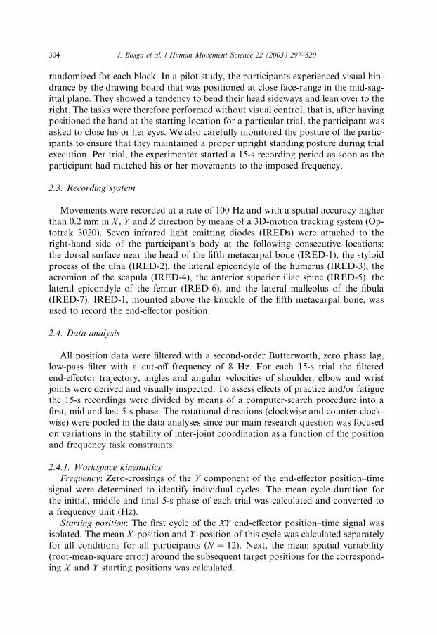
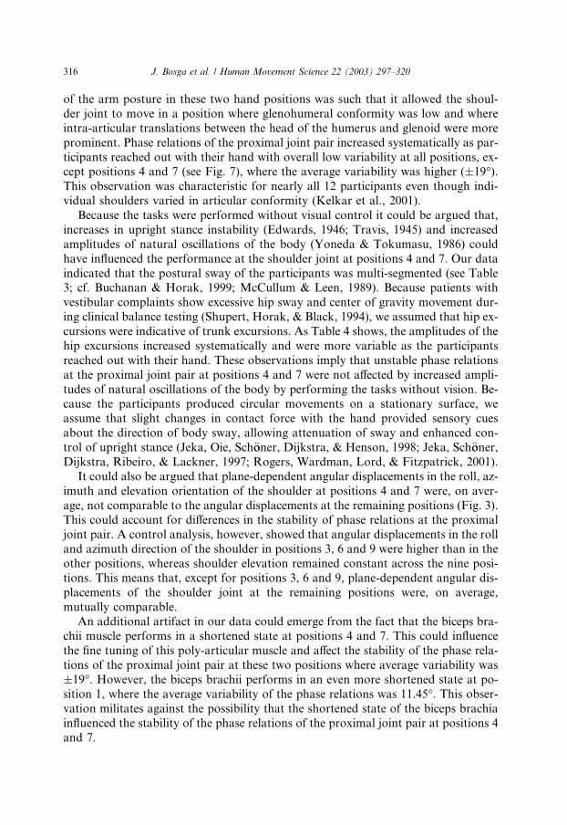
Fig. 2 shows nine, two-dimensional plots of typical hand paths as realized in the
experiment. The circular hand movements were performed by participant number
three, in a clockwise direction at a frequency of 1 Hz. The figure demonstrates that
perfect circles were almost never produced. We suggest that the distortion results
Table 1
Means and standard deviations of the realized frequencies during the initial, middle and final phase of tri-
als for the three (1, 1.5, and 2 Hz) imposed frequencies pooled over the nine positions and both movement
directions
Realized frequency Imposed frequency
1 Hz 1.5 Hz 2 Hz
Mean [SD] Mean [SD] Mean [SD]
Initial phase 1.03 [0.05] 1.53 [0.06] 2.01 [0.07]
Middle phase 1.02 [0.05] 1.52 [0.06] 2.00 [0.06]
Final phase 1.01 [0.05] 1.52 [0.06] 2.01 [0.07]
Table 2
Main effects of orientation, imposed position and frequency mode (see text) on mean circle size, standard
deviation of the curvature of hand movements (SdK) and the mean plane-dependent angular amplitudes of
the shoulder joint (Mpdh)
Factor Level Circle size mean
[SD]
SdK mean
[SD]
Mpdh mean
[SD]
Orientation Roll 5.98 [3.71]�
Elevation 13.75 [6.47]
Azimuth 2.47 [1.39]
Imposed position 1 114.65 [49.97] 0.021 [0.013]�� 6.98 [6.74]�
2 107.18 [49.42] 0.023 [0.015] 6.81 [5.71]
3 113.54 [51.98] 0.023 [0.016] 8.47 [6.06]
4 110.37 [50.43] 0.022 [0.013] 6.76 [6.66]
5 120.45 [65.75] 0.021 [0.015] 7.29 [7.22]
6 106.33 [49.29] 0.024 [0.015] 7.84 [5.60]
7 119.70 [54.46] 0.021 [0.012] 7.42 [6.83]
8 112.04 [57.60] 0.024 [0.015] 6.84 [6.35]
9 117.03 [54.79] 0.023 [0.014] 8.19 [6.43]
Frequency mode 1 Hz 133.73 [58.87] 0.020 [0.013]�� 8.27 [7.17]��
1.5 Hz 112.86 [57.27] 0.021 [0.013] 7.37 [6.73]
2 Hz 93.84 [34.38] 0.027 [0.016] 6.57 [5.12]
Standard deviations of the dependent variables are added between braces. �p < 0:01, ��p < 0:05.
308 J. Bosga et al. / Human Movement Science 22 (2003) 297–320
from failure of the CNS to account for changes in anisotropy of viscosity and inertia
(Pfann, Corcos, Moore, & Hasan, 2002).
Deterioration of the circular hand movements was assessed by analyzing the stan-
dard deviation of the curvature of hand movements (SdK). Control analyses showedthat the factor imposed position affected the SdK (see Table 2; F ð8; 88Þ ¼ 3:61,p < 0:05). The SdK varied between 0.021 (positions 1, 5 and 7) and 0.024 (positions
6 and 8). Movement frequency displayed a significant effect on the SdK (see Table 2;
F ð2; 22Þ ¼ 8:39, p < 0:05); post-hoc analysis showed that the deterioration of the cir-
cular hand movements was significantly higher for the 2 Hz frequency mode than the
lower frequency modes.
3.1.4. Plane dependent angular displacements
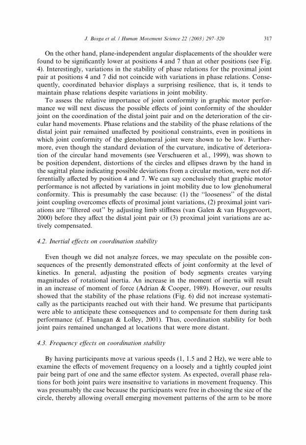
Control analyses showed that the mean plane-dependent angular displacements ofthe shoulder (Mpdh in Table 2) varied strongly in the different planar dimensions
(F ð2; 22Þ ¼ 55:20, p < 0:01). Fig. 3 displays the average angular displacements in
the roll, elevation and azimuth direction of the shoulder joint as a function of posi-
tion. The effect of position (Table 2) was significant (F ð8; 88Þ ¼ 7:55, p < 0:01). Post-hoc analysis showed that the displacements in the roll and azimuth direction were
significantly higher in positions 3, 6 and 9 than in the other positions. The elevation
remained relatively constant across the nine positions. Frequency also affected the
displacements in the roll, elevation and azimuth orientation (F ð2; 22Þ ¼ 4:60,p < 0:05; see Table 2). Post-hoc analysis for the roll displayed significantly higher
displacements in the lowest frequency mode (1 Hz). For the elevation, all the three
frequency modes were significantly different, displaying decreasing elevation with
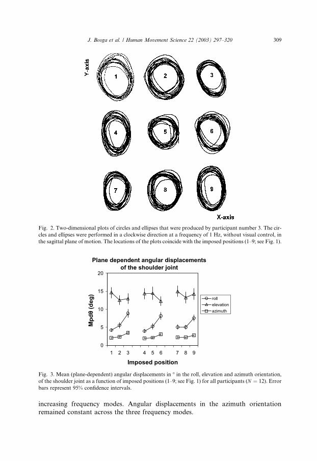
Fig. 2. Two-dimensional plots of circles and ellipses that were produced by participant number 3. The cir-
cles and ellipses were performed in a clockwise direction at a frequency of 1 Hz, without visual control, in
the sagittal plane of motion. The locations of the plots coincide with the imposed positions (1–9; see Fig. 1).
Fig. 3. Mean (plane-dependent) angular displacements in � in the roll, elevation and azimuth orientation,
of the shoulder joint as a function of imposed positions (1–9; see Fig. 1) for all participants (N ¼ 12). Error
bars represent 95% confidence intervals.
J. Bosga et al. / Human Movement Science 22 (2003) 297–320 309
increasing frequency modes. Angular displacements in the azimuth orientation
remained constant across the three frequency modes.
310 J. Bosga et al. / Human Movement Science 22 (2003) 297–320
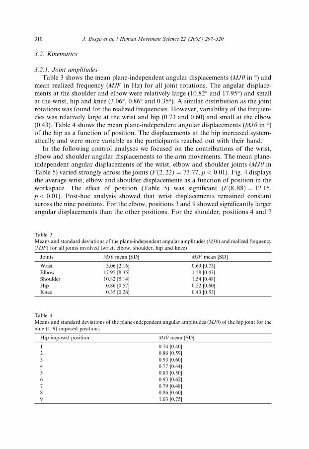
3.2. Kinematics
3.2.1. Joint amplitudes
Table 3 shows the mean plane-independent angular displacements (MJh in �) andmean realized frequency (MJF in Hz) for all joint rotations. The angular displace-ments at the shoulder and elbow were relatively large (10.82� and 17.95�) and small
at the wrist, hip and knee (3.06�, 0.86� and 0.35�). A similar distribution as the joint
rotations was found for the realized frequencies. However, variability of the frequen-
cies was relatively large at the wrist and hip (0.73 and 0.60) and small at the elbow
(0.43). Table 4 shows the mean plane-independent angular displacements (MJh in �)of the hip as a function of position. The displacements at the hip increased system-
atically and were more variable as the participants reached out with their hand.
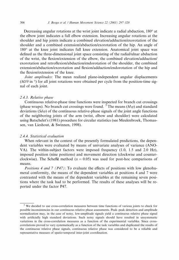
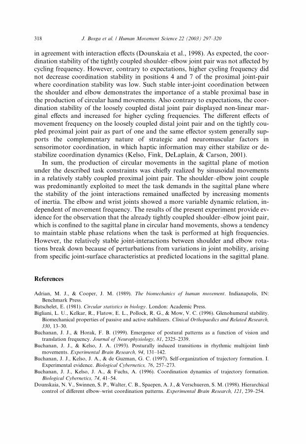
In the following control analyses we focused on the contributions of the wrist,elbow and shoulder angular displacements to the arm movements. The mean plane-
independent angular displacements of the wrist, elbow and shoulder joints (MJh in
Table 5) varied strongly across the joints (F ð2; 22Þ ¼ 73:77, p < 0:01). Fig. 4 displays
the average wrist, elbow and shoulder displacements as a function of position in the
workspace. The effect of position (Table 5) was significant (F ð8; 88Þ ¼ 12:15,p < 0:01). Post-hoc analysis showed that wrist displacements remained constant
across the nine positions. For the elbow, positions 3 and 9 showed significantly larger
angular displacements than the other positions. For the shoulder, positions 4 and 7
Table 3
Means and standard deviations of the plane-independent angular amplitudes (MJh) and realized frequency
(MJF ) for all joints involved (wrist, elbow, shoulder, hip and knee)
Joints MJh mean [SD] MJF mean [SD]
Wrist 3.06 [2.16] 0.69 [0.73]
Elbow 17.95 [8.35] 1.58 [0.43]
Shoulder 10.82 [5.14] 1.54 [0.48]
Hip 0.86 [0.57] 0.52 [0.60]
Knee 0.35 [0.26] 0.43 [0.53]
Table 4
Means and standard deviations of the plane-independent angular amplitudes (MJh) of the hip joint for the
nine (1–9) imposed positions
Hip imposed position MJh mean [SD]
1 0.74 [0.40]
2 0.86 [0.59]
3 0.93 [0.60]
4 0.77 [0.44]
5 0.83 [0.50]
6 0.93 [0.62]
7 0.79 [0.48]
8 0.86 [0.60]
9 1.03 [0.75]
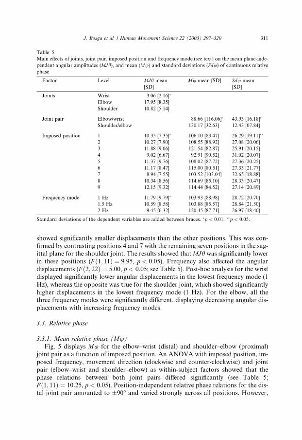
Table 5
Main effects of joints, joint pair, imposed position and frequency mode (see text) on the mean plane-inde-
pendent angular amplitudes (MJh), and mean (Mu) and standard deviations (Sdu) of continuous relativephase
Factor Level MJh mean
[SD]
Mu mean [SD] Sdu mean
[SD]
Joints Wrist 3.06 [2.16]�
Elbow 17.95 [8.35]
Shoulder 10.82 [5.14]
Joint pair Elbow/wrist 88.66 [116.06]� 43.93 [16.18]�
Shoulder/elbow 130.17 [32.63] 12.43 [07.84]
Imposed position 1 10.35 [7.35]� 106.10 [83.47] 26.79 [19.11]��
2 10.27 [7.90] 108.55 [88.92] 27.08 [20.06]
3 11.88 [9.06] 121.54 [82.87] 25.91 [20.15]
4 9.02 [6.67] 92.91 [90.52] 31.02 [20.07]
5 11.37 [9.76] 108.02 [87.72] 27.36 [20.25]
6 11.17 [8.47] 115.00 [80.51] 27.33 [21.77]
7 8.94 [7.55] 103.52 [103.04] 32.65 [18.88]
8 10.34 [8.56] 114.69 [85.10] 28.33 [20.47]
9 12.15 [9.32] 114.44 [84.52] 27.14 [20.89]
Frequency mode 1 Hz 11.79 [9.79]� 103.93 [88.98] 28.72 [20.70]
1.5 Hz 10.59 [8.58] 103.88 [85.57] 28.84 [21.50]
2 Hz 9.45 [6.32] 120.45 [87.71] 26.97 [18.40]
Standard deviations of the dependent variables are added between braces. �p < 0:01, ��p < 0:05.
J. Bosga et al. / Human Movement Science 22 (2003) 297–320 311
showed significantly smaller displacements than the other positions. This was con-
firmed by contrasting positions 4 and 7 with the remaining seven positions in the sag-
ittal plane for the shoulder joint. The results showed thatMJh was significantly lowerin these positions (F ð1; 11Þ ¼ 9:95, p < 0:05). Frequency also affected the angulardisplacements (F ð2; 22Þ ¼ 5:00, p < 0:05; see Table 5). Post-hoc analysis for the wristdisplayed significantly lower angular displacements in the lowest frequency mode (1
Hz), whereas the opposite was true for the shoulder joint, which showed significantly
higher displacements in the lowest frequency mode (1 Hz). For the elbow, all the
three frequency modes were significantly different, displaying decreasing angular dis-
placements with increasing frequency modes.
3.3. Relative phase
3.3.1. Mean relative phase (Mu)Fig. 5 displays Mu for the elbow–wrist (distal) and shoulder–elbow (proximal)
joint pair as a function of imposed position. An ANOVA with imposed position, im-
posed frequency, movement direction (clockwise and counter-clockwise) and joint
pair (elbow–wrist and shoulder–elbow) as within-subject factors showed that the
phase relations between both joint pairs differed significantly (see Table 5;
F ð1; 11Þ ¼ 10:25, p < 0:05). Position-independent relative phase relations for the dis-tal joint pair amounted to �90� and varied strongly across all positions. However,
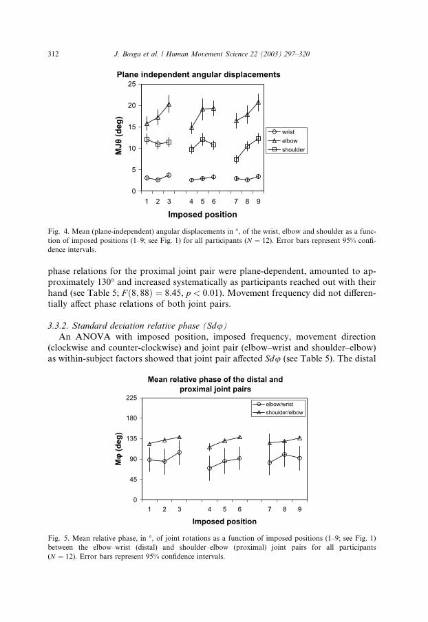
Fig. 4. Mean (plane-independent) angular displacements in �, of the wrist, elbow and shoulder as a func-
tion of imposed positions (1–9; see Fig. 1) for all participants (N ¼ 12). Error bars represent 95% confi-
dence intervals.
312 J. Bosga et al. / Human Movement Science 22 (2003) 297–320
phase relations for the proximal joint pair were plane-dependent, amounted to ap-
proximately 130� and increased systematically as participants reached out with their
hand (see Table 5; F ð8; 88Þ ¼ 8:45, p < 0:01). Movement frequency did not differen-tially affect phase relations of both joint pairs.
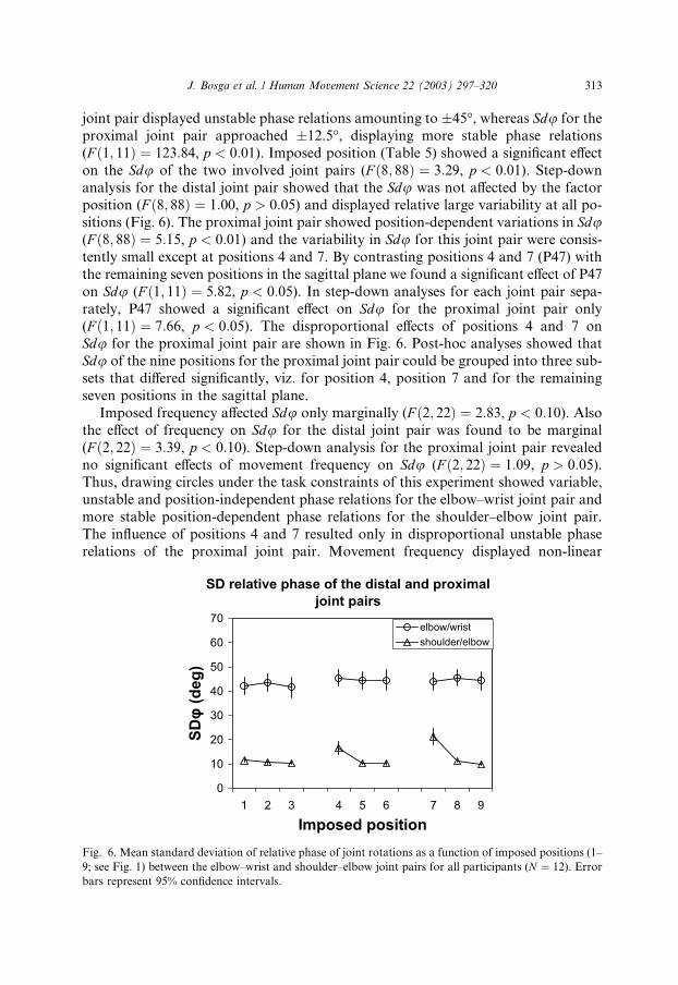
3.3.2. Standard deviation relative phase (Sdu)An ANOVA with imposed position, imposed frequency, movement direction
(clockwise and counter-clockwise) and joint pair (elbow–wrist and shoulder–elbow)
as within-subject factors showed that joint pair affected Sdu (see Table 5). The distal
Fig. 5. Mean relative phase, in �, of joint rotations as a function of imposed positions (1–9; see Fig. 1)
between the elbow–wrist (distal) and shoulder–elbow (proximal) joint pairs for all participants
(N ¼ 12). Error bars represent 95% confidence intervals.
J. Bosga et al. / Human Movement Science 22 (2003) 297–320 313
joint pair displayed unstable phase relations amounting to �45�, whereas Sdu for the
proximal joint pair approached �12.5�, displaying more stable phase relations
(F ð1; 11Þ ¼ 123:84, p < 0:01). Imposed position (Table 5) showed a significant effect
on the Sdu of the two involved joint pairs (F ð8; 88Þ ¼ 3:29, p < 0:01). Step-downanalysis for the distal joint pair showed that the Sdu was not affected by the factorposition (F ð8; 88Þ ¼ 1:00, p > 0:05) and displayed relative large variability at all po-
sitions (Fig. 6). The proximal joint pair showed position-dependent variations in Sdu(F ð8; 88Þ ¼ 5:15, p < 0:01) and the variability in Sdu for this joint pair were consis-
tently small except at positions 4 and 7. By contrasting positions 4 and 7 (P47) with
the remaining seven positions in the sagittal plane we found a significant effect of P47
on Sdu (F ð1; 11Þ ¼ 5:82, p < 0:05). In step-down analyses for each joint pair sepa-
rately, P47 showed a significant effect on Sdu for the proximal joint pair only
(F ð1; 11Þ ¼ 7:66, p < 0:05). The disproportional effects of positions 4 and 7 onSdu for the proximal joint pair are shown in Fig. 6. Post-hoc analyses showed that
Sdu of the nine positions for the proximal joint pair could be grouped into three sub-
sets that differed significantly, viz. for position 4, position 7 and for the remaining
seven positions in the sagittal plane.
Imposed frequency affected Sdu only marginally (F ð2; 22Þ ¼ 2:83, p < 0:10). Also
the effect of frequency on Sdu for the distal joint pair was found to be marginal
(F ð2; 22Þ ¼ 3:39, p < 0:10). Step-down analysis for the proximal joint pair revealed
no significant effects of movement frequency on Sdu (F ð2; 22Þ ¼ 1:09, p > 0:05).Thus, drawing circles under the task constraints of this experiment showed variable,
unstable and position-independent phase relations for the elbow–wrist joint pair and
more stable position-dependent phase relations for the shoulder–elbow joint pair.
The influence of positions 4 and 7 resulted only in disproportional unstable phase
relations of the proximal joint pair. Movement frequency displayed non-linear
Fig. 6. Mean standard deviation of relative phase of joint rotations as a function of imposed positions (1–
9; see Fig. 1) between the elbow–wrist and shoulder–elbow joint pairs for all participants (N ¼ 12). Error
bars represent 95% confidence intervals.
314 J. Bosga et al. / Human Movement Science 22 (2003) 297–320
marginal effects on the stability of the phase relations for the elbow–wrist joint pair,
with relatively lower phase relations in the 2 Hz frequency mode.
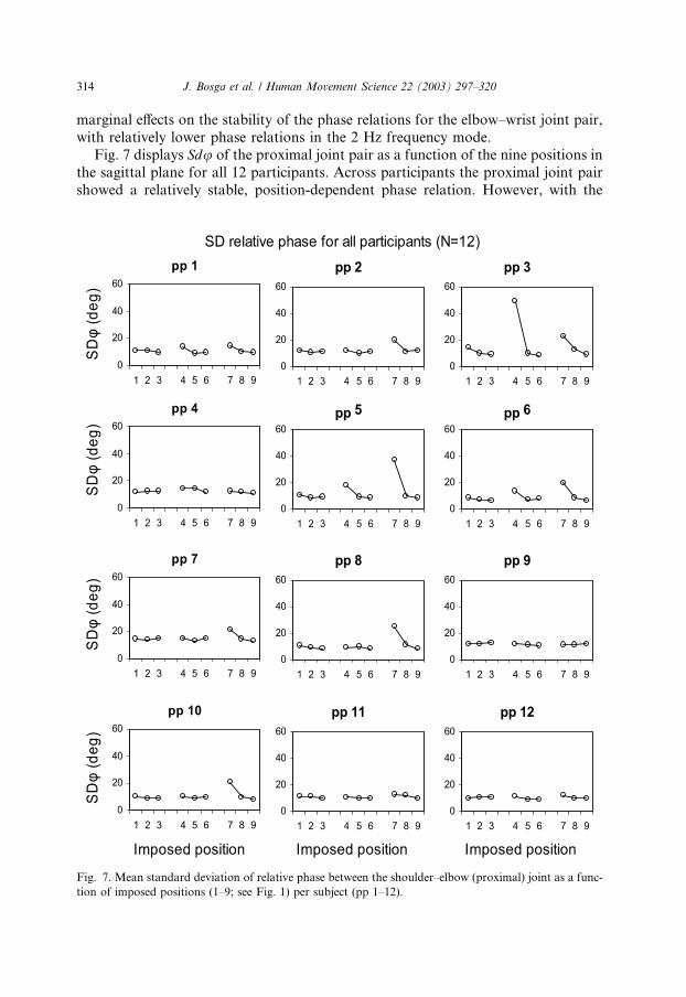
Fig. 7 displays Sdu of the proximal joint pair as a function of the nine positions in
the sagittal plane for all 12 participants. Across participants the proximal joint pair
showed a relatively stable, position-dependent phase relation. However, with the
Fig. 7. Mean standard deviation of relative phase between the shoulder–elbow (proximal) joint as a func-
tion of imposed positions (1–9; see Fig. 1) per subject (pp 1–12).
J. Bosga et al. / Human Movement Science 22 (2003) 297–320 315
exception of participant nine all participants showed disproportional distortions of
the stable baseline profile at positions 4 and 7.
4. Discussion
In this study, we focused on the coordination of shoulder, elbow and wrist joints
in circular drawing movements performed in the sagittal plane of motion. The aim
was to assess the variations in the stability of joint control of articular rotation an-
gles across three cycling frequencies and over a wide range of drawing positions in
the sagittal plane. In performing the drawing task with the hand in the mid-sagittal
plane of motion, the arm effectively possesses six degrees of freedom, whereas two
functional degrees of freedom suffice for the task. The hand movements can be pro-duced at all the investigated positions by primarily varying shoulder and elbow flex-
ion/extension. Consequently, the arm system is overspecified and task-specific
structural units (synergies) are created in joint space (Soechting et al., 1986). One
may hypothesize therefore, that, at the level of movement planning, these structural
units between joint rotations restrict the number of possible kinematic solutions
(Greene, 1972). Meeting task demands in general requires the production of ade-
quate hand-position changes by means of appropriate postural changes (Rosen-
baum, Loukopoulos, Meulenbroek, Vaughan, & Engelbrecht, 1995). Positionalchanges in the shoulder coincide with intra-articular positional changes or transla-
tions in the shoulder joint (Soslowsky et al., 1992). However, the conformity between
the humeral head and the glenoid is slightly mismatched in certain positions of the
shoulder joint (Kelkar et al., 2001; Warner et al., 1998), while translations are more
pronounced during active motions in positions where articular conformity is low
(Karduna et al., 1997; Wuelker et al., 1994). The present findings indicate that these
joint-surface characteristics induce position-dependent variations in the stability of
joint interactions.The analysis of the mean and standard deviations of relative phase confirmed that
the coordination between shoulder and elbow (proximal joint couple) depended on
drawing location (see Fig. 1). Furthermore, the proximal joint couple overall showed
a relatively stable phase relation, which was contained within a range of �12�around a mean relative phase of �130� and increased systematically as participants
reached out with their hand (see Figs. 6 and 7). In contrast, phase relations for the
distal joint couple were position-independent and showed a large variability of �44�with an average phase difference of �90� (see Figs. 6 and 7). These results are also inline with observations by Lacquaniti et al. (1987) supporting the view that the prox-
imal joint pair generates the whole movement but corrections needed to fulfil the
task requirements are generated at the distal joint pair.
4.1. Spatial effects on coordination stability
With the hand performing circular movements at nine different positions, posi-
tions 4 and 7 (see Fig. 1), were of particular interest in this study. The configuration
316 J. Bosga et al. / Human Movement Science 22 (2003) 297–320
of the arm posture in these two hand positions was such that it allowed the shoul-
der joint to move in a position where glenohumeral conformity was low and where
intra-articular translations between the head of the humerus and glenoid were more
prominent. Phase relations of the proximal joint pair increased systematically as par-
ticipants reached out with their hand with overall low variability at all positions, ex-cept positions 4 and 7 (see Fig. 7), where the average variability was higher (�19�).This observation was characteristic for nearly all 12 participants even though indi-
vidual shoulders varied in articular conformity (Kelkar et al., 2001).
Because the tasks were performed without visual control it could be argued that,
increases in upright stance instability (Edwards, 1946; Travis, 1945) and increased
amplitudes of natural oscillations of the body (Yoneda & Tokumasu, 1986) could
have influenced the performance at the shoulder joint at positions 4 and 7. Our data
indicated that the postural sway of the participants was multi-segmented (see Table3; cf. Buchanan & Horak, 1999; McCullum & Leen, 1989). Because patients with
vestibular complaints show excessive hip sway and center of gravity movement dur-
ing clinical balance testing (Shupert, Horak, & Black, 1994), we assumed that hip ex-
cursions were indicative of trunk excursions. As Table 4 shows, the amplitudes of the
hip excursions increased systematically and were more variable as the participants
reached out with their hand. These observations imply that unstable phase relations
at the proximal joint pair at positions 4 and 7 were not affected by increased ampli-
tudes of natural oscillations of the body by performing the tasks without vision. Be-cause the participants produced circular movements on a stationary surface, we
assume that slight changes in contact force with the hand provided sensory cues
about the direction of body sway, allowing attenuation of sway and enhanced con-
trol of upright stance (Jeka, Oie, Sch€ooner, Dijkstra, & Henson, 1998; Jeka, Sch€ooner,Dijkstra, Ribeiro, & Lackner, 1997; Rogers, Wardman, Lord, & Fitzpatrick, 2001).
It could also be argued that plane-dependent angular displacements in the roll, az-
imuth and elevation orientation of the shoulder at positions 4 and 7 were, on aver-
age, not comparable to the angular displacements at the remaining positions (Fig. 3).This could account for differences in the stability of phase relations at the proximal
joint pair. A control analysis, however, showed that angular displacements in the roll
and azimuth direction of the shoulder in positions 3, 6 and 9 were higher than in the
other positions, whereas shoulder elevation remained constant across the nine posi-
tions. This means that, except for positions 3, 6 and 9, plane-dependent angular dis-
placements of the shoulder joint at the remaining positions were, on average,
mutually comparable.
An additional artifact in our data could emerge from the fact that the biceps bra-chii muscle performs in a shortened state at positions 4 and 7. This could influence
the fine tuning of this poly-articular muscle and affect the stability of the phase rela-
tions of the proximal joint pair at these two positions where average variability was
�19�. However, the biceps brachii performs in an even more shortened state at po-
sition 1, where the average variability of the phase relations was 11.45�. This obser-vation militates against the possibility that the shortened state of the biceps brachia
influenced the stability of the phase relations of the proximal joint pair at positions 4
and 7.
J. Bosga et al. / Human Movement Science 22 (2003) 297–320 317
On the other hand, plane-independent angular displacements of the shoulder were
found to be significantly lower at positions 4 and 7 than at other positions (see Fig.
4). Interestingly, variations in the stability of phase relations for the proximal joint
pair at positions 4 and 7 did not coincide with variations in phase relations. Conse-
quently, coordinated behavior displays a surprising resilience, that is, it tends tomaintain phase relations despite variations in joint mobility.
To assess the relative importance of joint conformity in graphic motor perfor-
mance we will next discuss the possible effects of joint conformity of the shoulder
joint on the coordination of the distal joint pair and on the deterioration of the cir-
cular hand movements. Phase relations and the stability of the phase relations of the
distal joint pair remained unaffected by positional constraints, even in positions in
which joint conformity of the glenohumeral joint were shown to be low. Further-
more, even though the standard deviation of the curvature, indicative of deteriora-tion of the circular hand movements (see Verschueren et al., 1999), was shown to
be position dependent, distortions of the circles and ellipses drawn by the hand in
the sagittal plane indicating possible deviations from a circular motion, were not dif-
ferentially affected by position 4 and 7. We can say conclusively that graphic motor
performance is not affected by variations in joint mobility due to low glenohumeral
conformity. This is presumably the case because: (1) the ‘‘looseness’’ of the distal
joint coupling overcomes effects of proximal joint variations, (2) proximal joint vari-
ations are ‘‘filtered out’’ by adjusting limb stiffness (van Galen & van Huygevoort,2000) before they affect the distal joint pair or (3) proximal joint variations are ac-
tively compensated.
4.2. Inertial effects on coordination stability
Even though we did not analyze forces, we may speculate on the possible con-
sequences of the presently demonstrated effects of joint conformity at the level of
kinetics. In general, adjusting the position of body segments creates varyingmagnitudes of rotational inertia. An increase in the moment of inertia will result
in an increase of moment of force (Adrian & Cooper, 1989). However, our results
showed that the stability of the phase relations (Fig. 6) did not increase systemati-
cally as the participants reached out with their hand. We presume that participants
were able to anticipate these consequences and to compensate for them during task
performance (cf. Flanagan & Lolley, 2001). Thus, coordination stability for both
joint pairs remained unchanged at locations that were more distant.
4.3. Frequency effects on coordination stability
By having participants move at various speeds (1, 1.5 and 2 Hz), we were able to
examine the effects of movement frequency on a loosely and a tightly coupled joint
pair being part of one and the same effector system. As expected, overall phase rela-
tions for both joint pairs were insensitive to variations in movement frequency. This
was presumably the case because the participants were free in choosing the size of the
circle, thereby allowing overall emerging movement patterns of the arm to be more
318 J. Bosga et al. / Human Movement Science 22 (2003) 297–320
in agreement with interaction effects (Dounskaia et al., 1998). As expected, the coor-
dination stability of the tightly coupled shoulder–elbow joint pair was not affected by
cycling frequency. However, contrary to expectations, higher cycling frequency did
not decrease coordination stability in positions 4 and 7 of the proximal joint-pair
where coordination stability was low. Such stable inter-joint coordination betweenthe shoulder and elbow demonstrates the importance of a stable proximal base in
the production of circular hand movements. Also contrary to expectations, the coor-
dination stability of the loosely coupled distal joint pair displayed non-linear mar-
ginal effects and increased for higher cycling frequencies. The different effects of
movement frequency on the loosely coupled distal joint pair and on the tightly cou-
pled proximal joint pair as part of one and the same effector system generally sup-
ports the complementary nature of strategic and neuromuscular factors in
sensorimotor coordination, in which haptic information may either stabilize or de-stabilize coordination dynamics (Kelso, Fink, DeLaplain, & Carson, 2001).
In sum, the production of circular movements in the sagittal plane of motion
under the described task constraints was chiefly realized by sinusoidal movements
in a relatively stably coupled proximal joint pair. The shoulder–elbow joint couple
was predominantly exploited to meet the task demands in the sagittal plane where
the stability of the joint interactions remained unaffected by increasing moments
of inertia. The elbow and wrist joints showed a more variable dynamic relation, in-
dependent of movement frequency. The results of the present experiment provide ev-idence for the observation that the already tightly coupled shoulder–elbow joint pair,
which is confined to the sagittal plane in circular hand movements, shows a tendency
to maintain stable phase relations when the task is performed at high frequencies.
However, the relatively stable joint-interactions between shoulder and elbow rota-
tions break down because of perturbations from variations in joint mobility, arising
from specific joint-surface characteristics at predicted locations in the sagittal plane.
References
Adrian, M. J., & Cooper, J. M. (1989). The biomechanics of human movement. Indianapolis, IN:
Benchmark Press.
Batschelet, E. (1981). Circular statistics in biology. London: Academic Press.
Bigliani, L. U., Kelkar, R., Flatow, E. L., Pollock, R. G., & Mow, V. C. (1996). Glenohumeral stability.
Biomechanical properties of passive and active stabilizers. Clinical Orthopaedics and Related Research,
330, 13–30.
Buchanan, J. J., & Horak, F. B. (1999). Emergence of postural patterns as a function of vision and
translation frequency. Journal of Neurophysiology, 81, 2325–2339.
Buchanan, J. J., & Kelso, J. A. (1993). Posturally induced transitions in rhythmic multijoint limb
movements. Experimental Brain Research, 94, 131–142.
Buchanan, J. J., Kelso, J. A., & de Guzman, G. C. (1997). Self-organization of trajectory formation. I.
Experimental evidence. Biological Cybernetics, 76, 257–273.
Buchanan, J. J., Kelso, J. A., & Fuchs, A. (1996). Coordination dynamics of trajectory formation.
Biological Cybernetics, 74, 41–54.
Dounskaia, N. V., Swinnen, S. P., Walter, C. B., Spaepen, A. J., & Verschueren, S. M. (1998). Hierarchical
control of different elbow–wrist coordination patterns. Experimental Brain Research, 121, 239–254.
J. Bosga et al. / Human Movement Science 22 (2003) 297–320 319
Edwards, A. S. (1946). Body sway and vision. Journal of Experimental Psychology, 36, 526–535.
Fink, P. W., Foo, P., Jirsa, V. K., & Kelso, J. A. (2000). Local and global stabilization of coordination by
sensory information. Experimental Brain Research, 134, 9–20.
Flanagan, J. R., & Lolley, S. (2001). The inertial anisotropy of the arm is accurately predicted during
movement planning. Journal of Neuroscience, 21, 1361–1369.
Gielen, C. C. A. M., van Ingen Schenau, G. J., Tax, T., & Theeuwen, M. (1990). The activation of mono-
and bi-articular muscles in multi-joint movements. In J. M. Winters, & S. L. Y. Woo (Eds.), Multiple
muscle systems. Biomechanics and movement organization. New York: Springer-Verlag.
Greene, P. H. (1972). Problems of organization of motor systems. In R. Rosen, & F. Snell (Eds.), Progress
in theoretical biology (pp. 304–338). New York: Academic Press.
Hamrick, M. W. (1996). Articular size and curvature as determinants of carpal joint mobility and stability
in strepsirhine primates. Journal of Morphology, 230, 113–127.
Jeka, J. J., & Kelso, J. A. (1995). Manipulating symmetry in the coordination dynamics of human
movement. Journal of Experimental Psychology: Human Perception and Performance, 21, 360–374.
Jeka, J., Oie, K., Sch€ooner, G., Dijkstra, T., & Henson, E. (1998). Position and velocity coupling of postural
sway to somatosensory drive. Journal of Neurophysiology, 79, 1661–1674.
Jeka, J. J., Sch€ooner, G., Dijkstra, T., Ribeiro, P., & Lackner, J. R. (1997). Coupling of fingertip
somatosensory information to head and body sway. Experimental Brain Research, 113, 475–483.
Kapandji, A. I. (1974). The physiology of the joints. Edinburgh: Churchill Livinstone.
Karduna, A. R., Williams, G. R., Williams, J. L., & Iannotti, J. P. (1997). Glenohumeral joint translations
before and after total shoulder arthroplasty. A study in cadavera. Journal of Bone and Joint Surgery,
79, 1166–1174.
Kelkar, R., Wang, V. M., Flatow, E. L., Newton, P. M., Ateshian, G. A., Bigliani, L. U., Pawluk, R., &
Mow, V. C. (2001). Glenohumeral mechanics: A study of articular geometry, contact, and kinematics.
Journal of Shoulder and Elbow Surgery, 10, 73–84.
Kelso, J. A. (1984). Phase transitions and critical behavior in human bimanual coordination. American
Journal of Physiology, 246, R1000–R1004.
Kelso, J. A., Buchanan, J. J., & Wallace, S. A. (1991). Order parameters for the neural organization of
single, multijoint limb movement patterns. Experimental Brain Research, 85, 432–444.
Kelso, J. A., Fink, P. W., DeLaplain, C. R., & Carson, R. G., (2001). Haptic information stabilizes and
destabilizes coordination dynamics. In Proceedings of the Royal Society of London, Vol. 268. Series B:
Biological sciences (pp. 1207–1213).
Lacquaniti, F., Ferrigno, G., Pedotti, A., Soechting, J. F., & Terzuolo, C. (1987). Changes in spatial scale
in drawing and handwriting: Kinematic contributions by proximal and distal joints. Journal of
Neuroscience, 7, 819–828.
Lacquaniti, F., & Soechting, J. F. (1982). Coordination of arm and wrist motion during a reaching task.
Journal of Neuroscience, 2, 399–408.
Lacquaniti, F., Soechting, J. F., & Terzuolo, S. A. (1986). Path constraints on point-to-point arm
movements in three-dimensional space. Neuroscience, 17, 313–324.
McCullum, G., & Leen, T. K. (1989). Form and exploration of mechanical stability limits in erect stance.
Journal of Motor Behavior, 21, 244.
Meulenbroek, R. G., Thomassen, A. J., van Lieshout, P. H., & Swinnen, S. P. (1998). The stability of pen-
joint and interjoint coordination in loop writing. Acta Psychologica, 100, 55–70.
Pfann, K. D., Corcos, D. M., Moore, C. G., & Hasan, Z. (2002). Circle-drawing movements at different
speeds: Role of inertial anisotropy. Journal of Neurophysiology, 88, 2399–2407.
Rogers, M. W., Wardman, D. L., Lord, S. R., & Fitzpatrick, R. C. (2001). Passive tactile sensory input
improves stability during standing. Experimental Brain Research, 136, 514–522.
Rosenbaum, D. A., Loukopoulos, L. D., Meulenbroek, R. G., Vaughan, J., & Engelbrecht, S. E. (1995).
Planning reaches by evaluating stored postures. Psychological Review, 102, 28–67.
Shupert, C. L., Horak, F. B., & Black, F. O. (1994). Hip sway associated with vestibulopathy. Journal of
Vestibular Research, 4, 231–244.
Soechting, J. F. (1984). Effect of target size on spatial and temporal characteristics of a pointing movement
in man. Experimental Brain Research, 54, 121–132.
320 J. Bosga et al. / Human Movement Science 22 (2003) 297–320
Soechting, J. F., Lacquaniti, F., & Terzuolo, C. A. (1986). Coordination of arm movements in three-
dimensional space. Sensorimotor mapping during drawing movement. Neuroscience, 17, 295–311.
Soslowsky, L. J., Flatow, E. L., Bigliani, L. U., & Mow, V. C. (1992). Articular geometry of the
glenohumeral joint. Clinical Orthopaedics and Related Research, 285, 181–190.
Swinnen, S. P., Jardin, K., Verschueren, S., Meulenbroek, R., Franz, L., Dounskaia, N., & Walter, C. B.
(1998). Exploring interlimb constraints during bimanual graphic performance: Effects of muscle
grouping and direction. Behavioural Brain Research, 90, 79–87.
Travis, R. C. (1945). An experimental analysis of dynamic and static equilibrium. Journal of Experimental
Psychology, 35, 216–234.
van Bolhuis, B. M., van Gielen, C. C. A. M., & van Ingen Schenau, G. J. (1998). Activation patterns of
mono- and bi-articular arm muscles as a function of force and movement direction of the wrist in
humans. Journal of Physiology, 508(Pt 1), 313–324.
van Galen, G. P., & van Huygevoort, M. (2000). Error, stress and the role of neuromotor noise in space
oriented behaviour. Biological Psychology, 51, 151–171.
Verschueren, S. M., Swinnen, S. P., Cordo, P. J., & Dounskaia, N. V. (1999). Proprioceptive control of
multijoint movement: Unimanual circle drawing. Experimental Brain Research, 127(2), 171–181.
Warner, J. J., Bowen, M. K., Deng, X. H., Hannafin, J. A., Arnoczky, S. P., & Warren, R. F. (1998).
Articular contact patterns of the normal glenohumeral joint. Journal of Shoulder and Elbow Surgery, 7,
381–388.
Warwick, R., & Williams, P. L. (1973). Gray�s anatomy (35 ed.). Edinburgh: Longman Group Ltd.
Wilk, K. E., Arrigo, C. A., & Andrews, J. R. (1997). Current concepts: The stabilizing structures of the
glenohumeral joint. Journal of Orthopaedic and Sports Physical Therapy, 25, 364–379.
Wuelker, N., Schmotzer, H., Thren, K., & Korell, M. (1994). Translation of the glenohumeral joint with
simulated active elevation. Clinical Orthopaedics and Related Research, 309, 193–200.
Yoneda, S., & Tokumasu, K. (1986). Frequency analysis of body sway in the upright posture, statistical
study in cases of peripheral vestibular disease. Acta Otolaryngologica, 102, 87–92.