Embed Size (px)
Citation preview
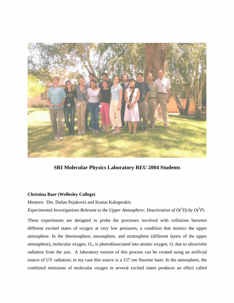
SRI Molecular Physics Laboratory REU 2004 Students
Christina Baer (Wellesley College)
Mentors: Drs. Dušan Pejakovi� and Kostas Kalogerakis
Experimental Investigations Relevant to the Upper Atmosphere: Deactivation of O(1D) by O(3P)
These experiments are designed to probe the processes involved with collisions between
different excited states of oxygen at very low pressures, a condition that mimics the upper
atmosphere. In the thermosphere, mesosphere, and stratosphere (different layers of the upper
atmosphere), molecular oxygen, O2, is photodissociated into atomic oxygen, O, due to ultraviolet
radiation from the sun. A laboratory version of this process can be created using an artificial
source of UV radiation; in my case this source is a 157 nm fluorine laser. In the atmosphere, the
combined emissions of molecular oxygen in several excited states produces an effect called
nightglow.
A green line located at 557.7 nm can be distinguished in spectra of the nightglow. The
appearance of this line, characteristic of oxygen, demonstrated that the airglow could be
associated with the photodissociation of O2. In addition, another spectral line characteristic to
oxygen is visible in the nightglow spectra, located at ~630 nm. While referred to as the “red
line,” this spectral feature actually consists of two separate red lines occurring at 630 and 634
nm.
In my experiments, atomic oxygen is produced by the photodissociation of molecular oxygen at
157 nm, the product atomic states are O(1D) and O(3P). The transition from the excited O(1D)
state to O(3P), the ground state, produces a 630 nm emission (the red line) which can be detected
by a photomultiplier tube (PMT) with the help of an interference filter or a monochromator. By
varying the intensity of the laser light or composition of gas in the cell, it is possible to control
the concentration of atomic oxygen in the resulting gas mixture. The evolution of the O(1D)
concentration over time will be determined by studying the time dependence of the red line
intensity. From this data, it is possible to determine the O(1D) + O(3P) rate coefficient.
As of August 20th the initial experimental goals determined for this summer have been met.
Despite our difficulties maintaining laser power from day to day, we managed to take data which
allows us to calculate a preliminary rate constant for the deactivation of O(1D) by O(3P). Our
initial values indicate that the constant is about 2 orders of magnitude slower than previously
thought. Many more experimental checks must be made before we are certain that our data
actually corresponds to the O(1D)+O(3P) collision. Analyzing the data and coming to understand
the theory behind the O(1D) experiment was a truly valuable experience; forcing myself to work
through the differential equations was not particularly pleasant but my understanding of the
processes underlying the experiment definitely improved. In addition to working on the
O(1D)+O(3P) experiment, I also had the opportunity to work with Rhiannon on her experiment
concerning ammonia ice and hydrocarbons. It was a great opportunity for me to be involved
with such different experiments; taking data on both setups exposed me to many different
experimental techniques and allowed me to have more hands-on time in the lab. I only wish I
had more time to follow through with my experiments, I feel like I've come to a deeper
understanding of my experiment just in time to leave. I have really enjoyed working for Kostas,
his enthusiasm, patience and inherent teaching ability greatly enriched my entire experience at
SRI. The REU program offered me the chance to work with professional scientists in a research
environment, an opportunity that is not available at Wellesley. Participating in experiments and
interacting on a daily basis with the scientists in MPL allowed me to see what a career as a
research scientist entails and I can't emphasize enough the importance of that experience.
Zachary Campbell (Texas A&M University)
Mentors: Drs. Dušan Pejakovi� and Richard Copeland
Laboratory Studies Relevant to the Airglow
The project I worked on this summer involved laboratory studies of processes relevant to the
upper atmosphere. Specifically, we investigated vibrationally excited molecular oxygen in its
ground and first excited electronic states. These studies were funded by NASA, and are relevant
to observations carried out by the SABER instrument of the TIMED satellite. Knowledge
gathered from this project will further our understanding of the emission from vibrationally
excited water molecules in the mesosphere, enabling the remote monitoring of H2O
concentration profiles.
Laboratory experiments were performed on mixtures of N2, O2, O3, and CO2 gases. We excited
the oxygen molecules in the mixture with one laser and detected the excited molecules with a
second laser. We used REMPI (Resonance Enhanced Multiphoton Ionization) techniques in
which only a particular excited species was ionized and detected by measuring the ion current.
Varying the delay between the laser pulses allowed us to study the dependence of the
concentration of particular O2 excited states.
In the first part of the project, I became well acquainted with the experimental apparatus and
assisted Dr. Dušan Pejakovi� and Dr. Rich Copeland in performing measurements of the rate
constants of the reactions O2 (X, υ = 1) + O and O2 (a, υ = 1) + O. In the second part of the
project, I independently performed measurements of the rate constants of the reactions O2 (a,
υ = 1) + O2 and O2 (a, υ = 1) + CO2 at temperatures of 240 K and room temperature. Our
measured room temperature rate constants are consistent with earlier SRI measurements, and the
O2 (a, υ = 1) + O2 rate constant does not show significant variation between room temperature
and 240 K. However, the O2 (a, υ = 1) + CO2 rate coefficient at 240 K is ~20% lower than that
at room temperature.
Yu Gu (Cornell University)
Mentors: Drs. Kenneth Kotz and Gregory Faris
Optical Microfluidics
Microfluidics is a new technology that has the potential to revolutionize analytical chemistry and
biotechnology. Previously at SRI, we had demonstrated a droplet-based microfluidics technique
using an Argon laser beam to move droplets containing water and red dye. My goals for this
summer were to reconstruct the setup and reproduce previous results with an infrared laser beam.
Using the infrared laser is advantageous over using the visible beam because the IR beam is non-
destructive and does not require the use of a dye since it’s directly absorbed by water.
I have accomplished three goals this summer. 1) I built a setup with a tunable diode laser
with a range of 1520-1580nm, an Erbium-doped fiber amplifier, and a custom-designed
microscope. 2) I showed that water droplets from 100um to 1mm could be moved and combined
using the infrared beam. 3) I repeated a simple protein detection assay performed earlier by Ken
(Horseradish Peroxidase + ABTS) with the new setup. I have also started the design process for
performing performing further molecular analysis. In particular, I have done drawings for the
creation of a doughnut-shaped beam using an axicon lens. The first eight weeks of the summer
were devoted to building the setup and the microscope. I spent three weeks performing
experiments and brainstorming about molecular analysis.
Future work includes increasing the range in droplet size, maintaining a high contact
angle, and facilitating molecular analysis.
Andy Knyazik (University of California-Santa Cruz)
Mentors: Drs. Kenneth Kotz and Gregory Faris
Stimulated Rayleigh and Brillouin Spectroscopy
Stimulated scattering occurs due to light – matter interactions. The two types of stimulated
scattering phenomena that we tried to measure this summer were Rayleigh and Brillouin
scattering. Brillouin scattering is produced when light scatters off an acoustic wave, whereas
Rayleigh scattering is due to light scattering from entropy or thermal waves. These
measurements can be used to determine bulk properties of materials such as density, mixing
fraction, compressibility, speed of sound, temperature, acoustic damping rate, dynamic viscosity
and thermal diffusivity.
My goal for the summer was to build an apparatus that will be used to demonstrate effects of
Rayleigh and Brillouin Spectroscopy in supercritical fluids. The name of the technique is
stimulated gain spectroscopy, which is when a powerful pump beam interferes with a probe
beam. The signal spectrum of the probe beam is then measured, in which I plan to see traits of
Rayleigh and Brillouin scattering. Throughout these 12 weeks, I intended to learn a lot about
optical equipment and its practical applications, by using it to create our apparatus. I learned to
work with mirrors, lenses, spatial filters, isolators, amplifiers, polarizers, half- and quarter-
waveplates, and fibers. I believe I have achieved most of my goals for this summer, as I will
illustrate below.
I have built a system that took the output of an Nd: YAG laser operating at 1064 nm, separated it
into probe and pulse beams, and interfered these in a sample cell further along the optical path.
To create a pump beam I double passed a weak narrow-band laser through two gain amplifiers
operating at 10 Hz. I optimized the two spatial filters that followed each gain amplifier, which
would only keep the desired fundamental spatial mode passing through and block all other
amplified modes. The probe laser was generated from the seed laser with a beam-splitter. I sent
the probe beam through a single-mode fiber to filter out unwanted modes, and later on focused it
inside a cell, where it interfered with the focused pulse beam. The probe beam then went inside
another fiber where the output was monitored with a photodiode connected to an oscilloscope in
order to detect the scattering of interest.
Optimizing the amplification while keeping lasing under control took a lot of time, and therefore
I did not collect any scattering signals inside supercritical fluids. The losses that the probe beam
has when it passes through a single mode fiber are also very disappointing: I am only getting 25
to 30% of the power through the fiber. The apparatus otherwise is optimized and is done well
enough to perform the scattering measurements.
Rhiannon Meharchand (Florida State University)
Mentors: Drs. Kostas Kalogerakis and James Boulter
Spectroscopic Characterization of Ammonia Ices Relevat to the Atmosphere of Jupiter
This summer, I performed spectroscopic characterization of ice mixtures of ammonia and
hydrocarbons. This involved extensive use of infrared absorption spectroscopy and cryogenic
temperatures. My main goal was to learn how to use the apparatus, perform certain experiments,
and increase my knowledge of all data collection and analysis techniques. I am proud to say:
mission accomplished (sort of).
I began the summer by trying to climb the steepest learning curve imaginable. I spent a lot of
time figuring out what spectroscopy was, and why on Earth (or Jupiter, for that matter) anyone
would need it. I also spent a good deal of time familiarizing myself with the apparatus, and
trying not to break things. I learned about Jupiter, and quite a bit about various spectroscopy
experiments that had taken place with ammonia. Everything was quite confusing in the
beginning, but things started to work out for the better as the summer continued.
By mid-summer, I had figured out how to run experiments by myself and not break too many
things (although I'm pretty sure the ion gauge and turbo-molecular pump hate me a little). I spent
a lot of time collecting data, in an attempt to figure out how to get spectra that look like the
literature. There were also a few weeks during the middle of the summer where I attempted to
figure out what the data meant and where to get the constants and figures needed for
calculations. This led me to the conclusion that Google hates me a little too.
Throughout the summer we took various trips to companies and research facilities in the bay
area, which was very enlightening. I had never seen the for-profit/industrial side of research, and
I feel like a whole new horizon of opportunity was laid out for me during our trips.
The end of summer rush was exhilarating. Experiments started to work, things started to make
sense, and I started to become a little sad that everything would be ending soon. All considered,
I discovered the following things:
1) It is possible to obtain and suppress ammonia features in the infrared absorption spectra by
coating the ammonia films with hydrocarbons.
2) 12 weeks may seem like a long time, but it is nothing in the world of research.
3) I like the world of research.
I never quite believed that this was a field that I could go into, but this summer has shown me
that anything is possible once you just get over how little you know and jump in headfirst. I'm
much more confident in my abilities now, and much more aware of what skills I will need for the
real world. I had a wonderful summer, made a lot of new friends, and discovered a little bit
about myself in the process. Thank you so much for the opportunity.
Nader Moussa (North Carolina State University)
Mentors: Dr. Jochen Marschall
Thermal Properties of Polycrystalline Ultra-High Temperature Ceramics
This summer, I worked with Dr. Marschall to develop a computational model of thermal
properties of polycrystalline Ultra-High Temperature Ceramics. This model was developed to
explain experimental uncertainties in laboratory samples of the material by exploring the
parameters that affected net material properties. The model implemented a large network of
crystalline grains, statistically distributing constituent materials of the UHTC into an
interconnected lattice. Further developments included accounting for alignment and anisotropy
of the crystal grains, alternative geometry and connectivity of the network, and exploration of the
conduction and resistance to heat transfer between individual cells.
Some of the difficulties I faced this summer involved the implementation of the computer model.
I worked in FORTRAN-77, which was a good experience with scientific computation, very
different from my school curriculum in computer science (which is geared towards "modern
business programming" techniques). The idiosyncrasies of this language provided a fun
challenge. I also applied many techniques from diverse fields to implement my geometric
model, which used Truncated Octahedron shapes instead of cubes for the individual ceramic
grains. Turning these complex shapes into program-code exercised my skills in vectors and
matrices. Lastly, I developed a good understanding of the timing, scheduling, and other issues
involved in a research environment. These skills are independent of the project I worked on, and
will be very portable to future work.
My experience at SRI was fantastic. The work was stimulating and fun; the environment was
conducive to productivity, with a great deal of self-direction. This gave me the opportunity to
work at my own pace, which I think made for a balance between productivity and learning. In
summary, I enjoyed working here this summer, and will definitely consider coming back in the
future.
Adam Percival (Reid College)
Mentors: Drs. Jeanne Haushalter, Xudong Xiao, and Gregory Faris
Upconverting Chelates
Upconverting chelates are a new type of optical reporter, recently invented at SRI. In the process
of upconversion, two lower energy photons are absorbed by a lanthanide ion that is held by a
chelating agent. A higher energy photon is then emitted and detected. These chelates can be
attached to an antibody, which makes them useful in biological assays such as detecting the
presence of viruses. Upconverting chelates are potentially extremely useful for biological
imaging as their use produces no autofluorescence, they are small, have low phototoxicity, and
do not photobleach. While there are other reporters that exhibit some of these capabilities, such
as single-photon fluorescence, upconverting phosphors, and downconverting chelates,
upconverting chelates offer unique capabilities compared to these alternative methods. The
overall goal of the upconverting chelate research at SRI is to demonstrate their feasibility as a
biological reporter.
The primary goal of the summer was to complete an upconverting chelate-based
immunoassay in a microscopic format including matrix effects for a cell culture. Completion of
this assay would meet the requirements of our funding milestone, allowing further work in this
area to proceed. To assist in achieving this goal, I performed a number of experiments. For
erbium chelates, I helped investigate different chelating agents (DPA, DTPA, and DOTA) and
excitation mechanisms (one-color excitation at both 810 nm and 980 nm, as well as two color
810 nm plus 980 nm). We found that we could produce upconversion using all three excitation
methods, and that the best chelating agent was DPA, followed by DTPA and then DOTA.
Secondly, we spent some time investigating the potential of silver island films to enhance the
signal from lanthanides. In preliminary experiments using Eu-DTPA, we were able observe an
increase in signal strength by a factor of ten in the chelate solution on a silver island film slide, as
compared to an ordinary glass slide. In addition, we observed a significantly decreased
fluorescence lifetime in the chelates on a silver island film compared with those on glass, as
predicted by the theory of silver nanoparticle enhancement of electromagnetic fields. This
provides us with further experimental confirmation that we are observing a true enhancement of
our signal from the silver island film.
Finally, I performed a number of experiments relating to a co-fluorescent solution that can be
used for enhancing the signal from our chelates. Despite difficulties in reproducing the results
from our experiments, and a number of problems resulting from europium contamination, we
were able to consistently show a significant increase in our signal strength due to the
co-fluorescent solution. Unfortunately, to date it has been difficult to quantify this enhancement
and determine our detection limit, due to poor reproducibility. We also are not yet certain of the
extent of the europium contaminant in gadolinium of different purities, or of how the signal
strength changes as the europium concentration varies. As a result, the co-fluorescence technique
is not quite to the point where it can be really useful in our assay, although several experiments
that attempt to resolve these issues are currently in progress.
Núria Queraltó��������Ramon Llull University, Spain)
Mentors: Dr. Jochen Marschall
Oxidation of High-Temperature Ceramics
Stability of materials is an important issue in the reentry of spacecrafts into the atmosphere.
Molecular oxygen is dissociated by the shock wave generating atomic oxygen which can interact
with the surface in two ways: (i) either it can recombine and, with the exothermic energy of this
reaction, heat the materials, (ii) or it can oxidize the surface materials because atomic oxygen is
very reactive. Resistant materials are therefore needed to build spacecrafts. Ultrahigh
temperature ceramics (UHTC) like HfB2/SiC or ZrB2/SiC have an extremely low vapor pressure
and resistance to oxidation. Additionally they are extremely hard and melt at high temperatures.
This combination of properties makes UHTC an interesting candidate for building spaceships.
In order to study all these processes I’ve been involved this summer in the research project
"Oxidation of refractory ceramics in dissociated oxygen" with Dr. Jochen Marschall. My main
goal was to study the effect of dissociated oxygen on the oxidation of Si, polycrystalline SiC and
LPCVD Si3N4. These systems are less complicated than UHTC and allow a better understanding
of the processes involved in the oxidation.

One of the steps was to investigate the oxidation environment because we really didn’t know
how many oxygen atoms are involved in the process. We ran some experiments measuring
pressure and temperature at different points of the system. We have concluded that to know
exactly the amount of oxygen atoms we would need a laser technique. In any case, we have
estimated the loss of oxygen atoms due to the recombination process along the tube.
Another step was to run some experiments changing the oxidation conditions like time or
temperature. With these results we have been able to fit the data to the oxidation equation of
Deal and Grove. From this part we have extracted some rate constants that have helped us to
understand better the overall process.
We have used two gases to obtain atomic oxygen: molecular oxygen (O2) and nitrogen oxide
(N2O). The first one was less dissociated than the second one because the bond between the
nitrogen and the oxygen of the nitrogen oxide is weaker than the bond of the molecular oxygen,
which is not 100% covalent. According to this, when we used N2O instead of O2 the samples
were more oxidized.
There’s a lot of controversy about which is the species that is diffusing through the silica layer.
Everybody argues about three candidates: O2, O, O2-. After reading in the literature we believe
that molecular oxygen is the one diffusing. In this hypothesis atomic oxygen remains in the
surface when we dissociate the molecular oxygen and then recombines into molecular oxygen
and diffuses.
Nevena Rakuljic��������University of California, San Diego)
Mentors: Drs. Ken Kotz and Gregory Faris
Oprical Imaging for Cancer Detection
Optical Imaging for Cancer Detection is a research project based on the use of the optical
systems to detect growing tumors in rodents. In order to grow, tumors create blood vessels
through a process called angiogenesis. Unlike healthy vessels, these vessels of the tumor
neovasculature leak, have abnormal branching, and misdirected flow. Due to these anomalous
anatomical structures, tumors tend to have oxygenation characteristics that differ from the
surrounding tissue. Using spectroscopy, and the fact that near infrared light passes through the
tissue easily, one can look at the hemoglobin and determine oxygenation status.
There are two measurement techniques that are used at SRI to characterize a tissue—a CW
imaging and fiber optic frequency domain instrument. My job this summer has been working on
the imaging setup for the frequency domain. I built a light intensifier circuit which detects light
coming out of the rodent that allows us to separate absorption from scattering, and thus to
quantify the characteristics of a tissue. The modulation frequency of the light and the detector is
in the 100 MHz range. The detector is based on modulating the gain of an image intensifier at a
frequency offset from the modulation of our source light, which allows us to look at the
heterodyne detection of the signals.
The intensifier acts as a mixer of the frequency offset optical signal and the 100 MHz amplified
signal. This information allows us to compare RF amplitude and phase before and after entering
the tissue, as well as the change in DC amplitude. Plugging this information into the frequency
domain equation, we are able to quantify the values of absorption and scattering of the tissue,
which are the numbers that we seek.
The stability of the light source that has been mentioned in my goals statement has been
addressed as well. Moreover, as we have realized, laser diodes are stable enough for our
experiment (in terms of temperature), and thus all that needs to be done is purchase a couple of
diodes at different NIR wavelengths, with the necessary power of 1 Watt or more.
I have been imposed with many problems that I had to solve, such as impedance matching,
dealing with RF pick-up and increasing the modulation depth of our signal. My electronics
background has helped me, but without help of my mentors would not have been sufficient to
successfully complete this portion of the project. Working in SRI has definitely been beneficial
to my future career, and has helped me get a better grasp in the field of applied electronics.
Brock South (Rose-Hulman Institute of Technology)
Mentor: Dr. Harald Oser
Selective Mass-Spectrometric Detection of Chemical Agents During Real-Time Air-Intake
Monitoring
During my term at SRI, I worked with MPL’s nanosecond laser photoionization time-of-flight
mass spectrometer (TOFMS). The ongoing goal of the project is to apply this particular mass
spectrometric technique to the problem of on-line chemical agent detection. In the end, the
project wants to turn out a system that can, for example, sit in an air intake and perform real-time
monitoring of the incoming air, screening for deadly chemical agents. Currently, the search
continues for the optimum method to use in order to detect chemical agents—the system must be
sensitive, fast, and selective.
My contribution to this larger project for the most part dealt with improving the latter
characteristic: selectivity. I set out to determine if the fragmentation patterns of the chemical
agent simulant molecules change in a reproducible way as the wavelength of the ionizing
photons changes. This could provide a highly selective means to identify particular agent
molecules if there proved to be fragment pattern changes unique to each molecule. I collected
and analyzed three-dimensional signal intensity vs. wavelength and mass spectra of two agent
simulants under various conditions of temperature, laser focusing, and ionizing wavelength. The
end result of my studies showed that each fragment of these simulant molecules behaved in the
same way when the wavelength was scanned over the range of 270 nm to 250 nm. This means
that there were no patterns appearing that could be used to uniquely identify the simulants in this
wavelength region. Consequently, I showed that wavelength scanning in the investigated
wavelength range was not a good route to take. My studies, however, opened up another possible
route having to do with the power density of the laser beam in the ionization region which
ultimately could greatly simplify the current system without limiting its potential usefulness as a
detector.
In addition to my project goals, I had many personal goals for this passing summer. I was able to
absorb knowledge pertaining to a variety of fields—organic chemistry, laser physics, chemical
agent studies, and mass spectroscopy. I had never worked with or even seen a mass spec system
before this summer, and I certainly had not even a clue how they worked. Now I can say that I
have a comfortable working knowledge of time-of-flight mass spectrometers—at the least, I
could probably pick up and use similar time-of-flight mass specs without much training or
hassle. In working with this system, it was required of me to learn some simple organic
chemistry naming rules. I had seen chemical names quite a bit in my schoolwork, but it wasn’t
until I sat down with them here that I actually figured out that they really do correspond well to
the structure of the molecules. Also, while working with the system I became reasonably familiar
with the mass spec’s laser system. Over the course of the summer I picked up a great deal of
knowledge about chemical agents—what they are, what they do, how to detect them, and so on.
If nothing else, that information is interesting, if perhaps not directly applicable in the near-term.
What I will probably be using very soon, however, are the basic lab skills I honed during these
months. These range from simple laser safety techniques to the habit of keeping a thorough lab
notebook—honestly, the latter hadn’t been a priority of mine until I learned firsthand the
impossibility of keeping your figures and procedures just by memory even over the course of a
week.
Indara Suarez (Pasadena City College)
Mentor: Dr. Harald Oser
Photoionization Studies of Explosives and Explosive Related Compounds
In cases where endangerment of civilians exists, such as landmines, it is necessary to have a
rapid, selective, and sensitive method of detection for explosives and Explosives Related
Compounds (ERCs). Due to the low vapor pressure and instability of the explosives, it is a
challenging task to develop a fast method of detection. Current methods of detection are
unsuitable for the rapid detection of these materials. The presented research examines the
selectivity and sensitivity of the detection of explosives and ERCs and its limitations with the
Resonantly Enhanced Multiphoton Ionization (REMPI) Time-of-Flight (TOF) Mass
Spectrometer (MS) technique when combined with a tunable femto-second laser system.
Previous experiments were done using a nanosecond jet-REMPI TOF MS system. These
experiments showed that the parent molecule (ERC or explosive molecule) dissociates into
fragments, mainly into NO, before ionization. These experiments were unable to show a
significant difference between the NO fragments from the explosive (or ERC) and NO from
compounds found in the atmosphere. In our current experiments, we utilize a femto-second Multi
Photon Ionization (MPI) TOF MS system to successfully ionize the parent molecule before it
fragments. This summer I set-up the first femto-second laser system at SRI. From the set-up, I
learned many of the properties of each component of this system. I also learned about how
different factors in the environment surrounded the system, such as temperature, influenced the
performance of the system. Furthermore, I read many papers on systems similar to it and their
perspective results when performing similar experiments. This allowed me to learn a lot about
the structure of the molecules I was experimenting on and helped me understand the results.
With the use of the femto-second MPI TOF MS approach we have obtained a distinguishable
mass spectral signature of the parent molecule itself. In addition to the parent molecule, we were
also able to see some structural information in these spectra. Experiments done with
nitrobenzene, nitrotoluenes (ortho, para, meta), and 1-3 dinitrobenzene. With the nitrotoluenes,
the structural information in the spectra allowed us to distinguish between these three isomers.
In the case of the ortho-nitrotoluene, we saw a clear signature of the ortho-effect, as our highest
peak in the spectra was that of the parent molecule with an OH missing. The experiments done
with the 1-3 dinitrobenzene were a bit trickier, since the 1-3 dinitrobenzene comes in crystals
whereas the nitrotoluenes come are liquid. Since we were using an oven to increase the
temperature of the molecules so that they would vaporize into a micro-meter capillary, which
would then lead them into the ionization chamber, crystals caused a problem. We could not put
the 1-3 dinitrobenzene crystals in solution because the solvent we used, methanol, introduced too
much background noise. We decided to try to liquidize and vaporize the crystals in the oven.
This was very successful, but it caused some problems. There was background noise from the
air of the capillary, however it was only lower masses (highest air-mass was 63 amu). The other
problem was that at times the crystals would get stuck in the capillary. At one point, we had to
change it. The experiments performed with the 1-3 dinitrobenzene allowed us to map out the
approach to molecules with lower vapor pressure (also in the form of crystals) like
Trinitrotoluene (TNT). Also, since there is no previous literature on 1-3 dinitrobenze, these were
some good preliminary results. And for the nitrotoluenes we were able to verify previous
literature results. These were the first results obtained from the femto-second laser system at
SRI. The next molecules we will approach with this system will be explosives, such as TNT and
RDX. We will then extend the research to multi-mix real world samples, since the purpose of
this research is to be able to apply it to the real world, and go beyond explosives, maybe to
chemical agents. My experience at SRI was very fulfilling. In addition to learning a lot about
this project, I learned about how research is carried through and how research teams work. I was
also introduced to a new field in physics and will consider this field as a career option. I have
walked out of SRI with more confidence as a student and with a variety of new knowledge.
Allison Widhalm (University of Southen California)
Mentors: Drs. Brian Sharpee and Tom Slanger
Analysis Of Data From The Keck 10-Meter Telescopes
Large ground-based astronomical telescopes simultaneously record light from stellar objects and
from the earth’s nightglow, a faint emission that originates with the chemistry of the upper
atmosphere. Astronomers find this glow to be an annoyance, and do their best to make it go
away, but in fact it contains an enormous amount of information on atmospheric processes, and
is proving to be a valuable resource to aeronomers studying the atmosphere. We take advantage
of the superb tools used by the astronomers to obtain the highest quality spectra ever obtained of
the night sky. These spectra, contain emissions from oxygen atoms and molecules, nitrogen
atoms, sodium, potassium, and OH. The OH emission is intense, and its study is important, as it
relates to atmospheric temperature, chemistry, spectroscopy, and dynamics.
In addition, the large telescopes have been used to study the atmosphere of Venus, and we have
found both similarities and differences in the emission from the two environments, with the
common thread being emission from oxygen atoms and molecules. We study the various
emissions from both the earth and Venus to learn how the atmosphere changes in an hour, a
night, a season, and a solar cycle. Work on this project involves learning how to handle the data
files, and represents a unique opportunity to explore our environment as made accessible by the
world’s largest telescopes.
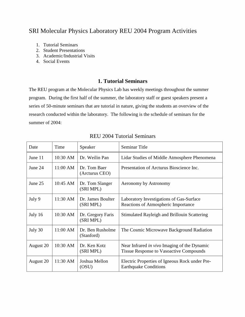
SRI Molecular Physics Laboratory REU 2004 Program Activities
1. Tutorial Seminars 2. Student Presentations 3. Academic/Industrial Visits 4. Social Events
1. Tutorial Seminars The REU program at the Molecular Physics Lab has weekly meetings throughout the summer
program. During the first half of the summer, the laboratory staff or guest speakers present a
series of 50-minute seminars that are tutorial in nature, giving the students an overview of the
research conducted within the laboratory. The following is the schedule of seminars for the
summer of 2004:
REU 2004 Tutorial Seminars
Date Time Speaker Seminar Title
June 11 10:30 AM Dr. Weilin Pan Lidar Studies of Middle Atmosphere Phenomena
June 24 11:00 AM Dr. Tom Baer (Arcturus CEO)
Presentation of Arcturus Bioscience Inc.
June 25 10:45 AM Dr. Tom Slanger (SRI MPL)
Aeronomy by Astronomy
July 9 11:30 AM Dr. James Boulter (SRI MPL)
Laboratory Investigations of Gas-Surface Reactions of Atmospheric Importance
July 16 10:30 AM Dr. Gregory Faris (SRI MPL)
Stimulated Rayleigh and Brillouin Scattering
July 30 11:00 AM Dr. Ben Rusholme (Stanford)
The Cosmic Microwave Background Radiation
August 20 10:30 AM Dr. Ken Kotz (SRI MPL)
Near Infrared in vivo Imaging of the Dynamic Tissue Response to Vasoactive Compounds
August 20 11:30 AM Joshua Mellon (OSU)
Electric Properties of Igneous Rock under Pre-Earthquake Conditions
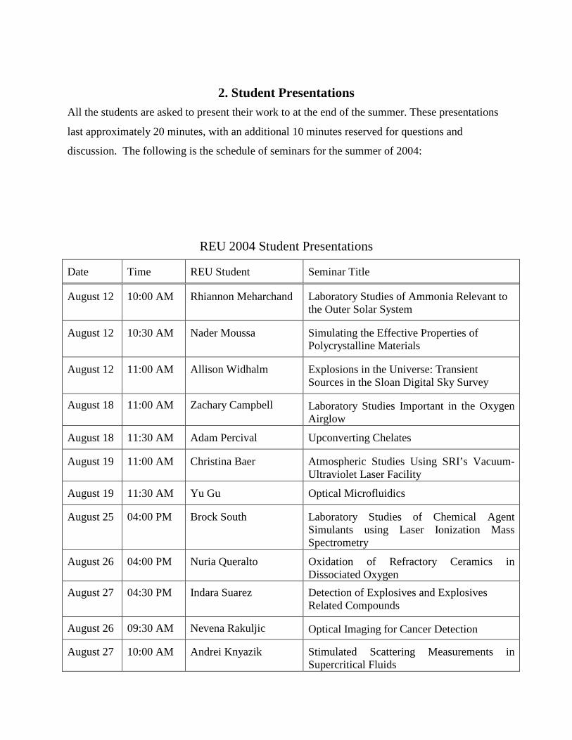
2. Student Presentations All the students are asked to present their work to at the end of the summer. These presentations
last approximately 20 minutes, with an additional 10 minutes reserved for questions and
discussion. The following is the schedule of seminars for the summer of 2004:
REU 2004 Student Presentations
Date Time REU Student Seminar Title
August 12 10:00 AM Rhiannon Meharchand Laboratory Studies of Ammonia Relevant to the Outer Solar System
August 12 10:30 AM Nader Moussa Simulating the Effective Properties of Polycrystalline Materials
August 12 11:00 AM Allison Widhalm Explosions in the Universe: Transient Sources in the Sloan Digital Sky Survey
August 18 11:00 AM Zachary Campbell Laboratory Studies Important in the Oxygen Airglow
August 18 11:30 AM Adam Percival Upconverting Chelates
August 19 11:00 AM Christina Baer Atmospheric Studies Using SRI’s Vacuum-Ultraviolet Laser Facility
August 19 11:30 AM Yu Gu Optical Microfluidics
August 25 04:00 PM Brock South Laboratory Studies of Chemical Agent Simulants using Laser Ionization Mass Spectrometry
August 26 04:00 PM Nuria Queralto Oxidation of Refractory Ceramics in Dissociated Oxygen
August 27 04:30 PM Indara Suarez Detection of Explosives and Explosives Related Compounds
August 26 09:30 AM Nevena Rakuljic Optical Imaging for Cancer Detection
August 27 10:00 AM Andrei Knyazik Stimulated Scattering Measurements in Supercritical Fluids
3. Academic / Industrial Visits
a) On June 29, 2004, the students attended a public lecture entitled “Our lopsided Universe: The Matter with anti-Matter” (speaker: Steve Sekula, MIT/BABAR) at the Stanford Linear Accelerator Center (SLAC).
b) On June 30, 2004, we visited and had a tour at SLAC in the morning, followed by a visit and tour of the laboratories of Profs. Steve Chu and Hongjie Dai at Stanford’s Bio-X, Applied Physics, and Chemistry Departments.
c) Visit to Google Headquarters in Mountain View on (7/23/2004, host: Dyana Wong)
d) Visit to Intuitive Surgical in Sunnyvale (8/2/2004, host: Tom Nixon)
e) Visit to Arcturus Bioscience in Mountain View (8/12/2004, host: Tom Baer)
f) Tour and operation of SRI’s Scanning Electron Microscope (SEM) Facility (7/28/2004, host: Jordi Perez)
g) Visit to SRI’s Artificial Intelligence group and introduction to their robotics technologies (8/4/2004, host: Regis Vincent)
h) Tour of SRI’s Engineering Demo Room and introduction to SRI’s artificial muscle and diamagnetic levitation technologies (8/10/2004, host: Roy Kornbluh)
4. Social Events
Besides several weekend outings and trips the students organized on their own, MPL hosted the annual pool party at the Huestis residence, two birthday parties for the four students who had birthdays in the summer, an ice cream “happy hour”, a pizza lunch and payday meeting, and two farewell gatherings. In addition, the students attended several SRI events (e.g. All-Hands meeting with CEO, New Staff luncheon, SRI Summer BBQ). Finally, MPL sponsored one weekend outing to Muir Woods and dinner in San Francisco.