Embed Size (px)
Citation preview

http://sph.sagepub.com/
ApproachSports Health: A Multidisciplinary
http://sph.sagepub.com/content/2/1/39The online version of this article can be found at:
DOI: 10.1177/1941738109338548
2010 2: 39 originally published online 1 January 2009Sports Health: A Multidisciplinary ApproachMichael M. Reinold and Thomas J. Gill
Part 1: Physical Characteristics and Clinical ExaminationCurrent Concepts in the Evaluation and Treatment of the Shoulder in Overhead-Throwing Athletes,
Published by:
http://www.sagepublications.com
On behalf of:
American Orthopaedic Society for Sports Medicine
can be found at:Sports Health: A Multidisciplinary ApproachAdditional services and information for
http://sph.sagepub.com/cgi/alertsEmail Alerts:
http://sph.sagepub.com/subscriptionsSubscriptions:
http://www.sagepub.com/journalsReprints.navReprints:
http://www.sagepub.com/journalsPermissions.navPermissions:
by Michael Reinold on January 11, 2010sph.sagepub.comDownloaded from

39
vol. 2 • no. 1 SPORTS HEALTH
Current Concepts in the Evaluation and Treatment of the Shoulder in Overhead-Throwing Athletes, Part 1: Physical Characteristics and Clinical Examination Michael M. Reinold, PT, DPT, ATC, CSCS,* and Thomas J. Gill, MD
The overhead-throwing athlete is a challenging sports medicine patient. The repetitive microtraumatic stresses imposed on the athlete’s shoulder joint complex during the throwing motion constantly places the athlete at risk for injury. These stresses may effect several adaptations to normal shoulder range of motion, strength, and scapula position. The clinician should therefore appreciate the unique physical characteristics of the overhead-throwing athlete to accurately evaluate and treat throwing-related injuries.
Keywords: glenohumeral joint; scapula; baseball
[ Sports Physical Therapy ]
From the Boston Red Sox Baseball Club, Boston, Massachusetts, and the Division of Sports Medicine, Department of Orthopedic Surgery, Massachusetts General Hospital, Boston, Massachusetts*Address correspondence to Michael M. Reinold, PT, DPT, ATC, CSCS, Boston Red Sox Baseball Club, Fenway Park, 4 Yawkey Way, Boston, MA 02215.No potential conflict of interest declared.DOI: 10.1177/1941738109338548© 2010 The Author(s)
T he overhead-throwing athlete is a unique and compli-cated sports medicine patient. The repetitive micro-traumatic stresses placed on the athlete’s shoulder joint
complex during the throwing motion challenges the physio-logic limits of the surrounding tissues. During the overhead-throwing motion, the athlete places excessive stresses on the shoulder at the end range of motion, with tremendous angu-lar velocities. Fleisig et al28,29 reported the angular velocity of the overhead throw reaches over 7000 degrees per second, which is the fastest recorded human movement. This motion results in high forces being generated at the shoulder joint, where the dynamic and static stabilizing structures of the shoulder are vulnerable.28,29 Fleisig et al28 also reported anterior forces up to 1 times body weight during external rotation (ER; late cocking) and up to 1.5 times body weight during the follow-through phase (distracting the joint). These forces are likely similar for other overhead-throwing athletes, such as football quarterbacks, softball players, and tennis players.
Consequently, the preventative care and treatment of these athletes are challenging. Injury may occur because of muscle fatigue, muscle weakness, strength imbalances, loss of motion, soft tissue flexibility, alterations in throwing mechanics, and
poor static stability. Because the overhead-throwing athlete is unique, the knowledge of the normal physical characteris-tics, biomechanics, and pathomechanisms of throwing-related injuries is imperative to accurately assess and treat potential injuries.
CLINICAL EXAMINATION
The overhead-throwing athlete exhibits several different physical characteristics—specifically, shoulder range of motion, scapular position, laxity, strength, and proprioception (Table 1). These characteristics must be understood to accurately assess what is a normal physical adaptation rather than pathology.
History
A thorough history of the patient’s complaints, mechanism of injury, and chronicity of symptoms is advantageous and can often lead the clinician to the appropriate examination pro-cess. The injured overhead-throwing athlete generally presents with pain, agitated by throwing and subsiding with inactivity; the athlete also tends to be asymptomatic during all activi-
by Michael Reinold on January 11, 2010sph.sagepub.comDownloaded from

40
Reinold and Gill Jan • Feb 2010
ties other than overhead throwing. Injuries may occur through acute mechanisms in which the athlete attributes the onset of the symptoms to a specific throw. Throwing injuries are typ-ically the result of chronic, repetitive throwing. Patients often report a gradual onset, with no history of an acute episode of injury. It can be helpful to ask what phase of the throw elicits the most symptoms.
Symptoms may initially be subtle and may not alter the patient’s performance. As the symptoms progress, the patient may complain that his or her shoulder is “difficult to warm up” or “get loose” during sport participation, with vague dis-comfort in the shoulder throughout the throwing motion. There is often a loss of throwing velocity and a lack of com-mand while pitching, which becomes more notable as symp-toms worsen. The chronicity of symptoms often establishes the severity of the injury, and the repetitive nature of these athletic activities often results in a gradual progression of pathology and a decline in performance. A player’s injury history, pitch counts in recent games, and number of innings pitched in previous years will give an indication of recent workloads and fatigue levels.
As symptoms progress, the patient can often localize the source and timing of discomfort. However, symptoms are typ-ically vague and diffuse, likely because of a combination of pathologies that are present in the throwing shoulder. The positions that are most provocative in overhead throwers are the fully externally rotated cocked position and the ball release
position (Figure 1). These positions correlate to phases of the throwing motion when stresses on the shoulder are highest.28
Palpation of the entire shoulder girdle may also elicit symp-toms and help differentiate involved structures. The postero-superior glenohumeral joint line, subacromial space, greater tuberosity, and acromioclavicular joint and tendon of the long head of the biceps should be palpated for tenderness. For acro-mioclavicular disorders or bicipital tendonitis, the subjective examination and palpation may be enough to diagnose the pathology.
Range of Motion
One of the most distinguishing characteristics of overhead-throwing athletes is glenohumeral range of motion. Most ath-letes exhibit excessive ER and decreased internal rotation (IR) at 90° of abduction in the throwing shoulder.10,13,43,70,89 This has been shown in baseball players70,89 and tennis players25,26 dur-ing passive motion70,89 and active motion.25,26 Meister et al55 also found this adaptation in adolescent baseball players, noting that the loss of IR was gradual but most dramatic between the ages of 13 and 14 years old.
Wilk et al89 reported passive range of motion characteristics of the shoulder in 372 professional baseball players: 129° ± 10° of ER and 61° ± 9° of IR in the throwing shoulder at 90° abduc-tion. ER was an average of 7° greater, and IR an average of 7° less, in the dominant arm when compared to the nondominant
Table 1. The physical characteristics of the shoulder in the asymptomatic overhead-throwing athlete. a
Examination Component Specific Measurement Normative Value
Range of motion External rotation at 90° abductionInternal rotation at 90° abductionTotal motion
129°-137° (7°-9° > than ND)18,70,89
54°-61° (7°-9° < than ND)18,70,89
183°-198° (bilaterally equal)18,70,89
Joint laxity Sulcus signAnterior translationPosterior translation
61% of pitchers, positive sulcus10
2.8 mm (bilaterally equal)11,12
5.4 mm (bilaterally equal)11,12
Resting scapula position Upward rotationAnterior tiltProtraction
6° on D73,79,80
20° on D73,79,80
39° on D73,79,80
Muscular strength External rotationInternal rotationAbductionAdductionScapular retractionScapular protractionScapular elevationScapular depression
0%-14% < on D58,69,84,86
3%-9% > on D58,69,84,86
Bilaterally equal58,69,84,86
10%-30% > on D58,69,84,86
0%-3% > on D58,92
0% to -4% < on D58,92
Bilaterally equal58,92
22% > on D58,92
Proprioception Joint reposition sense -2° error < on D2,81,82,b
aND, nondominant extremity; D, dominant extremity.bJoint reposition sense decreased by 2° of error.
by Michael Reinold on January 11, 2010sph.sagepub.comDownloaded from

41
vol. 2 • no. 1 SPORTS HEALTH
arm. Thus, total rotation range of motion at 90° of abduction is bilaterally equal in asymptomatic overhead throwers (Figure 2).
The cause of this adaptation has not been established. Numerous theories regarding the altered range of motion pattern observed in overhead-throwing athletes have been reported.† Several authors have documented humeral osseous retroversion in the thrower’s shoulder and attribute the altered range of motion to bony adaptations.18,63,67 Others have theorized that excessive ER and limited IR are due to anterior capsular lax-ity and posterior capsule tightness,15 although no clinical studies have confirmed these findings to date.
The theory of posterior capsular tightness has come into ques-tion from other researchers who have determined that range of motion in baseball pitchers—specifically, a loss of IR—does not correlate with an alteration in posterior glenohumeral transla-tion.11,12 Borsa et al12 studied glenohumeral translation in a series of 43 asymptomatic professional baseball pitchers. The authors reported that posterior translation was twice that of ante-rior translation. There was also no difference in the amount of translation between the dominant shoulder and the nondom-inant shoulder. The authors were unable to show a correlation between a loss of IR range of motion and posterior laxity.
Reinold et al70 recently examined the passive range of motion of the shoulder in 31 professional baseball pitchers, before and immediately after pitching. The researchers reported that rota-tional range of glenohumeral motion is immediately affected by
overhead throwing. Mean IR range of motion after pitching significantly decreased (73° ± 16° before, 65° ± 11° after) and total rotation motion decreased (average, 9°). Mean ER before throwing (133° ± 11°) did not significantly change after throw-ing (131° ± 10°). The researchers hypothesized that this decrease in IR range of motion is due to large eccentric forces being gen-erated in the external rotators (particularly, the infraspinatus and teres minor) during the follow-through phase of throwing. The authors attribute the acute loss of motion to microscopic mus-cle damage due to eccentric contractions of the posterior shoul-der musculature. Eccentric muscular contractions have been cor-related to a rise in passive muscular tension and a loss of joint range of motion.66 Anecdotally, baseball players often describe generalized tightness in the musculature of their posterior shoul-der after pitching. The muscles responsible for ER of the shoulder exhibit high eccentric muscle activity31,32,41,42,75 during the throwing motion as the shoulder internally rotates between 6000 and 7000 degrees per second.23,28,64 Yanagisawa et al93 showed long-last-ing T2 elevations on magnetic resonance imaging of the supra-spinatus, infraspinatus, and teres minor following baseball pitch-ing. The authors attributed these findings to muscle damage that resulted from eccentric muscle contractions. Previous stud-ies examining the effect of repetitive eccentric contractions have shown a subsequent loss of joint range of motion in the upper and lower extremities following testing.38,65,71
The observed range of motion adaptations are likely due to osseous adaptations in the humeral physes of young athlete’s throwing shoulder.55,68,70 In addition, throwing itself results in
Figure 1. The 2 critical instances of potential injury during the throwing motion: A, the moment of full arm cocking when the shoulder reaches maximal external rotation. During this moment, 67 N⋅m of internal rotation torque and 310 N of anterior force are applied to the shoulder. B, the moment of ball release as the shoulder begins to decelerate. Forces at this moment include 1090 N of compressive force at the shoulder joint to prevent subluxation. (From Fleisig GS, Dillman CJ, Andrews JR. Kinetics of baseball pitching with implications about injury mechanisms. Am J Sports Med. 1995;23:233-239.)
†References 10-12, 15, 18, 55, 59, 63, 67, 70, 89.
by Michael Reinold on January 11, 2010sph.sagepub.comDownloaded from

42
Reinold and Gill Jan • Feb 2010
an acute loss of IR motion, most likely attributed to muscular tightness of the posterior shoulder muscles from the high levels of eccentric contraction while the arm decelerates.70
An evaluation (unpublished data, 2008) of shoulder range of motion before and after the competitive season in 20 profes-sional baseball pitchers was conducted. The season consisted of 2 months of spring training and 6 months of the compet-itive season, with pitchers averaging 122 innings. Over the course of the season, these pitchers performed a daily stretch-ing program designed to maintain their range of motion, but they avoided stretching and mobilizing their posterior capsule. The stretching program was performed daily with 3 to 5 rep-etitions of 10 seconds in shoulder flexion, ER and IR at 90° abduction, and cross-body horizontal adduction. At the sea-son end, there was no change in passive IR motion. Based on these results, a loss of IR may be a consequence of the eccen-tric nature of throwing, and a stretching program may help prevent loss of IR. Shoulder ER increased an average of almost 5° over the course of the season, despite the avoidance of aggressive ER stretching. Total rotation motion also increased by 5° in the throwing shoulder, which may be explained by the repetitive attenuation of the anterior capsule and other structures of the shoulder over the course of a season.40
When evaluating range of glenohumeral motion, stan-dard goniometric measurements of active and passive motion should be performed for all planes of movement. Total rota-tion motion should be calculated and compared to the non-dominant shoulder at 90°. Reinold et al70 found that goniomet-ric measurements of passive ER and IR at 90° of abduction were reliable in overhead-throwing athletes (intratester reli-ability intraclass correlation coefficients were .81 and .87). However, bilateral comparisons of ER and IR are not useful.
If the total rotation motion decreases on the throwing side, careful measurements of range of motion should be made to determine if IR has been lost. A loss of IR with a hard end-point may represent other pathologies, such as a throw-er’s exostosis27 (ie, calcification of the posteroinferior gleno-humeral capsular attachment due to chronic traction stress). If total motion increases, the status of the static stabilizers should be assessed.
Joint Laxity
The excessive motion observed in overhead-throwing athletes is commonly attributed to an increase in glenohumeral laxity.53,68,89 This increased motion may represent excessive ER due to ante-rior capsular laxity.40 Excessive laxity may be the result of repet-itive throwing (acquired laxity)68 or congenital laxity.10
Bigliani et al10 reported laxity measurements in 72 profes-sional baseball pitchers and 76 positional players. Sixty-one percent of pitchers and 47% of positional players exhibited a positive sulcus sign, indicating laxity of the superior glenohu-meral ligament. This laxity was present bilaterally, suggesting a congenital origin.
Borsa et al11,12 recently assessed anterior and posterior cap-sular laxity in professional baseball pitchers using an objective mechanical translation device and reported that posterior cap-sular laxity was significantly greater than anterior capsular lax-ity despite gross limitations of passive or active IR. The partic-ipant in this study who had the least IR range of motion had the greatest amount of posterior translation. Total translation (anterior and posterior) was equal bilaterally, indicating that the throwing shoulder was not more lax than the nonthrow-ing shoulder.
Figure 2. The total motion concept: The dominant shoulder (A) of overhead-throwing athletes exhibits a greater external rotation (ER) and lesser internal rotation (IR), compared to the nondominant shoulder (B). However, the total motion (external and internal rotation) is equal bilaterally.
by Michael Reinold on January 11, 2010sph.sagepub.comDownloaded from

43
vol. 2 • no. 1 SPORTS HEALTH
Assessing Laxity
The overhead-throwing athlete has acquired laxity from throw-ing that is often superimposed on underlying congenital lax-ity.68,89 To assess shoulder laxity, the clinician should begin with an exam for generalized joint laxity: hyperextension of the elbow, knee, fifth finger, apposition of the thumb, and trunk flexion.8 For the shoulder, a sulcus test is performed at 0° of abduction. In this position, inferior translation is resisted by the superior glenohumeral ligament. Excessive mobility is thought to indicate generalized glenohumeral hypermobility.90,91
Next, assessment of glenohumeral translation is performed with standard anterior drawer,30 anterior fulcrum (Figure 3),85 posterior drawer,30 and posterior fulcrums85 at 0°, 45°, and 90° of abduction to assess all aspects of the glenohu-meral ligament complex. Another important test to perform is the Lachman test of the shoulder (Figure 4).5 The shoulder is abducted overhead to approximately 120° to 135° of abduc-tion and full ER and then translated anteriorly. The examiner notes the amount of humeral translation as well as the end-point of translation, in comparison to the nondominant shoul-der. In this position, the integrity of the inferior glenohumeral ligament and anterior-inferior capsule is tested. The ante-rior drawer and fulcrum maneuvers can be repeated at 45° of abduction (to test the middle glenohumeral ligament) and in adduction (to assess the superior glenohumeral ligament). Special tests for gross instability, such as the apprehension/relocation sign,39 should be performed to assess the integrity of the static stabilizing structures. It is not uncommon for an overhead-throwing athlete to have a capsulolabral defect from chronic microtrauma.
The anterior Lachman and anterior fulcrum tests are 2 of the most important tests to perform because they assess the ante-rior stabilizing structures of the shoulder in the full ER position,
similar to the vulnerable maximal arm-cocking position during throwing. The apprehension test is an essential part of the anterior stability examination.
Scapular Position
Evaluation of scapular position is an important component of the clinical examination of the overhead-throwing athlete. Past reports have documented alterations in resting scapula position
Figure 3. The anterior fulcrum test for anterior shoulder laxity and instability: A, the shoulder is positioned in approximately 90° of abduction and external rotation; B, as the arm is brought into horizontal abduction, an anterior force is applied to the glenohumeral joint in a fulcrum maneuver. The examiner notes the amount of translation and end feel in comparison to the opposite extremity.85
Figure 4. The anterior Lachman test for anterior shoulder laxity and instability: The shoulder is positioned in approximately 120° to 135° of abduction and external rotation. The proximal and distal aspects of the shoulder are translated anteriorly. The examiner notes the amount of translation and end feel in comparison to the opposite extremity.5
by Michael Reinold on January 11, 2010sph.sagepub.comDownloaded from

44
Reinold and Gill Jan • Feb 2010
in symptomatic patients, which may contribute to some shoul-der pathologies.16 The combination of scapular depression, ante-rior tilt, and protraction may contribute to shoulder pathology.16
Bastan et al7 reported that the asymptomatic thrower’s scap-ula is more protracted and anteriorly tilted at rest, compared to the nonthrowing side. Seitz et al73 confirmed these findings in a study using an electromagnetic tracking system that mea-sured scapular position in 41 asymptomatic professional base-ball pitchers. Results indicated that in asymptomatic pitchers, the scapula rests in 6° of superior rotation, 20° of anterior tilt-ing, and 39° of protraction.
These studies dispute the clinical impression that a protracted and anteriorly tilted scapular position is indicative of pathology. Macrina et al50 noted that the scapular is more protracted after throwing than before. A protracted scapular position may be a normal adaptation to throwing, which, if untreated, may pro-gressively increase over the course of a season. This scapular positioning may be similar to the humeral adaption of IR.
This adaptive scapular position may alter scapular and gle-nohumeral range of motion and strength. An increase in anterior tilt of the scapula correlated with an increase in glenohumeral IR in the dominant shoulder of 98 asymptomatic professional baseball pitchers.80 A protracted, anterior-tilted scapula also correlated to a significant decrease in serratus anterior and lower trapezius strength in asymptomatic baseball pitchers.79
Just as overhead-throwing athletes have adaptations in gleno-humeral motion, asymptomatic baseball pitchers have an adap-tive depressed, anteriorly tilted, and protracted scapula (Tables 1 and 2).71,75-77 Measuring scapular position using a digital incli-nometer (Figure 5) allows comparisons to normative data.73,78-
80 Testing can be performed with the arm in various degrees of shoulder abduction and rotation to assess scapular position. The superomedial border of the scapula should be palpated
during abduction to detect “snapping scapular syndrome” asso-ciated with scapulothoracic bursitis.51
Muscular Strength
Several investigators have examined muscle strength parameters in the overhead-throwing athlete.‡ Isokinetic testing on profes-sional baseball pitchers’ throwing shoulders during spring training showed ER peak torque at an average of 6% lower (P < .05) than that of the nonthrowing shoulders at 90° of abduction.84,86 IR peak torque of the throwing shoulder was 3% higher on average (P < .05) than that of the nonthrowing shoul-der. The mean optimal ratio between ER and IR peak torque at 90° of abduction during isokinetic testing was between 66% and 75%. Adduction torque of the throwing shoulder was 14% greater than that of the nonthrowing shoulder.
The muscle strength profiles of professional baseball pitchers using a handheld dynamometer have been studied (unpublished data, 2009). A 7% dominant-side increase in IR force and a slight decrease in ER and abduction force (1% to 2% each) was seen before the competitive baseball season.86 Over the course of the 8-month season (2 months of preseason and 6 months of com-petition), a 3% to 4% decrease in force in all planes of motion was seen. Abduction force decreased by 16% at the midpoint of the season and 21% by the end of the season. All players partic-ipated in a shoulder injury prevention program designed to min-imize loss of strength over the course of a season. These results suggest that although testing of the rotator cuff did not signifi-cantly change, the loss of abduction strength may be related to rotator cuff fatigue. Fatigue may result in an inability of the rota-tor cuff to center and stabilize the glenohumeral joint, poten-tially resulting in subacromial impingement.
Figure 5. Clinical measurements of anterior/posterior tilt (A) and upward/downward rotation (B) of the scapula using a digital inclinometer, which is placed along the medial border (to measure tilt) and along the spine of the scapula (to measure rotation).
‡References 1, 6, 13, 17, 19, 36, 84, 88.
by Michael Reinold on January 11, 2010sph.sagepub.comDownloaded from

45
vol. 2 • no. 1 SPORTS HEALTH
In another study, ER and IR force at 0° and 90° of abduc-tion was compared in 23 professional baseball pitchers using a handheld dynamometer.69 A decrease in ER and IR force of approximately 20% was noted at 90° of abduction, indicating that the 90° abducted position may be better suited for manual strength testing.
Strength of the scapular muscles also plays a vital role dur-ing overhead throwing.22 When compared to positional players, professional pitchers and catchers have exhibited significantly greater force during scapular protraction and elevation.92
Manual muscle testing with a handheld dynamometer is used for ER and IR at 0° and 90° of coronal plane abduction and scapular plane elevation (full can) for the shoulder. Elevation, posterior tilt, protraction, and retraction are tested for the scapula. A handheld dynamometer is valuable for detecting subtle differences that are often present in overhead-throwing athletes and that may be missed with manual muscle testing. The adaptations that occur from repetitive throwing preclude the meaningful use of bilateral comparisons (Table 2).
The timing of the strength examination must be considered when assessing results. Pitchers often have profound weakness on manual strength testing for 2 days following a start, as well as at the end of the season, presumably due to cuff fatigue.
Proprioception
The overhead thrower relies on enhanced proprioception to dynamically stabilize the glenohumeral joint in the presence of capsular laxity and excessive range of motion.20,24,29,68,87,89 One study tested shoulder proprioception in 20 healthy over-head-throwing athletes by joint repositioning.2 The dominant shoulder exhibited diminished proprioception and improved proprioception toward end range of motion.72 Proprioception significantly decreased after throwing to fatigue, although deficits returned to normal within 10 minutes after throwing.81
To assess proprioception one can use repositioning in several patterns of movement (Figure 6). For example, ER can be tested with the athlete’s eyes closed. The athlete assumes the supine position, and the shoulder is abducted to 90°. The athlete’s shoulder is passively rotated to a point within his or her ER range, and it is held for 3 to 5 seconds before returning to the starting position. The athlete is then instructed to reproduce the previous position, and the dif-ference between the 2 angles is calculated as the error. This measurement is repeated at various points within the range of motion, with an emphasis toward end range, where
Table 2. The effects of acute and chronic throwing on the physical characteristics of the shoulder in the asymptomatic overhead-throwing athlete.a
Examination Component: Measurement
Normative Value
Before Throwing Immediately After Throwing Over the Course of a Season
Range of motionExternal rotationInternal rotationTotal motion
137°70
54°70
191°70
No change70
45°70
180°70
Increase of 5°70
No change70
Increase of 5°70
Muscular strengthExternal rotationInternal rotationFull canAbductionAdductionScapular retractionScapular posterior tilt
0%-14% < on D58,69,84,86
3%-9% > on D58,69,84,86
Bilaterally equal58,69,84,86
Bilaterally equal58,69,84,86
10%-30% > on D58,69,84,86
0%-3% > on D92
0%-3% > on D92
-11%44
-18%44
-6%44
-12%44
-11%44
-4%44
-4%44
-3% to -4% -3% to -4% -3% to -4%-16% to -21% -3% to -4%
Resting scapular positionUpward rotationAnterior tiltProtraction
6°73,79,80
20°73,79,80
39°73,79,80
No change50
No change50
8%50
ProprioceptionJoint reposition sense -2° error2,81,82,b -4° error82,c
aD, dominant extremity.bJoint reposition sense decreased by 2° of error.cJoint reposition sense decreased by 4° of error.
by Michael Reinold on January 11, 2010sph.sagepub.comDownloaded from

46
Reinold and Gill Jan • Feb 2010
proprioception is arguably most important. This measure-ment technique can also be used for shoulder flexion, abduction, proprioceptive neuromuscular facilitation diagonal patterns, and scapula position.
Testing for Rotator Cuff Injuries
Injuries to the rotator cuff can range from tendonitis to a full-thickness tear. Progressive degeneration can occur in ath-letes with poor strength and poor injury prevention. Young athletes often present with inflammation from overuse, with poor muscle strength, and with a stability imbalance between the rotator cuff and scapula. Experience suggests that over the course of a season or career, this degeneration may result in partial-thickness undersurface tearing. If untreated, full-thickness rotator cuff tears can develop.3 Internal impinge-ment of the supraspinatus and infraspinatus on the postero-superior aspect of the glenoid rim during abduction and ER may cause pain in the thrower.83 The rotator cuff is active in resisting glenohumeral subluxation and decelerating the arm. Patients with internal impingement often respond to
conservative treatment. If the pathology progresses, vague discomfort along the deltoid insertion is common, especially in older athletes.
Examination should include the Neer61 and Hawkins34 impingement tests to detect subacromial inflammation. The empty can test can be used to evaluate the athlete’s tolerance of overload to the supraspinatus.
Meister et al54 described an internal impingement sign. With the athlete supine, the arm is abducted to 90° and maximally externally rotated. This maneuver compresses the posterosu-perior rotator cuff tendons against the posterosuperior gle-noid rim. The athlete will often report a vague “deep dis-comfort”; the test is considered positive if posterior humeral translation causes a decrease in symptoms (Figure 7). The fact that this relocation test is indicative of internal impinge-ment lends credibility to the theory that anterior capsular lax-ity/microinstability is a likely contributing factor to inter-nal impingement. In a series of 69 athletes, Meister et al54 reported a sensitivity of 95% and a specificity of 100% in detecting articular-side rotator cuff path ology using an appre-hension-relocation test.
Figure 6. Clinical assessment of joint repositioning skill: A, with the patient’s eyes closed, the examiner passively brings the joint to a point within the patient’s available range of motion. This position is measured and documented, and the joint is brought back to the starting position. B, the patient is instructed to attempt to reproduce the precise position. Measurements are taken and compared to the original measurement to determine the of degree error.
by Michael Reinold on January 11, 2010sph.sagepub.comDownloaded from

47
vol. 2 • no. 1 SPORTS HEALTH
Figure 7. The internal impingement sign: A, the shoulder is positioned in 90° of abduction and full external rotation. In this position, a patient with internal impingement will complain of posterosuperior shoulder pain. B, the examiner may then place a posteriorly directed force on the anterior aspect of the glenohumeral joint to relocate the humeral head within the glenoid fossa. The patient will report a reduction of symptoms in this position.54
Detecting full-thickness rotator cuff tears based on the ath-lete’s strength alone is difficult. The majority of overhead-throwing athletes with full-thickness rotator cuff tears will present with pain in the lateral aspect of their shoulders, weakness in empty can testing, and positive impingement signs. They usually do not present with drop arm37 or lag signs.35
Superior Labral Injuries
Superior labral (SLAP) lesions can be difficult to detect because of the presence of concomitant pathology. Andrews et al4 reported that 45% of patients (73% of baseball pitchers) with SLAP lesions had concomitant partial-thickness tears of the supraspinatus. Mileski and Snyder56 reported that 29% of their patients with SLAP lesions exhibited partial-thickness tears, 11% had complete cuff tears, and 22% had Bankart lesions. Kim et al48 prospectively analyzed SLAP lesions in 139 cases and found that type I is typically associated with rotator cuff pathology whereas type III and IV are associated with trau-matic instability. With type II SLAP lesions, older patients tend to have associated rotator cuff pathology, and younger patients are more likely to have instability. Labral pathologies may result from repetitive overuse but can also result from a single traumatic event, such as a fall onto the outstretched arm, sudden traction, or a blow to the shoulder.
Special tests have been described to detect labral pathol-ogy, including active compression,62 compression-rotation (or grind),76 Speed’s,76 dynamic Speed’s,91 clunk,4 crank,49 anterior slide,45 biceps load,47 biceps load II,46 pronated load,91 pain pro-vocation,57 and resisted supination ER.60
Dessaur and Magarey21 and Jones and Galluch44 reviewed and noted that the majority of studies reporting highly accurate
tests for SLAP lesions were of low quality and were not sup-ported by other researchers.52,77
The discrepancy in accurately testing for SLAP lesions may be due to the difficulty in comparing patient populations. The testing for SLAP lesions in the overhead-throwing ath-lete should attempt to reproduce the peel-back mechanism.91 As the shoulder externally rotates in the abducted posi-tion, torsion occurs at the insertion of the long head of the biceps into the labrum—peeling back the superior portion.14 Tests that mimic the peel-back mechanism14,74 include biceps load,47 biceps load II,46 pronated load,91 pain provocation,57 and resisted supination ER.60 Tests that do not re-create this mech-anism may produce false negatives.62 The presence of deep and diffuse glenohumeral joint pain is most indicative of the presence of a SLAP lesion. Posterior symptoms may be indic-ative of rotator cuff strain. The active compression test is use-ful to localize pain and to establish a starting point for specific SLAP testing.
Two new tests to detect SLAP lesions include the pronated load91 test and the resisted supination ER test.60 For the pro-nated load test, the athlete assumes the supine position with the shoulder abducted to 90° and externally rotated. The fore-arm is then fully pronated to increase tension on the biceps and the labral attachment. When maximal ER is achieved, a resisted isometric contraction of the biceps is used to simu-late the peel-back mechanism (Figure 8). This test combines active biceps contraction46,47,57 with the passive ER in the pro-nated position.
For the resisted supination ER test (Figure 9), the patient is positioned in 90° of shoulder abduction, 65° to 70° of elbow flexion, and neutral forearm rotation.60 Maximal active supi-nation is resisted while passively externally rotating the
by Michael Reinold on January 11, 2010sph.sagepub.comDownloaded from

48
Reinold and Gill Jan • Feb 2010
shoulder. This test simulates the peel-back mechanism of SLAP injuries by placing maximal tension on the long head of the biceps.60 A preliminary study of 40 patients revealed sensitivity (82.8%), specificity (81.8%), positive predictive
value (92.3%), negative predictive value (64.3%), and diag-nostic accuracy (82.5%).60
IMAGING
Basic examination includes standard radiographs for the over-head-throwing athlete: the West Point, axillary, Stryker notch, and IR/ER views in the true anteroposterior plane of the shoulder (Grashey views).
Magnetic resonance arthrography may also be performed to provide further detail of the soft tissue structures; it is the imaging technique of choice for suspected rotator cuff tears, SLAP lesions, and capsular disruptions.
The diagnostic accuracy of magnetic resonance imaging for SLAP lesions is unclear,33,72 and definitive diagnosis may require arthroscopy. Bencardino et al9 retrospectively reviewed preop-erative magnetic resonance arthrography following shoulder arthroscopy, reporting sensitivity (89%), specificity (91%), and accuracy (90%; 47 of 52 patients) in detecting SLAP lesions.
CLINICAL IMPLICATIONS
The physical characteristics (Table 1) of the overhead-throwing athlete are important factors to consider during a physical exam-ination. Acute and chronic adaptations may occur following throwing and over the course of a competitive season (Table 2) that are not necessarily pathologic.
CONCLUSION
The overhead-throwing athlete presents with several nor-mal anatomical adaptations that make the physical examina-tion challenging. Adaptations of range of motion, strength, and scapular position are common and not necessarily pathologic.
NATA Members: Receive 3 free CEUs each year when you subscribe to Sports Health and take and pass the related online quizzes! Not a subscriber? Not a member? The Sports Health–related CEU quizzes are also available for purchase. For more information and to take the quiz for this article, visit www.nata.org/sportshealthquizzes.
REFERENCES 1. Alderink GJ, Kuck DJ. lsokinetic shoulder strength of high school and
college-aged pitchers. J Orthop Sports Phys Ther. 1986;7(4):163-172. 2. Allegrucci M, Whitney SL, Lephart SM, Irrgang JJ, Fu FH. Shoulder kinesthe-
sia in healthy unilateral athletes participating in upper extremity sports. J Orthop Sports Phys Ther. 1995;21(4):220-226.
3. Andrews JR, Broussard TS, Carson WG. Arthroscopy of the shoulder in the management of partial tears of the rotator cuff: a preliminary report. Arthroscopy. 1985;1(2):117-122.
4. Andrews JR, Carson WG Jr, McLeod WD. Glenoid labrum tears related to the long head of the biceps. Am J Sports Med. 1985;13(5):337-341.
5. Andrews JR, Timmerman LA, Wilk KE. Baseball. In: Pettrone FA, ed. Athletic Injuries of the Shoulder. New York: McGraw-Hill; 1995:323-331.
6. Bartlett LR, Storey MD, Simons BD. Measurement of upper extremity torque production and its relationship to throwing speed in the competitive athlete. Am J Sports Med. 1989;17(1):89-91.
7. Bastan M, Reinold MM, Wilk KE, Crenshaw K. Scapular position in profes-sional baseball pitchers: a 3-dimension clinical measure. J Orthop Sports Phys Ther. 2006;36(1):A67.
Figure 8. The pronated load test for superior labral lesions: The shoulder is positioned in 90° of abduction and full external rotation while the forearm is placed in full pronation. Once full passive external rotation is achieved, the patient is instructed to begin an active isometric contraction of his or her biceps, in an attempt to simulate a peel-back superior labral lesion.91
Figure 9. The resisted supination external rotation test for superior labral lesions: The shoulder is placed in 90° abduction and full passive external rotation. The elbow is flexed to approximately 65°. As the shoulder reaches full passive external rotation, the patient is instructed to begin an active contraction against resistance into forearm supination, in an attempt to simulate a peel-back superior labral lesion.60
by Michael Reinold on January 11, 2010sph.sagepub.comDownloaded from
49
vol. 2 • no. 1 SPORTS HEALTH
8. Beighton P, Horan F. Orthopaedic aspects of the Ehlers-Danlos syndrome. J Bone Joint Surg Br. 1969;51(3):444-453.
9. Bencardino JT, Beltran J, Rosenberg ZS, et al. Superior labrum anterior- posterior lesions: diagnosis with MR arthrography of the shoulder. Radiology. 2000;214(1):267-271.
10. Bigliani LU, Codd TP, Connor PM, Levine WN, Littlefield MA, Hershon SJ. Shoulder motion and laxity in the professional baseball player. Am J Sports Med. 1997;25(5):609-613.
11. Borsa PA, Dover GC, Wilk KE, Reinold MM. Glenohumeral range of motion and stiffness in professional baseball pitchers. Med Sci Sports Exerc. 2006;38(1):21-26.
12. Borsa PA, Wilk KE, Jacobson JA, et al. Correlation of range of motion and glenohumeral translation in professional baseball pitchers. Am J Sports Med. 2005;33(9):1392-1399.
13. Brown LP, Niehues SL, Harrah A, Yavorsky P, Hirshman HP. Upper extrem-ity range of motion and isokinetic strength of the internal and external shoulder rotators in major league baseball players. Am J Sports Med. 1988; 16(6):577-585.
14. Burkhart SS, Morgan CD. The peel-back mechanism: its role in producing and extending posterior type II SLAP lesions and its effect on SLAP repair rehabilitation. Arthroscopy. 1998;14(6):637-640.
15. Burkhart SS, Morgan CD, Kibler WB. The disabled throwing shoulder: spec-trum of pathology, part I: pathoanatomy and biomechanics. Arthroscopy. 2003;19(4):404-420.
16. Burkhart SS, Morgan CD, Kibler WB. The disabled throwing shoulder: spec-trum of pathology, part III: The SICK scapula, scapular dyskinesis, the kinetic chain, and rehabilitation. Arthroscopy. 2003;19(6):641-661.
17. Cook EE, Gray VL, Savinar-Nogue E, Medeiros J. Shoulder antagonistic strength ratios: a comparison between college-level baseball pitchers and nonpitchers. J Orthop Sports Phys Ther. 1987;8(9):451-461.
18. Crockett HC, Gross LB, Wilk KE, et al. Osseous adaptation and range of motion at the glenohumeral joint in professional baseball pitchers. Am J Sports Med. 2002;30(1):20-26.
19. Davies GJ, ed. Compendium of Isokinetic Clinical Usage. Onalaska, WI: S&S Publishing; 1992.
20. Davies GJ, Dickoff-Hoffman S. Neuromuscular testing and rehabilitation of the shoulder complex. J Orthop Sports Phys Ther. 1993;18(2):449-458.
21. Dessaur WA, Magarey ME. Diagnostic accuracy of clinical tests for superior labral anterior posterior lesions: a systematic review. J Orthop Sports Phys Ther. 2008;38(6):341-352.
22. DiGiovine NM, Jobe FW, Pink M. An electromyographic analysis of the upper extremity in pitching. J Shoulder Elbow Surg. 1992;1:15-25.
23. Dillman CJ, Fleisig GS, Andrews JR. Biomechanics of pitching with emphasis upon shoulder kinematics. J Orthop Sports Phys Ther. 1993;18(2):402-408.
24. Ellenbecker TS. Rehabilitation of shoulder and elbow injuries in tennis play-ers. Clin Sports Med. 1995;14(1):87-110.
25. Ellenbecker TS, Roetert EP, Bailie DS, Davies GJ, Brown SW. Glenohumeral joint total rotation range of motion in elite tennis players and baseball pitch-ers. Med Sci Sports Exerc. 2002;34(12):2052-2056.
26. Ellenbecker TS, Roetert EP, Piorkowski PA, Schulz DA. Glenohumeral joint internal and external rotation range of motion in elite junior tennis players. J Orthop Sports Phys Ther. 1996;24(6):336-341.
27. Ferrari JD, Ferrari DA, Coumas J, Pappas AM. Posterior ossification of the shoulder: the Bennett lesion: etiology, diagnosis, and treatment. Am J Sports Med. 1994;22(2):171-176.
28. Fleisig GS, Andrews JR, Dillman CJ, Escamilla RF. Kinetics of baseball pitching with implications about injury mechanisms. Am J Sports Med. 1995;23(2):233-239.
29. Fleisig GS, Barrentine SW, Escamilla RF, Andrews JR. Biomechanics of over-hand throwing with implications for injuries. Sports Med. 1996;21(6):421-437.
30. Gerber C, Ganz R. Clinical assessment of instability of the shoulder: with special reference to anterior and posterior drawer tests. J Bone Joint Surg Br. 1984;66(4):551-556.
31. Glousman RE, Barron J, Jobe FW, Perry J, Pink M. An electromyographic analysis of the elbow in normal and injured pitchers with medial collateral ligament insufficiency. Am J Sports Med. 1992;20(3):311-317.
32. Gowan ID, Jobe FW, Tibone JE, Perry J, Moynes DR. A comparative elec-tromyographic analysis of the shoulder during pitching: professional versus amateur pitchers. Am J Sports Med. 1987;15(6):586-590.
33. Green MR, Christensen KP. Magnetic resonance imaging of the glenoid labrum in anterior shoulder instability. Am J Sports Med. 1994;22(4):493-498.
34. Hawkins RJ, Kennedy JC. Impingement syndrome in athletes. Am J Sports Med. 1980;8(3):151-158.
35. Hertel R, Ballmer FT, Lombert SM, Gerber C. Lag signs in the diagnosis of rotator cuff rupture. J Shoulder Elbow Surg. 1996;5(4):307-313.
36. Hinton RY. Isokinetic evaluation of shoulder rotational strength in high school baseball pitchers. Am J Sports Med. 1988;16(3):274-279.
37. Hoppenfeld S, ed. Physical Examination of the Spine and Extremities. Norwalk, CT: Appleton-Century-Crofts; 1976.
38. Jamurtas AZ, Theocharis V, Tofas T, et al. Comparison between leg and arm eccentric exercises of the same relative intensity on indices of muscle dam-age. Eur J Appl Physiol. 2005;95(2-3):179-185.
39. Jobe FW, Bradley JP. The diagnosis and nonoperative treatment of shoulder injuries in athletes. Clin Sports Med. 1989;8(3):419-438.
40. Jobe FW, Kvitne RS, Giangarra CE. Shoulder pain in the overhand or throw-ing athlete: the relationship of anterior instability and rotator cuff impinge-ment. Orthop Rev. 1989;18(9):963-975.
41. Jobe FW, Moynes DR, Tibone JE, Perry J. An EMG analysis of the shoulder in pitching: a second report. Am J Sports Med. 1984;12(3):218-220.
42. Jobe FW, Tibone JE, Perry J, Moynes D. An EMG analysis of the shoulder in throwing and pitching: a preliminary report. Am J Sports Med. 1983;11(1):3-5.
43. Johnson L. Patterns of shoulder flexibility among college baseball players. J Athl Train. 1992;27(1):44-49.
44. Jones GL, Galluch DB. Clinical assessment of superior glenoid labral lesions: a systematic review. Clin Orthop Relat Res. 2007;455:45-51.
45. Kibler WB. Specificity and sensitivity of the anterior slide test in throwing athletes with superior glenoid labral tears. Arthroscopy. 1995;11(3):296-300.
46. Kim SH, Ha KI, Ahn JH, Choi HJ. Biceps load test II: A clinical test for SLAP lesions of the shoulder. Arthroscopy. 2001;17(2):160-164.
47. Kim SH, Ha KI, Han KY. Biceps load test: a clinical test for superior labrum anterior and posterior lesions in shoulders with recurrent anterior disloca-tions. Am J Sports Med. 1999;27(3):300-303.
48. Kim TK, Queale WS, Cosgarea AJ, McFarland EG. Clinical features of the different types of SLAP lesions: an analysis of one hundred and thirty-nine cases. J Bone Joint Surg Am. 2003;85(1):66-71.
49. Liu SH, Henry MH, Nuccion SL. A prospective evaluation of a new phys-ical examination in predicting glenoid labral tears. Am J Sports Med. 1996;24(6):721-725.
50. Macrina LC, Wilk K, Geus J, Porterfield R. The effect of pitching on scap-ula position on professional baseball players. J Orthop Sports Phys Ther. 2007;37(1):A69.
51. Manske RC, Reiman MP, Stovak ML. Nonoperative and operative manage-ment of snapping scapula. Am J Sports Med. 2004;32(6):1554-1565.
52. McFarland EG, Kim TK, Savino RM. Clinical assessment of three com-mon tests for superior labral anterior-posterior lesions. Am J Sports Med. 2002;30(6):810-815.
53. Meister K. Injuries to the shoulder in the throwing athlete: part one: biomechanics/pathophysiology/classification of injury. Am J Sports Med. 2000;28(2):265-275.
54. Meister K, Buckley B, Batts J. The posterior impingement sign: diagnosis of rotator cuff and posterior labral tears secondary to internal impingement in overhand athletes. Am J Orthop. 2004;33(8):412-415.
55. Meister K, Day T, Horodyski M, Kaminski TW, Wasik MP, Tillman S. Rotational motion changes in the glenohumeral joint of the adolescent/Little League baseball player. Am J Sports Med. 2005;33(5):693-698.
56. Mileski RA, Snyder SJ. Superior labral lesions in the shoulder: pathoanatomy and surgical management. J Am Acad Orthop Surg. 1998;6(2):121-131.
57. Mimori K, Muneta T, Nakagawa T, Shinomiya K. A new pain provocation test for superior labral tears of the shoulder. Am J Sports Med. 1999;27(2):137-142.
58. Mullaney MJ, McHugh MP, Donofrio TM, Nicholas SJ. Upper and lower extremity muscle fatigue after a baseball pitching performance. Am J Sports Med. 2005;33(1):108-113.
59. Myers JB, Laudner KG, Pasquale MR, Bradley JP, Lephart SM. Glenohumeral range of motion deficits and posterior shoulder tightness in throwers with pathologic internal impingement. Am J Sports Med. 2006;34(3):385-391.
60. Myers TH, Zemanovic JR, Andrews JR. The resisted supination external rota-tion test: a new test for the diagnosis of superior labral anterior posterior lesions. Am J Sports Med. 2005;33(9):1315-1320.
61. Neer CS II, Welsh RP. The shoulder in sports. Orthop Clin North Am. 1977;8(3):583-591.
62. O’Brien SJ, Pagnani MJ, Fealy S, McGlynn SR, Wilson JB. The active com-pression test: a new and effective test for diagnosing labral tears and acro-mioclavicular joint abnormality. Am J Sports Med. 1998;26(5):610-613.
by Michael Reinold on January 11, 2010sph.sagepub.comDownloaded from

50
Reinold and Gill Jan • Feb 2010
63. Osbahr DC, Cannon DL, Speer KP. Retroversion of the humerus in the throw-ing shoulder of college baseball pitchers. Am J Sports Med. 2002;30(3):347-353.
64. Pappas AM, Zawacki RM, Sullivan TJ. Biomechanics of baseball pitching: a preliminary report. Am J Sports Med. 1985;13(4):216-222.
65. Prasartwuth O, Taylor JL, Gandevia SC. Maximal force, voluntary activation and muscle soreness after eccentric damage to human elbow flexor muscles. J Physiol. 2005;567(pt 1):337-348.
66. Proske U, Morgan DL. Muscle damage from eccentric exercise: mecha-nism, mechanical signs, adaptation and clinical applications. J Physiol. 2001;537(pt 2):333-345.
67. Reagan KM, Meister K, Horodyski MB, Werner DW, Carruthers C, Wilk K. Humeral retroversion and its relationship to glenohumeral rotation in the shoulder of college baseball players. Am J Sports Med. 2002;30(3):354-360.
68. Reinold MM, Wilk KE, Dugas JR, Andrews JR. Internal impingement. In: Wilk KE, Reinold MM, Andrews JR, eds. The Athletes Shoulder. 2nd ed. Philadelphia, PA: Churchill Livingston Elsevier; 2009:123-142.
69. Reinold MM, Wilk KE, Hooks TR, Andrews JR. Comparison of bilateral shoulder external and internal rotation strength at 0 degrees and 90 degrees of abduction in professional baseball pitchers. J Orthop Sports Phys Ther. 2003;33(2):A51.
70. Reinold MM, Wilk KE, Macrina LC, et al. Changes in shoulder and elbow passive range of motion after pitching in professional baseball players. Am J Sports Med. 2008;36(3):523-527.
71. Reisman S, Walsh LD, Proske U. Warm-up stretches reduce sensations of stiffness and soreness after eccentric exercise. Med Sci Sports Exerc. 2005;37(6):929-936.
72. Safran MR, Borsa PA, Lephart SM, Fu FH, Warner JJ. Shoulder proprioception in baseball pitchers. J Shoulder Elbow Surg. 2001;10(5):438-444.
73. Seitz AL, Thigpen CA, Reinold MM, Gill TJ. Comparison of three dimen-sional scapular kinematics in professional baseball pitchers in dominant and nondominant shoulders. Phys Ther. In press.
74. Seneviratne A, Montgomery K, Bevilacqua B, Zikria B. Quantifying the extent of a type II SLAP lesion required to cause peel-back of the glenoid labrum: a cadaveric study. Arthroscopy. 2006;22(11):1163, e1161-e1166.
75. Sisto DJ, Jobe FW, Moynes DR, Antonelli DJ. An electromyographic analysis of the elbow in pitching. Am J Sports Med. 1987;15(3):260-263.
76. Snyder SJ, Karzel RP, Del Pizzo W, Ferkel RD, Friedman MJ. SLAP lesions of the shoulder. Arthroscopy. 1990;6(4):274-279.
77. Stetson WB, Templin K. The crank test, the O’Brien test, and routine mag-netic resonance imaging scans in the diagnosis of labral tears. Am J Sports Med. 2002;30(6):806-809.
78. Thigpen CA. Validity of a clinical measure of posterior tilting in professional baseball pitchers. J Orthop Sports Phys Ther. 2008;38(1):A4.
79. Thigpen CA, Reinold MM, Padua DA, Schneider RS, Distefano LJ, Gill TJ. 3-d scapular position and muscle strength are related in professional baseball pitchers. J Athl Train. 2008;43(2):S49.
80. Thigpen CA, Reinold MM, Padua DA, Schneider RE, Gill TJ. Total arc of motion affects shoulder girdle strength and scapula tilt in professional pitch-ers. J Orthop Sports Phys Ther. 2009;39(1):A108.
81. Tripp BL, Yochem EM, Uhl TL. Functional fatigue and upper extremity sensorimotor system acuity in baseball athletes. J Athl Train. 2007; 42(1):90-98.
82. Tripp BL, Yochem EM, Uhl TL. Recovery of upper extremity sensorimotor system acuity in baseball athletes after a throwing-fatigue protocol. J Athl Train. 2007;42(4):452-457.
83. Walch G, Liotard JP, Boileau P, Noel E. Postero-superior glenoid impinge-ment: another shoulder impingement. Rev Chir Orthop Reparatrice Appar Mot. 1991;77(8):571-574.
84. Wilk KE, Andrews JR, Arrigo CA. The abductor and adductor strength characteristics of professional baseball pitchers. Am J Sports Med. 1995;23(3):307-311.
85. Wilk KE, Andrews JR, Arrigo CA. The physical examination of the gle-nohumeral joint: emphasis on the stabilizing structures. J Orthop Sports Phys Ther. 1997;25(6):380-389.
86. Wilk KE, Andrews JR, Arrigo CA, Keirns MA, Erber DJ. The strength char-acteristics of internal and external rotator muscles in professional baseball pitchers. Am J Sports Med. 1993;21(1):61-66.
87. Wilk KE, Arrigo CA, Andrews JR. Current concepts: the stabiliz-ing structures of the glenohumeral joint. J Orthop Sports Phys Ther. 1997;25(6):364-379.
88. Wilk KE, Arrigo CA, Andrews JR. Isokinetic testing of the shoulder abduc-tors and adductors: windowed vs nonwindowed data collection. J Orthop Sports Phys Ther. 1992;15(2):107-112.
89. Wilk KE, Meister K, Andrews JR. Current concepts in the rehabilitation of the overhead throwing athlete. Am J Sports Med. 2002;30(1):136-151.
90. Wilk KE, Reinold MM, Dugas JR, Andrews JR. Rehabilitation following thermal-assisted capsular shrinkage of the glenohumeral joint: current con-cepts. J Orthop Sports Phys Ther. 2002;32(6):268-292.
91. Wilk KE, Reinold MM, Dugas JR, Arrigo CA, Moser MW, Andrews JR. Current concepts in the recognition and treatment of superior labral (SLAP) lesions. J Orthop Sports Phys Ther. 2005;35(5):273-291.
92. Wilk KE, Suarez K, Reed J. Scapular muscular strength values in professional baseball players. Phys Ther. 1999;79(5):S81-S82.
93. Yanagisawa O, Niitsu M, Takahashi H, Itai Y. Magnetic resonance imaging of the rotator cuff muscles after baseball pitching. J Sports Med Phys Fitness. 2003;43(4):493-499.
For reprints and permission queries, please visit SAGE’s Web site at http://www.sagepub.com/journalsPermissions.nav.
by Michael Reinold on January 11, 2010sph.sagepub.comDownloaded from

http://sph.sagepub.com/Approach
Sports Health: A Multidisciplinary
http://sph.sagepub.com/content/2/2/101The online version of this article can be found at:
DOI: 10.1177/1941738110362518
2010 2: 101Sports Health: A Multidisciplinary ApproachMichael M. Reinold, Thomas J. Gill, Kevin E. Wilk and James R. Andrews
Part 2: Injury Prevention and TreatmentCurrent Concepts in the Evaluation and Treatment of the Shoulder in Overhead Throwing Athletes,
Published by:
http://www.sagepublications.com
On behalf of:
American Orthopaedic Society for Sports Medicine
can be found at:Sports Health: A Multidisciplinary ApproachAdditional services and information for
http://sph.sagepub.com/cgi/alertsEmail Alerts:
http://sph.sagepub.com/subscriptionsSubscriptions:
http://www.sagepub.com/journalsReprints.navReprints:
http://www.sagepub.com/journalsPermissions.navPermissions:
by Michael Reinold on December 29, 2010sph.sagepub.comDownloaded from

101
vol. 2 • no. 2 SPORTS HEALTH
T he overhead throwing athlete is an extremely challenging patient in sports medicine, with unique physical characteristics as the result of sport competition. The
repetitive microtraumatic stresses placed on the athlete’s shoulder joint complex during the throwing motion challenges the physiologic limits of the surrounding tissues.
Consequently, it is imperative to emphasize the preventative care and treatment of these athletes. Injury may occur because of muscle fatigue, muscle weakness and imbalances, alterations in throwing mechanics, and/or altered static stability. A comprehensive program designed for the overhead athlete is necessary to avoid injury and maximize performance. Unfortunately, not all injuries may be prevented, because the act of throwing oftentimes exceeds the ultimate tensile strength of the stabilizing structures of the shoulder.17,18
Part 1 of this series, on the examination and treatment of the overhead athlete, described the unique physical characteristics and examination process for the injured athlete.49 Part 2 descibes a proper treatment program and emphasizes the unique physical characteristics and stresses observed during the act of throwing.
PrinciPles of injury Prevention and treatment Programs
Several general principles should be incorporated into the development of injury prevention and treatment programs for the thrower’s shoulder. Injury prevention and treatment programs share considerable overlap given that both are based on similar principles.
Maintain Range of Motion
The first principle involves maintaining appropriate “thrower’s motion” at the glenohumeral joint. The shoulder in overhead athletes exhibits excessive motion, ranging from 129° to 137° of external rotation (ER), 54° to 61° of internal rotation (IR), and 183° to 198° of total ER-IR motion.49 Although the dominant shoulder has greater ER and less IR, the combined total motion should be equal bilaterally.49,54,69 More important, the act of throwing reduces IR and total motion (Figure 1).54 Studies by Ruotolo et al57 and Myers et al43 have both shown that a loss of total motion correlates with a greater risk of injury.
Current Concepts in the Evaluation and Treatment of the Shoulder in Overhead Throwing Athletes, Part 2: Injury Prevention and TreatmentMichael M. Reinold, PT, DPT, SCS, ATC, CSCS,*†‡ Thomas J. Gill, MD,†‡ Kevin E. Wilk, PT, DPT,§ and James R. Andrews, MD||
The overhead throwing athlete is an extremely challenging patient in sports medicine. The repetitive microtraumatic stresses imposed on the athlete’s shoulder joint complex during the throwing motion constantly place the athlete at risk for injury. Treatment of the overhead athlete requires the understanding of several principles based on the unique physical characteris-tics of the overhead athlete and the demands endured during the act of throwing. These principles are described and incor-porated in a multiphase progressive rehabilitation program designed to prevent injuries and rehabilitate the injured athlete, both nonoperatively and postoperatively.
Keywords: glenohumeral joint; scapula; rotator cuff; internal impingement; superior labral anterior posterior lesion; baseball
[ Sports Physical Therapy ]
From the †Boston Red Sox Baseball Club, Boston, Massachusetts, the ‡Division of Sports Medicine, Department of Orthopedic Surgery, Massachusetts General Hospital, Boston, Massachusetts, §Champion Sports Medicine, Birmingham, Alabama, and ||Andrews Sports Medicine and Orthopaedic Center, Birmingham, Alabama*Address correspondence to Michael M. Reinold, PT, DPT, SCS, ATC, CSCS, 4 Yawkey Way, Boston, MA 002215-3496 (e-mail: [email protected]).No potential conflict of interest declared.DOI: 10.1177/1941738110362518© 2010 The Author(s)
by Michael Reinold on December 29, 2010sph.sagepub.comDownloaded from

102
Reinold et al Mar • Apr 2010
Thus, it is important to maintain motion over the course of a season. Reinold et al54 theorized that the loss of IR and total motion after throwing is the result of eccentric muscle damage as the external rotators and other posterior musculature attempt to decelerate the arm during the throwing motion. In general, total motion should be maintained equal to that of the nondominant shoulder by frequently performing gentle stretching. Caution should be emphasized against overaggressive stretching in an attempt to gain mobility, in favor of stretching techniques to maintain mobility.
It is equally important to regain full range of motion following injury and surgery. Time frames vary for each injury. Athletes who are attempting to return to throwing before regaining full motion have a difficult time returning to competition without symptoms. The clinician should ensure that full motion has been achieved before allowing the initiation of an interval throwing program.
Maintain Strength of the Glenohumeral and Scapulothoracic Musculature
Because the act of throwing is so challenging for the static and dynamic stabilizing structures of the shoulder, strengthening of the entire upper extremity—including shoulder, scapula, elbow, and wrist—is essential for the overhead thrower. A proper program is designed per the individual needs of each athlete, the unique stress of the throwing motion, and the available research on strengthening each muscle.48 Emphasizing the external rotators, scapular retractors, and lower trapezius is important according to electromyographic studies of the throwing motion.48,50,52 These exercises serve as a foundation for the strengthening program, to which skilled and advanced techniques may be superimposed.
Emphasize Dynamic Stabilization and Neuromuscular Control
The excessive mobility and compromised static stability observed within the glenohumeral joint often result in injuries to the capsulolabral and musculotendinous structures of the throwing shoulder. Efficient dynamic stabilization and neuromuscular control of the glenohumeral joint is necessary for overhead athletes to avoid injuries.12 This involves neuromuscular control: efferent (motor) output in response to afferent (sensory) stimulation. It is one of the most overlooked yet crucial components of injury prevention and treatment programs for the overhead athlete.
Neuromuscular control of the shoulder involves not only the glenohumeral but also the scapulothoracic joint. The scapula provides a base of support for muscular attachment and dynamically positions the glenohumeral joint during upper extremity movement. Scapular strength and stability are essential to proper function of the glenohumeral joint.
Neuromuscular control techniques should be included in rehabilitation programs for the overhead athlete—specifically, rhythmic stabilization, reactive neuromuscular control drills, closed kinetic chain, and plyometric exercises.12,16,48,51,69,73 Closed kinetic chain exercises stress the joint in a load-bearing position, resulting in joint approximation.12 The goal is to stimulate receptors and facilitate co-contraction of the shoulder force couples.46
Plyometric exercises provide quick, powerful movements by a prestretch of the muscle, thereby activating the stretch shortening cycle.20,62,73 Plyometric exercises increase the speed of the myotactic/stretch reflex, desensitize the Golgi tendon organ, and increase neuromuscular coordination.73
Figure 1. The total motion concept. The combination of external rotation (ER) and internal rotation (IR) equals total motion and is equal bilaterally in overhead athletes, although shifted posteriorly in the dominant (A) versus nondominant (B) shoulder. Pathological loss of internal rotation will result in a loss of total motion (C).
by Michael Reinold on December 29, 2010sph.sagepub.comDownloaded from

103
vol. 2 • no. 2 SPORTS HEALTH
Core and Lower Body Training
The lower extremities are vital in the development of force during the throwing motion. Core stabilization drills and lower body training further enhance the transfer of kinetic energy and proximal stability with distal mobility of the upper extremity. Any deficits in strength, endurance, or neuromuscular control of the lower body will have a significant impact on the forces of the upper extremity and the athlete’s ability to produce normal pitching mechanics.
Core stabilization is based on the kinetic chain concept: Imbalance at any point of the kinetic chain results in pathology. Movement patterns such as throwing require a precise interaction of the entire kinetic chain to become efficient. An imbalance of strength, flexibility, endurance, or stability anywhere within the chain may result in fatigue, abnormal arthrokinematics, and subsequent compensation.
Off-Season Preparation
The off-season is a valuable time for the athlete to rest, regenerate, and prepare for the rigors of an upcoming season. The main components of a player’s off-season include an initial period of rest, followed by a progressive full-body strength and conditioning program. The goal of the off-season is to build enough strength, power, and endurance to compete without the negative effects of fatigue or weakness from overtraining or undertraining. Whereas the timing of the in- and off-season components of an athlete’s yearly cycle may vary greatly among athletes at different skill levels, the concepts and goals for the off-season remain the same. Training is based on Matveyev’s periodization concept with individualized attention to each athlete’s specific goals (Figure 2).35
At the conclusion of a competitive season, athletes should remain physically active while taking time away from their sports. Recreational activities are encouraged, such as swimming, golfing, cycling, and jogging. This is also a valuable time to rehabilitate any lingering injury that may have been managed through the season.
The remainder of the off-season is used to build a baseline of strength, power, endurance, and neuromuscular control—the goal of which is to maximize physical performance before the start of sport-specific activities. Doing so will ensure that the athlete has adequate physical fitness to withstand the demands of the competitive season.
In-Season Maintenance
Equally as important as preparing for the competitive season is maintaining gains in strength and conditioning during the season. The chronic, repetitive nature of a long season often results in a decline in physical performance.
Whereas a full-body strength and conditioning program is imperative, attention should be paid to the throwing shoulder and the muscles of the glenohumeral and scapulothoracic
joints. Any fatigue or weakness in these areas can lead to injury through a loss of dynamic stability.
An in-season maintenance program should focus on strength and dynamic stability while adjusting for the workload of a competitive season.
rehabilitation Progression
In addition to eliminating pain and inflammation, the rehabilitation process for throwing athletes must restore motion, muscular strength, and endurance, as well as proprioception, dynamic stability, and neuromuscular control (Table 1). As the athlete advances, sport-specific drills are added to prepare for a return to competition. Neuromuscular control drills are performed throughout, advancing as the athlete progresses, to provide a continuous challenge to the neuromuscular control system.
Acute Phase
The acute phase of rehabilitation begins immediately following injury or surgery by abstaining from throwing activities. The duration of the acute phase depends on the chronicity of the injury and the healing constraints of the involved tissues.
Figure 2. The concept of periodization as defined by Matveyev35 (A) and customized per the schedule of a professional baseball player (B).
by Michael Reinold on December 29, 2010sph.sagepub.comDownloaded from

104
Reinold et al Mar • Apr 2010
Table 1. Treatment guidelines for the overhead athlete.a
Phase 1: Acute Phase
Goals Diminish pain and inflammationImprove posterior flexibilityReestablish posterior strength and dynamic stability (muscular balance)Control functional stresses/strains
Treatment Abstain from throwing until pain-free full ROM and full strength—specific time determined by physician
Modalities Iontophoresis (disposable patch highly preferred)PhonophoresisElectrical stimulation and cryotherapy as needed
Flexibility Improve IR ROM at 90° abduction to normal total motion valuesEnhance horizontal adduction flexibilityGradually stretch into ER and flexion—do not force into painful ER
Exercises Rotator cuff strengthening (especially ER) with light-moderate weight • Tubing ER/IR • Side ER
Scapular strengthening exercises • Retractors • Depressors • Protractors
Manual strengthening exercises • Side ER • Supine ER at 45° of abduction • Prone row • Side flexion in the scapular plane
Dynamic rhythmic stabilization exercisesProprioception trainingElectrical stimulation to posterior cuff as needed during exercisesClosed kinetic chain exercisesMaintain core, lower body, and conditioning throughoutMaintain elbow, wrist, and forearm strength
Criteria to progress to phase 2
Minimal pain or inflammationNormalized IR and horizontal adduction ROMBaseline muscular strength without fatigue
Phase 2: Intermediate Phase
Goals Progress strengthening exercisesRestore muscular balance (ER/IR)Enhance dynamic stabilityMaintain flexibility and mobilityImprove core stabilization and lower body strength
Flexibility Control stretches and flexibility exercises • Especially for IR and horizontal adduction • Gradually restore full ER
(continued)
by Michael Reinold on December 29, 2010sph.sagepub.comDownloaded from

105
vol. 2 • no. 2 SPORTS HEALTH
Exercises Progress strengthening exercisesFull rotator cuff and scapula shoulder isotonic program—begin to advance weightInitiate dynamic stabilization program
• Side ER with RS • ER tubing with end range RS • Wall stabilization onto ball • Push-ups onto ball with stabilization
May initiate 2-hand plyometric throws • Chest pass • Side to side • Overhead soccer throws
Criteria to progress to phase 3
Full, pain-free ROMFull 5/5 strength with no fatigue
Phase 3: Advanced Strengthening Phase
Goals Aggressive strengthening programProgress neuromuscular controlImprove strength, power, and enduranceInitiate light throwing activities
Exercises Stretch prior to exercise program—continue to normalize total motionContinue strengthening program aboveReinitiate upper-body programDynamic stabilization drills
• ER tubing with end-range RS at 90° abduction • Wall stabs in 90° of abduction and 90° of ER • Wall dribble with RS in 90° of abduction and 90° of ER
Plyometrics • Two-hand drills • One-hand drills (90/90 throws, deceleration throws, throw into bounce-back) • Stretch postexercise
Criteria to progress to phase 4
Full ROM and strengthAdequate dynamic stabilityAppropriate rehabilitation progression to this point
Phase 4: Return-to-Activity Phase
Goals Progress to throwing programContinue strengthening and flexibility exercisesReturn to competitive throwing
Exercises Stretching and flexibility drillsShoulder programPlyometric programDynamic stabilization drillsProgress to interval throwing programGradually progress to competitive throwing as tolerated
aROM, range of motion; IR, internal rotation; ER, external rotation; RS, rhythmic stabilizations; 90/90, 90° of abduction and 90° of external rotation.
Table 1. (continued)
by Michael Reinold on December 29, 2010sph.sagepub.comDownloaded from

106
Reinold et al Mar • Apr 2010
Range of motion exercises are promptly performed in a restricted range, according to the theory that motion assists in the enhancement and organization of collagen tissue and the stimulation of joint mechanoreceptors and that it may assist in the neuromodulation of pain.58-60 The rehabilitation program should allow for progressive loads, beginning with gentle passive and active-assisted motion.
Flexibility exercises for the posterior shoulder musculature are also performed early. The posterior shoulder is subjected to extreme repetitive eccentric contractions during throwing, which may result in soft tissue adaptations and loss of IR motion,49,54 which may not be due to posterior capsular tightness. Conversely, it appears that most throwers exhibit significant posterior laxity when evaluated.8,9 Thus, common stretches should include horizontal adduction across the body, IR stretching at 90° of abduction, and the sleeper stretch (Figures 3 and 4).
The cross-body horizontal adduction stretch may be performed in a straight plane or integrated with IR at the glenohumeral joint (Figure 4). Overaggressive stretching with the sleeper stretch should be avoided (Figure 3). Frequent, gentle stretching yields far superior results than does the occasional aggressive stretch. Stretches or joint mobilizations for the posterior capsule should not be performed unless the capsule has been shown to be mobile on clinical examination.
The rehabilitation specialist should assess the resting position and mobility of the scapula. Throwers frequently exhibit a posture of rounded shoulders and a forward head. This posture is associated with muscle weakness of the scapular retractors and deep neck flexor muscles owing to prolonged elongation or sustained stretches.48,65 In addition, the scapula may appear protracted and anteriorly tilted. An anteriorly tilted scapula contributes to a loss of glenohumeral IR.7,32 This
scapular position is associated with tightness of the pectoralis minor, upper trapezius, and levator scapula muscles and weakness of the lower trapezius, serratus anterior, and deep neck flexor muscle groups.48,65 Tightness of these muscles can lead to axillary artery occlusion and neurovascular symptoms, such as arm fatigue, pain, tenderness, and cyanosis.44,56,64 Muscle weakness may result in improper mechanics or shoulder symptoms. Stretching, soft tissue mobilization, deep tissue lengthening, muscle energy, and other manual techniques may be needed in these athletes.
Depending on the severity of the injury, strengthening often begins with submaximal, pain-free isometrics for all shoulder and scapular movements. Isometrics should be performed at multiple angles throughout the available range of motion, with emphasis on contraction at the end.
Manual rhythmic stabilization drills are performed for internal and external rotators with the arm in the scapular plane at 30° of abduction (Figure 5). Alternating isometric contractions facilitate co-contraction of the anterior and posterior rotator cuff musculature. Rhythmic stabilization drills may also be performed with the patient supine and with the arm elevated to approximately 90° to 100° and positioned at 10° of horizontal abduction (Figure 6). This position is chosen for the initiation of these drills due to the combined centralized line of action of both the rotator cuff and deltoid musculature, generating a humeral head compressive force during muscle contraction.45,68 The rehabilitation specialist employs alternating isometric contractions in the flexion, extension, horizontal abduction, and horizontal adduction planes of motion. As the patient progresses, the drills can be performed at variable degrees of elevation, such as 45° and 120°.
Active range of motion activities are permitted when adequate muscle strength and balance have been achieved. With the athlete’s eyes closed, the rehabilitation specialist
Figure 3. A, the sleeper stretch for glenohumeral internal rotation; B, the body should be positioned so that the shoulder is in the scapular plane.
by Michael Reinold on December 29, 2010sph.sagepub.comDownloaded from

107
vol. 2 • no. 2 SPORTS HEALTH
Figure 4. A, cross-body horizontal adduction stretch; B, the clinician may also perform the stretch with the shoulder in internal rotation.
Figure 5. Rhythmic stabilization drills for internal and external rotation with the arm at 90° of abduction and neutral rotation (A) and 90° of external rotation (B).
by Michael Reinold on December 29, 2010sph.sagepub.comDownloaded from

108
Reinold et al Mar • Apr 2010
passively moves the upper extremity in the planes of flexion, ER, and IR; pauses; and then returns the extremity to the starting position. The patient is then instructed to actively reposition the upper extremity to the previous location. The rehabilitation specialist may perform these joint-repositioning activities throughout the available range of motion.
Basic closed kinetic chain exercises are also performed during the acute phase. Exercises are initially performed below shoulder level. The athlete may perform weight shifts in the anterior/posterior and medial/lateral directions. Rhythmic stabilizations may also be performed during weight shifting. As the athlete progresses, a medium-sized ball may be placed on the table and weight shifts may be performed on the ball. Load-bearing exercises can be advanced from the table to the quadruped position (Figure 7).
Modalities such as ice, high-voltage stimulation, iontophoresis, ultrasound, and nonsteroidal anti-inflammatory medications may be employed as needed to control pain and inflammation. Iontophoresis may be particularly helpful in reducing pain and inflammation during this phase of rehabilitation.
Intermediate Phase
The intermediate phase begins once the athlete has regained near-normal passive motion and sufficient shoulder strength balance. Lower extremity, core, and trunk strength and stability are critical to efficiently perform overhead activities by transferring and dissipating forces in a coordinated fashion. Therefore, full lower extremity strengthening and core stabilization activities are performed during the intermediate phase. Emphasis is placed on regaining proprioception, kinesthesia, and dynamic stabilization throughout the athlete’s
full range of motion, particularly at end range. For the injured athlete midseason, it is common to begin in the intermediate phase or at least progress to this phase within the first few days following injury. The goals of the intermediate phase are to enhance functional dynamic stability, reestablish neuromuscular control, restore muscular strength and balance, and regain full range of motion for throwing.
During this phase, the rehabilitation program progresses to aggressive isotonic strengthening activities with emphasis on restoration of muscle balance. Selective muscle activation is also used to restore muscle balance and symmetry. The shoulder external rotator muscles and scapular retractor, protractor, and depressor muscles are isolated through a fundamental exercise program for the overhead thrower.48,70-72 This exercise program is based on the collective information derived from electromyographic research of numerous investigators.¶ These patients frequently exhibit ER weakness and benefit from side lying ER and prone rowing into ER. Both exercises elicit high levels of muscular activity in the posterior cuff muscles.52
Drills performed in the acute phase may be progressed to include stabilization at end ranges of motion with the patient’s eyes closed. Rhythmic stabilization exercises are performed during the early part of the intermediate phase. Proprioceptive neuromuscular facilitation exercises are performed in the athlete’s available range of motion and so progress to include full arcs of motion. Rhythmic stabilizations may be incorporated in various degrees of elevation during the proprioceptive neuromuscular facilitation patterns to promote dynamic stabilization.
Manual-resistance ER is also performed during the intermediate phase. By applying manual resistance during specific exercises, the rehabilitation specialist can vary the amount of resistance throughout the range of motion and incorporate concentric and eccentric contractions, as well as rhythmic stabilizations at end range (Figure 8). As the athlete
Figure 6. Rhythmic stabilization drills for flexion and extension with the arm elevated to 100° of flexion in the scapular plane.
Figure 7. Rhythmic stabilization drills for the throwing shoulder while weightbearing in the quadruped position.
¶ References 6, 13, 41, 48, 50, 52, 66, 74
by Michael Reinold on December 29, 2010sph.sagepub.comDownloaded from
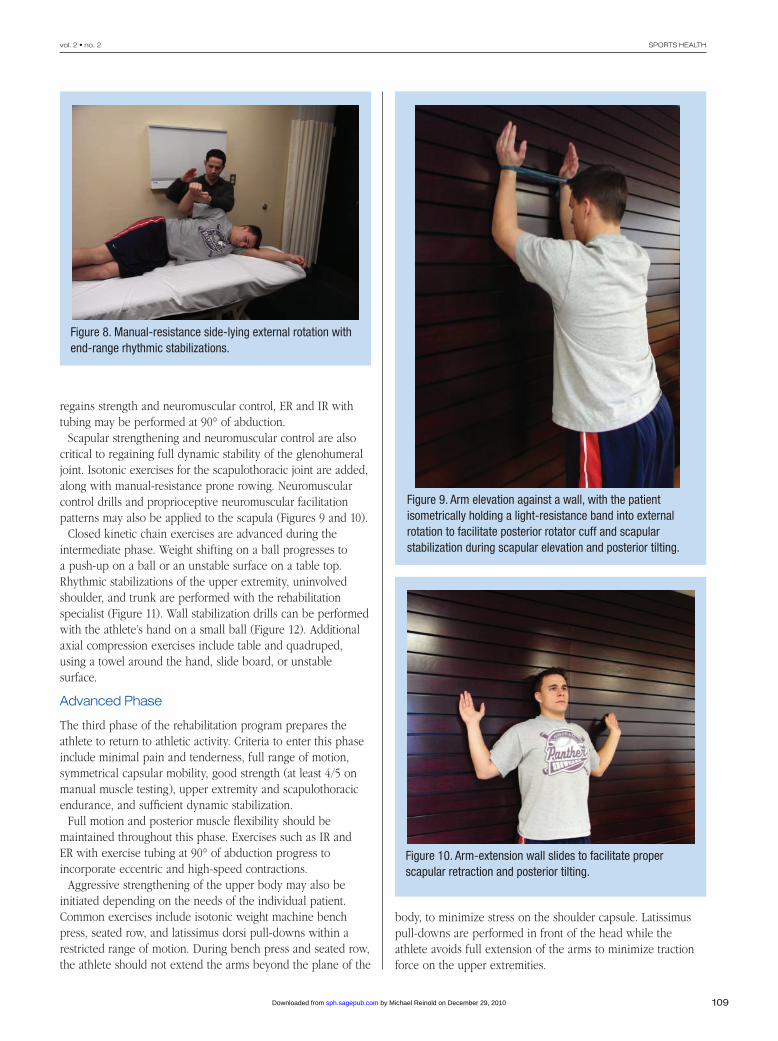
109
vol. 2 • no. 2 SPORTS HEALTH
regains strength and neuromuscular control, ER and IR with tubing may be performed at 90° of abduction.
Scapular strengthening and neuromuscular control are also critical to regaining full dynamic stability of the glenohumeral joint. Isotonic exercises for the scapulothoracic joint are added, along with manual-resistance prone rowing. Neuromuscular control drills and proprioceptive neuromuscular facilitation patterns may also be applied to the scapula (Figures 9 and 10).
Closed kinetic chain exercises are advanced during the intermediate phase. Weight shifting on a ball progresses to a push-up on a ball or an unstable surface on a table top. Rhythmic stabilizations of the upper extremity, uninvolved shoulder, and trunk are performed with the rehabilitation specialist (Figure 11). Wall stabilization drills can be performed with the athlete’s hand on a small ball (Figure 12). Additional axial compression exercises include table and quadruped, using a towel around the hand, slide board, or unstable surface.
Advanced Phase
The third phase of the rehabilitation program prepares the athlete to return to athletic activity. Criteria to enter this phase include minimal pain and tenderness, full range of motion, symmetrical capsular mobility, good strength (at least 4/5 on manual muscle testing), upper extremity and scapulothoracic endurance, and sufficient dynamic stabilization.
Full motion and posterior muscle flexibility should be maintained throughout this phase. Exercises such as IR and ER with exercise tubing at 90° of abduction progress to incorporate eccentric and high-speed contractions.
Aggressive strengthening of the upper body may also be initiated depending on the needs of the individual patient. Common exercises include isotonic weight machine bench press, seated row, and latissimus dorsi pull-downs within a restricted range of motion. During bench press and seated row, the athlete should not extend the arms beyond the plane of the
body, to minimize stress on the shoulder capsule. Latissimus pull-downs are performed in front of the head while the athlete avoids full extension of the arms to minimize traction force on the upper extremities.
Figure 8. Manual-resistance side-lying external rotation with end-range rhythmic stabilizations.
Figure 9. Arm elevation against a wall, with the patient isometrically holding a light-resistance band into external rotation to facilitate posterior rotator cuff and scapular stabilization during scapular elevation and posterior tilting.
Figure 10. Arm-extension wall slides to facilitate proper scapular retraction and posterior tilting.
by Michael Reinold on December 29, 2010sph.sagepub.comDownloaded from

110
Reinold et al Mar • Apr 2010
Plyometrics for the upper extremity may be initiated during this phase to train the upper extremity to dissipate forces. The chest pass, overhead throw, and alternating side-to-side throw with a 3- to 5-lb (1.6- to 2.3-kg) medicine ball are initially performed with 2 hands. Two-hand drills progress to 1-hand drills over 10 to 14 days. One-hand plyometrics include baseball-style throws in the 90/90 position (90° of abduction and 90° of ER) with a 2-lb (0.9-kg) ball, deceleration flips (Figure 13), and stationary and semicircle wall dribbles. Wall dribbles progress to the 90/90 position. They are beneficial for upper extremity endurance while overhead.
Dynamic stabilization and neuromuscular control drills should be reactive, functional, and in sport-specific positions.
Figure 11. Transitioning weightbearing rhythmic stabilization exercises to nonweightbearing positions simulating the landing (A), arm-cocking (B), and ball-release (C) phases of the throwing motion.
Figure 12. Rhythmic stabilization drills in the 90° abducted and 90° external rotation position on an unstable surface in the closed kinetic chain position against the wall.
Figure 13. Plyometric deceleration ball flips. The patient catches the ball over the shoulder and decelerates the arm (similar to the throwing motion) before flipping back and returning to the starting position.
by Michael Reinold on December 29, 2010sph.sagepub.comDownloaded from

111
vol. 2 • no. 2 SPORTS HEALTH
Concentric and eccentric manual resistance may be applied as the athlete performs ER with exercise tubing, with the arm at 0° abduction. Rhythmic stabilizations may be included at end range, to challenge the athlete to stabilize against the force of the tubing as well as the therapist, and may progress to the 90/90 position (Figure 14). Rhythmic stabilizations may also be applied at end range during the 90/90 wall-dribble exercise. These drills are designed to impart a sudden perturbation to the throwing shoulder near end range to develop the athlete’s ability to dynamically stabilize the shoulder.
Muscle endurance exercises should be emphasized because the overhead athlete is at greater risk for shoulder and/or elbow injuries when pitching fatigued.34 Endurance drills include wall dribbling, ball flips (Figure 15), wall arm circles, upper-body cycle, or isotonic exercises using lower weights for higher repetitions. Murray et al42 demonstrated the effects of fatigue on the entire body during pitching using kinematic and kinetic motion analysis. Once the thrower is fatigued, shoulder ER decreases ball velocity and leads to lower extremity knee flexion, and shoulder adduction torque decreases. Muscle fatigue also affects proprioception.67 Once the rotator cuff muscles are fatigued, the humeral head migrates superiorly when arm elevation is initiated.11 Muscle fatigue was the predisposing factor that correlated best with shoulder injuries in Little League pitchers.34 Thus, endurance drills appear critical for the overhead thrower.
Return-to-Activity Phase
Upon completion of the rehabilitation program—including minimal pain or tenderness, full range of motion, balanced
Figure 14. Rhythmic stabilization drills during exercise tubing at 90° of abduction and 90° of external rotation (A) and during wall dribbles (B).
Figure 15. Ball flips for endurance of the external rotators (A) and scapular retractors (B).
by Michael Reinold on December 29, 2010sph.sagepub.comDownloaded from

112
Reinold et al Mar • Apr 2010
capsular mobility, adequate proprioception, and dynamic stabilization—the athlete may begin the return-to-activity phase.
The return to throwing starts with a long-toss program (Appendix 1, available at http://sph.sagepub.com/supplemental) designed to increase distance and number of throws.55 Athletes typically begin at 30 to 45 ft (9 to 14 m) and progress to 60, 90, and 120 ft (18, 27, and 37 m). Pitchers begin a mound throwing program (Appendix 2, available at http://sph.sagepub.com/supplemental), whereas positional players progress to greater distances of long-toss and positional drills. Throwing off the mound includes a gradual increase in the number and intensity of effort and, finally, type of pitch. A player will typically throw 3 times a week with a day off in between, performing each step 2 or 3 times before progressing.
The interval throwing program is supplemented with a high-repetition, low-resistance maintenance exercise program for the rotator cuff and scapula. All strengthening, plyometric, and neuromuscular control drills should be performed 3 times per week (with a day off in between) on the same day as the ISP. The athlete should warm up, stretch, and perform 1 set of each exercise before the interval sport program, followed by 2 sets of each exercise after the program. This provides an adequate warm-up but ensures maintenance of necessary range of motion and flexibility of the upper extremity.
Nonthrowing days are used for lower extremity, cardiovascular, core stability training, range of motion, and light strengthening exercises emphasizing the posterior rotator cuff and scapular muscles. The cycle is repeated throughout the week with the seventh day designated for rest, light range of motion, and stretching exercises.
Internal Posterosuperior Glenoid Impingement
Posterosuperior glenoid impingement (internal impingement) is one of the most frequently observed conditions in the overhead throwing athlete,# and may be caused by excessive anterior shoulder laxity.51 The primary goal of the rehabilitation program is to enhance dynamic stabilization to control anterior humeral head translation while restoring flexibility to the posterior rotator cuff muscles. A careful approach is warranted because aggressive stretching of the anterior and inferior glenohumeral structures may result in increased anterior translation.
The scapular musculature—specifically, the middle trapezius and lower trapezius—is an area of special focus. Once the thrower begins the interval throwing program, the clinician or pitching coach should frequently observe the athlete’s throwing mechanics. Throwers with internal impingement occasionally allow their arms to lag behind the scapula (excessive horizontal abduction). This hyperangulation of the arm may lead to excessive strain on the anterior capsule and internal
impingement of the posterior rotator cuff.21,22,51,55,69 The best treatment for internal impingement is a nonoperative program.
Subacromial Impingement
Primary subacromial impingement in the young overhead throwing athlete is unusual22,37 but may occur with primary hyperlaxity or loss of dynamic stability.22
The nonoperative treatment is similar to that of internal impingement, emphasizing scapular strengthening. Impingement patients exhibit less posterior tilting than do those without impingement.32 The rehabilitation program should include pectoralis minor stretching, inferior trapezius strengthening to ensure posterior scapular tilting, and minimizing forward head posture. Excessive scapular protraction produces anterior scapular tilt and diminishes the acromial-humeral space, whereas scapular retraction increases it.63
Subacromial impingement can be treated conservatively with or without a subacromial injection. The injection is used to relieve pain and inflammation, which in turn allows the patient to more effectively perform a therapy program after a period of rest.
Overuse Syndrome Tendinitis
Throwers may exhibit the signs of overuse tendinitis in the rotator cuff and/or long head of the biceps brachii muscles,4,23,37,49 especially early in the season, when the athlete’s arm may not be in optimal condition.
The thrower will often complain of bicipital pain, referred to as groove pain. The biceps brachii appears to be moderately active during the overhead throwing motion. Bicipital tendinitis usually represents a secondary condition in the overhead thrower. The primary disorder may be instability, a superior labral anterior posterior (SLAP) lesion, or other pathology. The rehabilitation of this condition focuses on improving dynamic stabilization of the glenohumeral joint through muscular training drills. A glenohumeral joint capsule–biceps reflex is present in the feline.19,28 Investigators demonstrated that the biceps brachii was the first muscle to respond to stimulation of the capsule (2.7 milliseconds). The biceps brachii may be activated to a greater extent when the thrower exhibits hyperlaxity or inflammation of the capsule. Nonoperative rehabilitation usually consists of a reduction in throwing activities, the reestablishment of dynamic stability and modalities to reduce bicipital inflammation.
Posterior Rotator Cuff Tendinitis
The successful treatment of rotator cuff tendinitis depends on its differentiation from internal impingement. Subjectively, the athlete notes posterior shoulder pain during ball release or the deceleration phase of throwing. In athletes with internal impingement, the pain occurs during late cocking and early acceleration. During throwing, excessive forces must be dissipated and opposed by the rotator cuff muscles.17 There #References 2,15,31,36,49,51,69
by Michael Reinold on December 29, 2010sph.sagepub.comDownloaded from

113
vol. 2 • no. 2 SPORTS HEALTH
is often significant weakness of the infraspinatus, lower trapezius, and middle trapezius, as well as tightness of the external rotators.
Once strength levels have improved, eccentric muscle training should be emphasized for the external rotators and lower trapezius (Figure 16). Electromyographic activity in the teres minor is 84% maximal voluntary contraction and 78% maximal voluntary contraction in the lower trapezius, during the deceleration phase of a throw and should thus be the focus of the strengthening program.14
Acquired Microinstability
The anterior capsule undergoes significant tensile stress in the late cocking and early acceleration phases of the throwing motion. This stress can lead to gradual stretching of the capsular collagen over time, leading to increased anterior capsular laxity. Several authors25,26,29,30,69 have proposed that repetitive strain on the anterior capsule causes anterior capsular laxity and may worsen internal impingement. Reinold and Gill (unpublished data, 2009) noted that professional baseball pitchers exhibit an increase of 5° of ER at the end of a season in comparison to preseason measurements despite the avoidance of aggressive stretching.49
Mihata et al40 demonstrated in the cadaveric model that excessive ER results in elongation of the anterior band of the
inferior glenohumeral ligament complex and an increase in anterior and inferior translation. Anterior displacement may cause the undersurface of the rotator cuff musculature to impinge on the posterosuperior glenoid rim.
Several arthroscopic procedures (capsular plication and thermal capsular shrinkage) have been developed in an attempt to reduce capsular laxity without overconstraining the joint.5,30,33,47 Rehabilitation following these procedures is designed to gradually restore motion, strength, and neuromuscular control.53,71 Immediately following surgery, restricted passive motion is allowed but not overaggressive stretching. Excessive ER, elevation, or extension is not allowed. By week 6, 75° of ER is the goal. By week 8, 90° of ER at 90° of abduction should be achieved. Usually between weeks 6 and 8, flexion is 170° to 180°. In the case of the overhead athlete—particularly, the pitcher—ER to 115° is needed. This is usually achieved gradually, no earlier than week 12. The athlete is not stretched aggressively past 115° to 120° of ER. Rather, the athlete regains “normal” motion through functional activities within the rehabilitation program, such as plyometrics. The lack of full ER in the overhead athlete is a common cause of symptoms during rehabilitation and throwing.
Isometrics begin in the first 7 to 10 days in a submaximal, nonpainful manner. At approximately 10 to 14 days postoperatively, a light isotonic program begins emphasizing ER and scapular strengthening. At week 5, the athlete is allowed to progress to the full rotator cuff and scapula exercise program with plyometrics at 8 weeks using 2 hands and restricting ER. After 10 to 14 days, 1-hand drills begin. An aggressive strengthening program is allowed at week 12 and is adjusted according to the patient’s response. A gradual return to throwing is expected at week 16, with a return to overhead sports by 9 to 12 months.55
SLAP Lesions
SLAP lesions involve detachment of glenoid labrum–biceps complex from the glenoid rim. These injuries occur through a variety of mechanisms, including falls, traction, motor vehicle accidents, and sports.72 Overhead throwing athletes commonly present with a type II SLAP lesion with the biceps tendon detached from the glenoid rim and a peel back lesion.10
Conservative management of SLAP lesions is often unsuccessful—particularly, type II and type IV lesions with labral instability and underlying shoulder instability. With surgical repair, the initial rehabilitative concern is to ensure that forces on the repaired labrum are controlled. The extent of the lesion, its location, and number of suture anchors are considered when developing a rehabilitation program. The patient sleeps in a shoulder immobilizer and wears a sling during the day for the first 4 weeks following surgery. Range of motion is performed below 90° of elevation to avoid strain on the labral repair. During the first 2 weeks, IR and ER range of motion exercises are performed passively in the
Figure 16. Manual-resistance eccentric contraction of the lower trapezius.
by Michael Reinold on December 29, 2010sph.sagepub.comDownloaded from
114
Reinold et al Mar • Apr 2010
scapular plane (15° of ER and 45° of IR). Initial ER range of motion is performed cautiously to minimize strain on the labrum. At 4 weeks postoperative, the patient begins IR and ER range of motion activities at 90° of shoulder abduction. Flexion can be performed above 90° of elevation. Motion is gradually increased to restore full range of motion (90° to 100° of ER at 90° of abduction) by 8 weeks and progresses to thrower’s motion (115° to 120° of ER) through week 12.
Isometric exercises are performed submaximally immediately postoperatively to prevent atrophy. Biceps contractions are not permitted for 8 weeks following repair. ER/IR exercise tubing is initiated at week 3 or 4 and so progresses to include lateral raises, full can, prone rowing, and prone horizontal abduction by week 6. A full isotonic exercise program for the rotator cuff and scapulothoracic musculature is initiated by week 7 or 8.
Resisted biceps activity (elbow flexion and forearm supination) is not allowed for the first 8 weeks to protect healing of the biceps anchor. Aggressive strengthening of the biceps is avoided for 12 weeks following surgery. Weightbearing exercises are not performed for at least 8 weeks to avoid compression and shearing forces on the healing labrum. Two-hand plyometrics, as well as more advanced strengthening activities, are allowed between 10 and 12 weeks, progressing to an interval sport program at week 16. Return to play following the surgical repair of a type II SLAP lesion typically occurs at approximately 9 to 12 months following surgery.
conclusion
The overhead athlete can present with several different pathologies owing to forces acting during throwing. Care of these patients requires a thorough understanding of the unique physical characteristics of this population. Rehabilitation should follow a gradual and sequential progression. Injury prevention and rehabilitation programs should both emphasize range of motion, flexibility, rotator cuff and scapula strength, posture, and dynamic stabilization. Programs should be individualized per time of season, level of play, and type of injury.
NATA Members: Receive 3 free CEUs each year when you subscribe to Sports Health and take and pass the related online quizzes! Not a subscriber? Not a member? The Sports Health–related CEU quizzes are also available for purchase. For more information and to take the quiz for this article, visit www.nata.org/sportshealthquizzes.
references1. Andrews JR, Angelo RL. Shoulder arthroscopy for the throwing athlete. Tech
Orthop. 1988;3:75-79.2. Andrews JR, Broussard TS, Carson WG. Arthroscopy of the shoulder in
the management of partial tears of the rotator cuff: a preliminary report. Arthroscopy. 1985;1(2):117-122.
3. Andrews JR, Kupferman SP, Dillman CJ. Labral tears in throwing and racquet sports. Clin Sports Med. 1991;10(4):901-911.
4. Andrews JR, Timmerman LA, Wilk KE. Baseball. In: Pettrone FA, ed. Athletic Injuries of the Shoulder. New York: McGraw-Hill; 1995.
5. Bigliani LU, Kurzweil PR, Schwartzbach CC, Wolfe IN, Flatow EL. Inferior capsular shift procedure for anterior-inferior shoulder instability in athletes. Am J Sports Med. 1994;22(5):578-584.
6. Blackburn TA, McLeod WD, White B. Electromyographic analysis of posterior rotator cuff exercises. J Athl Train. 1990;25:40-45.
7. Borich MR, Bright JM, Lorello DJ, Cieminski CJ, Buisman T, Ludewig PM. Scapular angular positioning at end range internal rotation in cases of glenohumeral internal rotation deficit. J Orthop Sports Phys Ther. 2006;36(12):926-934.
8. Borsa PA, Dover GC, Wilk KE, Reinold MM. Glenohumeral range of motion and stiffness in professional baseball pitchers. Med Sci Sports Exerc. 2006;38(1):21-26.
9. Borsa PA, Wilk KE, Jacobson JA, et al. Correlation of range of motion and glenohumeral translation in professional baseball pitchers. Am J Sports Med. 2005;33(9):1392-1399.
10. Burkhart SS, Morgan CD. The peel-back mechanism: its role in producing and extending posterior type II SLAP lesions and its effect on SLAP repair rehabilitation. Arthroscopy. 1998;14(6):637-640.
11. Chen SK, Wickiewicz TL, Otis JC. Glenohumeral kinematics in a muscle fatigue model. Orthop Trans. 1995;18:1126.
12. Davies GJ, Dickoff-Hoffman S. Neuromuscular testing and rehabilitation of the shoulder complex. J Orthop Sports Phys Ther. 1993;18(2):449-458.
13. Decker MJ, Hintermeister RA, Faber KJ, Hawkins RJ. Serratus anterior muscle activity during selected rehabilitation exercises. Am J Sports Med. 1999;27(6):784-791.
14. DiGiovine NM, Jobe FW, Pink M. An electromyographic analysis of the upper extremity in pitching. J Shoulder Elbow Surg. 1992;1:15-25.
15. Dugas JR, Andrews JR. Throwing injuries in the adult. In: DeLee JC, Drez D, Miller MD, eds. DeLee and Drez’s Orthopaedic Sports Medicine. Philadelphia, PA: Elsevier Science; 2003:1236-1249.
16. Ellenbecker TS. Rehabilitation of shoulder and elbow injuries in tennis players. Clin Sports Med. 1995;14(1):87-110.
17. Fleisig GS, Andrews JR, Dillman CJ, Escamilla RF. Kinetics of baseball pitching with implications about injury mechanisms. Am J Sports Med. 1995;23(2):233-239.
18. Fleisig GS, Barrentine SW, Escamilla RF, Andrews JR. Biomechanics of overhand throwing with implications for injuries. Sports Med. 1996;21(6):421-437.
19. Guanche C, Knatt T, Solomonow M, Lu Y, Baratta R. The synergistic action of the capsule and the shoulder muscles. Am J Sports Med. 1995;23(3):301-306.
20. Heiderscheit BC, McLean KP, Davies GJ. The effects of isokinetic vs. plyometric training on the shoulder internal rotators. J Orthop Sports Phys Ther. 1996;23(2):125-133.
21. Jobe CM. Posterior superior glenoid impingement: expanded spectrum. Arthroscopy. 1995;11(5):530-536.
22. Jobe CM. Superior glenoid impingement: current concepts. Clin Orthop Relat Res. 1996;330:98-107.
23. Jobe FW. Impingement problems in the athlete. Instr Course Lect. 1989;38:205-209.
24. Jobe FW, Bradley JP. Rotator cuff injuries in baseball: prevention and rehabilitation. Sports Med. 1988;6(6):378-387.
25. Jobe FW, Giangarra CE, Kvitne RS, Glousman RE. Anterior capsulolabral reconstruction of the shoulder in athletes in overhand sports. Am J Sports Med. 1991;19(5):428-434.
26. Jobe FW, Kvitne RS, Giangarra CE. Shoulder pain in the overhand or throwing athlete: the relationship of anterior instability and rotator cuff impingement. Orthop Rev. 1989;18(9):963-975.
27. Jobe FW, Pink M. Classification and treatment of shoulder dysfunction in the overhead athlete. J Orthop Sports Phys Ther. 1993;18(2):427-432.
28. Knatt T, Guanche C, Solomonow M, Lu Y, Baratta R, Zhou BH. The glenohumeral-biceps reflex in the feline. Clin Orthop Relat Res. 1995;314:247-252.
29. Kvitne RS, Jobe FW. The diagnosis and treatment of anterior instability in the throwing athlete. Clin Orthop Relat Res. 1993;291:107-123.
30. Kvitne RS, Jobe FW, Jobe CM. Shoulder instability in the overhand or throwing athlete. Clin Sports Med. 1995;14(4):917-935.
31. Levitz CL, Dugas J, Andrews JR. The use of arthroscopic thermal capsulorrhaphy to treat internal impingement in baseball players. Arthroscopy. 2001;17(6):573-577.
32. Lukasiewicz AC, McClure P, Michener L, Pratt N, Sennett B. Comparison of 3-dimensional scapular position and orientation between subjects with and without shoulder impingement. J Orthop Sports Phys Ther. 1999;29(10):574-583.
by Michael Reinold on December 29, 2010sph.sagepub.comDownloaded from
115
vol. 2 • no. 2 SPORTS HEALTH
33. Lusardi DA, Wirth MA, Wurtz D, Rockwood CA Jr. Loss of external rotation following anterior capsulorrhaphy of the shoulder. J Bone Joint Surg Am. 1993;75(8):1185-1192.
34. Lyman S, Fleisig GS, Andrews JR, Osinski ED. Effect of pitch type, pitch count, and pitching mechanics on risk of elbow and shoulder pain in youth baseball pitchers. Am J Sports Med. 2002;30(4):463-468.
35. Matveyev LP. Perodisienang das sportlichen training. Berlin: Beles & Wernitz; 1972.
36. Mazoue CG, Andrews JR. Repair of full-thickness rotator cuff tears in professional baseball players. Am J Sports Med. 2006;34(2):182-189.
37. Meister K. Injuries to the shoulder in the throwing athlete. Part one: biomechanics/pathophysiology/classification of injury. Am J Sports Med. 2000;28(2):265-275.
38. Meister K. Injuries to the shoulder in the throwing athlete. Part two: evaluation/treatment. Am J Sports Med. 2000;28(4):587-601.
39. Meister K. Internal impingement in the shoulder of the overhand athlete: pathophysiology, diagnosis, and treatment. Am J Orthop. 2000;29(6):433-438.
40. Mihata T, Lee Y, McGarry MH, Abe M, Lee TQ. Excessive humeral external rotation results in increased shoulder laxity. Am J Sports Med. 2004;32(5):1278-1285.
41. Moseley JB Jr, Jobe FW, Pink M, Perry J, Tibone J. EMG analysis of the scapular muscles during a shoulder rehabilitation program. Am J Sports Med. 1992;20(2):128-134.
42. Murray TA, Cook TD, Werner SL, Schlegel TF, Hawkins RJ. The effects of extended play on professional baseball pitchers. Am J Sports Med. 2001;29(2):137-142.
43. Myers JB, Laudner KG, Pasquale MR, Bradley JP, Lephart SM. Glenohumeral range of motion deficits and posterior shoulder tightness in throwers with pathologic internal impingement. Am J Sports Med. 2006;34(3):385-391.
44. Nuber GW, McCarthy WJ, Yao JS, Schafer MF, Suker JR. Arterial abnormalities of the shoulder in athletes. Am J Sports Med. 1990;18(5):514-519.
45. Poppen NK, Walker PS. Forces at the glenohumeral joint in abduction. Clin Orthop Relat Res. 1978;135:165-170.
46. Prokopy MP, Ingersoll CD, Nordenschild E, Katch FI, Gaesser GA, Weltman A. Closed-kinetic chain upper-body training improves throwing performance of NCAA Division I softball players. J Strength Cond Res. 2008;22(6):1790-1798.
47. Protzman RR. Anterior instability of the shoulder. J Bone Joint Surg Am. 1980;62(6):909-918.
48. Reinold MM, Escamilla RF, Wilk KE. Current concepts in the scientific and clinical rationale behind exercises for glenohumeral and scapulothoracic musculature. J Orthop Sports Phys Ther. 2009;39(2):105-117.
49. Reinold MM, Gill TJ. Current concepts in the evaluation and treatment of the shoulder in overhead throwing athletes: Part 1: Physical characteristics and clinical examination. Sports Health. 2010;2:39-50.
50. Reinold MM, Macrina LC, Wilk KE, et al. Electromyographic analysis of the supraspinatus and deltoid muscles during 3 common rehabilitation exercises. J Athl Train. 2007;42(4):464-469.
51. Reinold MM, Wilk KE, Dugas JR, Andrews JR. Internal Impingement. In: Wilk KE, Reinold MM, Andrews JR, eds. The Athletes Shoulder. 2nd ed. Philadelphia, PA: Churchill Livingston Elsevier; 2009:123-142.
52. Reinold MM, Wilk KE, Fleisig GS, et al. Electromyographic analysis of the rotator cuff and deltoid musculature during common shoulder external rotation exercises. J Orthop Sports Phys Ther. 2004;34(7):385-394.
53. Reinold MM, Wilk KE, Hooks TR, Dugas JR, Andrews JR. Thermal-assisted capsular shrinkage of the glenohumeral joint in overhead athletes: a 15- to 47-month follow-up. J Orthop Sports Phys Ther. 2003;33(8):455-467.
54. Reinold MM, Wilk KE, Macrina LC, et al. Changes in shoulder and elbow passive range of motion after pitching in professional baseball players. Am J Sports Med. 2008;36(3):523-527.
55. Reinold MM, Wilk KE, Reed J, Crenshaw K, Andrews JR. Interval sport programs: guidelines for baseball, tennis, and golf. J Orthop Sports Phys Ther. 2002;32(6):293-298.
56. Rohrer MJ, Cardullo PA, Pappas AM, Phillips DA, Wheeler HB. Axillary artery compression and thrombosis in throwing athletes. J Vasc Surg. 1990;11(6):761-768.
57. Ruotolo C, Price E, Panchal A. Loss of total arc of motion in collegiate baseball players. J Shoulder Elbow Surg. 2006;15(1):67-71.
58. Salter RB, Bell RS, Keeley FW. The protective effect of continuous passive motion in living articular cartilage in acute septic arthritis: an experimental investigation in the rabbit. Clin Orthop Relat Res. 1981;159:223-247.
59. Salter RB, Hamilton HW, Wedge JH, et al. Clinical application of basic research on continuous passive motion for disorders and injuries of synovial joints: a preliminary report of a feasibility study. J Orthop Res. 1984;1(3):325-342.
60. Salter RB, Simmonds DF, Malcolm BW, Rumble EJ, MacMichael D, Clements ND. The biological effect of continuous passive motion on the healing of full-thickness defects in articular cartilage: an experimental investigation in the rabbit. J Bone Joint Surg Am. 1980;62(8):1232-1251.
61. Scarpinato DF, Bramhall JP, Andrews JR. Arthroscopic management of the throwing athlete’s shoulder: indications, techniques, and results. Clin Sports Med. 1991;10(4):913-927.
62. Schulte-Edelmann JA, Davies GJ, Kernozek TW, Gerberding ED. The effects of plyometric training of the posterior shoulder and elbow. J Strength Cond Res. 2005;19(1):129-134.
63. Solem-Bertoft E, Thuomas KA, Westerberg CE. The influence of scapular retraction and protraction on the width of the subacromial space: an MRI study. Clin Orthop Relat Res. 1993;296:99-103.
64. Sotta RP. Vascular problems in the proximal upper extremity. Clin Sports Med. 1990;9(2):379-388.
65. Thigpen CA, Reinold MM, Padua DA, Schneider RS, Distefano LJ, Gill TJ. 3-D scapular position and muscle strength are related in professional baseball pitchers. J Athl Train. 2008;43(2):S49.
66. Townsend H, Jobe FW, Pink M, Perry J. Electromyographic analysis of the glenohumeral muscles during a baseball rehabilitation program. Am J Sports Med. 1991;19(3):264-272.
67. Voight ML, Hardin JA, Blackburn TA, Tippett S, Canner GC. The effects of muscle fatigue on and the relationship of arm dominance to shoulder proprioception. J Orthop Sports Phys Ther. 1996;23(6):348-352.
68. Walker PS, Poppen NK. Biomechanics of the shoulder joint during abduction in the plane of the scapula [proceedings]. Bull Hosp Joint Dis. 1977;38(2):107-111.
69. Wilk KE, Meister K, Andrews JR. Current concepts in the rehabilitation of the overhead throwing athlete. Am J Sports Med. 2002;30(1):136-151.
70. Wilk KE, Reinold MM, Andrews JR. Rehabilitation of the thrower’s elbow. Clin Sports Med. 2004;23(4):765-801.
71. Wilk KE, Reinold MM, Dugas JR, Andrews JR. Rehabilitation following thermal-assisted capsular shrinkage of the glenohumeral joint: current concepts. J Orthop Sports Phys Ther. 2002;32(6):268-292.
72. Wilk KE, Reinold MM, Dugas JR, Arrigo CA, Moser MW, Andrews JR. Current concepts in the recognition and treatment of superior labral (SLAP) lesions. J Orthop Sports Phys Ther. 2005;35(5):273-291.
73. Wilk KE, Voight ML, Keirns MA, Gambetta V, Andrews JR, Dillman CJ. Stretch-shortening drills for the upper extremities: theory and clinical application. J Orthop Sports Phys Ther. 1993;17(5):225-239.
74. Worrell TW, Corey BJ, York SL, Santiestaban J. An analysis of supraspinatus EMG activity and shoulder isometric force development. Med Sci Sports Exerc. 1992;24(7):744-748.
For reprints and permissions queries, please visit SAGE’s Web site at http://www.sagepub.com/journalsPermissions.nav.
by Michael Reinold on December 29, 2010sph.sagepub.comDownloaded from