Embed Size (px)
Citation preview
INFECTION AND IMMUNITY, July 2011, p. 2829–2838 Vol. 79, No. 70019-9567/11/$12.00 doi:10.1128/IAI.00097-11Copyright © 2011, American Society for Microbiology. All Rights Reserved.
Spontaneous Phthiocerol Dimycocerosate-Deficient Variants ofMycobacterium tuberculosis Are Susceptible to Gamma
Interferon-Mediated Immunity�
Meghan A. Kirksey,1†‡ Anna D. Tischler,3*† Roxane Simeone,2§ Katherine B. Hisert,1¶Swapna Uplekar,3 Christophe Guilhot,2 and John D. McKinney1,3
Laboratory of Infection Biology, The Rockefeller University, New York, New York 100211; Institut de Pharmacologie et deBiologie Structurale, Centre National de la Recherche Scientifique and Universite P. Sabatier (Unite Mixte de
Recherche 5089), 31077 Toulouse Cedex, France2; and Global Health Institute,Swiss Federal Institute of Technology Lausanne (EPFL),
CH-1015 Lausanne, Switzerland3
Received 28 January 2011/Returned for modification 17 March 2011/Accepted 3 May 2011
Onset of the adaptive immune response in mice infected with Mycobacterium tuberculosis is accompanied byslowing of bacterial replication and establishment of a chronic infection. Stabilization of bacterial numbersduring the chronic phase of infection is dependent on the activity of the gamma interferon (IFN-�)-induciblenitric oxide synthase (NOS2). Previously, we described a differential signature-tagged mutagenesis screendesigned to identify M. tuberculosis “counterimmune” mechanisms and reported the isolation of three mutantsin the H37Rv strain background containing transposon insertions in the rv0072, rv0405, and rv2958c genes.These mutants were impaired for replication and virulence in NOS2�/� mice but were growth-proficient andvirulent in IFN-��/� mice, suggesting that the disrupted genes were required for bacterial resistance to anIFN-�-dependent immune mechanism other than NOS2. Here, we report that the attenuation of these strainsis attributable to an underlying transposon-independent deficiency in biosynthesis of phthiocerol dimycocer-osate (PDIM), a cell wall lipid that is required for full virulence in mice. We performed whole-genomeresequencing of a PDIM-deficient clone and identified a spontaneous point mutation in the putative polyketidesynthase PpsD that results in a G44C amino acid substitution. We demonstrate by complementation with thewild-type ppsD gene and reversion of the ppsD gene to the wild-type sequence that the ppsD(G44C) pointmutation is responsible for PDIM deficiency, virulence attenuation in NOS2�/� and wild-type C57BL/6 mice,and a growth advantage in vitro in liquid culture. We conclude that PDIM biosynthesis is required for M.tuberculosis resistance to an IFN-�-mediated immune response that is independent of NOS2.
Pathogenic mycobacteria possess a unique array of complexcell wall-associated lipids. The most abundant of these lipids,the phthiocerol dimycocerosates (PDIMs) (Fig. 1), are amongthe best characterized (23). PDIMs contain long-chain diolsesterified by methyl-branched fatty acid chains. As early as1974, it was recognized that a spontaneously arising PDIM-deficient variant of the laboratory strain H37Rv was attenuatedin a guinea pig model of infection (11). Shortly thereafter, itwas shown that the in vivo survival of an avirulent Mycobacte-rium tuberculosis strain was enhanced by coating the bacteriawith cholesterol oleate and purified PDIM (16). A genetic linkbetween PDIM production and virulence was not establisheduntil a quarter century later, when a large chromosomal locus
was identified as playing an essential role in the biosynthesisand export of PDIM (3, 4, 6). Transposon insertions within thefadD26, fadD28, mmpL7, and drrC genes, and in the putativetranscriptional promoter region upstream of the fadD26 gene,were identified in strains deficient in surface-localized PDIM.The fadD26 and fadD28 mutants apparently fail to synthesizePDIM, whereas the mmpL7 and drrC mutants produce PDIMbut accumulate it intracellularly, thus implicating these genesin transmembrane transport of PDIM to the cell surface.Strains deficient in production or surface localization of PDIMare markedly attenuated for growth in the lungs of intrave-nously (3, 6) or intranasally (28) infected mice, and in gammainterferon (IFN-�)-activated but not in nonactivated macro-phages (28).
Mutant strains that lack surface-localized PDIM display en-hanced membrane permeability (3), but the precise role ofPDIM in virulence of M. tuberculosis is unclear. The attenu-ated growth of PDIM-deficient M. tuberculosis in IFN-�-acti-vated macrophages is reversed by treatment of the infectedmacrophages with N�-nitro-L-arginine methyl ester (L-NAME), a small-molecule inhibitor of the mammalian induc-ible nitric oxide synthase (NOS2), suggesting that PDIM mightplay a role in countering the impact of this important hostantimicrobial mechanism (28). However, PDIM-deficient bac-teria do not show increased sensitivity to reactive nitrogen
* Corresponding author. Mailing address: Global Health Institute,Swiss Federal Institute of Technology Lausanne (EPFL), Station 19,CH-1015 Lausanne, Switzerland. Phone: (41) (0)21 693 0626. Fax: (41)(0)21 693 1790. E-mail: [email protected].
‡ Present address: Department of Anesthesiology, Weill CornellMedical Center, New York, NY 10021.
§ Present address: Institute Pasteur, Pathogenomique Mycobacteri-enne Integree, 75724 Paris Cedex 15, France.
¶ Present address: Division of Pulmonary and Critical Care Medi-cine, University of Washington School of Medicine, Seattle, WA98195-6522.
† These authors contributed equally to this work.� Published ahead of print on 16 May 2011.
2829
on March 22, 2018 by guest
http://iai.asm.org/
Dow
nloaded from
species (RNS) in vitro (3), suggesting that M. tuberculosis mightbe sensitized to RNS during intracellular growth or that theimpact of L-NAME on intracellular bacteria might be indirect,possibly via modulation of expression of host (9) or pathogen(29, 35) genes that are RNS responsive. A detailed compara-tive analysis of PDIM-proficient and PDIM-deficient M. tuber-culosis strains in macrophages revealed that PDIM participatesin receptor-dependent phagocytosis and inhibition of phago-some acidification (2). PDIM insertion in model membranescaused alterations in membrane fluidity, suggesting that phys-ical changes to the host cell membrane caused by interactionwith PDIM may influence the uptake and ultimate cellulardestination of M. tuberculosis (2).
Whereas all M. tuberculosis clinical isolates apparently pro-duce PDIM, only a subset of isolates produce the closely re-lated phenolic glycolipids (PGLs). The PGLs comprise aPDIM lipid core that is terminated by a glycosylated aromaticnucleus (26). A 7-bp deletion in the pks15/1 polyketide syn-thase locus, resulting in a translational frameshift, is responsi-ble for the lack of PGL production by the H37Rv laboratory
strain and all other strains of the Euro-American M. tubercu-losis lineage (5, 10). This frameshift abolishes production ofthe Pks15/1 polyketide synthase that is responsible for biosyn-thesis of the PGL precursor p-hydroxyphenylalkanoate fromp-hydroxybenzoic acid (5). In the absence of Pks15/1 function,glycosylated p-hydroxybenzoic acid methyl esters (p-HBADs)accumulate and are released into the culture medium (5, 25).Three glycosyltransferases, encoded by the rv2962c, rv2958c,and rv2957 genes, are thought to add, successively, two rham-nosyl residues and one fucosyl residue to the p-hydroxybenzoicacid moiety (Fig. 1B) (25). M. tuberculosis H37Rv produces thetriglycosylated p-HBAD (p-HBAD-II). Disruption of rv2962abolishes p-HBAD production entirely; disruption of therv2958c or rv2957 gene results in the production of monogly-cosylated p-HBAD (p-HBAD-I) (25).
In M. tuberculosis strains that produce PGL, a role for PGLin immune modulation and virulence has been reported (26).Whether the p-HBAD moieties secreted by PGL-negative M.tuberculosis strains might also play a role in immune modula-tion and virulence is not known. Previously, we described a
FIG. 1. PDIM and p-HBAD biosynthesis in M. tuberculosis. (A) Genomic locus responsible for PDIM and p-HBAD-II biosynthesis. In M.tuberculosis H37Rv and other members of the Euro-American lineage, a frameshift mutation (red triangle) disrupts the pks15/1 open readingframe. Other M. tuberculosis lineages have an intact pks15/1 locus that encodes a functional polyketide synthase. (B) Structures of p-HBAD-II andPDIM. The polyketide synthase Pks15/1 adds malonyl coenzyme A (malonyl-CoA) units to p-hydroxybenzoic acid to generate p-hydroxyphenyl-alkanoic acid derivatives, which are precursors of PGL biosynthesis (5). M. tuberculosis 37Rv lacks Pks15/1 activity and produces tri-glycosylatedp-hydroxybenzoic acid (p-HBAD-II) and PDIM instead. The Rv2962c, Rv2958c, and Rv2957 glycosyl transferases add rhamnose3 rhamnose3fucose to p-hydroxyphenylalkanoic acid (25), and the Rv2959c methyltransferase O-methylates the C2 ring position of the proximal rhamnosylresidue (24).
2830 KIRKSEY ET AL. INFECT. IMMUN.
on March 22, 2018 by guest
http://iai.asm.org/
Dow
nloaded from
genetic screen that was designed to identify M. tuberculosisgenes involved in countering IFN-�-dependent host immunemechanisms other than NOS2 (12). Disruption of these “coun-terimmune” (cim) genes severely attenuated growth and viru-lence in NOS2�/� mice but had little or no impact on bacterialgrowth and virulence in IFN-��/� mice. One of the cim mu-tants identified in this study contained a transposon (Tn) in-sertion in the rv2958c gene, suggesting that secretedp-HBAD-II might, like full-length PGL, modulate the interac-tion of the bacterium and the host immune response. Here, wedescribe further studies to elucidate the role of the rv2962c,rv2958c, and rv2957 glycosyltransferases in M. tuberculosis vir-ulence and counterimmunity. In contrast to our previous re-port, we find that these genes are dispensable for bacterialgrowth and survival in wild-type (C57BL/6) and NOS2�/�
mice. We demonstrate that the phenotypes we had previouslyascribed to disruption of the rv2958c gene are attributable,instead, to the spontaneous loss of PDIM production in therv2958c::Tn mutant. Whole-genome resequencing of a sponta-neous PDIM-deficient variant that arose during in vitro culti-vation of M. tuberculosis H37Rv identified a single-nucleotidepolymorphism (SNP) in the ppsD gene, which encodes a mod-ular polyketide synthase putatively involved in PDIM biosyn-thesis (34). We demonstrate by complementation and rever-sion that the spontaneous point mutation in the ppsD gene isresponsible for both the defect in PDIM production and at-tenuation of virulence in mice. We additionally show that thespontaneous PDIM deficient variant has an in vitro growthadvantage that allows it to replace the PDIM-proficient paren-tal strain during subculture. We suggest that spontaneous lossof PDIM production is likely to be a common phenomenonthat calls for a reexamination of published genetic studies of M.tuberculosis in which complementation analysis was not doneor was unsuccessful.
MATERIALS AND METHODS
Bacteriology. M. tuberculosis strains from the McKinney lab were H37Rv(parental strain) and Tn5370 (Tn) mutagenized derivatives rv0072::Tn,rv0405::Tn, and rv2958c::Tn, described previously (12). M. tuberculosis strainsfrom the Guilhot lab were H37Rv (parental strain), �rv2957, �rv2958c, �rv2959c,and �rv2962c, described previously (24, 25). Bacteria were cultured at 37°C inMiddlebrook 7H9 broth (Difco) containing 10% albumin-dextrose-saline, 0.5%glycerol, and 0.05% Tween 80 or on Middlebook 7H10 agar (Difco) containing10% oleic acid-albumin-dextrose-catalase (BD Biosciences) and 0.5% glycerol.Cycloheximide was added at 10 �g ml�1 to prevent fungal contamination, asneeded. Kanamycin (15 �g ml�1), hygromycin (50 �g ml�1), and sucrose (2%)were added to the growth media, as needed. Frozen stocks were prepared bygrowing liquid cultures in 7H9 broth to mid-log phase (optical density at 600 nm[OD600] � 0.5) and freezing in aliquots at �80°C after the addition of glycerol(15% [vol/vol]).
Mouse infections. Male and female C57BL/6, NOS2�/�, and IFN-��/� mice,5 to 8 weeks of age, were purchased from Jackson Laboratories and housed inThe Rockefeller University’s Laboratory Animal Research Center or the EPFLCenter of Phenogenomics under specific-pathogen-free conditions. Bacteriawere grown to mid-log phase (OD600 � 0.5) in 7H9 broth, collected by centrif-ugation (2,500 � g, 15 min), resuspended in phosphate-buffered saline containing0.05% Tween 80 (PBST), and centrifuged at a low speed (150 � g, 5 min) toremove clumps. The declumped supernatant was adjusted to an OD600 of 0.1(�108 CFU ml�1) and further diluted 2-fold before being loaded into thenebulizer. Mice were infected by the aerosol route with �100 CFU using acustom-built aerosol exposure chamber (Department of Mechanical Engineer-ing, University of Wisconsin, Madison, WI) and an exposure time of 15 min, asdescribed previously (37). Infected mice were euthanized by CO2 overdose.Bacterial CFU were enumerated by plating serially diluted lung homogenates on
7H10 agar and counting colonies after 3 to 4 weeks at 37°C. For survival exper-iments, infected mice were monitored twice daily, and animals that showed signsof illness (ruffled fur, immobility, hunched posture, labored breathing) wereeuthanized by CO2 overdose and scored as “died.” The animal protocols forthese studies were reviewed and approved by the Institutional Animal Care andUse Committee (IACUC) of The Rockefeller University and by the chief vet-erinarian of the Swiss Federal Institute of Technology Lausanne (EPFL), theService de la Consommation et des Affaires Veterinaires of the Canton of Vaud,and the Swiss Office Veterinaire Federal.
p-HBAD-II glycosylation and PDIM production. p-HBAD-II biosynthesis bywild-type and mutant strains of M. tuberculosis was analyzed by thin-layer chro-matography (TLC) of extracted and purified glycolipids, as described previously(25). PDIM biosynthesis was analyzed by growing bacteria to mid-log phase andlabeling 10 ml of culture for 24 to 48 h with 10 �Ci of [1-14C]-propionate (specificactivity of 54 Ci mol�1 [Amersham] or 55.9 Ci mol�1 [Campro Scientific]).Apolar lipids were extracted essentially as described previously (32). Labeledcells were collected by centrifugation (2,500 � g, 10 min), resuspended in 5 ml of10:1 (vol/vol) methanol/0.3% NaCl, and 5 ml of petroleum ether was added.Samples were vortexed vigorously for 4 min and phase-separated by centrifuga-tion (750 � g, 10 min). The upper layer was removed, and the extraction wasrepeated with an additional 5 ml of petroleum ether. Remaining bacteria in thecombined petroleum ether fraction were killed by the addition of an equalvolume of chloroform. Extracts were reduced to �10 ml by overnight evapora-tion and spotted (25 to 30 �l) on a silica gel 60 F254 TLC plate (5 by 10 cm;Merck). TLC plates were developed in petroleum ether/diethyl ether (9:1 [vol/vol]), air-dried, and visualized using a Typhoon PhosphorImager (AmershamBiosciences).
Whole-genome resequencing. High-quality genomic DNA was prepared fromPDIM-positive and PDIM-negative isolates of H37Rv by the cetyltrimethylam-monium bromide (CTAB)-lysozyme method (17). Genomic DNA fragment se-quencing libraries were prepared using a genomic DNA sample prep kit (Illu-mina) according to the manufacturer’s instructions, with 5 �g of purifiedgenomic DNA. Each genomic DNA fragment library was sequenced on one laneof an Illumina genome analyzer IIx sequencing chip using a single-read clustergeneration kit v2 (Illumina) and a 36-cycle sequencing kit v2 (Illumina). Imageanalysis and base calling were done using the Illumina Pipeline software package,v1.32.
A total of 6.2 and 5.3 million reads 36 bases in length were obtained for thePDIM-positive and PDIM-negative H37Rv clones, respectively. These sequencereads were mapped to the reference M. tuberculosis H37Rv sequence using Maqv0.7.1 via ungapped alignments allowing up to two mismatches per read. Thismethod mapped 95% and 89% of the PDIM-positive and PDIM-negative se-quence reads, respectively, to the H37Rv genome. Maq was also used for SNPcalling and filtering out low-quality SNPs using the SNP filter designed forsingle-end reads (18). SNPs identified in the ppsA and ppsD genes were furtherconfirmed by PCR and sequencing using the primer pairs ppsAF/ppsAR andppsDF/ppsDR (Table 1).
Plasmid construction. Oligonucleotide primers used for plasmid construc-tion are listed in Table 1. For complementation of the ppsD(G44C) mutation,the full-length ppsD gene was amplified from PDIM-positive H37Rv genomicDNA using primers ppsDF2 and ppsDR2 that contain EcoRI and HpaI
TABLE 1. Oligonucleotide primersa
Name Sequence (5 to 3)
ppsAF................GCGAGGACCTGGTCGGTATCppsAR ...............GGCCTTGTTGAGGTTGGTCppsBF ................GAACTCTGCCACGAGCTGGppsBR................GCACCGATGACGAGCTGppsDF................GTCTTAATTAAGGAAACCCTGGGACTCGACppsDR ...............TAGGCGCGCCGCCAAGTGAATTGCCACCAGppsDF2..............CTGGAATTCTAAGAAGGAGATATACATATGAC
AAGTCTGGCGGAGCppsDR2 .............CTGGTTAACTCGGGGATGCTCACAGGTCppsDR4 .............AAGCTTGAGGGCGGATGTGATMVinsF .............AGCGAGGACAACTTGAGCppsDF4..............CGATTCGCGCTCAGCTAGACpJGR .................AAATGCCGATATCCTATTGGCpJGF..................GTGGACCTCGACGACCTC
a Restriction enzyme sites are underlined. The strong ribosome-binding site inprimer ppsDF2 is indicated in italics.
VOL. 79, 2011 SPONTANEOUS PDIM DEFICIENCY IN M. TUBERCULOSIS 2831
on March 22, 2018 by guest
http://iai.asm.org/
Dow
nloaded from
restriction sites, respectively. The ppsDF2 primer additionally harbors astrong ribosome-binding site immediately upstream of the translational startsite of the ppsD gene. The resulting PCR products were cloned in pCR2.1-TOPO (Invitrogen) and sequenced. Clones were identified that containedwild-type ppsD sequence between a unique NcoI restriction site and the 3end of the ppsD gene and between a unique HindIII site and the unique NcoIsite. The first 979 bp of the ppsD gene 5 to the unique HindIII site wereamplified by PCR from PDIM-positive H37Rv genomic DNA using primersppsDF2 and ppsDR4, cloned in pCR2.1-TOPO, and sequenced. The ppsD5-HindIII fragment was digested out of the pCR2.1 cloning vector withEcoRI and HindIII and cloned downstream of the strong constitutive hsp60promoter in pMV361, a vector that integrates at the attB site on the M.tuberculosis chromosome and contains a Kanr marker for selection (33). Theresulting pMV361-ppsD 5-HindIII construct was digested with HindIII andHpaI, and the HindIII-NcoI and NcoI-3 fragments of the ppsD gene wereligated together in the plasmid. The resulting full-length ppsD complemen-tation construct, pAT223, was confirmed by sequencing.
For allelic exchange of the ppsD(G44C) point mutation, the wild-type ppsDsequence approximately 450 bp up- and downstream of the point mutationwas amplified from PDIM-positive H37Rv genomic DNA using primersppsDF and ppsDR, which contain PacI and AscI restriction sites, respec-tively. The resulting PCR product was digested with PacI and AscI and ligatedto PacI/AscI-digested pJG1100, a suicide vector that contains Kanr and Hygr
markers for selection of recombinants and the sacB counterselectable markerthat confers sucrose sensitivity, to generate pAT221. The construct was con-firmed by sequencing.
Strain construction. Oligonucleotide primers used in PCR confirmation ofstrains are listed in Table 1. For complementation of the ppsD(G44C) pointmutation with pAT223, the plasmid was electroporated into the PDIM-negativeH37Rv ppsD(G44C) strain, and Kanr colonies were selected. The presence ofpAT223 was confirmed by PCR using primers MVinsF and ppsDR4. Reversionof the ppsD(G44C) point mutation to the wild-type sequence was accomplishedby a two-step homologous recombination method. The pAT221 allelic exchangevector was electroporated into the PDIM-negative H37Rv ppsD(G44C) strain,and Kanr Hygr colonies were selected. Isolates containing the pAT221 vectorintegrated at the ppsD gene were identified by PCR with the primer pairsppsDF4/pJGR and pJGF/ppsDR4. These isolates were grown in 7H9 mediumwithout antibiotic selection to mid-log phase and plated on 7H10 agar containing2% sucrose to select isolates that had undergone a second recombination. Ex-cision of the plasmid in sucrose-resistant isolates was confirmed by PCR usingprimers ppsDF4/ppsDR4, and the resulting PCR product was sequenced todetermine whether the wild-type ppsD sequence or the ppsD(G44C) mutantsequence was present.
Statistics. Student’s unpaired t test (one-tailed) was used to assess statisticalsignificance of pairwise comparisons between groups of mice infected withPDIM-positive or PDIM-negative bacteria. The Mantel-Cox log-rank test wasused for comparison of Kaplan-Meier plots of mouse survival. P values werecalculated using GraphPad Prism 4.0 software (GraphPad Software, Inc.). Pvalues of 0.05 were considered significant.
RESULTS
A strain of M. tuberculosis with a Tn insertion in the rv2958cgene is attenuated in wild-type, NOS2�/�, and IFN-��/� mice.Previously, we reported the results of a pilot signature-taggedmutagenesis screen to identify M. tuberculosis genes involved incountering the impact of IFN-�-dependent immune mecha-nisms other than NOS2 (12). This was accomplished by parallelscreening of Tn-induced mutants in intravenously infectedgene knockout mice to identify mutants that were attenuatedfor growth and virulence in NOS2�/� mice but unimpaired forgrowth and virulence in IFN-��/� mice. One of the mutantsidentified in this screen contained a Tn insertion in the rv2958cgene, encoding a putative rhamnosyl transferase involved inbiosynthesis of p-HBAD-II (25). To confirm the phenotypesobserved in the screen, we infected mice by the aerosol routewith the H37Rv parental strain or with the rv2958c::Tn mutantderived from it. Consistent with the results of the screen,growth of the rv2958c::Tn mutant was attenuated in C57BL/6(wild-type) mice (Fig. 2A) and in NOS2�/� mice (Fig. 2B). Incontrast to our previous results, however, we found that growthof the rv2958c::Tn mutant was also somewhat attenuated inIFN-��/� mice (Fig. 2C). We do not know the reason for thisdiscrepancy. A possible explanation is that screening and re-testing of mutants in our previous report were done in miceinfected by the intravenous route (12), whereas the experi-ments reported here were done in mice infected by the respi-ratory route, which is the natural route of infection.
Role of p-HBAD-II in M. tuberculosis growth in C57BL/6 andNOS2�/� mice. Concurrent with our report describing theresults of our pilot screen to identify M. tuberculosis “counter-immune” (cim) mutants (12), another group reported the con-struction of deletion mutations in the M. tuberculosis rv2962c,rv2958c, and rv2957 genes and the roles of the putative glycosyltransferases encoded by these genes in the biosynthesis ofp-HBAD-II (Fig. 1) (25). In a separate report, the same groupalso described the construction of a deletion mutation in therv2959c gene, encoding a methyltransferase responsible forO-methylation of the first rhamnosyl residue linked to thep-hydroxybenzoic acid moiety of p-HBAD-II (Fig. 1) (24). Inorder to elucidate the contributions of these genes to M. tu-
FIG. 2. Growth kinetics of M. tuberculosis strains H37Rv (wild type) and rv2958c::Tn in wild-type and immunodeficient mice. C57BL/6 (A),NOS2�/� (B), and IFN-��/� (C) mice were aerosol infected with M. tuberculosis strains H37Rv (squares) or rv2958c::Tn (circles). These strainswere described previously (12). Groups of mice were sacrificed at the indicated time points, and bacterial CFU were enumerated by plating lunghomogenates on 7H10 agar and scoring colonies after 3 to 4 weeks of incubation at 37°C. Symbols represent means (n � 4 or 5 mice per groupper time point); error bars indicate standard errors. Asterisks indicate a statistically significant difference (P 0.05) between the groups.Representative results of two independent experiments are shown.
2832 KIRKSEY ET AL. INFECT. IMMUN.
on March 22, 2018 by guest
http://iai.asm.org/
Dow
nloaded from
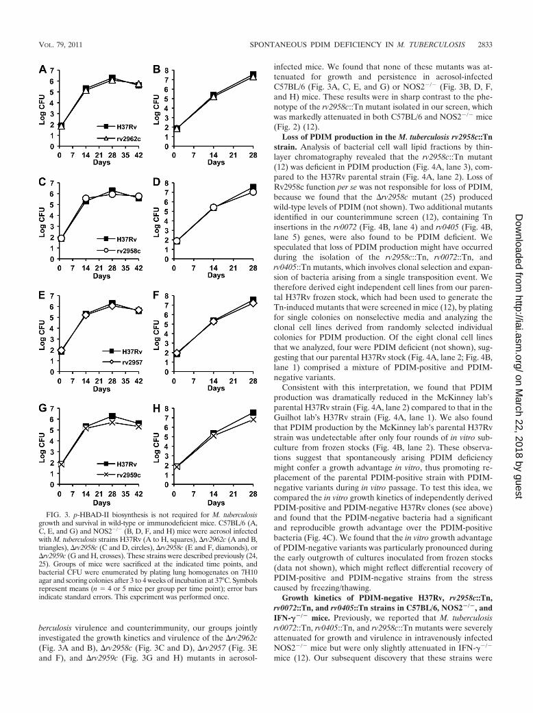
berculosis virulence and counterimmunity, our groups jointlyinvestigated the growth kinetics and virulence of the �rv2962c(Fig. 3A and B), �rv2958c (Fig. 3C and D), �rv2957 (Fig. 3Eand F), and �rv2959c (Fig. 3G and H) mutants in aerosol-
infected mice. We found that none of these mutants was at-tenuated for growth and persistence in aerosol-infectedC57BL/6 (Fig. 3A, C, E, and G) or NOS2�/� (Fig. 3B, D, F,and H) mice. These results were in sharp contrast to the phe-notype of the rv2958c::Tn mutant isolated in our screen, whichwas markedly attenuated in both C57BL/6 and NOS2�/� mice(Fig. 2) (12).
Loss of PDIM production in the M. tuberculosis rv2958c::Tnstrain. Analysis of bacterial cell wall lipid fractions by thin-layer chromatography revealed that the rv2958c::Tn mutant(12) was deficient in PDIM production (Fig. 4A, lane 3), com-pared to the H37Rv parental strain (Fig. 4A, lane 2). Loss ofRv2958c function per se was not responsible for loss of PDIM,because we found that the �rv2958c mutant (25) producedwild-type levels of PDIM (not shown). Two additional mutantsidentified in our counterimmune screen (12), containing Tninsertions in the rv0072 (Fig. 4B, lane 4) and rv0405 (Fig. 4B,lane 5) genes, were also found to be PDIM deficient. Wespeculated that loss of PDIM production might have occurredduring the isolation of the rv2958c::Tn, rv0072::Tn, andrv0405::Tn mutants, which involves clonal selection and expan-sion of bacteria arising from a single transposition event. Wetherefore derived eight independent cell lines from our paren-tal H37Rv frozen stock, which had been used to generate theTn-induced mutants that were screened in mice (12), by platingfor single colonies on nonselective media and analyzing theclonal cell lines derived from randomly selected individualcolonies for PDIM production. Of the eight clonal cell linesthat we analyzed, four were PDIM deficient (not shown), sug-gesting that our parental H37Rv stock (Fig. 4A, lane 2; Fig. 4B,lane 1) comprised a mixture of PDIM-positive and PDIM-negative variants.
Consistent with this interpretation, we found that PDIMproduction was dramatically reduced in the McKinney lab’sparental H37Rv strain (Fig. 4A, lane 2) compared to that in theGuilhot lab’s H37Rv strain (Fig. 4A, lane 1). We also foundthat PDIM production by the McKinney lab’s parental H37Rvstrain was undetectable after only four rounds of in vitro sub-culture from frozen stocks (Fig. 4B, lane 2). These observa-tions suggest that spontaneously arising PDIM deficiencymight confer a growth advantage in vitro, thus promoting re-placement of the parental PDIM-positive strain with PDIM-negative variants during in vitro passage. To test this idea, wecompared the in vitro growth kinetics of independently derivedPDIM-positive and PDIM-negative H37Rv clones (see above)and found that the PDIM-negative bacteria had a significantand reproducible growth advantage over the PDIM-positivebacteria (Fig. 4C). We found that the in vitro growth advantageof PDIM-negative variants was particularly pronounced duringthe early outgrowth of cultures inoculated from frozen stocks(data not shown), which might reflect differential recovery ofPDIM-positive and PDIM-negative strains from the stresscaused by freezing/thawing.
Growth kinetics of PDIM-negative H37Rv, rv2958c::Tn,rv0072::Tn, and rv0405::Tn strains in C57BL/6, NOS2�/�, andIFN-��/� mice. Previously, we reported that M. tuberculosisrv0072::Tn, rv0405::Tn, and rv2958c::Tn mutants were severelyattenuated for growth and virulence in intravenously infectedNOS2�/� mice but were only slightly attenuated in IFN-��/�
mice (12). Our subsequent discovery that these strains were
FIG. 3. p-HBAD-II biosynthesis is not required for M. tuberculosisgrowth and survival in wild-type or immunodeficient mice. C57BL/6 (A,C, E, and G) and NOS2�/� (B, D, F, and H) mice were aerosol infectedwith M. tuberculosis strains H37Rv (A to H, squares), �rv2962c (A and B,triangles), �rv2958c (C and D, circles), �rv2958c (E and F, diamonds), or�rv2959c (G and H, crosses). These strains were described previously (24,25). Groups of mice were sacrificed at the indicated time points, andbacterial CFU were enumerated by plating lung homogenates on 7H10agar and scoring colonies after 3 to 4 weeks of incubation at 37°C. Symbolsrepresent means (n � 4 or 5 mice per group per time point); error barsindicate standard errors. This experiment was performed once.
VOL. 79, 2011 SPONTANEOUS PDIM DEFICIENCY IN M. TUBERCULOSIS 2833
on March 22, 2018 by guest
http://iai.asm.org/
Dow
nloaded from

PDIM deficient (Fig. 4A), presumably due to selection of pre-existing PDIM-negative variants in our parental H37Rv stockduring Tn mutagenesis, suggested that the attenuation of thesemutants in mice might be due, at least in part, to their PDIMdeficiency. To test this idea, we compared the growth kineticsof these mutants with a PDIM-negative H37Rv clone in aero-sol-infected mice (Fig. 5). We found that the rv0072::Tn (Fig.5A to C), rv0405::Tn (Fig. 5D to F), and rv2958c::Tn (Fig. 5Gto I) mutants grew with kinetics similar to those of the PDIM-negative H37Rv clone in C57BL/6 (Fig. 5A, D, and G),NOS2�/� (Fig. 5B, E, and H), and IFN-��/� (Fig. 5C, F, andI) mice. These observations suggest that the in vivo phenotypeswe reported previously for these mutants (12) were probablydue to the spontaneous loss of PDIM production in thesestrains, rather than disruption of the rv2958c, rv0072, or rv0405genes per se.
Growth kinetics of PDIM-positive and PDIM-negativeclones of H37Rv in C57BL/6, NOS2�/�, and IFN-��/� mice.To further test the idea that PDIM deficiency might contributeto the in vivo attenuation of our rv2958c::Tn, rv0072::Tn, andrv0405::Tn mutants, we compared the growth kinetics ofPDIM-positive and PDIM-negative clones of H37Rv in aero-sol-infected mice. We found that, compared to the PDIM-positive H37Rv clone, the PDIM-negative H37Rv clone wasmarkedly attenuated for growth in C57BL/6 (Fig. 6A),NOS2�/� (Fig. 6B), and IFN-��/� (Fig. 6C) mice. Virulence ofthe PDIM-negative H37Rv clone, measured in terms of sur-vival time of infected mice, was also attenuated in NOS2�/�
mice (P � 0.0005) and, to a lesser extent, in IFN-��/� mice(P � 0.002) (Fig. 6D). Median survival time (MST) ofNOS2�/� mice was �200 days after infection with PDIM-negative H37Rv, compared to 64 days postinfection forNOS2�/� mice infected with PDIM-positive H37Rv (Fig. 6D).The MST of IFN-��/� mice was also longer for mice infectedwith PDIM-negative H37Rv (MST, 82 days) compared to mice
infected with PDIM-positive H37Rv (MST, 59 days) (Fig. 6D).These data strongly suggest that the in vivo attenuation of the“counterimmune” mutants described in our previous report(12) can be attributed to the fortuitous loss of PDIM produc-tion in these strains.
Whole-genome resequencing of PDIM-negative H37Rv iden-tifies a point mutation, ppsD(G44C), responsible for PDIMdeficiency. Since our results demonstrated a correlation be-tween spontaneous loss of the ability to produce PDIM andattenuation in the mouse model of infection, we sought toidentify the mutation responsible for PDIM deficiency in aPDIM-negative clone. Analysis of the PDIM biosynthesis locus(Fig. 1) by Southern blotting did not identify any major inser-tions or genetic rearrangements (data not shown), suggestingthat a point mutation might be the cause of PDIM deficiency.To identify putative point mutations that could contribute toPDIM deficiency, we performed whole-genome resequencingof PDIM-positive and PDIM-negative H37Rv clones using anIllumina genome analyzer platform. In comparison to theH37Rv reference sequence, our PDIM-positive H37Rv clonehad 161 putative SNPs and 15 putative small sequence inser-tions or deletions (indels). The PDIM-negative H37Rv clonehad 151 putative SNPs and 15 putative indels. These sequencealterations included 72 polymorphisms (57 SNPs and 15 indels)that were recently identified by whole-genome resequencing ofsix H37Rv isolates and that are likely to be errors within theH37Rv reference sequence (14). Analysis of the remainingsequence polymorphisms, excluding those in the highly repet-itive GC-rich PE_PGRS coding regions, revealed 22 putativeSNPs unique to the PDIM-negative H37Rv clone.
Among the SNPs present only in the PDIM-negative H37Rvisolate were two mutations in genes required for PDIM bio-synthesis: a putative A to C at position 2544 in the ppsA geneand a G to T at position 130 in the ppsD gene. Only the G toT in the ppsD gene was predicted to be nonsynonymous, gen-
FIG. 4. PDIM deficiency confers an in vitro growth advantage in M. tuberculosis H37Rv. (A and B) Thin-layer chromatographic analysis ofPDIM biosynthesis. Bacteria were labeled with [14C]propionate, which preferentially labels PDIM (6), and cell wall lipids were extracted andseparated by thin-layer chromatography. (A) M. tuberculosis strains. H37Rv, Guilhot lab (25) (lane 1); H37Rv, McKinney lab (12) (lane 2);rv2958c::Tn (12) (lane 3). (B) M. tuberculosis strains, McKinney lab (12). H37Rv (lane 1), H37Rv after subculture (lane 2), rv2958c::Tn (lane 3),rv0072::Tn (lane 4), rv0405::Tn (lane 5). (C) Independently derived subclones of PDIM-positive H37Rv (squares) and PDIM-negative H37Rv(circles) were grown in 7H9 broth with aeration at 37°C. Growth of the cultures was monitored by withdrawing aliquots and measuring the OD600at the indicated time points (plotted on the primary y axis). The (PDIM-positive OD600)/(PDIM-negative OD600) ratios at each time point areplotted on the secondary y axis (diamonds). Results are representative of three independent experiments.
2834 KIRKSEY ET AL. INFECT. IMMUN.
on March 22, 2018 by guest
http://iai.asm.org/
Dow
nloaded from

erating a glycine-to-cysteine transition at position 44 in thePpsD protein. PpsD is a modular polyketide synthase, and theG44C point mutation identified in our PDIM-negative clone islocated within the putative �-ketoacyl acyl carrier protein syn-thase enzymatic domain (34). We reasoned that the G44Cmutation might interfere with the activity of PpsD, since anadditional cysteine residue could cause formation of aberrantdisulfide bonds, thereby preventing proper protein folding orinterfering with the function of the active site cysteine residue.We confirmed that the ppsD(G44C) point mutation wasunique to the PDIM-negative clone by PCR and sequencing.We were not able to confirm the point mutation in the ppsAgene by sequencing of a PCR product, suggesting that thereare likely to be some false positives among the SNPs identifiedby whole-genome resequencing.
We attempted to restore PDIM production to the PDIM-negative H37Rv ppsD(G44C) mutant using two approaches.We complemented the ppsD(G44C) mutation in trans by pro-
viding a wild-type copy of the ppsD gene under the control ofa strong constitutive promoter on the vector pMV361, whichintegrates in the M. tuberculosis chromosome at the uniqueattB site. We also reverted the ppsD(G44C) mutation in thePDIM-negative clone to the wild-type ppsD sequence by atwo-step homologous recombination method. Both strainswere tested for the ability to produce PDIM in vitro by thin-layer chromatography of extractable cell wall lipids. Produc-tion of PDIM was restored by either complementation or re-version (ppsD rev) of the ppsD(G44C) point mutation (Fig.7A), confirming that this spontaneous mutation is responsiblefor the deficiency in PDIM production of our PDIM-negativeH37Rv clone.
The ppsD(G44C) point mutation enhances growth of H37Rvin vitro. Since the ppsD(G44C) spontaneous point mutationwas responsible for PDIM deficiency, we sought to establishwhether the in vitro and in vivo growth characteristics of ourPDIM-negative H37Rv clone could also be attributed to this
FIG. 5. Growth kinetics of PDIM-negative H37Rv and M. tuberculosis rv0072::Tn, rv0405::Tn, and rv2958c::Tn mutants in wild-type andimmunodeficient mice. C57BL/6 (A, D, and G), NOS2�/� (B, E, and H), and IFN-��/� (C, F, and I) mice were aerosol infected with M. tuberculosisH37Rv PDIM-negative variant (A to I, squares), rv0072::Tn (A to C, triangles), rv0405::Tn (D to F, diamonds), or rv2958c::Tn (G to I, circles).The PDIM-negative variant of H37Rv is described herein; the Tn mutant strains were described previously (12). Groups of mice were sacrificedat the indicated time points, and bacterial CFU were enumerated by plating lung homogenates on 7H10 agar and scoring colonies after 3 to 4 weeksof incubation at 37°C. Symbols represent means (n � 4 or 5 mice per group per time point); error bars indicate standard errors. This experimentwas performed once.
VOL. 79, 2011 SPONTANEOUS PDIM DEFICIENCY IN M. TUBERCULOSIS 2835
on March 22, 2018 by guest
http://iai.asm.org/
Dow
nloaded from
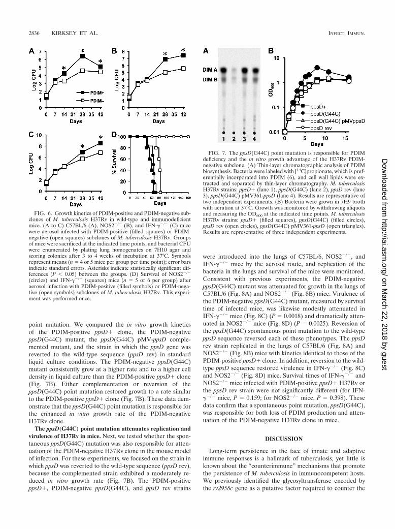
point mutation. We compared the in vitro growth kineticsof the PDIM-positive ppsD clone, the PDIM-negativeppsD(G44C) mutant, the ppsD(G44C) pMV-ppsD comple-mented mutant, and the strain in which the ppsD gene wasreverted to the wild-type sequence (ppsD rev) in standardliquid culture conditions. The PDIM-negative ppsD(G44C)mutant consistently grew at a higher rate and to a higher celldensity in liquid culture than the PDIM-positive ppsD clone(Fig. 7B). Either complementation or reversion of theppsD(G44C) point mutation restored growth to a rate similarto the PDIM-positive ppsD clone (Fig. 7B). These data dem-onstrate that the ppsD(G44C) point mutation is responsible forthe enhanced in vitro growth rate of the PDIM-negativeH37Rv clone.
The ppsD(G44C) point mutation attenuates replication andvirulence of H37Rv in mice. Next, we tested whether the spon-taneous ppsD(G44C) mutation was also responsible for atten-uation of the PDIM-negative H37Rv clone in the mouse modelof infection. For these experiments, we focused on the strain inwhich ppsD was reverted to the wild-type sequence (ppsD rev),because the complemented strain exhibited a moderately re-duced in vitro growth rate (Fig. 7B). The PDIM-positiveppsD , PDIM-negative ppsD(G44C), and ppsD rev strains
were introduced into the lungs of C57BL/6, NOS2�/�, andIFN-��/� mice by the aerosol route, and replication of thebacteria in the lungs and survival of the mice were monitored.Consistent with previous experiments, the PDIM-negativeppsD(G44C) mutant was attenuated for growth in the lungs ofC57BL/6 (Fig. 8A) and NOS2�/� (Fig. 8B) mice. Virulence ofthe PDIM-negative ppsD(G44C) mutant, measured by survivaltime of infected mice, was likewise modestly attenuated inIFN-��/� mice (Fig. 8C) (P � 0.0018) and dramatically atten-uated in NOS2�/� mice (Fig. 8D) (P � 0.0025). Reversion ofthe ppsD(G44C) spontaneous point mutation to the wild-typeppsD sequence reversed each of these phenotypes. The ppsDrev strain replicated in the lungs of C57BL/6 (Fig. 8A) andNOS2�/� (Fig. 8B) mice with kinetics identical to those of thePDIM-positive ppsD clone. In addition, reversion to the wild-type ppsD sequence restored virulence in IFN-��/� (Fig. 8C)and NOS2�/� (Fig. 8D) mice. Survival times of IFN-��/� andNOS2�/� mice infected with PDIM-positive ppsD H37Rv orthe ppsD rev strain were not significantly different (for IFN-��/� mice, P � 0.159; for NOS2�/� mice, P � 0.398). Thesedata confirm that a spontaneous point mutation, ppsD(G44C),was responsible for both loss of PDIM production and atten-uation of the PDIM-negative H37Rv clone in mice.
DISCUSSION
Long-term persistence in the face of innate and adaptiveimmune responses is a hallmark of tuberculosis, yet little isknown about the “counterimmune” mechanisms that promotethe persistence of M. tuberculosis in immunocompetent hosts.We previously identified the glycosyltransferase encoded bythe rv2958c gene as a putative factor required to counter the
FIG. 6. Growth kinetics of PDIM-positive and PDIM-negative sub-clones of M. tuberculosis H37Rv in wild-type and immunodeficientmice. (A to C) C57BL/6 (A), NOS2�/� (B), and IFN-��/� (C) micewere aerosol-infected with PDIM-positive (filled squares) or PDIM-negative (open squares) subclones of M. tuberculosis H37Rv. Groupsof mice were sacrificed at the indicated time points, and bacterial CFUwere enumerated by plating lung homogenates on 7H10 agar andscoring colonies after 3 to 4 weeks of incubation at 37°C. Symbolsrepresent means (n � 4 or 5 mice per group per time point); error barsindicate standard errors. Asterisks indicate statistically significant dif-ferences (P 0.05) between the groups. (D) Survival of NOS2�/�
(circles) and IFN-��/� (squares) mice (n � 5 or 6 per group) afteraerosol infection with PDIM-positive (filled symbols) or PDIM-nega-tive (open symbols) subclones of M. tuberculosis H37Rv. This experi-ment was performed once.
FIG. 7. The ppsD(G44C) point mutation is responsible for PDIMdeficiency and the in vitro growth advantage of the H37Rv PDIM-negative subclone. (A) Thin-layer chromatographic analysis of PDIMbiosynthesis. Bacteria were labeled with [14C]propionate, which is pref-erentially incorporated into PDIM (6), and cell wall lipids were ex-tracted and separated by thin-layer chromatography. M. tuberculosisH37Rv strains: ppsD (lane 1), ppsD(G44C) (lane 2), ppsD rev (lane3), ppsD(G44C) pMV361-ppsD (lane 4). Results are representative oftwo independent experiments. (B) Bacteria were grown in 7H9 brothwith aeration at 37°C. Growth was monitored by withdrawing aliquotsand measuring the OD600 at the indicated time points. M. tuberculosisH37Rv strains: ppsD (filled squares), ppsD(G44C) (filled circles),ppsD rev (open circles), ppsD(G44C) pMV361-ppsD (open triangles).Results are representative of three independent experiments.
2836 KIRKSEY ET AL. INFECT. IMMUN.
on March 22, 2018 by guest
http://iai.asm.org/
Dow
nloaded from

impact of IFN-�-dependent immune mechanisms other thanNOS2 (12). Here, we tested mutants harboring targeted dele-tions of genes that are required for biosynthesis of secretedp-HBAD, including the rv2958c gene, for their ability to rep-licate in the lungs of mice. Although none of the �rv2957,�rv2958c, �rv2959c, or �rv2962c mutants were attenuated forgrowth, it remains possible that secreted p-HBADs play a rolein virulence modulation that is not reflected in the bacterialgrowth kinetics, as shown previously for PGL (26).
We attribute the discrepancy between the phenotypes of therv2958c::Tn and �rv2958c mutants in the mouse infectionmodel to the spontaneous loss of PDIM production in ourH37Rv parental strain and the rv2958c::Tn mutant derivedfrom it. Our observations demonstrate a role for PDIM incountering the impact of an IFN-�-dependent, NOS2-indepen-dent immune mechanism, in addition to the previously postu-lated role for PDIM in protecting the bacteria from the cidalactivity of RNS (28). Consistent with this idea, we found thatthe survival time of IFN-��/� and NOS2�/� mice was notsignificantly different (P � 0.07) after infection with the PDIM-positive H37Rv clone. In contrast, following infection with thePDIM-negative H37Rv clone, NOS2�/� mice survived signif-icantly longer than IFN-��/� mice (P 0.001). Our results arealso consistent with the results of a recent screen for mutantsthat specifically alter growth of M. tuberculosis in NOS2�/�
mice, which identified two independent mutations in the drrAgene encoding a putative PDIM transporter (22). The immunemechanism responsible for this IFN-�-dependent, NOS2-inde-pendent attenuation and the countermechanism by whichPDIM confers protection are currently unknown.
We demonstrated that a point mutation, ppsD(G44C), in aPDIM-negative clone derived from our H37Rv parental strainwas responsible for both a defect in PDIM production andattenuation in mice. To our knowledge, this is the first directevidence that PpsD is required for PDIM biosynthesis andvirulence. We consistently observed a low level of res-idual PDIM production by the ppsD(G44C) mutant. TheppsD(G44C) mutant was a clonal isolate from a single colony,suggesting that weak PDIM production is a property of thisstrain. It is possible that reversion of the G44C point mutationor compensatory mutations elsewhere in ppsD that restorePDIM synthesis occur spontaneously at some low frequency. Itis unlikely that such strains would become a significant fractionof the population, however, since the ppsD(G44C) mutantgrows at a higher rate. We therefore favor the possibility thatthe PpsDG44C mutant protein retains some residual activitythat enables weak PDIM production. The low level of PDIMproduced by the ppsD(G44C) mutant is apparently not suffi-cient, however, to support normal replication in the lungs ofmice.
A correlation was previously observed between PDIM defi-ciency and a growth advantage in liquid culture (7). Similarly,our PDIM-negative ppsD(G44C) mutant had an enhanced invitro growth rate and grew to a higher cell density than PDIM-positive clones. The in vitro growth advantage of such sponta-neous PDIM-negative variants could explain why they are ableto supplant the parental strain during repeated cycles ofgrowth in vitro, for example, during mutant strain construction.Indeed PDIM-negative variants were isolated with significantlyhigher frequency in a culture that was serially passaged than ina nonpassaged control (7). It has also been suggested thatspontaneous PDIM deficient variants might be selected bygenetic manipulations involving electroporation or bacterio-phage infections (11, 26). Since our results indicate that thesespontaneous PDIM-deficient variants are attenuated for viru-lence, care should be taken to minimize the number of pas-sages during genetic manipulation of M. tuberculosis and toensure that strains used in animal infection studies are PDIMproficient.
Spontaneously arising PDIM-deficient variants have beendescribed previously (1, 7, 8, 11, 15, 19–21) and are probablymore common than has been reported or realized. Althoughspontaneous PDIM deficiency has most frequently been asso-ciated with the H37Rv strain, it has also recently been de-scribed for the M. tuberculosis Erdman strain and the clinicalisolate HN878 (7, 20). Many of the attenuated M. tuberculosismutants described in the scientific literature have not beencomplemented, and in other cases, complementation restoredvirulence only partially. Some of these mutants might haveacquired unrecognized secondary mutations causing PDIM de-ficiency, which could be a factor contributing to their attenu-ation. Moreover, a number of genes have been implicated inPDIM synthesis, based on the PDIM-negative phenotypes ofthe corresponding mutants, in the absence of confirmatorycomplementation (for example, see references 13, 27, 28, 30,
FIG. 8. Reversion of the ppsD(G44C) point mutation restores wild-type levels of growth and virulence in mice. (A to D) Mice were aerosolinfected with ppsD (filled squares), ppsD(G44C) (filled circles), orppsD rev (open circles) strains of M. tuberculosis H37Rv. (A and B)Bacterial growth in the lungs of C57BL/6 (A) and NOS2�/� (B) mice.Groups of mice were sacrificed at the indicated time points, and bac-terial CFU were enumerated by plating lung homogenates on 7H10and scoring colonies after 3 to 4 weeks of incubation at 37°C. Symbolsrepresent means (n � 4 mice per group); error bars indicate standarderrors. Asterisks indicate statistically significant differences (P 0.05)in comparisons of ppsD(G44C) versus ppsD and ppsD(G44C) versusppsD rev strains. This experiment was performed once. (C and D)Survival of IFN-��/� (C) and iNOS�/� (D) mice (n � 5 mice pergroup). This experiment was performed once.
VOL. 79, 2011 SPONTANEOUS PDIM DEFICIENCY IN M. TUBERCULOSIS 2837
on March 22, 2018 by guest
http://iai.asm.org/
Dow
nloaded from

31, and 36). Some of these mutant strains might contain un-recognized secondary mutations that are the true cause of theirPDIM deficiency. This phenomenon might also explain theobservation that Mycobacterium leprae produces PDIM despitelacking functional copies of certain polyketide biosynthesisgenes that were reported to be essential for PDIM productionin M. tuberculosis (discussed in reference 36).
Construction and characterization of random or targetedgene-disrupted mutants is a powerful technique to assign bio-logical functions to biochemical pathways in mycobacteria.However, our findings underscore the idea that any functionalassignment must be tentative in the absence of complementa-tion analysis, even in cases where the mutation is not polar onthe expression of neighboring genes.
ACKNOWLEDGMENTS
We thank Peter Giannakis and Laetitia Martin for expert technicalassistance with animal experiments and Keith Harshman and JeromeThomas at the University of Lausanne core facility for performing thewhole-genome resequencing reactions.
This work was supported by a Robert D. Watkins Graduate Fellow-ship from the American Society for Microbiology (M.A.K.), the Wil-liam Randolph Hearst Endowed Scholarship Fund (K.B.H.), NIHMSTP grant GM07739 (M.A.K. and K.B.H.) for the Tri-InstitutionalMD/PhD Program of Weill-Cornell Medical School, Rockefeller Uni-versity, and the Sloan-Kettering Institute, an Irvington Institute Post-doctoral Fellowship of the Cancer Research Institute (A.D.T.), Sys-temsX, The Swiss Initiative in Systems Biology (S.U.), and NationalInstitutes of Health grant AI046392 (J.D.M.).
REFERENCES
1. Andreu, N., and I. Gibert. 2008. Cell population heterogeneity in Mycobac-terium tuberculosis H37Rv. Tuberculosis (Edinb.) 88:553–559.
2. Astarie-Dequeker, C., et al. 2009. Phthiocerol dimycocerosates of M. tuber-culosis participate in macrophage invasion by inducing changes in the orga-nization of plasma membrane lipids. PLoS Pathog. 5:e1000289.
3. Camacho, L. R., et al. 2001. Analysis of the phthiocerol dimycocerosate locusof Mycobacterium tuberculosis: evidence that this lipid is involved in the cellwall permeability barrier. J. Biol. Chem. 276:19845–19854.
4. Camacho, L. R., D. Ensergueix, E. Perez, B. Gicquel, and C. Guilhot. 1999.Identification of a virulence gene cluster of Mycobacterium tuberculosis bysignature-tagged transposon mutagenesis. Mol. Microbiol. 34:257–267.
5. Constant, P., et al. 2002. Role of the pks15/1 gene in the biosynthesis ofphenolglycolipids in the Mycobacterium tuberculosis complex: evidence thatall strains synthesize glycosylated p-hydroxybenzoic acid methyl esters andthat strains devoid of phenolglycolipids harbor a frameshift mutation in thepks15/1 gene. J. Biol. Chem. 277:38148–38158.
6. Cox, J. S., B. Chen, M. McNeil, and W. R. Jacobs, Jr. 1999. Complex lipiddetermines tissue-specific replication of Mycobacterium tuberculosis in mice.Nature 402:79–83.
7. Domenech, P., and M. B. Reed. 2009. Rapid and spontaneous loss of ph-thiocerol dimycocerosate (PDIM) from Mycobacterium tuberculosis grown invitro: implications for virulence studies. Microbiol. 155:3532–3543.
8. Domenech, P., et al. 2004. The role of MmpL8 in sulfatide biogenesis andvirulence of Mycobacterium tuberculosis. J. Biol. Chem. 279:21257–21265.
9. Ehrt, S., et al. 2001. Reprogramming of the macrophage transcriptome inresponse to interferon-� and Mycobacterium tuberculosis: signaling roles ofnitric oxide synthase-2 and phagocyte oxidase. J. Exp. Med. 194:1123–1140.
10. Gagneux, S., et al. 2006. Variable host-pathogen compatibility in Mycobac-terium tuberculosis. Proc. Natl. Acad. Sci. U. S. A. 103:2869–2873.
11. Goren, M. B., O. Brokl, and W. B. Schaefer. 1974. Lipids of putative rele-vance to virulence in Mycobacterium tuberculosis: phthiocerol dimycocerosateand the attenuation indicator lipid. Infect. Immun. 9:150–158.
12. Hisert, K. B., et al. 2004. Identification of Mycobacterium tuberculosis coun-terimmune (cim) mutants in immunodeficient mice by differential screening.Infect. Immun. 72:5315–5321.
13. Hotter, G. S., et al. 2005. Transposon mutagenesis of Mb0100 at the ppe1-nrplocus in Mycobacterium bovis disrupts phthiocerol dimycocerosate (PDIM)
and glycosylphenol-PDIM biosynthesis, producing an avirulent strain withvaccine properties at least equal to those of M. bovis BCG. J. Bacteriol.187:2267–2277.
14. Ioerger, T. R., et al. 2010. Variation among genome sequences of H37Rvstrains of Mycobacterium tuberculosis from multiple laboratories. J. Bacteriol.192:3645–3653.
15. Kana, B. D., et al. 2008. The resuscitation-promoting factors of Mycobacte-rium tuberculosis are required for virulence and resuscitation from dormancybut are collectively dispensable for growth in vitro. Mol. Microbiol. 67:672–684.
16. Kondo, E., and K. Kanai. 1976. A suggested role of a host-parasite lipidcomplex in mycobacterial infection. Jpn. J. Med. Sci. Biol. 29:199–201.
17. Larsen, M. H., K. Biermann, S. Tandberg, T. Hsu, and W. R. J. Jacobs. 2007.Genetic manipulation of Mycobacterium tuberculosis. Curr. Protoc. Micro-biol. Chapter 10:Unit 10A.2.
18. Li, H., J. Ruan, and R. Durbin. 2008. Mapping short DNA sequencing readsand calling variants using mapping quality scores. Genome Res. 18:1851–1858.
19. Manjunatha, U. H., et al. 2006. Identification of a nitroimidazo-oxazine-specific protein involved in PA-824 resistance in Mycobacterium tuberculosis.Proc. Natl. Acad. Sci., U. S. A. 103:431–436.
20. Marrero, J., K. Y. Rhee, D. Schnappinger, K. Pethe, and S. Ehrt. 2010.Gluconeogenic carbon flow of tricarboxylic acid cycle intermediates is criticalfor Mycobacterium tuberculosis to establish and maintain infection. Proc.Natl. Acad. Sci., U. S. A. 107:9819–9824.
21. Matsunaga, I., et al. 2004. Mycobacterium tuberculosis pks12 produces anovel polyketide presented by CD1c to T cells. J. Exp. Med. 200:1559–1569.
22. Murry, J. P., A. K. Pandey, C. M. Sassetti, and E. J. Rubin. 2009. Phthioceroldimycocerosate transport is required for resisting interferon-�-independentimmunity. J. Infect. Dis. 200:774–782.
23. Onwueme, K. C., C. J. Vos, J. Zurita, J. A. Ferreras, and L. E. Quadri. 2005.The dimycocerosate ester polyketide virulence factors of mycobacteria. Prog.Lipid Res. 44:259–302.
24. Perez, E., et al. 2004. Molecular dissection of the role of two methyltrans-ferases in the biosynthesis of phenolglycolipids and phthiocerol dimycosero-sate in the Mycobacterium tuberculosis complex. J. Biol. Chem. 279:42584–42592.
25. Perez, E., et al. 2004. Characterization of three glycosyltransferases involvedin the biosynthesis of the phenolic glycolipid antigens from the Mycobacte-rium tuberculosis complex. J. Biol. Chem. 279:42574–42583.
26. Reed, M. B., et al. 2004. A glycolipid of hypervirulent tuberculosis strainsthat inhibits the innate immune response. Nature 431:84–87.
27. Rousseau, C., et al. 2003. Virulence attenuation of two Mas-like polyketidesynthase mutants of Mycobacterium tuberculosis. Microbiology 149:1837–1847.
28. Rousseau, C., et al. 2004. Production of phthiocerol dimycocerosates pro-tects Mycobacterium tuberculosis from the cidal activity of reactive nitrogenintermediates produced by macrophages and modulates the early immuneresponse to infection. Cell. Microbiol. 6:277–287.
29. Schnappinger, D., et al. 2003. Transcriptional adaptation of Mycobacteriumtuberculosis within macrophages: insights into the phagosomal environment.J. Exp. Med. 198:693–704.
30. Sirakova, T. D., V. S. Dubey, M. H. Cynamon, and P. E. Kolattukudy. 2003.Attenuation of Mycobacterium tuberculosis by disruption of a mas-like geneor a chalcone synthase-like gene, which causes deficiency in dimycocerosylphthiocerol synthesis. J. Bacteriol. 185:2999–3008.
31. Sirakova, T. D., V. S. Dubey, H.-J. Kim, M. H. Cynamon, and P. E. Kolat-tukudy. 2003. The largest open reading frame (pks12) in the Mycobacteriumtuberculosis genome is involved in pathogenesis and dimycocerosyl phthioc-erol synthesis. Infect. Immun. 71:3794–3801.
32. Slayden, R. A., and C. E. I. Barry. 2001. Analysis of the lipids of Mycobac-terium tuberculosis, p. 229–245. In T. Parish and N. G. Stoker (ed.), Myco-bacterium tuberculosis protocols. Humana Press, Totowa, NJ.
33. Stover, C. K., et al. 1991. New use of BCG for recombinant vaccines. Nature351:456–460.
34. Trivedi, O. A., et al. 2005. Dissecting the mechanism and assembly of acomplex virulence mycobacterial lipid. Mol. Cell 17:631–643.
35. Voskuil, M. I., et al. 2003. Inhibition of respiration by nitric oxide induces aMycobacterium tuberculosis dormancy program. J. Exp. Med. 198:705–713.
36. Waddell, S. J., et al. 2005. Inactivation of polyketide synthase and relatedgenes results in the loss of complex lipids in Mycobacterium tuberculosisH37Rv. Lett. Appl. Microbiol. 40:201–206.
37. Wiegeshaus, E. H., D. N. McMurray, A. A. Grover, G. E. Harding, and D. W.Smith. 1970. Host-parasite relationships in experimental airborne tubercu-losis. III. Relevance of microbial enumeration to acquired resistance inguinea pigs. Am. Rev. Respir. Dis. 102:422–429.
Editor: J. L. Flynn
2838 KIRKSEY ET AL. INFECT. IMMUN.
on March 22, 2018 by guest
http://iai.asm.org/
Dow
nloaded from