Embed Size (px)
Citation preview
Spontaneous Chronic Meningo-Encephalitis of RabbitsAuthor(s): Jean OliverSource: The Journal of Infectious Diseases, Vol. 30, No. 1 (Jan., 1922), pp. 91-94Published by: Oxford University PressStable URL: http://www.jstor.org/stable/30080534 .
Accessed: 22/05/2014 10:37
Your use of the JSTOR archive indicates your acceptance of the Terms & Conditions of Use, available at .http://www.jstor.org/page/info/about/policies/terms.jsp
.JSTOR is a not-for-profit service that helps scholars, researchers, and students discover, use, and build upon a wide range ofcontent in a trusted digital archive. We use information technology and tools to increase productivity and facilitate new formsof scholarship. For more information about JSTOR, please contact [email protected].
.
Oxford University Press is collaborating with JSTOR to digitize, preserve and extend access to The Journal ofInfectious Diseases.
http://www.jstor.org
This content downloaded from 195.78.109.34 on Thu, 22 May 2014 10:37:01 AMAll use subject to JSTOR Terms and Conditions
SPONTANEOUS CHRONIC MENINGO-ENCEPHALITIS OF RABBITS
Jean Oliver
From the Pathological Laboratory, School of Medicine, Stanford University, San Francisco, Calif.
During the course of the last year we have been studying reactions which follow arsphenamin administration to rabbits. After each experi- ment, tissues were taken from all the organs as a routine measure and examined for any lesions that might be present.
In the first group of experiments, animals were given repeated large doses of arsphenamin extending over a period of 10 days, and, as a rule, such animals died from the effects of this procedure. Lesions of various organs were found and especially a peculiar inflammatory process in the brain. A later group of experiments in which the animals died immediately or in the course of a few hours
following the injection of massive doses of arsphenamin, also showed these same lesions of the central nervous system, and as in these cases sufficient time had not elapsed for the development of changes of this
type, it seemed fairly certain that the lesions must be due to a disease
spontaneously occurring in the rabbits. Supposedly normal animals from our stock-room were therefore killed and their brains examined. Brains were also obtained from the public markets. In both instances about 20*^ of the animals showed the same lesions as had been encoun- tered in the experimental animals.
No gross lesions of the meninges or in frontal sections of the brain could be made out. Microscopic examination, however, showed a
widespread inflammatory process most frequently in the cerebral cortex, but also occurring in the neighborhood of the basal ganglions and the medulla.
The pia in all cases showed a varying degree of infiltration with "round cells." This consisted in most part of lymphocytes, though a few plasma cells were occasionally found. The process was often most marked in the cerebral sulci and in such places extended to the depths of the fissures. An extension into the substance of the cerebral cortex was also seen around the small vessels of the pia, which penetrate it. Here a "jacket" of lymphocytes, often of considerable thickness, was
Received for publication Aug. 29, 1921.
This content downloaded from 195.78.109.34 on Thu, 22 May 2014 10:37:01 AMAll use subject to JSTOR Terms and Conditions
92 Jean Oliver
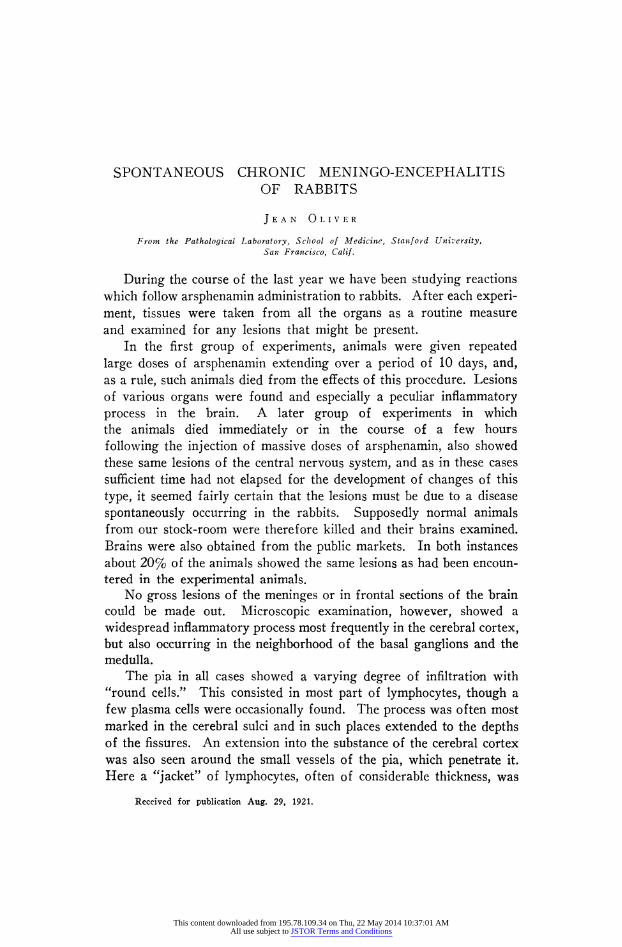
commonly found (fig. 1). Similar perivascular infiltrations were found
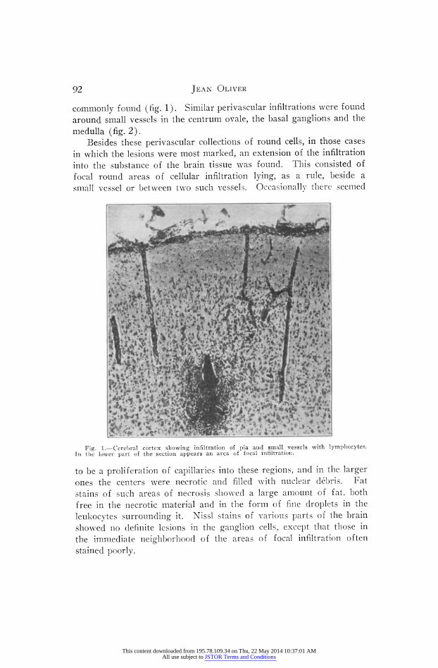
around small vessels in the centrum ovale, the basal ganglions and the medulla (fig. 2). ll ih
Besides these perivascular collections of round cells, in those cases in which the lesions were most marked, an extension of the infiltration into the substance of the brain tissue was found. This consisted of focal round areas of cellular infiltration lying, as a rule, beside a small vessel or between two such vessels. Occasionallv there seemed
Fig. 1.?Cerebral cortex showing infiltration of pia and small vessels with lymphocytes. In the lower part of the section appears an area of focal infiltration.
to be a proliferation of capillaries into these regions, and in the larger ones the centers were necrotic and filled with nuclear debris. Fat stains of such areas of necrosis showed a large amount of fat, both free in the necrotic material and in the form of fine droplets in the
leukocytes surrounding it. Nissl stains of various parts of the brain showed no definite lesions in the ganglion cells, except that those in the immediate neighborhood of the areas of focal infiltration often stained poorly.
This content downloaded from 195.78.109.34 on Thu, 22 May 2014 10:37:01 AMAll use subject to JSTOR Terms and Conditions
Meningo-Encephalitis of Rabbits 93
The distribution of these inflammatory processes in a single case varied widely depending on the severity of the disease. The least affected showed only a meningeal involvement, others, meningeal and
perivascular, while in the most severe cases focal areas of infiltration and necrosis were present. In some ainmals these processes were most marked in the cerebrum, in others the centrum ovale or basal
ganglions were more involved. One frequently found combination which anatomically resembles very closely "lethargic encephalitis" of
Fig. 2.?Perivascular infiltration with lymphocytes around vessels in neighborhood of basal ganglions.
man, was a slight meningeal and a more marked perivascular infiltra- tion in the region of the basal ganglions, with no areas of focal infil- tration and necrosis.
Sections stained for bacteria with the Giemsa, Gram-Weigert and carbol-fuchsin methods showed no organisms in any of the lesions.
The rabbits in our stock-room have been examined for functional disturbances. Their gait is normal, and various reflexes?accommoda- tion of the pupil to light, for instance?are apparently normal. They
This content downloaded from 195.78.109.34 on Thu, 22 May 2014 10:37:01 AMAll use subject to JSTOR Terms and Conditions
94 Jean Oliver
seem to be as lively as normal rabbits and none have died recently. None of those examined had had "snuffles" recently.
These rabbits are bought in comparatively small numbers from several widely separated sources, and as a rule are used in the course of a week or 10 days. It seems, therefore, on account of the chronicity of the lesions and also from the fact that the same process was found in rabbits from the public markets, that the disease is of widespread distribution in this region. The disease has apparently not been of constant duration, as in 1917 Dr. E. C. Dickson in some experiments on botulism, using rabbits from the same stock-room, examined 60
experimental animals without encountering the condition. In some experiments on the results of injections of streptococci
into rabbits, Bull1 has described similar lesions to those mentioned. He also examined three control rabbits which had died of a bacillary septicemia associated with snuffles. In one of these animals the same lesions were found.
Our findings show how prevalent such spontaneous lesions may be and also that they may be unassociated with snuffles or with any other demonstrable disturbance in the animals' health. Though the disease seems to be of little importance as far as the general health of the rabbit is concerned, the importance of the lesion as a source of confusion in experimental procedures is obvious. This is espe- cially true as the animals rarely die, and as there is apparently no simple means of determining from a clinical examination whether the rabbit is healthy or not.
1 Jour. Exper. Med., 1917, 25, p. 557.
This content downloaded from 195.78.109.34 on Thu, 22 May 2014 10:37:01 AMAll use subject to JSTOR Terms and Conditions