Embed Size (px)
Citation preview
7
Spondylodiscitis and HIV – Diagnosis and Treatment Strategies
Jan Siewe, Kourosh Zarghooni and Rolf Sobottke Department for Orthopaedic and Trauma Surgery, University of Cologne,
Germany
1. Introduction
1.1 Spinal infections 1.1.1 History
Infection of the spine is a disease with a long, well-documented history. Excavations of human skeletons from an archaeological site from the Iron Age showed likely cases of individual spinal infection (Tayles & Buckley, 2004). Tuberculosis of the spine is often described as Pott’s disease; the pattern of the disease was reported by Percivall Pott in the year 1779 (Sir D’Arcy Power, 1923). A case series of 102 patients with pyogenic spinal osteomyelitis was reported in the year 1936 (Kulowski, 1936).
1.1.2 Anatomical distribution
Spinal infections can affect several anatomical structures and are described as spondylitis, discitis, spondylodiscitis, pyogenic facet arthropathy, epidural infections, meningitis, polyradiculopathy, and myelitis (Tali, 2004). When the diagnosis is ascertained, often radiological inflammatory signs both in the vertebra and the disc can be observed. The point of origin of the bacterial inflammation in the spine cannot be determined. Therefore spondylitis and spondylodiscitis are often used synonymously.
1.1.3 Epidemiology
Spondylodiscitis remains rare, but an increasing incidence of vertebral infections has been observed due to increasingly susceptible populations as well as the availability of more effective diagnostic tools (Gouliouris et al., 2010). In addition, a number of causative factors are collaborating in this increase, including the HIV-epidemic, especially in Sub-Saharan Africa, the large number of intravenous drug abusers, the currently widely-used aspiration and catheter techniques, and the recurrence of tuberculosis in industrialized nations. In general, patients in an immunocompromised state have a higher risk of contracting spondylodiscitis.
1.1.4 Clinical course
Initially, the clinical course is characterized by nonspecific back pain. Therefore a delay in diagnosis by several months is commonly seen. The patients are often treated for degenerative diseases of the spine, and this misguided therapy interferes with early accurate
www.intechopen.com
HIV Infection in the Era of Highly Active Antiretroviral Treatment and Some of Its Associated Complications
122
diagnosis. Pain can occur at rest as well as during movement. Eventually, patients will show the systemic signs of fever and weight loss. (Frangen et al., 2006; Tali, 2004)
1.2 Spinal infections and HIV HIV positive patients are more susceptible to other opportunistic infections (Nichols et al., 1989) some of which may affect the musculoskeletal system. However, the occurrence of osteoarticular infections in patients with HIV appears comparatively low, although, the prevalence increases when patients with a history of intravenous drug abuse are included in the group. In that case, infections of the spine becomes more prevalent (Busch et al., 2007). Furthermore, in patients with HIV infection, the lethality of musculoskeletal infectious diseases has been reported at about 20% (Vassilopoulos et al., 1997). The incidence of spinal infections is significantly higher in patients with HIV infection than in HIV negative patients. This predilection persists even when intravenous drug users are eliminated from analysis (Weinstein et al., 2005).
2. Diagnosis
2.1 Infection pathway Generally for spinal infections, the aetiology must be differentiated into exogenous and endogenous sources. Pathogenic inoculation is usually endogenous. A focus of infection somewhere in the body results in haematogenous spread of the pathogen. Inoculation occurs within the bone marrow of the vertebral body, close to the endplates and near the anterior longitudinal ligament in particular. This pattern is based on the distinct vascularization of the subchondral bone and the paravertebral blood vessel supply (Müller et al., 2004; Wiley & Trueta, 1959). Frequently, the primary focus of infection is not detectable when the spinal infection is first identified. The exogenous pathway can be initiated iatrogenically or by injury. Infiltrations, spine surgery, as well as invasive diagnostic procedures can be causative agents (Frangen et al., 2006). In HIV positive patients, the endogenous pathway is of primary consideration (Sobottke et al., 2009). In addition, the treating physician should consider that involvement of the posterior spinal column is more common with tuberculous and fungal spondylitis (Gouliouris et al., 2010).
2.2 Pathogens Generally, it is important to distinguish non-tuberculosis (non-specific) versus tuberculosis (specific) spondylodiscitis. Various pathogens have been associated with spondylodiscitis (bacterial, mycobacterial, fungal, and parasitic). Nevertheless, Staphylococcus aureus is the predominant agent causing the non-tuberculosis cases in 20-84% of cases. Tuberculosis is the most common cause of spinal infection worldwide, and accounts for 9%-46% of cases in developed countries. Skeletal system involvement occurs in 1%-3% of all tuberculosis infections. The spine is involved in approximately half of these cases (Gouliouris et al. 2010; Tuli, 2007). There is evidence suggesting that the frequency of vertebral tuberculosis in HIV positive patients is similar to the HIV-negative individuals. In a population of 2519 patients, only 1% of patients with vertebral osteomyelitis were HIV positive. Of these, vertebral tuberculosis developed in 31% (Grammatico et al., 2008). Other authors have reported spinal tuberculosis in about 35% of HIV positive patients with spondylodiscitis (Weinstein et al. 2005). Our own results identified spinal tuberculosis in 30% of HIV positive patients. Declaredly, 25% of the pathogens in this study could not be detected (Sobottke et al. 2009).
www.intechopen.com
Spondylodiscitis and HIV - Diagnosis and Treatment Strategies
123
However, the reported mortality rate associated with tuberculosis is higher in HIV positive patients (39/100,000) than in HIV negative individuals (26/100,000) (World Health Organization, 2009).
2.3 Differential diagnosis Differential diagnoses include erosive intervertebral osteochondrosis, vertebral fractures, ankylosing spondyloarthritis, avascular necrosis, haemophilia, chronic recurrent multifocal osteomyelitis, and Scheuermann’s disease. Furthermore, the following conditions must also be considered as differential diagnosis: dialysis arthropathy, Charcot joint, rheumatoid arthritis, pseudarthrosis, and primary and secondary cancer lesions including vertebral lymphoma, multiple lymphoma, chordoma, and metastases (Tali et al. 2003). Pyelitis and pathologies of the kidney can also cause back pain. An accurate physical examination and detailed evaluation of the medical history is essential.
2.4 Physical examination The inspection of the patient should focus on infected lesions (e.g. skin, intravenous drug abuse) as the origin of the spinal infection. Evaluation for neurological deficits is essential. Distinguishing symptoms of spinal infection include pain on heel strike or axial compression and percussion. If the patient is still able to stand, the relief posture might attract attention. Loading of the ventral column of the spine, inclination, and returning to a stand are often described as painful.
2.5 Laboratory examinations Erythrocyte sedimentation rate (ESR) is a sensitive marker for infection, but lacks specificity. Over the course of this disease, the fall in ESR appears to be a good prognostic marker. However, an unchanged or rising ESR is more difficult to interpret, and it should be looked at in conjunction with other parameters such as C-reactive protein (CRP). In most reports, the ESR is elevated in over 90% of cases with a mean value ranging from 43 mm/h to 87 mm/h (Gouliouris et al., 2010; Carragee et al.; 1997a). Similarly, C-reactive protein (CRP) levels are increased in the large majority of cases with spondylodiscitis. CRP has been suggested as the preferred marker for monitoring response to treatment (Gouliouris et al., 2010; Hsieh et al.; 2004). The leucocyte count appears to be the least useful of the inflammatory markers. It is high in only one third to one half of affected patients. Especially immunocompromised and older (>60 years) patients can show normal white cell counts (Gouliouris et al., 2010; Carragee et al. 1997b). In patients with with HIV, the CD4 blood count is crucial in determining the clinical course of spondylodiscitis. Discitis and/or osteomyelitis occur in HIV positive patients with a mild-to-moderate decrease (>200 cells/ µL) in the CD4-T-cell count, and the infection responds to appropriate antibiotics. Patients with a more severe decline in CD4 count (50-200 cells/µL) are more prone to develop spinal tuberculosis, and patients with very low CD4 counts (<50 cells/µL) are more likely to develop epidural abscesses. The probability of mixed infections rises with a CD4 T-cell count less than 100 µL. A protocol for evaluating HIV positive patients who have a suspected spinal infection should be based on a CD4-T cell count, white blood cell count, erythrocyte sedimentation rate, and C-reactive protein level. In addition, blood cultures should be obtained in all patients (Sobottke et al., 2009; Weinstein & Eismont, 2005).
www.intechopen.com
HIV Infection in the Era of Highly Active Antiretroviral Treatment and Some of Its Associated Complications
124
2.6 Radiology 2.6.1 X-ray
X-ray is the first procedure often recommended for people with back pain. For the diagnosis of vertebral osteomyelitis, plain radiographs have a sensitivity of 82%, a specificity of 57%, and an accuracy of 73% (Modic et al., 1985). In the early clinical stages of the disease, plain radiographs do not indicate spondylodiscitis and are non-specific. At this stage, only subtle changes such as endplate demineralization and/or irregularity may be noticed, or radiographs may be completely normal (Maiuri et al., 1997; Price et al., 1983, Sharif et al., 1989). Radiographically, the progression of infection is characterized by further destruction of the vertebral body affecting the opposite end plate, and eventual extension of inflammation through the anterior, lateral, and posterior surfaces. Although paravertebral soft tissue mass with displacement of the surrounding structures may be seen, soft tissue contrast resolution is poor. Identification of destruction of two neighbouring vertebral bodies extending from a narrowed intervertebral disc leads to the correct diagnosis (Jevtic, 2004; Sammak et al., 1999). However, even in later stages, signs of the disease on radiographs may be slight and may be difficult or impossible to distinguish from degenerative diseases.
2.6.2 MRI (magnetic resonance imaging)
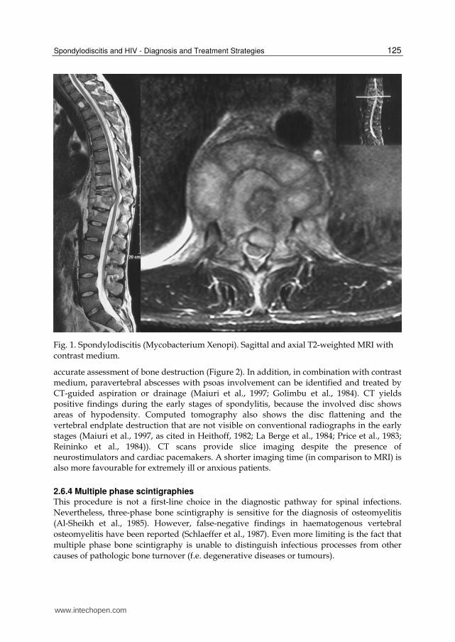
Magnetic resonance imaging is the study of choice for the diagnosis of spinal infections. Common MRI findings in infectious spondylitis are hypointensity of the involved tissue on T1-weighted images, hyperintensity on T2-weighted images, destruction of two or more adjacent vertebral bodies with involvement of the intervening disc, and epidural and paraspinal extension and/or abscesses. In addition, MRI provides a diagnostic view of the paravertebral and spinal space (Chang et al., 2006; An et al., 1991; Modic et al. 1985). In HIV positive patients, the treating physician must deal with the specific type of infection (e.g. vertebral tuberculosis, Figure 1). The following parameters can help distinguish TB from pyogenic spondylitis. The most distinctive finding of TB spondylitis is a pattern of mainly bone destruction with relative preservation of the disc. The vertebral body shows focal and heterogeneous contrast enhancement. In addition, a well-defined paraspinal area of abnormal signal intensity is often detectable. On the sagittal views, an intraosseous rim enhancement of the vertebra may occur. In comparison, pyogenic spondylitis is characterized by a pattern of discitis (disc destruction) with mild to moderate peri-discal bone destruction. The vertebral body shows a relatively diffuse and homogenous contrast enhancement. Furthermore, an ill-defined paraspinal area of abnormal signal intensity with peri-discal rim enhancement provides a hint to the correct diagnosis (Chang et al., 2006). In any case, the use of contrast medium during the procedure is highly recommended. MRI is the gold standard diagnostic to detect spondylodiscitis. Nevertheless, in early stages it may show only subtle, non-specific subchondral changes to the endplate. This can lead to a misdiagnosis (e.g. Modic I, degenerative endplate change). If the clinical course raises suspicions of an infectious process of the spine, a second MRI after 2-3 weeks is highly recommended (Dunbar et al., 2010).
2.6.3 CT (computed tomography)
Generally, the radiologic diagnosis of spondylodiscitis is based on MRI findings. Nevertheless, valuable information may be provided by computed tomography. CT enables
www.intechopen.com
Spondylodiscitis and HIV - Diagnosis and Treatment Strategies
125
Fig. 1. Spondylodiscitis (Mycobacterium Xenopi). Sagittal and axial T2-weighted MRI with contrast medium.
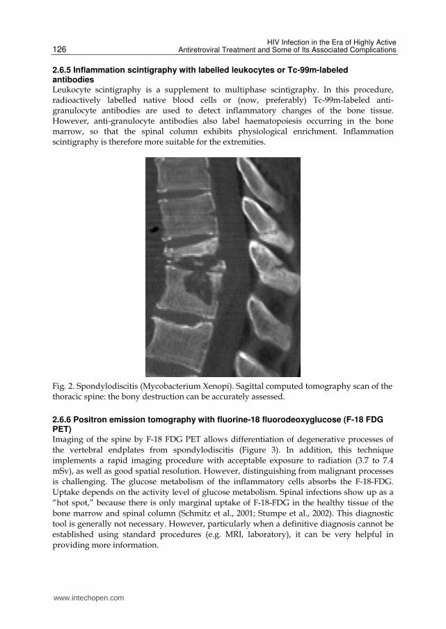
accurate assessment of bone destruction (Figure 2). In addition, in combination with contrast medium, paravertebral abscesses with psoas involvement can be identified and treated by CT-guided aspiration or drainage (Maiuri et al., 1997; Golimbu et al., 1984). CT yields positive findings during the early stages of spondylitis, because the involved disc shows areas of hypodensity. Computed tomography also shows the disc flattening and the vertebral endplate destruction that are not visible on conventional radiographs in the early stages (Maiuri et al., 1997, as cited in Heithoff, 1982; La Berge et al., 1984; Price et al., 1983; Reininko et al., 1984)). CT scans provide slice imaging despite the presence of neurostimulators and cardiac pacemakers. A shorter imaging time (in comparison to MRI) is also more favourable for extremely ill or anxious patients.
2.6.4 Multiple phase scintigraphies
This procedure is not a first-line choice in the diagnostic pathway for spinal infections. Nevertheless, three-phase bone scintigraphy is sensitive for the diagnosis of osteomyelitis (Al-Sheikh et al., 1985). However, false-negative findings in haematogenous vertebral osteomyelitis have been reported (Schlaeffer et al., 1987). Even more limiting is the fact that multiple phase bone scintigraphy is unable to distinguish infectious processes from other causes of pathologic bone turnover (f.e. degenerative diseases or tumours).
www.intechopen.com
HIV Infection in the Era of Highly Active Antiretroviral Treatment and Some of Its Associated Complications
126
2.6.5 Inflammation scintigraphy with labelled leukocytes or Tc-99m-labeled antibodies Leukocyte scintigraphy is a supplement to multiphase scintigraphy. In this procedure, radioactively labelled native blood cells or (now, preferably) Tc-99m-labeled anti-granulocyte antibodies are used to detect inflammatory changes of the bone tissue. However, anti-granulocyte antibodies also label haematopoiesis occurring in the bone marrow, so that the spinal column exhibits physiological enrichment. Inflammation scintigraphy is therefore more suitable for the extremities.
Fig. 2. Spondylodiscitis (Mycobacterium Xenopi). Sagittal computed tomography scan of the thoracic spine: the bony destruction can be accurately assessed.
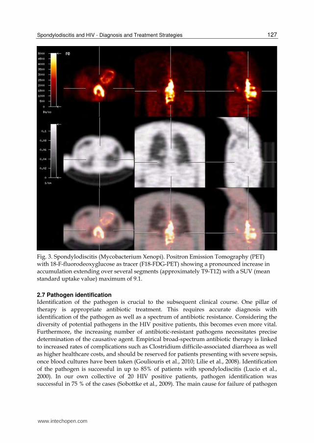
2.6.6 Positron emission tomography with fluorine-18 fluorodeoxyglucose (F-18 FDG PET) Imaging of the spine by F-18 FDG PET allows differentiation of degenerative processes of the vertebral endplates from spondylodiscitis (Figure 3). In addition, this technique implements a rapid imaging procedure with acceptable exposure to radiation (3.7 to 7.4 mSv), as well as good spatial resolution. However, distinguishing from malignant processes is challenging. The glucose metabolism of the inflammatory cells absorbs the F-18-FDG. Uptake depends on the activity level of glucose metabolism. Spinal infections show up as a “hot spot,” because there is only marginal uptake of F-18-FDG in the healthy tissue of the bone marrow and spinal column (Schmitz et al., 2001; Stumpe et al., 2002). This diagnostic tool is generally not necessary. However, particularly when a definitive diagnosis cannot be established using standard procedures (e.g. MRI, laboratory), it can be very helpful in providing more information.
www.intechopen.com
Spondylodiscitis and HIV - Diagnosis and Treatment Strategies
127
Fig. 3. Spondylodiscitis (Mycobacterium Xenopi). Positron Emission Tomography (PET) with 18-F-fluorodeoxyglucose as tracer (F18-FDG-PET) showing a pronounced increase in accumulation extending over several segments (approximately T9-T12) with a SUV (mean standard uptake value) maximum of 9.1.
2.7 Pathogen identification Identification of the pathogen is crucial to the subsequent clinical course. One pillar of therapy is appropriate antibiotic treatment. This requires accurate diagnosis with identification of the pathogen as well as a spectrum of antibiotic resistance. Considering the diversity of potential pathogens in the HIV positive patients, this becomes even more vital. Furthermore, the increasing number of antibiotic-resistant pathogens necessitates precise determination of the causative agent. Empirical broad-spectrum antibiotic therapy is linked to increased rates of complications such as Clostridium difficile-associated diarrhoea as well as higher healthcare costs, and should be reserved for patients presenting with severe sepsis, once blood cultures have been taken (Gouliouris et al., 2010; Lilie et al., 2008). Identification of the pathogen is successful in up to 85% of patients with spondylodiscitis (Lucio et al., 2000). In our own collective of 20 HIV positive patients, pathogen identification was successful in 75 % of the cases (Sobottke et al., 2009). The main cause for failure of pathogen
www.intechopen.com
HIV Infection in the Era of Highly Active Antiretroviral Treatment and Some of Its Associated Complications
128
identification is previous use of systemic antibiotic treatment. If possible, antibiotic therapy should be initiated only after sample materials are obtained. In cases where the patient is already receiving antibiotic treatment and the health status is stable, the medication should be discontinued for at least 3 days prior to culture. In such cases, we believe that attempts to identify the pathogens are more likely to be successful.
2.7.1 Blood culture Infections of the spine are generally monomicrobial, and often have a haematogenous source. Therefore, blood cultures are a simple and cost effective method for identifying bacterial pathogens of spinal infections. A positive culture can be expected in 40%-60% of clinically defined cases of pyogenic spondylodiscitis (Gouliouris et al., 2010; Sapico, 1996). However, the previous administration of systemic antibiotic therapy severely handicaps the ability to identify the causative agent. In such cases, temporary interruption of antibiotic administration is necessary prior to performing the blood culture. It is recommended to repeat the blood culture up to 3 times for definite identification.. The pathogen is often successfully identified, not only in the acute phase of fever or the presence of sepsis, but also in clinically bland cases of afebrile patients (Nolla et al., 2002). Nevertheless, there is a high incidence of infective endocarditis (26%) reported during enterococcal and streptococcal spondylodiscitis. Routine echocardiography should be performed when these pathogens are suspected (Mulleman et al., 2006).
2.7.2 Biopsy
Other alternatives for identifying the pathogen are use of a percutaneous punch under anaesthesia, and CT-guided fine needle aspiration. The latter can be performed while concomitantly placing a drain to reduce pressure on the abscess. In obtaining a histological diagnosis for cases of malignant disease, a percutaneous spinal biopsy is accurate. In cases of spondylodiscitis, however, the reported accuracy is more variable. Identification of the pathogen is possible in 40%-73% of cases (Rankine et al., 2004; Shaltot et al., 1982, Borowski et al, 1998). Nonetheless, spinal biopsy leads to a direct change in management for 35% of patients, and is still worthwhile even if the patient has already started on antibiotics. However, success of the procedure for identification of the pathogen is much greater prior to starting antibiotics (Rankine et al., 2004). If antibiotics have already been initiated, the treating physician should consider stopping this treatment for 2-3 days prior to the biopsy. If the first attempt to identify the pathogen by percutaneous biopsy fails, a second procedure should be considered. Friedman et al. identified microbiological growth in 50% of cases after disc space biopsy in patients with spontaneous spondylodiscitis. Repeat biopsies brought this rate up to 79% (Friedman et al. 2002). Even more promising is open surgical biopsy, if the first set of cultures is negative (Lew & Waldvogel, 2004).
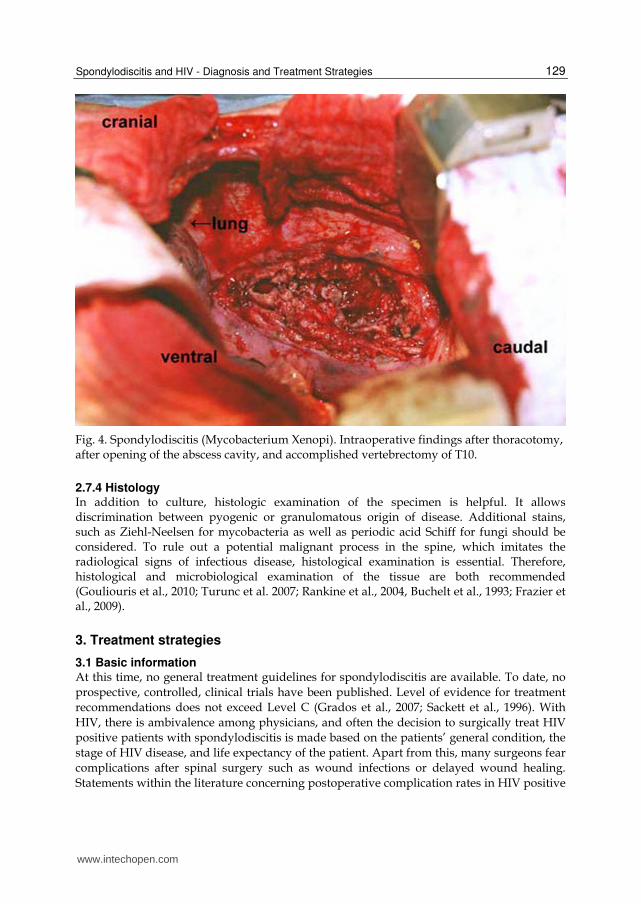
2.7.3 Intraoperative sampling The highest probability for probe acquisition enabling identification of the pathogen is surgical sampling (Figure 4). Open bone biopsies can yield the microbial aetiology of spondylodiscitis in almost 100% of cases (Jimenz-Mejias et al., 1999). In this case, it is recommended to obtain at least 2 probes for histological and microbiological examination (Lucio et al., 2000).
www.intechopen.com
Spondylodiscitis and HIV - Diagnosis and Treatment Strategies
129
Fig. 4. Spondylodiscitis (Mycobacterium Xenopi). Intraoperative findings after thoracotomy, after opening of the abscess cavity, and accomplished vertebrectomy of T10.
2.7.4 Histology In addition to culture, histologic examination of the specimen is helpful. It allows discrimination between pyogenic or granulomatous origin of disease. Additional stains, such as Ziehl-Neelsen for mycobacteria as well as periodic acid Schiff for fungi should be considered. To rule out a potential malignant process in the spine, which imitates the radiological signs of infectious disease, histological examination is essential. Therefore, histological and microbiological examination of the tissue are both recommended (Gouliouris et al., 2010; Turunc et al. 2007; Rankine et al., 2004, Buchelt et al., 1993; Frazier et al., 2009).
3. Treatment strategies
3.1 Basic information At this time, no general treatment guidelines for spondylodiscitis are available. To date, no prospective, controlled, clinical trials have been published. Level of evidence for treatment recommendations does not exceed Level C (Grados et al., 2007; Sackett et al., 1996). With HIV, there is ambivalence among physicians, and often the decision to surgically treat HIV positive patients with spondylodiscitis is made based on the patients’ general condition, the stage of HIV disease, and life expectancy of the patient. Apart from this, many surgeons fear complications after spinal surgery such as wound infections or delayed wound healing. Statements within the literature concerning postoperative complication rates in HIV positive
www.intechopen.com
HIV Infection in the Era of Highly Active Antiretroviral Treatment and Some of Its Associated Complications
130
patients are inconsistent. However, the aim of treatment is to eradicate the infection, restore and preserve the structure and function of the spine, and alleviate pain (Gouliouris et al. 2010). Therefore, the affected segments must be immobilized, and appropriate antibiotic treatment is required. If necessary, surgical debridement and decompression of the spinal canal must also be considered. As previously mentioned, antibiotic treatment should be initiated after identification of the pathogen and according to the resistance profile. Bed rest is recommended if the pain level does not allow mobilisation of the patient, or if there is a high risk of spinal instability (Quinones-Hinojosa et al., 2004). Since the advent of antibiotics, mortality from this disease has dropped from up to 56% to less than 5% (Gouliouris et al., 2010; Bauman et al., 1923; Sapico et al.; 1979). Guidelines regarding the route of administration for and/or duration of antibiotic treatment do not exist for spondylodiscitis patients with or without HIV infection. Intravenous administration of antibiotics for at least 2-4 weeks is recommended (Lew & Waldvogel, 2004; Sobottke et al., 2008) If antibiotic treatment without direct pathogen identification is required, the medication selected should be appropriate for the most common pathogens causing spondylodiscitis, i.e. Staphylococcus aureus and Escherichia coli, after blood cultures are taken. The intravenous phase can be shortened when the organism is highly susceptible to antimicrobials, and the patient has negative blood cultures, normal motor function, and no evidence of endocarditis (Grados et al., 2007). Furthermore, observation of the inflammatory parameters are recommended. The optimal duration for antibiotic administration remains unclear. Overall, treatment for more than 12 weeks appears to be associated with a lower recurrence rate when compared with that of 4-8 weeks (Grados et al., 2007). The following antibiotics diffuse extremely well to bone tissue: fluoroquinolones, clindamycin, rifampicin, fusidic acid, and metronidazole. Fair diffusion can be contributed by ß-lactams, glycopeptides, phosphomycin, and sulphonamides (Grados et al., 2007). In case of tuberculosis spondylodiscitis, anti-tubercular chemotherapy should be started after histological and microbiological evidence is obtained, and should be carried out for 18-24 months. Spondylodiscitis caused by fungus should be treated with appropriate antimycotic medication. In advanced cases, antimycotic drug therapy is thought to be ineffective. An overriding indication for surgery is recommended, in particular when spinal structures show progressive destruction (van Ooij et al., 2000).
3.2 Conservative treatment In addition to antibiotic treatment, immobilization of the affected region of the spinal column is required. Reclining ortheses distribute stress over the unaffected segments and their joints. Thereby, release of the affected ventral column can be obtained. Wearing the orthesis, patients can be fully mobilized. In our study of HIV positive patients, only half of the conservatively-treated patients were supplied with a reclination brace, which was worn for an average of about 51 days. In addition, 4 of the patients undergoing surgery were also treated with such a reclination brace. The condition of 2 patients worsened under initial conservative treatment and so surgery was required (Sobottke et al., 2009). Generally, if conservative treatment yields no radiologically evident fusion reaction, continued destruction occurs, and/or there is no clinical improvement, surgery should be considered (Hsieh et al., 2004; Quinones-Hinojosa et al., 2004). When major defects exist in the ventral column, the lower lumbar column, or at the lumbosacral border, necessary fusion through non-operative measures can only be achieved by at least six weeks bed rest. Mobilisation of
www.intechopen.com
Spondylodiscitis and HIV - Diagnosis and Treatment Strategies
131
the patient is only recommended once osseous infiltration becomes visible (Sobottke et al., 2008, as cited in Eysel & Peters, 1997 & Cramer et al., 2003).
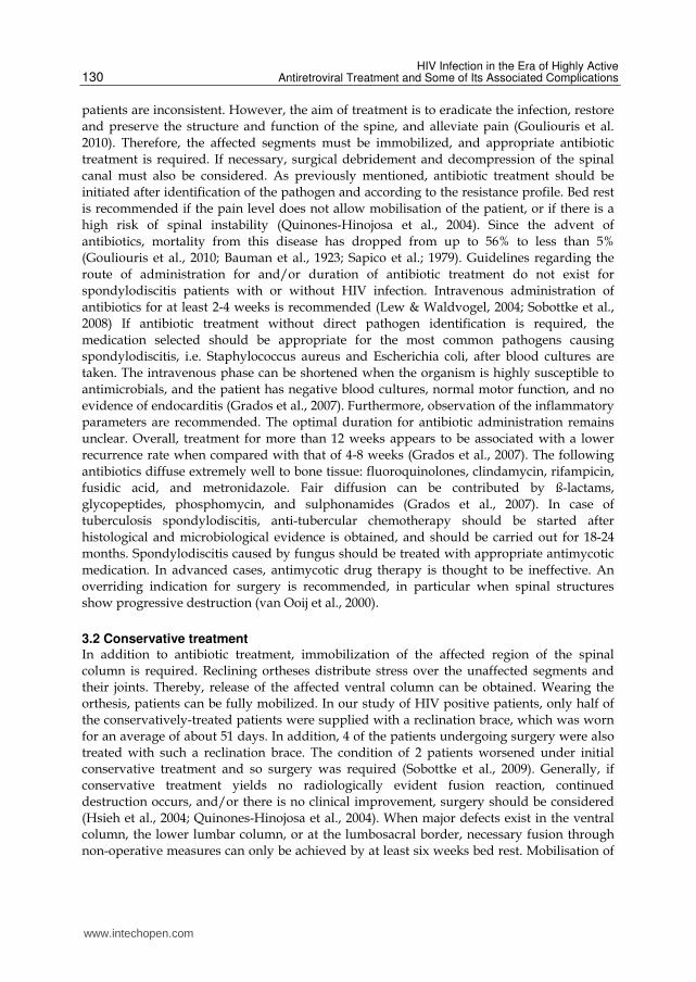
3.3 Surgical treatment 3.3.1 Drainage of abscesses Large abscesses, particularly those persisting even after antibiotic treatment, must be eliminated. One alternative is drain placement (Figure 5), inserted during an open procedure or CT-guided. Especially when the general state of health of the patient is poor and precludes open surgery, drain placement should be considered. It has been shown as an adequate technique to treat abscesses outside of the spine e. g. ilio-psoas muscle or pleural cavity (Grados et al., 2007).
Fig. 5. Spondylodiscitis (Mycobacterium Xenopi). Control X-ray after CT-guided draining of the paravertebral abscess.
www.intechopen.com
HIV Infection in the Era of Highly Active Antiretroviral Treatment and Some of Its Associated Complications
132
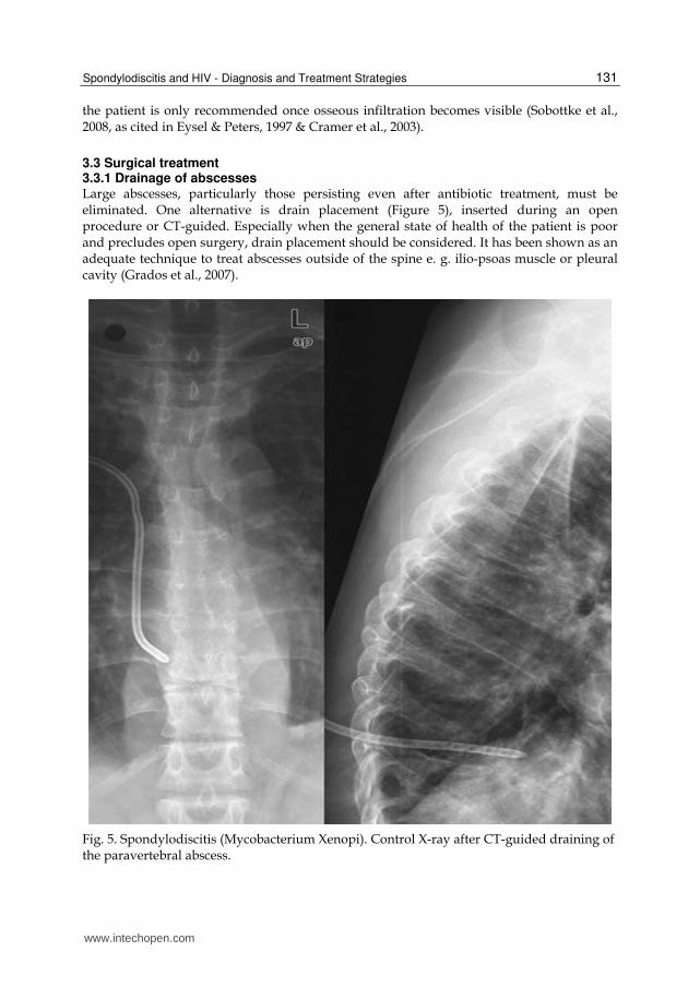
3.3.2 Surgery Surgical intervention should be considered when there is no response to therapy and patient is experiencing persistent and high intensity pain. The surgical procedure is aimed to relieve compression of the spinal cord or to drain epidural or paravertebral abscesses and to improve spinal stability (Lew & Waldvogel, 2004). It includes excision of the infected spinal and paravertebral tissue, retrieval of the pathogen for identification, and stabilisation of the spine via fusion of the affected segments. Surgical alternatives include dorsal instrumentation with pedicle screw-based systems, either minimally invasive, if no spinal decompression is required, or using open technique in combination with spinal canal decompression. Ventral approaches can be performed combined with dorsal instrumentation or alone using a ventral stabilization system (Figure 6). This approach enables treatment of the frequently-involved ventral column. Extensive debridement must be carried out on both the affected intervertebral disc space and the vertebral endplates. Spinal fusion can be achieved using autologous bone interposition or with cage fusion (Figure 6). Treatment of the cervical spine may include ventral stabilization by plate, cage, or autologous bone interposition. Instrumentation via dorsal approach is usually unnecessary (Müller et al., 2004). Generally, no evidence-based guidelines regarding surgical treatments are available (Sobottke et al., 2008). Of course, the implantation of
Fig. 6. Spondylodiscitis (Mycobacterium Xenopi). Control X-ray after debridement of the infected segment and ventral spondylodesis. It shows a properly inserted cage (titanium, Pyramesh, Medtronic) and an anterolateral monobar system (titanium, ART system, AMT).
www.intechopen.com
Spondylodiscitis and HIV - Diagnosis and Treatment Strategies
133
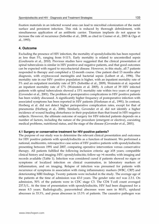
fixation materials in an infected wound area can lead to microbial colonization of the metal surface and persistent infection. This risk is reduced by thorough debridement, with simultaneous application of an antibiotic carrier. Titanium implants do not appear to increase the rate of recurrence (Sobottke et al., 2008, as cited in Cramer et al., 2003 & Oga et al., 1993).
4. Outcome
Excluding the presence of HIV infection, the mortality of spondylodiscitis has been reported as less than 5%, ranging from 0-11%. Early mortality is related to uncontrolled sepsis (Gouliouris et al., 2010). Previous studies have suggested that the clinical presentation of spinal tuberculosis is similar in HIV positive and negative patients, and that good outcomes can be expected with regard to mycobacterial disease. However, in this study, all 7 patients responded to therapy and completed a 12-month course. One patient died 13 months after diagnosis, with cryptococceal meningitis and bacterial sepsis (Leibert et al., 1996). The mortality rate in our HIV- positive population is higher, with an inpatient mortality rate of 5% and an outpatient mortality rate of 20% (Sobottke et al., 2009). Weinstein et al. reported an inpatient mortality rate of 17% (Weinstein et al. 2005). A cohort of 39 HIV infected patients with spinal tuberculosis showed a 15% mortality rate within two years of surgery (Govender et al., 2001). The problem of postoperative complications in HIV positive patients has been widely discussed. A significantly higher frequency of postoperative infection with associated symptoms has been reported in HIV patients (Hoekman et al., 1991). In contrast, Horberg et al. did not detect higher perioperative complication rates, except for that of pneumonia (Horberg et al., 2006). Similarly, Govender et al. did not identify a higher incidence of wound healing disturbance in their population than that found in HIV-negative subjects. However, the ultimate outcome of surgery for HIV-infected patients depends on a number of factors, including the nature of the procedure (emergent or elective), coexisting medical problems, nutritional status, and the stage of the disease (Govender et al., 2001).
4.1 Surgery or conservative treatment for HIV-positive patients? The purpose of our study was to determine the relevant clinical presentation and outcomes for HIV positive patients with spondylodiscitis as a function of treatment. We performed a national, multicentre, retrospective case series of HIV positive patients with spondylodiscitis presenting between 1991 und 2007, comparing operative intervention versus conservative therapy. All patients fulfilled the following inclusion criteria: age ≥18 years; compatible clinical history and imaging; HIV; spondylodiscitis; follow-up >6 months; with imaging and records available (Table 1). Infection was considered cured if patients showed no signs or symptoms of localized infection on clinical examination, in laboratory markers of inflammation, and on imaging. Relapse of infection was presumed for patients with recurrence of symptoms in association with rising inflammatory markers (WBC, CRP) and deteriorating MRI findings. Twenty patients were included in the study. The average age of the patients at the time of admission was 43.0 years. The gender ratio m:f was 2.3:1. On admission, 50% of the patients were in CDC stage C3. The CD4 T-cell count averaged 237.5/L. At the time of presentation with spondylodiscitis, HIV had been diagnosed for a mean 8.5 years. Radiologically, paravertebral abscesses were seen in 80.0%, epidural abscesses in 33.3%, and psoas abscesses in 13.3% of patients. The causative pathogen was
www.intechopen.com
HIV Infection in the Era of Highly Active Antiretroviral Treatment and Some of Its Associated Complications
134
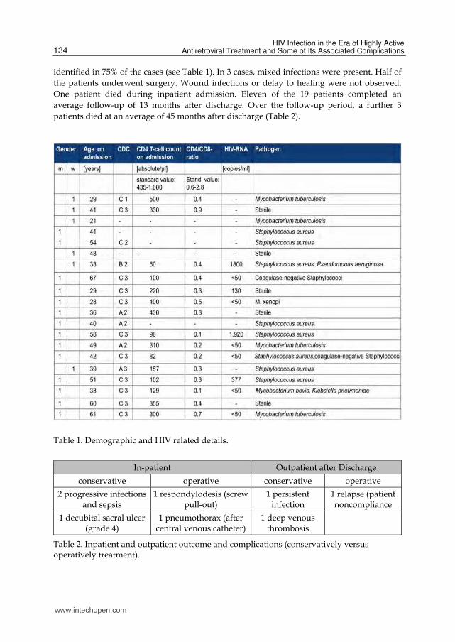
identified in 75% of the cases (see Table 1). In 3 cases, mixed infections were present. Half of the patients underwent surgery. Wound infections or delay to healing were not observed. One patient died during inpatient admission. Eleven of the 19 patients completed an average follow-up of 13 months after discharge. Over the follow-up period, a further 3 patients died at an average of 45 months after discharge (Table 2).
Table 1. Demographic and HIV related details.
In-patient Outpatient after Discharge
conservative operative conservative operative
2 progressive infections and sepsis
1 respondylodesis (screw pull-out)
1 persistent infection
1 relapse (patient noncompliance
1 decubital sacral ulcer (grade 4)
1 pneumothorax (after central venous catheter)
1 deep venous thrombosis
Table 2. Inpatient and outpatient outcome and complications (conservatively versus operatively treatment).
www.intechopen.com
Spondylodiscitis and HIV - Diagnosis and Treatment Strategies
135
5. Conclusion
An increasing incidence of spondylodiscitis has been observed. Factors implicated and collaborating in this increase include the HIV epidemic, particularly that in Sub-Saharan Africa, the large numbers of intravenous drug abusers, the currently widely-used aspiration and catheter techniques, as well as the recurrence of tuberculosis in industrialized nations. In principle, patients of all ages can contract spondylitis, but it appears that 50 to 70 year-old patients are most likely to fall ill from it. The peak age of disease in HIV positive patients lies substantially earlier: 10% are under 30 years of age when they first contract spondylitis. The estimated number of patients who are seropositive for human immunodeficiency virus is about 50 billion people worldwide, and continues to increase. Potential pathogens responsible for spondylitis include bacteria, fungi, and parasites (e. g. hydatidosus). As a general rule, the infection is of bacterial origin. However, especially in immune deficient patients, the possibility of infection stemming from fungi or atypical mycobacteria should be taken into account. The probability of an infection by MOTT (Mycobacteria Other Than Tuberculosis) strongly depends on the CD4 T-cell count. Highly Active Anti-Retroviral Therapy (HAART) has accomplished rapid increases of the CD4 T-cell count, and thus caused a so-called "immune reconstitution syndrome," which has resulted in atypical, creeping disease processes and at times even to spontaneous complete recovery. Several years may elapse between the inception of symptoms and the final diagnosis of spondylodiscitis. Primary factors resulting in successful treatment of spondylodiscitis are early diagnosis and rapid onset of treatment to prevent progressive stages with serious complications. Immobilisation of the affected spine segments, antibiotic therapy, and, depending on the extent of the disease, debridement, decompression, and stabilisation are basic requirements for successful treatment leading to a complete recovery from spondylodiscitis. Emergent surgical intervention is required for spondylodiscitis on the development of neurological deficits and/or sepsis. Further indications for surgery are instability, impending or already existing deformities, intraspinal space-occupying lesions, an unclear origin with the suspicion of malignancy, and lack of response to conservative therapy. Relative indications for surgical treatment include uncontrollable pain symptoms and lack of patient compliance with conservative therapy. The occurrence of spondylodiscitis in HIV positive patients is accompanied by high mortality. We recorded a hospital mortality of 5% and a total mortality of 20%. The occurrence of spondylodiscitis in HIV positive patients is associated with a low CD4 T-cell count. The probability of mixed infections rises with a CD4 T-cell counts below 100/L, but there is no correlation between a low CD4 T-cell count and probability of infection by MOTT. Because increased surgical morbidity is not evident among HIV positive patients, HIV infection or AIDS should not interfere with the decision to perform operative stabilisation of the affected spinal segments. The existence of HIV infection or AIDS in a patient with spondylodiscitis should not influence the decision for or against conservative or operative therapy. However, these patients should be treated in a specialized hospital by an experienced team of consultants.
6. Further research
To date, no global data has been collected or published regarding the aetiopathology, clinical course, and/or treatment strategies for spinal infections in patients also afflicted with HIV. This information, however, is a prerequisite for an understanding of the disease
www.intechopen.com
HIV Infection in the Era of Highly Active Antiretroviral Treatment and Some of Its Associated Complications
136
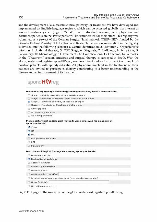
and the development of a successful clinical pathway for treatment. We have developed and implemented an English-language registry, which can be accessed globally via internet at www.clinicalsurveys.net (Figure 7). With an individual account, any physician can document patients online. Participants will be remunerated for their effort. This registry was submitted as a project of the German Surgical Trial network (CHIR–NET), funded by the German Federal Ministry of Education and Research. Patient documentation in the registry is divided into the following sections: 1. Centre identification, 2. Identifier, 3. Opportunistic infection, 4. Antiviral therapy, 5. CDC Stage, 6. Diagnosis, 7. Radiology, 8. Symptoms, 9. Laboratory, 10. Microbiology, 11. Treatment , 12. Complications, 13. Outcome, 14. Remarks. In the “Treatment” section, antibiotic and surgical therapy is surveyed in depth. With the global, web-based registry spondHIVreg, we have introduced an instrument to survey HIV-positive patients with spondylodiscitis. All physicians involved in the treatment of these patients are invited to participate, thereby contributing to a better understanding of the disease and an improvement of its treatment.
Fig. 7. Full page of the survey list of the global web-based registry SpondHIVreg.
www.intechopen.com
Spondylodiscitis and HIV - Diagnosis and Treatment Strategies
137
7. Acknowledgement
JS, KZ and RS are supported by the German Federal Ministry of Research and Education (BMBF grant 01KN1106). Peer Eysel conceived of the study, which forms the basis of this manuscript.
8. References
An, H. S.; Vaccaro, A. R.; Dolinskas, C. A.; Cotler, J. M.; Balderston, R. A. & Bauerle, W. B. (1991). Differentiation between spinal tumors and infections with magnetic resonance imaging. Spine, Vol. 16, No. 8 (August 1991), pp. 334-338, ISSN 0362-2436
Al-Sheikh, W.; Sfakianakis, G. N.; Mnaymneh, W.; Hourani, M.; Heal, A.; Duncan, R. C.; Burnett, A.; Ashkar, F. C. & Serafini, A. N. (1985). Subacute and chronic bone infections: diagnosis using In-111, Ga-67 and Tc-99m MDP bone scintigraphy, and radiography. Radiology, Vol. 155, No.2 (May 1985), pp. 501-506, ISSN 0033-8419
Borowski, A. M.; Crow, W. N.; Hdjipavlou, A. G.; Chaljub, G.; Mader, J.; Cesani, F. & vanSonnenberg, E. (1998). Interventional radiology case conference: the University of Texa Medical Branch. Percutaneous management fo pyogenic spondylodiscitis. American Journal of Radiology, Vol. 170, No. 6 (June 1998), pp. 1587-1592, ISSN 0361-803X
Buchelt, M.; Lack, W.; Kutschera, H. P.; Katterschafka, T.; Kiss, H.; Schneider, B. & Kotz, R. (1993). Comparision of tuberculous and pyogenic spondylitis. An analysis of 122 cases. Clinical Orthopaedics and related research, Vol. 296 (November 1993), pp. 192-199, ISSN 0009-921X
Busch, V. J.; Regez R. M.; Heere, B. & Willems, W. J. (2007) Osteoarticular infections in HIV-infected patients: 23 cases among 1,515 HIV-infected patients. Acta Orthopaedica, Vol. 78, No. 6, ( December 2007), pp786-790, ISSN 1745-3674.
Carragee, E. J.; Kim, D.; van der Vlugt, T. & Vittum, D. (1997a). The clinical use of erythrocyte sedimentation rate in pyogenic vertebral osteomyelitis. Spine, Vol. 22, No.18 (September 1997), pp. 2089-2093, ISSN 0362-2436
Carragee, E. J. (1997b). Pyogenic vertebral osteomyelitis. Journal of Bone and Joint Surgery, Vol. 79, No. 6, (June 1997), pp.874-880, ISSN 0021-9355
Chang, M. C.; Wu, H. T.; Lee, C. H.; Liu, C. L. & Chen, T. H. (2006). Tuberculous spondylitis and pyogenic spondylitis: comparatative magnetic resonance imaging features. Spine, Vol 31, No. 7 (April 2006), pp. 782-788, ISSN 0362-2436
Dunbar, J. A.; Sandoe, J. A., Rao, A. S.; Crimmins, D. W.; Baig, W. & Rankine, J. J. (2010). The MRI appearances of early vertebral osteomyelitis and discitis. Clinical Radiology, Vol. 65, No. 12 (December 2010), pp. 974-981, ISSN 0009-9260
Frangen, T. M; Kälicke, T.; Gottwald, M.; Andereya, S.; Andress, H. J.; Russe, O. J.; Müller, E. J.; Muhr, G. & Schinkel, C. (2006). Der Unfallchirurg, Vol. 109, No. 9 (September 2006), pp. 743-753. ISSN 0177-5537
Frazier, D. D.; Campbell, D. R.; Garvey, T. A.; Wiesel, S; Bohlmann, H. H. & Eismont, F. J.(2001). Journal of Bone and Joint Surgery, Vol. 83, No. A4 (April 2001), 560-565, ISSN 0021-9355
Friedman, J. A.; Maher, C. O.; Quast, L. M.; McClelland, R. L. & Ebersold, M. J. (2002). Spontaneous disc space infections in adults. Surgical Neurology, Vol. 57, No. 2 (February 2002), pp. 81-86, ISSN 0090-3019
Golimbu, C.; Firooznia, H. & Rafii M. (1984). CT of osteomyelitis of the spine. American Journal of Roentgenology, Vol. 142, No. 1 (January 1984), pp. 159-163. ISSN 0361-803X
www.intechopen.com
HIV Infection in the Era of Highly Active Antiretroviral Treatment and Some of Its Associated Complications
138
Gouliouris, T.; Aliju S. H. & Brown, N. M. (2010). Spondylodiscitis: update on diagnosis and management. Journal of Antimicrobial Chemotherapy, Vol. 65, No. 3, (November 2010), pp. 11-24, ISSN 0305-7453
Govender, S.; Parbhoo, A. H.; Kumar, K. P. & Annamalai, K. (2001). Anterior spinal decompression in HIV-positive patients with tuberculosis. A prospective study. Journal of Bone & Joint Surgery, Vol. 83, No. 6 (August 2001), pp. 864-867), ISSN 0301-620X
Grados, F.; Lescure, F. X.; Senneville, E.; Flipo, R. M.; Schmit, J. L.; Fardellone, P. (2007). Suggestions for managing pyogenic (non-tuberculous) discitis in adults. Joint, Bone, Spine, Vol 74, No. 2 (March 2007), pp. 133-139, ISSN 1297-319X
Grammatico, L.; Baron, S.; Rusch, E.; Lepage, B.; Surer, N.; Desenclos, J. C. & Besnier, J. M. (2008). Epidemiology of vertebral osteomyelitis (VO) in France: analysis of hospital-discharge data 2002-2003. Epidemiology and Infection, Vol. 136, No. 5 (May 2008), pp. 653-660, ISSN 0950-2688
Hoekman, P.; van de Perre, P.; Nelissen, J.; Kwisanga, B.; Bogaerts, J. & Kanyangabo, F. (1991). Increased frequency of infection after open reduction of fractures in patients who are seropositive for human immunodeficiency virus. Journal of Bone and Joint Surgery, Vol. 73, No. 5 (June 1991), ISSN 0021-9355
Horberg, M. A.; Hurley, L. B.; Klein, D. B; Follansbee, S.E.; Quesenberry, C.; Flamm, J. A.; Green, G. M. & Luu, T. (2006). Surgical outcomes in human immunodeficiency virus-infected patients in the era of highly active antiretroviral therapy. Archives of Surgery, Vol. 141, No. 12 (December 2006), pp. 1238-1245, ISSN 0272-5533
Hsieh, P. C.; Wienecke, R. J.; O’Shaughnessy, B. A.; Koski, T. R. & Ondra, S. L. (2004). Surgical strategies for vertebral osteomyelitis and epidural abscess. Neurosurgical Focus, Vol. 17, No. 6 (December 2004), E4, ISSN 1092-0684
Jevtic, V. (2004). Vertebral infection. European Radiology, Vol. 14, No. 3 (March 2004), pp. 43-52, ISSN 0938-7994
Jimenez-Mejias, M. E.; De Dios Colmenero, J.; Sanchez-Lora, F. J.; Palomino-Nicas, J.; Requera, J. M.; Garcia de la Heras, J.; Garcia-Ordonez, M. E.; Pachon, J. Postoperative spondylodiscitis: etiology, clinical findings, prognosis, and comparison with nonoperative pyogenic spondylodiscitis. Clinical Infectious diseases, Vol. 29, No. 2 (August 1999), pp.339-345, ISSN 1058-4838
Kulowski, J. (1936). Pyogenic osteomyelitis of the spine: an analysis and discussion of 102 cases. Journal of Bone and Joint Surgery, Vol. 18, No. 2, (April 1936), pp. 343-364. ISSN 0021-9355
Leibert, E.; Schluger, N. W.; Bonk, S. & Rom, W. N. (1996). Spinal tuberculosis in patients with human immunodeficiency virus infection: clinical presentation, therapy and outcome. Tubercle and lung disease: The official journal of the International Union against Tuberculosis and Lung Disease, Vol. 77, No. 4 (August 1996), pp. 329-34, ISSN 0962-8479
Lew, D. P.; Waldvogel, F. A. (2004). Osteomyelitis. Lancet, Vol. 364, No. 9431 (July 2004), pp. 369-379, ISSN 0140-6736
Lillie, P.; Thaker, H.; Moss, P.; Baruah, J.; Cullen, L.; Taylor, D. & Barlow, G. (2008). Healthcare associated discitis in the era of antimicrobial resistance. Journal of Clinical Rheumtology, Vol. 14, No. 4 (August 2008), pp. 234-237 ISSN 1076-1608
Lucio, E.; Adesokan, A.; Hadjipavlou, A. G.; Crow, W. N. & Adegboyega, P. A. (2000). Pyogenic spondylodiscitis: a radiologic/pathologic and culture correlation study.
www.intechopen.com
Spondylodiscitis and HIV - Diagnosis and Treatment Strategies
139
Archives of Pathology and Laboratory Medicine, Vol. 124, No. 5 (May 2000), pp. 712-716, ISSN 0003-9985
Maiuri, F.; Iaconetta, G.; Gallicchio, B.; Manto, A.; Briganti, F. (1997). Spondylodiscitis. Clinical and magnetic resonance diagnosis. Spine, Vol. 22, No. 15 (August 1997), pp. 1741-1746, ISSN 0362-2436
Modic, M. T.; Feiglin, D. H.; Piraino, D. W.; Boumphrey, F.; Weinstein, M. A.; Duchesneau, P. M. & Rehm, S. (1985). Vertebral osteomyelitis: assessment using MR. Radiology, Vol. 157, No. 1 (October 1985), pp. 157-166. ISSN 0033-8419
Mulleman, D.; Philippe, P.; Senneville, E.; Costes, C.; Fages, L.; Deprez, X.; Flipo, R. M. & Duquesnoy, B. (Streptococcal and enterococcal spondylodiscitis (vertebral osteomyelitis). High incidence of infective endocarditis in 50 cases. Journal of Rheumatology, Vol. 33, No. 1 (January 2006), pp.91-97, ISSN 0315-162X
Müller, E. J; Russe, O. J. & Muhr, G. (2004). Osteomyelitis of the spine. Der Orthopäde, Vol. 33, No. 3 (March 2004), pp. 305-315, ISSN 0085-4530
Nichols, L.; Balogh, K. & Silverman, M. (1989). Bacterial infections in the acquired immune deficiency syndrome. Clinicopathologic correlations in a series of autopsy cases. American Journal of Clinical Pathology, Vol. 92, No. 6, pp. 787-790, ISSN 0002-9173
Nolla, J. M.; Ariza, J.; Gomez-Vaquero, C.; Fiter, J.; Bermejo, J.; Valverde, J; Escofet, D. R. & Gudiol, F. (2002). Spontaneous pyogenic vertebral osteomyelitis in nondrug users. Seminars in Arthritis and Rheumatism, Vol. 31, No. 4 (February 2002), pp. 271-278, ISSN 0049-0172
Price, A. C.; Allen, J. H.; Eggers, F. M.; Shaff, M. I.; James, A. E. Jr. (1983). Intervertebral disc-space infection: CT-changes. Radiology, Vol.149, No. 3 (December 1983), pp. 725-9, ISSN 0033-8419
Quinones-Hinojosa, A.; Jun, P.; Jacobs, R., Rosenberg, W. S. & Weinstein, P. R. (2004). General principles in the medical and surgical management of spinal infections: a multidisciplinary approach. Neurosurgical focus, Vol. 17, No. 6 (December 2004), pp. E1, ISSN 1092-0684
Rankine, J. J. ; Barron, D. A.; Robinson, P.; Millner, P. A; Dickson, R. A. (2004). Therapeutic impact of percutaneous spinal biopsy in spinal infection. Postgraduate Medical Journal, Vol. 80, No. 948 (October 2004), pp. 607-609, ISSN 0032-5473
Sackett, D. L.; Rosenberg, W. M.; Gray, J. A.; Haynes, R. B. & Richardson, W. S. (1996). Evidence based medicine: what it is and what it isn’t. British Medical Journal, Vol. 312, No. 7023 (January 1996), pp. 71-72.
Sammak, B.; Abd El Bagi M.; Al Shahed, M.; Hamilton, D.; Al Nabulsi, J., Youssef, B. & Al Thagafi, M. (1999). Osteomyelitis: a review of currently used imaging techniques. European Radiology, Vol. 9, No. 5 (August 1999), ISSN 0938-7994
Sapico, F. L. & Montgomerie, J. Z. (1979). Pyogenic vertebral osteomyelitis: report on nine cases and review of the literature. Reviews of infectious diseases, Vol. 1, No. 5 (September-October 1979), pp. 754-776, ISSN 0162-0886
Sapico, F. L. (1996). Microbiology and antimicrobial therapy of spinal infections. The Orthopedic Clinics of North America, Vol. 27, No. 1 (January 1996), pp. 9-13, ISSN 0030-5898
Schlaeffer, F.; Mikolich, D. J. & Mates, S. M. (1987). Technetium Tc 99m diphosphonate bone scan. False-normal findings in elderly patients with hematogenous vertebral osteomyelitis. Archives of Internal Medicine, Vol. 147, No. 11 (November 1987), pp. 2024-2026.
www.intechopen.com
HIV Infection in the Era of Highly Active Antiretroviral Treatment and Some of Its Associated Complications
140
Schmitz, A.; Risse, J. H.; Grünwald, F.; Gassel, F. & Biersack, H. J.; Schmitt, O. (2001). Fluorine-18 fluorodeoxyglucose positron emission tomography findings in spondylodiscitis: preliminary results. European Spine Journal, Vol. 10, No. 6 (December 2001), pp. 534-539. ISSN 0940-6719
Shaltot, A.; Michell, P. A.; Betts, J. A.; Darby, A. J.; Gishen, P. (1982). Jamshidi needle biopsy of bone lesions. Clinical radiology, Vol. 33, No. 2 (March 1982), ISSN 0009-9260
Sharif, H. S.; Aideyan, O. A.; Clark, D. C.; Madkour, M. M.; Aabed, M. Y.; Mattsson, T. A.; al-Deeb, S. M. & Moutaery, K. R. (1989). Brucellar and tuberculous spondylitis: comparative imaging features. Radiology, Vol 171, No. 2 (May 1989), pp. 419-425, ISSN 0938-7994
Sir D’Arcy Power, K. B. E. (1923). Pott’s disease fo the spine. British Journal of Surgery, Vol. 11, No. 41 (July 1923), pp. 1-6, ISSN 1365-2168
Sobottke, R.; Seifert, H.; Fätkenheuer, G.; Schmidt, M.; Gossmann, A. & Eysel, P. (2008). Current diagnosis and treatment of spondylodiscitis. Deutsches Ärzteblatt, Vol. 105, No. 10 (March 2008), pp. 181-187, ISSN 1866-0452
Sobottke, R.; Zarghooni, K.; Krengel, M.; Delank, S.; Seifert, H; Fätgenheuer, G.; Ernestus, I.; Källicke, T.; Frangen, T.; Arasteh, K.; Oette, M. & Eysel, P. (2009). Treatment of spondylodiscitis in human immunodefiency virus-infected patients: a comparison of conservative and operative therapy. Spine, Vol. 34, No. 13 (June 2009), pp. E452-458, ISSN 0362-2436
Stumpe, K. D.; Zanetti, M.; Weishaupt, D., Hodler, J.; Boos, N. & von Schulthess, G. K. (2002). FDG positron emission tomography for differentiation of degenerative and infectious endplate abnormalities in the lumbar spine detected on MR imaging. American Journal of Roentgenology, Vol. 179, No. 5 (November 2002), pp. 1151-1157, ISSN 0361-803X
Tali, E. T. (2004). Spinal infections. European Journal of Radiology, Vol. 50, No. 2, (May 2004), pp. 120-133, ISSN 0720-048X
Tayles, N. & Buckley, H. R. (2004). Leprosy and tuberculosis in iron age southeast asia? American Journal of Physical Anthropology, Vol. 125, No. 3 (November 2004), pp. 239-256, ISSN 0002-9483
Turunc, T.; Demiroglu, Y. Z.; Uncu, H.; Colakoglu, S. & Arslan, H. (2007). A comparative analysis of tuberculous, brucellar and pyogenic spontaneous spondylodiscitis patients. Journal of Infection, Vol. 158, No. 63 (August 2007), pp.158-163, ISSN 0163-4453
Van Ooij, A.; Beckers, J. M.; Herpers, M. J. & Walenkamp, G. H. (2000). Surgical treatment of aspergillus spondylodiscitis. European Spine Journal, Vol. 9, No. 1 (February 2000), ISSN 0940-6719
Vassilopoulos, D.; Chalasani, P., Jurado, R. L.; Workowski, K. & Agudelo, C. A. (1997). Medicine, Vol. 76, No. 4 (July 1997), pp. 284-294, ISSN 0025-7974
Weinstein, M. A. & Eismont, F. J. (2005). Infections of the spine in patients with human immunodeficiency virus. Journal of Bone and Joint Surgery, Vol. 87, No. 3 (March 2005), pp. 604-609, ISSN 0021-9355
Wiley, A. M. & Trueta, J. (1959). The vascular anatomy of the spine and its relationship to pyogenic vertebral osteomyelits. Journal of Bone and Joint Surgery, Vol: 41-B (Novemeber 1959), pp. 796-809, ISSN 0301-620X
World Health Organization (2009). Global tuberculosis control epidemiology, strategy and financing. WHO report 2009.World Health Organization Press, Geneva, Switzerland.
www.intechopen.com
HIV Infection in the Era of Highly Active Antiretroviral Treatmentand Some of Its Associated ComplicationsEdited by Dr. Elaheh Aghdassi
ISBN 978-953-307-701-7Hard cover, 212 pagesPublisher InTechPublished online 14, November, 2011Published in print edition November, 2011
InTech EuropeUniversity Campus STeP Ri Slavka Krautzeka 83/A 51000 Rijeka, Croatia Phone: +385 (51) 770 447 Fax: +385 (51) 686 166www.intechopen.com
InTech ChinaUnit 405, Office Block, Hotel Equatorial Shanghai No.65, Yan An Road (West), Shanghai, 200040, China
Phone: +86-21-62489820 Fax: +86-21-62489821
Human immunodeficiency virus (HIV) infection is a complex illness affecting the immune system. Acquiredimmunodeficiency syndrome (AIDS) is an advanced form of HIV infection in which the patient has developedopportunistic infections or certain types of cancer and/or the CD4+ T cell count has dropped below 200/µL.More than 40 million persons around the world are infected with HIV, with approximately 14,000 new infectionsevery day. The disease causes 3 million deaths worldwide each year, 95% of them in developing countries.Optimal management of human immunodeficiency virus requires strict adherence to highly active antiretroviraltreatment (HAART) regimens, but the complexity of these regimens (e.g., pill burden, food requirements, druginteractions, and severe adverse effects) limits effective treatment. However, more patients with HIV aresurviving longer today because of these drugs. This allows further study of commonly associated adverseeffects. These may affect all body systems and range from serious toxicities to uncomfortable but manageableevents. This book reviews some of HAART-related metabolic and neurological complications.
How to referenceIn order to correctly reference this scholarly work, feel free to copy and paste the following:
Jan Siewe, Kourosh Zarghooni and Rolf Sobottke (2011). Spondylodiscitis and HIV – Diagnosis and TreatmentStrategies, HIV Infection in the Era of Highly Active Antiretroviral Treatment and Some of Its AssociatedComplications, Dr. Elaheh Aghdassi (Ed.), ISBN: 978-953-307-701-7, InTech, Available from:http://www.intechopen.com/books/hiv-infection-in-the-era-of-highly-active-antiretroviral-treatment-and-some-of-its-associated-complications/spondylodiscitis-and-hiv-diagnosis-and-treatment-strategies

© 2011 The Author(s). Licensee IntechOpen. This is an open access articledistributed under the terms of the Creative Commons Attribution 3.0License, which permits unrestricted use, distribution, and reproduction inany medium, provided the original work is properly cited.