Embed Size (px)
Citation preview

146 Iacob Spinal meningiomas
Spinal meningiomas. Personal experience and review of
literature
G. Iacob
Neurosurgery Clinic, Universitary Hospital, Bucharest, Romania
Abstract
Background: to present personal
experience in the surgical management of
spinal meningiomas, also the literature results
review too.
Methods: 32 patients (4 men and 28
women) harboring spinal meningiomas who
had undergone microsurgical resection were
treated between 2002 and 2012 in our
department. Clinical presentation, diagnosis,
histological examination, microsurgical
resection, functional outcome were evaluated,
defining potential prognosis factors associated
with these lesions.
Results: tumors site was intradural,
extramedullary with different topography: the
most common the thoracic region, postero-
lateral and antero-lateral. In all cases
neurologic improvement was noted after
operation, without instability, despite the
extent of preoperative deficits. Transient
motor deficits were observed in 2 thoracal
anterior placed tumors without mortality. In 2
cases with semimaligne meningioma (6,25%)
recurrence was noted at one and two years
after first operation, initial diagnosis was
transitional type meningioma.
Conclusion: benign spinal meningiomas
should have always early diagnosis and
microsurgical total resection for a good
outcome. For semimalignant or even
malignant cases, radiotherapy should be
considered.
Key words: spinal cord meningioma,
microsurgical resection, functional outcome,
recurrence.
Introduction
Spinal meningiomas occur after the fourth
decade of life, over 70% of the patients are
between the ages of 40 and 70 years with a
mean age of 50 years, with similar frequency as
the nerve sheat tumors, representing
approximately 25% of all spinal cord tumors,
40% of intradural extramedullary tumors,
without invading pia mater (1-8). Most
meningiomas have a significant predilection
for females 75% - 85% of cases; arising
primarily in the thoracic region -
approximately 80%; the cervical region is
affected less often; lumbar and sacral tumors
are relatively rare (9). Meningiomas typically
grow slowly and usually with benign character,
with a region of dural attachment, often seen
dorsal-lateral, in a globoid configuration;
rarely “en plaque meningiomas” - as a carpet-
like (10)(11). Clinical findings variate from
Romanian Neurosurgery (2014) XXI 2: 146 - 160 147
mild to significant neurologic dysfunction; the
most frequent clinical findings are back pain,
sensori-motor deficit and sphincter
dysfunction (9). The MRI study of the entire
neuraxis (12), with and without gadolinium
enhancement, using both T1 and T2 –
weighted images, in sagittal and axial planes,
was done in all cases, usefull for early
diagnosis, operative planning and long term
follow up. For a better prognosis, advances in
microsurgery, ultrasonic dissection,
peroperative monitoring should be used, since
total surgical removal is the treatment of
choice, generally curative, even when
preoperative neurological status is poor
(5)(9)(13). In this retrospective study I report
my personal experience and literature data
concerning this pathology.
Methods
In this retrospective study we include 32
patients (4 men and 28 women) who had
undergone microsurgical resection between
2002 and 2012 in our department, harboring
only spinal meningiomas (cranio-cervical
meningiomas with intracranial extension were
excluded). All these patients were examined
preoperatively including: age - the mean age
was 54,7 years (range 34–82 years), the mean
duration of symptoms was 13,7 months,
scoring of motor weakness (no patient
presented with paraplegia), sensory deficits,
pain: severe pain or dysesthetic syndrome
impairing patient’s quality of life, dysesthesias,
mild to moderate gait difficulty, bladder and
bowel function. All patients were preoperative
assessed by magnetic resonance imaging
(MRI) of the spine with injection of
gadolinium: spinal levels were determined
using sagittal T1 or T2 sequences; topography,
tumor insertion were assessed on axial
sequences, computed tomography (CT) and
electromyography (EMG). Neuroimaging
studies evaluate: tumor location, cord edema,
extent of spinal cord compression, site of dural
attachment and calcification.
We used on the day of hospital admission,
intravenous dexamethasone, furosemid,
antalgic drugs continued for 5 days
postoperatively up to patients discharge. After
informed consent of the patient was signed,
operation was done the day after hospital
admission. All meningiomas were operated
using a microsurgical technique via a posterior
approach with the goal of spinal cord
decompression. Antibiotic prophylaxy was
done only in the operation day. Anesthesia was
maintained with continuous propofol infusion
(20 ml/h). No muscle relaxants were used after
induction and intubation. ECG, pulse
oximetry, invasive blood pressure,
temperature, end-tidal carbon dioxide
concentration, were monitored. The patients
were positioned prone. After careful
preoperative planning and radiologic
intraoperatory control, a midline skin incision
was performed extending two levels above and
below the extent of the lesion. A
monosegmental or multisegmental
laminectomy above and below the extent of
the tumor was performed, completed with
partial facetectomy on tumor side in order to
increase the viewing angle in only two cases.
Dura was open longitudinal under operating
microscope and fixed to the sides with
moderate tension in order to expose, assure
148 Iacob Spinal meningiomas
hemostasis, avoidind motor deficits.
Arachnoid - sometimes with calcifications was
open, dentate ligament was sectioned. On
inspection tumor and spinal cord
vascularisation are identified. Dura mater on
tumor side was gently handled in order to
identify lateral tumor attachments and tumor
debulking starts using sharp dissection using a
microsurgical technique and minimal bipolar
electrocoagulation in order to avoid thermal
and mechanical injury to the spinal cord. After
tumor completely removal and careful
hemostasis, the dura is coagulated in all cases
and primarily closed in a watertight manner.
No spinal stabilization was used. The
pathologic examination was reported in all
cases. The mean follow-up was 24 months;
referring to clinical control postoperatively
immediate after operation, at discharge;
clinical and MRI control 1 months
postoperatively and 1, respectively 2 years after
operation.
Results
Tumors site was thoracal, intradural,
extramedullary, most common, postero-
lateral 22 cases, antero-lateral 4 cases, anterior
to the spinal cord 2 cases; cervical intradural,
extramedullary, postero-lateral 3 cases,
anterior to the spinal cord 1 case. Mean tumor
size was 35/30/25 mm. The most common
presenting symptom was motor and sensory
deficits, back pain, unsteady gait, sphincter
dysfunction, whereas no patient presented
with paraplegia. All meningiomas were
operated using a microsurgical technique via a
posterior approach, with complete tumor
excision. Histopathology revealed the
presence of meningiomas WHO grade I
lesions: meningotheliomatous type 26 cases,
psammomatous type 2 cases, transitional type
2 cases, fibrous 1 case, microcystic 1 case. In all
cases resection was complete according to
Simpson (despite Simpson’s score, frequently
used since 1957 for intracranial
meningiomas), is not validated in spinal
meningiomas: grade I in 26 cases, grade II in 6
cases. Transient motor deficits were observed
in 2 thoracal anterior placed tumors without
mortality. In two patients I have had a CSF
leak. Postoperative all patients had marked
neurological improvement, without
instability. After a mean follow-up period of 24
months, only in 2 cases (6,25%) despite initial
diagnosis was transitional type meningioma,
recurrence were noted, one and two years after
first operation – histopathological diagnosis
was semimaligne meningioma.
Illustrative Case
A 44-years-old woman presented with a 2-
years history of cervical and right shoulder
pain, a 2-week history of progressive upper
and lower right paresthesias, followed by mild
right brahial weakness. Neurological
examination revealed mild hypoesthesias in
the right C5-C8 and left C6-C8 dermatomes;
motor deficits C5-C7, right more than left,
ASIA 3 grade; right Babinski and mild
amiotrophy involving tenar emminence and
interosseous right muscles. The patient
underwent spinal MR examinations pre and
postoperative MR imaging in a 1,5-Tesla MR
system (General Electric). Sagittal T1 and T2 -
weighted images and axial T2 – weighted MR
images of the cervical spine revealed an
unique, right antero-laterally located
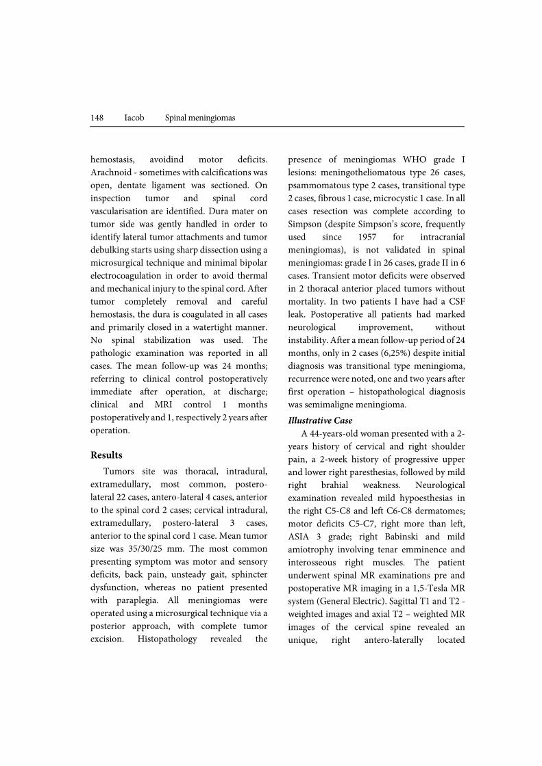
Romanian Neurosurgery (2014) XXI 2: 146 - 160 149
intradural, extramedullary space-occupying
lesion at the C2-C3 level; without extradural
component; the lesion was hypointense on T1-
weighted images, hyperintense on T2-
weighted images and presented intense
enhancement (figures 1- 3).
Figure 1 - Preoperative MRI: sagittal T2w FSE sequences reveals intradural extramedular tumor at C2 - C3 level
with homogene hyperintense T2w signal
Figure 2 - Preoperative MRI: sagittal T1w sequences without and with paramagnetic contrast media reveals the
tisular nature and the homogenous enhancement of the tumor
Figure 3 - Preoperative MRI: axial T2w sequences at the tumor level
150 Iacob Spinal meningiomas
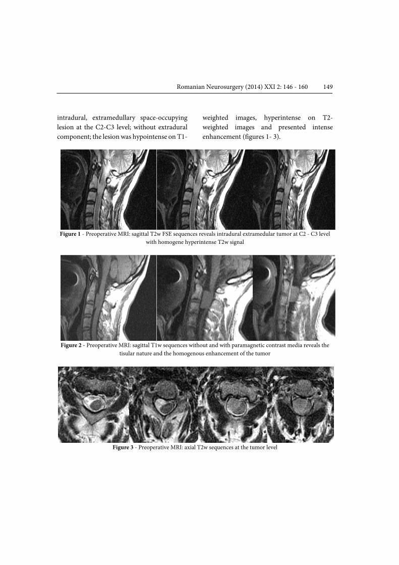
After induction of general anesthesia, the
patient was positioned prone, on chest rolls,
with head fixed in three point Mayfield head
holder. Using a posterior approach, the
posterior arch of C1, the lamina C2 and C3
were exposed and removed, without extending
bony resection to the right transverse foramen
C1-C3, allowing excellent visualisation of the
tumor and ventral dura well past the midline.
Dura was opened in a rostral-to-caudal
direction, preserving the arachnoid and tacked
laterally using 4-0 sutures to maximize
exposure, disposing Gelfoam and cotton
surgical strips laterally to assure hemostasis by
venous bleeding. The arachnoid was then
opened, dissected off the tumor and adjacent
cord. We disclosed a 3 × 2 cm, well
demarcated, red, intradural, unhomogenous,
bleeding mass, located antero-lateral,
displacing the spinal cord. Tumor was
adherent and displaced both anterior and
posterior right roots C2-C3: also tumor was
inserted on the dura mater anterior. Using the
operating microscope, the arachnoid was
opened and tumor was dissected, gradually
debulked along the lateral aspect of the spinal
canal and gently extracted from the antero-
lateral dural attachments. All nerve roots were
preserved and the mass was completely
removed. No methods of spinal reconstruction
and instrumentation were used. (Figure 4)
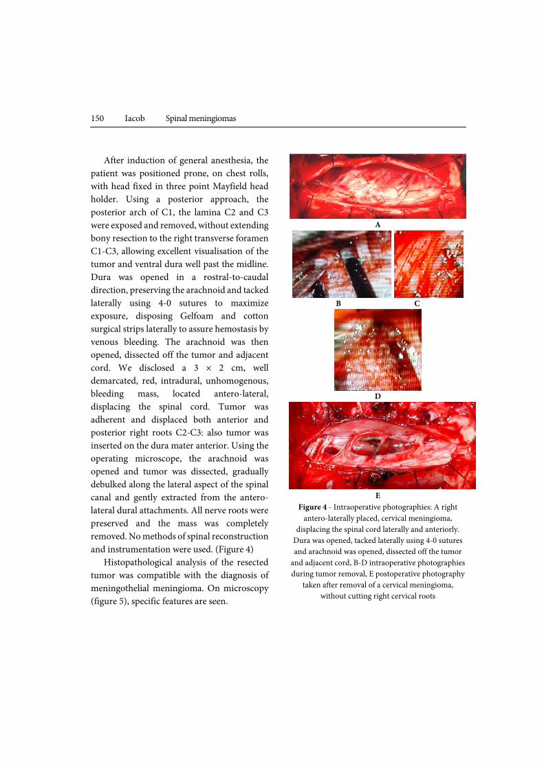
Histopathological analysis of the resected
tumor was compatible with the diagnosis of
meningothelial meningioma. On microscopy
(figure 5), specific features are seen.
A
B C
D
E
Figure 4 - Intraoperative photographies: A right
antero-laterally placed, cervical meningioma,
displacing the spinal cord laterally and anteriorly.
Dura was opened, tacked laterally using 4-0 sutures
and arachnoid was opened, dissected off the tumor
and adjacent cord, B-D intraoperative photographies
during tumor removal, E postoperative photography
taken after removal of a cervical meningioma,
without cutting right cervical roots
Romanian Neurosurgery (2014) XXI 2: 146 - 160 151
A
B
C
Figure 5 - Meningothelial meningioma microscopy
photographies:
A.H&E staining, X 100: tumour cells form lobules
surrounded by thin collagenous septae
B.H&E staining, X 200: solid areas of cells with
poorly defined cell membranes (syncytial
appeareance); like normal arachnoid, tumour cells
are uniform, with oval or round nuclei with pale
central clearing (slight tendency for the chromatin to
be marginated at the periphery)
C.H&E staining, X 200: perilobular collagen and
reticulin are variable; usually nodular vascular
thickening are seen
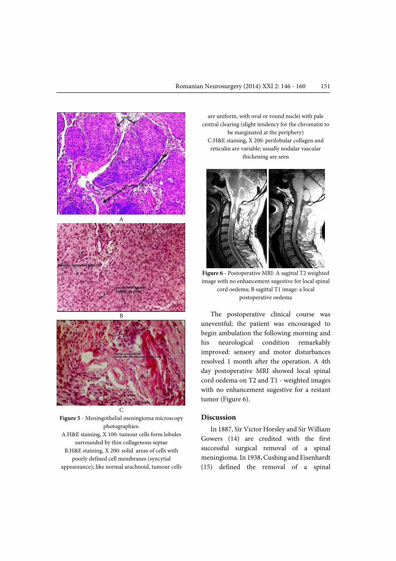
Figure 6 - Postoperative MRI: A sagittal T2 weighted
image with no enhancement sugestive for local spinal
cord oedema; B sagittal T1 image: a local
postoperative oedema
The postoperative clinical course was
uneventful; the patient was encouraged to
begin ambulation the following morning and
his neurological condition remarkably
improved: sensory and motor disturbances
resolved 1 month after the operation. A 4th
day postoperative MRI showed local spinal
cord oedema on T2 and T1 - weighted images
with no enhancement sugestive for a restant
tumor (Figure 6).
Discussion
In 1887, Sir Victor Horsley and Sir William
Gowers (14) are credited with the first
successful surgical removal of a spinal
meningioma. In 1938, Cushing and Eisenhardt
(15) defined the removal of a spinal
152 Iacob Spinal meningiomas
meningioma as “one the most gratifying of all
operative procedures”. This affirmation was
related both to the tendency of these tumors to
develop in the lateral or postero-lateral surface
of the spinal cord and to their extraaxial
development, which renders spinal
meningiomas easily dissectable from the
spinal cord. Spinal meningiomas are usually
solitary - 98%, represent 25% to 46% of all
primary spinal cord tumors, less frequently
than intracranial ones and account for
approximately 7.5–12.7% of all meningiomas
(1), with a peak incidence in the fifth and sixth
decades; similar as in my series; the mean age
was 54,7 years (range 34–82 years)(2- 8)(16).
The second most common, spinal, intradural,
extramedullary tumors after the nerve sheath
tumors; spinal meningiomas typically do not
disrupt the pia, are infrequently adherent to
the spinal cord and can be resect safely (17).
Occasionally, spinal meningiomas grow in a
sheet-like or collar-like manner around the
spinal cord, infiltrating the pia mater (18).
Multiple spinal meningiomas can occur rarely:
1 to 2%, most often associated with
neurofibromatosis type 2, especially in the
pediatric population, unrelated with
neurofibromatosis (19)(20) or after
Tamoxifen, which is a non-steroidal mixed
estrogen drug widely used for patients with
metastatic and primary breast cancer; utilized
in the treatment of malignant gliomas with
some efficacy (21).
There is a female predominance in the
adult population: female to male ratio is 2:1 in
intracranial meningiomas and 9:1 for spinal
meningiomas, no sex predilection in children
(22) (23). In my present series, the
female/male ratio was 7: 1. Female
predominance could be explained by
hormonal factors as evidenced by
progesterone and estrogen receptors
frequently found on histological examination,
as well as the reports of an association between
meningioma and breast cancer or tumor
growth and hormonal phases: pregnancy,
menopause (24). Pregnancy frequently
increase their size, explained by the discovery
that a high percentage of meningiomas have
progesterone receptors, whereas only a few
have estrogen receptors. Several other receptor
types have been found in some meningiomas,
including receptors for androgens, insulin-like
growth factor 1, epidermal growth factor,
somatostatin, and dopamine. Other
predisposing factors, primarily evaluated in
cases of cranial meningiomas, may contribute
to meningioma formation, although their roles
appear to be minimal or controversial:
previous radiation therapy, trauma and
exposure to papovavirus, SV40, BK or herpes
viruses (23).
The presumed site of origin of spinal
meningiomas from arachnoidal cap cells
located in the leptomeninges at the spinal
nerve root exit zones, adiacent to the
denticulate ligaments or entry zones of arteries
in the spinal canal; but also from
meningothelial cells making up the arachnoid
villi near the dorsal root ganglia explaining
why these tumors frequently arise in a lateral
location to the spinal cord (25). The most
common topography is the thoracic spine
80%, (laterally 45–71%, postero-laterally 10–
31% or antero-laterally to the spinal cord15–
27%) as in my series (1) (3). Cervical
Romanian Neurosurgery (2014) XXI 2: 146 - 160 153
meningiomas (9) have a tendency to be more
antero-lateral to the spinal cord 15%, than
thoracic ones, especially in patients younger
than 50 years of age, representing a true
surgical challenge and uncommon on the
lumbo-sacral spine 2–10% (3). Spinal
meningiomas usually are benign tumors with
a capsule, typically grow in a globoid
configuration with a region of dural
attachment. They are classically found in an
intradural extramedullary location, but can be
extradural (10)(16)(26); purely extradural
tumours account for only 3.5–7.0% of all
spinal meningiomas and occur more
commonly in children than in adults or both
extra- and intradural components mimiking
on a dumbbell appearance in 5% of cases (27).
There is also a very rare variety of spinal
meningioma called ‘‘en plaque’’ (first spinal
case was reported by Friedberg in 1972
(10)(11); term referring to “flat spreading
carpets of tumor” Cushing and Eisenhardt
(15) These variety are different from classic
meningiomas: extensive tumor base,
infiltration of surrounding structures, not
respect tissue planes, ossifications, difficult to
resect completely; also more likely to cause
spinal arachnoiditis (18). Intramedullary
meningiomas are exceedingly rare; only four
cases have been reported to date (23). Genetic
studies has found in meningiomas a
translocation involving the area of
chromosome 22, also known to harbor the
gene responsible for neurofibromatosis type 2
(as like as in sporadic unilateral acoustic
neuromas, sporadic spinal schwannomas and
ependymomas). The abnormal loci for the
meningioma and NF-2 genes are both on the
distal end of the myoglobin locus of
chromosome 22, but in separate regions. The
proximity of these loci may explain the
frequent association of the two tumors. Several
oncogenes (gene sequences that actively
induce tumor formation) also have been found
in meningiomas: DNA coding for the Ha-ras,
C-mos, myc, C-erb B and six oncogenes. Based
on deletions on chromosome 22, there is a two
to fourfold increase in the rate of meningiomas
seen in women with breast cancer as compared
with age-matched control subjects (23).
Clinical findings in spinal meningiomas
variate from mild to significant neurologic
dysfunction, with many patterns of clinical
presentation depending on: tumor location,
the rate of tumor growth - onset and
development are insidious, generally with a
long clinical history until a diagnosis is made;
in my present series was 13,7 months and
correlates well with the time reported in the
literature 12–24 months (9); dimension
(usually they are small), tumor aggressiveness,
the extent of spinal cord compression,
myelopathy with vascular compromise with
sensorimotor deficits, hyperactive deep
tendon reflexes, Babinski sign, gait ataxia and
weakness and the least common local or
radicular pain, sphincter disturbances
(2)(5)(12)(17). For cervical meningioma
clinical findings are insidiously installed,
asymmetric, occasionally remitting: occipital
headache, neck pain; radicular symptoms 20%
of patients, progression of motor and sensory
deficits starting in one arm, greater ipsilateral
to the side of the lesion and gradually
involving the other extremities up to
progressive tetraparesis with spasticity,
154 Iacob Spinal meningiomas
atrophy and clumsiness of the hands,
pathological reflexes, urinary incontinence -
15% of patients; because most cervical
meningiomas are located laterally or antero-
laterally, displacing the spinal cord and the
anterior spinal artery (23)(26). In younger
patients, they might have an aggressive clinical
course (3).
The current standard diagnostic study for a
spinal meningioma is MRI of the entire
neuraxis (in order to reveal tumor in other
locations) with and without gadolinium
enhancement, using both T1 and T2 –
weighted images, in sagittal and axial planes,
usefull for early diagnosis, operative planning,
improving outcome and long term follow (12)
(28-31). MRI study can delineate: tumor
location and extensions vertically and laterally,
extradural components, relation to the dura
(site of dural attachment), spinal cord,
vertebral artery (especially in the case of high
cervical and foramen magnum tumors);
presence of cord edema, intratumoral signal
changes such as necrosis, hematoma or
calcification. In general, spinal meningiomas
are well circumscribed with dural tail sign and
broad base attachment; usually isointense to
the spinal cord (T1 weighted images:
isointense to slightly hypointense, possible
heterogenous texture and T2 weighted images:
isointense to slightly hyperintense with similar
signal characteristics to intracranial
meningiomas). 15% of patients with calcified
meningiomas are hypointense on T1 and T2;
T1 weighted images + Gadolinium: immediate
and moderate homogeneous enhancement or
only minimal contrast enhancement (6) (32).
Cervical meningiomas are located
intradurally, most frequently antero-laterally,
without extension into the neural foramen
(23). They are: iso or hypointense in relation
to the spinal cord on the unenhanced T1-
weighted images, slightly hiperintense on T2 –
weighted images, with homogeneously
enhance after the intravenous administration
of gadolinium. There is no pathognomonic
picture on MR images for atypical and invasive
spinal meningiomas: heterogeneously
enhancement or in a ring-like fashion, solitary
or multiple lesions (32). In limited cases, three-
dimensional computed tomography-high
resolution/myelography is indicated alone or
in addition to MR imaging: see
contraindications for MR imaging, bone
destruction of osseous structures. Spinal CT
indicate calcifications, isodense or moderately
hyperdense mass, hyperostosis less common
as in the intracranial forms. The main
differential diagnosis of spinal meningiomas
includes intradural extramedullary
schwannomas. Statistically significant
predictors of meningioma are: female, thoracic
location - especially postero-lateral,
calcification, dural tail, broad dural contrast,
lack of foraminal extension, widening (27).
Distinctions for a diagnosis of schwannoma or
neurofibroma include: lumbar location,
relation to the nerve root, widening of neural
foramen, fluid signal intensity on T2 weighted
images, rim enhancement on MR and
scalloping on CT, not associated with a broad
dural base (6)(30). Angiography can reveal a
tumor blush with pathological vessels or early
venous drainage. Plain x-rays is usually
normal, occasionally show bone erosions, fine
calcifications within the spinal canal in only 2
Romanian Neurosurgery (2014) XXI 2: 146 - 160 155
to 5% of cases with higher frequency in the en
plaque type; these studies are usefull for
differential diagnosis when bony
abnormalities are consistent with metastatic
diseases or neural foraminal enlargement
suggest the diagnosis of nerve sheath tumor
(3). For tetraparetic patients with cervical
meningiomas, preoperative pulmonary
function tests are mandatory (23).
Surgery is the treatment of choice,
generally curative, suitable in the majority of
the cases, even in patients with severe
preoperative neurological deficits or advanced
age, the goal of treatment is total surgical
removal, including radical removal of their
dural base without causing spinal instability
and restoration of normal neurological
function (4)(5)(9)(33-36). To facilitate tumor
removal and diminish the risk of
intraoperative spinal cord damage, at surgery:
general anesthesia, high-dose corticosteroids,
the operating microscope, the irrigating
bipolar forceps to minimize heat transfer to
the spinal cord, ultrasonic dissection,
peroperative monitoring somato-sensory-
evoked potential (SSEP) - easily recordable
without adjusting the anesthetic regimen,
transcranial motor-evoked potentials
(TcMEPs) and continuous free running
electromyography (EMG) - evaluate the
pyramidal motor pathways, giving an
immediate and conclusive feedback of motor
tract integrity should be used (35). Generally
surgical steps are: safe approach, gentle spinal
cord decompression without rotation, by
gradual tumor debulking, tumor capsule
dissection with progressively removal. To
improve Simpson resection grade (obtaining a
Simpson Grade I resection still is a desiderate,
affecting recurrence rate and survival), even
for calcified meningiomas "en plaque", dural
attachment is in most reported studies:
coagulated, separated: the inner layer is
resected together with the tumor, preservind
the outer layer or resection of the dural
attachment with suturing of a patchgraft.
However, in large series, there is no significant
superiority of dural base resection over
patients with dural base coagulation because
recurrence is wery low (36)(41).
Alternative approaches are:
-posterior approach: more convenient, less
invasive, technically more easy for dorsally
placed tumors who can be removed totally,
well-tolerated especially in older patients,
allow wide enough exposure of the tumor and
the dural attachment, even for those large
tumors, use a standard hemilaminectomy,
laminectomy or laminoplasty – postero-lateral
approach (33), at one or two levels, with lateral
extension even for anterior and antero-lateral
meningiomas (35) if intraoperative
monitoring are used ! (transcranial motor-
evoked potentials (TcMEPs), somatosensory-
evoked potential (SSEP) and free running
electromyography (EMG), with fewer
potential complications, eliminate the
necessity of vertebrectomy; is facilitating
complete tumor excision with good results.
Posterior approach disadvantages are: limited
space to expose the tumor and the need for
spinal cord manipulation
-anterior approach indicated in anterior
cervical meningioma or by a transthoracic
approach for anterior spinal thoracic
menigiomas (33)(36-39). Such approach has
156 Iacob Spinal meningiomas
many advantages: large bony window of
access through a corpectomy, good tumor
visualization in front of the spinal cord, good
control over bleeding during meningioma
resection, no spinal cord manipulation during
meningioma resection, preserving anterior
spinal artery and nerve roots vessels. Anterior
approach disadvantages are: time consuming,
generate the necessity to fixation–fusion of the
spine both anterior on several levels and
sometimes posterior (posterior fixation is
recommended by many authors after three
levels of corpectomy)(40), cerebrospinal fluid
leakage which could be avoided by watertight
dural closure, application of fibrin glue, filling
the dead space with muscle fascia and
prophylactic lumbar CSF drainage (49).
For high cervical spinal meningiomas-
from the C3 cephalad (23) special
considerations are made:
- surgical indication must be reserved to
patients with signs or symptoms of spinal cord
compression installed rapidely in spite of
corticosteroid administration. For the
asymptomatic patient or for patients with NF-
1 and multiple tumors, a closed followed up
conservatively with serial MRIs should be
performed.
- high cervical meningiomas tend to be
intradural, entirely extradural or have both
intra-and extradural components.
-tumors located posteriorly or postero-
lateral to the spinal cord are approached
through the postero-lateral approach, a lateral
extension of the standard midline posterior
approach. These technique enables the
approach of most intradural extramedullary
tumors, enlargement toward the opposite side
for those located more anteriorly, especially
for large tumors and may offer controll to both
vertebral arteries, especially in high cervical
meningiomas with extradural component,
where to achieve a complete resection, the
vertebral artery should be mobilized.
-tumors placed ventral or ventro-lateral to
the spinal cord are removed through a straight
forward anterior approach, providing a wide
field, also a good dural closure.
- after total removal, significant
neurological improvements are usually
observed, whereas tumor recurrence rate has
been reported to be 1.3–14%.
Spinal meningioma are histopathologically
usually benign (more than 95% being classified
as WHO grade I lesions); are slowly growing
tumors, without pia mater invasion–
important anatomical detail, which improves
the ability to resect them safely.
Meningotheliomatous, Roux et al. 44% in their
series (41) and psammomatous subtypes
(20%) are the most common, similar to my
series; also immunohistochemical staining
positive for vimentin and epithelial membrane
antigen, progesterone receptor activity was
found frequently Another histological
subtypes cited in the literature are: fibroblastic,
chordoid, transitional, vacuolated, clear cell,
but also atypical or WHO grade II, malignant
or WHO grade III, meningosarcoma (32). In
younger patients, meningiomas grow in a
more aggressive pattern, especially in cervical
topography, frequently with an extradural
component, are very invasive, angioblastic,
involving the bone and cervical soft tissue,
including the vertebral artery. There are not
valid data about the rate of atypical and
Romanian Neurosurgery (2014) XXI 2: 146 - 160 157
malignant meningioma in the spinal canal.
Despite Ki-67 index value, known as predictor
of intracranial meningiomas for recurrence
and overall survival, for spinal meningiomas,
the value of this index in predicting behavior
has not been yet fully elucidated and further
studies are needed (42)(43). Comparing spinal
and intracranial meningiomas the
proliferation rates of intracranial
meningiomas were significant higher (Ki-67
3.6% versus spinal Ki-67 of 2.48%)
Particular situation are leated to spinal en
plaque meningioma, who may not always be
totally resectable because this variety tends to
invade the arachnoid layer (18)(27) see Caroli
et al. (11) in only 3 of the patients from a series
of 7 cases of en plaque spinal meningiomas.
For partially resected tumors recurrence is the
rule. Similar in patients with heavily calcified
meningiomas with a ventral or ventro-lateral
location, a partial vertebrectomy and/or
costotransversectomy approach allows safer
tumor manipulation and removal, with
subsequent instrumentation; especially those
tumors occurring at the cervico-thoracic
and/or thoraco-lumbar junction, dorsal
stabilization should be considered to prevent
junctional kyphosis. Generally the prognosis
of benign spinal meningioma with complete
resection is very good, even when preoperative
neurological status is poor; improvement are
seen over 90% of the cases immediately or
gradual up to 18 months, with a good long
term functional outcome (2)(5)(9)(13). Spinal
menigioma prognosis are more favorable
compared to intracranial localizations: mean
6.2% for morbidity and 2.1% for mortality;
related to the preoperative neurological status,
the patient's age, the duration of symptoms,
tumor location in the spinal canal: anterior or
antero-lateral, total removal of the lesion,
histological tumor grade, Simpson resection
grade, invasion of pia mater (9)(44).
Meningiomas located directly anteriorly, a low
preoperative Karnofsky score, a short history,
calcified tumor (45) with en plaque extension,
cervical location with an extradural
component (which are often more vascular
and aggressive), tumor progression, invasion
of the arachnoid/pia, arachnoid scarring (46);
also reoperation for recurrent tumor may
expose to transiently neurological worsening
after surgery. Nevertheless even after
successful surgery, a possible increase of the
motor deficit or even permanent deficit
(paraplegia) could be seen if spinal
meningiomas are located anteriorly to the
spinal cord. Klekamp J, Samii M.(46) found
11.2% of complication in 117 cases of spinal
cord meningiomas and Solero et al. (47) found
3.5% in 174 cases; also sensory disturbances,
dysesthesia syndrome progressing to
myelopathy (46).
Recurrence after spinal meningioma
resection has been reported to be 1.3–14%
(4)(48); for meningiomas that have been
totally resected is 1.3% at 5 years and 6% at 14
years, related to: subtotal resection, anterior
location, calcification, malignancy proved
histologically, multiplicity of lesions, young
age. Recurrence in the patients with dural base
coagulation was so low, that no significant
superiority of dural base resection over dural
base coagulation could be found (41).
Postoperatory complications which could
be avoided by a meticulous technique (46)(49)
158 Iacob Spinal meningiomas
are: CSF leak (2 cases in my series),
pseudomeningocele development, wound
infections, aseptic meningitis, arachnoiditis
(as a result of blood left in the subarachnoid
space or as a result of placement of dural
substitutes of animal origin), syringomyelia,
spinal destabilization, as well as other routine
complications of spinal surgery and general
anesthesia. Less frequent complications are
pulmonary embolism, pneumonia,and
myocardial infarction. pulmonary embolism.
In invasive and high-grade spinal
meningiomas therapeutic alternatives are:
extensive tumor resection, completed with
radiosurgery (50) or stereotactic CyberKnife
frameless stereotactic radiosurgery as a single
fraction therapy (51)(52), combined with
chemotherapy (as hydroxyurea), hormonal
manipulation with tamoxifen (antiestrogen),
mifepristone (antiprogesterone), recombinant
interferon α2b.
Conclusions
My personal experience based on this
retrospective study argue that even when
tumor is late discovered and neurological
compromise exist, in the high cervical area or
anterior spine topography, spinal
meningiomas can be successfully resected
today with favorably outcomes, low
complication rates, offering a better life
quality. Early diagnosis by MRI,
corticosteroids, microsurgery techniques,
intraoperative electrophysiological
monitoring are mandatory. For anterior spine
approaches if an anterior approach is chosed,
reconstruction and instrumentation methods
should be used. In cases of semimalignant or
malignant transformation, close follow-up
and adjuvant therapies must be considered.
Correspondence
Dr. Luis Rafael Moscote. Universidad de
Cartagena, Cartagena de Indias, Colombia.
e-mail: [email protected]
References
1. Albanese V, Platania N - Spinal intradural
extramedullary tumors. Personal experience; J Neurosurg
Sci 2002, 46:18-24
2. Gezen F, Kahraman S, Canakci Z - Review of 36 cases
of spinal cord meningioma. Spine 2000, 25:727-731
3. Cohen-Gadol AA, Zikel OM, Koch CA - Spinal
meningiomas in patients younger than 50 years of age: a
21-year experience, J Neurosurg (Spine) 2003, 98:258-263
4. Gottfried, O.N., et al. - Spinal meningiomas: surgical
management and outcome, Neurosurg Focus 2003, 14
(6), e2
5. Morandi, X., Haegelen, C., et al., - Results in the
operative treatment of elderly patients with spinal
meningiomas. Spine 2004 29 (19), 2191-2194
6. De Verdelhan O, et al. - MR imaging features of spinal
schwannomas and meningiomas; J.Neroradiol 2005,
32:42-49
7. Haegelen, C., et al. - Results of spinal meningioma
surgery in patients with severe preoperative neurological
deficits. Eur Spine 2005, J 14 (5), 440-444
8. Cavanaugh, D.A., et al. - Intraspinal meningioma in a
101-year-old: should age determine the aggressiveness of
intervention?, Surg.Neurol. 2008, 69 (2), 130-134
9. Riad H., S. Knafo S. et al. - Spinal meningiomas:
Surgical outcome and literature review, Neurochirurgie
2013, 59, 30-34
10.Achari G, et al. - Extradural meningioma en-plaque of
the cervical cord, Neurol Res. 2000; 22: 551-553
11.Caroli E, Acqui M, Roperto R, et al. Spinal en-plaque
meningiomas: a comtemprary experience. Neurosurgery.
2004; 55: 1275-1279
12.Saraceni, C., Harrop, J.S., - Spinal meningioma:
chronicles of contemporary neurosurgical diagnosis and
management, Clin Neurol Neurosurg 2009, 111 (3), 221-
226
13.Yoon, S.H., et al. - Surgical outcome of spinal canal
Romanian Neurosurgery (2014) XXI 2: 146 - 160 159
meningiomas, J Korean Neurosurg Soc 2007, 42 (4), 300-
3004
14.Mulholland R.C. - Sir William Gowers 1845-1915,
Spine 1996, 21: 1106-1110
15.Cushing H. &Eisenhardt L. - Meningiomas: Their
Clasification, Regional Behaviour, Life History and
Surgical End Results, Springfield 1938, IL: Charles C.
Thomas
16.Dagain, .A., et al. - Extradural spinal meningioma: case
report. Neurochirurgie 2009, 55 (6), 565-568
17.Sandalcioglu, I.E., et al. - Spinal meningiomas: critical
review of 131 surgically treated patients. Eur Spine J 2008,
17 (8), 1035-1041
18.Gamache F, Wang J, Deck M, Heise C - Unusual
appearance of an en plaque meningioma of the cervical
spinal canal. A case report and literature review, Spine
2001, 26(5): E87-E89
19.Silva J.A., Holanda M.M., et al. - Multiple
meningiomas within the spinal canal: case report with 23
tumors, Arq Neuropsiquiatr.2005; 63: 166-170
20.Keskin F., Kalkan E., Karatas Y - A Case of Upper
Thoracic Intradural-Extramedullary Multiple
Meningiomas, Neurosurg Q 2013; 23: 224-225
21.Colazza G.B. et al.- Multiple spinal meningiomas after
tamoxifen therapy: a case report, Neurol Sci 2003, 24: 37-
39
22.Peker S, et al. - Spinal meningiomas: evaluation of 41
patients. J NeurosurgSci 2005, 49:7-11
23.Cooper P.R., Wienecke R.J., White B.J. - Spinal
Meningiomas, in Batjer H.H., Loftus Ch.M. - Textbook of
Neurological Surgery, Lippincott Williams & Wilkins,
2003, vol. II, 1857-1864.
24.Barnholtz-Sloan J.S., Kruchko C. - Meningiomas:
causes and risk factors. Neurosurg Focus 2007, 23 (4), E2.
25.Setzer M., Vatter H., et al. - Management of spinal
meningiomas: surgical results and a review of the
literature, Neurosurg. Focus 2007, 23 (4): E14
26.Frank, B.L., et al. - Cervical extradural meningioma:
case report and literature review. J Spinal Cord Med 2008,
31 (3), 302-305
27.Messori A., Rychlicki F., Salvolini U. - Spinal epidural
en-plaque meningioma with an unusual pattern of
calcification in 14-year-old girl: case report and review of
the literature, Neuroradiology 2002, 44: 256-260
28.Abul-Kasim K, et al. - Intradural spinal tumors:
current classification and MRI features. Neuroradiology
2008, 50:301-314
29.Alorainy I.A - Dural tail sign in spinal meningiomas,
Eur J Radiol. 2006, 60: 387-391
30.Gebauer G.P., Farjoodi P., et al. - Magnetic resonance
imaging of spinal tumours: classification, differential
diagnosis and spectrum of disease, J. Bone Jnt.Surg Am
2008, 90: 146-162
31.Lee J.Y. - Radiological findings of spinal
schwannomas and meningiomas: focus on
discrimination of two disease entities, Eur Radiol 2009,
19: 2707-2715
32.Schaller, B., - Spinal meningioma: relationship
between histological subtypes and surgical outcome? J
Neurooncol 2005, 75 (2), 157-161
33.Gambardella G, Gervasio O, Zaccone C - Approaches
and surgical results in the treatment of ventral thoracic
meningiomas. Review of our experience with a postero-
lateral combined transpedicular-transarticular approach.
ActaNeurochir (Wien) 2003, 145:385-392
34.Sacko O. et al. - Spinal meningioma surgery in elderly
patients with paraplegia or severe paraparesis:a
multicenter study. Neurosurgery 2009, 64:503-509
35.Voulgaris S. et al. - Posterior approach to ventrally
located spinal meningiomas, Springer-Verlag 2010, Eur
Spine J 2010, 19:1195-1199
36.Payer M. - The anterior approach to anterior cervical
meningiomas: review illustrated by a case, ActaNeurochir
(Wien) (2005) 147: 555-560
37.D'Aliberti G., Talamonti G., Villa F. - Anterior
approach to thoracic and lumbar spine lesions: results in
145 consecutive cases. J Neurosurg Spine 2008, 9: 466-482
38.Banczerowski P., Lipoth L., Vajda J., Veres R. - Surgery
of ventral intradural midline cervical spinal pathologies
via anterior cervical approach: our experience. Ideggyogy
Sz 2003, 56: 115-118
39.Brunon J., Fuentes J.M. - Anterior and antero-lateral
surgery of the lower cervical spine (25 years after H.
Verbiest), 2: Indications, results, complications,
Neurochirurgie 1996, 42(4-5): 229-248
40.Singh K, Vaccaro A, et al. - Biomechanical
comparison of cervical spine reconstructive techniques
after a multilevel corpectomy of the cervical spine. Spine
2003, 28(20): 2352-2358
41.Roux F.X, Nataf F., Pinaudeau M., et al. - Intraspinal
meningiomas: review of 54 cases with discussion of poor
prognosis factors and modern therapeutic management.
Surg Neurol. 1996;46:458-464
160 Iacob Spinal meningiomas
42.Bruna J, et al. - Ki-67 proliferative index predicts
clinical outcome in patients with atypical or anaplastic
meningioma. Neuropathology 2007, 27:114-120
43.Roser F, Nakamura M, Bellinzona M - Proliferation
potential of spinal meningiomas. Eur Spine J 2006,
15:211-215
44.Pena M., Galasko C.S., Barrie J.L. - Delay in diagnosis
of intradural spinal tumors. Spine 1992, 17: 1110-1116,
45.Naderi, S., et al., - Ossified thoracic spinal meningioma
in childhood: a case report and review of the literature.
Clin Neurol Neurosurg 2001, 103 (4), 247-249
46.Klekamp J, Samii M. - Surgical results for spinal
meningiomas, Surg. Neurol. 1999; 52:552-562; also
Klekamp J, Samii M. - Meningiomas, Surgery of the spinal
Tumors, Springer 2007, 4.5.1., 248-260
47.Solero C.L., Fornari M., Giombini S., et al. - Spinal
meningiomas: review of 174 operated cases.
Neurosurgery 1989; 125:153-160
48.Nadkarni, B., et al. - Recurrent spinal meningioma: a
case report withreview of the literature. J OrthopSurg
(Hong Kong) 2005, 13 (3), 326-329
49.Misra, S.N., - Avoidance of structural pitfalls in spinal
meningioma resection. Neurosurg Focus 2003, 14 (6), e1
50.Gerszten P.C., Burton S.A., Ozhasoglu C. -
Radiosurgery for benign intradural spinal tumors,
Neurosurgery 2008, 62:887-895
51.Robert L.D., Mi-Ryeong R. et al. - CyberKnife
Radiosurgery for Benign Intradural Extramedullary
Spinal Tumor, Neurosurgery, 2006, 58, 4, 674-685
52.Chang UK, Rhee CH, et al. - Radiosurgery using the
Cyberknife for benign spinal tumors: Korea Cancer
Center Hospital experience. J Neuroonc. 2011 Jan; 101(1):
91-9


![Surgical Excition of Spinal Intradural Meningiomas through ... · Unilateral hemilaminectomy was firstly reported by Taylor [1] and then it was popularized by Eggert et al. [2] and](https://img.pdfslide.us/doc/110x75/5acb40107f8b9ad13e8b5b58/surgical-excition-of-spinal-intradural-meningiomas-through-hemilaminectomy-was.jpg)


![Foramen magnum meningiomas: detailed surgical ......Meningiomas are common neoplasms representing 14.3 to 19% of all intracranial tumors [63]. Among all the meningiomas, only 1.8 to](https://img.pdfslide.us/doc/110x75/60aa2d3285131731732f9abe/foramen-magnum-meningiomas-detailed-surgical-meningiomas-are-common-neoplasms.jpg)


![Surgical management of clinoidal meningiomas: 10 cases ... · sphenoid wing or inner sphenoid wing meningiomas[1,2]. However, accumulating anatomical knowledge and clinical experience](https://img.pdfslide.us/doc/110x75/5eca8277e895a04bfa1c336b/surgical-management-of-clinoidal-meningiomas-10-cases-sphenoid-wing-or-inner.jpg)