Embed Size (px)
Citation preview
CLINICAL ARTICLEJ Neurosurg 130:435–442, 2019
Tumors involving the cavernous sinus (CS) are rare lesions, which represent less than 1% of all intra-cranial tumors. Meningiomas are the most common
tumors of the CS (MCSs; 41%), accounting for 1.8% of all intracranial meningiomas.19,20 The symptoms caused by MCSs are related to compression of neighboring cranial nerves (CNs): VI (80%), V, IV, III (30%), and II.10,22
There are many heterogeneities for the definition of MCSs. MCSs with an extracavernous extension (sella, tem-poral, clivus), sphenocavernous meningiomas secondarily invading the CS, and meningiomas strictly confined to the CS are studied and described without any anatomical distinctions and without any details on the initial location,
although both are different entities. In this work we have exclusively studied the meningiomas confined to the CS.
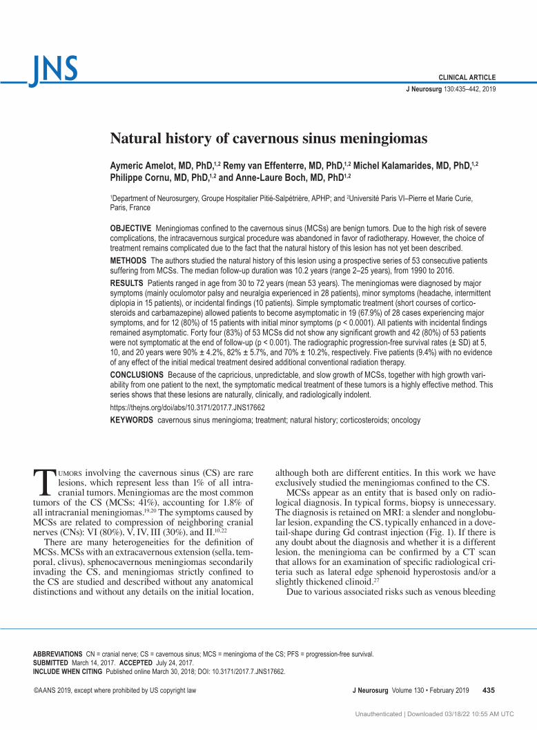
MCSs appear as an entity that is based only on radio-logical diagnosis. In typical forms, biopsy is unnecessary. The diagnosis is retained on MRI: a slender and nonglobu-lar lesion, expanding the CS, typically enhanced in a dove-tail-shape during Gd contrast injection (Fig. 1). If there is any doubt about the diagnosis and whether it is a different lesion, the meningioma can be confirmed by a CT scan that allows for an examination of specific radiological cri-teria such as lateral edge sphenoid hyperostosis and/or a slightly thickened clinoid.27
Due to various associated risks such as venous bleeding
ABBREVIATIONS CN = cranial nerve; CS = cavernous sinus; MCS = meningioma of the CS; PFS = progression-free survival.SUBMITTED March 14, 2017. ACCEPTED July 24, 2017.INCLUDE WHEN CITING Published online March 30, 2018; DOI: 10.3171/2017.7.JNS17662.
Natural history of cavernous sinus meningiomasAymeric Amelot, MD, PhD,1,2 Remy van Effenterre, MD, PhD,1,2 Michel Kalamarides, MD, PhD,1,2 Philippe Cornu, MD, PhD,1,2 and Anne-Laure Boch, MD, PhD1,2
1Department of Neurosurgery, Groupe Hospitalier Pitié-Salpétrière, APHP; and 2Université Paris VI–Pierre et Marie Curie, Paris, France
OBJECTIVE Meningiomas confined to the cavernous sinus (MCSs) are benign tumors. Due to the high risk of severe complications, the intracavernous surgical procedure was abandoned in favor of radiotherapy. However, the choice of treatment remains complicated due to the fact that the natural history of this lesion has not yet been described.METHODS The authors studied the natural history of this lesion using a prospective series of 53 consecutive patients suffering from MCSs. The median follow-up duration was 10.2 years (range 2–25 years), from 1990 to 2016.RESULTS Patients ranged in age from 30 to 72 years (mean 53 years). The meningiomas were diagnosed by major symptoms (mainly oculomotor palsy and neuralgia experienced in 28 patients), minor symptoms (headache, intermittent diplopia in 15 patients), or incidental findings (10 patients). Simple symptomatic treatment (short courses of cortico-steroids and carbamazepine) allowed patients to become asymptomatic in 19 (67.9%) of 28 cases experiencing major symptoms, and for 12 (80%) of 15 patients with initial minor symptoms (p < 0.0001). All patients with incidental findings remained asymptomatic. Forty four (83%) of 53 MCSs did not show any significant growth and 42 (80%) of 53 patients were not symptomatic at the end of follow-up (p < 0.001). The radiographic progression-free survival rates (± SD) at 5, 10, and 20 years were 90% ± 4.2%, 82% ± 5.7%, and 70% ± 10.2%, respectively. Five patients (9.4%) with no evidence of any effect of the initial medical treatment desired additional conventional radiation therapy.CONCLUSIONS Because of the capricious, unpredictable, and slow growth of MCSs, together with high growth vari-ability from one patient to the next, the symptomatic medical treatment of these tumors is a highly effective method. This series shows that these lesions are naturally, clinically, and radiologically indolent.https://thejns.org/doi/abs/10.3171/2017.7.JNS17662KEYWORDS cavernous sinus meningioma; treatment; natural history; corticosteroids; oncology
J Neurosurg Volume 130 • February 2019 435©AANS 2019, except where prohibited by US copyright law
Unauthenticated | Downloaded 03/18/22 10:55 AM UTC
A. Amelot et al.
J Neurosurg Volume 130 • February 2019436
(which is difficult to control), wounds in the internal ca-rotid artery, intratumoral bleeding, and morbidity caused by accidental damage to the oculomotor nerve or trigemi-nal nerve, surgical approaches to the CS have practically been abandoned.23 Surgery is sometimes necessary and recommended only for extracavernous portions of sphe-nocavernous meningiomas. Radiation therapy is now the standard treatment of MCSs, but its mid- and long-term complications should not be underestimated.
What complicates the choice of a treatment strategy for MCSs is an essential point: the poorly studied natural his-tory of the disease. Indeed, it is not conceivable to random-ize therapeutic lesions that lead to little or no symptoms, or for which the symptoms are fluctuating. Unfortunately, while the knowledge of the natural history of MCSs is the prerequisite for adapting a therapy, it is also the least stud-ied aspect in the literature. No study specifically dedicated to the growth of meningiomas confined to the CS could be found in the literature. All radiotherapy series analyze the development of tumors under treatment and claim to have obtained frequent stabilization or decreases in tumor volume. However, these series never compare the results to those of a control group, i.e., untreated MCSs.
The aim of our work was to determine the natural his-tory of MCSs through a consecutive series of 53 patients with this lesion. All patients were treated symptomati-cally, presenting a low expandability of their tumor, and were closely followed up both radiographically and clini-cally. This prospective study, with a median follow-up > 10 years, will aid in gaining insights into the treatment of such tumors.
MethodsThe local ethics committee of La Pitié Salpétrière Hos-
pital approved this study. Written consent was obtained and recorded in each patient’s medical file.
Study PopulationFrom 1990 to 2016, 90 adults harboring MCSs were
diagnosed by 3 neurosurgeons and their cases were sub-sequently followed. Fifty-three patients treated for menin-giomas confined to the CS, newly diagnosed using brain MRI, were included. Thirty-seven patients who had a me-
ningioma that expanded beyond the CS with an extracav-ernous portion (i.e., sphenoid, sella, temporal, clinoid, etc.) or invading the CS secondarily were excluded from this study.
At diagnosis, all patients were clinically evaluated with particular attention to both an ophthalmological (visual acuity, visual field, and coordimetric examinations accord-ing to Hess-Lancaster) and CN examination. Medication history investigations focused on hormonal intake (such as cyproterone acetate or treatment of menopause) were performed. Radiological examination included brain MRI, and sometimes, if deemed necessary, a CT scan.
Follow-UpAll patients were followed-up prospectively with a
neurological examination and a neuro-ophthalmological assessment at 6 weeks to assess the effectiveness of symp-tomatic treatment when it was established. Furthermore, follow-up examinations were systematically performed at 6 months, every year for 3 years, then every 2 years after-ward. Each time, the patient underwent MRI and a clinical examination. The median follow-up duration of our series was 10.8 ± 5.55 years (range 2–25.2 years). Six patients (11%) were followed < 5 years, 19 patients (35.9%) be-tween 5 and 10 years, and 28 patients (52%) > 10 years, including 10 patients (18.5%) > 15 years, with a maximum of 25 years. In the course of follow-up evaluations, some patients worsened clinically or radiologically because of their tumor. In these cases, the initial course of observa-tion without intervention was modified accordingly to in-terventional radiotherapy: 3 patients by radiotherapy (36 Gy), and 2 others by Gamma Knife radiosurgery (16 Gy). Even if these cases could be considered outside the bounds of analysis of natural history in the strict sense, the study of such patients was continued.
MRI and Tumor VolumeDuring initial evaluation and follow-up, the tumor vol-
ume was assessed by 3D gradient echo Tl-weighted post-Gd sequences, with 0.7-mm slice thickness and no gap, by manual segmentation using OsiriX software (version 4.1.2, OsiriX). Tumor progression was defined as a > 20% increase in volume, and tumors that showed < 20% reduc-
FIG. 1. Axial brain T1-weighted MRI with Gd in CS meningiomas showing examples in patients with an incidental finding (A), minor symptoms (B), and major symptoms (C).
Unauthenticated | Downloaded 03/18/22 10:55 AM UTC
J Neurosurg Volume 130 • February 2019 437
A. Amelot et al.
tion and < 20% increase in volume were categorized as stable.4
Statistical AnalysesStatistical analyses were performed using Stata (ver-
sion 11, StataCorp). Associations between different group parameters or patient groups were analyzed with the Fish-er’s exact test. Sex and initial clinical presentation were considered categorical variables, while age and lesion vol-ume were considered continuous variables. Clinical and radiographic progression-free survival (PFSs) were plot-ted according to the Kaplan-Meier method. All tests were 2-sided, and p values < 0.05 were considered significant.
ResultsEpidemiology
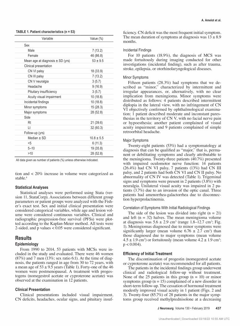
From 1990 to 2014, 53 patients with MCSs were in-cluded in the study and evaluated. There were 46 women (87%) and 7 men (13%; sex ratio 6:1). At the time of diag-nosis, the patients ranged in age from 30 to 72 years, with a mean age of 53 ± 9.5 years (Table 1). Forty-one of the 46 women were postmenopausal. A treatment with proges-togens (nomegestrol acetate or cyproterone acetate) was observed at the examination in 12 patients.
Clinical PresentationClinical presentations included visual impairment,
CN deficits, headaches, ocular signs, and pituitary insuf-
ficiency. CN deficit was the most frequent initial symptom. The mean duration of symptoms at diagnosis was 13 ± 8.9 months.
Incidental FindingsFor 10 patients (18.9%), the diagnosis of MCS was
made fortuitously during imaging conducted for other investigations (incidental finding), such as after trauma, stroke, epilepsia, or otorhinolaryngological diseases.
Minor SymptomsFifteen patients (28.3%) had symptoms that we de-
scribed as “minor,” characterized by intermittent and irregular appearances, or, alternatively, with no clear implication from meningioma. Minor symptoms were distributed as follows: 4 patients described intermittent diplopia in the lateral view, with no infringement of CN VI objectively confirmed by ophthalmological examina-tion; 1 patient described moderate and inconstant pares-thesias in the territory of CN V, with no facial nerve pain or hypoesthesia; another patient complained of visual acuity impairment; and 9 patients complained of simple retroorbital headache.
Major SymptomsTwenty-eight patients (53%) had a symptomatology at
diagnosis that can be qualified as “major,” that is, perma-nent or debilitating symptoms and clearly attributable to the meningioma. Twenty-three patients (40.7%) presented with impaired oculomotor nerve function: 14 patients (26.4%) had CN VI palsy, 7 patients (13%) had CN III palsy, and 2 patients had both CN VI and CN II palsy. No abnormality of CN IV was detected (Table 1). Trigeminal signs and symptoms were present in 2 patients (3.8%) with neuralgia. Unilateral visual acuity was impaired in 2 pa-tients (3.7%) due to an invasion of the optic canal. Three patients had amenorrhea-galactorrhea due to disconnec-tion hyperprolactinemia.
Correlation of Symptoms With Initial Radiological FindingsThe side of the lesion was divided into right (n = 21)
and left (n = 32) halves. The mean meningioma volume at diagnosis was 5.6 ± 2.9 cm3 (range 1.8–12.7 cm3; Fig. 1). Meningiomas diagnosed due to minor symptoms were significantly larger (mean volume 6.76 ± 2.7 cm3) than those diagnosed due to major symptoms (mean volume 4.5 ± 1.9 cm3) or fortuitously (mean volume 4.2 ± 1.9 cm3; p < 0.004).
Efficiency of Initial TreatmentThe discontinuation of progestin (nomegestrol acetate
or cyproterone acetate) was recommended for all patients.The patients in the incidental findings group underwent
clinical and radiological follow-up without treatment. None of the 25 patients in this group (n = 10) or minor symptoms group (n = 15) complained of a new disorder in short-term follow-up. The cessation of hormonal treatment modestly improved visual acuity in 1 patient (Figs. 2 and 3). Twenty-four (85.7%) of 28 patients in the major symp-toms group received methylprednisolone at a decreasing
TABLE 1. Patient characteristics (n = 53)
Variable Value (%)
Sex Male 7 (13.2) Female 46 (86.8)Mean age at diagnosis ± SD (yrs) 53 ± 9.5Clinical presentation CN VI palsy 18 (33.9) CN III palsy 7 (13.2) CN V neuralgia 3 (5.7) Headache 9 (16.9) Pituitary insufficiency 3 (5.7) Acuity visual impairment 10 (18.8)Incidental findings 10 (18.8)Minor symptoms 15 (28.3)Major symptoms 28 (52.8)Side Rt 21 (39.6) Lt 32 (60.3)Follow-up (yrs) Median ± SD 10.8 ± 5.5 <5 6 (11.3) 5–10 19 (35.8) >10 28 (52.8)
All data given as number of patients (%) unless otherwise indicated.
Unauthenticated | Downloaded 03/18/22 10:55 AM UTC
A. Amelot et al.
J Neurosurg Volume 130 • February 2019438
dose for 6 weeks; 3 patients (5.6%) received bromocriptine for endocrine disorders, and 1 patient (1.8%) received car-bamazepine for neuralgia.
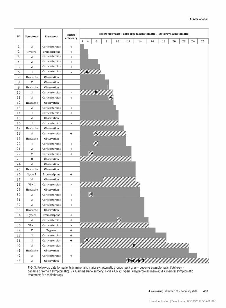
Symptom ImprovementAt the control consultation at 6 weeks, 22 patients
(78.6%) improved and demonstrated a regression of their major symptoms (p < 0.0001, Pearson’s chi-square test) as a direct consequence of their medical treatment (cor-ticosteroids/carbamazepine or bromocriptine). These pa-tients were distributed as follows: 13 patients with CN VI palsy, 4 patients with CN III palsy, 2 patients with CN V neuralgia, and 3 patients with disconnection hyperpro-lactinemia. After cessation of this initial symptomatic treatment, 18 patients (64.3%) did not experience relapse of their disorders; 1 had a recurrence of CN V neuralgia, which required resumption of carbamazepine treatment, and 3 patients with initial CN VI palsy relapsed after ces-sation of corticosteroids, but the installation of an eyeglass prism to limit diplopia allowed them to resume a normal life (Fig. 2).
No Symptom ImprovementSix patients did not improve after the initial symptom-
atic treatment: 2 patients had CN VI palsy associated with visual acuity loss, 3 patients had CN III palsy, and 1 pa-tient had isolated CN VI palsy.
Clinical Correlation With Radiological FindingsThe mean volume of meningiomas in patients whose
condition was improved by medical treatment was 4.4 cm3 ± 1.8 cm3 versus 4.3 ± 2.06 cm3 in patients who did not improve (p = 0.92, Student t-test).
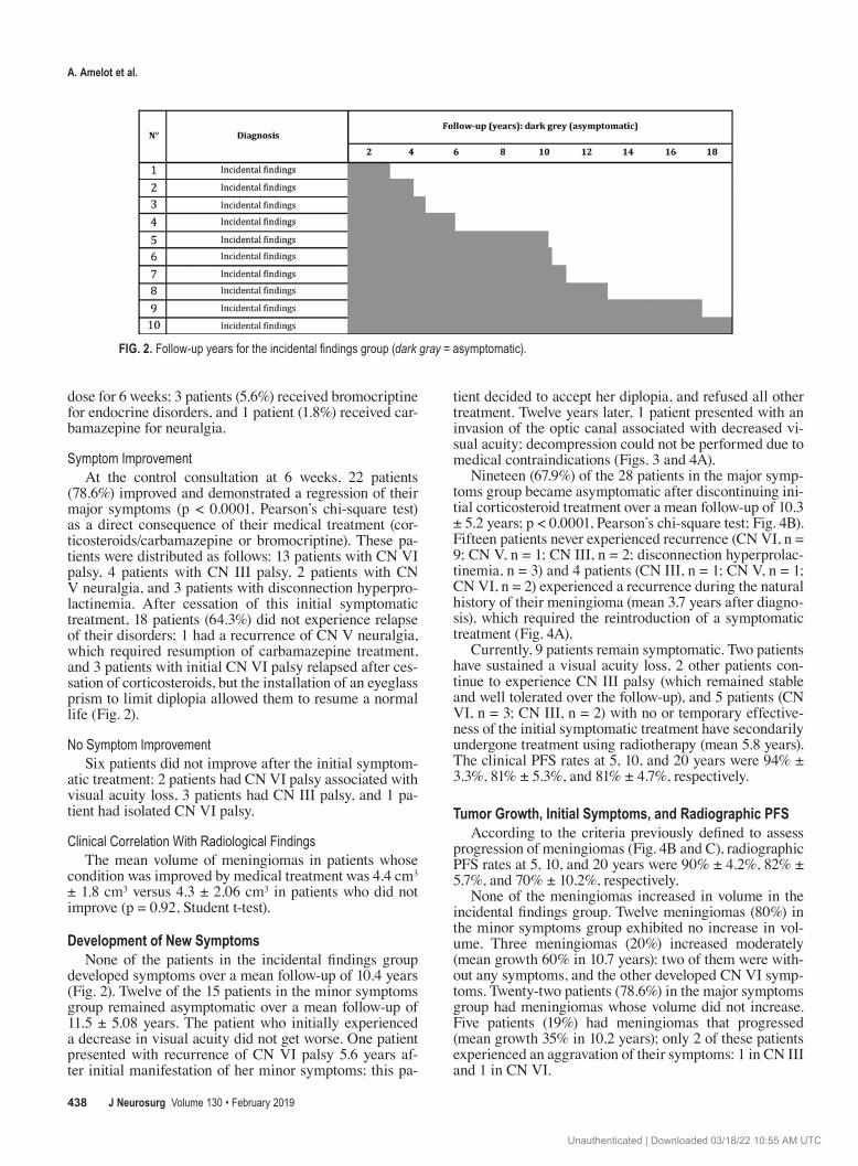
Development of New SymptomsNone of the patients in the incidental findings group
developed symptoms over a mean follow-up of 10.4 years (Fig. 2). Twelve of the 15 patients in the minor symptoms group remained asymptomatic over a mean follow-up of 11.5 ± 5.08 years. The patient who initially experienced a decrease in visual acuity did not get worse. One patient presented with recurrence of CN VI palsy 5.6 years af-ter initial manifestation of her minor symptoms; this pa-
tient decided to accept her diplopia, and refused all other treatment. Twelve years later, 1 patient presented with an invasion of the optic canal associated with decreased vi-sual acuity; decompression could not be performed due to medical contraindications (Figs. 3 and 4A).
Nineteen (67.9%) of the 28 patients in the major symp-toms group became asymptomatic after discontinuing ini-tial corticosteroid treatment over a mean follow-up of 10.3 ± 5.2 years; p < 0.0001, Pearson’s chi-square test; Fig. 4B). Fifteen patients never experienced recurrence (CN VI, n = 9; CN V, n = 1; CN III, n = 2; disconnection hyperprolac-tinemia, n = 3) and 4 patients (CN III, n = 1; CN V, n = 1; CN VI, n = 2) experienced a recurrence during the natural history of their meningioma (mean 3.7 years after diagno-sis), which required the reintroduction of a symptomatic treatment (Fig. 4A).
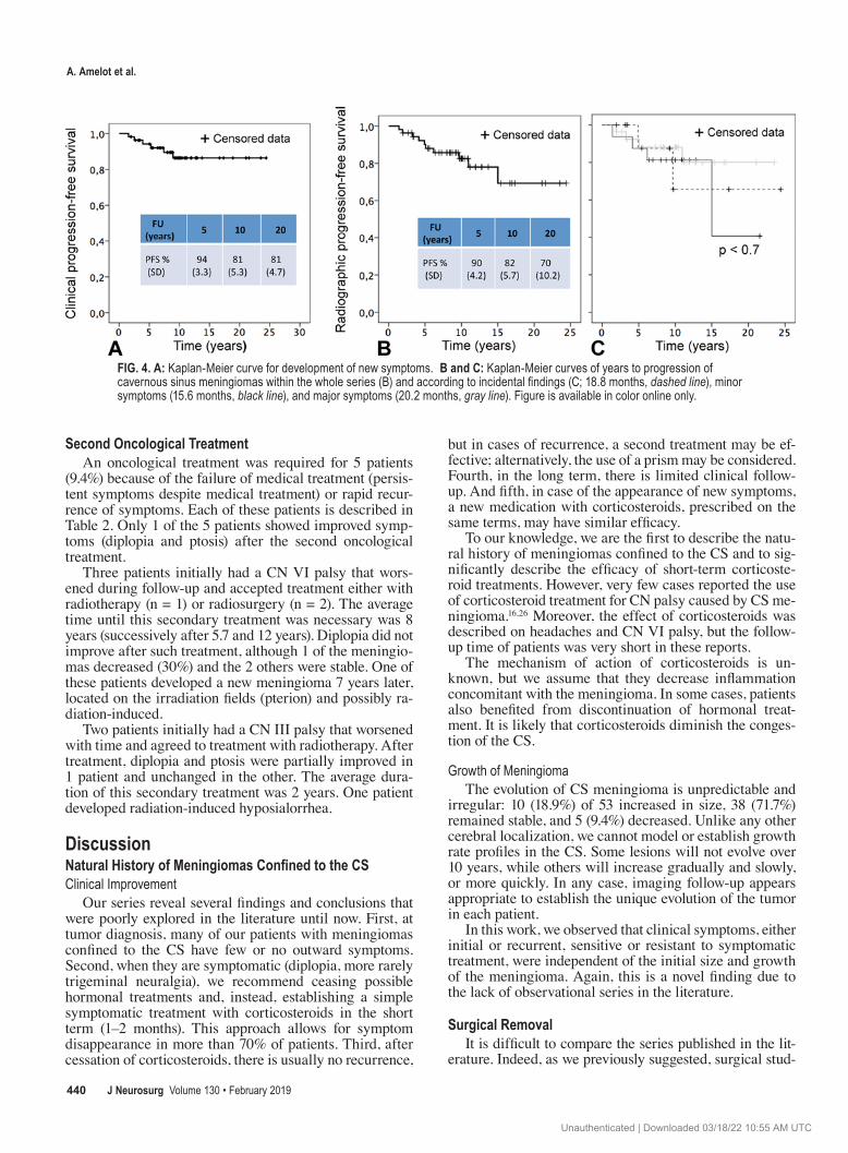
Currently, 9 patients remain symptomatic. Two patients have sustained a visual acuity loss, 2 other patients con-tinue to experience CN III palsy (which remained stable and well tolerated over the follow-up), and 5 patients (CN VI, n = 3; CN III, n = 2) with no or temporary effective-ness of the initial symptomatic treatment have secondarily undergone treatment using radiotherapy (mean 5.8 years). The clinical PFS rates at 5, 10, and 20 years were 94% ± 3.3%, 81% ± 5.3%, and 81% ± 4.7%, respectively.
Tumor Growth, Initial Symptoms, and Radiographic PFSAccording to the criteria previously defined to assess
progression of meningiomas (Fig. 4B and C), radiographic PFS rates at 5, 10, and 20 years were 90% ± 4.2%, 82% ± 5.7%, and 70% ± 10.2%, respectively.
None of the meningiomas increased in volume in the incidental findings group. Twelve meningiomas (80%) in the minor symptoms group exhibited no increase in vol-ume. Three meningiomas (20%) increased moderately (mean growth 60% in 10.7 years): two of them were with-out any symptoms, and the other developed CN VI symp-toms. Twenty-two patients (78.6%) in the major symptoms group had meningiomas whose volume did not increase. Five patients (19%) had meningiomas that progressed (mean growth 35% in 10.2 years); only 2 of these patients experienced an aggravation of their symptoms: 1 in CN III and 1 in CN VI.
FIG. 2. Follow-up years for the incidental findings group (dark gray = asymptomatic).
Unauthenticated | Downloaded 03/18/22 10:55 AM UTC
J Neurosurg Volume 130 • February 2019 439
A. Amelot et al.
FIG. 3. Follow-up data for patients in minor and major symptomatic groups (dark gray = become asymptomatic, light gray = became or remain symptomatic). g = Gamma Knife surgery; II–VI = CNs; HyperP = hyperprolactinemia; M = medical symptomatic treatment; R = radiotherapy.
Unauthenticated | Downloaded 03/18/22 10:55 AM UTC
A. Amelot et al.
J Neurosurg Volume 130 • February 2019440
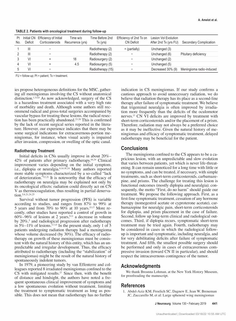
Second Oncological TreatmentAn oncological treatment was required for 5 patients
(9.4%) because of the failure of medical treatment (persis-tent symptoms despite medical treatment) or rapid recur-rence of symptoms. Each of these patients is described in Table 2. Only 1 of the 5 patients showed improved symp-toms (diplopia and ptosis) after the second oncological treatment.
Three patients initially had a CN VI palsy that wors-ened during follow-up and accepted treatment either with radiotherapy (n = 1) or radiosurgery (n = 2). The average time until this secondary treatment was necessary was 8 years (successively after 5.7 and 12 years). Diplopia did not improve after such treatment, although 1 of the meningio-mas decreased (30%) and the 2 others were stable. One of these patients developed a new meningioma 7 years later, located on the irradiation fields (pterion) and possibly ra-diation-induced.
Two patients initially had a CN III palsy that worsened with time and agreed to treatment with radiotherapy. After treatment, diplopia and ptosis were partially improved in 1 patient and unchanged in the other. The average dura-tion of this secondary treatment was 2 years. One patient developed radiation-induced hyposialorrhea.
DiscussionNatural History of Meningiomas Confined to the CSClinical Improvement
Our series reveal several findings and conclusions that were poorly explored in the literature until now. First, at tumor diagnosis, many of our patients with meningiomas confined to the CS have few or no outward symptoms. Second, when they are symptomatic (diplopia, more rarely trigeminal neuralgia), we recommend ceasing possible hormonal treatments and, instead, establishing a simple symptomatic treatment with corticosteroids in the short term (1–2 months). This approach allows for symptom disappearance in more than 70% of patients. Third, after cessation of corticosteroids, there is usually no recurrence,
but in cases of recurrence, a second treatment may be ef-fective; alternatively, the use of a prism may be considered. Fourth, in the long term, there is limited clinical follow-up. And fifth, in case of the appearance of new symptoms, a new medication with corticosteroids, prescribed on the same terms, may have similar efficacy.
To our knowledge, we are the first to describe the natu-ral history of meningiomas confined to the CS and to sig-nificantly describe the efficacy of short-term corticoste-roid treatments. However, very few cases reported the use of corticosteroid treatment for CN palsy caused by CS me-ningioma.16,26 Moreover, the effect of corticosteroids was described on headaches and CN VI palsy, but the follow-up time of patients was very short in these reports.
The mechanism of action of corticosteroids is un-known, but we assume that they decrease inflammation concomitant with the meningioma. In some cases, patients also benefited from discontinuation of hormonal treat-ment. It is likely that corticosteroids diminish the conges-tion of the CS.
Growth of MeningiomaThe evolution of CS meningioma is unpredictable and
irregular: 10 (18.9%) of 53 increased in size, 38 (71.7%) remained stable, and 5 (9.4%) decreased. Unlike any other cerebral localization, we cannot model or establish growth rate profiles in the CS. Some lesions will not evolve over 10 years, while others will increase gradually and slowly, or more quickly. In any case, imaging follow-up appears appropriate to establish the unique evolution of the tumor in each patient.
In this work, we observed that clinical symptoms, either initial or recurrent, sensitive or resistant to symptomatic treatment, were independent of the initial size and growth of the meningioma. Again, this is a novel finding due to the lack of observational series in the literature.
Surgical RemovalIt is difficult to compare the series published in the lit-
erature. Indeed, as we previously suggested, surgical stud-
FIG. 4. A: Kaplan-Meier curve for development of new symptoms. B and C: Kaplan-Meier curves of years to progression of cavernous sinus meningiomas within the whole series (B) and according to incidental findings (C; 18.8 months, dashed line), minor symptoms (15.6 months, black line), and major symptoms (20.2 months, gray line). Figure is available in color online only.
Unauthenticated | Downloaded 03/18/22 10:55 AM UTC
J Neurosurg Volume 130 • February 2019 441
A. Amelot et al.
ies propose heterogeneous definitions for the MSC, gather-ing all meningiomas involving the CS without anatomical distinction.1,3,4,8 As now acknowledged, surgery of the CS is a hazardous treatment associated with a very high rate of morbidity and death. Although some authors still rec-ommend radical and gross-total surgeries accompanied by vascular bypass for treating these lesions, the radical resec-tion has been practically abandoned.23,28 This is confirmed by the lack of recent surgical series reported in the litera-ture. However, our experience indicates that there may be some surgical indications for extracavernous-portion me-ningiomas, for instance, when visual acuity is impaired after invasion, compression, or swelling of the optic canal.
Radiotherapy TreatmentInitial deficits in CNs usually improve in about 20%–
42% of patients after primary radiotherapy.18,24 Clinical improvement varies depending on the initial symptom, i.e., diplopia or neuralgia.6,14,18,24 Many authors reported more stable symptoms characterized by a so-called “lack of deterioration.”12,21 It is noteworthy that the efficacy of radiotherapy on neuralgia may be explained not only by its oncological effects; radiation could directly act on CN V as thermocoagulation, thus resulting in partial denerva-tion.5,6,21,24,25
Survival without tumor progression (PFS) is variable according to studies, and ranges from 87% to 99% at 5 years and from 70% to 90% at 10 years.15,17 More re-cently, other studies have reported a control of growth in 60%–96% of lesions at 2 years,9,11 a decrease in volume by 20%,5 and radiological progression after radiotherapy in 5%–11% of lesions.5,7,15 In the present study, only 1 of 5 patients undergoing radiation therapy had a meningioma whose volume decreased (by 30%). The efficacy of radio-therapy on growth of these meningiomas must be consis-tent with the natural history of this entity, which has an un-predictable and irregular development. Thus, the efficacy attributed to radiotherapy (including the “stabilization” of meningiomas) might be the result of the natural history of spontaneously indolent tumors.
In 1979, a pioneering study by van Effenterre and col-leagues reported 8 irradiated meningiomas confined to the CS with mitigated results.27 Since then, with the benefit of distance and hindsight, the authors have noted a fre-quent spontaneous clinical improvement of symptoms and a low spontaneous evolution without treatment, limiting the treatment to symptomatic treatment as long as pos-sible. This does not mean that radiotherapy has no further
indication in CS meningiomas. If our study confirms a cautious approach to avoid unnecessary radiation, we do believe that radiation therapy has its place as a second-line therapy after failure of symptomatic treatment. We believe that trigeminal neuralgia is often improved by irradia-tion more frequently than the deficits of the oculomotor nerves.13 CN VI deficits are improved by treatment with short-term corticosteroids and/or the placement of a prism. Therefore, radiation may not always be a preferred choice as it may be ineffective. Given the natural history of me-ningiomas and efficacy of symptomatic treatment, delayed radiotherapy may be beneficial for the patient.
ConclusionsThe meningioma confined to the CS appears to be a ca-
pricious lesion, with an unpredictable and slow evolution that varies between patients, yet which is never life-threat-ening. It can remain unnoticed for a long time with little or no symptoms, and can be treated, if necessary, with simple treatments, such as short-term corticosteroids, carbamaze-pine, and prisms. The challenge for treating this lesion is functional outcomes (mostly diplopia and neuralgia); con-sequently, the motto “First, do no harm” should guide our treatment. We propose the following approach. First, as a first-line symptomatic treatment, cessation of any hormone therapy (nomegestrol acetate or cyproterone acetate), car-bamazepine for neuralgic pain, short-term corticosteroids for diplopia, and prism placement in the case of failure. Second, follow up long-term clinical and radiological out-comes. Third, if diplopia recurs, symptomatic short-term treatment may be tried again. Fourth, radiotherapy may be considered in cases in which the radiological follow-up is important and symptomatic, including neuralgia, and for very debilitating deficits after failure of symptomatic treatment. And fifth, the smallest possible surgery should be performed and only in cases of extracavernous com-pressive invasion (toward CN II in particular), and should respect the intracavernous contingence of the tumor.
AcknowledgmentsWe thank Breanna Lohman, at the New York History Museum,
for proofreading the manuscript.
References 1. Abdel-Aziz KM, Froelich SC, Dagnew E, Jean W, Breneman
JC, Zuccarello M, et al: Large sphenoid wing meningiomas
TABLE 2. Patients with oncological treatment during follow-up
Pt No.
Initial CN Deficit
Efficiency of Initial Corticosteroids
Time w/o Recurrence (yrs)
Time Before 2nd Tx (yrs)
Efficiency of 2nd Tx on CN Deficit
Lesion Vol Evolution After 2nd Tx (yrs FU) Secondary Complication
1 III − Radiotherapy (2) + (partially) Unchanged (3)2 III − Radiotherapy (2) − Unchanged (6) Pituitary deficiency3 VI + 18 Radiosurgery (2) − Unchanged (2)4 VI + 4.5 Radiosurgery (5) − Unchanged (5)5 VI − Radiotherapy (15) − Decreased 30% (9) Meningioma radio-induced
FU = follow-up; Pt = patient; Tx = treatment.
Unauthenticated | Downloaded 03/18/22 10:55 AM UTC
A. Amelot et al.
J Neurosurg Volume 130 • February 2019442
involving the cavernous sinus: conservative surgical strate-gies for better functional outcomes. Neurosurgery 54:1375–1384, 2004
2. De Jesús O, Sekhar LN, Parikh HK, Wright DC, Wagner DP: Long-term follow-up of patients with meningiomas involving the cavernous sinus: recurrence, progression, and quality of life. Neurosurgery 39:915–920, 1996
3. DeMonte F, Smith HK, Al-Mefty O: Outcome of aggres-sive removal of cavernous sinus meningiomas. J Neurosurg 81:245–251, 1994
4. Dombi E, Ardern-Holmes SL, Babovic-Vuksanovic D, Barker FG, Connor S, Evans DG, et al: Recommendations for imaging tumor response in neurofibromatosis clinical trials. Neurology 81 (21 Suppl 1):S33–S40, 2013
5. Hafez RF, Morgan MS, Fahmy OM: Stereotactic Gamma Knife surgery safety and efficacy in the management of symptomatic benign confined cavernous sinus meningioma. Acta Neurochir (Wien) 157:1559–1564, 2015
6. Hasegawa T, Kida Y, Yoshimoto M, Koike J, Iizuka H, Ishii D: Long-term outcomes of Gamma Knife surgery for cavern-ous sinus meningioma. J Neurosurg 107:745–751, 2007
7. Hashimoto N, Rabo CS, Okita Y, Kinoshita M, Kagawa N, Fujimoto Y, et al: Slower growth of skull base meningiomas compared with non-skull base meningiomas based on volu-metric and biological studies. J Neurosurg 116:574–580, 2012
8. Heth JA, Al-Mefty O: Cavernous sinus meningiomas. Neuro-surg Focus 14(6):3, 2003
9. Kimball MM, Friedman WA, Foote KD, Bova FJ, Chi YY: Linear accelerator radiosurgery for cavernous sinus menin-giomas. Stereotact Funct Neurosurg 87:120–127, 2009
10. Klinger DR, Flores BC, Lewis JJ, Barnett SL: The treatment of cavernous sinus meningiomas: evolution of a modern ap-proach. Neurosurg Focus 35(6):E8, 2013
11. Kobayashi T, Kida Y, Mori Y: Long-term results of stereo-tactic gamma radiosurgery of meningiomas. Surg Neurol 55:325–331, 2001
12. Leber KA, Berglöff J, Pendl G: Dose-response tolerance of the visual pathways and cranial nerves of the cavernous sinus to stereotactic radiosurgery. J Neurosurg 88:43–50, 1998
13. Martínez Moreno NE, Gutiérrez-Sárraga J, Rey-Portolés G, Jiménez-Huete A, Martínez Álvarez R: Long-term outcomes in the treatment of classical trigeminal neuralgia by Gamma Knife radiosurgery: a retrospective study in patients with minimum 2-year follow-up. Neurosurgery 79:879–888, 2016
14. Morita A, Coffey RJ, Foote RL, Schiff D, Gorman D: Risk of injury to cranial nerves after gamma knife radiosurgery for skull base meningiomas: experience in 88 patients. J Neuro-surg 90:42–49, 1999
15. Nicolato A, Foroni R, Alessandrini F, Maluta S, Bricolo A, Gerosa M: The role of Gamma Knife radiosurgery in the management of cavernous sinus meningiomas. Int J Radiat Oncol Biol Phys 53:992–1000, 2002
16. Phang SY, Whitfield P: Postpartum regression of a pre-sumed cavernous meningioma. Case Rep Oncol Med 2016:2649426, 2016
17. Pichierri A, Santoro A, Raco A, Paolini S, Cantore G, Delfini R: Cavernous sinus meningiomas: retrospective analysis and proposal of a treatment algorithm. Neurosurgery 64:1090–1101, 2009
18. Pollock BE, Stafford SL, Link MJ, Garces YI, Foote RL: Single-fraction radiosurgery of benign cavernous sinus me-ningiomas. J Neurosurg 119:675–682, 2013
19. Radhakrishnan K, Mokri B, Parisi JE, O’Fallon WM, Sunku J, Kurland LT: The trends in incidence of primary brain tu-mors in the population of Rochester, Minnesota. Ann Neurol 37:67–73, 1995
20. Razek AA, Castillo M: Imaging lesions of the cavernous si-nus. AJNR Am J Neuroradiol 30:444–452, 2009
21. Roche PH, Régis J, Dufour H, Fournier HD, Delsanti C, Pel-let W, et al: Gamma knife radiosurgery in the management of cavernous sinus meningiomas. J Neurosurg 93 (3 Suppl 3):68–73, 2000
22. Sindou M: Les tumeurs de la loge caverneuse. Neurochirur-gie 41:143–144, 1995
23. Sindou M, Wydh E, Jouanneau E, Nebbal M, Lieutaud T: Long-term follow-up of meningiomas of the cavernous sinus after surgical treatment alone. J Neurosurg 107:937–944, 2007
24. Spiegelmann R, Cohen ZR, Nissim O, Alezra D, Pfeffer R: Cavernous sinus meningiomas: a large LINAC radiosurgery series. J Neurooncol 98:195–202, 2010
25. Tishler RB, Loeffler JS, Lunsford LD, Duma C, Alexander E III, Kooy HM, et al: Tolerance of cranial nerves of the cav-ernous sinus to radiosurgery. Int J Radiat Oncol Biol Phys 27:215–221, 1993
26. Toro J, Burbano LE, Reyes S, Barreras P: Cavernous si-nus syndrome: need for early diagnosis. BMJ Case Rep 2015:bcr2014206999, 2015
27. van Effenterre R, Bataïni JP, Cabanis EA, Iba-Zizen MT: High energy radiotherapy in the treatment of meningiomas of the cavernous sinus. Acta Neurochir Suppl (Wien) 28:464–467, 1979
28. Wanibuchi M, Akiyama Y, Mikami T, Iihoshi S, Miyata K, Horita Y, et al: Radical removal of recurrent malignant men-ingeal tumors of the cavernous sinus in combination with high-flow bypass. World Neurosurg 83:424–430, 2015
DisclosuresThe authors report no conflict of interest concerning the materi-als or methods used in this study or the findings specified in this paper.
Author ContributionsConception and design: Amelot, Boch. Acquisition of data: Amelot, van Effenterre, Kalamarides, Boch. Analysis and inter-pretation of data: Amelot, van Effenterre, Boch. Drafting the article: Amelot, van Effenterre, Cornu, Boch. Critically revising the article: Amelot, Kalamarides, Cornu, Boch. Reviewed submit-ted version of manuscript: Amelot, van Effenterre, Cornu, Boch. Statistical analysis: Amelot. Administrative/technical/material support: Amelot, van Effenterre, Kalamarides, Boch. Study super-vision: Amelot, Kalamarides, Boch.
CorrespondenceAymeric Amelot: Groupe Hospitalier Pitié-Salpêtrière, Paris, France. [email protected].
Unauthenticated | Downloaded 03/18/22 10:55 AM UTC