Embed Size (px)
Citation preview

Spinal Cord and Reflex
Syahruramdhani
School of NursingMedical Faculty and Health Sciences
Universitas Muhammadiyah Yogyakarta

Objectives
1. Describe the gross and microscopic anatomy of the spinal cord;2. Name the major conduction pathways of the spinal cord and state
their functions;3. Define reflex and explain how reflexes differ from other motor
actions;4. Describe the general components of a typical reflex arc; and5. Explain how the basic types of somatic reflexes function.

Spinal Cord
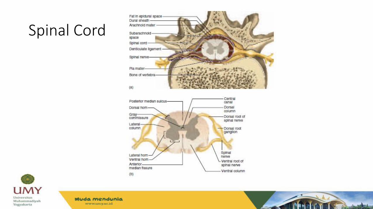
• The spinal cord is a cylinder of nervous tissue that begins at the foramen magnum and passes through the vertebral canal as far as the inferior margin of the first lumbar vertebra (L1).
• The spinal cord is divided into cervical, thoracic, lumbar, and sacral regions.
• The spinal cord serves three principal functions:1. Conduction 2. Locomotion3. Reflexes

Spinal Cord
Spinal Cord
Spinal Cord
• The spinal cord, like the brain, consists of two kinds of nervous tissue called gray and white matter.
• Gray matter has a relatively dull color because it contains little myelin. It contains the somas, dendrites, and proximal parts of the axons of neurons.
• It is the site of synaptic contact between neurons, and therefore the site of all synaptic integration (information processing) in the central nervous system.

• White matter contains an abundance of myelinated axons, which give it a bright, pearly white appearance.
• It is composed of bundles of axons, called tracts, that carry signals from one part of the CNS to another.

Spinal Cord
• Knowledge of the locations and functions of the spinal tracts is
essential in diagnosing and managing spinal cord injuries.
• Ascending tracts carry sensory information up the cord and
descending tracts conduct motor impulses down.
• All nerve fibers in a given tract have a similar origin, destination, and
function.

Spinal Cord
Nature of Reflex
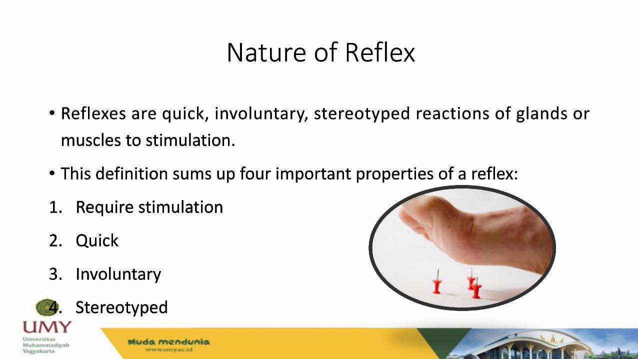
• Reflexes are quick, involuntary, stereotyped reactions of glands or muscles to stimulation.
• This definition sums up four important properties of a reflex:
1. Require stimulation
2. Quick
3. Involuntary
4. Stereotyped

Reflexes
• Somatic reflexes/spinal reflexes
Example: the quick withdrawal of your hand from a hot stove or the lifting of your foot when you step on something sharp.
Many somatic reflexes are initiated by proprioceptors, organs that monitor the position and movements of body parts.
• Visceral reflexes
Example: high blood pressure activates a visceral baroreflex.
Somatic Reflexes
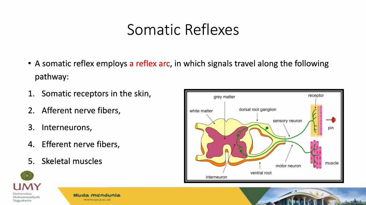
• A somatic reflex employs a reflex arc, in which signals travel along the following pathway:
1. Somatic receptors in the skin,
2. Afferent nerve fibers,
3. Interneurons,
4. Efferent nerve fibers,
5. Skeletal muscles

Muscle Spindle
• Muscle spindles are proprioceptors embedded in the skeletal muscles that
respond to stretching of the muscle.
• A spindle contains 3 to 12 modified muscle fibers and a few nerve fibers, all
wrapped in a fibrous capsule.
• The muscle fibers within a spindle are called intrafusal fibers, while those
of the rest of the muscle are called extrafusal fibers.


Muscle Spindle
• Muscle spindles have three types of nerve fibers:
• 1. Primary afferent fibers,
• 2. Secondary afferent fibers, and
• 3. Gamma motor neurons.

The strestch reflex
• The tendency of a muscle to contract when it is stretched.
• For example, if your head starts to tip forward, it stretches muscles such as the
semispinalis and splenius capitis of the nuchal region (back of your neck).
• Reciprocal inhibition, a reflex phenomenon that prevents muscles from working
against each other by inhibiting antagonists.

The golgi tendon reflex
• Golgi tendon organs are proprioceptors located in a tendon near its
junction with a muscle.
• The Golgi tendon reflex is a response to excessive tension on the tendon.
• The Golgi tendon reflex also functions when some parts of a muscle
contract more than others.


The flexor (withdrawal) reflex
• A flexor reflex is the quick contraction of flexor muscles resulting in the
withdrawal of a limb from an injurious stimulus.
• For example, suppose you are wading in a lake and step on a broken bottle with
your right foot.
• The protective function of this reflex requires more than a quick jerk like a tendon
reflex, so it involves more complex neural pathways.


The crossed extensor reflex
• The crossed extensor reflex is the contraction of extensor muscles in the
limb opposite from the one that is withdrawn.
• To produce this reflex, branches of the afferent nerve fibers cross from the
stimulated side of the body to the contralateral side of the spinal cord.

Referensi
• Saladin K (2003). Anatomy and Physiology: The Unity of Form and
Function, 3rd ed. Chapter 23, Pages: 503-508.