Embed Size (px)
Citation preview
Invertebrate Reproduction and Development, 34: 1 (1 998) 13-23 Balaban, Philadelphia/Rehovot 0168-8 170/98/$05.00 (9 1998 Balaban
Spermatozoa1 ultrastructure in three species of hydrothermal vent crab, in the genera Bythograea, Austinograea and Segonzacia
(Decapoda, Brachyura, Bythograeidae)
C.C. TUDGEI*, B.G.M. JAMIESON~, M. SEGONZAC~ and D. ~ U r N o l 4 rustac ace an Laboratory, Museum of Victoria, 71 Victoria Crescent, Abbotsford, Vic. 3067, Australia
Tel. +61 (3) 9284 0236; Fax +61 (3) 9416 0475; E-mail: [email protected] 2~oology Department, The University of Queensland, Brisbane 4072, Australia
'IFREMER, Centre de Brest, CENTOB, BP 70, F-29280 PlouzanP, France '~aboratoire de Zoologie (Arthropodes), Muse'um national dlHistoire naturelle,
61 rue Buffon, 75231 Paris, Cedex 05, France
Received 1 March 1997; Accepted 18 July 1997
Summary
The ultrastructural investigation of the spermatozoa1 morphology of the hydrothermal vent crabs, Bythograea thermydron, Austinograea alayseae and Segonzacia mesatlantica (family Bytho- graeidae) reveals a consistent familial sperm type and a close similarity between the three genera and the deep water trapeziid, Calocarcinus aj?icanus. Association of the bythograeids with Calocarcinus is supported (apart from general similarity) by three synapomorphies: modification of the xanthid ring, development of a well developed periopercular rim (weakly shared with some xanthid and potamid members) and, as a particularly striking Imk, the unique spiral configuration of the contents of the outer acrosome zone. Calocarcinus is distinguished by shortening of the posterior dense zone into a true xanthid ring and, less so, by a flatter operculum. Bythograea and Segonzacia are apomorphic in the independent loss of the ragged outer acrosome profile. Their sister genus Austinograea has no distinct apomorphies (with the possible exception of an apical perforation in the operculum). The similarity between the spermatozoa of Calocarcinus and the investigated bythograeids and the dissimilarity between Calocarcinus and other trapeziids is evidence for possible inclusion of Calocarcinus in the Bythograeidae. This hypothesized relationship of Calocarcinus within the Bythograeidae sensu lato and its deep water distribution suggests origin of hydrothermal bythograeids from Calocarcinus-like deep water xanthoids which may have entered the hydrothermal system in or after the Eocene. Further investigation of somatic morphology is required to test this putative relationship.
Key words: Spermatozoa, ultrastructure, Bythograeoidea, hydrothermal vents, Calocarcinus
*corresponding author.
Dow
nloa
ded
by [
UQ
Lib
rary
] at
19:
52 0
5 O
ctob
er 2
011
14 C.C. Tudge et al. / IRD 34 (1998) 13-23
Introduction
The taxonomic and phylogenetic relationships of the Bythograeidae have been the subject of some debate. Williams (1980), in erecting the family Bythograeidae for Bythograea thermydron, noted that it exhibited some characters of the families Portunidae and Xanthidae, and superficial resemblance to the family Potamidae. He considered it to be assignable to the tribe Brachygnatha, between the Portunidae and Potamidae, within the superfamily Brachyrhyncha, in the system of Balss (1957) and to the brachyrhynchan superfamily Portunoidea in the system of Glaessner (1969). However, it fitted comfortably with none of the recognized superfamilies, hence erection of a new superfamily, the Bythograeiodea.
Guinot (1 977,1978) replaced the earlier systems with a classification of Brachyura, on the basis of the position of the genital pores, into Podotremata, Heterotremata and Thoracotremata, a system endorsed (Jamieson and Tudge, 1990; Jamieson, 1991, 1994) from spermatozoal ultrastructure. Guinot (1988) questioned the apparent similarity of bythograeids to khwater crabs of the superfamily Potamoidea and, in particular, the relationship invoked by Delamare Deboutteville and Guinot (1981) with cavemicolous freshwater crabs of the family Pseudothelphusidae in Guatemala and Mexico. These, like the bythograeids, tend to reduce the eyes and pigmentation. However, Guinot (1988) considered that the morphology of the American freshwater crabs furnished no real argument for filiation with the Bythograeidae. On the other hand, bythograeoids offered no special similarities with bathyal and abyssal crabs and appeared to form an isolated group among these. Guinot (1 988) suggested that bythograeoids are not derived from an ancient bathyal group which adapted to a hydrothermal regime but speculated that they arose from brachyuran stock from shallower water which became adapted to hydrothermal conditions. She concluded that bytho- graeoids are carcinised true heterotremes forming a special superfamily like the Majoidea, Potamoidea, Portunoidea etc. The characteristics of the first zoeal larva described by Van Dover and colleagues (1984) support establishment by Williams (1 980) of the superfamily Bythograeoidea.
Guinot's conclusions found agreement in the comment of Williams, cited by Guinot (1990), who denied alignment of bythograeids with Mesozoic crabs, regarding them as having a "modem appearance" and stating that "despite the great antiquity of deep hydrothermal systems in the world ocean, the brachyurans did not adapt themselves to these systems until sometime during the Cenozoic".
In contrast to these views of a post-Mesozoic (approx. 65 mya) origin of bythograeids, Newman (1985) argued that because there does not seem to be an extant taxon from which the bythograeoid level of organization could have been derived, it seemed reasonable that they originated as part of the Mesozoic radiation, when Austinograea, Bythograea and Cyana- graea or their ancestor(s) first could have entered the hydrothermal environment. He noted, further, that the radiation which produced the portunoids and xanthoids occurred, according to Glaessner (1969), near the end of the Mesozoic.
The discovery of hydrothermal vents and the brachyuran crabs associated with them have attracted much interest. Notable examples include publications on their biogeography and evolution (Hessler and Wilson, 1983; Newman, 1985; Tunnicliffe, 1988), elements of their larval development (Van Dover et al., 1984), reproductive biology (Van Dover et al., 1985), their microdistribution and comparisons between hydrothermal sites in different oceans (Segonzac, 1992; Van Dover, 1995) and their physiology (Airries and Childress, 1994).
Since the erection of the Bythograeidae (Williams, 1980) to accommodate the single species Bythograea thermydron, several species have been added to the family, in the genera Cyanagraea de Saint Laurent (1984), Segonzacia Guinot (1989) and Austinograea Hessler and Martin (1989). All members of the family have been found in association with deep water hydrothermal vents in the Pacific Ocean, with one species (Segonzacia mesatlantica) discovered in the mid-Atlantic (Guinot, 1989); a further species of Bythograea , B. laubieri, from the southern part of the East Pacific Ridge has recently been described (Guinot and Segonzac, 1997).
The Bythograeidae are generally placed in the Section Heterotremata of Guinot's classification based on gonopore position (Guinot, 1977,1978) and are usually placed in the proximity of the brachyuran families Portunidae, Xanthidae or Potamidae (Williams, 1980; Feldmann and McLay, 1993). Bytho- graeids possess very few classical morphological characters to indicate their true relationships and the genera, though widely dispersed in the world (Bythograea and Cyanagraea in the East Pacific, Austinograea in the West Pacific, Segonzacia in the mid-Atlantic) have the same carapace, walking legs and the same overall facies. The family is extremely homogeneous in general morphology.
Many brachyurans have been investigated for spermatozoal ultrastructure (see Jamieson, 199 1, 1994; Felgenhauer and Abele, 199 1 ; Jamieson et al., 1995 for reviews and contained references) including numerous
Dow
nloa
ded
by [
UQ
Lib
rary
] at
19:
52 0
5 O
ctob
er 2
011
C. C. Tudge el al. / IRD 34 (1 998) 13-23 15
representatives from the section Heterotremata (of which the Bythograeidae are members). More recently, the heterotremes Potamonautes perlatus sidneyii (Potamidae), Trapezia cymodoce (Trapeziidae), Calocarcinus aJi.icanus (?Trapeziidae), Australo- carcinus riparius (Goneplacidae) and the further potamids Potamon fluviatile and P. ibericum (Potamidae) have been studied (see Jamieson, 1993a, 1993b; Jamieson et al., 1993; Jamieson and Guinot, 1996; Guinot et al., 1997, respectively).
In the present study sperm ultrastructure of three species of bythograeid crabs from hydrothermal vents, Austinograea alayseae Guinot, 1990, Segonzacia mesatlantica (Williams, 1988), and Bythograea thermydron Williams, 1980, is described, and com- parisons are made with the sperm of other crabs, including Calocarcinus and Trapezia, with a view to aiding determination of relationships.
Materials and Methods
The specimen of Austinograea alayseae was collected in May, 1989 during the "BIOLAU" cruise at 'Vai Lili', west of the islands of Tonga, Pacific Ocean. It was netted at 1750 m at the hydrothermal site 'Ride de Valu Fa' (176"38'W, 22'34's). The specimen is one of numerous paratype males (ex-MNHN B24055). The specimen of B. thermydron was collected during the "Hero" cruise in 199 1 at 'Rift de Galapagos' in the Pacific Ocean (1 3 ON). Specimens of both genera were originally fixed in formalin and later preserved in ethanol. The single male specimen of each species was dissected in Paris in July, 1992, and the reproductive tract was subjected to the standard fixation procedure for transmission electron microscopy outlined below.
The specimens of Segonzacia mesatlantica were collected from the 'Snake Pit' hydrothermal vent site (23 ON, 3480 m) on the Mid-Atlantic Ridge, during the Franco-American cruise "MAR 93" in June, 1993. The gonads of two individuals were fixed in cold 2% glutaraldehyde in cacodylate buffer before being transferred to cacodylate buffer for transport to Brisbane, Australia. Upon arrival the material was further processed according to the fixation procedure outlined below.
Portions of the testis were fixed in 3% glutaral- dehyde in 0.1 M phosphate buffer (pH 7.2) for 1 h at 4°C. They were washed in phosphate buffer (three washes in 15 min), postfixed in similarly buffered 1% osmium tetroxide for 80 min, and washed in buffer and dehydrated through ascending concentrations of ethanol (20-100%). After being infiltrated and embedded in Spun's epoxy resin (Spurr, 1969), thin sections (50-80 nm thick) were cut on a LKB 2128
UMIV microtome with a diamond knife. Sections were placed on carbon-stabilized colloidin-coated 200pm mesh copper grids and stained according to the abbreviated method of Daddow (1 986): Reynold's lead citrate for 30 s, rinsed in distilled water, stained in 6% aqueous uranyl acetate for 1 min, stained again with lead citrate for 30 s and further rinsed in distilled water. Micrographs were taken on an Hitachi 300 trans- mission electron microscope at 75 kV.
Results
General morphology In all three species of hydrothermal vent crab many
spermatozoa are contained within thin walled, oval to oblong-ovate spermatophores of various sizes. An ultrastructural description of the general bythograeid spermatozoon will be given and generic differences noted.
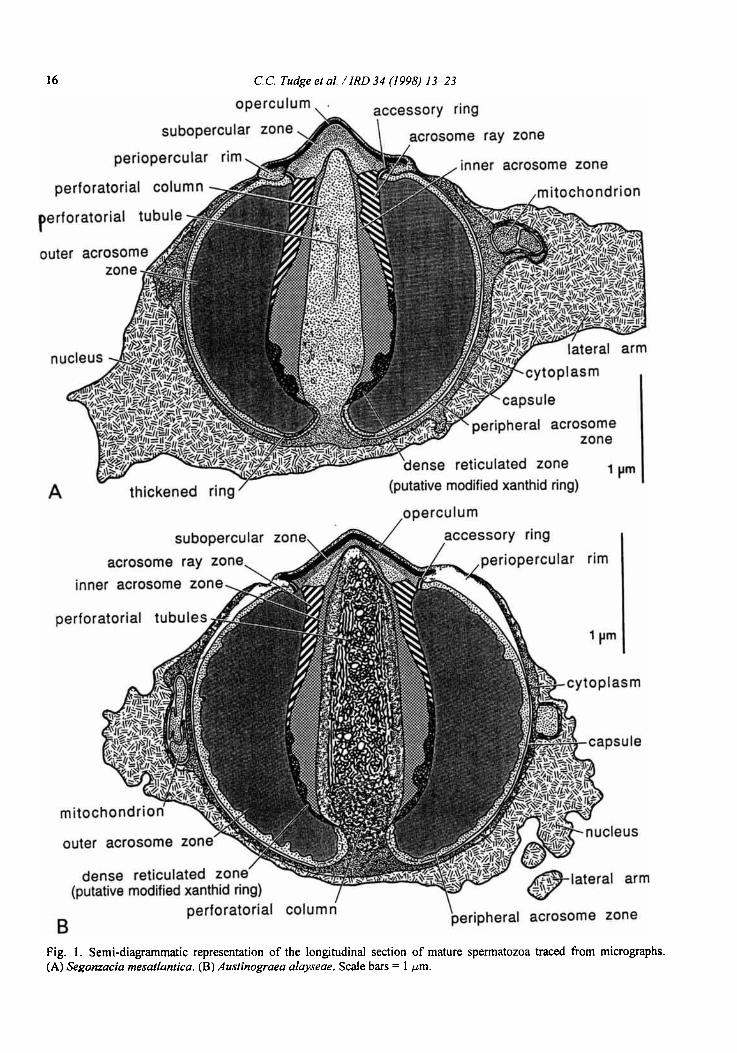
The spermatozoa are typically brachyuran in gross morphology. An acrosome vesicle, forming most of its bulk, with concentric zonation and capped apically by a dense operculum, is posteriorly embedded in a thin layer of cytoplasm which in turn is embedded in the nucleus. The acrosome vesicle is centrally penetrated, almost to its apex, by a cylindrical perforatorial column. The nuclear material forms several lateral projections or "arms" (refer to Figs. 1A and B throughout). The spherical form of the acrosome is typical of the Heterotremata sensu lato.
Acrosome The acrosome vesicle is spherical in form and of
similar dimensions in all three species (Figs. 2A,B, 3A,B, 4E,F,H). In the acrosome of A. alayseae the mean length is 2.9pm (n=5) and the mean width 2.8pm (n=7); in S. mesatlantica the mean length is 3.1 pm (n=7) and the mean width 3.1 pm (n=6); in B. thermydron the mean length is 3.1 pm (n=2) and the mean width 2.9pm (n=3). The apical pole of the acrosome vesicle is covered by an extremely electron- dense cap or operculum (approx. 1.3 pm in diameter). This operculum is a shallow, pointed dome with the rim forming a small overhanging lip (Figs. 2A, 3A, 4A-C, E-G). The operculum is imperforate in S. mesatlantica, B. thermydron and probably in A. alayseae, though a small central perforation was observed in a single micrograph of the latter (Fig. 4B). Immediately subjacent to the dense operculum is a slightly less electron-dense, finely granular region, the subopercular zone. e lateral edge of the subopercular zone is bordered by two closely apposed
Dow
nloa
ded
by [
UQ
Lib
rary
] at
19:
52 0
5 O
ctob
er 2
011
C.C. Tudge et al. / IRD 34 (1998) 13-23
subopercular zon crosome ray zone
perforatorial column
outer acrosom
peripheral acrosome zone
ense reticulated zone (putative modified xanthid ring)
' )rm
1 operculum
perforatorial column \ peripheral acrosome zone
Fig. 1 . Semi-diagrammatic representation of the longitudinal section of mature spermatozoa traced from micrographs. (A) Segonzacia rnesatlantica. ( B ) Auslinograea alayseae. Scale bars = 1 pm.
Dow
nloa
ded
by [
UQ
Lib
rary
] at
19:
52 0
5 O
ctob
er 2
011

C. C. Tudge et al. / IRD 34 (1 998) 13-23
Fig. 2. Micrographs of the mature spermatozoa of Segomacia mesatlantica (Glutaraldehyde fixed). (A) Longitudinal section through a spermatozoon. (B) Transverse section through the widest part of the acrosome vesicle. ac, accessory ring; ar, acrosome ray zone; ca, capsule; cy, cytoplasm; dr, dense reticulated zone; ia, inner acrosome zone; la, lateral arm of nucleus; m, mitochondrion; n, nucleus; o, operculum; oa, outer acrosome zone; p, perforatorial column; pa, peripheral acrosome zone; pr, periopercular rim; pt, perforatorial tubules; so, subopercular zone; tr, thickened ring. Scale bars = 1 pm.
Dow
nloa
ded
by [
UQ
Lib
rary
] at
19:
52 0
5 O
ctob
er 2
011

C. C. Tudge et al. / IRD 34 (1 998) 13-23
Fig. 3. Micrographs of the mature spermatozoa of Austinograea alayseae (Formalin fixed). ( A ) Longitudinal section through a spermatozoon. (B) Transverse section through the acrosome vesicle. ac, accessory ring; ar, acrosome ray zone; ca, capsule; cy, cytoplasm; dr, dense reticulated zone; ia, inner acrosome zone; la, lateral arm of nucleus; m, mitochondrion; n, nucleus; o, operculum; oa, outer acrosome zone; p, perforatorial column; pa, peripheral acrosome zone; pr, periopercular rim; pt, perforatorial tubules; so, subopercular zone. Scale bars = 1 pm.
Dow
nloa
ded
by [
UQ
Lib
rary
] at
19:
52 0
5 O
ctob
er 2
011

C. C. Tudge et al. / IRD 34 (1 998) 13-23 19
electron-dense rings, separated by a thin electron-pale zone, collectively termed the accessory ring. This accessory ring is clearly evident in the indentation formed below the overhanging lateral lip of the operculum (Figs. 2A, 3A, 4A,B,G).
The contents of the acrosome vesicle form five concentric zones around the central columnar perfora- torium. The two innermost zones form an electron- dense cylinder which extends from the subopercular zone to the constricted base of the perforatorium. In the two formalin fixed species (A. alayseae and B. thermydron) the innermost zones are not as electron- dense but the internal divisions are still very apparent (Figs. 3A, 4E,F). In all three species the interiormost of the two dense zones (inner acrosome zone) surrounds the perforatorium for nearly its entire length as a homogeneous ring which is thickest midway along the perforatorium but is attenuated both anteriorly and posteriorly. Immediately exterior to this inner acrosome zone is another electron-dense cylinder which is more heterogeneous and distinctive in form. The anterior and posterior ends of this zone are the broadest, thinning midway to correspond with the thickest region of the inner acrosome zone. Anteriorly this zone is composed of radiating and alternating bands of electron-pale and electron-dense material, comprising the acrosome ray zone (evident in Segonzacia, Fig. 2A). These rays or bands appear continuous with the posterior region which is less regular in form and appears as a reticulated pattern of dark and light. In micrographs of the formalin fixed species (A. alayseae and B. thermydron) the equivalent of the acrosome ray zone lacks this distinctive banding, but nevertheless appears continuous with the intact lower reticulated zone (Figs. 3A, 4A,B,E,F).
The remainder of the acrosome vesicle is made up of a coarsely granular but homogeneous region called the outer acrosome zone. In micrographs of longi- tudinal sections of this zone some faint, lateral striations are apparent (Figs. 2A, 3A). In transverse sections these striations can be seen to be arranged in a distinct spiral pattern (not apparent, possibly because of the poor fixation, in B. thermydron), radiating outwards from the central cylinder of the perforatorial column and inner acrosome zones (Figs. 2B, 3B). The exterior boundary of the outer acrosome zone, where it meets the peripheral acrosome zone, is smooth and uniform in S. mesatlantica (Fig. 2B) and B. thermy- dron (Fig. 4H), but has an irregular outline in A. alayseae (Fig. 3B).
At the region where the large outer acrosome zone meets the constricted base of the perforatorium there is a thin electron-dense ring, the thickened ring. The electron-pale, thin peripheral zone occurs immediately
beneath the thickened capsule or acrosome boundary and extends from the accessory ring to the thickened ring. The thickened ring and peripheral acrosome zone are obvious in the micrographs of the glutaraldehyde fwed Segonzacia (Figs. 2A,B) but are not apparent (thickened ring) (Figs. 3A, 4E) or less apparent (peri- pheral acrosome zone) (Figs. 4F,H) in the two formalin fixed species.
The outer capsule appears bilayered (more obviously in Segonzacia and Austinograea) (Figs. 2A,B, 3A,B, 4A,B). At the overhanging lip of the operculum the capsule splits; the inner layer joins with the accessory ring and the outer or upper layer overlies the operculum. A swelling occurs at the point of separation creating a small raised ring around the operculum-the periopercular rim. The formalin fixed species have the membranes distorted in this area and can show exaggerated swelling of the periopercular rim (Figs. 3A, 4A,B,G), but this is presumed partly to be an artefact of fixation.
The perforatorial chamber and enclosed perfora- torial material in all three species averages 0.6pm in diameter and extends from the posterior end of the acrosome vesicle, where it invaginates, to a position immediately below the operculum in the subopercular zone. It is constricted at its base, then widens to form a cylindrical spike, slightly tapering to a rounded point. The contents of the perforatorium are electron-pale and coarsely granular and in the formalin fixed species, conspicuous, electron-dense, convoluted tubules extend nearly its fill length (Figs. 3A,B, 4A-F). These perforatorial tubules tend to be longitudinal in orientation and comprise a network of small dense tubules (0.03 ym in diameter) interspersed with larger, spherical electron-lucent vesicles (0.08 ym in dia- meter).
Cytoplasm The reduced cytoplasm in all three species occurs
as a thin veneer over the outer surface of the acrosome vesicle, excepting the opercular region, and basally appears continuous with the contents of the perfora- torium. Irregular swellings occur in the cytoplasm and these contain membrane bound bodies which may represent degenerate mitochondria (Figs. 2A,B, 3B). No recognizable cristae are evident, even in the glutaraldehyde fixed Segonzacia. In Segonzacia some small lattice-like arrangements indicative of micro- tubular arrays are occasionally present in the cytoplasm (Fig. 2A).
The remainder of the cytoplasm consists of a homogeneous, coarsely granular matrix of medium electron density.
Dow
nloa
ded
by [
UQ
Lib
rary
] at
19:
52 0
5 O
ctob
er 2
011

C. C. Tudge et a/. / IRD 34 (1 998) 13-23
Dow
nloa
ded
by [
UQ
Lib
rary
] at
19:
52 0
5 O
ctob
er 2
011
C. C. Tudge ef al. / IRD 34 (1 998) 13-23 21
Nucleus The nuclear material cups the entire acrosome
vesicle, excepting the opercular region, and forms four to seven large lateral extensions or arms. The nuclear contents consist of electron-dense strands or fibrils in an electron-lucent matrix. The densest aggregations form reticulate clumps nearest the acrosome vesicle but reduce in density in the lateral arms where they tend to become longitudinally arranged (Figs. 2A,B). This bipartite arrangement is seen in both glutaraldehyde and formalin fixed specimens. In the latter, the nuclear material is contracted and adjacent to the acrosome vesicle is extremely dense and the lateral arms are reduced (Figs. 3A,B, 4E,H).
Discussion
The evidence from spermatozoal ultrastructure sheds some light on bythograeid relationships. The three bythograeid species studied share the following spermatozoal characteristics: similar acrosome dimen- sions (approximately 3 pm diameter), an overhanging operculum, a periopercular rim, an accessory ring, an anterior acrosome ray zone connected to a posterior dense reticulated zone (here considered to be a putative modified xanthid ring), spiral arrangement of the outer acrosome zone (not visible in Bythograea), a thickened ring (a heterotreme synapornorphy) and a thin peri- pheral zone. Besides possible fixation artefacts the only spermatological differences between them are the ragged appearance of the peripheral acrosome zone in Austinograea (Fig. 3B) and possibly the apical perforation in the operculum in the same species (Fig. 4B).
Fig. 4 (A- D). Micrographs of the mature spermatozoa of Ausfinograea alayseae (Formalin fixed). (A,B) Longitudinal sections (LSs) through the opercular region showing expanded periopercular rim and perforation in tip of operculum (B only). (C) Transverse section (TS) through the opercular region. (D) Detail of TS of perforatorial column and inner acrosome zone. (E- H) Micrographs of the mature spermatozoa of Bythograea thermydron (Formalin fixed). ( E ) LS of acrosome vesicle. (F) LS of poorly fixed acrosome vesicle. (G) Detail of LS of opercular region showing periopercular rim and accessory ring. (H) Partial TS of acrosome vesicle and nucleus. ac, accessory ring; ar, acrosome ray zone; ca, capsule; cy, cytoplasm; dr, dense reticulated zone; ia, inner acrosome zone; n, nucleus; o, operculum; oa, outer acrosome zone; p, perforatorial column; pa, peripheral acrosome zone; pr, periopercular rim; pt, perforatorial tubules; so, subopercular zone; tr, thickened ring. Scale bars = I p m (except where indicated).
Of all the heterotremes studied to date the one which shares the most spermatological characters with the bythograeids is Calocarcinus afiicanus (see Jamieson et a]., 1993). These characters are an overhanging operculum, a periopercular rim, an accessory ring, an anterior acrosome ray zone connected to a posterior dense zone (considered a true xanthid ring in Calocarcinus), spiral arrangement of the outer acrosome zone (Austinograea and Segonzacia only), a thickened ring and a thin peripheral zone with a ragged outline (shared with Austinograea only). Calocarcinus differs from the three bythograeids in having a flatter operculum and a true xanthid ring.
Calocarcinus has formerly been placed in the family Trapeziidae. Another trapeziid, Trapezia cymodoce (see Jamieson, 1993b, as T. coerulea) has been investigated for spermatozoal ultrastructure and it appears that the three bythograeids and Calocarcinus share no exclusive spermatozoal synapomorphies with Trapezia. A further four species of xanthoid crab (the xanthid genera Atergatis, Etisus, Liagore and Pilodius, see Jamieson, 1989a) share some spermatozoal features with the investigated bythograeids and Calocarcinus. The shared characters are an over- hanging operculum, a periopercular rim (in some species only), an accessory ring, an anterior acrosome ray zone connected to a posterior xanthid ring (most clearly defined in Calocarcinus), a thickened ring and a peripheral zone with a ragged outline (shared with Atergatis and Pilodius only). In contrast, the investi- gated xanthids tend to have a flatter operculum and a true xanthid ring.
With respect to the comments of Williams (1980) and Guinot (1988) that the Bythograeidae had a superficial resemblance to some members in the Potamoidea, spermatological comparisons were made with Potamonautes perlatus sydneyii and Potamon spp. (Jamieson, 1993a; Guinot et al., 1997, respect- ively). Spermatozoal similarities are a periopercular rim (though weakly developed in the potamids), the ubiquitous eubrachyuran thickened ring (reduced in the potamids) and possibly an overhanging operculum, none of which is an exclusive synapomorphy. The remainder of the heterotremes investigated for spermatozoal ultrastructure, including Portunus pela- gicus and Australocarcinus riparius (Jamieson, 1989b; Jamieson and Tudge, 1990; Jamieson and Guinot, 1996), share no exclusive spermatozoal synapo- morphies with the three bythograeids and Calocarcinus.
Spermatozoal evidence thus suggests a relationship of bythograeids with Calocarcinus, and wider relationships within the Xanthoidea. Association of
Dow
nloa
ded
by [
UQ
Lib
rary
] at
19:
52 0
5 O
ctob
er 2
011

22 C.C. Tudge et al. / IRD 34 (1998) 13-23
bythograeids with Calocarcinus is supported (apart Acknowledgements from general similarity) by three -~pomorphies: modification of the xanthid ring (the ring was first described as an autapomorphy for the xanthids by Jamieson, 1989a), development of a well developed periopercular rim and, as a particularly striking link, the unique spiral configuration of the contents of the outer acrosome zone (not discernible in Bythograea). Calocarcinus is distinguished by shortening of the posterior dense zone into a true xanthid ring and, less so, by a flatter operculum. Bythograea and Segonzacia are apomorphic in the independent loss of the ragged outer acrosome profile, and their sister genus Austinograea has no distinct apomorphies (with the possible exception of an apical perforation in the operculum). The close similarity between the sperma- tozoa of Calocarcinus and the investigated bytho- graeids and the dissimilarity between Calocarcinus and another trapeziid, Trapezia, presents the possibility of removal of Calocarcinus from the Trapeziidae and subsequent erection of its own family, or inclusion in the Bythograeidae.
General somatic morphology does not, on first analysis, support relationships of bythograeids with Calocarcinus. The taxonomic status of Trapezia and affiliated genera within the Xanthoidea remains an enigma, as does the previously supposed relationship of Calocarcinus to Trapezia. Relationship of bytho- graeids to Calocarcinus, here deduced from spermatozoa1 evidence, requires corroboration from other, notably morphological and molecular, evidence.
Calocarcinus is atypical with respect to the trapeziids Trapezia and Tetralia (although all are coral symbionts) in extending into deep in addition to shallow waters. The possible relationship of Calo- carcinus, within the Bythograeids sensu lato and its deep water distribution, suggests an origin of hydro- thermal bythograeids from Calocarcinus-like deep water xanthoids. These may have entered the hydro- thermal system in or after the Eocene (the beginning of the Cenozoic), as intimated by Glaessner (1969) and Williams (cited in Guinot, 1990).
We may add that invasion of the hydrothermal system by more than one recognizably bythograeid genus seems unlikely (unless bythograeids are in fact extant in a wider range of habitats than the hydrothermal vents) and would require extinction of bythograeids in non-hydrothermal habitats. It is more parsimonious to assume that a single ancestral genus or species invaded and subsequently gave rise in situ to the hydrothermal Bythograeidae irrespective of the date of that invasion.
- The authors wish to thank the mission chiefs for
making possible the collection of the specimens of Bythograea thermydron, Austinograea alayseae and Segonzacia mesatlantica: D. Desbruyhres (IFREMER) for Hero '91 cruise, A.-M. Alayse (IFREMER) for Biolau cruise, C. Van Dover (WHOI) and A. Fiala (UniversitC Paris VI) for the MAR 93 cruise. Professors J. SchrCvel (Laboratoire de Biologie Parasitaire, Protistologie et Helminthologie) and Y. Coineau (Laboratoire de Zoologie, Arthopodes), of the Mustum national d'Histoire naturelle, Paris, are gratefully acknowledged for use of laboratory facilities. Mrs. L. Daddow (Zoology Department, The University of Queensland, Australia) gave excellent technical assistance with the electron microscopy. This work was initiated in Paris when C.C. Tudge was the recipient of a French Government Scientific Fellow- ship for 1992. It was also funded by an Australian Research Council grant to B.G.M. Jarnieson.
References
Airries, C.N. and Childress, J.J., Homeoviscous properties implicated by the interactive effects of pressure and temperature on the hydrothermal vent crab Bythograea thermydron. Biol. Bull., 187 (1994) 208-214.
Balss, H., Decapoda. VIII. Systematik. In: Klassen und Ordnungen des Tierreichs, Vol. 5, H.G. Bronns (ed.), Leipzig and Heidelberg, Winter, 1957, pp. 1505-1672.
Daddow, L., An abbreviated method of the double lead stain technique. J. Submicr. Cytol., 18 (1986) 221-224.
Delamare Deboutteville, C. and Guinot, D., ConsidBrations sur les Bythograeoidea Williams, nouvelle superfamille de crabes de la dorsale Pacifique Est. VIIe RCunion des Carcinologistes de langue franpise, Banyuls-sur-Mer, France, 1 4 June 1981 (Abstract).
Feldmann, R.M. and McLay, C.L., Geological history of brachyuran decapods from New Zealand. J. Crust. Biol., 13 (1993) 443455.
Felgenhauer, B.E. and Abele, L.G., Morphological diversity of decapod spermatozoa. In: Crustacean Sexual Biology, R.T. Bauer and J.W. Martin (eds.), Columbia University Press, New York, 1991, pp. 322-341.
Glaessner, M.F., Decapoda. In: Treatise on Invertebrate Paleontology, Part R, Arthropods 4, R.C. Moore (ed.), Geological Society of America and University of Kansas, Conneticut and New York, 1969, pp. 399-533.
Guinot, D., Propositions pour une nouvelle classification des crustacks decapodes brachyoures. C. R. Acad. Sci., Paris D., 285 (1977) 1049-1052.
Guinot, D., Principes d'une classification Bvolutive des crustaces d6capodes brachyoures. Bull. Biol. Fr. Belg., 112 (1978) 21 1-292.
Dow
nloa
ded
by [
UQ
Lib
rary
] at
19:
52 0
5 O
ctob
er 2
011

Dow
nloa
ded
by [
UQ
Lib
rary
] at
19:
52 0
5 O
ctob
er 2
011