Embed Size (px)
Citation preview
Spectrochimica Acta Part A: Molecular and Biomolecular Spectroscopy 131 (2014) 388–397
Contents lists available at ScienceDirect
Spectrochimica Acta Part A: Molecular andBiomolecular Spectroscopy
journal homepage: www.elsevier .com/locate /saa
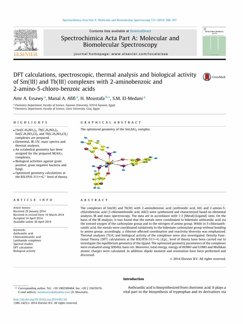
DFT calculations, spectroscopic, thermal analysis and biological activityof Sm(III) and Tb(III) complexes with 2-aminobenzoic and2-amino-5-chloro-benzoic acids
http://dx.doi.org/10.1016/j.saa.2014.04.1341386-1425/� 2014 Elsevier B.V. All rights reserved.
⇑ Corresponding author. Tel.: +20 1002589404; fax: +20 2 35676576.E-mail address: [email protected] (H. Moustafa).
Amr A. Essawy a, Manal A. Afifi a, H. Moustafa b,⇑, S.M. El-Medani a
a Chemistry Department, Faculty of Science, Fayoum University, 63514 Fayoum, Egyptb Chemistry Department, Faculty of Science, Cairo University, Giza, Egypt
h i g h l i g h t s
� (Sm[C7H6NO2]3, Tb[C7H6NO2]3,Sm[C7H5NO2Cl]3 and Tb[C7H5NO2Cl],)complexes are prepared.� Elemental, IR, UV, mass spectra and
thermal analyses.� An octahedral geometry has been
assigned for the prepared M(AA)3
complexes.� Biological activities against gram
positive, gram negative bacteria andfungi.� Optimized geometry calculations at
the B3LYP/6-311++G�� level of theory.
g r a p h i c a l a b s t r a c t
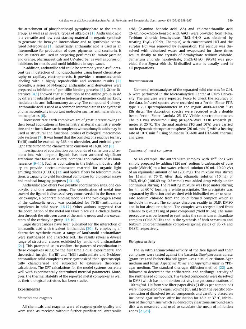
The optimized geometry of the Sm(AA)3 complex.
a r t i c l e i n f o
Article history:Received 29 January 2014Received in revised form 16 March 2014Accepted 14 April 2014Available online 30 April 2014
Keywords:Anthranilic acidChloroanthranilic acidLanthanide complexesSpectral studiesDFT calculationBiological activity
a b s t r a c t
The complexes of Sm(III) and Tb(III) with 2-aminobenzoic acid (anthranilic acid, AA) and 2-amino-5-chlorobenzoic acid (5-chloroanthranilic acid, AACl) were synthesized and characterized based on elementalanalysis, IR and mass spectroscopy. The data are in accordance with 1:3 [Metal]:[Ligand] ratio. On thebasis of the IR analysis, it was found that the metals were coordinated to bidentate anthranilic acid viathe ionised oxygen of the carboxylate group and to the nitrogen of amino group. While in 5-chloroanth-ranilic acid, the metals were coordinated oxidatively to the bidentate carboxylate group without bondingto amino group; accordingly, a chlorine-affected coordination and reactivity-diversity was emphasized.Thermal analyses (TGA) and biological activity of the complexes were also investigated. Density Func-tional Theory (DFT) calculations at the B3LYP/6-311++G (d,p)_ level of theory have been carried out toinvestigate the equilibrium geometry of the ligand. The optimized geometry parameters of the complexeswere evaluated using SDDALL basis set. Moreover, total energy, energy of HOMO and LUMO and Mullikanatomic charges were calculated. In addition, dipole moment and orientation have been performed anddiscussed.
� 2014 Elsevier B.V. All rights reserved.
Introduction
Anthranilic acid is biosynthesized from chorismic acid. It plays avital part in the biosynthesis of tryptophan and its derivatives via
A.A. Essawy et al. / Spectrochimica Acta Part A: Molecular and Biomolecular Spectroscopy 131 (2014) 388–397 389
the attachment of phosphoribosyl pyrophosphate to the aminegroup, as well as in several types of alkaloids [1]. Anthranilic acidis a versatile and low cost starting material in organic synthesisto generate the benzyne intermediate and to synthesize benzo-fused heterocycles [1]. Industrially, anthranilic acid is used as anintermediate for production of dyes, pigments, and saccharin. Itand its esters are used in preparing perfumes to imitate jasmineand orange, pharmaceuticals and UV-absorber as well as corrosioninhibitors for metals and mold inhibitors in soya sauce.
In addition, anthranilic acid could be commonly used as fluores-cent tag in detection of monosaccharides using liquid chromatog-raphy or capillary electrophoresis. It provides a monosaccharidelabeling with a highly reproducible and accurate results [2].Recently, a series of N-benzoyl anthranilic acid derivatives wereprepared as inhibitors of penicillin binding proteins [3]. Other lit-eratures [4,5] showed that substitution of the amino group in AAby different substituted aryl or heteroaryl moieties could markedlymodulate the anti-inflammatory activity. The compound N-pheny-lanthranilic acid is used as a common intermediate in the synthesisof pharmaceutically important molecules such as antimalarials andantineoplatics [6].
Fluorescent rare earth complexes are of great interest owing totheir broad applications in biochemistry, material chemistry, medi-cine and so forth. Rare earth complexes with carboxylic acids may beused as structural and functional probes of biological macromole-cule systems [7]. It was found that the complex of a reactive ternaryTb(III) could be excited by 365 nm ultraviolet, and emitted greenlight attributed to the characteristic emission of Tb(III) ion [8].
Investigation of coordination compounds of samarium and ter-bium ions with organic ligands has been attracted significantattentions that focus on several potential applications of its lumi-nescence [9–11]. Such as application in the lighting industry, abil-ity to provide electroluminescent material for organic lightemitting diodes (OLEDs) [12] and optical fibers for telecommunica-tions, a capacity to yield functional complexes for biological assaysand medical imaging purposes [13–15].
Anthranilic acid offers two possible coordination sites, one car-boxylic and one amino group. The coordination of metal ionstoward the ligand is discussed very controversial in the literature.For example, a bidentate binding mode via the two oxygen atomsof the carboxylic group was postulated for Tb(III) anthranilatecomplexes in solid state [16,17]. Other authors suggested thatthe coordination of the metal ion takes place via a chelate forma-tion through the nitrogen atom of the amino group and one oxygenatom of the carboxylic group [18,19].
Large discrepancies have been published for the interaction ofanthranilic acid with trivalent lanthanides [20]. By employing analternative synthetic route, a range of lanthanoid anthranilateswere synthesized and characterized. The results reveal a diverserange of structural classes exhibited by lanthanoid anthranilates[21]. This prompted us to confirm the pattern of coordination inthese complexes using for the first time a dual experimental andtheoretical insight. Sm(III) and Tb(III) anthranilate and 5-chloro-anthranilate solid complexes were synthesized then spectroscopi-cally characterized and subjected to extensive theoreticalcalculations. The DFT calculations for the model systems correlatewell with experimentally determined metrical parameters. More-over, the thermal stability of the reported metal complexes as wellas their biological activities has been studied.
Experimental
Materials and reagents
All chemicals and reagents were of reagent grade quality andwere used as received without further purification. Anthranilic
acid, (2-amino benzoic acid, AA) and chloroanthranilic acid(2-amino-5-chloro benzoic acid, AACl) were provided from Fluka.Terbium chloride hexahydrate, TbCl3�6H2O was obtained bytreating Tb4O7 (99.9%, Chempur) with concentrated HCl and thesurplus HCl was removed by evaporation. The residue was dis-solved with deionized water and evaporated for three timesresults finally to the crystals of hexahydrate terbium chloride.Samarium chloride hexahydrate, SmCl3�6H2O (99.9%) was pro-vided from Sigma–Aldrich. Bi-distilled water is usually used inall preparations.
Instrumentation
Elemental microanalyses of the separated solid chelates for C, H,N were performed in the Microanalytical Center at Cairo Univer-sity. The analyses were repeated twice to check the accuracy ofthe data. Infrared spectra were recorded on a Perkin–Elmer FTIRtype 1650 spectrophotometer in the region 4000–400 cm�1 asKBr discs. The absorption spectra were recorded with a doublebeam Perkin–Elmer Lambda 25 UV–Visible spectrophotometer.The pH was measured using pHs-JAN-WAY 3330 research pHmeter at 25 �C. The thermal analysis (TG and DTA) were carriedout in dynamic nitrogen atmosphere (20 mL min�1) with a heatingrate of 10 �C min�1 using Shimadzu TG-60H and DTA-60H thermalanalyzers.
Synthesis of metal complexes
As an example, the anthranilate complex with Tb3+ ions wassimply prepared by adding (126 mg) sodium bicarbonate of puregrade (Aldrich) to a hot water–ethanol solution (30 mL, 10:20 v/v)of an equimolar amount of AA (206 mg). The mixture was stirredfor 15 min at 70 �C. After that, ethanolic solution (10 mL) ofTbCl3�6H2O (186.69 mg, 0.5 mmol) was added drop wisely undercontinuous stirring. The resulting mixture was kept under stirringfor 4 h at 60 �C forming a white precipitate. The precipitate wasseparated by filtration and washed with bidistilled water to sepa-rate sodium chloride from the solid formed complex which isinsoluble in water. The complex dissolves readily in DMF, DMSOand in hot absolute ethanol. The complex was recrystallized fromethanol to give 231 mg of pure compound (Yield 81.6%). The sameprocedure was performed to synthesize the samarium anthranilatecomplex (Yield 80.3%) and in the synthesis of both samarium andterbium chloroanthranilate complexes giving yields of 85.7% and84.8%, respectively.
Biological activity
The in vitro antimicrobial activity of the free ligand and theircomplexes were tested against the bacteria: Staphylococcus aureus(gram +ve) and Escherichia coli (gram �ve) in Mueller Hinton-Agarmedium and fungi: Aspergillus flavus and Aspergillus niger in YPD-agar medium. The standard disc-agar diffusion method [22] wasfollowed to determine the antibacterial and antifungal activity ofthe synthesized compounds. The tested compounds were dissolvedin DMF (which has no inhibition activity), to get concentrations of100 mg/mL. Uniform size filter paper disks (3 disks per compound)were impregnated by equal volume (0.1 mL) from the specific con-centration of dissolved tested compounds and carefully placed onincubated agar surface. After incubation for 48 h at 37 �C, inhibi-tion of the organisms which evidenced by clear zone surround eachdisk was measured and used to calculate the mean of inhibitionzones [21,23].
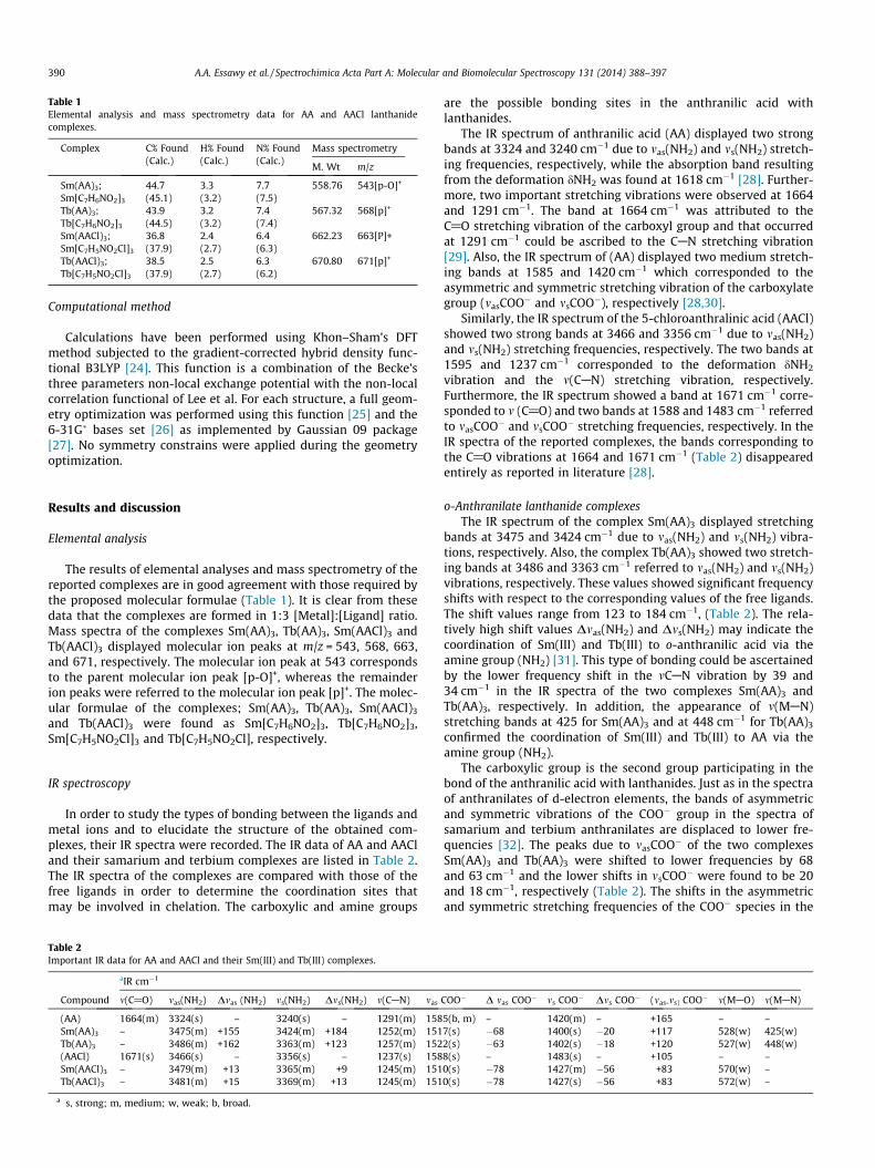
Table 1Elemental analysis and mass spectrometry data for AA and AACl lanthanidecomplexes.
Complex C% Found(Calc.)
H% Found(Calc.)
N% Found(Calc.)
Mass spectrometry
M. Wt m/z
Sm(AA)3; 44.7 3.3 7.7 558.76 543[p-O]+
Sm[C7H6NO2]3 (45.1) (3.2) (7.5)Tb(AA)3; 43.9 3.2 7.4 567.32 568[p]+
Tb[C7H6NO2]3 (44.5) (3.2) (7.4)Sm(AACl)3; 36.8 2.4 6.4 662.23 663[P]+Sm[C7H5NO2Cl]3 (37.9) (2.7) (6.3)Tb(AACl)3; 38.5 2.5 6.3 670.80 671[p]+
Tb[C7H5NO2Cl]3 (37.9) (2.7) (6.2)
390 A.A. Essawy et al. / Spectrochimica Acta Part A: Molecular and Biomolecular Spectroscopy 131 (2014) 388–397
Computational method
Calculations have been performed using Khon–Sham’s DFTmethod subjected to the gradient-corrected hybrid density func-tional B3LYP [24]. This function is a combination of the Becke’sthree parameters non-local exchange potential with the non-localcorrelation functional of Lee et al. For each structure, a full geom-etry optimization was performed using this function [25] and the6-31G� bases set [26] as implemented by Gaussian 09 package[27]. No symmetry constrains were applied during the geometryoptimization.
Results and discussion
Elemental analysis
The results of elemental analyses and mass spectrometry of thereported complexes are in good agreement with those required bythe proposed molecular formulae (Table 1). It is clear from thesedata that the complexes are formed in 1:3 [Metal]:[Ligand] ratio.Mass spectra of the complexes Sm(AA)3, Tb(AA)3, Sm(AACl)3 andTb(AACl)3 displayed molecular ion peaks at m/z = 543, 568, 663,and 671, respectively. The molecular ion peak at 543 correspondsto the parent molecular ion peak [p-O]+, whereas the remainderion peaks were referred to the molecular ion peak [p]+. The molec-ular formulae of the complexes; Sm(AA)3, Tb(AA)3, Sm(AACl)3
and Tb(AACl)3 were found as Sm[C7H6NO2]3, Tb[C7H6NO2]3,Sm[C7H5NO2Cl]3 and Tb[C7H5NO2Cl], respectively.
IR spectroscopy
In order to study the types of bonding between the ligands andmetal ions and to elucidate the structure of the obtained com-plexes, their IR spectra were recorded. The IR data of AA and AACland their samarium and terbium complexes are listed in Table 2.The IR spectra of the complexes are compared with those of thefree ligands in order to determine the coordination sites thatmay be involved in chelation. The carboxylic and amine groups
Table 2Important IR data for AA and AACl and their Sm(III) and Tb(III) complexes.
aIR cm�1
Compound m(C@O) mas(NH2) Dmas (NH2) ms(NH2) Dms(NH2) m(CAN) mas
(AA) 1664(m) 3324(s) – 3240(s) – 1291(m) 158Sm(AA)3 – 3475(m) +155 3424(m) +184 1252(m) 151Tb(AA)3 – 3486(m) +162 3363(m) +123 1257(m) 152(AACl) 1671(s) 3466(s) – 3356(s) – 1237(s) 158Sm(AACl)3 – 3479(m) +13 3365(m) +9 1245(m) 151Tb(AACl)3 – 3481(m) +15 3369(m) +13 1245(m) 151
a s, strong; m, medium; w, weak; b, broad.
are the possible bonding sites in the anthranilic acid withlanthanides.
The IR spectrum of anthranilic acid (AA) displayed two strongbands at 3324 and 3240 cm�1 due to mas(NH2) and ms(NH2) stretch-ing frequencies, respectively, while the absorption band resultingfrom the deformation dNH2 was found at 1618 cm�1 [28]. Further-more, two important stretching vibrations were observed at 1664and 1291 cm�1. The band at 1664 cm�1 was attributed to theC@O stretching vibration of the carboxyl group and that occurredat 1291 cm�1 could be ascribed to the CAN stretching vibration[29]. Also, the IR spectrum of (AA) displayed two medium stretch-ing bands at 1585 and 1420 cm�1 which corresponded to theasymmetric and symmetric stretching vibration of the carboxylategroup (masCOO� and msCOO�), respectively [28,30].
Similarly, the IR spectrum of the 5-chloroanthralinic acid (AACl)showed two strong bands at 3466 and 3356 cm�1 due to mas(NH2)and ms(NH2) stretching frequencies, respectively. The two bands at1595 and 1237 cm�1 corresponded to the deformation dNH2
vibration and the m(CAN) stretching vibration, respectively.Furthermore, the IR spectrum showed a band at 1671 cm�1 corre-sponded to m (C@O) and two bands at 1588 and 1483 cm�1 referredto masCOO� and msCOO� stretching frequencies, respectively. In theIR spectra of the reported complexes, the bands corresponding tothe C@O vibrations at 1664 and 1671 cm�1 (Table 2) disappearedentirely as reported in literature [28].
o-Anthranilate lanthanide complexesThe IR spectrum of the complex Sm(AA)3 displayed stretching
bands at 3475 and 3424 cm�1 due to mas(NH2) and ms(NH2) vibra-tions, respectively. Also, the complex Tb(AA)3 showed two stretch-ing bands at 3486 and 3363 cm�1 referred to mas(NH2) and ms(NH2)vibrations, respectively. These values showed significant frequencyshifts with respect to the corresponding values of the free ligands.The shift values range from 123 to 184 cm�1, (Table 2). The rela-tively high shift values Dmas(NH2) and Dms(NH2) may indicate thecoordination of Sm(III) and Tb(III) to o-anthranilic acid via theamine group (NH2) [31]. This type of bonding could be ascertainedby the lower frequency shift in the mCAN vibration by 39 and34 cm�1 in the IR spectra of the two complexes Sm(AA)3 andTb(AA)3, respectively. In addition, the appearance of m(MAN)stretching bands at 425 for Sm(AA)3 and at 448 cm�1 for Tb(AA)3
confirmed the coordination of Sm(III) and Tb(III) to AA via theamine group (NH2).
The carboxylic group is the second group participating in thebond of the anthranilic acid with lanthanides. Just as in the spectraof anthranilates of d-electron elements, the bands of asymmetricand symmetric vibrations of the COO� group in the spectra ofsamarium and terbium anthranilates are displaced to lower fre-quencies [32]. The peaks due to masCOO� of the two complexesSm(AA)3 and Tb(AA)3 were shifted to lower frequencies by 68and 63 cm�1 and the lower shifts in msCOO� were found to be 20and 18 cm�1, respectively (Table 2). The shifts in the asymmetricand symmetric stretching frequencies of the COO� species in the
COO� D mas COO� ms COO� Dms COO� (mas-ms) COO� m(MAO) m(MAN)
5(b, m) – 1420(m) – +165 – –7(s) �68 1400(s) �20 +117 528(w) 425(w)2(s) �63 1402(s) �18 +120 527(w) 448(w)8(s) – 1483(s) – +105 – –0(s) �78 1427(m) �56 +83 570(w) –0(s) �78 1427(s) �56 +83 572(w) –

A.A. Essawy et al. / Spectrochimica Acta Part A: Molecular and Biomolecular Spectroscopy 131 (2014) 388–397 391
IR spectra of lanthanide complexes indicate that o-anthranilate iscoordinated to Sm(III) and Tb(III) via the oxygen of the carboxylategroup. This is confirmed by the appearance of new mMAO stretch-ing bands at 528 and 527 cm�1 for Sm(III) and Tb(III) complexes,respectively.
5-Chloroanthranilate lanthanide complexesThe IR spectra of the complexes Sm(AACl)3 and Tb(AACl)3 dis-
played stretching medium bands at 3479 and 3481 cm�1 due tomas(NH2) and at 3365 and 3369 cm�1 due to ms(NH2) with lowershifts with respect to those of the free ligands (Table 2). The rela-tively small shift values of Dmas(NH2) and Dms(NH2) may indicatethat the amine group (NH2) in the chloroanthranilate may not par-ticipate in the coordination to Sm(III) and Tb(III) in their chloro-anthranalate complexes. This could be ascertained from theabsence of m(MAN) stretching band in the IR spectra of the twocomplexes Sm(AACl)3 and Tb(AACl)3. Comparing the Dmas(NH2)and Dms(NH2) values of the anthranilate and the chloroanthranilateSm(III) and Tb(III) complexes, revealed that the shift values for theanthranilate complexes (123–184 cm�1) are higher than the corre-sponding values of the chloroanthranilate complexes (9–15 cm�1).This indicates the participation of the anthranilate NH2 group inthe coordination to samarium and terbium while in chloroanthran-ilate complexes; chlorine atom greatly dimensioned the contribu-tion of this coordination site.
The carboxylate group can participate in bonding of the5-chloroanthranilate with lanthanides. On complexation, themasCOO� and msCOO� bands in samarium and terbium complexesshowed lower shifts by 78 and 56 cm�1, respectively, with respect
C
NH2
O
O
C
NH2
O
O
CO
O
H2N
Sm
(a)
Sm(AA)3
NH2Cl
C O
O
H2N
Cl
C
O
O H2N
Cl
C
O
O
Sm
(b)
Sm(AACl)3
Scheme 1. The proposed structures of: (a) samarium and terbium
to the corresponding species in the ligands. The shift values of thebands to lower frequency indicate the degree of bond covalency.The lower shift of the peak masCOO� (1585 cm�1) was found to be40–50 cm�1 for Cd, Ni, Co, Zn, Cu anthranilates [33]. Whereas inSm (III) and Tb (III) anthranilate and 5-chloroanthranilate com-plexes; the shift values were found to be 63–78 cm�1. The rela-tively higher shifts in the antisymmetric and symmetricstretching frequencies of the COO� species in the IR spectra ofthe 5-chloroanthranilate complexes may indicate the coordinationof the ligand to Sm(III) and Tb(III) via the two carboxylate oxygenatoms forming four membered ring. This is confirmed by theappearance of new mMAO stretching band at 570 and 572 cm�1
in the IR spectra of Sm(III) and Tb(III) complexes, respectively.According to elemental analysis data and interpretation of IR
spectra, samarium (III) and terbium (III) ions were expected to becoordinated to the o-anthranilate ion through a nitrogen atom ofNH2 group and oxygen of the carboxylate group [34], forming sixmembered ring in each complex, Sm(AA)3 or Tb(AA)3. On the otherhand, Sm(III) and Tb(III) ions were bonded to the 5-chloroanthran-ilate ions through the two oxygen of the carboxylate ion formingfour membered ring in Sm(AACl)3 or Tb(AACl)3 complex. The pro-posed structures of the reported complexes are shown inScheme 1.
Thermogravimetric analysis
Thermogravimetry provides more insight into the compositionand structure of the complexes. Thermal studies could be carriedout using thermogravimetry (TG) and (DTG) techniques. Thermal
C
NH2
O
O
C
NH2
O
O
CO
O
H2N
Tb
Tb(AA)3
NH2Cl
C O
O
H2N
Cl
C
O
O H2N
Cl
C
O
O
Tb
Tb(AACl)3
anthranilate complexes and (b) chloroanthranilate complexes.
Table 3Thermal analysis data for samarium and terbium complexes.
Complex Decomposition step, K DTG max, K an Weight loss% Mol. wt Found (Calc.) Species eliminated Solid residue Found, (calc.) %
Sm(AA)3 Sm[C21H18N3O6] 330–637 598 1 10.04 56.10 (56.08) C3H6N 29.82 (29.78)637–726 671 1 35.82 200.15 (200.17) C11H6NO3 SmO726–923 735, 875 2 24.32 135.89 (136.13) C7H6NO2
Sm(AACl)3 Sm[C21H15N3O6Cl3] 563–626 605 1 9.87 65.36 (65.57) C2H6 + ½ Cl2 34.40 (34.19)626–774 719 1 49.98 331.01 (331.17) C11H6N3 + Cl2 + 5/2 O2 SmO + 5C774–936 825 1 5.93 39.27 (39.06) C3H3
Tb(AA)3 Tb[C21H18N3O6] 423–700 674 1 63.72 361.47 (364.36) C20H18N3 + 2 O2 36.28 (35.76)TbO2 + C
Tb(AACl)3 Tb[C21H15N3O6Cl3] 321–520 403 1 5.20 34.90 (35.5) ½ Cl2 35.31 (35.62)520–1074 746 1 59.49 399.00 (396.31) C17H15N3 + 2O2 + Cl2 TbO2 + 4C
a n = Number of decomposition steps.
300 325 350 375
0.5
1.0
1.5
2.0 (A)32
1
AA Sm3+- AA Tb3+- AA
Abs
orba
nce
Wavelength, nm
300 325 350 375 400
0.5
1.0
1.5
2.0 (B) 3
2
1
AACl Sm3+- AACl Tb3+- AACl
Abs
orba
nce
Wavelength, nm
Fig. 1. (a) UV absorption spectra of AA (spectrum 1), Sm3+-(AA)3 (spectrum 2) andTb3+-(AA)3 (spectrum 3). (b) UV absorption spectra of AACl (spectrum 1), Sm3+-(AACl)3 (spectrum 2) and Tb3+-(AACl)3 (spectrum 3). [Experimental conditions:concentration of ligand and complexes were 1 � 10�5 mol. L�1, solvent = ethanol].
392 A.A. Essawy et al. / Spectrochimica Acta Part A: Molecular and Biomolecular Spectroscopy 131 (2014) 388–397
analysis is used to: (i) get information about the thermal stabilityof the metal complexes, (ii) suggest the presence of crystallineand hydrated water molecules and (iii) suggest a general schemefor thermal decomposition of the complexes. The results of thermaldecomposition of the metal complexes indicated the absence ofany type of water molecules as shown in Table 3.
Thermogravimetric analysis of Sm(AA)3 and Tb(AA)3 complexesThe TG plot of Sm(AA)3, Sm[C21H18N3O6], complex displayed
four decomposition steps. Since the third and fourth decomposi-tion steps were confused, three resolved and well-defined decom-position steps could be considered. The first decomposition stepoccurred in the temperature range 330–637 K with a net weightloss of 10.04% corresponding to elimination of (C3H6N) species(10.04% cal). The second decomposition peak occurred in the tem-perature range 637–726 K with a weight loss of 35.82% and corre-sponded to the material decomposition (C11H6NO3) moieties(35.82% cal). The third decomposition step occurred in the temper-ature range 726–923 K with a weight loss of 24.32% correspondingto the elimination of (C9H4N2) species (24.36% cal) to give finallythe residue SmO (29.82%).
Thermal studies of the complex Tb(AA)3, Tb[C21H18N3O6], werecarried out using thermogravimetry (TG) and (DTG) techniques.The TG plot of the complex showed one decomposition step inthe temperature range 423–700 K giving a net weight loss of63.72% corresponding to elimination of (C20H18N3 + 2O2) species(64.22% cal) to give finally the residue (TbO2 + C) with 36.28%.
Thermogravimetric analysis of Sm(AACl)3 and Tb(AACl)3 complexesThe TG plot of Sm(AACl)3, Sm[C21H15N3O6Cl3], complex dis-
played three resolved and well-defined decomposition steps. Thefirst decomposition step occurred in the temperature range563–626 K with a net weight loss of 9.87% corresponding to elim-ination of (C2H6 + ½Cl2) species (9.90% cal). The second decompo-sition peak occurred in the temperature range 626–774 K with aweight loss of 49.98% and corresponded to the material decompo-sition (C11H6N3 + Cl2 + 5/2O2) moieties (50.01% cal). The thirddecomposition step occurred in the temperature range774–936 K with a weight loss of 5.93% corresponding to the elim-ination of (C3H3) species (5.90% cal) to give finally the residue(SmO + 5C) species (34.40%).
The TG plot of Tb(AACl)3, Tb[C21H15N3O6Cl3], complex displayedtwo resolved and well-defined decomposition steps. The firstdecomposition step occurred in the temperature range321–520 K with a net weight loss of 5.20% corresponding to elim-ination of ½Cl2 species (5.29% cal). The second decomposition peakoccurred in the temperature range 520–1074 K with a weight lossof 59.49% and corresponded to the material decomposition(C17H15N3 + Cl2 + 2O2) moieties (59.08% cal) to give finally theresidue (TbO2 + 4C) species (35.31%).
The sum of the eliminated and solid residual species for all lan-thanide complexes confirmed their molecular formulae (Table 3).
Absorption spectra
The UV absorption spectra of 1 � 10�5 mol. L�1 ethanolic solu-tion of AA, AACl and their tris-samarium and terbium complexesare presented in Fig. 1. Fig. 1A reveals a broad UV absorption bandat kmax = 331 nm with e value around 21780 mol.�1 L cm�1. Thisband could be assigned to p–p� transition in AA (spectrum 1).
A.A. Essawy et al. / Spectrochimica Acta Part A: Molecular and Biomolecular Spectroscopy 131 (2014) 388–397 393
Upon addition of Sm(III) ions, a slight blue shift and enhancementof the absorbance value due to complex formation, (spectrum 2). Asimilar behavior was also observed in case of Tb3+ ions (spectrum3). Moreover, the estimated e values for the Sm(AA)3 and Tb(AA)3
complexes amount, respectively to 31,600 and 34,000 mol.�1
L cm�1 with a noticeable increase compared to free ligand. Com-pared to AA ligand and their Tb(III) or Sm(III) complexes, the mon-itored UV absorption spectra of ligand AACl (kmax 348 nm,e = 30,760 mol.�1 L cm�1), their Tb(III) and Sm(III) counterparts(Fig. 1B) indicate increased blue shift by 4–7 nm and higher esti-mated e values 38200 and 42,000 mol.�1 L cm�1, respectively.These results reflect and further confirm the role of Cl atom in rein-forcing lanthanoid-AACl coordination through two oxygens of car-boxylate group rather than the preferential O- and N-coordinationsites elucidated in lanthanoid-AA complexation.
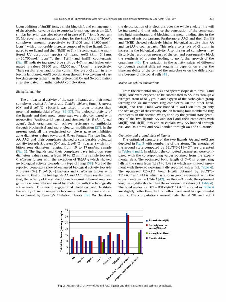
Biological activity
The antibacterial activity of the parent ligands and their metalcomplexes against A. flavus and Canidia albicans fungi, S. aureus(G+) and E. coli (G�) bacteria was tested in order to assess theirpotential antimicrobial effects [35–37]. The biological activity ofthe ligands and their metal complexes were also compared withtetracycline (Antibacterial agent) and Amphotericin B (Antifungalagent). Such organisms can achieve resistance to antibioticsthrough biochemical and morphological modification [27]. In thepresent work all the synthesized complexes gave no inhibitionzone diameters values towards A. flavus fungus. The two ligandsAA, AACl and their complexes showed a considerable biologicalactivity towards S. aureus (G+) and E. coli (G�) bacteria with inhi-bition zone diameters ranging from 10 to 17 mm/mg sample(Fig. 2). The ligands and their complexes gave inhibition zonediameters values ranging from 10 to 12 mm/mg sample towardsC. albicans fungus with the exception of Tb(AA)3 which showedno biological activity towards this type of fungi [38]. Most of thereported complexes showed enhanced biological activity towardsS. aureus (G+), E. coli (G�) bacteria and C. albicans fungus withrespect to that of the free ligands AA and AACl. These results meanthat, the activity of the studied ligands against different microor-ganisms is generally enhanced by chelation with the biologicallyactive metal. This would suggest that chelation could facilitatethe ability of such complexes to cross a cell membrane and canbe explained by Tweedy’s Chelation Theory [39]. On chelation,
Fig. 2. Antimicrobial activity of AA and AACl ligand
the delocalization of p-electrons over the whole chelate ring willbe increased and that enhance the penetration of the complexesinto lipid membranes and blocking the metal binding sites in theenzymes of microorganisms. Furthermore, AACl and their Sm(III)and Tb(III) showed relatively higher biological activity than AAand Ln-(AA)3 counterparts. This refers to a role of Cl atom inincreasing the biological activity. Also, the tested complexes maydisturb the respiration process of the cell and consequently blockthe synthesis of proteins leading to no further growth of theorganisms [40]. The variation in the activity values of differentcompounds against different organisms depends on either theimpermeability of the cells of the microbes or on the differencesin ribosome of microbial cells [41].
Molecular orbital calculations
From the elemental analysis and spectroscopic data, Sm(I) andTb(I) ions were expected to be coordinated to AA ions through anitrogen atom of NH2 group and oxygen of the carboxylate groupforming the six membered ring complexes. On the other hand,Sm(I) and Tb(I) ions were bonded to AACl ion through onlythe two oxygen of the carboxylate ion forming four membered ringcomplexes. In this section, we try to study the ground state geom-etry of the two ligands AA and AACl and their complexes withSm(I) and Tb(I) ions and to explain why AA bonded throughN10 and O8-atoms, and AACl bonded through O8 and O9-atoms.
Geometry and ground state of ligandsThe optimized structure of the two ligands AA and AACl are
depicted in Fig. 3 with numbering of the atoms. The energies ofthe ground state computed by B3LYP/6-311++G�� are presentedin Tables 4 and 5. In addition, the computed parameters were com-pared with the corresponding values obtained from the experi-mental data. The optimized bond length of CAC in phenyl ringfalls in the range from 1.393 to 1.428 Å which are in good agree-ment with those of experimentally reported values (c.f. Table 4).The optimized C2ACl11 bond length obtained by B3LYP/6-311++G�� is 1.741 Å which is also in good agreement with theexperimental value 1.744 Å [42]. For the CAO bonds, the optimizedlength is slightly shorter than the experimental values (c.f. Table 4).The bond angles for DFT – B3LYP/6-311++G�� reported in Table 4are slightly better than the HF-method compared to experimentalresults. The computations overestimate the <HNH and <OCO
s and their samarium and terbium complexes.

Fig. 3. Optimized geometry, vector of dipole moment, numbering system, net charges and HOMO and LUMO charge density maps for AA and AACl ligands using B3LYP/6-311G��.
394 A.A. Essawy et al. / Spectrochimica Acta Part A: Molecular and Biomolecular Spectroscopy 131 (2014) 388–397
angles. From the analysis of geometric parameters; we notice suchdifferences between calculated and measured values. These dis-crepancies can be explained by the fact that the calculationsassume an isolated molecule where the intermolecular columbicinteractions with the neighboring molecules are absent, whereasthe experimental results correspond to interact molecules in thecrystal lattice for a similar compound.
The ligand AA is considered as electron donor, whereas, ligandAACl is electron acceptor as indicated from the EHOMO and ELUMO
(c.f. Table 5). The ligand AA is more reactive than AACl as reflectedfrom energy gap values (c.f. Table 5). From the computed netcharge on active centers, it was found that the most negative cen-ters in AA are N10 and O8. Whereas in ligand AACl, the most neg-ative centers are O8 and O9. The reason why AA ligand bonded
through N10 and O8 while AACl ligand bonded through O8 andO9-atoms. The computed dipole moment of AA ligand is 8.38D.Insertion of Cl-atom in C5-atom as in AACl increases the dipolemoment to 10.19D indicating that the vector of the dipole momentof Cl-atom is in the same direction of the vector of AA-ligand.
Geometric parameters of the complexesTables 6–9 in addition to Fig. 4 present the optimized geometry,
numbering system, the vector of the dipole moment, the energetic,dipole moment, energy gap, energy of HOMO and LUMO, netcharge on active centers, bond lengths, bond angles and dihedralangles of all metal complexes studied in this work. In the com-plexes of Sm(AA)3 and Tb(AA)3 the metal ion coordinates withN10, O12, N27, O26 and N42, O39 to form six-member ring.
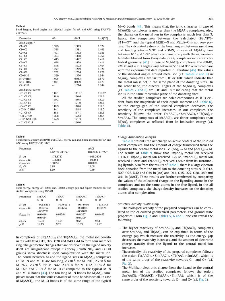
Table 4Bond lengths, Bond angles and dihydral angles for AA and AACl using B3LYP/6-311++G��.
Parameter AA AACI Expt[57]
Bond length, ÅC1AC2 1.399 1.399 1.374C1AC6 1.398 1.391 1.403C2AC3 1.401 1.392 1.385C3AC4 1.393 1.388 1.366C4AC5 1.415 1.422 1.411C5AC6 1.428 1.429 1.411C6AC7 1.546 1.522 1.464C7AO8 1.277 1.269 1.320C7AO9 1.252 1.264 1.229C5AN10 1.369 1.370 1.364N10AH15 1.006 0.983 0.879N10AH16 1.031 0.999 0.879C2ACl11 – 1.714 1.744
Bond angle, degree<C1 C6 C5 118.1 1119.0 119.67<C1 C2 C5 118.6 120.4 121.6<C2 C3 C4 120.2 119.5 120.9<C3 C4 C5 121.1 121.0 121.6<C4 C5 C6 118.9 118.6 117.3<C5 N10 H16 114.1 118.8 118.5<C1 C6 C7 118.8 117.1 121.5<O8 C7 O9 126.8 122.3 121.4<H15 N10 H16 124.9 121.3 119.1<C1 C2 Cl11 – 120.0
Table 5Total energy, energy of HOMO and LUMO, energy gap and dipole moment for AA andAACl using B3LYP/6-311++G��.
Parameter AA AAClB3LYP/6-31++G�� B3LYP/6-31++G��
ET, au �475.6737 �935.2676EHOMO, au �0.06282 �0.6454ELUMO, au 0.08358 0.07876Eg, eV 3.98 19.68l, D 8.39 10.19
Table 6Total energy, energy of HOMO and, LUMO, energy gap and dipole moment for thestudied complexes using SDDALL.
Parameter Sm(AA) Tb(AA) Sm(AACl) Tb(AACl)OAN OAN OAO OAO
ET, au �905.6308 �1070.4615 �947.9705 �1113.142EHOMO, au �0.35752 �0.34257 �0.31084 �0.30478
�0.35751 �0.31082ELUMO, au 0.04446 0.04504 0.04397 0.04493
0.04454 0.04399Eg, eV 10.93 10.54 9.65 9.51l, D 19.13 9.70 13.03 12.97
A.A. Essawy et al. / Spectrochimica Acta Part A: Molecular and Biomolecular Spectroscopy 131 (2014) 388–397 395
In complexes of Sm(AACl)3 and Tb(AACl)3, the metal ion coordi-nates with O14, O15, O27, O28 and O40, O44 to form four memberring. The geometric changes that are observed in the ligand moietyitself are insignificant except C (phenyl) with NH2 and COOHgroups show shortness upon complexation with the metal ion.The bonds between M and the ligand sites in M(AA)3 complexesi.e. MAN and MAO are too long, 2.730 Å for MAN10, 2.758 Å forMAN27, 2.728 Å for MAN42, 2.160 Å for MAO12, 2.182 Å forMAO26 and 2.171 Å for MAO39 compared to the typical MANand MAO bonds [43]. The too long MAN bonds for M(AA)3 com-plexes mean that the ionic character of these bonds is small. In caseof M(AACl)3, the MAO bonds is of the same range of the typical
MAO bonds [44]. This means that, the ionic character in case ofM(AACl)3 complexes is greater than the M(AA)3 complexes. Also,the charge on the metal ion in the complex is much less than 3,hence, the comparison between the calculated (B3LYP/6-311++G��) and the typical M(I)AN and M(I)AO is not very pre-cise. The calculated values of the bond angles (between metal ionand binding sites) < MNC and <OMN, in case of M(AA)3 varybetween 61� and 124� which compare nicely with the experimen-tal data obtained from X-ray data for Oh complexes indicates octa-hedral geometry [45]. In case of M(AACl)3 complexes, the <OMO,<MOC and <OCO angles vary between 55� and 95� which comparewith the experimental data reported in literature [46]. The valuesof the dihedral angles around metal ion (c.f. Tables 7 and 8) inM(AA)3 complexes, are far from 0.0� or 180� which indicate thatthe metal ion is not in the same plane of the donating sites. Onthe other hand, the dihedral angles of the M(AACl)3 complexes(c.f. Tables 7 and 8) are 0.0� and 180� indicating that the metalion is in the same molecular plane of the donating sites.
All the studied complexes are polar compounds as it is evi-dent from the magnitude of their dipole moment (c.f. Table 6).As the energy gap of the studied complexes decreases, thereactivity of the complexes increases. In our complexes, thereactivity follows the order Tb(AACl)3 > Sm(AACl)3 > Tb(AA)3 >Sm(AA)3. The complexes of M(AACl)3 are donor complexes thanM(AA)3 complexes as reflected from its ionization energy (c.f.Table 6).
Charge distribution analysisTable 9 presents the net charge on active centers of the studied
metal complexes and the amount of charge transferred from theligands to the central metal ions, i.e. (AA)3 ? M and (AACl)3 ? M.The results of Table 9 show that Sm(AA)3 metal ion received1.116 e, Tb(AA)3 metal ion received 1.227e, Sm(AACl)3 metal ionreceived 1.599e and Tb(AACl)3 received 1.592e from its surround-ing ligands. Also from the results of Table 9, there is a large electronback-donation from the metal ion to the donating sites N10, O12,N27, O26, N42 and O39 in (AA) and O14, O15, O27, O28, O40 andO41 in (AACl). These results are further confirmed by comparingthe values of the calculated charge on the liganding atoms in thecomplexes and on the same atoms in the free ligand. In the allstudied complexes, the charge density increases on the donatingatoms after complexation.
Structure activity relationshipThe biological activity of the prepared complexes can be corre-
lated to the calculated geometrical parameters and ground stateproperties. From Fig. 2 and Tables 5, 6 and 9 one can reveal thefollowing:
– The higher reactivity of Sm(AACl)3 and Tb(AACl)3 complexesover Sm(AA)3 and Tb(AA)3 can be explained in terms of theenergy gap which measure the reactivity, as the energy gapdecreases the reactivity increases, and the amount of electroniccharge transfer from the ligand to the central metal ionincreases.
– Theoretically, the reactivity of the prepared complexes followsthe order: Tb(AACl)3 > Sm(AACl)3 > Tb(AA)3 > Sm(AA)3 which isof the same order of the reactivity towards G� and G+ (c.f.Fig. 2).
– The Mullikan electronic charge from the ligand to the centralmetal ion of the studied complexes follows the order:Sm(AACl)3 > Tb(AACl)3 > Tb(AA)3 > Sm(AA)3 which is of thesame order of the reactivity towards G� and G+ (c.f. Fig. 2).
Fig. 4. Final geometry, numbering system and vector of The dipole moment for metals complexes using SDDALL.
Table 7Bond lengths, bond angles and dihydral angles for the metal-AA complexes usingSDDLL.
Parameter AA Parameter AA
Sm Tb Sm Tb
MAN10 2.730 2.614 C24AO25 1.214 1.233MAO12 2.160 2.128 C37AO38 1.208 1.222MAN27 2.758 2.789 <O12 M N10 68.8 70.1MAO26 2.182 2.183 <O26MN27 65.7 67.0MAO39 2.171 2.158 <O39 M N42 64.5 61.6MAN42 2.728 2.569 <M N42 C31 124.1 115.6C31AN42 1.460 1.459 <N42 C31 C36 125.8 122.2C13AC36 1.397 1.401 <C31 C36 C37 115.6 122.8C36AC37 1.504 1.499 <C36 C37 O39 �157.8 114.5C37AO39 1.306 1.312 <M N10 C4 C3 54.7 �163.7O12AC7 1.314 1.306 <M O12 C7 C5 58.7 �62.4C5AC7 1.500 1.513 <M O26 C24 C16 58.2 �86.7C4AC5 1.409 1.407 <M N27 C21 C20 �160.6 161.8C4AN10 1.453 1.464 <M O39 C37 C36 57.3 �6.3C21AN27 1.454 1.455 <M N42 C31 C32 �167.1 131.1C16AC21 1.396 1.395C16AC24 1.502 1.489C24AO26 1.308 1.304C7AO11 1.225 1.225
Table 8Bond lengths, bond angles and dihydral angles for the metal-AACl complexes usingSDDLL.
Parameter AA-CI Parameter AA-CI
Sm Tb Sm Tb
MAO14 2.377 2.357 <M O15 C13 C4 179.0 179.7MAO15 2.381 2.327 <O15 C13 C4 C3 0.11 0.44MAO27 2.405 2.364 <M O28 C26 C22 �179.9 �179.9MAO28 2.359 2.320 <O28 C26 C22 C21 �0.3 0.11MAO40 2.355 2.326 <M O40 C39 C38 �179.7 �179.5MAO41 2.398 2.357 <O40 C34 C38 C33 0.384 �0.11C4AC13 1.448 1.446C22AC26 1.448 1.446C38AC39 1.450 1.447C2ACl 8 1.829 1.830C20ACl 29 1.829 1.830C24ACl 43 1.830 1.830<O14 M O15 54.8 55.5<M O14 C13 27.5 27.8<O14 C13 O15 116.4 115.3<M O15 C13 94.3 95.3<O27 M O28 54.7 55.5<O40 M O41 63.5 62.9
396 A.A. Essawy et al. / Spectrochimica Acta Part A: Molecular and Biomolecular Spectroscopy 131 (2014) 388–397
Table 9Net charges on metals and active centers of the studied complexes.
Center AACl AA Center AA CI
Sm Tb Sm Tb
M 1.884 1.773 M 1.401 1.408N10 �0.933 �0.951 O14 �0.784 �0.769O12 �0.971 �0.900 O15 �0.689 �0.677O11 �0.525 �0.514 O27 �0.777 �0.776O26 �0.980 �0.884 O28 �0.690 �0.670N27 �0.912 �0.916 O40 �0.691 �0.678O25 �0.513 �0.563 O41 �0.779 �0.774N42 �0.923 �0.948 N7 �0.864 �0.870O39 �0.967 �0.988 N23 �0.866 �0.894O38 �0.502 �0.525 N42 �0.870 �0.868(AA)3 ? M 1.116 1.227 (AACl)3 ? M 1.599 1.592
A.A. Essawy et al. / Spectrochimica Acta Part A: Molecular and Biomolecular Spectroscopy 131 (2014) 388–397 397
Conclusions
Four solid lanthanide complexes of Sm(III) and Tb(III) withAntharnilic acid (AA) and chloroanthanilic acid (AACl) were pre-pared and isolated. According to elemental analysis, IR, mass spec-troscopy, and thermal analyses, these complexes are structurallyformulated in 1:3 [Metal]:[Ligand] ratio. From the IR spectra, it isclear that the active site in AA is O8 and N10 but in AACl is O8and O9. Therefore, a chlorine-affected coordination and reactiv-ity-diversity was emphasized. The complexes have not water mol-ecules inside or outside the coordination sphere of the centralmetal according to thermal analysis. The theoretical calculationsof the ligand and their complexes show a difference in the geomet-ric parameters between calculated and experimental results. Thesediscrepancies can be explained by the fact that the calculationsassume an isolated molecule where the intermolecular columbicinteraction with the neighboring molecules are absent, whereasthe experimental results corresponds to interacting molecules inthe crystal lattice for a similar compound. Also the calculationshows that, the ligand AA is considered as electron donor and morereactive than ligand AACl. Insertion of chlorine atom as in AAClligand increases the value of the dipole moment. The values ofthe dihedral angles around metal ion in M(AA)3 complexes arefar from 0.0� or 180� which indicate that the metal ion is not inthe same plane of the donating sites. On the other hand, the dihe-dral angles of the M(AACl)3 complexes are 0.0� and 180� indicatingthat the metal ion is in the same molecular plane of the donatingsites. All the studied complexes are polar as it is evident fromthe magnitude of their dipole moment. The reactivity of the com-plexes follows the order Tb(AACl)3 > Sm(AACl)3 > Tb(AA)3 >Sm(AA)3. Most of the reported complexes showed enhanced bio-logical activity towards S. aureus (G+) and E. coli (G�) bacteriaand C. albicans fungus with respect to that of the free ligands AAand AACl.
References
[1] P. Wiklund, J. Bergman, Curr. Org. Chem. 3 (2006) 379–402.[2] H. Stepan, E. Staudacher, Anal. Biochem. 418 (2011) 24–29.[3] I. Sosic, S. Turk, M. Sinreih, N. Trošt, O. Verlaine, A. Amoroso, A. Zervosen, A.
Luxen, B. Joris, S. Gobec, Acta Chim. Slov. 59 (2012) 380–388.[4] B. Goel, T. Ram, R. Tyagi, E. Bansal, A. Kumar, D. Mukherjee, J.N. Sinha, Eur. J.
Med. Chem. 34 (1999) 265–269.
[5] S. Sharma, V.K. Srivastava, A. Kumar, Eur. J. Med. Chem. 37 (2002) 689–697.[6] Y. Sarrafi, M. Mohadeszadeh, K. Alimohammadi, Chin. Chem. Lett. 20 (2009)
784–788.[7] A.M. Farag, S.G. Teoh, H. Osman, C.S. Yeap, H.K. Fun, Acta Cryst. E67 (2011) o37.[8] Z. Aiqin, P. Qiliang, J. Husheng, L. Xuguang, X. Bingshe, J. Rare Earth 30 (2012)
10–16.[9] V.F. Shul’gin, S.V. Abkhairova, O.V. Konnik, S.B. Meshkova, Z.M. Topilova, E.B.
Rusanov, G.G. Aleksandrov, I.L. Eremenko, Russ. J. Inorg. Chem. 58 (6) (2013)678–683.
[10] N.P. Kuzmina, S.V. Eliseeva, Russ. J. Inorg. Chem. 51 (2006) 73–88.[11] V.F. Shul’gin, S.V. Abkhairova, O.V. Konnik, S.B. Meshkova, Z.M. Topilova, M.A.
Kiskin, I.L. Eremenko, J. Inorg. Chem. 57 (2012) 420–426.[12] V.F. Shul’gin, O.V. Konnik, S.V. Abkhairova, A.N. Gusev, S.B. Meshkova, A.V.
Kiriyak, E.B. Rusanov, M. Hasegawa, W. Linert, Inorg. Chim. Acta 402 (2013)33–38.
[13] J.P. Leonard, C.B. Nolan, F. Stomeo, T.H. Gunnlaugsson, Top. Curr. Chem. 281(2007) 1–43.
[14] K. Binnemans, in: K.A. Gschneidner Jr., J.C.G. Bünzli, V.K. Pecharsky (Eds.),Rare-Earth Beta-Diketonates, Handbook on the Physics and Chemistry of RareEarth, Elsevier, Amsterdam, 2005, pp. 107–272.
[15] S.V. Eliseeva, J.C.G. Bünzli, Chem. Soc. Rev. 39 (2010) 189–227.[16] P.C.R. Soares-Santos, R.A. Sá Ferreira, T. Trindade, L.D. Carlos, H.I.S. Nogueira, J.
Alloys Compd. 451 (2008) 575–577.[17] M. Hilder, M. Lezhnina, M.L. Cole, C.M. Forsyth, P.C. Junk, U.H. Kynast, J.
Photochem. Photobiol. A 217 (2011) 76–86.[18] M. Forsyth, K. Wilson, T. Behrsing, C. Forsyth, G.B. Deacon, A. Phanasgoankar,
Corrosion 58 (2002) 953–960.[19] Y. Kim, S.S. Yun, Thermochim. Acta 59 (1982) 299–303.[20] G.B. Deacon, M. Forsyth, P.C. Junk, S.G. Leary, G.J. Moxey, Polyhedron 25 (223)
(2006) 379–386.[21] M. Shakiv, Y. Azim, H.T.N. Chishti, S. Parveen, Spectrochim. Acta A 65 (2006)
490–496.[22] N. Sari, S. Arslan, E. Logoglu, I. Sakiyan, J. Anim. Sci. 16 (2003) 283–288.[23] G.G. Mohamed, Spectrochim. Acta A 64 (2006) 188–195.[24] (a) A.D. Becke, J. Chem. Phys. 98 (1993) 5648–5652;
(b) A.D. Becke, J. Chem. Phys. 98 (1993) 1372–1377.[25] (a) C. Lee, W. Yang, R.G. Parr, Phys. Rev. B Condens. Matter. 37 (1988) 785–
789;(b) B. Miehlich, A. Savin, H. Stolt, H. Preuss, Chem. Phys. Lett. 157 (1989) 200–206.
[26] B.B. Stefanov, G. Liu, A. Liashenko, P. Piskorz, I. Komaromi, R.L. Martin, D.J. Fox,T. Keith, M.A. Al-Laham, C.Y. Peng, A. Nanayakkara, M. Challacombe, P.M.W.Gill, B. Johnson, W. Chen, M.W. Wong, C. Gonzalez, J.A. Pople, Gaussian, Inc.,Pittsburgh PA. 2003.
[27] M.J. Frisch, G.W. Trucks, H.B. Schlegel, G.E. Scuseria, et al., Gaussian, Inc.,Wallingford CT, 2009.
[28] W. Brzyska, Z. Rzaczynska, Monatsh. Chem. 119 (1988) 147–156.[29] G.J. Efremova, R.T. Butshkova, Koord Khim. 3 (1977) 1184–1189.[30] V.L. Dorofeev, Chem. Acta 38 (2004) 45–49.[31] G.G. Mohamed, A.A. Soliman, Thermochim. Acta 421 (2004) 151–159.[32] A.G. Hill, C. Curran, J. Phys. Chem. 64 (1960) 1519–1522.[33] S.S. Sandhu, B.S. Manhas, R.M. Mittal, S.S. Parmar, Indian J. Chem. 7 (1969)
286–289.[34] A.A. Soliman, G.G. Mohamed, Spectrochim. Acta A 91 (2012) 11–17.[35] Z.L. You, H.L. Zhu, W.S. Liu, Z. Anorg, Allg. Chem. 630 (2004) 1617–1622.[36] S. Yamada, Coord. Chem. Rev. 190 (1999) 537–555.[37] S. Chang, L. Jones, C.M. Wang, L.M. Henling, R.H. Grubbs, Organometallics 17
(1998) 3460–3465.[38] S. Duraiswamy, R.E. Michael, Z. Matthias, N. Karup-pannan, Polyhedron 26
(2007) 4314–4320.[39] N. Dharmaraj, P. Viswanathamurthi, K. Nataragan, Transition Met. Chem. 26
(2001) 105–109.[40] P.G. Lawrence, P.L. Harold, O.G. Francis, Antibiot. Chemother. 5 (1980) 1597–
1600.[41] A.C. Dros, R.W.J. Zijlstra, P.T. Van Duijmen, A.L. Spek, H. Kooijman, R.M. Kellogg,
Tetrahedron Lett. 54 (1998) 77–87.[42] H. Takagawa, S. Ohba, Y. Saito, Acta Cryst. C42 (1986) 1880–1881.[43] L. Armelao, S. Quici, F. Brigelletti, G. Accorsi, G. Bottaroi, M. Cavazzini, E.
Tendello, Coord. Chem. Rev. 254 (2010) 487–505.[44] S. Tanase, J. Reedijk, Coord. Chem. Rev. 250 (2006) 2501–2510.[45] S.S. Zumdahl, Chemistry for Chemical and Biological Science, University
Science Books, USA, 2000.[46] A.I. Boldyrev, V.V. Zhdankin, J. Simons, P.J. Stang, J. Am. Chem. Soc. 114 (1992)
10569–10572.