Embed Size (px)
Citation preview
Specific and General HLA-DR BindingMotifs: Comparison of Algorithms
Francisco Borras-Cuesta, Jose-Javier Golvano,Marta Garcıa-Granero, Pablo Sarobe,Jose-Ignacio Riezu-Boj, Eduardo Huarte, andJuan-Jose Lasarte
ABSTRACT: Using panels of peptides well characterizedfor their ability to bind to HLA DR1, DRB1*1101, orDRB1*0401 molecules, algorithms were deduced to pre-dict binding to these molecules. These algorithms consistof blocks of 8 amino acids containing an amino acidanchor (Tyr, Phe, Trp, Leu, Ile, or Val) at position i anddifferent amino acid combinations at positions i12 toi17 depending on the class II molecule. The sensitivity(% of correctly predicted binder peptides) and specificity(% of correctly predicted non-binder peptides) of thesealgorithms, were tested against different independentpanels of peptides and compared to other algorithmsreported in the literature. Similarly, using a panel of 232peptides able to bind to one or more HLA molecules aswell as 43 non-binder peptides, we deduced a generalmotif for the prediction of binding to HLA-DR mole-cules. The sensitivity and specificity of this general motifwas dependent on the threshold score used for the predic-tions. For a score of 0.1, the sensitivity and specificity
were 84.7% and 69.8%, respectively. This motif wasvalidated against several panels of binder and non-binderpeptides reported in the literature, as well as against 35,15-mer peptides from hepatitis C virus core protein, thatwere synthesized and tested in a binding assay against apanel of 19 HLA-DR molecules. The sensitivities andspecificities against these panels of peptides were similarto those attained against the panels used to deduce thealgorithm. These results show that comparison of binderand non-binder peptides, as well as correcting for therelative abundance of amino acids in proteins, is a usefulapproach to deduce performing algorithms to predictbinding to HLA molecules. Human Immunology 61,266–278 (2000). © American Society for Histocompat-ibility and Immunogenetics, 2000. Published by ElsevierScience Inc.
KEYWORDS: T cell determinant; class II molecules;algorithm; MHC; binding motif; prediction of binding
ABBREVIATIONSAPC antigen presenting cellMHC major histocompatibility complex
HCV hepatitis C virusTDh T-helper cell determinant
INTRODUCTIONDeterminants recognized by T-helper cells (TDh) arepeptides of 8 to 23 amino acids (commonly 14 to 15 inaverage) that are able to bind to MHC class II molecules.TDh originate generally in endocytic cell compartmentsafter processing of foreign protein antigens, that have
entered the antigen presenting cell (APC) following im-munization with an antigen or after infection, althoughpeptides derived from self proteins also bind to class IImolecules. Presentation of the MHC class II-peptidecomplex by an APC, followed by its recognition by theT cell receptor, leads to T cell activation. See [1] for athorough review on these findings.
It has been shown [2] that TDh peptides bind to asingle site in a groove of the MHC class II molecules.Also, the association pattern of peptides to differentallele variants of murine Ia is a reflection of the MHCrestriction of the immune response [3, 4]. Severalworkers have attempted to unravel the relevant struc-
From the Universidad de Navarra, Facultad de Medicina, Departamentode Medicina Interna, Pamplona, Spain.
Address reprint requests to: Francisco Borras-Cuesta, Universidad deNavarra, Facultad de Medicina, Departamento de Medicina Interna, Apar-tado 177, Pamplona, Spain; Tel: (34) 948 425600 Ext. 6366; Fax: (34)948 425649.
Received August 25, 1999; accepted September 15, 1999.
Human Immunology 61, 266–278 (2000)0198-8859/00/$–see front matter© American Society for Histocompatibility and Immunogenetics, 2000
Published by Elsevier Science Inc. PII S0198-8859(99)00153-6
tural requirements of TDh peptides to interact withmurine [5–7] or human [8 –14] class II molecules. TheX-ray structure of a class II molecule [15] has con-firmed that the groove is occupied by a single peptide,and also, that its structure is similar to the one foundfor class I molecules [16]. However, the binding site ofclass I molecules accommodates peptides of a length of8 to 10 amino acids [17], whereas peptides eluted fromclass II molecules may vary between 8 to 23 aminoacids [18].
Because TDh play a fundamental role in the inductionof humoral [19] and cytotoxic responses [20–24] theiridentification is of paramount importance. Severalgroups have published algorithms for the prediction ofTDh from proteins. These algorithms are based on sev-eral principles: (i) amino acid sequence patterns [25]; (ii)tendency of peptides to form amphypathic alpha helices[26]; (iii) tendency of peptides to form strip-of-helices[27]; (iv) amino acid sequence analogies with known Tcell epitopes [7, 28]; (v) peptides eluted from M13phages display libraries [29, 30]; (vi) elution of peptidesfrom HLA-DR molecules [31]; and (vii) prediction ofpeptide binding by comparison with a model peptide[32]. However, as discussed in the present publication,these algorithms have a wide range of sensitivities andspecificities.
TDh peptides have an hydrophobic amino acid residuenear the N-terminus that plays the role of anchor forbinding to class II molecules [14, 33]. The shorter TDhpeptides are usually 9 amino acids long [34], an obser-vation that has been confirmed by others [29, 30] wheneluting peptides from DR1, HLA DRB1*1101, andHLA DRB1*0401 molecules expressed by filamentousM13 phages. In all three cases the eluted peptides were9 amino acids long. Most of these peptides contained anhydrophobic anchor amino acid [13] (Tyr, Phe, Trp, Leu,Ile, or Val), at the N-terminus or one residue away fromthe N-terminus. Since the anchor residue could be situ-ated at position 2, the minimal binding block mighthave a length of 8 amino acids. It occurred to us that bycomparing blocks of 8 amino acids, in groups of binderand non-binder peptides to different HLA-DR mole-cules, algorithms could be developed for the predictionof peptide binding to these molecules. Similarly, bycomparing blocks of peptides that were able to bind toone or more HLA-DR molecules (DR1, DR2, DR5,DR52a) with 43 blocks of peptides that were unable tobind to these four HLA-DR restrictions [33], we de-duced an algorithm for the prediction of peptide bindingto either of these molecules. These developments arediscussed in detail below and are the main aim of thepresent publication.
MATERIALS AND METHODSPeptide SynthesisThirty-five 15-mer peptides from hepatitis C virus coreprotein genotype 1b [35] were synthesized manually bythe solid phase method of Merrifield [36] using the Fmocalternative [37] and a multiple solid phase peptide syn-thesizer [38]. Peptide HA(306–320) (CPKYVKQNTLKLATG) from Influenza A/Texas/77 virus hemaggluti-nin was synthesized manually, and biotinylated (whilestill attached to the resin) with an excess of N-hydroxy-succinimide-conjugated biotin (NH-LC-biotin) (Pierce).Completion of biotinylation was assessed by the ninhy-drin test of Kaiser [39]. All peptides were at least 80%pure as assessed by HPLC.
Cells and mAbEBV-transformed B lymphoblastoid cell lines (EBV-BLCL) BGE, TISI, FPAF, and MOU were obtained fromthe European Collection of Animal Cell Cultures(ECACC, PHLS, Salisbury, UK). The L243 anti-DR andW6/32 anti-class I hybridomas were obtained from theAmerican Type Culture Collection (ATCC, Manasas,VA, USA). The remaining EBV-BLCL (issued from theTenth Histocompatibility Workshop), the 33.1 anti-DQand the B7/21 anti-DP mAb were provided by Dr.Ghislaine Sterkers. The RM3 cell line (DR-, DQ-, DP-)derived from the human Burkitt lymphoma cell line Rajiwas provided by Dr. Bernard Benichou. EBV-BLCL andthe RM3 cell line were grown in RPMI 1640 (WhittakerBioproducts) supplemented with 10% FCS (INCFlow), penicillin and streptomycin. The 19 HLA-DRhomozygous EBV-BLCL used in the binding assay were:JESTOM (DRB1*0101), MZ070782 (DRB1*0102),MGAR (DRB1*1501/ DRB5*0101), BGE (DRB1*1502/ DRB5*0102), KAS011 (DRB1*1601/ DRB5*0201), VAVY (DRB1*0301/ DRB3*0101), RSH(DRB1*0302/ DRB3*0101), BOLETH (DRB1*0401/DRB4*0101), YAR (DRB1*0402/ DRB4*0101),SWEIG (DRB1*1101/ DRB3*0202), JVM (DRB1*1102/ DRB3*0202), TISI (DRB1*1103/ DRB3*0202),FPAF (DRB1*1104/ DRB3*0202), CB6B (DRB1*1301/ DRB3*0202), SLE005 (DRB1*1302/ DRB3*0301), AMALA (DRB1*1402/ DRB3*0101), DRB1*0101MOU (DRB1*0701/ DRB4*0101), OLGA(DRB1*0802), DKB (DRB1*0901/ DRB4*0101).
Peptide Binding AssaysBinding assays were performed as previously described[9, 14]. Briefly, 3.5 3 105 EBV-BLCL were co-incu-bated during 4 hours with biotinylated HA(306-320)(10 mM) and unbiotinilated HA(306-320) (150 mM) orwith biotinilated HA(306-320) (10 mM), and the pep-tide to be tested (150 mM). Cells were washed twice at
267Peptide Binding to HLA-DR Molecules
4°C with 2 ml of PBS/0.1% BSA, re-suspended in 5mg/ml of streptavidin-fluorescein conjugate (Pierce), andincubated at 4°C with 2 ml of PBS/0.1% BSA. Follow-ing re-suspension in 300 ml PBS/0.1% BSA, the cellsurface fluorescence was measured by flow cytometry ona FACScan analyzer (Becton Dickinson Immunocyto-chemistry Systems, Mountain View, CA, USA). Themean fluorescence of 5000 stained cells was determined.Dead cells were excluded from the analysis by stainingwith propidium iodide (1 mg/ml). Background was mea-sured as above but in the absence of biotinylated peptide.This value was subtracted from all measurements. Allassays were performed by triplicate. To compensate dif-ferences in HLA-DR expression between EBV-BLCL, thefluorescence obtained with biotinylated peptide and fluo-resceinated streptavidin, was divided by the fluorescenceobtained after staining the corresponding EBV-BLCLwith an excess of FITC-conjugated L243 mAb (BectonDickinson). Specificity was demonstrated by inhibitionof binding using anti-DR, anti-DQ, anti-DP, and anti-class I mAb (kindly provided by Dr. Ghislaine Sterkers)by competition with unbiotinilated HA(306–320) (datanot shown).
The relative inhibitory capacity (RI%) of peptidesfrom HCV core protein was calculated according to theformula:
RI% 5 100 3 (signal inhibition with peptide tested)/ (signal inhibition with unbiotinylated HA(306–320)).The fluorescence intensities of triplicate samples wereusually within 5% of the mean and always within 10%.
Statistical Methods
One sample Kolmogorov-Smirnov (Lilliefors) test wasused to assess normality. For variables not normallydistributed, Mann-Whitney’s U was used. For variablesnormally distributed, Student’s t test for independentsamples was used. One sample proportion Z test wasused to compare amino acid relative frequencies observedto the relative frequencies expected. x2 test was used tomeasure the association between binary (6) variables.
Average Amino Acid Frequencies
Average amino acid frequencies, used throughout thisarticle, were calculated from the amino acid compositionof 23406 protein sequences (8224555 amino acids) fromthe Swissprot data base. These frequencies were the fol-lowing: Ala (7.8%), Cys (1.8%), Asp (5.3%), Glu(6.3%), Phe (4.0%), Gly (7.3%), His (2.3%), Ile (5.5%),Lys (5.9%), Leu (9.1%), Met (2.3%), Asn (4.4%), Pro(5.1%), Gln (4.1%), Arg (5.2%), Ser (7.0%), Thr (5.8%),Val (6.6%), Trp (1.3%), Tyr (3.2%).
Relative Abundance of Amino Acids in Binderand Non-Binder Peptides to DifferentHLA-DR Molecules
By comparing the amino acid sequences from blocks of 8amino acids in binder and non-binder peptides to differ-ent HLA-DR molecules, containing an anchor residue attheir N-terminal position (Tyr, Phe, Trp, Leu, Ile, orVal), algorithms were deduced to predict peptide bind-ing to these molecules. Thus, 77 blocks of 8 amino acidsencompassed by 43 peptides, that according toO’Sullivan et al. [33], were unable to bind to DR1, DR2,DR5, or DR52a, were compared with similar blocksfrom 60, 52, and 52 binder peptides, eluted from DR1,DRB1*1101, and DRB1*0401 molecules, respectively,expressed by filamentous M13 phages [29, 30]. For thecase of DR1, besides the 43 peptides that were unable tobind to any of the four HLA-DR tested, another 28non-binder peptides to DR1 [33] were considered.
Comparisons between binder and non-binder blockswere done as follows: The percentage of abundance (rel-ative frequency) of the 20 natural amino acids at posi-tions (i11 to i17) of blocks of 8 amino acids containingan anchor residue at position i, was calculated both in thepopulation of binder and non-binder peptides (data notshown). The frequency of abundance of these amino acidsin 23406 proteins from the Swissprot data base was alsocalculated. To assess that differences in relative frequen-cies of each amino acid at every position of the block, inthe group of binders as well as in non-binders, respect tothe reference frequencies (those obtained from theSwissprot data base) were not due to chance alone, onesample proportion Z values for each amino acid at eachposition in binder and non-binder peptides were calcu-lated. The frequencies of every amino acid at positionsi11 to i17 in the blocks of binders and non-binderswere then divided by the frequency of the correspondingamino acid in the Swissprot data base. These relativefrequencies of abundance thus calculated, in conjunctionwith the Z values, were used to define amino acidshaving enhancing, deleterious or neutral effect on bind-ing of peptides to HLA-DR molecules. Thus, an aminoacid was considered to have an enhancing effect, whenthe ratio between its relative frequency in binders andnon-binders was equal or greater than 1.75 (75% greaterthan the observed value in non-binders) and also, that itsZ value was greater than 1.28 in binders but not innon-binders. Similarly, an amino acid was considereddeleterious when, the ratio between its relative frequencyin non-binders and binders was equal or greater than1.75 and its Z value was lower than 21.28 in binder butnot in non-binder blocks. The Z values of 1.28 and of21.28 are associated with a p , 0.1, which was consid-ered as significant. Those amino acids that did not be-
268 F. Borras-Cuesta et al.
long to either of the above two groups, were consideredas having no effect on binding.
RESULTSIn order to develop algorithms for the prediction ofpeptide binding to DR1, DRB1*0401 and DRB1*1101molecules, we studied the ratios of relative abundance ofamino acids in panels of well identified binder andnon-binder peptides to these molecules. The procedureused is specified in Methods and the algorithms thus
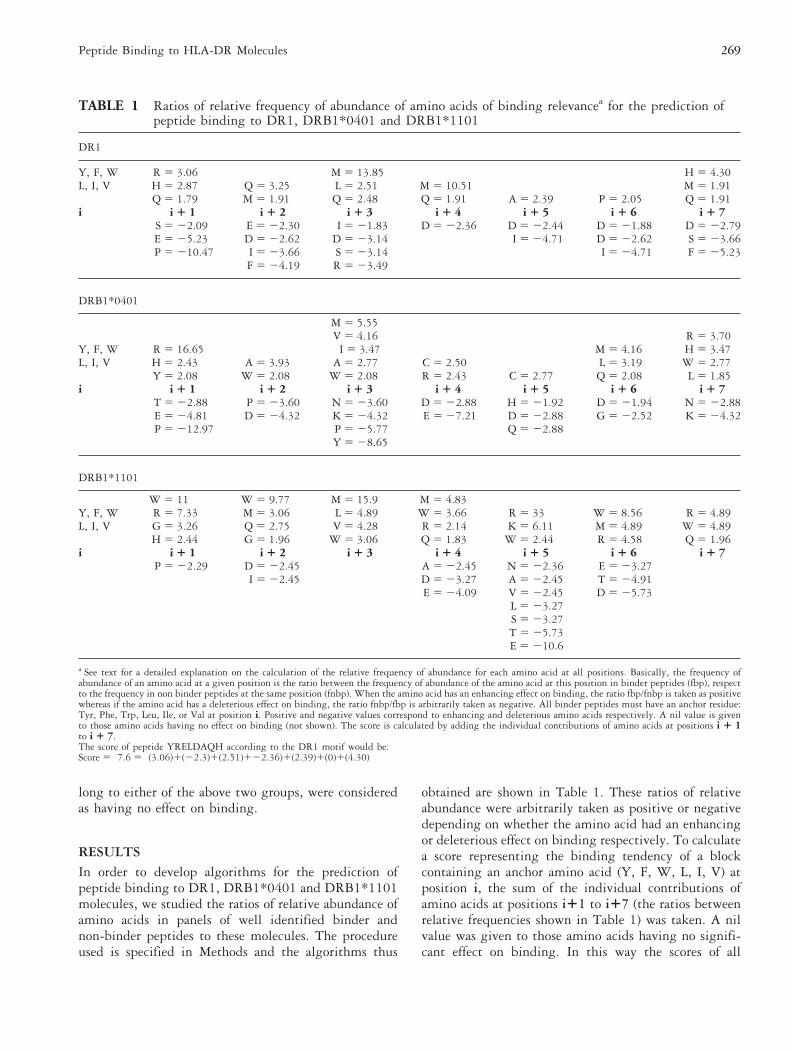
obtained are shown in Table 1. These ratios of relativeabundance were arbitrarily taken as positive or negativedepending on whether the amino acid had an enhancingor deleterious effect on binding respectively. To calculatea score representing the binding tendency of a blockcontaining an anchor amino acid (Y, F, W, L, I, V) atposition i, the sum of the individual contributions ofamino acids at positions i11 to i17 (the ratios betweenrelative frequencies shown in Table 1) was taken. A nilvalue was given to those amino acids having no signifi-cant effect on binding. In this way the scores of all
TABLE 1 Ratios of relative frequency of abundance of amino acids of binding relevancea for the prediction ofpeptide binding to DR1, DRB1*0401 and DRB1*1101
DR1
Y, F, W R 5 3.06 M 5 13.85 H 5 4.30L, I, V H 5 2.87 Q 5 3.25 L 5 2.51 M 5 10.51 M 5 1.91
Q 5 1.79 M 5 1.91 Q 5 2.48 Q 5 1.91 A 5 2.39 P 5 2.05 Q 5 1.91i i 1 1 i 1 2 i 1 3 i 1 4 i 1 5 i 1 6 i 1 7
S 5 22.09 E 5 22.30 I 5 21.83 D 5 22.36 D 5 22.44 D 5 21.88 D 5 22.79E 5 25.23 D 5 22.62 D 5 23.14 I 5 24.71 D 5 22.62 S 5 23.66P 5 210.47 I 5 23.66 S 5 23.14 I 5 24.71 F 5 25.23
F 5 24.19 R 5 23.49
DRB1*0401
M 5 5.55V 5 4.16 R 5 3.70
Y, F, W R 5 16.65 I 5 3.47 M 5 4.16 H 5 3.47L, I, V H 5 2.43 A 5 3.93 A 5 2.77 C 5 2.50 L 5 3.19 W 5 2.77
Y 5 2.08 W 5 2.08 W 5 2.08 R 5 2.43 C 5 2.77 Q 5 2.08 L 5 1.85i i 1 1 i 1 2 i 1 3 i 1 4 i 1 5 i 1 6 i 1 7
T 5 22.88 P 5 23.60 N 5 23.60 D 5 22.88 H 5 21.92 D 5 21.94 N 5 22.88E 5 24.81 D 5 24.32 K 5 24.32 E 5 27.21 D 5 22.88 G 5 22.52 K 5 24.32P 5 212.97 P 5 25.77 Q 5 22.88
Y 5 28.65
DRB1*1101
W 5 11 W 5 9.77 M 5 15.9 M 5 4.83Y, F, W R 5 7.33 M 5 3.06 L 5 4.89 W 5 3.66 R 5 33 W 5 8.56 R 5 4.89L, I, V G 5 3.26 Q 5 2.75 V 5 4.28 R 5 2.14 K 5 6.11 M 5 4.89 W 5 4.89
H 5 2.44 G 5 1.96 W 5 3.06 Q 5 1.83 W 5 2.44 R 5 4.58 Q 5 1.96i i 1 1 i 1 2 i 1 3 i 1 4 i 1 5 i 1 6 i 1 7
P 5 22.29 D 5 22.45 A 5 22.45 N 5 22.36 E 5 23.27I 5 22.45 D 5 23.27 A 5 22.45 T 5 24.91
E 5 24.09 V 5 22.45 D 5 25.73L 5 23.27S 5 23.27T 5 25.73E 5 210.6
a See text for a detailed explanation on the calculation of the relative frequency of abundance for each amino acid at all positions. Basically, the frequency ofabundance of an amino acid at a given position is the ratio between the frequency of abundance of the amino acid at this position in binder peptides (fbp), respectto the frequency in non binder peptides at the same position (fnbp). When the amino acid has an enhancing effect on binding, the ratio fbp/fnbp is taken as positivewhereas if the amino acid has a deleterious effect on binding, the ratio fnbp/fbp is arbitrarily taken as negative. All binder peptides must have an anchor residue:Tyr, Phe, Trp, Leu, Ile, or Val at position i. Positive and negative values correspond to enhancing and deleterious amino acids respectively. A nil value is givento those amino acids having no effect on binding (not shown). The score is calculated by adding the individual contributions of amino acids at positions i 1 1to i 1 7.The score of peptide YRELDAQH according to the DR1 motif would be:Score 5 7.6 5 (3.06)1(22.3)1(2.51)122.36)1(2.39)1(0)1(4.30)
269Peptide Binding to HLA-DR Molecules
potential blocks contained by the peptides screened werecalculated (see legend to Table 1 for a calculation exam-ple).
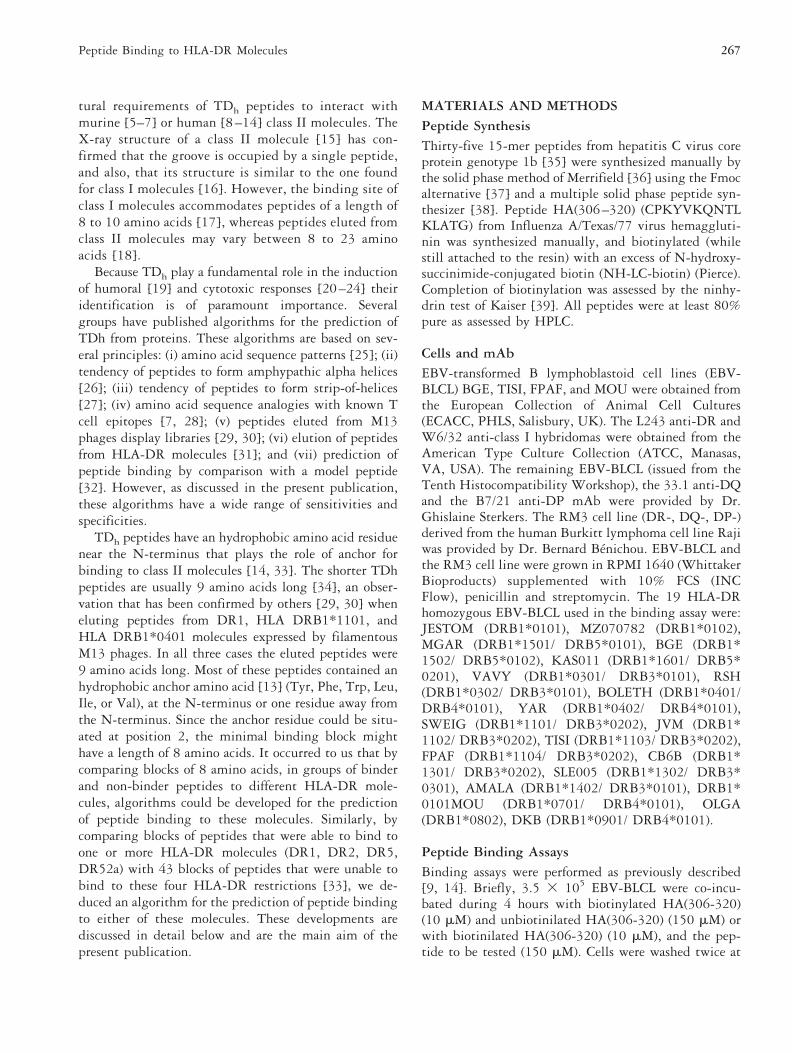
As shown below, in order to validate the three algo-rithms from Table 1, we tested their sensitivity andspecificity against different panels of well identifiedbinder and non-binder peptides reported by several re-search groups. We have defined the sensitivity and spec-ificity of the predictions as the percentage of correctlypredicted binder and non-binder peptides, respectively.We also tested these algorithms against 35, 15-mersynthetic peptides from HCV core protein whose bind-ing ability to 19 HLA-DR molecules was measured by
flow cytometry (Fig. 1). Moreover, using the same panelsof peptides, the sensitivities and specificities of thesealgorithms were compared with those attained withother algorithms reported in the literature [32, 40–42].
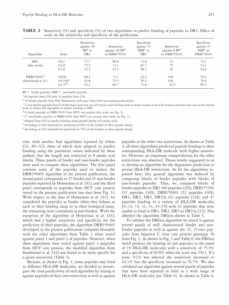
In Table 2, we compare the predictions made with theDR1 algorithm, deduced in the present publication,with another algorithm reported by Southwood et al.[42], which we adapted to predict binding using 8amino acid blocks instead of 9 amino acid blocks re-ported by these authors. Calculations were carried outusing the amino acid parameter values indicated in Fig.2 of their publication but using only the first 8 aminoacids. In Table 2, we compare the sensitivities and spec-ificities of our algorithm with those attained with themodified algorithm of Southwood et al. [42]. For a givenspecificity, the sensitivity of our algorithm was higherthan that attained with the modified algorithm of South-wood et al. [42], both against the set of 94 binderpeptides to DR1, as well as to another set of 30 peptidesthat bind to DRB1*0101/0102 (taken from Prof. Ram-mensee data base published at web page: http://www.uni-tuebingen.de/uni/kxi) (Table 2). However, both al-gorithms behave similarly when tested against binderand non-binder peptides to DRB1*0101 from HCV coreprotein (Table 2).
In Table 3 we compare the predictions made with thealgorithm DRB1*0401 deduced in the present publica-
FIGURE 1 Relative binding capacity of 35 peptides fromHCV core protein to 19 different HLA-DR molecules asstudied by flow cytometry. This was calculated by the inhibi-tion of the binding of biotinylated peptide HA(306-320) toHLA-DR molecules in competition experiments. The relativeinhibitory capacity (RI%) of peptides was calculated accordingto the Formula:
RI% 5 1003 (signal inhibition with peptide tested)/(signalinhibition with unbiotinylated HA(306-320)).
The fluorescence intensities of triplicate samples were usuallywithin 5% of the mean and always within 10%. See Methodsfor a detailed explanation of the calculation of RI%. A plussign indicates that at a score $ 0.1, the peptide is predicted asbinder by the DRGen algorithm.
270 F. Borras-Cuesta et al.
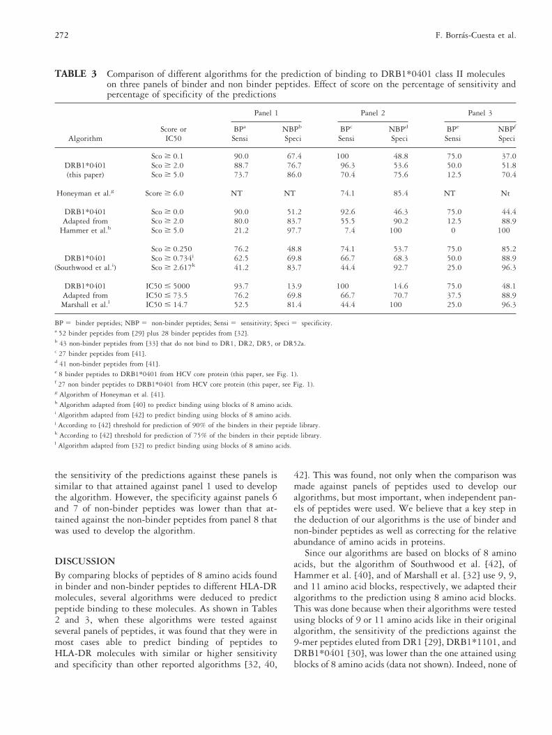
tion, with another four algorithms reported by others[32, 40–42], three of which were adapted to predictbinding using the parameter values indicated by theseauthors, but the length was restricted to 8 amino acidblocks. Three panels of binder and non-binder peptideswere used to compare these algorithms. The first panelcontains some of the peptides used to deduce theDRB1*0401 algorithm of the present publication, thesecond panel corresponds to 27 binder and 41 non-binderpeptides reported by Honeyman et al. [41], and the thirdpanel corresponds to peptides from HCV core proteintested in the present publication (see data from Fig. 1).With respect to the data of Honeyman et al. [41], weconsidered the peptides as binder when they behave assuch in their binding assay or in their biological assay,the remaining were considered as non-binders. With theexception of the algorithm of Honeyman et al. [41],which had a higher sensitivity and specificity for theprediction of their peptides, the algorithm DRB1*0401developed in the present publication compares favorablywith the other algorithms from Table 3 when testedagainst panel 1 and panel 2 of peptides. However, whenthese algorithms were tested against panel 3 (peptidesfrom HCV core protein, the modified algorithm fromSouthwood et al. [42] was found to be more specific fora given sensitivity (Table 3).
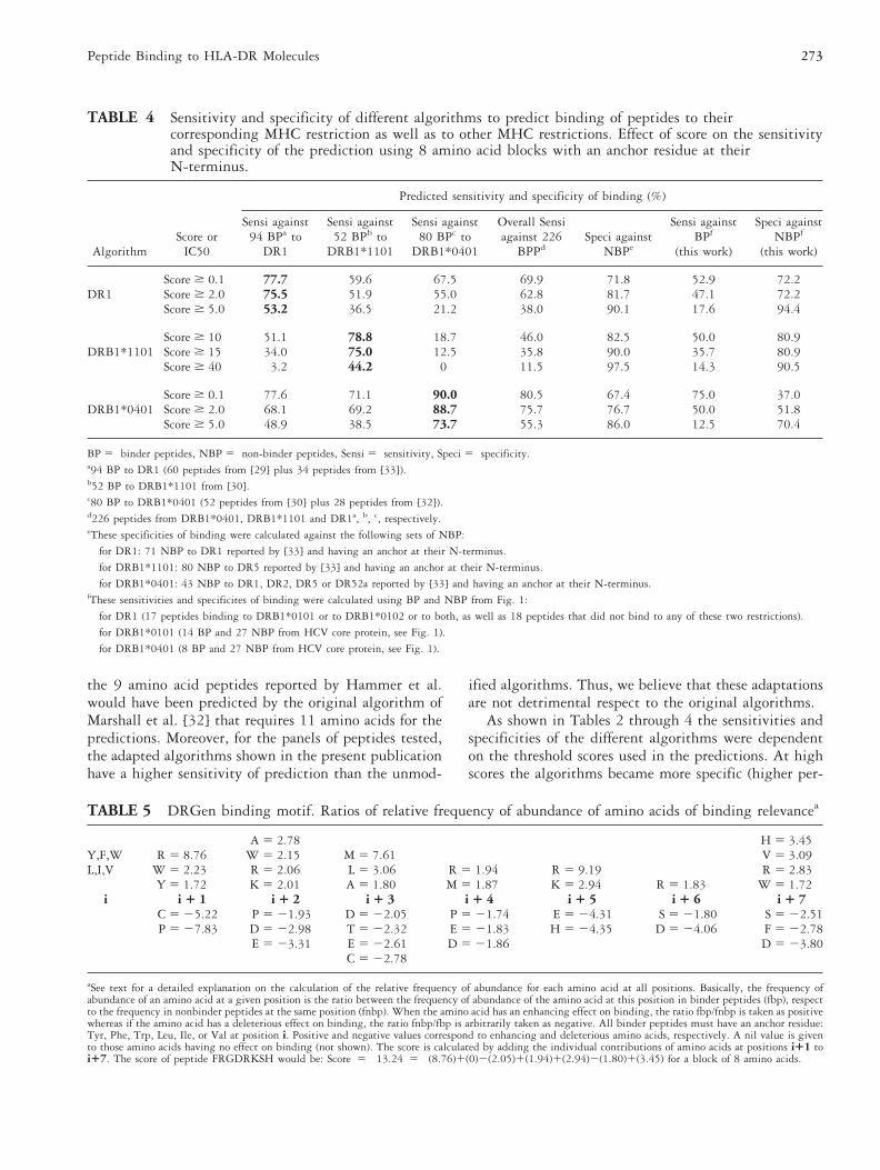
Because, as shown in Fig. 1, some peptides may bindto different HLA-DR molecules, we decided to investi-gate the cross predictivity of each algorithm by testing itagainst peptides of their own restriction as well as against
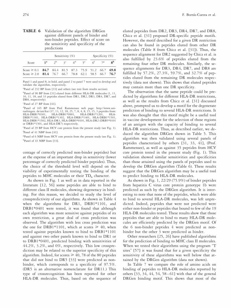
peptides of the other two restrictions. As shown in Table4, all three algorithms predicted peptide binding to theircorresponding HLA-DR molecule with higher sensitiv-ity. However, an important crossprediction for the otherrestrictions was observed. Theses results suggested to usto develop an algorithm for the degenerate prediction toseveral HLA-DR restrictions. As for the algorithms re-ported here, this general algorithm was deduced bycomparing blocks of binder peptides with blocks ofnon-binder peptides. Thus, we compared blocks ofbinder peptides to DR1 (60 peptides [29]), DRB1*1101(52 peptides [30]), DRB1*0401 (52 peptides [30]),DR1, DR2, DR5, DR52a (63 peptides [33]), and 47peptides binding to a variety of HLA-DR molecules[9–12, 14, 31, 33, 43–55] with 43 peptides that wereunable to bind to DR1, DR2, DR5 or DR52a [33]. Thisafforded the algorithm DRGen shown in Table 5.
To validate the DRGen algorithm we tested it againstseveral panels of well characterized binder and non-binder peptides as well as against the 35, 15-mer pep-tides from hepatitis C virus core protein genotype 1bfrom Fig. 1. As shown in Fig. 1 and Table 6, the DRGenmotif predicts the binding of core peptides to the panelof 19 HLA-DR molecules with a sensitivity of 75.9%and a specificity of 69.8% when the score was Ä0.1. If ascore Ä2.0 was selected the sensitivity decreased to62.1% but the specificity increased to 76.7%. We alsovalidated our algorithm against several panels of peptidesthat have been reported to bind to a wide range ofHLA-DR molecules (see Table 6). As shown in Table 6,
TABLE 2 Sensitivity (%) and specificity (%) of two algorithms to predict binding of peptides to DR1. Effect ofscore on the sensitivity and specificity of the predictions
Algorithm Score
Sensitivityagainst 94
BPa toDR1
Sensitivityagainst 30 BPb
to DRB1*0101
Specificityagainst 71NBPc to
DR1
Sensitivityagainst 8 BPd
to DRB1*0101
Specificityagainst 27NBPe to
DRB1*0101
DR1 $0.1 77.7 80.0 71.8 75 74.1(this work) $2.0 75.5 73.3 81.7 75 74.1
$5.0 53.2 43.3 90.1 25 92.6
DRB1*0101f $0.01 68.1 73.3 32.4 100 59.2(Southwood et al.) $0.183g 63.8 73.3 56.3 100 70.4
$1.57h 52.1 66.7 71.8 62.5 85.5
BP 5 binder peptides, NBP 5 non-binder peptides.a 60 peptides from [29] plus 34 peptides from [33].b 30 binder peptides from Prof. Rammensee web page: http//www.uni-tuebingen.de/uni/kxi.c 71 non-binder peptides from [33] that had at least one core of 8 amino acids binding with an anchor residue at their N-terminus and that were used in the presentwork to deduce the algorighm to predict binding to DR1.d 8 binder peptides to DRB1*0101 from HCV core protein (this work, see Fig. 1).e 27 non-binder peptides to DRB1*0101 from HCV core protein (this work, see Fig. 1).f Adapted from [42] to predict binding using peptide blocks of 8 amino acids.g According to [42] threshold for prediction of 90% of the binders in their peptide library.h According to [42] threshold for prediction of 75% of the binders in their peptide library.
271Peptide Binding to HLA-DR Molecules
the sensitivity of the predictions against these panels issimilar to that attained against panel 1 used to developthe algorithm. However, the specificity against panels 6and 7 of non-binder peptides was lower than that at-tained against the non-binder peptides from panel 8 thatwas used to develop the algorithm.
DISCUSSIONBy comparing blocks of peptides of 8 amino acids foundin binder and non-binder peptides to different HLA-DRmolecules, several algorithms were deduced to predictpeptide binding to these molecules. As shown in Tables2 and 3, when these algorithms were tested againstseveral panels of peptides, it was found that they were inmost cases able to predict binding of peptides toHLA-DR molecules with similar or higher sensitivityand specificity than other reported algorithms [32, 40,
42]. This was found, not only when the comparison wasmade against panels of peptides used to develop ouralgorithms, but most important, when independent pan-els of peptides were used. We believe that a key step inthe deduction of our algorithms is the use of binder andnon-binder peptides as well as correcting for the relativeabundance of amino acids in proteins.
Since our algorithms are based on blocks of 8 aminoacids, but the algorithm of Southwood et al. [42], ofHammer et al. [40], and of Marshall et al. [32] use 9, 9,and 11 amino acid blocks, respectively, we adapted theiralgorithms to the prediction using 8 amino acid blocks.This was done because when their algorithms were testedusing blocks of 9 or 11 amino acids like in their originalalgorithm, the sensitivity of the predictions against the9-mer peptides eluted from DR1 [29], DRB1*1101, andDRB1*0401 [30], was lower than the one attained usingblocks of 8 amino acids (data not shown). Indeed, none of
TABLE 3 Comparison of different algorithms for the prediction of binding to DRB1*0401 class II moleculeson three panels of binder and non binder peptides. Effect of score on the percentage of sensitivity andpercentage of specificity of the predictions
AlgorithmScore or
IC50
Panel 1 Panel 2 Panel 3
BPa NBPb BPc NBPd BPe NBPf
Sensi Speci Sensi Speci Sensi Speci
Sco $ 0.1 90.0 67.4 100 48.8 75.0 37.0DRB1*0401 Sco $ 2.0 88.7 76.7 96.3 53.6 50.0 51.8(this paper) Sco $ 5.0 73.7 86.0 70.4 75.6 12.5 70.4
Honeyman et al.g Score $ 6.0 NT NT 74.1 85.4 NT Nt
DRB1*0401 Sco $ 0.0 90.0 51.2 92.6 46.3 75.0 44.4Adapted from Sco $ 2.0 80.0 83.7 55.5 90.2 12.5 88.9
Hammer et al.h Sco $ 5.0 21.2 97.7 7.4 100 0 100
Sco $ 0.250 76.2 48.8 74.1 53.7 75.0 85.2DRB1*0401 Sco $ 0.734j 62.5 69.8 66.7 68.3 50.0 88.9
(Southwood et al.i) Sco $ 2.617k 41.2 83.7 44.4 92.7 25.0 96.3
DRB1*0401 IC50 # 5000 93.7 13.9 100 14.6 75.0 48.1Adapted from IC50 # 73.5 76.2 69.8 66.7 70.7 37.5 88.9Marshall et al.l IC50 # 14.7 52.5 81.4 44.4 100 25.0 96.3
BP 5 binder peptides; NBP 5 non-binder peptides; Sensi 5 sensitivity; Speci 5 specificity.a 52 binder peptides from [29] plus 28 binder peptides from [32].b 43 non-binder peptides from [33] that do not bind to DR1, DR2, DR5, or DR52a.c 27 binder peptides from [41].d 41 non-binder peptides from [41].e 8 binder peptides to DRB1*0401 from HCV core protein (this paper, see Fig. 1).f 27 non binder peptides to DRB1*0401 from HCV core protein (this paper, see Fig. 1).g Algorithm of Honeyman et al. [41].h Algorithm adapted from [40] to predict binding using blocks of 8 amino acids.i Algorithm adapted from [42] to predict binding using blocks of 8 amino acids.j According to [42] threshold for prediction of 90% of the binders in their peptide library.k According to [42] threshold for prediction of 75% of the binders in their peptide library.l Algorithm adapted from [32] to predict binding using blocks of 8 amino acids.
272 F. Borras-Cuesta et al.
the 9 amino acid peptides reported by Hammer et al.would have been predicted by the original algorithm ofMarshall et al. [32] that requires 11 amino acids for thepredictions. Moreover, for the panels of peptides tested,the adapted algorithms shown in the present publicationhave a higher sensitivity of prediction than the unmod-
ified algorithms. Thus, we believe that these adaptationsare not detrimental respect to the original algorithms.
As shown in Tables 2 through 4 the sensitivities andspecificities of the different algorithms were dependenton the threshold scores used in the predictions. At highscores the algorithms became more specific (higher per-
TABLE 4 Sensitivity and specificity of different algorithms to predict binding of peptides to theircorresponding MHC restriction as well as to other MHC restrictions. Effect of score on the sensitivityand specificity of the prediction using 8 amino acid blocks with an anchor residue at theirN-terminus.
AlgorithmScore or
IC50
Predicted sensitivity and specificity of binding (%)
Sensi against94 BPa to
DR1
Sensi against52 BPb to
DRB1*1101
Sensi against80 BPc to
DRB1*0401
Overall Sensiagainst 226
BPPdSpeci against
NBPe
Sensi againstBPf
(this work)
Speci againstNBPf
(this work)
DR1Score $ 0.1 77.7 59.6 67.5 69.9 71.8 52.9 72.2Score $ 2.0 75.5 51.9 55.0 62.8 81.7 47.1 72.2Score $ 5.0 53.2 36.5 21.2 38.0 90.1 17.6 94.4
DRB1*1101Score $ 10 51.1 78.8 18.7 46.0 82.5 50.0 80.9Score $ 15 34.0 75.0 12.5 35.8 90.0 35.7 80.9Score $ 40 3.2 44.2 0 11.5 97.5 14.3 90.5
DRB1*0401Score $ 0.1 77.6 71.1 90.0 80.5 67.4 75.0 37.0Score $ 2.0 68.1 69.2 88.7 75.7 76.7 50.0 51.8Score $ 5.0 48.9 38.5 73.7 55.3 86.0 12.5 70.4
BP 5 binder peptides, NBP 5 non-binder peptides, Sensi 5 sensitivity, Speci 5 specificity.a94 BP to DR1 (60 peptides from [29] plus 34 peptides from [33]).b52 BP to DRB1*1101 from [30].c80 BP to DRB1*0401 (52 peptides from [30] plus 28 peptides from [32]).d226 peptides from DRB1*0401, DRB1*1101 and DR1a, b, c, respectively.eThese specificities of binding were calculated against the following sets of NBP:
for DR1: 71 NBP to DR1 reported by [33] and having an anchor at their N-terminus.
for DRB1*1101: 80 NBP to DR5 reported by [33] and having an anchor at their N-terminus.
for DRB1*0401: 43 NBP to DR1, DR2, DR5 or DR52a reported by [33] and having an anchor at their N-terminus.fThese sensitivities and specificites of binding were calculated using BP and NBP from Fig. 1:
for DR1 (17 peptides binding to DRB1*0101 or to DRB1*0102 or to both, as well as 18 peptides that did not bind to any of these two restrictions).
for DRB1*0101 (14 BP and 27 NBP from HCV core protein, see Fig. 1).
for DRB1*0401 (8 BP and 27 NBP from HCV core protein, see Fig. 1).
TABLE 5 DRGen binding motif. Ratios of relative frequency of abundance of amino acids of binding relevancea
A 5 2.78 H 5 3.45Y,F,W R 5 8.76 W 5 2.15 M 5 7.61 V 5 3.09L,I,V W 5 2.23 R 5 2.06 L 5 3.06 R 5 1.94 R 5 9.19 R 5 2.83
Y 5 1.72 K 5 2.01 A 5 1.80 M 5 1.87 K 5 2.94 R 5 1.83 W 5 1.72i i 1 1 i 1 2 i 1 3 i 1 4 i 1 5 i 1 6 i 1 7
C 5 25.22 P 5 21.93 D 5 22.05 P 5 21.74 E 5 24.31 S 5 21.80 S 5 22.51P 5 27.83 D 5 22.98 T 5 22.32 E 5 21.83 H 5 24.35 D 5 24.06 F 5 22.78
E 5 23.31 E 5 22.61 D 5 21.86 D 5 23.80C 5 22.78
aSee text for a detailed explanation on the calculation of the relative frequency of abundance for each amino acid at all positions. Basically, the frequency ofabundance of an amino acid at a given position is the ratio between the frequency of abundance of the amino acid at this position in binder peptides (fbp), respectto the frequency in nonbinder peptides at the same position (fnbp). When the amino acid has an enhancing effect on binding, the ratio fbp/fnbp is taken as positivewhereas if the amino acid has a deleterious effect on binding, the ratio fnbp/fbp is arbitrarily taken as negative. All binder peptides must have an anchor residue:Tyr, Phe, Trp, Leu, Ile, or Val at position i. Positive and negative values correspond to enhancing and deleterious amino acids, respectively. A nil value is givento those amino acids having no effect on binding (not shown). The score is calculated by adding the individual contributions of amino acids at positions i11 toi17. The score of peptide FRGDRKSH would be: Score 5 13.24 5 (8.76)1(0)2(2.05)1(1.94)1(2.94)2(1.80)1(3.45) for a block of 8 amino acids.
273Peptide Binding to HLA-DR Molecules
centage of correctly predicted non-binder peptides) butat the expense of an important drop in sensitivity (lowerpercentage of correctly predicted binder peptides). Thus,the choice of the threshold level will depend on theviability of experimentally testing the binding of thepeptides to MHC molecules or their TDh character.
As shown in Fig. 1 as well as in data reported in theliterature [12, 56] some peptides are able to bind todifferent class II molecules, showing degeneracy in bind-ing. For this reason, we decided to study the level ofcrosspredictivity of our algorithms. As shown in Table 4when the algorithms for DR1, DRB1*1101, andDRB1*0401 were tested, it was found that althougheach algorithm was more sensitive against peptides of itsown restriction, a great deal of cross prediction wasobserved. The algorithm with less cross prediction wasthe one for DRB1*1101, which at scores Ä 40, whentested against peptides known to bind to DRB1*1101and against two other panels known to bind to DR1 orto DRB1*0401, predicted binding with sensitivities of44.2%, 3.2%, and 0%, respectively. This low crosspre-diction may be related to the superior specificity of thisalgorithm. Indeed, for scores Ä 40, 78 of the 80 peptidesthat did not bind to DR5 [33] were predicted as non-binder, which corresponds to a specificity of 97.5%.(DR5 is an alternative nomenclature for DR11.) Thistype of crossrecognition has been reported for otherHLA-DR molecules. Thus, based on the sequence of
eluted peptides from DR2, DR3, DR4, DR7, and DR8,Chicz et al. [31] proposed DR-specific peptide motifs.However, the motif described for a given DR restrictioncan also be found in peptides eluted from other DRmolecules (Table 8 from Chicz et al. [31]). Thus, thesequence alignment for DR2 suggested by Chicz et al. isalso fulfilled by 23.6% of peptides eluted from theremaining four other DR molecules. Similarly, the se-quence alignments for DR3, DR4, DR7, and DR8 arefulfilled by 57.2%, 27.3%, 59.7%, and 32.7% of pep-tides eluted from the remaining DR molecules respec-tively (data not shown). This shows that eluted peptidesmay contain more than one DR specificity.
The observation that the same peptide could be pre-dicted by algorithms for different HLA-DR restrictions,as well as the results from Chicz et al. [31] discussedabove, prompted us to develop a motif for the degenerateprediction of binding to several HLA-DR restrictions. Itwas also thought that this motif might be a useful toolin vaccine development for the selection of those regionsof an antigen with the capacity of binding to severalHLA-DR restrictions. Thus, as described earlier, we de-duced the algorithm DRGen shown in Table 5. Thisalgorithm was then validated using several panels ofpeptides characterized by others [31, 33, 41], (Prof.Rammensee), as well as against 35 peptides from HCVcore protein tested in the present study (Fig. 1). Thisvalidation showed similar sensitivities and specificitiesthan those attained using the panels of peptides used todevelop the DRGen algorithm (Table 6). These resultssuggest that the DRGen algorithm may be a useful toolto predict binding to HLA-DR molecules.
As shown in Fig. 1, 22/29 (75.9%) of binder peptidesfrom hepatitis C virus core protein genotype 1b werepredicted as such by the DRGen algorithm. It is inter-esting to note that none of the peptides, which were ableto bind to several HLA-DR molecules, was left unpre-dicted. Indeed, peptides that were not predicted wereeither non-binder or peptides that bound to few of the 19HLA-DR molecules tested. These results show that thosepeptides that are able to bind to many HLA-DR mole-cules are efficiently predicted by the DRGen motif. Ofthe 6 non-binder peptides 4 were predicted as non-binder but the other 3 were predicted as binder.
Other researchers [25, 26] have published algorithmsfor the prediction of binding to MHC class II molecules.When we tested their algorithms using the program “Tsites” [57] it was found that for a given specificity thesensitivity of these algorithms was well below that at-tained by the DRGen algorithm (data not shown).
In Table 7 we compare the role of amino acids onbinding of peptides to HLA-DR molecules reported byothers [33, 34, 44, 54, 58–61] with that of the generalDRGen binding motif. This shows that most of the
TABLE 6 Validation of the algorithm DRGenagainst different panels of binder andnon-binder peptides. Effect of score onthe sensitivity and specificity of thepredictions
Score
Sensitivity (%) Specificity (%)
1a 2b 3c 4d 5e 6f 7g 8h
Score $ 0.1 84.7 84.4 81.5 87.3 75.9 51.2 66.7 69.8Score $ 2.0 81.4 76.7 66.7 78.8 62.1 58.5 66.7 76.7
Panel 1 and panel 8, in bold, and panel 2 to panel 7 were used to develop andvalidate the algorithm, respectively.aPanel of 232 BP from [33] and others (see text from results section).bPanel of 90 BP from [31] eluted from different HLA-DR molecules (1, 11,16, 11, 18, and 33 peptides eluted from DR1, DR2, DR3, DR4, DR7, andDR8, respectively).cPanel of 27 BP from [41].dPanel of 165 BP from Prof. Rammensee web page: http://www.uni-tuebingen. de/uni/kxi (37, 13, 12, 18, 25, 7, 8, 4, 8, 15, 15, 3 peptides from:HLA-DRB1*0301, HLA-DRB1*0901, HLA-DRB1*1201, HLA-DRB1*1301, HLA-DRB1*1302, HLA-DRB1*1401, HLA-DRB1*1501,HLA-DRB1*1502, HLA-DR3, HLA-DRB5*0101, HLA-DR2 (DRB5*0101or DRB1*1501, and HLA-DR7) respectively.ePanel of 29 BP from HCV core protein from the present study (see Fig. 1).fPanel of 41 NBP from [41].gPanel of 6 NBP from HCV core protein from the present study (see Fig. 1).hPanel of 43 NBP from [33].
274 F. Borras-Cuesta et al.
effects reported are compatible with our peptide bindingmotif. It is clear that, in addition to an anchor residue,binder peptides should have few, if any, amino acidsgiving negative interactions with the MHC. It has beensuggested that the absence of “negative” amino acids maybe more important than the presence of amino acidsgiving a “positive” interaction with MHC [58]. Also, thereported enhanced binding of peptide HEL(104–120) atacidic pH [59], where the net charge is less negative, isin agreement with the disrupting effect on binding ofAsp and Glu at several positions of the peptide at higherpH [60] where the net charge is more negative.
The crystal structure of an HLA-DR1 molecule com-plexed with peptide HA(306-318) shows that residuesTyr308, Gln311, Thr313, Leu314, and Leu316 ofHA(306-318) interact respectively with pockets 1, 4, 6,7, and 9 of the HLA-DR1 molecule (See Fig. 4 frompublication of Stern et al. [62]). If we consider thatTyr308 corresponds to the anchor at position i of ourpeptide motif, then pockets 1, 4, 6, 7 and 9 of the MHCmay interact with positions i, i13, i15, i16, and i18of the motif, respectively. Thus, the enhancing or dis-rupting effect of amino acids at positions i13, i15, i16,and i18 of the motif should be compatible with theamino acids delimiting pockets 4, 6, 7, and 9 of theMHC, respectively. Thus, pocket 1 is formed mainly byhydrophobic residues [62]. This is compatible with theinteraction with the reported hydrophobic anchor :Tyr,Phe, Trp, Leu, Ile, Val at position i.
Pocket 4 may interact with residue i13 of the DRGenmotif which favours Met and Leu at this position. Thispreference may be related with packing of Met or Leuwith the hydrophobic residues Phe/His/Tyrb13 (of someDR molecules) and Tyrb78 in the pocket. The deleteri-ous effect of Asp and Glu may be related with an elec-trostatic repulsion against a negatively charged amino
acid close to pocket 4, like Glub28 in DR1, and Aspb28in the remaining DR molecules.
Positions i15, i16, and i18, which interact withpockets 6, 7 and 9 of the MHC molecule, do not favorAsp residues (Table 5) suggesting the presence of nega-tively charged residues at/or near these MHC pockets.For instance Glu a11 and Asp a66 from pocket 6 andAsp b57 from pocket 9 and/or with Glu b9 in most DRmolecules. The presence of these negatively charged res-idues of the MHC molecule, might also explain theenhancing effect on binding of Arg in those positionswhere the negatively charged residues have a deleteriouseffect. Thus, the general amino acid topology of thebinder peptide DRGen motif favors hydrophobic andpositively charged amino acids, which have chemicalaffinity for hydrophobic and negatively charged aminoacids respectively (Table 8). Also, the strong deleteriouseffect due to Pro, at position i11 may be related to thereported effect on the orientation of the peptide chain byPro [63, 64] that may prevent the amino acids from thepeptide to interact with the HLA-DR molecule.
To summarize, the published HLA-DR structure [62]suggests that its general topology may be complemen-tary to the topology associated with the proposedDRGen peptide binding motif. This motif is able topredict with high sensitivity and specificity those regionsof an antigen that may bind to HLA-DR molecules. Thisprediction, in conjunction with a better understanding ofantigen processing and of immunodominancy, due tohigher affinity of peptides for HLA-DR molecules and/orhigher affinity of the complex: peptide-(class II mole-cule) by the T cell receptor, may allow in not too distantfuture, the identification of the most relevant TDh fromantigens. This is of paramount importance because TDh
are essential for the induction of humoral and cellular
TABLE 7 Effect of amino acids at positions i to i17 on binding of peptides to MHC class II molecules
i i 1 1 i12 i13 i14 i15 i16 i17 Ref
Y1F1W1 K1R1 K1R1 [54]Y1F1W1 K1R1 [34]
Absense of negative interactions at all positions, more important than presence of positive interactions [58]Enhanced binding of peptide HEL (104-120) at acidic pH [59]
E2 E2 [44]Y1 D2 D2 D2 D2 D2 D2 [60]
E2 [12]E2 [61]
Y,F,W,I,L,V R1W1Y1 A1K1R1W1 A1L1M1 M1R1 K1R1 R1 H1R1V1W1 Thisanchor C2P2 D2E2P2 C2D2E2T2 D2E2P2 E2H2 D2S2 D2F2S2 work
1Enhancing effect on binding.
2Deleterious effect on binding.
275Peptide Binding to HLA-DR Molecules

immune responses, and consequently, for the design offuture vaccines.
ACKNOWLEDGMENTS
This work was partly supported by grants from the Departa-mento de Salud, Gobierno de Navarra, Project: No 840, byC.I.C.Y.T project: SAF97-0223, and from AIRTEL 97.
REFERENCES
1. Germain RN, Margulies DH: The biochemistry and cellbiology of antigen processing and presentation. Annu RevImmunol 11:403, 1993.
2. Guillet JG, Lai MZ, Briner TJ, Smith JA, Gefter ML:Interaction of peptide antigens and class II major histo-compatibility complex antigens. Nature 324(6094):260,1986.
3. Babbitt BP, Allen PM, Matsueda G, Haber E, UnanueER: Binding of immunogenic peptides to Ia histocompat-ibility molecules. Nature 317(6035):359, 1985.
4. Buus S, Sette A, Colon SM, Miles C, Grey HM: Therelation between major histocompatibility complex(MHC) restriction and the capacity of Ia to bind immu-nogenic peptides. Science 235(4794):1353, 1987.
5. Sette A, Buus S, Colon S, Smith JA, Miles C, Grey HM:Structural characteristics of an antigen required for itsinteraction with Ia and recognition by T cells. Nature328(6129):395, 1987.
6. Sette A, Adorini L, Appella E, Colon SM, Miles C, TanakaS, Ehrhardt C, Doria G, Nagy ZA, Buus S, et al.: Struc-tural requirements for the interaction between peptideantigens and I-Ed molecules. J Immunol 143(10):3289,1989.
7. Sette A, Buus S, Appella E, Smith JA, Chesnut R, MilesC, Colon SM, Grey HM: Prediction of major histocom-patibility complex binding regions of protein antigens bysequence pattern analysis. Proc Natl Acad Sci USA 86(9):3296, 1989.
8. Busch R, Rothbard JB: Detection of peptide-MHC class IIcomplexes on the surface of intact cells. J Immunol Meth-ods 134(1):1, 1990.
9. Busch R, Strang G, Howland K, Rothbard JB: Degeneratebinding of immunogenic peptides to HLA-DR proteinson B cell surfaces. Int Immunol 2(5):443, 1990.
10. Jardetzky TS, Gorga JC, Busch R, Rothbard J, StromingerJL, Wiley DC: Peptide binding to HLA-DR1: a peptidewith most residues substituted to alanine retains MHCbinding. Embo J 9(6):1797, 1990.
11. Roche PA, Cresswell P: High-affinity binding of an in-fluenza hemagglutinin-derived peptide to purified HLA-DR. J Immunol 144(5):1849, 1990.
12. O’Sullivan D, Arrhenius T, Sidney J, Del Guercio MF,Albertson M, Wall M, Oseroff C, Southwood S, ColonSM, Gaeta FC, Sette A: On the interaction of promiscuousantigenic peptides with different DR alleles. Identifica-
tion of common structural motifs. J Immunol 147(8):2663, 1991.
13. Krieger JI, Karr RW, Grey HM, Yu WY, O’Sullivan D,Batovsky L, Zheng ZL, Colon SM, Gaeta FC, Sidney J,Albertson M, Del Guercio MF, Chesnut RW, Sette A:Single amino acid changes in DR and antigen defineresidues critical for peptide-MHC binding and T cellrecognition. J Immunol 146(7):2331, 1991.
14. Hill CM, Hayball JD, Allison AA, Rothbard JB: Confor-mational and structural characteristics of peptides bindingto HLA-DR molecules. J Immunol 147(1):189, 1991.
15. Brown JH, Jardetzky TS, Gorga JC, Stern LJ, Urban RG,Strominger JL, Wiley DC: Three-dimensional structure ofthe human class II histocompatibility antigen HLA-DR1[see comments]. Nature 364(6432):33, 1993.
16. Bjorkman PJ, Saper MA, Samraoui B, Bennett WS,Strominger JL, Wiley DC: Structure of the human class Ihistocompatibility antigen, HLA-A2. Nature 329(6139):506, 1987.
17. Matsumura M, Fremont DH, Peterson PA, Wilson IA:Emerging principles for the recognition of peptide anti-gens by MHC class I molecules [see comments]. Science257(5072):927, 1992.
18. Chicz RM, Urban RG, Lane WS, Gorga JC, Stern LJ,Vignali DA, Strominger JL: Predominant naturally pro-cessed peptides bound to HLA-DR1 are derived fromMHC-related molecules and are heterogeneous in size.Nature 358(6389):764, 1992.
19. Milich DR: Synthetic T and B cell recognition sites:implications for vaccine development. Adv Immunol 45:195, 1989.
20. Keene JA, Forman J: Helper activity is required for the invivo generation of cytotoxic T lymphocytes. J Exp Med155(3):768, 1982.
21. Leist TP, Cobbold SP, Waldmann H, Aguet M, Zinker-nagel RM: Functional analysis of T lymphocyte subsets inantiviral host defense. J Immunol 138(7):2278, 1987.
22. Fayolle C, Deriaud E, Leclerc C: In vivo induction ofcytotoxic T cell response by a free synthetic peptide re-quires CD41 T cell help. J Immunol 147(12):4069,1991.
23. Lasarte JJ, Sarobe P, Gullon A, Prieto J, Borras Cuesta F:Induction of cytotoxic T lymphocytes in mice against theprincipal neutralizing domain of HIV-1 by immunizationwith an engineered T-cytotoxic-T-helper synthetic pep-tide construct. Cell Immunol 141(1):211, 1992.
24. Shirai M, Pendleton CD, Ahlers J, Takeshita T, NewmanM, Berzofsky JA: Helper-cytotoxic T lymphocyte (CTL)determinant linkage required for priming of anti-HIVCD81 CTL in vivo with peptide vaccine constructs. J Im-munol 152(2):549, 1994.
25. Rothbard JB, Taylor WR: A sequence pattern common toT cell epitopes. EMBO J 7(1):93, 1988.
26. Margalit H, Spouge JL, Cornette JL, Cease KB, Delisi C,Berzofsky JA: Prediction of immunodominant helper T
276 F. Borras-Cuesta et al.
cell antigenic sites from the primary sequence. J Immunol138(7):2213, 1987.
27. Stille CJ, Thomas LJ, Reyes VE, Humphreys RE: Hydro-phobic strip-of-helix algorithm for selection of T cell-presented peptides. Mol Immunol 24(10):1021, 1987.
28. Guillet JG, Hoebeke J, Lengagne R, Tate K, Borras-Herrera F, Strosberg AD, Borras-Cuesta F: Haplotypespecific homology scanning algorithm to predict T-cellepitopes from protein sequences. J Mol Recognit 4(1):17,1991.
29. Hammer J, Takacs B, Sinigaglia F: Identification of amotif for HLA-DR1 binding peptides using M13 displaylibraries. J Exp Med 176(4):1007, 1992.
30. Hammer J, Valsasnini P, Tolba K, Bolin D, Higelin J,Takacs B, Sinigaglia F: Promiscuous and allele-specificanchors in HLA-DR-binding peptides. Cell 74(1):197,1993.
31. Chicz RM, Urban RG, Gorga JC, Vignali DA, Lane WS,Strominger JL: Specificity and promiscuity among natu-rally processed peptides bound to HLA-DR alleles. J ExpMed 178(1):27, 1993.
32. Marshall KW, Wilson KJ, Liang J, Woods A, Zaller D,Rothbard JB: Prediction of peptide affinity to HLADRB1*0401. J Immunol 154(11):5927, 1995.
33. O’Sullivan D, Sidney J, Appella E, Walker L, Phillips L,Colon SM, Miles C, Chesnut RW, Sette A: Characteriza-tion of the specificity of peptide binding to four DRhaplotypes. J Immunol 145(6):1799, 1990.
34. O’Sullivan D, Sidney J, Del Guercio MF, Colon SM, SetteA: Truncation analysis of several DR binding epitopes.J Immunol 146(4):1240, 1991.
35. Lasarte JJ, Garcia Granero M, Lopez A, Casares N, GarciaN, Civeira MP, Borras Cuesta F, Prieto J: Cellular immu-nity to hepatitis C virus core protein and the response tointerferon in patients with chronic hepatitis C. Hepatol-ogy 28(3):815, 1998.
36. Merrifield RB: Solid phase peptide synthesis. I. The syn-thesis of a tetrapeptide. J Am Chem Soc 85:2149, 1963.
37. Atherton E, Logan JC, Sheppard RC: Peptide synthesis II.Procedures for solid phase synthesis using N-fluorenylmethoxycarbonyl aminoacids on polyamide supports. Syn-thesis of substance P and of acyl carrier protein 65-74decapeptide. J Chem Soc Perkin Trans 1:538, 1989.
38. Borras Cuesta F, Golvano J, Sarobe P, Lasarte JJ, Prieto I,Szabo A, Guillaume JL, Guillet JG: Insights on the aminoacid side-chain interactions of a synthetic T-cell determi-nant. Biologicals 19(3):187, 1991.
39. Kaiser E, Colescott RL, Bossinger CD, Cook PI: Color testfor detection of free terminal amino groups in the solid-phase synthesis of peptides. Anal Biochem 34(2):595,1970.
40. Hammer J, Bono E, Gallazzi F, Belunis C, Nagy Z,Sinigaglia F: Precise prediction of major histocompatibil-ity complex class II-peptide interaction based on peptideside chain scanning. J Exp Med 180(6):2353, 1994.
41. Honeyman MC, Brusic V, Stone NL, Harrison LC: Neuralnetwork-based prediction of candidate T-cell epitopes.Nat Biotechnol 16(10):966, 1998.
42. Southwood S, Sidney J, Kondo A, del Guercio MF, Ap-pella E, Hoffman S, Kubo RT, Chesnut RW, Grey HM,Sette A: Several common HLA-DR types share largelyoverlapping peptide binding repertoires. J Immunol160(7):3363, 1998.
43. Sinigaglia F, Guttinger M, Kilgus J, Doran DM, MatileH, Etlinger H, Trzeciak A, Gillessen D, Pink JR: Amalaria T-cell epitope recognized in association with mostmouse and human MHC class II molecules. Nature336(6201):778, 1988.
44. Panina Bordignon P, Tan A, Termijtelen A, Demotz S,Corradin G, Lanzavecchia A: Universally immunogenic Tcell epitopes: promiscuous binding to human MHC classII and promiscuous recognition by T cells. Eur J Immunol19(12):2237, 1989.
45. Hickling JK, Fenton CM, Howland K, Marsh SG, Roth-bard JB: Peptides recognized by class I restricted T cellsalso bind to MHC class II molecules. Int Immunol 2(5):435, 1990.
46. Karr RW, Yu W, Watts R, Evans KS, Celis E: The roleof polymorphic HLA-DR beta chain residues in presenta-tion of viral antigens to T cells. J Exp Med 172(1):273,1990.
47. Gyllensten U, Sundvall M, Ezcurra I, Erlich HA: Geneticdiversity at class II DRB loci of the primate MHC.J Immunol 146(12):4368, 1991.
48. Brown LR, Nygard NR, Graham MB, Bono C, BracialeVL, Gorka J, Schwartz BD, Braciale TJ: Recognition ofthe influenza hemagglutinin by class II MHC-restricted Tlymphocytes and antibodies. I. Site definition and impli-cations for antigen presentation and T lymphocyte recog-nition. J Immunol 147(8):2677, 1991.
49. Kilgus J, Jardetzky T, Gorga JC, Trzeciak A, Gillessen D,Sinigaglia F: Analysis of the permissive association of amalaria T cell epitope with DR molecules. J Immunol146(1):307, 1991.
50. Suhrbier A, Rodda SJ, Ho PC, Csurhes P, Dunckley H,Saul A, Geysen HM, Rzepczyk CM: Role of single aminoacids in the recognition of a T cell epitope. J Immunol147(8):2507, 1991.
51. Kropshofer H, Max H, Muller CA, Hesse F, Stevanovic S,Jung G, Kalbacher H: Self-peptide released from class IIHLA-DR1 exhibits a hydrophobic two-residue contactmotif. J Exp Med 175(6):1799, 1992.
52. Harris PE, Liu Z, Suciu-Foca N: MHC class II binding ofpeptides derived from HLA-DR 1. J Immunol 148(7):2169, 1992.
53. Olson RR, De Magistris MT, Di Tommaso A, Karr RW:Mutations in the third, but not the first or second, hy-pervariable regions of DR(beta 1*0101) eliminate DR1-restricted recognition of a pertussis toxin peptide. J Im-munol 148(9):2703, 1992.
277Peptide Binding to HLA-DR Molecules

54. Newcomb JR, Cresswell P: Characterization of endoge-nous peptides bound to purified HLA-DR molecules andtheir absence from invariant chain-associated alpha betadimers. J Immunol 150(2):499, 1993.
55. Chaye H, Ou D, Chong P, Gillam S: Human T- andB-cell epitopes of E1 glycoprotein of rubella virus. J ClinImmunol 13(2):93, 1993.
56. Panina-Bordignon P, Tan A, Termijtelen A, Demotz S,Corradin G, Lanzavecchia A: Universally immunogenic Tcell epitopes: promiscuous binding to human MHC classII and promiscuous recognition by T cells. Eur J Immunol19(12):2237, 1989.
57. Feller DC, de la Cruz VF: Identifying antigenic T-cellsites. Nature 349(6311):720, 1991.
58. Rothbard JB, Gefter ML: Interactions between immuno-genic peptides and MHC proteins. Annu Rev Immunol9:527, 1991.
59. Jensen PE: Enhanced binding of peptide antigen to puri-fied class II major histocompatibility glycoproteins atacidic pH. J Exp Med 174(5):1111, 1991.
60. Zeliszewski D, Golvano JJ, Gaudebout P, Dorval I, Frei-
del C, Gebuhrer L, Betuel H, Borras Cuesta F, Sterkers G:Implication of HLA-DR residues at positions 67, 71, and86 in interaction between HLA-DR11 and peptideHA306-320. J Immunol 151(11):6237, 1993.
61. Boehncke WH, Takeshita T, Pendleton CD, HoughtenRA, Sadegh Nasseri S, Racioppi L, Berzofsky JA, GermainRN: The importance of dominant negative effects ofamino acid side chain substitution in peptide-MHC mol-ecule interactions and T cell recognition. J Immunol150(2):331, 1993.
62. Stern LJ, Brown JH, Jardetzky TS, Gorga JC, Urban RG,Strominger JL, Wiley DC: Crystal structure of the humanclass II MHC protein HLA-DR1 complexed with an in-fluenza virus peptide. Nature 368(6468):215, 1994.
63. Chou PY, Fasman GD: Empirical predictions of proteinconformation. Annu Rev Biochem 47:251, 1978.
64. Garnier J, Osguthorpe DJ, Robson B: Analysis of theaccuracy and implications of simple methods for predict-ing the secondary structure of globular proteins. J MolBiol 120(1):97, 1978.
278 F. Borras-Cuesta et al.


![PROTEOCHEMOMETRIC ANALYSIS OF PEPTIDES BINDING TO … · [12], atopic myelitis [13]; allele HLA-DP3 – with susceptibility to juvenile rheumatoid arthri-tis [14], allele HLA-DP41](https://img.pdfslide.us/doc/110x75/5e324fe4062fa370e23fad36/proteochemometric-analysis-of-peptides-binding-to-12-atopic-myelitis-13-allele.jpg)