Embed Size (px)
Citation preview
Specificity of regional brain activity in anxiety types
during emotion processing
ANNA S. ENGELS,a,b WENDY HELLER,a,b APRAJITA MOHANTY,a,b
JOHN D. HERRINGTON,a,b MARIE T. BANICH,c ANDREW G. WEBB,d
and GREGORY A. MILLERa,b,e
aDepartment of Psychology, University of Illinois at Urbana-Champaign, Urbana, Illinois, USAbBeckman Institute Biomedical Imaging Center, University of Illinois at Urbana-Champaign, Urbana, Illinois, USAcDepartment of Psychology and Institute of Cognitive Science, University of Colorado at Boulder, Boulder, Colorado, USAdDepartment of Bioengineering, Pennsylvania State University, University Park, Pennsylvania, USAeDepartment of Psychiatry, University of Illinois at Urbana-Champaign, Urbana, Illinois, USA
Abstract
The present study tested the hypothesis that anxious apprehension involves more left- than right-hemisphere activity
and that anxious arousal is associated with the opposite pattern. Behavioral and fMRI responses to threat stimuli in an
emotional Stroop task were examined in nonpatient groups reporting anxious apprehension, anxious arousal, or
neither. Reaction times were longer for negative than for neutral words. As predicted, brain activation distinguished
anxious groups in a left inferior frontal region associated with speech production and in a right-hemisphere inferior
temporal area. Addressing a second hypothesis about left-frontal involvement in emotion, distinct left frontal regions
were associated with anxious apprehension versus processing of positive information. Results support the proposed
distinction between the two types of anxiety and resolve an inconsistency about the role of left-frontal activation in
emotion and psychopathology.
Descriptors: fMRI, Anxiety, Anxious apprehension, Anxious arousal, Emotion, Stroop
According to lifetime prevalence estimates from a recent large
survey of mental illness and its treatment in the United States,
anxiety disorders are the most commonly reported psychiatric
disorders, closely followed by mood disorders (Kessler et al.,
2005). Research, treatment, and public policy efforts regarding
anxiety and depression have typically focused on emotional
symptoms. However, these emotional states have significant ef-
fects on cognitive function as well, resulting in an unmeasured
but considerable toll on productivity and efficiency (for reviews,
see Levin, Heller, Mohanty, Herrington, & Miller, in press;
Mohanty & Heller 2002; Nitschke & Heller, 2002, 2005; Nits-
chke, Heller, & Miller, 2000). Anxiety has specific effects on
cognition. In particular, it has been strongly associated with an
attentional bias toward threatening stimuli (Compton, Heller,
Banich, Palmieri, & Miller, 2000; McNally, 1998; Nitschke &
Heller, 2002). In various paradigms, attention is captured by
ambiguous, emotional, or threatening information. Anxiety also
impairs performance on tasks that are difficult or stressful, be-
cause worrisome thoughts interfere with attention to task-
relevant information (e.g., McNally, 1998). These attentional
effects have been documented in trait and state anxiety (e.g.,
Egloff & Hock, 2001) and in every DSM-IV-TR (American
Psychiatric Association, 2000) anxiety disorder (for reviews, see
Heller, Koven, & Miller, 2003; Nitschke & Heller, 2002, 2005).
An implication is that, if someone is anxious, their attention is at
constant risk for diversion from the task at hand, slowing or
impairing performance.
Anxiety is not a monolithic construct. We have distinguished
two types of anxiety, anxious apprehension and anxious arousal,
associated with different patterns of brain activity. Anxious
apprehension is primarily characterized by worry and verbal
rumination (Barlow, 1991; Heller, Nitschke, Etienne, & Miller,
Aprajita Mohanty is now at the Institute for Neuroscience and Cog-
nitive Neurology and Alzheimer’s Disease Center, Northwestern Uni-
versity. John D. Herrington is now at Yale University School of
Medicine.
This report is based on amasters thesis submitted by the first author to
the University of Illinois at Urbana-Champaign. This research was sup-
ported by the National Institute of Drug Abuse (R21 DA14111), the
National Institute of Mental Health (R01 MH61358, T32 MH14257,
T32 MH19554), and the University of Illinois Beckman Institute and
Intercampus Research Initiative in Biotechnology. Anna S. Engels was a
predoctoral trainee in the Cognitive Psychophysiology training program
of the Department of Psychology, University of Illinois at Urbana–
Champaign, under NIMH Grant T32 MH19554. John D. Herrington
was a predoctoral trainee in the Quantitative Methods training program
of the Department of Psychology, University of Illinois at Urbana–
Champaign, under NIMH Grant T32 MH14257. The authors thank
Nancy Dodge, Rebecca Levin, Sarah Sass, Brad Sutton, Holly Tracy,
and Tracey Wszalek for their contributions to this project.Address reprint requests to: Gregory A. Miller, Department of Psy-
chology, University of Illinois at Urbana–Champaign, 603 E. DanielStreet, Champaign, IL 61820, USA. E-mail: [email protected]
Psychophysiology, 44 (2007), 352–363. Blackwell Publishing Inc. Printed in the USA.Copyright r 2007 Society for Psychophysiological ResearchDOI: 10.1111/j.1469-8986.2007.00518.x
352

1997). The timescale for perceived threat in anxious apprehen-
sion can range from immediate to distant future (Nitschke et al.,
2000). For example, the content of persistent worrisome
thoughts may include personal and emotional threats to self,
physical health, competence at work, or general world problems.
These worries are mentally rehearsed repeatedly without resolu-
tion and are difficult to dismiss (Mathews, 1990). In addition to
worry, physical symptoms often accompany anxious apprehen-
sion, including restlessness, fatigue, and muscle tension (Nits-
chke, Heller, Palmieri, & Miller, 1999).
Anxious arousal, in contrast, is characterized by somatic ten-
sion and physiological hyperarousal. Symptoms associated with
anxious arousal include pounding heartbeat, sweating, dizziness,
feeling of choking, and shortness of breath (Nitschke et al.,
1999). Threats that trigger anxious arousal are more likely to
reflect perceived immediate danger rather than events in the dis-
tant future (Nitschke et al., 2000). Although these two types of
anxiety are not mutually exclusive andmay be present in varying
degrees in different disorders, anxious apprehension is prominent
in generalized anxiety disorder and obsessive compulsive disor-
der, whereas anxious arousal is characteristic of panic attacks
and high-stress situations (Nitschke et al., 2000).
We have argued that an understanding of the patterns of brain
activity that characterize anxiety and depression directly informs
understanding of their cognitive consequences. Numerous stud-
ies employing electroencephalographic (EEG), event-related
brain potential (ERP), and hemodynamic (PET, fMRI) meth-
ods have shown that activity in regions of cortex specialized for
particular modes of information processing covaries with per-
formance on tasks that benefit from that type of computation. In
the vast majority of studies, increased activity is associated with
better performance (Heller, Nitschke, & Lindsay, 1997), whereas
decreased activity is associated with impaired performance. In
the few studies not showing such a relationship, the effects have
been attributed to decreases in use of nonessential brain areas
with practice, changes in cognitive strategies, or changes in task
demands (e.g., Haier et al., 1992). Because different emotions are
associated with changes in regional brain activity that may either
enhance or impair performance, depending on the role that a
particular brain region plays in the implementation of a task (for
reviews, see Compton et al., 2003; Heller et al., 2003; Levin et al.,
in press), it follows that the cognitive consequences of anxiety
and depression might drive or reflect changes in the levels of
activity in critical regions of cortex.
We have hypothesized that patterns of brain activity are dif-
ferent for anxious apprehension, anxious arousal, and depression
and that it is therefore critical to distinguish them in experimental
paradigms. In particular, anxious apprehension should be char-
acterized by increased left-hemisphere activity and anxious
arousal by increased right-hemisphere activity (Heller, Etienne,
& Miller, 1995; Heller & Nitschke, 1998; Heller, Nitschke,
Etienne, et al., 1997; Nitschke et al., 2000). This hypothesis is
consistentwith established specializations of the two hemispheres
as well as research on brain activity associated with various anx-
iety disorders and by EEG research directly comparing psycho-
metrically distinct anxiety groups (Nitschke et al., 1999).
The left hemisphere has been implicated in studies of obses-
sive–compulsive disorder (e.g., Baxter et al., 1987; Swedo et al.,
1989), generalized anxiety disorder (e.g., Wu et al., 1991; for a
review, see Nitschke & Heller, 2002), and trait anxiety (Tucker,
Antes, Stenslie, & Barnhardt, 1978). Compared to control par-
ticipants, psychometrically defined anxious apprehension
participants exhibited an EEG asymmetry in favor of the left
hemisphere during a resting condition (Heller, Nitschke, Etienne,
et al., 1997). Similarly, worrying about an impromptu speech
task was associated with greater left frontal EEG activity than
fearing a more imminent stressor (Hofmann et al., 2005). These
findings linking the left hemisphere to anxiety disorders that
feature worry and anxious apprehension are consistent with its
specialization for language. Thus, anxiety-related impairments in
various tasks could reflect ruminative activity in left-hemisphere
verbal processing circuits.
In contrast, the right hemisphere is involved in vigilance and
autonomic arousal (Compton et al., 2003; Heller, Nitschke,
Etienne, et al., 1997), and right-hemisphere regions such as tem-
porparietal cortex and inferior frontal cortex are involved in de-
tecting salient stimuli that are behaviorally relevant (for a review,
see Corbetta & Shulman, 2002). The right hemisphere has been
implicated in studies of patients with panic disorder or panic
symptoms (Reiman, Raichle, Butler, Herscovitch, & Robins,
1984; Swedo et al., 1989) and nonpatients in high-stress situa-
tions (Tucker, Roth, Arneson, & Buckingham, 1977). In non-
patients, exposure to threatening words enhanced left visual field
(right-hemisphere) processing in subsequent letter identification
tasks (Van Strien & Heijt, 1995; Van Strien & Morpurgo, 1992),
the threat of electric shock decreased the left hemisphere advan-
tage on a dichotic listening task of syllable identification (As-
bjornsen, Hugdahl, & Bryden, 1992), and performance under
exam stress indicated a decreased left-hemisphere advantage on a
letter discrimination task (Gruzelier & Phelan, 1991). To examine
whether presenting threatening information to one hemisphere
or the other would affect performance for each type of anxiety,
Compton et al. (2000) used a variation of the emotional Stroop
task. Positive, neutral, and threat words were presented in either
the left or right visual field, a colored square was presented in
either the same or opposite visual field, and participants were
instructed to identify the color of the square. Consistent with
other behavioral studies, an overall advantage of the left hemi-
sphere for color naming was reduced when emotional distracters
were present, due to an increase in performance of the right
hemisphere, in effect a better division of labor between hemi-
spheres. This finding was carried by the anxious apprehension
group; the anxious arousal group did not make such a strategic
adjustment. Thus, threat-related stimuli are likely to engage the
right hemisphere and to do so differentially as a function of type
of anxiety (for reviews, see Compton et al., 2000; Corbetta &
Shulman, 2002; Nitschke et al., 2000).
In the Heller, Nitschke, Etienne, et al. (1997) EEG study
mentioned above, participants were recruited on the basis of high
or low self-reported trait anxiety scores as a means of assessing
anxious apprehension, with both groups low in self-reported de-
pression. Anxious arousal was experimentally induced using
emotional narratives modeled on those of Miller et al. (1987).
During the emotional narratives, the anxious apprehension
group showed an increase in right but not left parietal activity,
whereas the control group displayed no hemispheric difference.
Thus, trait anxious apprehension served essentially as a risk fac-
tor for state anxious arousal. Nitschke et al. (1999) found dif-
ferent resting levels of EEG brain activity for groups
characterized by anxious apprehension and anxious arousal,
with the anxious arousal group showing more right- than
left-hemisphere activity. Increased frontal and posterior right-
hemisphere activity as measured by fMRI has also been
demonstrated in normal individuals in response to high-arousal
Regional brain specificity in anxiety types 353

negative words during an emotional Stroop task (Compton et al.,
2003). These and other findings (e.g., for a review of the literature
on brain damage, see Heller, Nitschke, Lindsay, 1997) point to
the importance of the right hemisphere in anxious arousal.
Overall, research supports the laterality pattern of greater left-
than right-hemisphere activity for anxious apprehension and
greater right- than left-hemisphere activity for anxious arousal.
Although this differential laterality pattern is established in the
EEG/ERP literature, it has not been examined with electromag-
netic or hemodynamic methods having the spatial resolution to
foster localization of the laterality effect(s).
The available picture for anxious apprehension versus anx-
ious arousal is difficult to reconcile with another literature, that
on regional brain activity associated with emotion. Because anx-
ious apprehension is presumably unpleasant, findings of in-
creased left prefrontal cortical activity in anxious apprehension
are seemingly at odds with findings of increased left prefrontal
cortical activity for positively valenced emotion or appetitive or
approach motivation (e.g., Davidson, 1992, 2004; Harmon-
Jones, 2004; Heller, Nitschke, & Miller, 1998; Tomarken, Da-
vidson, Wheeler, & Doss, 1992). Dorsolateral prefrontal cortex
(DLPFC), in particular, has been implicated in processing pos-
itively valenced emotional information and in the experience of
positive affect (Herrington et al., 2005). Enhanced DLPFC ac-
tivity has been posited to account for the improved performance
that has been observed on a variety of executive function tasks
during positive affect (Ashby, Isen, & Turken, 1999). An analysis
of the cognitive demands of anxious apprehension suggests that it
should involve brain areas associated with verbal rehearsal, such
as Broca’s area (Nitschke et al., 2000).
One possibility is that different prefrontal cortical regions are
involved in anxious apprehension and positive emotion process-
ing (Heller & Nitschke, 1998; Heller, Nitschke, Lindsay, 1997).
Many distinctions have been offered regarding functional spe-
cialization of subregions of the PFC (e.g., dorsolateral PFC in-
volved in manipulation of representations in working memory
vs. ventrolateral PFC involved in maintenance of representa-
tions; Petrides, 2000). It is thus possible that regions of PFC are
differentially involved in modulating anxious apprehension ver-
sus processing emotional information. It is also possible that
there could be overlap among regions involved, depending on the
cognitive demands of a particular task.
The present study examinedwhether patterns of brain activity
during processing of negative information distinguish groups
characterized by anxious apprehension, anxious arousal, or low
anxiety and whether regions involved in anxious apprehension
differ from those involved in processing positively valenced
emotional information. fMRI was used to provide better ana-
tomical resolution of relevant circuits than available low-density
EEG studies. In addition, by using groups of psychometrically
defined anxious apprehension and anxious arousal participants,
it was possible to avoid comorbidity and other problems fre-
quently associated with clinical samples and to isolate the di-
mensions of anxiety that are the target of this investigation. As
noted above, in individuals with clinical diagnoses the degree of
comorbidity among anxiety types and depression is typically
high, and we have argued that it is imperative to separate their
effects (e.g., Heller & Nitschke, 1998; Heller et al., 2003;
Herrington, Koven, Heller, Miller, & Nitschke, in press; Keller
et al., 2000; Nitschke et al., 1999). Selection procedures that
minimize the presence of other types of anxiety and depression
produce purer groups of participants characterized predomi-
nantly by only one type of anxiety. This experimental strategy
maximizes the likelihood that the brain areas that are specifically
active in anxious apprehension or anxious arousal can be iden-
tified and functionally distinguished.
The data reported here were part of a larger project that col-
lected EEG and fMRI data in separate sessions for both emo-
tional and color-word Stroop tasks. This report focuses on
behavioral and fMRI data for the emotional Stroop task. The
primary hypothesis was that the anxiety groups would show dis-
tinct patterns of brain activity. A substantial effect on overt per-
formance was not anticipated. In nonclinical samples the
impairment is typically attenuated, and even with a very large
sample statistical significance may depend on careful selection of
anxiety measures (e.g., N5 138 in Koven, Heller, Banich, &
Miller, 2003, in which anxiety sensitivity but not anxious appre-
hension or anxious arousal uniquely and significantly predicted
negative-word Stroop interference). The present focus was on
brainmechanisms. Specifically, in response to threateningwords,
more left frontal activity in Broca’s areawas expected for anxious
apprehension and more right PFC and posterior activity for
anxious arousal.
Secondarily, the present study included a replication and ex-
tension of the study by Herrington et al. (2005) to identify po-
tential differences between left-hemisphere brain regions
involved in anxious apprehension versus the processing of pos-
itive information. Specifically, Herrington et al. found greater
activity for left DLPFC for pleasant than for unpleasant words
during an emotional Stroop task. Region-of-interest (ROI) an-
alyses of valence and hemisphere involving this region and its
homologue in the right hemisphere found greater left than right
DLPFC activity for pleasant words superimposed on greater bi-
lateral activation for pleasant compared to unpleasant words.
The present study investigated whether a similar left dorsolateral
prefrontal region would emerge for positive compared to neg-
ative words as well as potential differences from areas active for
anxious apprehension.
Methods
Participants
Of 1099 college undergraduates screened for the study, partic-
ipants were 42 (24 female) paid volunteers (mean age5 18.71
years, SD5 0.80) recruited via group questionnaire screening
sessions. Participants were classified as high anxious apprehen-
sion (N5 11), high anxious arousal (N5 13), or control (N5 18)
on the basis of responses on the Penn State Worry Questionnaire
(PSWQ; Meyer, Miller, Metzger, & Borkovec, 1990; Molina &
Borkovec, 1994) and the Mood and Anxiety Symptom Ques-
tionnaire (MASQ; Watson, Clark, et al., 1995; Watson, Weber,
et al., 1995). The anxious apprehension group scored above the
80th percentile on the PSWQ and below the 50th percentile on
the MASQ Anxious Arousal scale. The anxious arousal group
scored above the 80th percentile on the MASQAnxious Arousal
scale and below the 50th percentile on the PSWQ. The control
group scored below the 50th percentile on both scales. (Data
from a subset of the control group were analyzed for a portion of
a report by Mohanty et al., 2007.) All subjects also scored below
the 50th percentile on a depressed-mood subscale (Nitschke,
Heller, Imig, McDonald, & Miller, 2001) of the MASQ
Anhedonic Depression scale. The PSWQ, MASQ-AA, and full
354 A.S. Engels et al.

MASQ-AD were administered again when participants came to
the laboratory session. As expected, the three groups differed
significantly on the two anxiety scales and did not differ on
MASQ-AD (see Table 1 formeans and standard deviations). The
groups did not differ in age, F(2,39)5 0.248, p5 .782, or gender
balance, w2(2, N5 42)5 2.379, p5 .304. All participants were
right-handed, native speakers of English with self-reported nor-
mal color vision. Participants were given a laboratory tour, in-
formed of the procedures of the study, and screened for
claustrophobia or other contraindications for MRI participa-
tion. Six participants (2 anxious apprehension, 1 anxious arous-
al, 3 control) were excluded from the study due to scanner
artifact, yielding the total of 42 participants.
Stimuli and Experimental Design
The emotional Stroop task consisted of blocks of pleasant or
unpleasant emotion words alternating with blocks of neutral
words. Each trial consisted of one word presented in one of four
ink colors (red, yellow, green, blue) on a black background, with
each color occurring equally often with eachword type (pleasant,
neutral, unpleasant). Participants were instructed to press one of
four buttons to indicate the color in which the word appeared on
the screen, with word meaning irrelevant to the task. To the
extent emotion stimuli attract attention, rapid and correct per-
formance depends on attention being directed away from word
meaning. Participants received 256 trials in 16 blocks (4 positive,
8 neutral, 4 negative) of 16 trials. Pilot studies for this project as
well as published studies show that a blocked design is more
effective in eliciting emotion-word Stroop interference than is an
intermixed design (e.g., Compton et al., 2000; Dalgleish, 1995;
Holle, Neely, & Heimberg, 1997).
Word presentation and reaction-time measurement were con-
trolled by STIM software (James Long Company, Caroga Lake,
NY). Stimuli were displayed using LCD goggles (Magnetic Res-
onance Technologies, Willoughby, OH). A trial began with the
presentation of a word for 1500 ms, followed by a fixation cross
for 275 to 725 ms (onset-to-onset ITI 2000 � 225 ms). Each
participant received one of eight orders designed specifically to
control stimulus order effects. In four of the eight presentation
orders, the first and third blocks were neutral words, with pleas-
ant and unpleasant blocks second or fourth, with valence order
counterbalanced across participants. This sequence of four
blocks occurred four times for a total of 16 blocks. The remain-
ing four presentation orders complemented this pattern, with the
first and third blocks being either pleasant or unpleasant emotion
words and the neutral words second and fourth. Pilot work
showed that in a blocked design there is an increased likelihood
that emotional words influence the reaction time for the neutral
words immediately following the emotional words. Thus, these
eight orders of presentation were designed to ensure that the
neutral and emotional words preceded each other equally often.
Stimulus familiarity was controlled by ensuring that nowordwas
repeated throughout the experiment. Within a block, each color
appeared four times, and trials were pseudorandomized such that
no more than two trials featuring the same color appeared in a
row. After every fourth block, there was a brief rest period. In
addition to the 16 word blocks, there were four fixation blocks,
one at the beginning, one at the end, and two in the middle of the
experiment. Instead of a word, a brighter fixation cross was pre-
sented for 1500 ms, followed by the fixation cross that followed
word stimuli.
The 256 word stimuli were selected from the Affective Norms
for English Words set (ANEW: Bradley & Lang, 1998). Sixty-
four pleasant (e.g., birthday, ecstasy, laughter), 64 unpleasant
(e.g., suicide, war, victim), and two sets of 64 neutral (e.g., hy-
drant, moment, carpet) words were carefully selected on the basis
of established norms for valence, arousal, and frequency of usage
in the English language (Bradley & Lang, 1998; Toglia & Battig,
1978) as well as for number of letters. In the ANEW rating sys-
tem, all words are considered ‘‘emotional’’ and located some-
where in a valence/arousal plane. Neutral words are toward the
low-arousal end of the arousal dimension, and the positive and
negative words selected for this study are on the high-arousal
end. Words ranged from three to eight letters in length and were
presented in capital letters using Tahoma 72-point font.
Image Acquisition
Instructions were read verbatim by experimenters to assure that
participants were provided with all relevant information about
the procedure before participating. The MRI technologist and
experimenter assisted the participant in correct placement of
earplugs, goggles, and protective headphones. During an initial
low-resolution acquisition of anatomical data, the participant
performed 64 practice trials. No participants failed to understand
the task instructions or the mapping between colors and buttons
after completing practice trials. Participants then completed the
emotion-word task (and a color-word task not reported here)
during functional MRI data acquisition.
MR data were collected using a research-dedicated 3T Sie-
mens Allegra. A gradient-echo echo-planar imaging (EPI)
sequence (TR5 2000 ms, TE5 25 ms, flip angle5 601,
FOV5 24 cm) provided 370 functional images (16 images per
block of 16 stimuli). Twenty oblique slices were acquired parallel
to the axial plane of the anterior and posterior commissures 7
Regional brain specificity in anxiety types 355
Table 1. Questionnaire Scores Used in Group Selection
Group
PSWQ MASQ-AA MASQ-AD
Time 1 Time 2 Time 1 Time 2 Time 1 Time 2
Anxious apprehension 69.00 (3.52) 62.91 (8.57) 20.18 (1.99) 20.91 (2.77) 13.09 (2.43) 14.00 (3.26)Anxious arousal 39.31 (7.33) 36.31 (10.18) 38.08 (4.33) 32.46 (9.89) 14.85 (1.82) 13.92 (1.44)Control 37.22 (8.68) 34.39 (8.42) 20.33 (1.85) 20.17 (2.20) 12.00 (2.30) 12.67 (2.68)
Note. Questionnaire scores (mean (SD)) for each groupatmass testing (Time 1) and laboratory session (Time 2). The anxious apprehension group scoredhigher than the other two groups on the Penn State Worry Questionnaire (PSWQ) (po.001), and the anxious arousal group and control group did notdiffer significantly from each other. The anxious arousal group scored higher than the other two groups on the Mood and Anxiety SymptomQuestionnaire Anxious Arousal scale (MASQ-AA) (po.001), and the anxious apprehension group and control group did not differ significantly fromeach other. On the MASQ Anhedonic Depression scales (MASQ-AD), the anxious arousal group scored higher than the control group at Time 1(p5 .003), but the three groups did not significantly differ at Time 2.
mm contiguous slices, in-plane 3.75�3.75 mm). After the EPI
sequence, a 128-slice high-resolution structural image was ac-
quired with an MPRAGE sequence (1.3 mm contiguous slices,
in-plane 1�1 mm) for registering each participant’s functional
data to standard space.
fMRI Data Reduction and Analysis
Prior to image processing, the first three volumes of each data set
were discarded to allow the MR signal to reach a steady state.
Functional image processing and analyses were done via FEAT
(FMRI Expert Analysis Tool, FMRIB’s Software Library,
http://www.fmrib.ox.ac.uk/analysis/research/feat/), part of the
FSL analysis package (http://www.fmrib.ox.ac.uk/fsl). Addi-
tional ROI analyses were carried out using locally written Mat-
lab programs (e.g., Herrington et al., 2005) and SPSS version
12.0.1 (SPSS Inc., Chicago IL).
Each fMRI time series was motion-corrected using
MCFLIRT (Jenkinson, Bannister, Brady, & Smith, 2002), tem-
porally filtered with a nonlinear high-pass filter (to remove drift
in signal intensity), mean-based intensity-normalized by the same
single scaling factor, and spatially smoothed using a 3D Gauss-
ian kernel (full-width-half-maximum 7 mm) prior to analysis.
MCFLIRT effectively adjusts for motion up to one voxel
(Jenkinson et al., 2002). No participants exhibited head motion
of more than one voxel.
After these preprocessing steps, regression analyses were per-
formed on each participant’s time series using FILM (FMRIB’s
Improved LinearModel) with autocorrelation correction (Wool-
rich, Ripley, Brady, & Smith, 2001). Statistical maps were gen-
erated by applying a regression analysis to each intracerebral
voxel. An explanatory variable (EV) was created for each re-
gressor following a customized square waveform that reflected
the presentation of each experimental condition. Four EVs were
included in the regression model, one for each condition (fixa-
tion, positive, neutral, negative). For each EV, the vector of as-
signed weights corresponding to the experimental condition of
interest was convolved with a gamma function to better approx-
imate the temporal course of the blood-oxygen-dependent
(BOLD) hemodynamic response (e.g., Aguirre, Zarahn, &D’Es-
posito, 1998; Miezin, Maccotta, Ollinger, Petersen, & Buckner,
2000). Each EV yielded a per-voxel effect-size parameter (b) es-timate (PE) map representing the magnitude of activity associ-
ated with that regressor. Functional activation maps, as well as
their corresponding structural MRI map, were transformed into
a common stereotaxic space (Talairach & Tournoux, 1988) using
FMRIB’s Linear Image Registration Tool (FLIRT; Jenkinson &
Smith, 2001; Jenkinson et al., 2002).
Inferential statistical analyses were carried out using FLAME
(FMRIB’s Local Analysis of Mixed Effects) and SPSS. To iden-
tify regions of interest for subsequent analysis, voxels with sig-
nificantly more activity for the negative than the neutral
condition were identified within each group via two-tailed, per-
voxel t tests on contrast b maps and then converted to z scores.
Monte Carlo simulations via AFNI’s AlphaSim program were
used to estimate the overall significance level (probability of a
false detection) for thresholding the 3D functional z-map image
(Ward, 2000). These simulations indicated that a cluster size of
206 in combination with a z value of 2.58 (p5 .01) provided an
overall familywise error rate of .05. Thus, the threshold z value
was set to � 2.58 and the minimum cluster size to 206.
To test hypotheses regarding group and laterality, clusters
where the response to negative words exceeded the response to
neutral words that survived this thresholding were defined as
ROIs for further analysis. (No regions in which response to
neutral words exceeded the response to negative words survived
this thresholding.) For each ROI, for each participant, the mean
b value of the negative-versus-neutral contrast (across voxels
within the ROI) was calculated, weighting each voxel equally.
Mean b values were also extracted from the homologous region
in the opposite hemisphere for each ROI.
To test the primary hypothesis that the anxiety groups would
show distinct patterns of brain activity, group variance was cap-
tured in two orthogonal contrasts reflecting the hypothesis and
applied to the negative-versus-neutral b values. The first contrast
pooled the two anxiety groups for comparison to controls, and
the second contrast compared the two anxiety groups to each
other. The secondary hypothesis, that left-hemisphere regions
involved in anxious apprehension and in processing of positive
information are distinct, did not rely on such a pattern of group
differences. Thus, a three-level group factor was used. The de-
pendent measure was activation for the positive, neutral, or neg-
ative condition minus fixation, with the three-level emotion
factor evaluated with orthogonal trend components following
Herrington et al. (2005). The linear trend (valence) compared
positive and negative activation, and the quadratic trend (arous-
al) pooled positive and negative for comparison to neutral
activation. Simple-effects ANOVAs explored significant inter-
actions as appropriate. All tests were two-tailed.
Results
Behavioral Data
Average reaction times (RT) were computed for negative- and
neutral-word trials. Every participant demonstrated greater than
80% performance accuracy on the task. A Group (anxious ap-
prehension, anxious arousal, control)�Emotion (negative, neu-
tral)MANOVAwas conducted. As expected, RTwas greater for
negative (mean5 680 ms, SD5 70 ms) than for neutral (mean5
664 ms, SD5 70 ms) words, F(1,39)5 9.343, p5 .004. Anxious
groups exhibited slightly greater interference than the control
group, although the difference did not approach significance.
That overt performance was equivalent among the groups indi-
cates comparable task engagement, avoiding some interpretive
problems often faced in clinical studies with large behavioral
differences.
A second Group (anxious apprehension, anxious arousal,
control) � Emotion (positive, neutral, negative) MANOVAwas
conducted to explore linear (valence) and quadratic (arousal)
orthogonal trends on the emotion factor for reaction time. Re-
sults revealed amain effect of emotion, F(2,38)5 5.232, p5 .010.
The arousal effect confirmed delayed responses for positive and
negative relative to neutral stimuli, F(1,39)5 10.468, p5 .002.
There was no valence effect on RT. Group differences did not
approach significance.
Primary Hypothesis: Group Differences Processing Negative
Words versus Neutral Words
Table 2 lists regions showing more activity for negative than for
neutral words in any group according to the cluster-size thresh-
old described above. The anxious apprehension group exhibited
activation in left-hemisphere inferior frontal gyrus (IFG) closely
approximating Broca’s area (Brodmann areas 44 and 45), middle
temporal gyrus, and inferior parietal lobule. The anxious arousal
group showed activation in right-hemisphere inferior temporal
356 A.S. Engels et al.

gyrus (ITG). The control group displayed activation in left
medial frontal gyrus (MFG) extending into rostral anterior
cingulate (rACC), left IFG, left superior temporal gyrus, and
right-hemisphere cerebellum. Activation in the amygdalae did
not survive thresholding procedures.
Each cluster and the homologous region in the other hemi-
sphere served as ROIs in orthogonal Group (anxious apprehen-
sion, anxious arousal, control) � Hemisphere (left, right)
ANOVAs that represented the primary hypothesis.1 Negative
words prompted left-lateralized activation in all ROIs except for
cerebellum (right-lateralized) and ITG (no lateralization). The
generally leftward lateralization presumably reflected language
processing prompted by the task.Main effects for group emerged
for the inferior temporal region identified in the anxious arousal
group and for the medial frontal region identified in the control
group. These main effects largely reflect how the ROIs were se-
lected and are not as informative as the interactions. Of primary
interest were Group�Hemisphere effects. Regions producing
neither group nor Group�Hemisphere effects are not discussed
further.
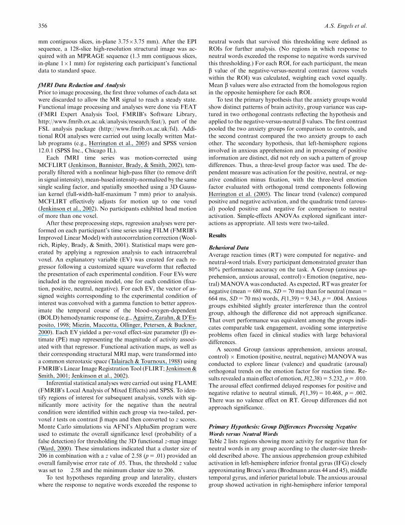
Figure 1 illustrates the critical Group�Hemisphere interac-
tion for IFG. The first contrast found no difference between the
pooled anxiety groups and controls. The second contrast, com-
paring the two anxiety groups, produced a Group�Hemisphere
effect, F(1,22)5 7.847, p5 .010, with more leftward asymmetry
in the anxious apprehension group, po.001, than in the anxious
arousal group, p5 .024, apparent in the right panel of Figure 1.
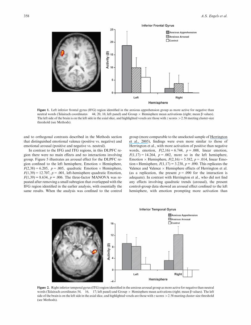
In ITG, Figure 2 shows relatively large responses and distinct
laterality patterns in the anxiety groups. Orthogonal contrasts
confirmed a larger response than in controls, F(1,40)5 9.206,
p5 .004, with the two anxious groups not differing overall. The
pooled anxiety groups versus controls did not differ by hemi-
sphere, but Figure 2 illustrates contrasting asymmetries for the
two anxiety groups, Group�Hemisphere, F(1,22)5 13.019,
p5 .002, with more left-hemisphere activation in the anxious
apprehension group, p5 .009, and more right-hemisphere acti-
vation in the anxious arousal group, p5 .039. An alternative
dissection of the interaction showed that the anxious arousal
group showed greater right-hemisphere activation than did the
anxious apprehension group, p5 .043, whereas the groups did
not differ in the left hemisphere.
Figure 3 indicates that the medial frontal cluster extending
into rACC was less active in the anxiety groups than in controls,
F(1,40)5 6.578, p5 .014, with no difference between the anxiety
groups. The Group�Hemisphere interactions did not approach
significance.
Group � Hemisphere ANOVAs were repeated for each of
these ROIs, examining activation for positive words relative to
neutral words. Thesewere done as omnibus (three-group) tests in
the absence of an a priori hypothesis about the groups and pos-
itive emotion, in order to test the emotion specificity of the
findings above for negative words. Left-hemisphere exceeded
right-hemisphere activation for all ROIs in Table 2 (all pso.017)
except for right ITG and right cerebellum. No significant group
or Group�Hemisphere effects emerged, indicating that group
effects were specific to negative-word activation.
Secondary Hypothesis: Functional Distinctions between
Left-Frontal Regions
To test the hypothesis that anxious apprehension and positive
affect are associated with activity in distinct regions of left PFC,
regions were evaluated for possible association with positive af-
fect, using the procedures of Herrington et al. (2005) to identify a
region in the control groupwithmore activation for positive than
negative words. A left DLPFC cluster that survived thresholding
(� 26, 18, 38; see Figure 4) overlapping with Herrington et al.’s
(� 32, 24, 42) emerged. This region and the homologous ROI
in the right hemisphere were subjected to a Group (anxious
apprehension, anxious arousal, control) � Emotion (positive, neu-
tral, negative, each relative to fixation) � Hemisphere MANOVA
Regional brain specificity in anxiety types 357
Table 2. Brain Regions More Active for Negative Than for Neutral Words, by Group
Region Cluster size Mean z
Location
Group Hemisphere Group � HemisphereX Y Z
High anxious apprehensionLeft inferior frontal gyrus (BA 44, 45, 9, 47) 1138 2.90 � 44 20 16 po .001 po .05Left inferior temporal gyrus (BA 20) 312 2.97 � 50 � 38 0 po .001 n.s.Left inferior parietal lobule (BA 7) 432 2.92 � 30 � 58 44 po .001 n.s.
High anxious arousalRight inferior temporal gyrus (BA 21, 20) 218 2.88 54 � 16 � 18 po .05 po .10
ControlRight cerebellum 308 2.88 20 � 82 � 18 po .01 n.s.Left inferior frontal gyrus (BA 47) 561 3.14 � 46 28 � 2 po .001 n.s.Left medial frontal gyrus (BA 10) 1007 3.04 � 2 54 10 po .05 po .001 n.s.Left superior temporal gyrus (BA 22) 914 3.11 � 54 � 52 22 po .05 po .10
Note. For each group, brain regions showing more activity for negative words than for neutral words. BA refers to Brodmann area in standard brainanatomy.Mean z is the average z value for the cluster.Whether each region displays an effect of group, hemisphere, or Group � Hemisphere at a po.10threshold is noted. Degrees of freedom were (2,39), (1,39), and (2,39) for the group, hemisphere, and Group � Hemisphere tests.
1In the ANOVAs, the left IFG region was that identified in the anx-ious apprehension group. Use of an ROI across groups and hemispheresdefined by activation in a single group and hemisphere potentially fostersregression to the mean and thus artifactual differences in group, hemi-sphere, and their interaction. Present results, however, cannot be ac-counted for that way, because the a priori predictions they support arenot subject to that problem and because the results do not generallyfollow regression toward the mean. For example, the ITG region wasdefined in the right hemisphere of the anxious arousal group, but theanxious apprehension group showed a significant contrary lateralizationin that ROI. Regression toward the mean, at worst, would produce nolateralization in that group. An analysis using a spherical ROI centeredon Broca’s area in IFG was done to avoid the possibility that IFG groupdifferences were an artifact due to regression to the mean, given how theoriginal IFG ROI was selected. This alternative analysis produced thesame significant Group � Hemisphere interaction (and main effect ofHemisphere) found using the IFG region identified in the anxious ap-prehension group, ruling out the regression to the mean confound.
and to orthogonal contrasts described in the Methods section
that distinguished emotional valence (positive vs. negative) and
emotional arousal (positive and negative vs. neutral).
In contrast to the IFG and ITG regions, in this DLPFC re-
gion there were no main effects and no interactions involving
group. Figure 5 illustrates an arousal effect for the DLPFC re-
gion confined to the left hemisphere, Emotion � Hemisphere,
F(2,38)5 6.205, p5 .005, quadratic Emotion � Hemisphere,
F(1,39)5 12.707, p5 .001, left-hemisphere quadratic Emotion,
F(1,39)5 8.634, p5 .006. The three-factor MANOVA was re-
peated after removing a small subregion that overlapped with the
IFG region identified in the earlier analysis, with essentially the
same results. When the analysis was confined to the control
group (more comparable to the unselected sample of Herrington
et al., 2005), findings were even more similar to those of
Herrington et al., with more activation of positive than negative
words, emotion, F(2,16)5 6.744, p5 .008, linear emotion,
F(1,17)5 14.264, p5 .002, more so in the left hemisphere,
Emotion � Hemisphere, F(2,16)5 5.582, p5 .014, linear Emo-
tion�Hemisphere, F(1,17)5 3.238, p5 .090. This replicates the
Valence and Valence � Hemisphere effects of Herrington et al.
(as a replication, the present p5 .090 for the interaction is
adequate). In contrast with Herrington et al., who did not find
any effects involving quadratic trends (arousal), the present
control-group data showed an arousal effect confined to the left
hemisphere, with emotion prompting more activation than
358 A.S. Engels et al.
Figure 1. Left inferior frontal gyrus (IFG) region identified in the anxious apprehension group as more active for negative than
neutral words (Talairach coordinates � 44, 20, 16; left panel) and Group � Hemisphere mean activations (right; mean b values).
The left side of the brain is on the left side in the axial slice, and highlighted voxels are those with z scores42.58 meeting cluster-size
threshold (see Methods).
Figure 2. Right inferior temporal gyrus (ITG) region identified in the anxious arousal group asmore active for negative than neutral
words (Talairach coordinates 54,� 16,� 17; left panel) and Group � Hemisphere mean activations (right; mean b values). The left
side of the brain is on the left side in the axial slice, and highlighted voxels are those with z scores42.58meeting cluster-size threshold
(see Methods).

neutral trials, quadratic Emotion�Hemisphere, F(1,17)5
9.740, p5 .006.
To confirm that the two left frontal clusters, the one in
DLFPC and the other in IFG, behaved differently, a direct
comparison was carried out with a Group (anxious apprehen-
sion, anxious arousal, and control)�Emotion (positive, neutral,
negative) � Hemisphere (left, right)�Region (IFG, DLPFC)
MANOVA, with orthogonal trends on the Emotion factor. The
numerous significant effects (see Table 3) will be briefly summa-
rized, because they largely recapitulated those reported above for
separate regions. Overall, positive and negative words prompted
greater activity than did neutral words, there wasmore activation
in the left than in the right hemisphere, and there was more in
IFG than in DLPFC. The difference between IFG and DLPFC
was larger for the left hemisphere. Positive and negative words
prompted more activation in IFG and less in DLPFC than did
neutral words, and the emotion effect was confined to the left
hemisphere. An Emotion � Hemisphere � Region effect was
qualified by a Group�Emotion�Hemisphere�Region interac-
tion, pursued with simple-effects tests. The groups differed
in IFG, Group�Emotion�Hemisphere, F(4,74)5 2.613,
p5 .042, not in DLPFC. In IFG, all groups showed significant-
ly greater left- than right-hemisphere activity for negative words,
but this difference was most pronounced for the anxious appre-
hension group (po.001). The anxious apprehension group also
produced greater left- than right-hemisphere activation for pos-
itive (po.001) and neutral (po.05) words, and within the left
hemisphere both positive and negative words showed greater
activation than neutral (po.001). Pairwise tests confirmed more
lateralization in the anxious apprehension group (po.001) than
in the anxious arousal group (p5 .012). Further dissection of the
interactions yielded a consistent pattern: Positive and negative
(but not neutral) words prompted leftward lateralization specific
to or larger in the anxious apprehension group. An analysis
comparing the two regions with overlapping voxels removed
yielded essentially the same results. Consistently, then, group
effects in PFC were confined to IFG, whereas valence effects
were confined to DLPFC. These findings indicate that distinct
regions in left PFC are involved in processing positive emotion
(DLPFC) and in modulating the effects of anxiety on the pro-
cessing of emotional information (IFG).
Discussion
The primary hypothesis was that the anxiety groups would show
distinct patterns of brain activity. The anxious groups’ hyperac-
tivation in ITG and hypoactivation in rACC suggests an exag-
gerated alarm response (Compton et al., 2003; Corbetta &
Shulman, 2002) accompanied by compromise of attentional
controlmechanisms (Bush, Luu, & Posner, 2000;Mohanty et al.,
2007). Moreover, as predicted, the anxious groups diverged in
response to negative words: The anxious apprehension group
exhibited more left-hemisphere activity in IFG and ITG, and the
anxious arousal group exhibited less leftward IFG asymmetry
and more right-hemisphere posterior activation. Thus, distinct
patterns of lateralized brain activity characterized psychometri-
cally distinct types of anxiety.
The secondary goal of the present study was a replication and
extension of Herrington et al. (2005), in order to distinguish left
frontal brain regions involved in anxious apprehension versus the
processing of positive information. The valence and Valence �
Regional brain specificity in anxiety types 359
Figure 3. Medial frontal gyrus (MFG) region (extending into rostral anterior cingulate cortex, rACC) identified in the control
group as more active for negative than neutral words (Talairach coordinates � 2, 54, 10) and Group � Hemisphere mean
activations (right; mean b values). Highlighted voxels are those with z scores 42.58 meeting cluster-size threshold (see Methods).
Figure 4. Coronal view of left inferior frontal gyrus (IFG, blue) region
for negative 4neutral (same cluster as is displayed in Figure 1’s axial
view) and left dorsolateral prefrontal cortex (DLPFC, yellow) for
positive 4neutral, displayed at Talairach coordinate y5 18.

Hemisphere effects in DLPFC from the earlier study were con-
firmed (even though subject sample, scanner field strength, and
much of the analysis software differed in the two studies). An-
alyses demonstrated that this region, most activated when at-
tention must be directed away from positive words, is
anatomically and functionally distinct from the region around
Broca’s area that is particularly active in individuals who exhibit
high levels of anxious apprehension.
Group Differences in Negative Emotion
Demonstration of exaggerated left-hemisphere activity in ante-
rior language areas when anxious apprehension participants
must direct their attention away from negative words has con-
siderable construct validity, given the strong linguistic compo-
nent in worry. Broca’s area is involved in speech production,
syntactic processing, phonological processing, and subvocal
articulatory rehearsal (Awh et al., 1996; Zatorre,Meyer, Gjedde,
& Evans, 1996). Inferior prefrontal regions have been implicated
in maintenance of verbal information (Fletcher & Henson,
2001; Wagner, 1999), and enhanced activity in these regions re-
flects a variety of verbal processes such as accessing word mean-
ing, holding language-related information online, and verbal
rehearsal, consistent with the ruminative style of cognition
that characterizes anxious apprehension. Worry may affect
attention and working memory by drawing from a limited
pool of resources (Eysenck & Calvo, 1992) or by interfering
with performance due to competition with or distraction from
attention to task-relevant information (Nitschke et al., 2000).
For example, anxious apprehension participants may be doing
more semantic processing of word stimuli, which interferes with
selective attention to the task-relevant attribute of color. In the
present study of nonpatients, there were no significant perfor-
mance differences among the groups. However, with a large
enough sample size, performance differences related to anxiety
may emerge as noted in the Introduction (Koven et al., 2003). It
might be expected, therefore, that worry would interfere with
optimum performance in a variety of circumstances, particularly
those that require or would benefit from high levels of selective
attention.
In addition to the IFG finding for anxious apprehension, the
opposing patterns of lateralization in ITG for the two anxious
groups further support the model presented in the Introduction.
Negative emotional words prompted greater right-hemisphere
temporoparietal activity specific to the anxious arousal group.
These results are consistent with hypotheses that the right hemi-
sphere houses an integrated system for responding to immediate
threat, promoting sympathetic nervous system activity, spatial
attention, visual scanning of the environment, and sensitivity to
meaningful nonverbal cues (Compton et al., 2003; Nitschke et
al., 2000). ITG is also implicated as important for object iden-
tification (the ‘‘what’’ system; Hermann, Seidenberg, Wyler, &
Haltiner, 1993), and right temporoparietal cortex is important
360 A.S. Engels et al.
Figure 5. Left dorsolateral prefrontal cortex (DLPFC) region identified in the control group as more active for positive than
negative words (as in Herrington et al., 2005) and the homologous ROI in the right hemisphere (left; Talairach coordinates � 26, 18,
38) and Emotion � Hemisphere mean activations (right).
Table 3. Group � Emotion � Hemisphere � Region MANOVA
for Negative-Word Activation
Effect df F p
Emotion 2,38 7.112 .002Quadratic Emotion 1,39 13.514 .001
Hemisphere 1,39 7.842 .008Region 1,39 49.971 o.001Region�Hemisphere 1,39 40.801 o.001Emotion�Region 2,38 20.634 o.001Quadratic Emotion�Region 1,39 42.260 o.001
Emotion � Hemisphere 2,38 15.190 o.001Quadratic Emotion�Hemisphere 1,39 29.366 o.001
Emotion � Hemisphere�Region 2,38 13.024 o.001Quadratic Emotion�Hemisphere�Region 1,39 20.252 o.001
Group�Emotion�Hemisphere�Region 4,74 4.173 .004Group�quadratic Emotion�Hemisphere�Region 2,39 8.516 .001
Note. Only effects exceeding p5 .05 are listed. For orthogonal trends onemotion, the linear trend component (none po.05 in this analysis) con-trasts positive and negative words, reflecting emotional valence, and thequadratic trend component contrasts those with neutral words, reflectingemotional arousal.

for detecting salient and unexpected stimuli (Corbetta &
Shulman, 2002). Differential engagement of this system for anx-
ious arousal is consistent with the cognition that characterizes
this type of anxiety. Present findgs are also consistent with pre-
vious fMRI results using an emotional Stroop task with unse-
lected subjects, showing activation as a function of emotional
arousal near the present ITG area (Compton et al., 2003). This
provides independent confirmation of relatively posterior activ-
ity associated with emotional arousal that we propose charac-
terizes the anxious arousal construct.
In contrast, the anxious apprehension group showed
more left-hemisphere activation in ITG, again consistent with a
more verbally mediated response to threatening cues. PFC
(particularly inferior frontal and middle frontal regions) is likely
to play a prominent role in the synthesis of attention and emo-
tion. According to a model of the neural network involved in
attention (Banich et al., 2000a, 2000b, 2001; Compton et al.,
2003; Milham & Banich, 2005; Milham et al., 2001, 2002;
Milham, Banich, & Barad, 2003; Milham, Banich, Claus, &
Cohen, 2003), these regions are important for imposing an
attentional set or bias for the information to be attended,
especially when processing of the to-be-attended information is
less automatic than that of the task-irrelevant information.
As attentional demands increase (e.g., the more difficult it is to
direct attention to task-relevant vs. task-irrelevant information),
the degree to which these prefrontal regions are called upon
increases. Frontal activity can therefore provide a metric for
how much attentional control is required to ignore the task-ir-
relevant information. In the context of the emotional Stroop
task, an interpretation of the greater left frontal activation ob-
served for the anxious apprehension group is that such individ-
uals find it difficult to suppress task-irrelevant emotional word
meaning and require more attentional control to impose an
attentional set.
Apprehension versus Pleasure: Functional Distinctions between
Left-Frontal Regions
Evidence here and elsewhere of exaggerated left frontal activa-
tion in anxious apprehension appears contradictory to reports of
increased left frontal activity associated with positive emotion or
appetitive or approach motivation cited in the Introduction.
Present data suggest a resolution, however, because at least in
this emotional Stroop task different regions are involved when a
stimulus contains positive emotional content versus those differ-
entially activated in individuals who have anxious apprehension.
Whereas left IFG was more active for the anxious apprehension
group for negative than for neutral words and was sensitive to
group differences, a replication of the analysis procedures of
Herrington et al. (2005) identified an anatomically and func-
tionally distinct region in left DLPFC that was more active when
a stimulus contained positive emotional content and showed no
group differentiation.
Present results encourage ongoing efforts to make functional
distinctions among PFC regions. In our work and that of
others, the term ‘‘dorsolateral prefrontal cortex’’ has often been
used broadly, including coordinates in IFG.However, functional
distinctions have been proposed between ventrolateral (BA 44,
45, 47), dorsolateral (BA 9, 46), and anterior (BA 8, 10) regions
of the frontal cortex (Fletcher & Henson, 2001; Milham et al.,
2002; Petrides, 2000). Ventrolateral areas (such as the IFG region
observed here as more active for the anxious apprehension
group) have been implicated in articulatory rehearsal and main-
tenance of verbal information, whereas dorsolateral areas are
implicated in manipulating information in working memory.
Present findings suggest extending this ventral/dorsal differenti-
ation to the notion that distinct regions of left PFC are involved
in processing positive emotional content and in modulating the
effects of anxious apprehension on processing related to negative
emotion.
Rostral Anterior Cingulate Cortex in the Emotional Stroop Task
Both anxiety groups showed less activation than the control
group in medial PFC extending into rACC (also, activation in
this region was negatively correlated with anxious apprehension
ratings). Considerable evidence implicates rACC in the assess-
ment of the salience of emotional information as well as the
regulation of emotional responses (see Bush et al., 2000; Moh-
anty et al., 2007). Activation in this region for the control group
supports results from an emotional counting Stroop task with
normal controls, showing greater activity in this region for neg-
ative than neutral words (Whalen et al., 1998). Research has also
demonstrated diminished activity in this region for participants
with PTSD (Shin et al., 2005). In addition, medial PFC activity
has been shown to correlate negatively with anxiety symptom
severity (Bishop, Duncan, Brett, & Lawrence, 2004; Shin et al.,
2005). Present results are consistent with these findings and sug-
gest reduced control in the presence of threatening stimuli for
participants with anxiety (Bishop et al., 2004; Whalen et al.,
1998). However, present results are inconsistent with other stud-
ies that have found enhanced rACC activity for anxiety (for a
review, see Hajcak,McDonald, & Simons, 2003). Results for the
present task, which relies on emotional self-regulation, make
sense in light of a proposal that rACC normally acts to down-
regulate amygdala activity, central to some types of anxiety
(Etkin, Egner, Peraza, Kandel, & Hirsch, 2006). Thus, present
anxious groups showed abnormally low rACC activation. In
these nonpatient individuals, a consequent amygdala hyperacti-
vation did not emerge here.
Present results provide support for the hypothesis that two
conceptually, psychometrically, and clinically distinct types of
anxiety are associated with different neural mechanisms. Fur-
thermore, a region specifically involved in anxious apprehension
differs anatomically and functionally from one specifically in-
volved in processing positive information. The consistency of
distinctions, across several measurement domains, between anx-
ious apprehension and anxious arousal indicates that they war-
rant separate, systematic assessment in psychopathology
research and in treatment development. Present data also sup-
port an integrationist (not merely interactionist) objection
(Miller, 1996) to the commonly assumed distinction between
cognition, emotion, and their relationship to shared and distinct
neural mechanisms. Functionally, it makes sense that anxious
apprehension engages Broca’s area, because this region is in-
volved in verbal production and rehearsal. Similarly, it is func-
tionally appropriate that anxious arousal fosters engagement of
right-hemisphere regions involved in responding to immediate
threat. Differential activation of these regions for each anxiety
type thus supports the contention that patterns of brain activity
associated with distinct emotional states implement the distinc-
tive computations associated with these states, rather than re-
flecting either cognition or emotion in some mutually exclusive
sense.
Regional brain specificity in anxiety types 361

REFERENCES
Aguirre, G. K., Zarahn, E., & D’Esposito, M. (1998). The variability ofhuman BOLD hemodynamic response. NeuroImage, 8, 360–369.
American Psychiatric Association. (2000). Diagnostic and statisticalmanual of mental disorders, Fourth edition, Text revision. Washing-ton, DC: American Psychiatric Association.
Asbjornsen, A., Hugdahl, K., & Bryden, M. P. (1992). Manipulations ofsubjects’ level of arousal in dichotic listening. Brain and Cognition, 19,183–194.
Ashby, F.G., Isen, A.M., & Turken, A.U. (1999). A neuropsychologicaltheory of positive affect and its influence on cognition. PsychologicalReview, 106, 529–550.
Awh, E., Jonides, J., Smith, E. E., Schumacher, E. H., Koeppe, R. A., &Katz, S. (1996). Dissociation of storage and rehearsal in verbal work-ing memory: Evidence from positron emission tomography. Psycho-logical Science, 7, 25–31.
Banich, M. T., Milham, M. P., Atchley, R. A., Cohen, N. J., Webb, A.,Wszalek, T., et al. (2000a). fMRI studies of Stroop tasks reveal uniqueroles of anterior and posterior brain systems in attentional selection.Journal of Cognitive Neuroscience, 12, 988–1000.
Banich, M. T., Milham, M. P., Atchley, R. A., Cohen, N. J., Webb, A.,Wszalek, T., et al. (2000b). Prefrontal regions play a predominant rolein imposing an attentional ‘set’: Evidence from fMRI.Cognitive BrainResearch, 10, 1–9.
Banich, M. T., Milham, M. P., Jacobson, B. L., Webb, A., Wszalek, T.,Cohen, N. J., et al. (2001). Attentional selection and the processing oftask-irrelevant information: Insights from fMRI examinations of theStroop task. In C. Casanova &M. Ptito (Eds.), Vision: From neuronsto cognition, Progress in Brain Research, Vol. 134, (pp. 459–470).Amsterdam: Elsevier Science.
Barlow, D. H. (1991). Disorders of emotion. Psychological Inquiry, 2,58–71.
Baxter, L. R., Phelps, M. E., Mazziotta, J. C., Guze, B. H., Schwartz, J.M., & Selin, C. E. (1987). Local cerebral glucose metabolic rates inobsessive-compulsive disorder. Archives of General Psychiatry, 44,211–218.
Bishop, S., Duncan, J., Brett, M., & Lawrence, A. D. (2004). Prefrontalcortical function and anxiety: Controlling attention to threat-relatedstimuli. Nature Neuroscience, 7, 184–188.
Bradley, M. M., & Lang, P. J. (1998). Affective norms for English words(ANEW). Gainesville, FL: University of Florida, NIMH Center forthe Study of Emotion and Attention.
Bush, G., Luu, P., & Posner, M. I. (2000). Cognitive and emotionalinfluences in anterior cingulate cortex. Trends in Cognitive Sciences, 4,215–222.
Compton, R. J., Banich, M. T., Mohanty, A., Milham,M. P., Miller, G.A., Scalf, P. E., et al. (2003). Paying attention to emotion: An fMRIinvestigation of cognitive and emotional Stroop tasks. Cognitive, Af-fective, and Behavioral Neuroscience, 3, 81–96.
Compton, R. J., Heller, W., Banich, M. T., Palmieri, P. A., & Miller,G. A. (2000). Responding to threat: Hemispheric asymmetriesand interhemispheric division of input. Neuropsychology, 14,254–264.
Corbetta, M., & Shulman, G. L. (2002). Control of goal-directed andstimulus-driven attention in the brain. Nature Reviews Neuroscience,3, 201–215.
Dalgleish, T. (1995). Performance on the emotional Stroop task ingroups of anxious, expert, and control subjects: A comparison ofcomputer and card presentation formats. Cognition & Emotion, 9,341–362.
Davidson, R. J. (1992). Emotion and affective style: Hemispheric sub-strates. Psychological Science, 3, 39–43.
Davidson, R. J. (2004). What does the prefrontal cortex ‘‘do’’ in affect:Perspectives on frontal EEG asymmetry research. Biological Psy-chology, 67, 219–233.
Egloff, B., & Hock, M. (2001). Interactive effects of state anxiety andtrait anxiety on emotional Stroop interference. Personality and Indi-vidual Differences, 31, 875–882.
Etkin, A., Egner, T., Peraza, D. M., Kandel, E. R., & Hirsch, J.(2006). Resolving emotional conflict: A role for the rostral anteriorcingulate cortex in modulating activity in the amygdala. Neuron, 51,871–882.
Eysenck, M. W., & Calvo, M. G. (1992). Anxiety and performance: Theprocessing efficiency theory. Cognition and Emotion, 6, 409–434.
Fletcher, P. C., & Henson, R. N. A. (2001). Frontal lobes and humanmemory: Insights from functional neuroimaging. Brain, 124,849–881.
Gruzelier, J., & Phelan, M. (1991). Stress induced reversal of a lexicaldivided visual-field asymmetry accompanied by retarded electrode-rmal habituation. International Journal of Psychophysiology, 11,269–276.
Haier, R. J., Siegel, B. V., Jr,MacLachlan, A., Soderling, E., Lottenberg,S., & Buchsbaum, M. S. (1992). Regional glucose metabolic changesafter learning a complex visuospatial/motor task: A positron emissiontomographic study. Brain Research, 570, 134–143.
Hajcak, G., McDonald, N., & Simons, R. F. (2003). Anxiety and error-related brain activity. Biological Psychology, 64, 77–90.
Harmon-Jones, E. (2004). Contributions from research on anger andcognitive dissonance to understanding the motivational functions ofasymmetrical frontal brain activity. Biological Psychology, 67, 51–76.
Heller, W., Etienne, M., & Miller, G. A. (1995). Patterns of perceptualasymmetry in depression and anxiety: Implications for neuropsycho-logical models of emotion and psychopathology. Journal of AbnormalPsychology, 104, 327–333.
Heller, W., Koven, N. S., &Miller, G. A. (2003). Regional brain activityin anxiety and depression, cognition/emotion interaction, and emo-tion regulation. In K. Hugdahl & R. J. Davidson (Eds.), The asym-metrical brain (pp. 533–564). Cambridge, MA: MIT Press.
Heller, W., &Nitschke, J. B. (1998). The puzzle of regional brain activityin depression and anxiety: The importance of subtypes and comor-bidity. Cognition and Emotion, 12, 421–447.
Heller, W., Nitschke, J. B., Etienne, M. A., & Miller, G. A. (1997).Patterns of regional brain activity differentiate types of anxiety. Jour-nal of Abnormal Psychology, 106, 376–385.
Heller,W., Nitschke, J. B., & Lindsay, D. L. (1997). Neuropsychologicalcorrelates of arousal in self-reported emotion.Cognition and Emotion,11, 383–402.
Heller, W., Nitschke, J. B., & Miller, G. A. (1998). Lateralization inemotion and emotional disorders.Current Directions in PsychologicalScience, 7, 26–32.
Hermann, B. P., Seidenberg, M., Wyler, A., & Haltiner, A. (1993). Dis-sociation of object recognition and spatial localization abilities fol-lowing temporal lobe lesions in humans. Neuropsychology, 7,343–350.
Herrington, J. D., Koven, N., Heller,W.,Miller, G. A., &Nitschke, J. B.(in press) Frontal asymmetry in emotion, motivation, personality andpsychopathology: Electrocortical and hemodynamic neuroimaging.In N. Allen & C. Pantelis (Eds.), Neurobiology of mental disorders.
Herrington, J. D., Mohanty, A., Koven, N. S., Fisher, J. E., Stewart,J. L., Banich, M. T., et al. (2005). Emotion-modulated performanceand activity in left dorsolateral prefrontal cortex.Emotion, 5, 200–207.
Hofmann, S. G.,Moscovitch, D. A., Litz, B. T., Kim,H.-J., Davis, L. L.,& Pizzagalli, D. A. (2005). The worried mind: Autonomic and pre-frontal activation during worrying. Emotion, 5, 464–475.
Holle, C., Neely, J. H., & Heimberg, R. G. (1997). The effects of blockedversus random presentation and semantic relatedness of stimuluswords on response to a modified Stroop task among social phobics.Cognitive Therapy and Research, 21, 681–697.
Jenkinson, M., Bannister, P., Brady, M., & Smith, S. (2002). Improvedoptimization for the robust and accurate linear registration and mo-tion correction of brain images. NeuroImage, 17, 825–841.
Jenkinson, M., & Smith, S. (2001). A global optimization method forrobust affine registration of brain images.Medical Image Analysis, 5,143–156.
Keller, J., Nitschke, J. B., Bhargava, T., Deldin, P. J., Gergen, J. A.,Miller, G. A., et al. (2000). Neuropsychological differentiation ofdepression and anxiety. Journal of Abnormal Psychology, 109, 3–10.
Kessler, R. C., Berglund, P., Demler, O., Jin, R., Merikangas, K. R., &Walters, E. E. (2005). Lifetime prevalence and age-of-onset distribu-tions of DSM-IV disorders in the National Comorbidity SurveyReplication. Archives of General Psychiatry, 62, 593–602.
Koven, N. S., Heller, W., Banich, M. T., & Miller, G. A. (2003). Re-lationships of distinct affective dimensions to performance on anemotional Stroop task.Cognitive Therapy and Research, 27, 671–680.
Levin, R. L., Heller,W.,Mohanty, A., Herrington, J. D., &Miller, G. A.(in press) Cognitive deficits in depression and functional specificity ofregional brain activity. Cognitive Therapy and Research.
362 A.S. Engels et al.

Mathews, A. (1990). Why worry? The cognitive function of anxiety.Behaviour Research and Therapy, 28, 445–468.
McNally, R. J. (1998). Information-processing abnormalities in anxietydisorders: Implications for cognitive neuroscience. Cognition andEmotion, 12, 479–495.
Meyer, T. J., Miller, M. L., Metzger, R. L., & Borkovec, T. D. (1990).Development and validation of the Penn State Worry Questionnaire.Behaviour Research and Therapy, 28, 487–495.
Miezin, F. M., Maccotta, L., Ollinger, J. M., Petersen, S. E., & Buckner,R. L. (2000). Characterizing the hemodynamic response: Effects ofpresentation rate, sampling procedure, and the possibility of orderingbrain activity based on relative thinking. NeuroImage, 11, 735–739.
Milham, M. P., & Banich, M. T. (2005). Anterior cingulate cortex: AnfMRI analysis of conflict specificity and functional differentiation.Human Brain Mapping, 25, 328–335.
Milham, M. P., Banich, M. T., & Barad, V. (2003). Competition forpriority in processing increases prefrontal cortex’s involvement in top-down control: An event-related fMRI study of the Stroop task. Cog-nitive Brain Research, 17, 212–222.
Milham, M. P., Banich, M. T., Claus, E., & Cohen, N. (2003). Practice-related effects demonstrate complementary roles of anterior cingulateand prefrontal cortices in attentional control. NeuroImage, 18,483–493.
Milham, M. P., Banich, M. T., Webb, A., Barad, V., Cohen, N. J.,Wszalek, T., et al. (2001). The relative involvement of anterior cingu-late and prefrontal cortex in attentional control depends on nature ofconflict. Cognitive Brain Research, 12, 467–473.
Milham,M. P., Erickson, K. I., Banich,M. T., Kramer, A. F.,Webb, A.,Wszalek, T., et al. (2002). Attentional control in the aging brain:Insights from an fMRI study of the Stroop task. Brain & Cognition,49, 277–296.
Miller, G. A. (1996). Presidential address: How we think about cogni-tion, emotion, and biology in psychopathology. Psychophysiology,33, 615–628.
Miller, G. A., Levin, D. N., Kozak, M. J., Cook, E. W., McLean, A., &Lang, P. J. (1987). Individual differences in imagery and the psycho-physiology of emotion. Cognition and Emotion, 1, 367–390.
Mohanty, A., Engels, A. S., Herrington, J. D., Heller, W., Ringo Ho,M.-H. R., Banich, M. T., et al. (2007). Differential engagement ofanterior cingulate cortex subdivisions for cognitive and emotionalfunction. Psychophysiology, 44, 343–351.
Mohanty, A., & Heller, W. (2002). The neuropsychology of depression:Affect, cognition, and neural circuitry. In H. D’Haenen, J. A. denBoer, H. Westenberg, & P. Willner (Eds.), Textbook of biologicalpsychiatry (pp. 791–802). Chichester, UK: Wiley.
Molina, S., & Borkovec, T. D. (1994). The Penn State Worry Question-naire: Psychometric properties and associated characteristics. InG. C.L. Davey & F. Tallis (Eds.), Worrying: Perspectives on theory, as-sessment and treatment (pp. 265–283). Chichester, UK: Wiley.
Nitschke, J. B., & Heller, W. (2002). The neuropsychology of anxietydisorders: Affect, cognition, and neural circuitry. In H. D’haenen, J.A. den Boer, H. Westenberg, & P. Willner (Eds.), Textbook of bio-logical psychiatry (pp. 975–988). Chichester, UK: Wiley.
Nitschke, J. B., & Heller, W. (2005). Distinguishing neural substrates ofheterogeneity among anxiety disorders. In M. Glabus (Ed.), Inter-national review of neurobiology, Vol. 67, Neuroimaging, Part B (pp.1–42). San Diego, CA: Elsevier.
Nitschke, J. B., Heller,W., Imig, J. C.,McDonald, R. P., &Miller, G. A.(2001). Distinguishing dimensions of anxiety and depression. Cogni-tive Therapy and Research, 25, 1–22.
Nitschke, J. B., Heller, W., & Miller, G. A. (2000). Anxiety, stress, andcortical brain function. In J. C. Borod (Ed.), The neuropsychology ofemotion (pp. 298–319). New York: Oxford University Press.
Nitschke, J. B., Heller, W., Palmieri, P. A., & Miller, G. A. (1999).Contrasting patterns of brain activity in anxious apprehension andanxious arousal. Psychophysiology, 36, 628–637.
Petrides, M. (2000). The role of the mid-dorsolateral prefrontal cortex inworking memory. Experimental Brain Research, 133, 44–54.
Reiman, E.M., Raichle, M. E., Butler, F. K., Herscovitch, P., &Robins,E. (1984). A focal brain abnormality in panic disorder, a severe formof anxiety. Nature, 310, 683–685.
Shin, L. M., Wright, C. I., Cannistraro, P. A., Wedig, M.M., McMullin,K.,Martis, B., et al. (2005). A functionalmagnetic resonance imagingstudy of amygdala and medial prefrontal cortex responses to overtlypresented fearful faces in Posttraumatic Stress Disorder. Archives ofGeneral Psychiatry, 62, 273–281.
Swedo, S. E., Schapiro, M. B., Grady, C. L., Cheslow, D. L., Leonard,H. L., Kumar, A., et al. (1989). Cerebral glucose metabolism inchildhood-onset obsessive-compulsive disorder. Archives of GeneralPsychiatry, 46, 518–523.
Talairach, J., & Tournoux, P. (1988). Co-planar stereotaxic atlas of thehuman brain (2nd edn.), New York: Thieme Medical Publishers.
Toglia, M. P., & Battig, W. F. (1978).Handbook of semantic word norms.Hillsdale, NJ: Erlbaum.
Tomarken, A. J., Davidson, R. J., Wheeler, R. E., & Doss, R. C. (1992).Individual differences in anterior brain asymmetry and fundamentaldimensions of emotion. Journal of Personality and Social Psychology,62, 676–687.
Tucker, D. M., Antes, J. R., Stenslie, C. E., & Barnhardt, T. M. (1978).Anxiety and lateral cerebral function. Journal of Abnormal Psychol-ogy, 87, 380–383.
Tucker, D. M., Roth, R. S., Arneson, B. A., & Buckingham, V. (1977).Right hemisphere activation during stress. Neuropsychologia, 15,697–700.
Van Strien, J. W., & Heijt, R. (1995). Altered visual field asymmetries forletter naming and letter matching as a result of concurrent presen-tation of threatening and nonthreatening words. Brain and Cognition,29, 187–203.
Van Strien, J. W., & Morpurgo, M. (1992). Opposite hemispheric ac-tivations as a result of emotionally threatening and non-threateningwords. Neuropsychologia, 30, 845–848.
Wagner, A.D. (1999).Workingmemory contributions to human learningand remembering. Neuron, 22, 19–22.
Ward, D. B. (2000). Simultaneous inference for FMRI data. http://afni.nimh.nih.gov/pub/dist/doc/manual/AlphaSim.pdf, accessedJuly 27, 2006.
Watson, D., Clark, L. A., Weber, K., Assenheimer, J. S., Strauss, M. E.,&McCormick, R. A. (1995). Testing a tripartite model: II. Exploringthe symptom structure of anxiety and depression in student, adult,and patient samples. Journal of Abnormal Psychology, 104, 15–25.
Watson, D., Weber, K., Assesnheimer, J. S., Clark, L. A., Strauss, M. E.,&McCormick, R. A. (1995). Testing a tripartite model: I. Evaluatingthe convergent and discriminant validity of anxiety and depressionsymptom scales. Journal of Abnormal Psychology, 104, 3–14.
Whalen, P. J., Bush, G., McNally, R. J., Wilhelm, S., McInerney, S. C.,Jenike,M. A., et al. (1998). The emotional counting stroop paradigm:A functional magnetic resonance imaging probe of the anteriorcingulate affective division. Biological Psychiatry, 44, 1219–1228.
Woolrich, M. W., Ripley, B. D., Brady, M., & Smith, S. M. (2001).Temporal autocorrelation in univariate linearmodeling of fMRI data.NeuroImage, 14, 1370–1386.
Wu, J. C., Buchsbaum, M. S., Hershey, T. G., Hazlett, E., Sicotte, N., &Johnson, J. C. (1991). PET in generalized anxiety disorder. BiologicalPsychiatry, 29, 1181–1199.
Zatorre, R. J.,Meyer, E., Gjedde, A., & Evans, A. C. (1996). PETstudiesof phonetic processing of speech: Review, replication, and reanalysis.Cerebral Cortex, 6, 21–30.
(Received October 1, 2006; Accepted January 8, 2007)
Regional brain specificity in anxiety types 363