Embed Size (px)
Citation preview
E u r o p e ’ s j o u r n a l o n i n f e c t i o u s d i s e a s e e p i d e m i o l o g y, p r e v e n t i o n a n d c o n t r o l
www.eurosurveillance.org
Special edition: Middle East Respiratory
Syndrome Coronavirus (MERS-CoV)
July 2013
•This issue presents descriptions of the assays for laboratory confirmation of MERS-CoV infections, case reports from the United Kingdom, Germany and France documenting the related contact investigations, public health measures and more.

Based at the European Centre for Disease Prevention and Control (ECDC), 171 83 Stockholm, Sweden
Telephone number+46 (0)8 58 60 11 38 or +46 (0)8 58 60 11 36
Fax number+46 (0)8 58 60 12 94
Editor-in-chiefInes Steffens
Scientific editors Kathrin HagmaierWilliamina WilsonKaren Wilson
Assistant editorsAlina BuzduganIngela Söderlund
Associate editors Andrea Ammon, Stockholm, SwedenTommi Asikainen, Frankfurt, GermanyMike Catchpole, London, United KingdomDenis Coulombier, Stockholm, SwedenChristian Drosten, Bonn, GermanyKarl Ekdahl, Stockholm, SwedenJohan Giesecke, Stockholm, SwedenHerman Goossens, Antwerp, BelgiumDavid Heymann, London, United KingdomHeath Kelly, Melbourne, AustraliaIrena Klavs, Ljubljana, SloveniaKarl Kristinsson, Reykjavik, IcelandDaniel Lévy-Bruhl, Paris, FranceRichard Pebody, London, United KingdomPanayotis T. Tassios, Athens, GreeceHélène Therre, Paris, FranceHenriette de Valk, Paris, FranceSylvie van der Werf, Paris, France
Design / LayoutFabrice Donguy / Arne Haeger
www.eurosurveillance.org
© Eurosurveillance, 2013
Albania: Alban Ylli, TiranaAustria: Reinhild Strauss, Vienna Belgium: Koen De Schrijver, Antwerp Belgium: Sophie Quoilin, BrusselsBosnia and Herzogovina: Nina Rodić Vukmir, Banja LukaBulgaria: Mira Kojouharova, Sofia Croatia: To be nominated Cyprus: To be nominated Czech Republic: Bohumir Križ, Prague Denmark: Peter Henrik Andersen, Copenhagen England and Wales: TBC, London Estonia: Kuulo Kutsar, Tallinn Finland: Outi Lyytikäinen, Helsinki France: Judith Benrekassa, Paris Germany: Jamela Seedat, BerlinGreece: Rengina Vorou, Athens Hungary: Ágnes Csohán, Budapest Iceland: Haraldur Briem, Reykjavik Ireland: Lelia Thornton, Dublin Italy: Paola De Castro, Rome Kosovo (under UNSCR 1244/99): Lul Raka, PristinaLatvia: Jurijs Perevoščikovs, Riga Lithuania: Milda Zygutiene, Vilnius Luxembourg: Thérèse Staub, Luxembourg The FYR of Macedonia: Elisaveta Stikova, SkopjeMalta: Tanya Melillo Fenech, VallettaMontenegro: Dragan Laušević, PodgoricaNetherlands: Paul Bijkerk, Bilthoven Norway: Hilde Klovstad, Oslo Poland: Malgorzata Sadkowska-Todys, Warsaw Portugal: Isabel Marinho Falcão, LisbonRomania: Daniela Pitigoi, Bucharest Serbia: Tatjana Pekmezovic, BelgradeSlovakia: Lukáš Murajda, Martin Slovenia: Alenka Kraigher, Ljubljana Spain: Elena Rodríguez Valín, Madrid Sweden: Christer Janson, Stockholm Turkey: Fehmaniz Temel, AnkaraUnited Kingdom: Norman MacDonald, GlasgowEuropean Commission: Paolo Guglielmetti, LuxembourgWorld Health Organization Regional Office for Europe: Nedret Emiroglu, Copenhagen
Editorial team Editorial advisors
1www.eurosurveillance.org
Contents
© Eurosurveillance
Illustration of Coronavirus, phylogenetic tree
Middle East Respiratory Syndrome Coronavirus (MERS-CoV)Note from the editors: A new virus bringing back memories from the past 2Eurosurveillance editorial team
RAPID COMMUNICATION
Severe respiratory illness caused by a novel coronavirus, in a patient transferred to the United Kingdom from the Middle East, September 2012 3A Bermingham et al.
The United Kingdom public health response to an imported laboratory confirmed case of a novel coronavirus in September 2012 8RG Pebody et al.
Evidence of person-to-person transmission within a family cluster of novel coronavirus infections, United Kingdom, February 2013 12The Health Protection Agency (HPA) UK Novel Coronavirus Investigation team
First cases of Middle East Respiratory Syndrome Coronavirus (MERS-CoV) infections in France, investigations and implications for the prevention of human-to-human transmission, France, May 2013 19A Mailles et al.
Novel coronavirus associated with severe respiratory disease: Case definition and public health measures 24N Danielsson et al.
Incubation period as part of the case definition of severe respiratory illness caused by a novel coronavirus 26H Nishiura et al.
Detection of a novel human coronavirus by real-time reverse-transcription polymerase chain reaction 30VM Corman et al.
Assays for laboratory confirmation of novel human coronavirus (hCoV-EMC) infections 36VM Corman et al.
Specific serology for emerging human coronaviruses by protein microarray 45C Reusken et al.
Laboratory capability for molecular detection and confirmation of novel coronavirus in Europe, November 2012 52D Palm et al.
SURVEILLANCE AND OUTBREAK REPORTS
Contact investigation of a case of human novel coronavirus infection treated in a German hospital, October-November 2012 56U Buchholz et al.
PERSPECTIVES
Transmission scenarios for Middle East Respiratory Syndrome Coronavirus (MERS-CoV) and how to tell them apart 63S Cauchemez et al.
2 www.eurosurveillance.org
Miscellaneous
Note from the editors: A new virus bringing back memories from the past
Eurosurveillance editorial team ([email protected])1
1. European Centre for Disease Prevention and Control (ECDC), Stockholm, Sweden
Citation style for this article: Eurosurveillance editorial team. Note from the editors: A new virus bringing back memories from the past. Euro Surveill. 2012;17(39):pii=20284. Available online: http://www.eurosurveillance.org/ViewArticle.aspx?ArticleId=20284
Article submitted on 27 September 2012 / published on 27 September 2012
In recent days, public health experts and healthcare workers around the world are alert following the dis-covery of a new human coronavirus causing severe respiratory illness. Two cases, both with connection to Saudi Arabia, were communicated through ProMED on 20 and 23 September respectively [1,2].
Many health professionals still have vivid memories of the alert that followed the death of an American businessman in a hospital in Hanoi, Vietnam, in early 2003 after having travelled to China, and the following outbreak of severe acute respiratory syndrome (SARS). This triggered worldwide alarm and containment meas-ures. During the outbreak, there was excellent col-laboration between global players and institutions, on various levels (i.e. public health institutions, labora-tories and hospitals) and new ways of communicating proved to be highly value for the exchange of informa-tion. The last case of SARS occurred in China in May 2004: thereafter the virus seemed to have disappeared and has not resurfaced since.
The public health world is currently looking closely into the two recent cases of coronovirus infection. Similar to SARS, the two patients had/have symptoms of severe respiratory illness and the virus comes from the same family, Coronaviridae. However, there are some marked differences. The virus is not the same: labora-tory analyses have proven that the new virus is not a
SARS-like virus. Furthermore, the two confirmed cases occurred with a gap of three months between them and there is no evidence of a direct epidemiological link.
Much remains unknown at the moment and information that would allow us to make a final judgment about the disease is missing. Two rapid communications in this issue give a timely account of the recommended public health measures and assays to detect the virus. On the basis of the limited evidence currently available, the risk for person-to-person transmission, as assessed by the European Centre for Disease Prevention and Control (ECDC) in a rapid risk assessment, is consid-ered low [3]. Eurosurveillance will continue to provide more information as it becomes available.
References1. ProMED-mail. Novel coronavirus - Saudi Arabia: human
isolate. Archive Number: 20120920.1302733. 20 Sep 2012. Available from: http://www.promedmail.org/?p=2400:1000
2. ProMED-mail. Novel coronavirus - Saudi Arabia (03): UK HPA, WHO, Qatar. Archive Number: 20120923.1305982. 23 Sept 2012. Available from: http://www.promedmail.org/?p=2400:1000
3. European Centre for Disease Prevention and Control (ECDC). Severe respiratory disease associated with a novel coronavirus, 24 September 2012. Rapid risk assessment. Stockholm: ECDC; 2012. Available from: http://www.ecdc.europa.eu/en/publications/Publications/RRA-Novel-coronavirus-final20120924.pdf Lorem ipsum dolor sit amet, consectetur adipiscing elit. Etiam in ligula vel lectus blandit euismod ut vel enim. Duis eget fringilla eros. Mauris laoreet felis non massa placerat tincidunt. Suspendisse neque arcu, malesuada dignissim condimentum et, gravida et lectus.
3www.eurosurveillance.org
Rapid communications
Severe respiratory illness caused by a novel coronavirus, in a patient transferred to the United Kingdom from the Middle East, September 2012
A Bermingham1, M A Chand ([email protected])1, C S Brown1,2, E Aarons3, C Tong3, C Langrish3, K Hoschler1, K Brown1, M Galiano1, R Myers1, R G Pebody1, H K Green1, N L Boddington1, R Gopal1, N Price3, W Newsholme3, C Drosten4, R A Fouchier5, M Zambon1
1. Health Protection Agency (HPA), London, United Kingdom2. Centre for Clinical Infection and Diagnostics Research, King’s College London, London, England3. Guy’s and St Thomas’ NHS Foundation Trust and King’s Health Partners, London, United Kingdom4. Institute of Virology, University of Bonn Medical Centre, Bonn, Germany5. Department of Virology, Erasmus Medical Centre, Rotterdam, the Netherlands
Citation style for this article: Bermingham A, Chand MA, Brown CS, Aarons E, Tong C, Langrish C, Hoschler K, Brown K, Galiano M, Myers R, Pebody RG, Green HK, Boddington NL, Gopal R, Price N, Newsholme W, Drosten C, Fouchier RA, Zambon M. Severe respiratory illness caused by a novel coronavirus, in a patient transferred to the United Kingdom from the Middle East, September 2012. Euro Surveill. 2012;17(40):pii=20290. Available online: http://www.eurosurveillance.org/ViewArticle.aspx?ArticleId=20290
Article submitted on 27 September 2012 / published on 4 October 2012
Coronaviruses have the potential to cause severe transmissible human disease, as demonstrated by the severe acute respiratory syndrome (SARS) outbreak of 2003. We describe here the clinical and virological fea-tures of a novel coronavirus infection causing severe respiratory illness in a patient transferred to London, United Kingdom, from the Gulf region of the Middle East.
IntroductionCoronaviruses are recognised causes of mild respira-tory tract infections in humans, first identified in the 1960s [1]. These large RNA viruses affect a wide range of animals including domestic and companion animals and bats [2]. Limited surveillance data show that bats host the greatest diversity of coronaviruses, varying by region and species [3], suggesting that they may be the natural reservoir.
The severe acute respiratory syndrome (SARS) out-break of 2003 – affecting over 8,000 people across three continents with a case fatality ratio of about 10% [4] – indicates the potential of an animal coronavirus to jump species and transmit from person to person caus-ing severe illness. This experience has raised aware-ness of the potential threat from zoonotic coronaviral infections and the need to adopt strict infection con-trol measures when such cases are found, especially in healthcare settings. We describe here the clinical fea-tures and diagnostic detection of a novel coronavirus infection in a severely ill adult transferred to London, United Kingdom, from the Gulf region of the Middle East for medical care.
Case historyOn 14 September 2012, the United Kingdom Health Protection Agency (HPA) Imported Fever Service was notified of a case of unexplained severe respiratory
illness in a London intensive care unit. The patient had recently transferred from Qatar and had a history of travel to Saudi Arabia.
He was a previously well 49 year-old man who devel-oped a mild undiagnosed respiratory illness while visiting Saudi Arabia during August 2012, which fully resolved. He subsequently presented to a physician in Qatar on 3 September, with cough, myalgia and arthralgia, and was prescribed oral antibiotics. Five days later, he was admitted to a Qatari hospital with fever (38.4 °C) and hypoxia, with oxygen saturation of 91% on room air. A chest X-ray showed bilateral lower zone consolidation. He was treated with ceftri-axone, azithromycin and oseltamivir. After 48 hours, he required intubation and ventilation and was trans-ferred by air ambulance to London. During transfer, he was clinically unstable, requiring manual ventilation.
On admission to intensive care in London, he remained severely hypoxic, achieving an arterial PaO2 of 6.5 kPA (normal range: 11–13 kPA) on 100% oxygen with opti-mised pressure ventilation, and required low-dose norepinephrine to maintain blood pressure. His white blood cell count was 9.1 x 10⁹/L (normal range: 4–11 x 109/L), C-reactive protein 350 mg/L (normal range: 0–10 mg/L) and creatinine 353 μmol/L (normal range: 53–97 μmol/L), with normal liver function and coagulation. He was treated with corticosteroids and broad-spec-trum antibiotics, initially meropenem, clarithromycin and teicoplanin. Colistin and liposomal amphotericin B were subsequently added.
His condition deteriorated between 11 and 20 September, with progressive hypoxia. His C-reactive protein level peaked at 440 mg/L and procalcitonin at 68 ng/ml (normal level: <0.5 ng/ml). His renal func-tion worsened and haemofiltration was initiated on 14

4 www.eurosurveillance.org
September. He was transferred to a specialist intensive care unit and on 20 September (day 17 of illness), extra-corporeal membrane oxygenation (ECMO) was started. As of 2 October, he remains stable but fully dependent on ECMO after 13 days (day 30 of illness).
Diagnostic approach Microbiological diagnostics in Qatar and London were used to look initially for common viral and bacterial causes of severe respiratory illness and subsequently for pathogens endemic in the Middle East (Table 1). By mid-September, the syndrome was considered most compatible with viral pneumonia. Upper and lower res-piratory tract samples were sent to the HPA Respiratory Virus Unit for extended influenza testing; all were neg-ative. On 20 September, a ProMED report described
a novel human coronavirus recovered from an adult male Saudi Arabian who died in June 2012 following acute respiratory illness, pneumonia and renal failure [5]. The Erasmus Medical Center (the Netherlands) had sequenced the virus and identified it as a previously undescribed coronavirus, related to known bat corona-viruses. Given that the patient described in our report had travelled to Saudi Arabia, HPA, in consultation with local clinicians, decided to investigate samples from the patient for the presence of the novel coronavirus.
Detection of a novel coronavirusWe used real-time PCR on upper (nose and throat swabs) and lower respiratory tract samples (sputum and tracheal aspirates) to test for a range of coronavi-ruses: OC43, 229E, NL63 and SARS-CoV. We also used
Source SampleDate of investigation (September 2012)
9 10 11 12 13 14 15 16 17 18 19 20 21 22 23 24 25
Qatar Broncho-alveolar lavage
London: ICU
Combined nose and throat swab
Local bacterial/viral testinga
Imported fever panel (blood/serum/urine/throat swab)b
Sputum
Nose swab
Throat swab
Tracheal aspirate
London: specialist ICU
Broncho-alveolar lavagec
Cerebrospinal fluid
Blood (EDTA/serum)
Stool
EDTA: ethylenediaminetetraacetic acid; ICU: intensive care unit; PCR: polymerase chain reaction.
Red = coronavirus detected (pan-coronavirus assay and real-time PCR assay for UpE and ORF1b (specific for novel coronavirus)Green = no pathogens detected, including testing by pan-coronavirus assayBlue = negative for all pathogens (not tested by pan-coronavirus assay)
a Included multiple blood and sputum cultures; urinalysis; atypical pneumonia screen; blood-borne virus screen; Epstein–Barr virus, cytomegalovirus, and varicella zoster virus; respiratory virus screen; mycobacterial respiratory screen; and tracheostomy site culture.
b Included dengue virus; West Nile virus; chikungunya virus; hantavirus; Sindbis virus; Rift Valley fever virus; sandfly viruses; Rickettsiae; Coxiella burnettii; Burkholderia mallei and B. pseudomallei.
c Negative for respiratory bacterial culture and mycobacterial stain and respiratory Influenza A/B, parainfluenza 1-4, RSV A/B, human metapneumovirus, enterovirus, rhinovirus, adenovirus, human bocavirus, and the human coronaviruses (NL63, 229E, OC43, HKU1).
Table 1Microbiological investigations performed on London patient with novel coronavirus infection, September 2012
5www.eurosurveillance.org
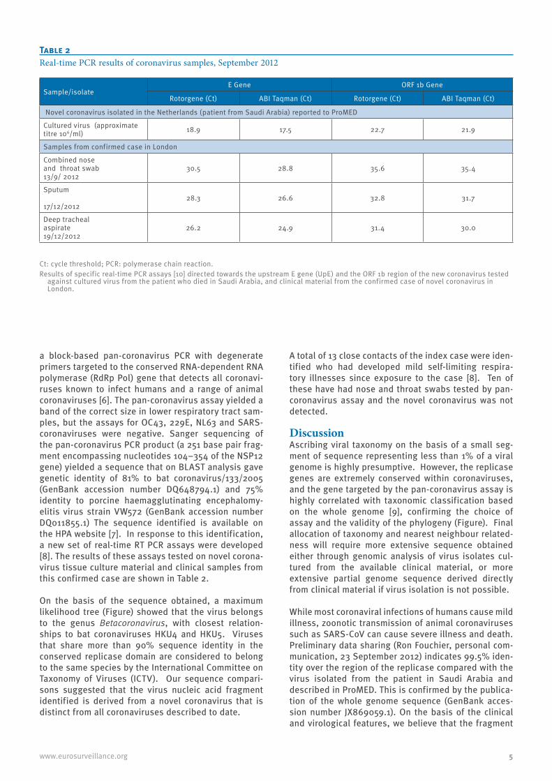
a block-based pan-coronavirus PCR with degenerate primers targeted to the conserved RNA-dependent RNA polymerase (RdRp Pol) gene that detects all coronavi-ruses known to infect humans and a range of animal coronaviruses [6]. The pan-coronavirus assay yielded a band of the correct size in lower respiratory tract sam-ples, but the assays for OC43, 229E, NL63 and SARS-coronaviruses were negative. Sanger sequencing of the pan-coronavirus PCR product (a 251 base pair frag-ment encompassing nucleotides 104–354 of the NSP12 gene) yielded a sequence that on BLAST analysis gave genetic identity of 81% to bat coronavirus/133/2005 (GenBank accession number DQ648794.1) and 75% identity to porcine haemagglutinating encephalomy-elitis virus strain VW572 (GenBank accession number DQ011855.1) The sequence identified is available on the HPA website [7]. In response to this identification, a new set of real-time RT PCR assays were developed [8]. The results of these assays tested on novel corona-virus tissue culture material and clinical samples from this confirmed case are shown in Table 2.
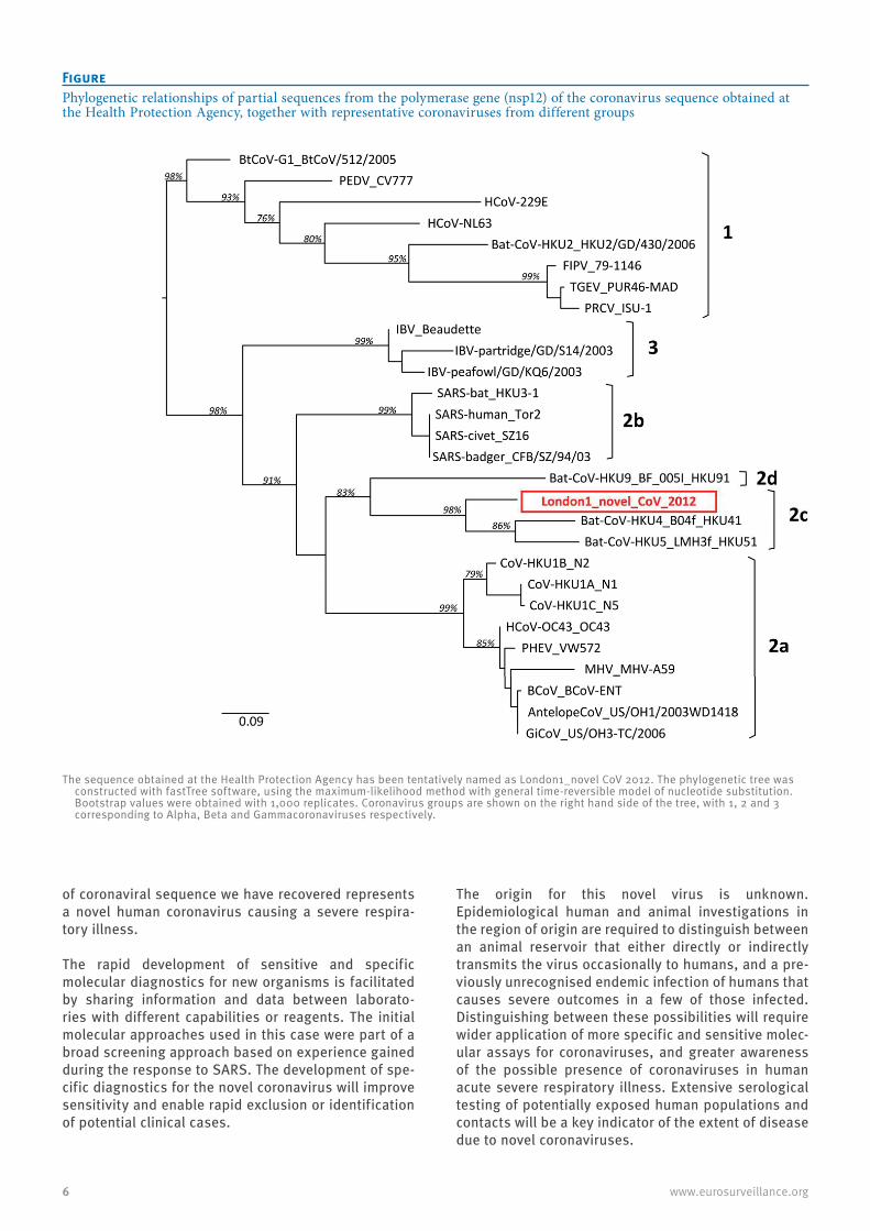
On the basis of the sequence obtained, a maximum likelihood tree (Figure) showed that the virus belongs to the genus Betacoronavirus, with closest relation-ships to bat coronaviruses HKU4 and HKU5. Viruses that share more than 90% sequence identity in the conserved replicase domain are considered to belong to the same species by the International Committee on Taxonomy of Viruses (ICTV). Our sequence compari-sons suggested that the virus nucleic acid fragment identified is derived from a novel coronavirus that is distinct from all coronaviruses described to date.
A total of 13 close contacts of the index case were iden-tified who had developed mild self-limiting respira-tory illnesses since exposure to the case [8]. Ten of these have had nose and throat swabs tested by pan-coronavirus assay and the novel coronavirus was not detected.
DiscussionAscribing viral taxonomy on the basis of a small seg-ment of sequence representing less than 1% of a viral genome is highly presumptive. However, the replicase genes are extremely conserved within coronaviruses, and the gene targeted by the pan-coronavirus assay is highly correlated with taxonomic classification based on the whole genome [9], confirming the choice of assay and the validity of the phylogeny (Figure). Final allocation of taxonomy and nearest neighbour related-ness will require more extensive sequence obtained either through genomic analysis of virus isolates cul-tured from the available clinical material, or more extensive partial genome sequence derived directly from clinical material if virus isolation is not possible.
While most coronaviral infections of humans cause mild illness, zoonotic transmission of animal coronaviruses such as SARS-CoV can cause severe illness and death. Preliminary data sharing (Ron Fouchier, personal com-munication, 23 September 2012) indicates 99.5% iden-tity over the region of the replicase compared with the virus isolated from the patient in Saudi Arabia and described in ProMED. This is confirmed by the publica-tion of the whole genome sequence (GenBank acces-sion number JX869059.1). On the basis of the clinical and virological features, we believe that the fragment
Sample/isolateE Gene ORF 1b Gene
Rotorgene (Ct) ABI Taqman (Ct) Rotorgene (Ct) ABI Taqman (Ct)
Novel coronavirus isolated in the Netherlands (patient from Saudi Arabia) reported to ProMED
Cultured virus (approximate titre 106/ml) 18.9 17.5 22.7 21.9
Samples from confirmed case in London
Combined nose and throat swab 13/9/ 2012
30.5 28.8 35.6 35.4
Sputum 17/12/2012
28.3 26.6 32.8 31.7
Deep tracheal aspirate19/12/2012
26.2 24.9 31.4 30.0
Ct: cycle threshold; PCR: polymerase chain reaction.Results of specific real-time PCR assays [10] directed towards the upstream E gene (UpE) and the ORF 1b region of the new coronavirus tested
against cultured virus from the patient who died in Saudi Arabia, and clinical material from the confirmed case of novel coronavirus in London.
Table 2Real-time PCR results of coronavirus samples, September 2012
6 www.eurosurveillance.org
of coronaviral sequence we have recovered represents a novel human coronavirus causing a severe respira-tory illness.
The rapid development of sensitive and specific molecular diagnostics for new organisms is facilitated by sharing information and data between laborato-ries with different capabilities or reagents. The initial molecular approaches used in this case were part of a broad screening approach based on experience gained during the response to SARS. The development of spe-cific diagnostics for the novel coronavirus will improve sensitivity and enable rapid exclusion or identification of potential clinical cases.
The origin for this novel virus is unknown. Epidemiological human and animal investigations in the region of origin are required to distinguish between an animal reservoir that either directly or indirectly transmits the virus occasionally to humans, and a pre-viously unrecognised endemic infection of humans that causes severe outcomes in a few of those infected. Distinguishing between these possibilities will require wider application of more specific and sensitive molec-ular assays for coronaviruses, and greater awareness of the possible presence of coronaviruses in human acute severe respiratory illness. Extensive serological testing of potentially exposed human populations and contacts will be a key indicator of the extent of disease due to novel coronaviruses.
Figure Phylogenetic relationships of partial sequences from the polymerase gene (nsp12) of the coronavirus sequence obtained at the Health Protection Agency, together with representative coronaviruses from different groups
The sequence obtained at the Health Protection Agency has been tentatively named as London1_novel CoV 2012. The phylogenetic tree was constructed with fastTree software, using the maximum-likelihood method with general time-reversible model of nucleotide substitution. Bootstrap values were obtained with 1,000 replicates. Coronavirus groups are shown on the right hand side of the tree, with 1, 2 and 3 corresponding to Alpha, Beta and Gammacoronaviruses respectively.
7www.eurosurveillance.org
References1. Tyrrell DA, Bynoe ML. Cultivation of a novel type of common-
cold virus in organ cultures. Br Med J. 1965;1(5448):1467-70. 2. Shi Z, Hu Z. A review of studies on animal reservoirs of the
SARS coronavirus. Virus Res. 2008;133(1):74-87. 3. Anderson LJ, Tong S. Update on SARS research and other
possibly zoonotic coronaviruses. Int J Antimicrob Agents. 2010;36 Suppl 1:S21-5.
4. World Health Organization (WHO). Summary table of SARS cases by country, 1 November 2002 - 7 August 2003. Geneva: WHO; 15 Aug 2003. Available from: http://www.who.int/csr/sars/country/2003_08_15/en/index.html
5. ProMED mail. Novel coronavirus - Saudi Arabia: human isolate. Archive Number: 20120920.1302733. Available from: http://www.promedmail.org/?p=2400:1000
6. Bermingham A, Heinen P, Iturriza-Gómara M, Gray J, Appleton H, Zambon MC. Laboratory diagnosis of SARS. Philos Trans R Soc Lond B Biol Sci. 2004;359(1447):1083-9.
7. Health Protection Agency (HPA). Partial genetic sequence information for scientists about the novel coronavirus 2012. London: HPA. [Accessed 2 Oct 2012]. Available from: http://www.hpa.org.uk/Topics/InfectiousDiseases/InfectionsAZ/NovelCoronavirus2012/respPartialgeneticsequenceofnovelcoronavirus/
8. Pebody RG, Chand MA, Thomas HL, Green HK, Boddington NL, Carvalho C, et al. The United Kingdom public health response to an imported laboratory confirmed case of a novel coronavirus in September 2012. Euro Surveill. 2012;17(40):pii=20292. Available from: http://www.eurosurveillance.org/ViewArticle.aspx?ArticleId=20292
9. Drexler JF, Gloza-Rausch F, Glende J, Corman VM, Muth D, Goettsche M, et al. Genomic characterization of severe acute respiratory syndrome-related coronavirus in European bats and classification of coronaviruses based on partial RNA-dependent RNA polymerase gene sequences J Virol. 2010;84(21):11336-49.
10. Corman VM, Eckerle I, Bleicker T, Zaki A, Landt O, Eschbach-Bludau M, et al. Detection of a novel human coronavirus by real-time reverse-transcription polymerase chain reaction. Euro Surveill. 2012;17(39):pii=20285. Available online: http://www.eurosurveillance.org/ViewArticle.aspx?ArticleId=20285
8 www.eurosurveillance.org
Rapid communications
The United Kingdom public health response to an imported laboratory confirmed case of a novel coronavirus in September 2012
R G Pebody ([email protected])1, M A Chand1, H L Thomas1,2,3, H K Green1, N L Boddington1, C Carvalho1,3, C S Brown1,4, S R Anderson1, C Rooney1, E Crawley-Boevey1, D J Irwin1, E Aarons5, C Tong5, W Newsholme5, N Price5, C Langrish5, D Tucker5, H Zhao1, N Phin1, J Crofts1, A Bermingham1, E Gilgunn-Jones1, K E Brown1, B Evans1, M Catchpole1, J M Watson1
1. Health Protection Agency (HPA), London, United Kingdom2. Field Epidemiology Training Programme (FETP), Health Protection Agency, London, United Kingdom3. European Programme for Intervention Epidemiology Training (EPIET), European Centre for Disease Prevention and Control,
(ECDC), Stockholm, Sweden4. Centre for Clinical Infection and Diagnostics Research, King’s College London, London, England5. Guy’s and St Thomas’ NHS Foundation Trust and King’s Health Partners, London, United Kingdom
Citation style for this article: Pebody RG, Chand MA, Thomas HL, Green HK, Boddington NL, Carvalho C, Brown CS, Anderson SR, Rooney C, Crawley-Boevey E, Irwin DJ, Aarons E, Tong C, Newsholme W, Price N, Langrish C, Tucker D, Zhao H, Phin N, Crofts J, Bermingham A, Gilgunn-Jones E, Brown KE, Evans B, Catchpole M, Watson JM. The United Kingdom public health response to an imported laboratory confirmed case of a novel coronavirus in September 2012. Euro Surveill. 2012;17(40):pii=20292. Available online: http://www.eurosurveillance.org/ViewArticle.aspx?ArticleId=20292
Article submitted on 27 September 2012 / published on 4 October 2012
On 22 September 2012, a novel coronavirus, very closely related to that from a fatal case in Saudi Arabia three months previously, was detected in a previously well adult transferred to intensive care in London from Qatar with severe respiratory illness. Strict respiratory isolation was instituted. Ten days after last exposure, none of 64 close contacts had developed severe dis-ease, with 13 of 64 reporting mild respiratory symp-toms. The novel coronavirus was not detected in 10 of 10 symptomatic contacts tested.
The outbreak of Severe Acute Respiratory Syndrome (SARS) in 2003, which led to 8,422 cases and 916 deaths worldwide [1], highlighted the potential for newly emerging zoonotic coronaviruses to transmit from person to person, especially in healthcare set-tings, and to cause severe human illness.
On 22 September 2012, the Health Protection Agency (HPA) in London, United Kingdom (UK), confirmed infec-tion with a novel coronavirus in a patient in a London hospital who had been transferred from Qatar 11 days previously. This patient represents the second con-firmed case of severe acute respiratory illness caused by this novel coronavirus. The first case was identified in a Saudi Arabian national who died in June 2012 [2,3]. We describe the exposure history, the public health response and follow-up of close contacts of the case in London.
Case exposure history and laboratory investigationsThe case is a previously well 49 year-old male, who travelled to Saudi Arabia from 31 July to 18 August 2012, where he, and several of his travelling compan-ions, developed rhinorrhoea and fever (Figure 1). On 18 August he travelled to Qatar, where his respiratory
symptoms resolved three days later. While in Qatar, he spent time on a farm, where he keeps camels and sheep, although no direct contact with these animals was reported.
On 3 September, he reported a mild respiratory ill-ness. Six days later, he required hospitalisation due to development of bilateral pneumonia. His condition worsened and he subsequently required intubation and ventilation. On 12 September, he was transferred by air ambulance to an intensive care unit in London, where acute renal impairment was also detected. Due to further deterioration, he was transferred to another London hospital on 20 September [3].
Following the report on proMED on 20 September 2012 [2] of the detection of a novel coronavirus (until fur-ther taxonomic denomination herewith referred to as hCoV-EMC) in a Saudi Arabian patient who had died from severe respiratory illness and renal failure, and as no diagnosis had been established despite investi-gations for common causes of pneumonia and patho-gens endemic to the Middle East, the patient in London was investigated for novel coronavirus infection. On 21 September, a coronavirus was detected in respira-tory tract samples using a pan-coronavirus PCR assay, and on 22 September sequencing of the PCR amplicon showed a sequence very closely related to the hCoV-EMC detected in the earlier patient from Saudi Arabia [4]. The virus belongs to the genus beta-coronavirus, with closest relationship to bat coronaviruses [4].
Public health managementThe identification of a novel coronavirus of the same group as the SARS-CoV, with two clinically severe human cases including one fatality, led to a public health response being mounted to isolate the case,

9www.eurosurveillance.org
identify and test close contacts and to prevent onward transmission. Once the patient was found to have a novel coronavirus infection, he was isolated in a nega-tive-pressure single room, and full personal protective equipment (PPE), including gowns, gloves, eye protec-tion and high filtration masks were worn by staff and other contacts. Interim case and close contact defini-tions were developed [5].
A possible case was defined as any person with acute respiratory syndrome which includes fever (≥38º C) or history of fever and cough requiring hospitalisation or with suspicion of lower airway involvement (clinical or radiological evidence of consolidation) not explained by another infection or aetiology with history of either travel to or residence in Saudi Arabia or Qatar or close contact with a confirmed case in the ten days before onset of illness
A close contact was defined as the following persons
• Healthcare and social care workers: worker who pro-vided direct clinical or personal care or examination of a symptomatic confirmed case or within close vicinity of an aerosol generating procedure AND who was not wearing full personal protective equipment (PPE) at the time. Full PPE is defined as correctly fit-ted high filtration mask (FFP3), gown, gloves and eye protection.
• Household: any person who has had prolonged face-to-face contact with the confirmed case(s) any time during the illness after onset in a household setting.
• Other close contacts: any person who has had pro-longed face-to-face contact with a confirmed case while symptomatic in any other enclosed setting and not wearing a mask e.g. school, visitor to the hospi-tal to the bed side of a symptomatic confirmed case.
These definitions were used as the basis for identify-ing further possible cases and contacts. Guidelines were developed on the investigation and public health management of these cases and their close contacts.
Identification and follow-up of individuals who had close contact with the case at any time during his symptomatic period from entry into the UK up until implementation of full isolation on 21 September (including healthcare workers and family), was rapidly initiated by HPA staff and staff from the London hos-pitals’ Infection Control Teams. Close contacts were followed up for a period of 10 days since the date of last exposure to the index case. If contacts developed respiratory illness in this period, they were asked to self-isolate in their homes (or were isolated in hospital if requiring admission).
The hospital in Qatar was informed to allow them to ini-tiate appropriate follow-up for those who had been in contact with the patient.
HPA rapidly developed and published advice to health professionals, the public and travellers [5]. The case was immediately reported under the International Health Regulations to the World Health Organisation and through the European Union Early Warning and Response System (EWRS). Extensive laboratory work was undertaken to characterise the virus and develop new diagnostic tools [3].
Initial epidemiological investigation and preliminary findingsClose contacts of the case were followed up to deter-mine the transmissibility of this novel coronavirus. This included collection of information on clinical illness, virological swabbing of contacts they had
Figure 1Timeline of disease and travel history of novel coronavirus case, London, August-September 2012
AugustJuly September
Patient
Location
Time
Saudi Arabia Qatar b
Hospitalised
United Kingdom (London)
Air ambulanceRegular flight
Mildly ill a Clinically well Clinically well
Ventilation required
Mildly ill
ECMOrequired
Onset 1 (rhinorrhea, fever)
Onset 2 (cough, arthralgia)
31 1 2 3 4 5 6 7 8 9 10 11 12 13 14 15 16 17 18 19 20 21 22 23 24 25 26 27 28 29 30 31 1 2 3 4 5 6 7 8 9 10 11 12 13 14 15 16 17 18 19 20 21 22 23 24 25 26
ECMO: Extracorporeal Membrane Oxygenation.
a According to relatives of the patient.b Contact with farm animals during stay (camels, sheep).
10 www.eurosurveillance.org
respiratory symptoms and collection of paired sera from all contacts to determine if there was evidence of recent infection.
It is likely that the patient’s infection was acquired in Qatar as he was in Qatar for the 16 days prior to the onset of his most recent respiratory illness in September. The earlier mild upper respiratory tract infection, which began during his visit to Saudi Arabia, resolved two weeks before onset of the present illness.
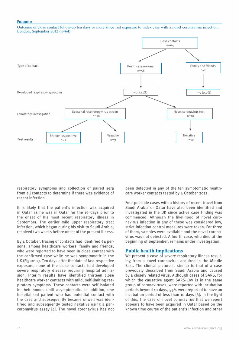
By 4 October, tracing of contacts had identified 64 per-sons, among healthcare workers, family and friends, who were reported to have been in close contact with the confirmed case while he was symptomatic in the UK (Figure 2). Ten days after the date of last respective exposure, none of the close contacts had developed severe respiratory disease requiring hospital admis-sion. Interim results have identified thirteen close healthcare worker contacts with mild, self-limiting res-piratory symptoms. These contacts were self-isolated in their homes until asymptomatic. In addition, one hospitalised patient who had potential contact with the case and subsequently became unwell was iden-tified and subsequently tested negative using a pan-coronavirus assay [4]. The novel coronavirus has not
been detected in any of the ten symptomatic health-care worker contacts tested by 4 October 2012.
Four possible cases with a history of recent travel from Saudi Arabia or Qatar have also been identified and investigated in the UK since active case finding was commenced. Although the likelihood of novel coro-navirus infection in any of these was considered low, strict infection control measures were taken. For three of them, samples were available and the novel corona-virus was not detected. A fourth case, who died at the beginning of September, remains under investigation.
Public health implications We present a case of severe respiratory illness result-ing from a novel coronavirus acquired in the Middle East. The clinical picture is similar to that of a case previously described from Saudi Arabia and caused by a closely related virus. Although cases of SARS, for which the causative agent SARS-CoV is in the same group of coronaviruses, were reported with incubation periods beyond 10 days, 95% were reported to have an incubation period of less than 10 days [6]. In the light of this, the case of novel coronavirus that we report appears to have been acquired in Qatar based on the known time course of the patient’s infection and other
Figure 2Outcome of close contact follow-up ten days or more since last exposure to index case with a novel coronavirus infection, London, September 2012 (n=64)
Close contacts n=64
Family and friends n=8
Healthcare workers n=56
n=13 (23%) n=0 (0.0%)
Seasonal respiratory virus screen n=10
Novel coronavirus test n=10
Negative n=9
Rhinovirus positive n=1
Negative n=10
Type of contact
Developed respiratory symptoms
Laboratory investigation
Test results
11www.eurosurveillance.org
available information, unless the illness had an unu-sual biphasic nature or a very long incubation period. After 10 days of follow-up, there has been no confirmed evidence of ongoing person-to-person transmission resulting in severe disease or milder laboratory con-firmed infection among close contacts, despite exten-sive active contact tracing. Completion of case-contact investigation, including serological testing when avail-able, will determine whether mild or asymptomatic infection among close contacts has occurred. In addi-tion, serological investigation in the countries of origin of the two confirmed cases should be considered to look for evidence of possible previous infection in the general population. Studies in animals are also neces-sary to determine whether there is an animal reservoir for this infection and what it might be.
Early detection and investigation of cases of severe respiratory illness among travellers returning from countries where infection with novel coronavirus has been reported and their close contacts will support the further elucidation of the epidemiological characteris-tics of this novel virus. An outbreak of severe respira-tory illness of unknown aetiology was reported from the Middle East earlier in 2012 [7]. Work needs to be undertaken to determine if a novel coronavirus has been circulating more widely in the general population in the Middle East already for some time or if the virus was more recently introduced from an unknown animal reservoir.
References1. World Health Organization (WHO). WHO final summary
SARS, 15 August 2003: Summary table of SARS cases by country, 1 November 2002 - 7 August 2003. Geneva; WHO; 2003. Avaliable from: http://www.who.int/csr/sars/country/2003_08_15/en/index.html
2. ProMED-mail. Novel coronavirus - Saudi Arabia: human isolate. Archive Number: 20120920.1302733. September 20 September 2012. Available from: http://www.promedmail.org/?p=2400:1000
3. Corman VM, Eckerle I, Bleicker T, Zaki A, Landt O, Eschbach-Bludau M, et al. Detection of a novel human coronavirus by real-time reverse-transcription polymerase chain reaction. Euro Surveill. 2012;17(39):pii=20285. Available from: http://www.eurosurveillance.org/ViewArticle.aspx?ArticleId=20285
4. Bermingham A, Chand MA, Brown CS, Aarons E, Tong C, Langrish C, et al. Severe respiratory illness caused by a novel coronavirus, in a patient transferred to the United Kingdom from the Middle East, September 2012. Euro Surveill. 2012;17(40):pii=20290. Available from: http://www.eurosurveillance.org/ViewArticle.aspx?ArticleId=20290
5. Health protection Agency (HPA). Algorithm for investigation and management of possible cases of severe acute respiratory illness associated with a novel coronavirus. London; HPA; 2012. Available from: http://www.hpa.org.uk/webw/HPAweb&Page&HPAwebAutoListName/Page/1317136202637
6. Lessler J, Reich NG, Brookmeyer R, Perl TM, Nelson KE, Cummings DA. Incubation periods of acute respiratory viral infections: a systematic review. Lancet Infect Dis. 2009; 9(5):291-300.
7. European Centre for Disease Prevention and Control (ECDC). Communicable Disease Threats Report (Week 18, 29 April-5 May 2012). Stockholm: ECDC; 2012. Available from: http://ecdc.europa.eu/en/publications/Publications/CDTR%20online%20version%204%20May%202012.pdf
12 www.eurosurveillance.org
Rapid communications
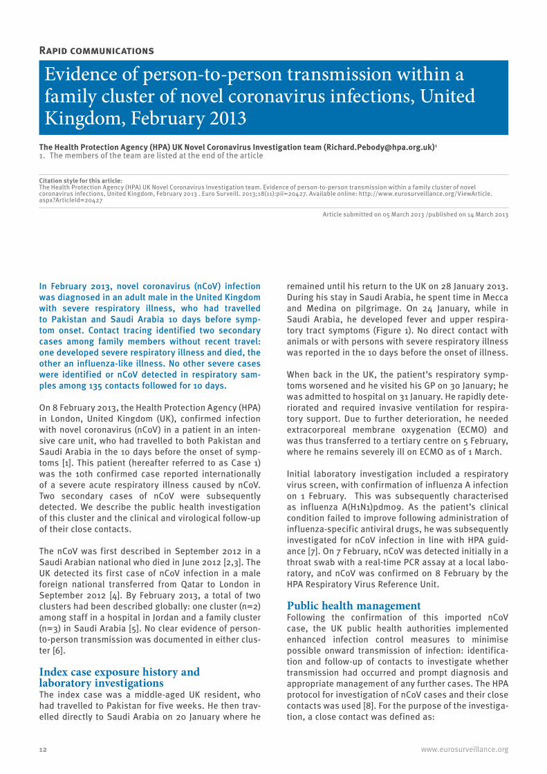
Evidence of person-to-person transmission within a family cluster of novel coronavirus infections, United Kingdom, February 2013
The Health Protection Agency (HPA) UK Novel Coronavirus Investigation team ([email protected])1
1. The members of the team are listed at the end of the article
Citation style for this article: The Health Protection Agency (HPA) UK Novel Coronavirus Investigation team. Evidence of person-to-person transmission within a family cluster of novel coronavirus infections, United Kingdom, February 2013 . Euro Surveill. 2013;18(11):pii=20427. Available online: http://www.eurosurveillance.org/ViewArticle.aspx?ArticleId=20427
Article submitted on 05 March 2013 /published on 14 March 2013
In February 2013, novel coronavirus (nCoV) infection was diagnosed in an adult male in the United Kingdom with severe respiratory illness, who had travelled to Pakistan and Saudi Arabia 10 days before symp-tom onset. Contact tracing identified two secondary cases among family members without recent travel: one developed severe respiratory illness and died, the other an influenza-like illness. No other severe cases were identified or nCoV detected in respiratory sam-ples among 135 contacts followed for 10 days.
On 8 February 2013, the Health Protection Agency (HPA) in London, United Kingdom (UK), confirmed infection with novel coronavirus (nCoV) in a patient in an inten-sive care unit, who had travelled to both Pakistan and Saudi Arabia in the 10 days before the onset of symp-toms [1]. This patient (hereafter referred to as Case 1) was the 10th confirmed case reported internationally of a severe acute respiratory illness caused by nCoV. Two secondary cases of nCoV were subsequently detected. We describe the public health investigation of this cluster and the clinical and virological follow-up of their close contacts.
The nCoV was first described in September 2012 in a Saudi Arabian national who died in June 2012 [2,3]. The UK detected its first case of nCoV infection in a male foreign national transferred from Qatar to London in September 2012 [4]. By February 2013, a total of two clusters had been described globally: one cluster (n=2) among staff in a hospital in Jordan and a family cluster (n=3) in Saudi Arabia [5]. No clear evidence of person-to-person transmission was documented in either clus-ter [6].
Index case exposure history and laboratory investigationsThe index case was a middle-aged UK resident, who had travelled to Pakistan for five weeks. He then trav-elled directly to Saudi Arabia on 20 January where he
remained until his return to the UK on 28 January 2013. During his stay in Saudi Arabia, he spent time in Mecca and Medina on pilgrimage. On 24 January, while in Saudi Arabia, he developed fever and upper respira-tory tract symptoms (Figure 1). No direct contact with animals or with persons with severe respiratory illness was reported in the 10 days before the onset of illness.
When back in the UK, the patient’s respiratory symp-toms worsened and he visited his GP on 30 January; he was admitted to hospital on 31 January. He rapidly dete-riorated and required invasive ventilation for respira-tory support. Due to further deterioration, he needed extracorporeal membrane oxygenation (ECMO) and was thus transferred to a tertiary centre on 5 February, where he remains severely ill on ECMO as of 1 March.
Initial laboratory investigation included a respiratory virus screen, with confirmation of influenza A infection on 1 February. This was subsequently characterised as influenza A(H1N1)pdm09. As the patient’s clinical condition failed to improve following administration of influenza-specific antiviral drugs, he was subsequently investigated for nCoV infection in line with HPA guid-ance [7]. On 7 February, nCoV was detected initially in a throat swab with a real-time PCR assay at a local labo-ratory, and nCoV was confirmed on 8 February by the HPA Respiratory Virus Reference Unit.
Public health managementFollowing the confirmation of this imported nCoV case, the UK public health authorities implemented enhanced infection control measures to minimise possible onward transmission of infection: identifica-tion and follow-up of contacts to investigate whether transmission had occurred and prompt diagnosis and appropriate management of any further cases. The HPA protocol for investigation of nCoV cases and their close contacts was used [8]. For the purpose of the investiga-tion, a close contact was defined as:
13www.eurosurveillance.org
•Aeroplane setting: the aircraft passengers in the same row and the two rows in front and behind a symptomatic case;
•Household setting: any person who had prolonged (>15 minutes) face-to-face contact with the confirmed case(s) any time during the illness in a household setting;
•Healthcare setting: either (i) a worker who provided direct clinical or personal care to or examined a symp-tomatic confirmed case or was within close vicinity of an aerosol-generating procedure AND who was not wearing full personal protective equipment (PPE) at the time; or (ii) a visitor to the hospital who was not wearing PPE at the bedside of a confirmed case; full PPE was defined as correctly fitted high filtration mask (FFP3), gown, gloves and eye protection;
•Other setting: any person who had prolonged (>15 minutes) face-to-face contact with a confirmed symp-tomatic case in any other enclosed setting.
Identification and follow-up of individuals who had close contact with the index case from entry into the UK at any time during his symptomatic period was rapidly initiated by the HPA together with staff from the two hospitals the patient had attended (includ-ing the Infection Prevention and Control Teams and Occupational Health).
Close contacts were followed up for a minimum period of 10 days after last exposure to the index case. Following the identification of two secondary nCoV cases among symptomatic family contacts of the index case, contact tracing was initiated for their respec-tive additional contacts. Follow-up included collection of information on the date and setting of contact with the index case, PPE use (healthcare workers) and any symptoms of respiratory infection in the 10 days after
last exposure. Contacts who developed any symptoms of acute respiratory infection in this period were asked to self-isolate in their homes (or were isolated in hospi-tal if admitted) until asymptomatic.
The airline provided details of passengers to the HPA to allow follow-up of those persons in the same row as the case and the two adjacent rows to the patient as per World Health Organization (WHO) guidance for severe acute respiratory syndrome (SARS) [9]. Passengers who were in the UK were followed up by the HPA to inform them of the potential exposure and determine whether they had developed symptoms of acute respir-atory illness in the 10 days post exposure. UK authori-ties informed relevant overseas national authorities directly about non-UK resident contacts on the flight through International Health Regulation mechanisms.
Laboratory investigationSymptomatic contacts had respiratory samples taken (nose and throat swab, and sputum if they had a pro-ductive cough) for testing for a panel of respiratory viruses (influenza virus, respiratory syncytial virus, parainfluenza virus types 1,2,3 and 4, adenovirus, rhinovirus, human metapneumovirus) and for nCoV. Criteria for laboratory confirmation of nCoV were Up E real-time PCR detection in two different laboratories [3] and detection of two other regions of the nCoV genome [3, HPA unpublished data].
In addition, nose and throat swabs were taken from a group of asymptomatic contacts of the three confirmed cases for nCoV testing to determine if there was evi-dence of asymptomatic carriage.
Paired serum samples are being taken from all house-hold and healthcare contacts regardless of symptoms
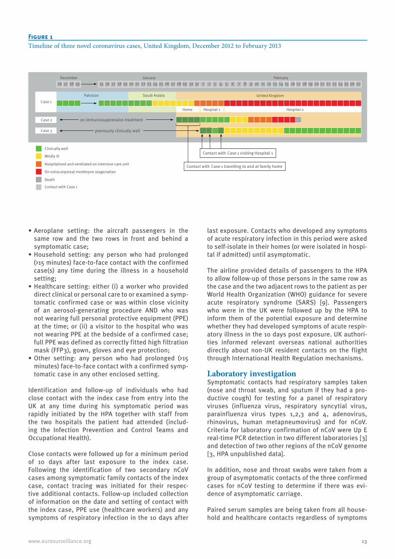
Figure 1Timeline of three novel coronavirus cases, United Kingdom, December 2012 to February 2013
Case 2
Case 3
Clinically well
Mildly ill
Hospitalised and ventilated on intensive care unit
On extracorporeal membrane oxygenation
Death
Contact with Case 1
December January February
Pakistan United Kingdom
Home Hospital 1 Hospital 2
Saudi Arabia
on immunosuppression treatment
previously clinically well
Contact with Case 1 travelling to and at family home
Contact with Case 1 visiting Hospital 1
Case 1
16 17 18 19 15 16 17 18 19 20 21 22 23 24 25 26 27 28 29 30 31 1 2 3 4 5 6 7 8 9 10 11 12 13 14 15 16 17 18 19 20 21 22 23 24 25 26 27
14 www.eurosurveillance.org
with the initial sample taken within seven days of last exposure and the second at least 21 days after the first. Once collected, samples will be tested for sero-logical reactivity to nCoV.
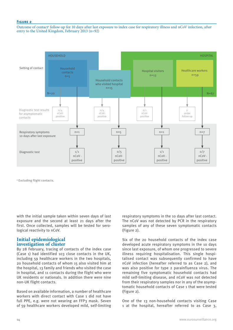
Initial epidemiological investigation of clusterBy 28 February, tracing of contacts of the index case (Case 1) had identified 103 close contacts in the UK, including 59 healthcare workers in the two hospitals, 20 household contacts of whom 15 also visited him at the hospital, 13 family and friends who visited the case in hospital, and 11 contacts during the flight who were UK residents or nationals. In addition there were nine non-UK flight contacts.
Based on available information, a number of healthcare workers with direct contact with Case 1 did not have full PPE, e.g. were not wearing an FFP3 mask. Seven of 59 healthcare workers developed mild, self-limiting
respiratory symptoms in the 10 days after last contact. The nCoV was not detected by PCR in the respiratory samples of any of these seven symptomatic contacts (Figure 2).
Six of the 20 household contacts of the index case developed acute respiratory symptoms in the 10 days since last exposure, of whom one progressed to severe illness requiring hospitalisation. This single hospi-talised contact was subsequently confirmed to have nCoV infection (hereafter referred to as Case 2), and was also positive for type 2 parainfluenza virus. The remaining five symptomatic household contacts had mild self-limiting disease, and nCoV was not detected from their respiratory samples nor in any of the asymp-tomatic household contacts of Case 1 that were tested (Figure 2).
One of the 13 non-household contacts visiting Case 1 at the hospital, hereafter referred to as Case 3,
Figure 2Outcome of contacta follow-up for 10 days after last exposure to index case for respiratory illness and nCoV infection, after entry to the United Kingdom, February 2013 (n=92)
Hospital visitors n=13
Diagnostic test results for asymptomatic contacts
0/4nCoV-
positive
0/1nCoV-
positive
2 under
follow-up
Healthcare workers
n=59
HOUSEHOLD
N=20
HOSPITAL
N=87
0/4nCoV-
positive
Setting of contact
n=1 n=1
Diagnostic test
n=5
1/1 nCoV -
positive
0/5 nCoV-
positive
1/1 nCoV - -
positive
n=7
0/7 nCoV
positive
Respiratory symptoms 10 days after last exposure
Household contacts who visited hospital
n=15
Household contacts
n=5
a Excluding flight contacts.
15www.eurosurveillance.org
developed an acute mild, respiratory illness, and nCoV was detected in a respiratory sample, as was type 2 parainfluenza virus.
Two of the 11 UK-based passengers reported respira-tory symptoms: one had recovered by the time of inter-view and did not have respiratory samples taken. In the other, nCoV was not detected from respiratory samples.
The periods of exposure of Case 2 and Case 3 to Case 1 and the timelines of their illnesses are represented in Figure 1.
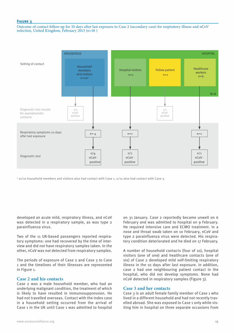
Case 2 and his contactsCase 2 was a male household member, who had an underlying malignant condition, the treatment of which is likely to have resulted in immunosuppression. He had not travelled overseas. Contact with the index case in a household setting occurred from the arrival of Case 1 in the UK until Case 1 was admitted to hospital
on 31 January. Case 2 reportedly became unwell on 6 February and was admitted to hospital on 9 February. He required intensive care and ECMO treatment. In a nose and throat swab taken on 10 February, nCoV and type 2 parainfluenza virus were detected. His respira-tory condition deteriorated and he died on 17 February.
A number of household contacts (four of 10), hospital visitors (one of one) and healthcare contacts (one of six) of Case 2 developed mild self-limiting respiratory illness in the 10 days after last exposure. In addition, case 2 had one neighbouring patient contact in the hospital, who did not develop symptoms. None had nCoV detected in respiratory samples (Figure 3).
Case 3 and her contactsCase 3 is an adult female family member of Case 1 who lived in a different household and had not recently trav-elled abroad. She was exposed to Case 1 only while vis-iting him in hospital on three separate occasions from
Figure 3Outcome of contact follow-up for 10 days after last exposure to Case 2 (secondary case) for respiratory illness and nCoV infection, United Kingdom, February 2013 (n=18 )
Hospital visitors n=1
n= 4 n=1
0/4 nCoV-positive
0/1 nCoV -
positive
n=1
0/1 nCoV -
positive
HOUSEHOLD
HOSPITAL
N=8
Diagnostic test
Setting of contact
Respiratory symptoms 10 days after last exposure
Diagnostic test results for asymptomatic contacts
0/5nCoV-
positive
0/1nCoV-
positive
Fellow patientn=1
Household members
and visitors n=10a
Healthcare workers
n=6
a 10/10 household members and visitors also had contact with Case 1, 2/10 also had contact with Case 3.
16 www.eurosurveillance.org
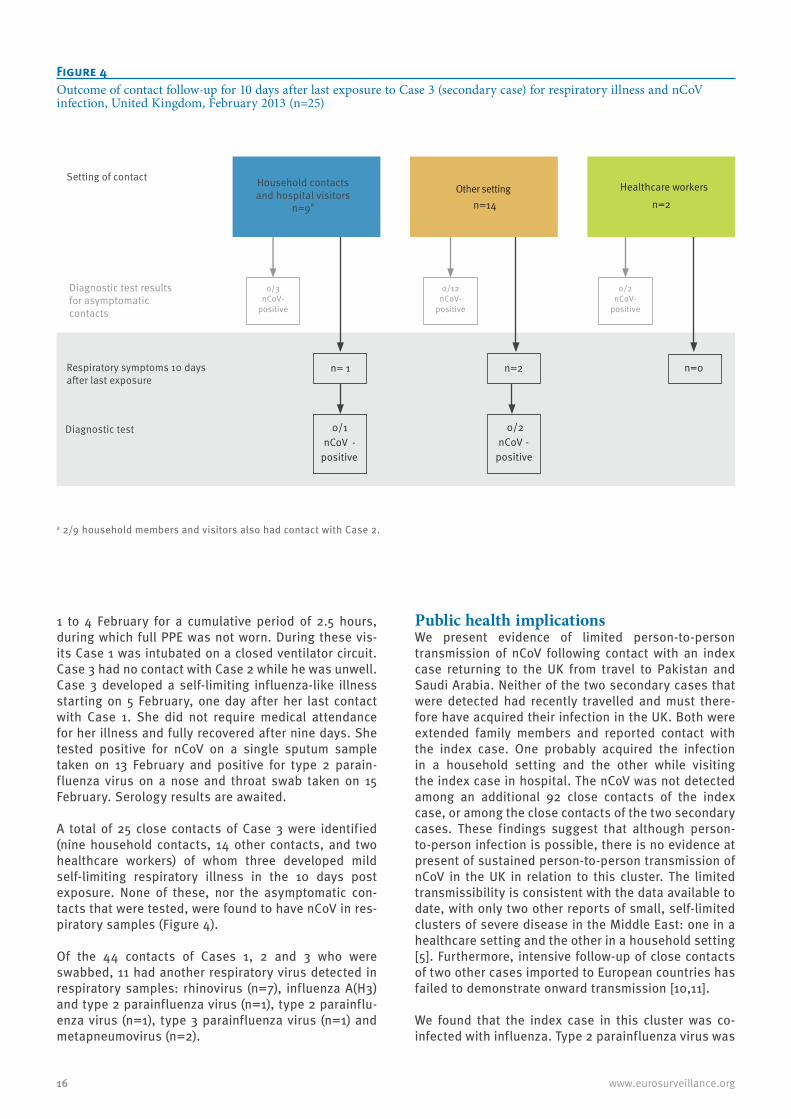
1 to 4 February for a cumulative period of 2.5 hours, during which full PPE was not worn. During these vis-its Case 1 was intubated on a closed ventilator circuit. Case 3 had no contact with Case 2 while he was unwell. Case 3 developed a self-limiting influenza-like illness starting on 5 February, one day after her last contact with Case 1. She did not require medical attendance for her illness and fully recovered after nine days. She tested positive for nCoV on a single sputum sample taken on 13 February and positive for type 2 parain-fluenza virus on a nose and throat swab taken on 15 February. Serology results are awaited.
A total of 25 close contacts of Case 3 were identified (nine household contacts, 14 other contacts, and two healthcare workers) of whom three developed mild self-limiting respiratory illness in the 10 days post exposure. None of these, nor the asymptomatic con-tacts that were tested, were found to have nCoV in res-piratory samples (Figure 4).
Of the 44 contacts of Cases 1, 2 and 3 who were swabbed, 11 had another respiratory virus detected in respiratory samples: rhinovirus (n=7), influenza A(H3) and type 2 parainfluenza virus (n=1), type 2 parainflu-enza virus (n=1), type 3 parainfluenza virus (n=1) and metapneumovirus (n=2).
Public health implications We present evidence of limited person-to-person transmission of nCoV following contact with an index case returning to the UK from travel to Pakistan and Saudi Arabia. Neither of the two secondary cases that were detected had recently travelled and must there-fore have acquired their infection in the UK. Both were extended family members and reported contact with the index case. One probably acquired the infection in a household setting and the other while visiting the index case in hospital. The nCoV was not detected among an additional 92 close contacts of the index case, or among the close contacts of the two secondary cases. These findings suggest that although person-to-person infection is possible, there is no evidence at present of sustained person-to-person transmission of nCoV in the UK in relation to this cluster. The limited transmissibility is consistent with the data available to date, with only two other reports of small, self-limited clusters of severe disease in the Middle East: one in a healthcare setting and the other in a household setting [5]. Furthermore, intensive follow-up of close contacts of two other cases imported to European countries has failed to demonstrate onward transmission [10,11].
We found that the index case in this cluster was co-infected with influenza. Type 2 parainfluenza virus was
Figure 4Outcome of contact follow-up for 10 days after last exposure to Case 3 (secondary case) for respiratory illness and nCoV infection, United Kingdom, February 2013 (n=25)
n= 1 n=0
n=2
0/1 nCoV -
positive
0/2 nCoV -
positive
Diagnostic test
Respiratory symptoms 10 days after last exposure
Diagnostic test results for asymptomatic contacts
0/3nCoV-
positive
0/12nCoV-
positive
0/2nCoV-
positive
Other setting
n=14
Healthcare workers
n=2
Setting of contact Household contacts and hospital visitors
n=9a
a 2/9 household members and visitors also had contact with Case 2.
17www.eurosurveillance.org
detected in the two secondary cases. This raises ques-tions about what roles these other infections might play in relation to nCoV transmissibility and/or the severity of the illness. In addition, as the index case was diag-nosed initially with influenza, this lead to a delay in recognition of nCoV. This highlights the importance of considering a diagnosis of nCoV in atypical cases (in this case the poor response to antiviral drugs), even if a putative alternative diagnosis has already been made. HPA guidance has been adapted accordingly [7].
Although the transmissibility patterns of nCoV and SARS have been different to date, confirmed cases of nCoV reported globally have suggested a clinical picture similar to SARS, in particular the presenta-tion with severe respiratory illness, with nine of the 15 cases reported globally to date having died [12]. Two of the three cases we describe fit this clinical picture: two required ECMO treatment and one of them died. However, the third case presented with an acute self-limiting respiratory infection that did not require hos-pitalisation or medical attention. This first reported case of a milder nCoV illness raises the possibility that the spectrum of clinical disease maybe wider than initially envisaged, and that a significant propor-tion of cases now or in the future might be milder or even asymptomatic. This highlights the importance of intensive contact tracing and virological and serologi-cal follow-up around all confirmed cases of nCoV. The application of recently developed serological assays in one case¬–contact study did not provide evidence of asymptomatic infection, although the contacts inves-tigated were exposed late in the case’s illness, when the viral load might be lower [11]. Paired sera are being gathered from contacts in this current investigation to determine whether there may have been more wide-spread mild or asymptomatic infection.
The fact that the two secondary cases acquired their infection from an imported sporadic case has enabled a preliminary estimation of the incubation and serial intervals. The timing of onset of symptoms in the index and the two secondary cases and of exposure sug-gests a putative incubation period ranging from one to nine days and a serial interval (time between onset of illness in index case and secondary case) of 13 to 14 days. Although the data are extremely limited, the observed upper range of the incubation period is per-haps more similar to that seen for SARS (usual range: two to 10 days) rather than seasonal coronavirus infec-tion (usual range: two to five days) [13]. It is therefore not possible to ascertain with certainty whether the index case acquired his infection in Saudi Arabia or in Pakistan, although previous nCoV cases have been linked to the Middle East. This highlights the impor-tance of gathering more information to determine risk factors for acquisition of infection.
All confirmed nCoV cases detected to date, apart from the two secondary cases in the UK cluster, spent time in the Middle East during the putative incubation
period. This, together with our observations of limited secondary transmission, highlights the importance of ongoing vigilance and rapid investigation of cases of severe respiratory illness in residents of and travellers from that area. Further work is required to determine how widely nCoV is circulating globally. In particular serological investigations are needed on the extent of recent infection in various populations, as well as viro-logical investigation of cases of severe undiagnosed respiratory illness in settings both in and beyond the Middle East.
Acknowledgements We would like to acknowledge Critical Care, Infection Prevention and Control and Occupational Health staff in-volved in patient care, and all health protection staff in-volved in the public health response.
Members of the • HPA West Midlands East Health Protection Unit (HPU):
Mamoona Tahir, Roger Gajraj, Madhu Bardhan, Huda Mohammed, Louise Dyke, Petra Charlemagne, Rea Alves.
• HPA West Midlands West HPU: David Kirrage, Dan Killalea, Kate James, Melinda Kemp.
• HPA West Midlands North HPU: Harsh Duggal, Robert Carr, Musarrat Afza, Nicholas Aigbogun, Bharat Sibal.
• HPA West Midlands Regional Epidemiology Unit: Ruth Harrell, Obaghe Edeghere, Keith Neal.
• HPA West Midlands Regional Director’s Office: Sue Ibbotson.
• Birmingham City Hospital: Nimal Wickramasinghe, Nick Sherwood.
• University Hospitals Birmingham: Beryl Oppenheim, Louise Hopton.
• HPA Specialist Microbiology Network Public Health Laboratory Birmingham: Husam Osman, Erasmus Smit, Sowsan Atabani, Judith Workman, Steve Wilson, Clair Overton-Lewis, Margaret Logan.
• HPA Greater Manchester HPU: Rosemary McCann, Marko Petrovic, Vinay Bothra, William Welfare.
• University Hospital of South Manchester: Barbara Isalska, Julian Barker, Alan Ashworth, Igor Fedor.
• HPA North West London HPU: Claude Seng, Deepti Kumar.• HPA South Yorkshire HPU: Suzanna Matthews.• HPA London Regional Director’s Office: Brian McCloskey.• University of Nottingham: Jonathan Nguyen-Van-Tam.• HPA Health Protection Services – Central Office: Paul
Cosford.• HPA Reference Microbiology Services: Alison Bermingham,
Joanna Ellis, Monica Galiano, Angie Lackenby, Richard Myers, Robin Gopal, Maria Zambon.
• HPA Colindale Health Protection Services: *Richard Pebody, Lucy Thomas, Nicki Boddington, Helen K Green, Hongxin Zhao, Iain Kennedy, Ibrahim Abubakar, Jane Jones, Nick Phin, Mike Catchpole, John M Watson.
Conflict of interestNone declared.
Authors’ contributionsThe HPA HPU and regional teams and NHS hospital teams were responsible for the collection of data and samples
18 www.eurosurveillance.org
on cases and their contacts. The HPA Microbiology service teams were responsible for testing and interpretation of re-sults from respiratory samples. National co-ordination of the investigation including design, data collation and analysis was undertaken by the HPS Colindale team in collaboration with other team members. HPS Colindale were responsible for the initial draft of the article. All co-authors provided comments and approved the final version.
References1. Novel coronavirus 2012 in the UK: situation at 19 February
2013. Health Protection Report. 2013;7(8). Available from: http://www.hpa.org.uk/hpr/archives/2013/hpr0813.pdf
2. ProMED-mail. Novel coronavirus - Saudi Arabia: human isolate. Archive Number: 20120920.1302733. 20 September 2012. Available from: http://www.promedmail.org/?archiveid=302733
3. Corman VM, Eckerle I, Bleicker T, Zaki A, Landt O, Eschbach-Bludau M, et al. Detection of a novel human coronavirus by real-time reverse-transcription polymerase chain reaction. Euro Surveill. 2012;17(39):pii=20285. Available from: http://www.eurosurveillance.org/ViewArticle.aspx?ArticleId=20285
4. Bermingham A, Chand MA, Brown CS, Aarons E, Tong C, Langrish C, et al. Severe respiratory illness caused by a novel coronavirus, in a patient transferred to the United Kingdom from the Middle East, September 2012. Euro Surveill. 2012;17(40):pii=20290. Available from: http://www.eurosurveillance.org/ViewArticle.aspx?ArticleId=20290
5. World Health Organization (WHO). Novel Coronavirus infection – update. Geneva: WHO; 30 Nov 2012. Available from: http://www.who.int/csr/don/2012_11_30/en/index.html
6. European Centre for Disease prevention and Control (ECDC). Rapid risk assessment. Severe respiratory disease associated with a novel coronavirus. Stockholm: ECDC; 19 February 2013. Available from: http://www.ecdc.europa.eu/en/publications/Publications/novel-coronavirus-rapid-risk-assessment-update.pdf
7. Health Protection Agency (HPA). HPS nCoV case algorithm. Version case_v13. London: HPA; 19 Feb 2013. Available from: http://www.hpa.org.uk/webc/HPAwebFile/HPAweb_C/1317136270914
8. Health Protection Agency (HPA). “The First Few Hundred (FF100)”. Enhanced Case and Contact Protocol v4.0. Epidemiological Protocols for Comprehensive Assessment of Early Novel Coronavirus Cases and their close contacts in the United Kingdom. London: HPA. [Accessed: 19 Feb 2013]. Available from: http://www.hpa.org.uk/webc/HPAwebFile/HPAweb_C/1317136300809
9. WHO recommended measures for persons undertaking international travel from areas affected by Severe Acute Respiratory Syndrome (SARS). Wkly Epidemiol Rec. 2003;78(14):97-9. PMid:12723281.
10. Pebody RG, Chand MA, Thomas HL, Green HK, Boddington NL, Carvalho C, et al. The United Kingdom public health response to an imported laboratory confirmed case of a novel coronavirus in September 2012. Euro Surveill. 2012;17(40):pii=20292. Available from: http://www.eurosurveillance.org/ViewArticle.aspx?ArticleId=20292
11. Buchholz U, Müller MA, Nitsche A, Sanewski A, Wevering N, Bauer-Balci T, et al. Contact investigation of a case of human novel coronavirus infection treated in a German hospital, October-November 2012. Euro Surveill. 2013;18(8):pii=20406. Available from: http://www.eurosurveillance.org/ViewArticle.aspx?ArticleId=20406. PMid:23449231.
12. World Health Organization (WHO). Novel coronavirus update. Geneva: WHO; 12 Mar 2013. Available from: http://www.who.int/csr/don/2013_03_12/en/index.html
13. Lessler J, Reich NG, Brookmeyer R, Perl TM, Nelson KE, Cummings DA. Incubation periods of acute respiratory viral infections: a systematic review. Lancet Infect Dis. 2009;9(5):291-300. http://dx.doi.org/10.1016/S1473-3099(09)70069-6.
19www.eurosurveillance.org
Rapid communications
First cases of Middle East Respiratory Syndrome Coronavirus (MERS-CoV) infections in France, investigations and implications for the prevention of human-to-human transmission, France, May 2013
A Mailles ([email protected])1, K Blanckaert2,3, P Chaud2,4, S van der Werf5, B Lina6, V Caro7, C Campese1, B Guéry8, H Prouvost4, X Lemaire9, M C Paty1, S Haeghebaert4, D Antoine1, N Ettahar10, H Noel1, S Behillil5, S Hendricx9, J C Manuguerra7, V Enouf6, G La Ruche1, Caroline Semaille1, B Coignard1, D Lévy-Bruhl1, F Weber1, C Saura1, D Che1, The investigation team11
1. Institut de veille sanitaire (InVS), Saint Maurice, France2. These authors contributed equally to this work3. Antenne Régionale de Lutte contre les Infections Nosocomiales (ARLIN), Lille, France4. Institut de Veille Sanitaire, Lille, France5. National Reference Center for influenza viruses (coordinating center) and Unit of Molecular Genetics of RNA Viruses,
coordinating center, Institut Pasteur, Paris, France6. National Reference Center for influenza viruses, Hospices Civils de Lyon and Virpath, Université Claude Bernard Lyon1,
Lyon, France7. Cellule d’Intervention Biologique d’Urgence (CIBU), Institut Pasteur, Paris, France8. Centre Hospitalier Régional et Universitaire, Université de Lille 2, Lille, France9. Centre hospitalier, Douai, France10. Centre Hospitalier, Valenciennes, France11. The members of the team are listed at the end of the article
Citation style for this article: Mailles A, Blanckaert K, Chaud P, van der Werf S, Lina B, Caro V, Campese C, Guéry B, Prouvost H, Lemaire X, Paty MC, Haeghebaert S, Antoine D, Ettahar N, Noel H, Behillil S, Hendricx S, Manuguerra JC, Enouf V, La Ruche G, Semaille C, Coignard B, Lévy-Bruhl D, Weber F, Saura C, Che D, The investigation team. First cases of Middle East Respiratory Syndrome Coronavirus (MERS-CoV) infections in France, investigations and implications for the prevention of human-to-human transmission, France, May 2013. Euro Surveill. 2013;18(24):pii=20502. Available online: http://www.eurosurveillance.org/ViewArticle.aspx?ArticleId=20502
Article submitted on 29 May 2013 / published on 13 June 2013
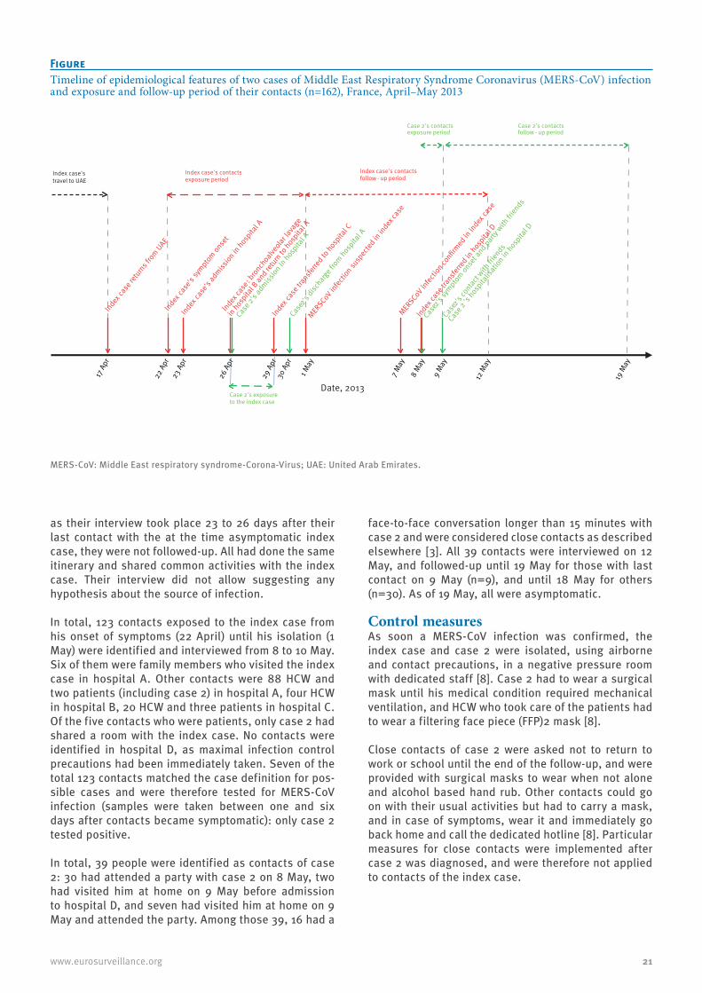
In May 2013, Middle East Respiratory Syndrome Coronavirus (MERS-CoV) infection was diagnosed in an adult male in France with severe respiratory illness, who had travelled to the United Arab Emirates before symptom onset. Contact tracing identified a second-ary case in a patient hospitalised in the same hospi-tal room. No other cases of MERS-CoV infection were identified among the index case’s 123 contacts, nor among 39 contacts of the secondary case, during the 10-day follow-up period.
On 7 May 2013, Middle East Respiratory syndrome-Cor-onavirus (MERS-CoV) infection was confirmed in France in a traveller who became ill after returning from the United Arab Emirates (index case). An investigation was immediately carried out among his contacts since onset of illness, as well as among individuals who had co-travelled with him to the United Arab Emirates. The aim of the investigation was to detect possible other cases and prevent human-to-human transmission. The secondary objective was to try to identify any likely cir-cumstances of exposure to the virus during his travel.
MERS-CoV is a novel virus among the genus Betacoronavirus, which was initially identified in Saudi Arabia in September 2012, in two patients with severe pneumonia [1]. As of 7 May 2013, when the case in France was identified, 30 cases had been confirmed as infected with the virus worldwide, including four
diagnosed in the United Kingdom (UK) and two in Germany [2,3].
Surveillance, contact tracing and case finding in France
French surveillance systemIn France, suspected cases of MERS-CoV infection have to be reported by attending physicians to regional health agencies and hospital infection control teams. After validation of the classification as a possible case by a French Institute for Public Health Surveillance (InVS) regional office (CIRE), located in a regional health agency, a standardised notification form includ-ing socio-demographical information, clinical details, and history of travel in at-risk countries is completed for each possible case.
Up to 17 May, a possible case was defined as follows:
(i) any patient with a history of travel in an at-risk coun-try, who presented with clinical signs and/or imaging consistent with acute respiratory distress syndrome (ARDS) or pulmonary infection, encompassing fever ≥38°C and cough within 10 days after return; (ii) any contact of a symptomatic possible or confirmed case, presenting with acute respiratory infection, what-ever the severity, with an onset of symptoms within 10
20 www.eurosurveillance.org
days of the last contact with a possible/confirmed case while symptomatic.
The list of at-risk countries, as defined in European Centre for Disease Prevention and Control (ECDC) rapid risk assessment dated 7 December 2012, included, Bahrain, Iran Iraq, Israel, Jordan, Kuwait, Lebanon, Palestine, Oman, Qatar, Saudi Arabia, Syria, United Arab Emirates, and Yemen [4].
For each possible case, respiratory samples (naso-pharyngeal aspiration/swab, bronchoalveolar lavage (BAL) fluid when indicated, or induced sputum) are col-lected and sent to the National Reference Centres for influenza (Institut Pasteur, Paris (coordinating centre) or Hospices civils, Lyon) to be tested for the presence of MERS-CoV genome by real-time reverse transcriptase polymerase chain reaction (RT-PCR) [5,6].
A confirmed case is defined as a possible case with a positive MERS-CoV RT-PCR on respiratory samples [5,6].
Moreover, as part of the usual surveillance of both emerging or nosocomial infections, any cluster of hos-pitalised patients or healthcare workers (HCW) pre-senting with severe respiratory infections, regardless of any history of travel in at-risk countries, has to be notified to Public Health Authorities.
Contact tracing and case findingThe contact tracing of all identified cases is imple-mented as soon as the diagnosis is confirmed. Contacts are defined as all people who provided healthcare to a confirmed case without individual protection, shared the same hospital room, lived in the same household or shared any leisure or professional activity with a confirmed case since this case’s onset of clinical symp-toms of MERS-CoV infection (respiratory, digestive or even isolated fever ≥38°C). All contacts are followed-up during a 10-day period (equal to the maximum incuba-tion period according to the knowledge of the disease at the time of the investigation described in this report) after their last contact with the confirmed case to check for clinical symptoms, and asked to measure their body temperature twice a day. The follow-up con-sists of daily calls from the InVS or CIRE for contacts who are not HCW or from the hospital infection control teams for HCW, to check for the occurrence of clinical symptoms and fever (≥38°C). Contacts are also pro-vided with a hotline number to call anytime in case of any symptom.
For confirmed cases with a history of travel in an at-risk country, a contact tracing of all members of the travel group (co-travellers) is implemented. If the con-firmed case had onset of symptoms during the travel, co-travellers are investigated as contacts. Because they potentially have been exposed to the same source of infection (co-exposed), co-travellers are followed-up during a 10-day period after their return from an
at-risk country. They are interviewed about the nature and date of their activities, exposure to people pre-senting with respiratory symptoms, food consumption and exposures to animals, and to aerosols during the travel, in order to investigate the source of infection.
The investigations are carried out with respect to French regulations (authorisation of the Commission Nationale Informatique et Libertés n°341194v42).
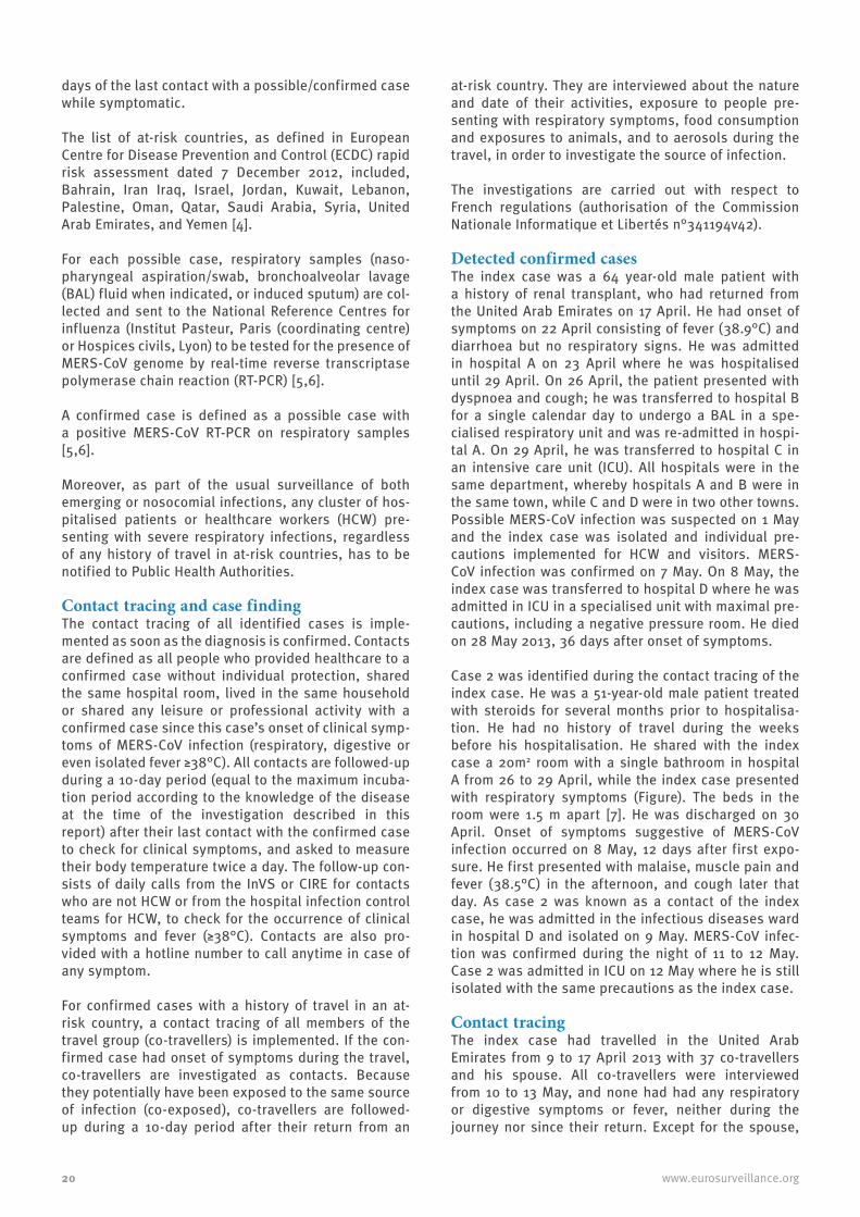
Detected confirmed casesThe index case was a 64 year-old male patient with a history of renal transplant, who had returned from the United Arab Emirates on 17 April. He had onset of symptoms on 22 April consisting of fever (38.9°C) and diarrhoea but no respiratory signs. He was admitted in hospital A on 23 April where he was hospitalised until 29 April. On 26 April, the patient presented with dyspnoea and cough; he was transferred to hospital B for a single calendar day to undergo a BAL in a spe-cialised respiratory unit and was re-admitted in hospi-tal A. On 29 April, he was transferred to hospital C in an intensive care unit (ICU). All hospitals were in the same department, whereby hospitals A and B were in the same town, while C and D were in two other towns. Possible MERS-CoV infection was suspected on 1 May and the index case was isolated and individual pre-cautions implemented for HCW and visitors. MERS-CoV infection was confirmed on 7 May. On 8 May, the index case was transferred to hospital D where he was admitted in ICU in a specialised unit with maximal pre-cautions, including a negative pressure room. He died on 28 May 2013, 36 days after onset of symptoms.
Case 2 was identified during the contact tracing of the index case. He was a 51-year-old male patient treated with steroids for several months prior to hospitalisa-tion. He had no history of travel during the weeks before his hospitalisation. He shared with the index case a 20m2 room with a single bathroom in hospital A from 26 to 29 April, while the index case presented with respiratory symptoms (Figure). The beds in the room were 1.5 m apart [7]. He was discharged on 30 April. Onset of symptoms suggestive of MERS-CoV infection occurred on 8 May, 12 days after first expo-sure. He first presented with malaise, muscle pain and fever (38.5°C) in the afternoon, and cough later that day. As case 2 was known as a contact of the index case, he was admitted in the infectious diseases ward in hospital D and isolated on 9 May. MERS-CoV infec-tion was confirmed during the night of 11 to 12 May. Case 2 was admitted in ICU on 12 May where he is still isolated with the same precautions as the index case.
Contact tracing The index case had travelled in the United Arab Emirates from 9 to 17 April 2013 with 37 co-travellers and his spouse. All co-travellers were interviewed from 10 to 13 May, and none had had any respiratory or digestive symptoms or fever, neither during the journey nor since their return. Except for the spouse,
21www.eurosurveillance.org
as their interview took place 23 to 26 days after their last contact with the at the time asymptomatic index case, they were not followed-up. All had done the same itinerary and shared common activities with the index case. Their interview did not allow suggesting any hypothesis about the source of infection.
In total, 123 contacts exposed to the index case from his onset of symptoms (22 April) until his isolation (1 May) were identified and interviewed from 8 to 10 May. Six of them were family members who visited the index case in hospital A. Other contacts were 88 HCW and two patients (including case 2) in hospital A, four HCW in hospital B, 20 HCW and three patients in hospital C. Of the five contacts who were patients, only case 2 had shared a room with the index case. No contacts were identified in hospital D, as maximal infection control precautions had been immediately taken. Seven of the total 123 contacts matched the case definition for pos-sible cases and were therefore tested for MERS-CoV infection (samples were taken between one and six days after contacts became symptomatic): only case 2 tested positive.
In total, 39 people were identified as contacts of case 2: 30 had attended a party with case 2 on 8 May, two had visited him at home on 9 May before admission to hospital D, and seven had visited him at home on 9 May and attended the party. Among those 39, 16 had a
face-to-face conversation longer than 15 minutes with case 2 and were considered close contacts as described elsewhere [3]. All 39 contacts were interviewed on 12 May, and followed-up until 19 May for those with last contact on 9 May (n=9), and until 18 May for others (n=30). As of 19 May, all were asymptomatic.
Control measures As soon a MERS-CoV infection was confirmed, the index case and case 2 were isolated, using airborne and contact precautions, in a negative pressure room with dedicated staff [8]. Case 2 had to wear a surgical mask until his medical condition required mechanical ventilation, and HCW who took care of the patients had to wear a filtering face piece (FFP)2 mask [8].
Close contacts of case 2 were asked not to return to work or school until the end of the follow-up, and were provided with surgical masks to wear when not alone and alcohol based hand rub. Other contacts could go on with their usual activities but had to carry a mask, and in case of symptoms, wear it and immediately go back home and call the dedicated hotline [8]. Particular measures for close contacts were implemented after case 2 was diagnosed, and were therefore not applied to contacts of the index case.
FigureTimeline of epidemiological features of two cases of Middle East Respiratory Syndrome Coronavirus (MERS-CoV) infection and exposure and follow-up period of their contacts (n=162), France, April–May 2013
MERS-CoV: Middle East respiratory syndrome-Corona-Virus; UAE: United Arab Emirates.
Index case's travel to UAE
Index ca
se re
turns f
rom UAE
Index ca
se's
symptom onse
t
Index ca
se: b
ronchoalv
eolar
lava
ge
in hospita
l B an
d retu
rn to hosp
ital A
Case 2
's ad
mission in
hospita
l A
Index ca
setra
nsferre
d to hosp
ital C
MERS-C
oV infec
tion su
spec
ted in
index
case
MERS-C
oV infec
tion co
nfirmed
in in
dex ca
se
Case2
's sy
mptom onset a
nd party w
ith fri
ends
Index ca
se tr
ansfe
rred in
hospita
l D
Case2
's co
ntact w
ith fri
ends
Case 2
's hosp
italis
ation in
hospita
l D
Case 2's exposure
to the index case
Index case's contacts
exposure period Index case's contacts
follow - up period
Case 2's contacts exposure period
Case 2's contacts follow - up period
Index ca
se's
admiss
ion in hosp
ital A
Case2
's disc
harge f
rom hospita
l A
17 A
pr
22 A
pr23
Apr
26 A
pr
29 A
pr30
Apr
1 May
7 May
8 M
ay
9 M
ay
12 M
ay
19 M
ay
Date, 2013
22 www.eurosurveillance.org
Both confirmed cases were notified to the ECDC and the World Health Organization (WHO), respectively on 8 May and 12 May.
Information about the disease and the outbreak was released to the public through the media, and to trav-ellers via flyers and posters disseminated in airports. Specific information about the patients’ management was disseminated to healthcare professionals through mailing lists and institutions’ websites.
Discussion and conclusionWe report the investigation of the first two cases of MERS-CoV diagnosed in France since the emergence of the virus was first described in Saudi Arabia in 2012 [1]. The index case diagnosed in France was imported from the United Arab Emirates, and the second case resulted from a nosocomial infection. Considering that both cases spent four days (26 to 29 April) in the same hospital room, the incubation period of case 2 ranged from nine to 12 days. This emphasises the need for gathering more clinical information from future and past cases to be able to determine precisely the incu-bation period.
As of 7 June 2013, 55 cases were identified worldwide since the beginning of the worldwide outbreak [9], suggesting a limited human-to-human transmission, even if we assume that some cases may have not been diagnosed.
The index case was initially admitted with an atypical presentation consisting of digestive symptoms but no respiratory signs. Therefore, MERS-CoV infection was not suspected until the patient was in ICU with severe pneumonia. This finding raised the importance of dis-seminating information about emerging diseases in all hospital settings, including those wards that are not specialised in infectious diseases or critical care.
In-hospital transmission has previously been described in England, in a family member who visited a confirmed case in hospital [10]. A hospital cluster suggestive of nosocomial transmission has also been reported in Saudi Arabia, although the details of the transmission are still under investigation [11]. In France, a second-ary infection was diagnosed in another hospitalised patient with underlying condition and long-term ster-oid treatment. The respiratory presentation of the index case strongly suggests an airborne transmission in the hospital room shared by both patients. However, some questions remain about the possible infectious-ness of other body fluids or clinical samples, including stools as the index case presented with diarrhoea at an early stage of his disease, and a cross transmission through contaminated surfaces, medical devices or hands of HCW cannot be ruled out. During the severe acute respiratory syndrome (SARS) outbreak in 2003, a cluster of infections was detected in inhabitants of the same building. Virus aerosols originating from a flat where the index case of the cluster had had digestive
symptoms, spread by drainage pipes, were assumed to be the origin of the infection of other cases in the cluster [12].
The large majority of reported MERS-CoV cases world-wide had underlying conditions and presented with severe respiratory infection requiring hospitalisation in ICU. Atypical presentations in immunocompromised patients may be really challenging for clinicians, espe-cially as digestive symptoms are very common in trav-ellers. Based on the index case’s clinical presentation and on knowledge acquired from the SARS outbreak [13], the French case definition for possible cases was extended on 17 May to improve the sensitivity of the surveillance system. It now includes severe febrile clin-ical signs or febrile diarrhoea in immunocompromised persons or in those with chronic underlying conditions, returning from an at-risk country [14].
Despite the identification of few infections since 2012, MERS-CoV has demonstrated a real potential for noso-comial transmission, and stringent recommendations have to be implemented around possible cases as soon as MERS-CoV infection is suspected. The chal-lenge presented by possible atypical presentations highlights the need for a better knowledge about both the virus and the disease.
Useful knowledge about the infection by MERS-CoV might be obtained from serological investigation in people who shared exposures of confirmed cases, or in contacts of confirmed cases. Such studies might help raising hypothesis about the extent of transmis-sion and risk factors for infection and fatal outcome and must be encouraged.
Acknowledgements David Briand, Mathilde Benassaya, Marie Lazzerini, and Clio Socratous of the National Reference Centre for influenza vi-ruses (Institut Pasteur) contributed to the diagnosis of cas-es. The authors are grateful to all clinicians and biologists who contribute to the surveillance of this emerging infection.
Investigation team InVS, Saint Maurice, France: Sandrine Barquin, Fatima Belghiti, Alexandre Blake, Gabriel Bedubourg, Isabelle Bonmarin, Jérémie Carré, Carole Debreyer, Gilles Delmas, Anne Gallay, Claire Gourier-Fréry, Etienne Lucas, Roselyne Pinget, Mathieu Tourdjmann, Sarah Tubiana, Dieter Van Cauteren, Annie Velter, Manuel Zubaran.InVS, Lille, France: Audrey Andrieu, Emile Baro, Guillaume Clement.National Reference Center for influenza viruses, Hospices Civils de Lyon and Virpath, Université Claude Bernard Lyon1, Lyon, France: Maude Bouscambert, Martine Valette.Centre Hospitalier Régional et Universitaire, Université de Lille 2, Lille, France: Karine Faure, Hugues Melliez, Fanny Vuotto.Centre hospitalier, Douai, France: Loubna El-Mansouf.Centre hospitalier, Valenciennes, France: Anne-Adélaide Cracco.
23www.eurosurveillance.org
Regional Health Agency, Lille, France: Jean-Philippe Legendre, Sandrine Segovia-Kueny.Centre hospitalier universitaire, Tourcoing, France: Eric Senneville.
Conflict of interestNone declared.
Authors’ contributionsAlexandra Mailles: wrote the manuscript. Alexandra Mailles, Christine Campese, coordinated the investigation of co-travellers of the index case. Pascal Chaud, Marie-Claire Paty, Caroline Semaille: coordinated the investigations of confirmed cases. Karine Blanckaert, Sylvie Hendricx: con-tact tracing and follow-up of healthcare workers. Pascal Chaud, Sylvie Haeghebaert: coordinated the contact trac-ing for index case and follow-up of healthcare workers and family contacts of the index case. Sylvie van der Werf, Bruno Lina, Valérie Caro, Sylvie Behillil, Jean-Claude Manuguerra, Vincent Enouf: implemented the biological diagnosis in France and carried out the diagnosis of all possible cases, in-cluding both confirmed case. Benoit Guéry, Xavier Lemaire, Nicolas Ettahar: clinical management of confirmed cases and symptomatic contacts. Delphine Antoine, Harold Noel, Guy La Ruche, Pascal Chaud, Hélène Prouvost coordinated the contact tracing and follow-up of contacts of case. Didier Che, Bruno Coignard, Daniel Levy-Bruh: expertise in risk assessment. Bruno Coignard, Daniel Levy-Bruhl, Françoise Weber: supervised the investigations and coordinated the relationship with health authorities in France and Europe. Christine Saura, Didier Che: implemented the surveillance system in France and coordinated all involved partners since September 2012. All: revised the manuscript and contributed with specific comments.
References1. Zaki AM, van Boheemen S, Bestebroer TM, Osterhaus AD,
Fouchier RA. Isolation of a novel coronavirus from a man with pneumonia in Saudi Arabia. N Engl J Med. 2012;367(19):1814-20. http://dx.doi.org/10.1056/NEJMoa1211721
2. Buchholz U, Müller MA, Nitsche A, Sanewski A, Wevering N, Bauer-Balci T, et al. Contact investigation of a case of human novel coronavirus infection treated in a German hospital, October-November 2012. Euro Surveill. 2013;18(8): pii=20406. Available from: http://www.eurosurveillance.org/ViewArticle.aspx?ArticleId=20406 PMid:23449231
3. Bermingham A, Chand MA, Brown CS, Aarons E, Tong C, Langrish C,et al. Severe respiratory illness caused by a novel coronavirus, in a patient transferred to the United Kingdom from the Middle East, September 2012. Euro Surveill. 2012;17(40):pii=20290. Available from: http://www.eurosurveillance.org/ViewArticle.aspx?ArticleId=20290
4. European Centre for Disease Prevention and Control (ECDC). Update: Severe respiratory disease associated with a novel coronavirus. Stockholm: ECDC; 7 Dec 2012. [Accessed 10 Jun 2013]. Available from: http://www.ecdc.europa.eu/en/publications/Publications/20121207-Novel-coronavirus-rapid-risk-assessment.pdf
5. Corman VM, Eckerle I, Bleicker T, Zaki A, Landt O, Eschbach-Bludau M, et al. Detection of a novel human coronavirus by real-time reverse-transcription polymerase chain reaction. Euro Surveill. 2012;17(39):pii=20285. Available from: http://www.eurosurveillance.org/ViewArticle.aspx?ArticleId=20285
6. Corman VM, Müller MA, Costabel U, Timm J, Binger T, Meyer B, et al. Assays for laboratory confirmation of novel human coronavirus (hCoV-EMC) infections. Euro Surveill. 2012;17(49):pii: 20334. Available from: http://www.eurosurveillance.org/ViewArticle.aspx?ArticleId=20334
7. Guery B, Poissy J, El Mansouf L, Séjourné C, Ettahar N, Lemaire X, et al. Clinical features and viral diagnosis of two cases of infection with Middle East Respiratory Syndrome coronavirus: a report of nosocomial transmission. Lancet. 2013 May 30. http://dx.doi.org/10.1016/S0140-6736(13)60982-4
8. Haut conseil de la santé publique (HCSP). Avis relatif à la prise en charge des patients suspects d’infections dues au nouveau coronavirus (HCoV-EMC). [Guidelines for the management of patients with suspected MERS-CoV]. Paris: HCSP; 19 Mar 2013. [Accessed 29 May 2013]. Available from: http://www.hcsp.fr/explore.cgi/telecharger/hcspa20130319_nouveaucoronavirus.pdf
9. World Health Organization (WHO). Middle East respiratory syndrome coronavirus (MERS-CoV) – update. Geneva: WHO; 07 Jun 2013. [Accessed 11 Jun 2013]. Available from: http://www.who.int/csr/don/2013_06_07/en/index.html
10. Health Protection Agency (HPA) UK Novel Coronavirus Investigation team. Evidence of person-to-person transmission within a family cluster of novel coronavirus infections, United Kingdom, February 2013. Euro Surveill. 2013;18(11):pii=20427. Available from: http://www.eurosurveillance.org/ViewArticle.aspx?ArticleId=20427
11. ProMED-mail. Novel Coronavirus - Eastern Mediterranean (21): Saudi Arabia. Archive Number: 20130509.1701527. 09 May 2013. [Accessed 29 May 2013]. Available from: http://www.promedmail.org/direct.php?id=20130509.1701527
12. Yu IT, Li Y, Wong TW, Tam W, Chan AT, Lee JH, et al. Evidence of airborne transmission of the severe acute respiratory syndrome virus. N Engl J Med. 2004;350(17):1731-9 http://dx.doi.org/10.1056/NEJMoa032867 PMid:15102999
13. Christian MD, Poutanen SM, Loutfy MR, Muller MP, Low DE. Severe acute respiratory syndrome. Clin Infect Dis. 2004;38(10):1420-7. http://dx.doi.org/10.1086/420743
14. European Centre for Disease Prevention and Control (ECDC). Severe respiratory disease associated with Middle East respiratory syndrome coronavirus (MERS-CoV). Stockholm: ECDC; 17 May 2013. [Accessed 29 May 2013]. Available from: http://www.ecdc.europa.eu/en/publications/Publications/risk-assessment-middle-east-respiratory-syndrome-coronavirus-MERS-CoV-17-may-2013.pdf
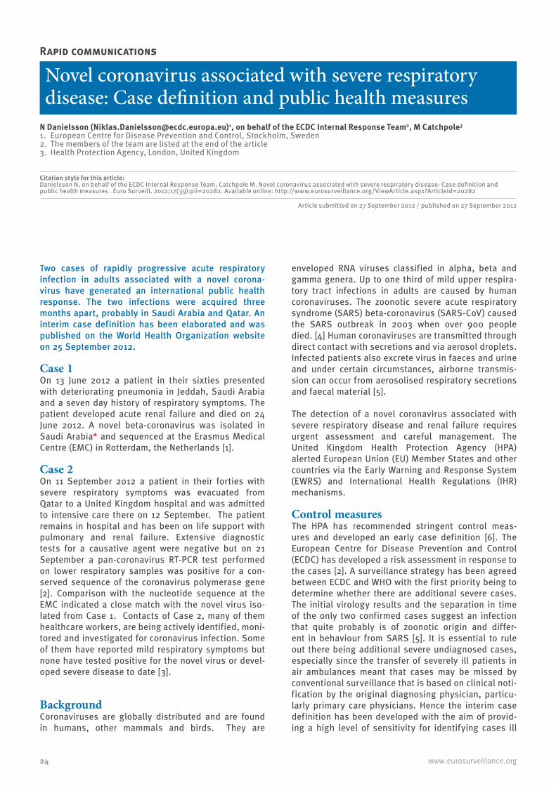
24 www.eurosurveillance.org
Rapid communications
Novel coronavirus associated with severe respiratory disease: Case definition and public health measures
N Danielsson ([email protected])1, on behalf of the ECDC Internal Response Team2, M Catchpole3
1. European Centre for Disease Prevention and Control, Stockholm, Sweden2. The members of the team are listed at the end of the article3. Health Protection Agency, London, United Kingdom
Citation style for this article: Danielsson N, on behalf of the ECDC Internal Response Team, Catchpole M. Novel coronavirus associated with severe respiratory disease: Case definition and public health measures . Euro Surveill. 2012;17(39):pii=20282. Available online: http://www.eurosurveillance.org/ViewArticle.aspx?ArticleId=20282
Article submitted on 27 September 2012 / published on 27 September 2012
Two cases of rapidly progressive acute respiratory infection in adults associated with a novel corona-virus have generated an international public health response. The two infections were acquired three months apart, probably in Saudi Arabia and Qatar. An interim case definition has been elaborated and was published on the World Health Organization website on 25 September 2012.
Case 1On 13 June 2012 a patient in their sixties presented with deteriorating pneumonia in Jeddah, Saudi Arabia and a seven day history of respiratory symptoms. The patient developed acute renal failure and died on 24 June 2012. A novel beta-coronavirus was isolated in Saudi Arabia* and sequenced at the Erasmus Medical Centre (EMC) in Rotterdam, the Netherlands [1].
Case 2On 11 September 2012 a patient in their forties with severe respiratory symptoms was evacuated from Qatar to a United Kingdom hospital and was admitted to intensive care there on 12 September. The patient remains in hospital and has been on life support with pulmonary and renal failure. Extensive diagnostic tests for a causative agent were negative but on 21 September a pan-coronavirus RT-PCR test performed on lower respiratory samples was positive for a con-served sequence of the coronavirus polymerase gene [2]. Comparison with the nucleotide sequence at the EMC indicated a close match with the novel virus iso-lated from Case 1. Contacts of Case 2, many of them healthcare workers, are being actively identified, moni-tored and investigated for coronavirus infection. Some of them have reported mild respiratory symptoms but none have tested positive for the novel virus or devel-oped severe disease to date [3].
BackgroundCoronaviruses are globally distributed and are found in humans, other mammals and birds. They are
enveloped RNA viruses classified in alpha, beta and gamma genera. Up to one third of mild upper respira-tory tract infections in adults are caused by human coronaviruses. The zoonotic severe acute respiratory syndrome (SARS) beta-coronavirus (SARS-CoV) caused the SARS outbreak in 2003 when over 900 people died. [4] Human coronaviruses are transmitted through direct contact with secretions and via aerosol droplets. Infected patients also excrete virus in faeces and urine and under certain circumstances, airborne transmis-sion can occur from aerosolised respiratory secretions and faecal material [5].
The detection of a novel coronavirus associated with severe respiratory disease and renal failure requires urgent assessment and careful management. The United Kingdom Health Protection Agency (HPA) alerted European Union (EU) Member States and other countries via the Early Warning and Response System (EWRS) and International Health Regulations (IHR) mechanisms.
Control measuresThe HPA has recommended stringent control meas-ures and developed an early case definition [6]. The European Centre for Disease Prevention and Control (ECDC) has developed a risk assessment in response to the cases [2]. A surveillance strategy has been agreed between ECDC and WHO with the first priority being to determine whether there are additional severe cases. The initial virology results and the separation in time of the only two confirmed cases suggest an infection that quite probably is of zoonotic origin and differ-ent in behaviour from SARS [5]. It is essential to rule out there being additional severe undiagnosed cases, especially since the transfer of severely ill patients in air ambulances meant that cases may be missed by conventional surveillance that is based on clinical noti-fication by the original diagnosing physician, particu-larly primary care physicians. Hence the interim case definition has been developed with the aim of provid-ing a high level of sensitivity for identifying cases ill
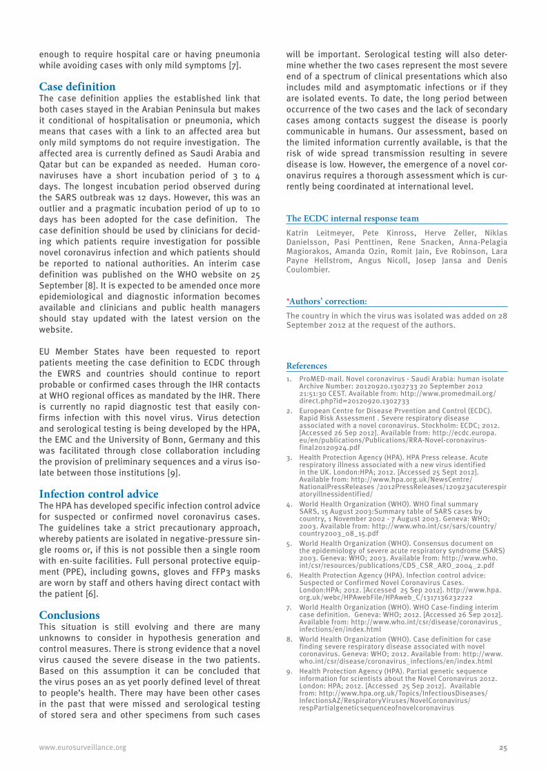
25www.eurosurveillance.org
enough to require hospital care or having pneumonia while avoiding cases with only mild symptoms [7].
Case definitionThe case definition applies the established link that both cases stayed in the Arabian Peninsula but makes it conditional of hospitalisation or pneumonia, which means that cases with a link to an affected area but only mild symptoms do not require investigation. The affected area is currently defined as Saudi Arabia and Qatar but can be expanded as needed. Human coro-naviruses have a short incubation period of 3 to 4 days. The longest incubation period observed during the SARS outbreak was 12 days. However, this was an outlier and a pragmatic incubation period of up to 10 days has been adopted for the case definition. The case definition should be used by clinicians for decid-ing which patients require investigation for possible novel coronavirus infection and which patients should be reported to national authorities. An interim case definition was published on the WHO website on 25 September [8]. It is expected to be amended once more epidemiological and diagnostic information becomes available and clinicians and public health managers should stay updated with the latest version on the website.
EU Member States have been requested to report patients meeting the case definition to ECDC through the EWRS and countries should continue to report probable or confirmed cases through the IHR contacts at WHO regional offices as mandated by the IHR. There is currently no rapid diagnostic test that easily con-firms infection with this novel virus. Virus detection and serological testing is being developed by the HPA, the EMC and the University of Bonn, Germany and this was facilitated through close collaboration including the provision of preliminary sequences and a virus iso-late between those institutions [9].
Infection control adviceThe HPA has developed specific infection control advice for suspected or confirmed novel coronavirus cases. The guidelines take a strict precautionary approach, whereby patients are isolated in negative-pressure sin-gle rooms or, if this is not possible then a single room with en-suite facilities. Full personal protective equip-ment (PPE), including gowns, gloves and FFP3 masks are worn by staff and others having direct contact with the patient [6].
ConclusionsThis situation is still evolving and there are many unknowns to consider in hypothesis generation and control measures. There is strong evidence that a novel virus caused the severe disease in the two patients. Based on this assumption it can be concluded that the virus poses an as yet poorly defined level of threat to people’s health. There may have been other cases in the past that were missed and serological testing of stored sera and other specimens from such cases
will be important. Serological testing will also deter-mine whether the two cases represent the most severe end of a spectrum of clinical presentations which also includes mild and asymptomatic infections or if they are isolated events. To date, the long period between occurrence of the two cases and the lack of secondary cases among contacts suggest the disease is poorly communicable in humans. Our assessment, based on the limited information currently available, is that the risk of wide spread transmission resulting in severe disease is low. However, the emergence of a novel cor-onavirus requires a thorough assessment which is cur-rently being coordinated at international level.
The ECDC internal response teamKatrin Leitmeyer, Pete Kinross, Herve Zeller, Niklas Danielsson, Pasi Penttinen, Rene Snacken, Anna-Pelagia Magiorakos, Amanda Ozin, Romit Jain, Eve Robinson, Lara Payne Hellstrom, Angus Nicoll, Josep Jansa and Denis Coulombier.
*Authors’ correction:The country in which the virus was isolated was added on 28 September 2012 at the request of the authors.
References1. ProMED-mail. Novel coronavirus - Saudi Arabia: human isolate
Archive Number: 20120920.1302733 20 September 2012 21:51:30 CEST. Available from: http://www.promedmail.org/direct.php?id=20120920.1302733
2. European Centre for Disease Prvention and Control (ECDC). Rapid Risk Assessment . Severe respiratory disease associated with a novel coronavirus. Stockholm: ECDC; 2012. [Accessed 26 Sep 2012]. Available from: http://ecdc.europa.eu/en/publications/Publications/RRA-Novel-coronavirus-final20120924.pdf
3. Health Protection Agency (HPA). HPA Press release. Acute respiratory illness associated with a new virus identified in the UK. London:HPA; 2012. [Accessed 25 Sept 2012]. Available from: http://www.hpa.org.uk/NewsCentre/NationalPressReleases /2012PressReleases/120923acuterespiratoryillnessidentified/
4. World Health Organization (WHO). WHO final summary SARS, 15 August 2003:Summary table of SARS cases by country, 1 November 2002 - 7 August 2003. Geneva: WHO; 2003. Available from: http://www.who.int/csr/sars/country/country2003_08_15.pdf
5. World Health Organization (WHO). Consensus document on the epidemiology of severe acute respiratory syndrome (SARS) 2003. Geneva: WHO; 2003. Available from: http://www.who.int/csr/resources/publications/CDS_CSR_ARO_2004_2.pdf
6. Health Protection Agency (HPA). Infection control advice: Suspected or Confirmed Novel Coronavirus Cases. London:HPA; 2012. [Accessed 25 Sep 2012]. http://www.hpa.org.uk/webc/HPAwebFile/HPAweb_C/1317136232722
7. World Health Organization (WHO). WHO Case-finding interim case definition. Geneva: WHO; 2012. [Accessed 26 Sep 2012]. Available from: http://www.who.int/csr/disease/coronavirus_infections/en/index.html
8. World Health Organization (WHO). Case definition for case finding severe respiratory disease associated with novel coronavirus. Geneva: WHO; 2012. Available from: http://www.who.int/csr/disease/coronavirus_infections/en/index.html
9. Health Protection Agency (HPA). Partial genetic sequence information for scientists about the Novel Coronavirus 2012. London: HPA; 2012. [Accessed 25 Sep 2012]. Available from: http://www.hpa.org.uk/Topics/InfectiousDiseases/InfectionsAZ/RespiratoryViruses/NovelCoronavirus/respPartialgeneticsequenceofnovelcoronavirus
26 www.eurosurveillance.org
Rapid communications
Incubation period as part of the case definition of severe respiratory illness caused by a novel coronavirus
H Nishiura ([email protected])1,2, K Mizumoto1,3, K Ejima1,4, Y Zhong1, B J Cowling1, R Omori1
1. School of Public Health, The University of Hong Kong, Hong Kong SAR, China2. PRESTO (Precursory Research for Embryonic Science and Technology), Japan Science and Technology Agency, Saitama, Japan3. Institute of Tropical Medicine and the Global Center of Excellence Program, Nagasaki University, Nagasaki, Japan4. Department of Mathematical Informatics, Graduate School of Information Science and Technology, The University of Tokyo,
Tokyo, Japan
Citation style for this article: Nishiura H, Mizumoto K, Ejima K, Zhong Y, Cowling BJ, Omori R. Incubation period as part of the case definition of severe respiratory illness caused by a novel coronavirus. Euro Surveill. 2012;17(42):pii=20296. Available online: http://www.eurosurveillance.org/ViewArticle.aspx?ArticleId=20296
Article submitted on 11 October 2012/ published on 18 October 2012
Non-specific symptoms of acute respiratory viral infections make it difficult for many countries without ongoing transmission of a novel coronavirus to rule out other possibilities including influenza before isolating imported febrile individuals with a possible exposure history. The incubation period helps differential diag-nosis, and up to two days is suggestive of influenza. It is worth including the incubation period in the case definition of novel coronavirus infection.
Introduction Two cases of severe respiratory infection have been confirmed as caused by a novel coronavirus [1]. The case definition has been issued by the World Health Organization (WHO), and is mainly based on acute respiratory illness, pneumonia (or suspicion of pulmo-nary parenchymal disease) and travel history [2]. To describe the clinical characteristics of the novel coro-navirus infection, the incubation period has played a key role in suspecting Saudi Arabia and Qatar as geographic locations of exposure for the two cases mentioned above [1,3]. The presumed length of the incubation period was compared with known incuba-tion periods of human coronavirus infections including that of severe acute respiratory syndrome (SARS) [3,4]. The present study intends to point out that the incu-bation period can be useful for all countries without ongoing transmission to distinguish the novel corona-virus infection from other viral respiratory infections, most notably influenza.
Methods
Motivating case studyA preschool child from Saudi Arabia was admitted to a Hong Kong hospital equipped with an isolation ward in early October 2012, suspected of novel coronavirus infection. It had fever, cough and vomiting, but did not have pneumonia. One close contact had had a fever two days earlier, but had recovered before the day of admission [5]. Assuming that the contact was the source of infection, the serial interval was two days, which is typically longer than the incubation period
[6,7], and thus, the incubation period is likely to have been two days or shorter. On the day following admis-sion, the child tested negative for the novel coronavi-rus, but positive for influenza A(H1N1)pdm09 [5].
A similar event, but involving two cases of severe pneumonia, occurred in Denmark: A cluster of febrile patients, some of whom had a travel history to Qatar and Saudi Arabia, was suspected of infection with the novel coronavirus. However, later laboratory testing revealed that the respiratory illnesses were caused by infection with an influenza B virus [8].
We believe that the distinction between coronavirus and influenza virus infections in these settings could have been facilitated by considering the length of the incubation period.
Bayesian modelLet be the probability density function of the incubation period t of virus i governed by parameter
. The incubation period distributions for a variety of acute upper respiratory viral infections have been fit-ted to log-normal distributions elsewhere [4,9] and are assumed known hereafter. The median incubation peri-ods of SARS, non-SARS human coronavirus infection, and influenza A and influenza B virus infections have been estimated at 4.0, 3.2, 1.4 and 0.6 days, respec-tively [4]. It should be noted that the median incuba-tion periods of influenza A and B have been estimated as shorter than those of coronaviruses. The incuba-tion period is assumed to be independent across different viruses i. Due to shortage of information, we ignore the time-dependence and geographic heteroge-neity in the risk of infection for all viruses. The poste-rior probability of novel coronavirus infection (which is labelled as i=1) given an incubation period t, Pr(novel coronavirus|t) is then obtained by using a Bayesian approach:
(1)
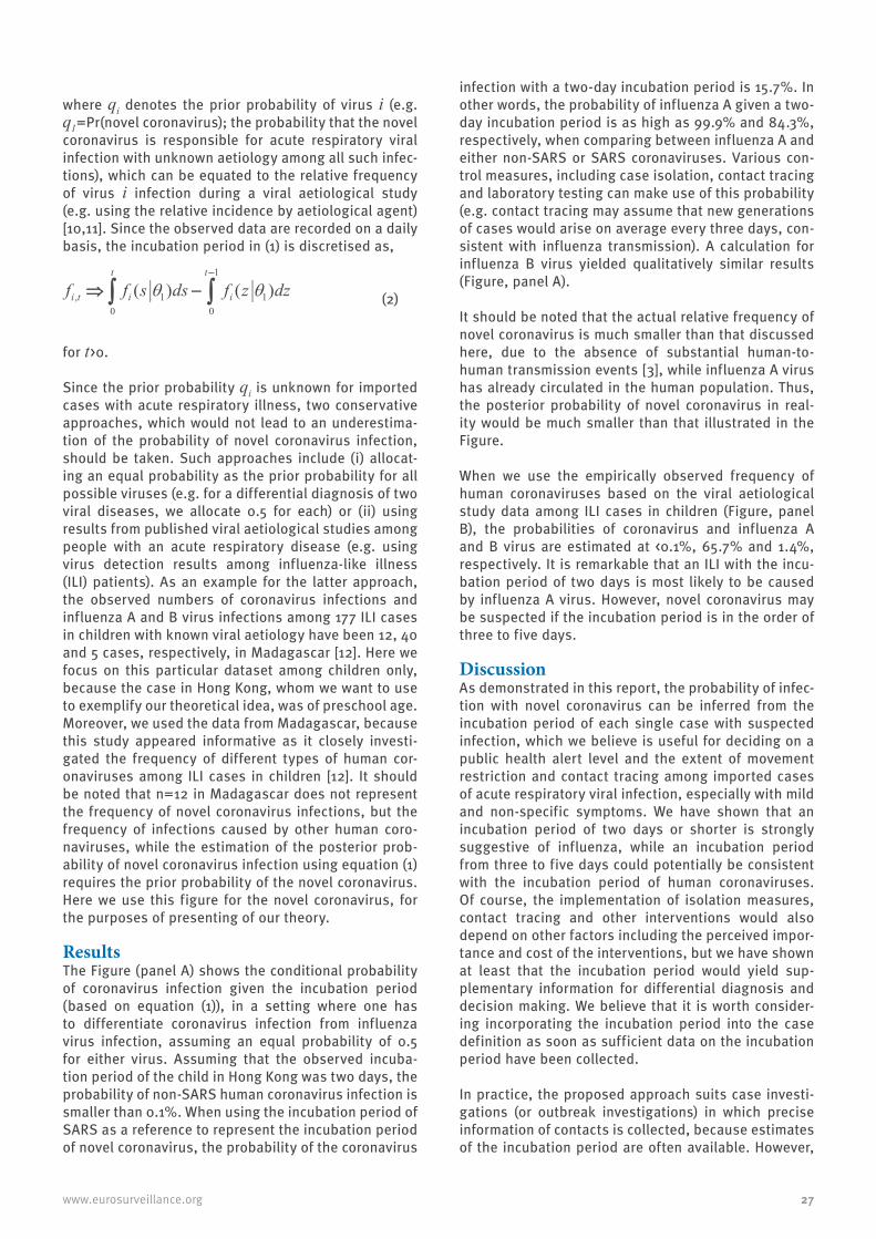
27www.eurosurveillance.org
where qi denotes the prior probability of virus i (e.g. q1=Pr(novel coronavirus); the probability that the novel coronavirus is responsible for acute respiratory viral infection with unknown aetiology among all such infec-tions), which can be equated to the relative frequency of virus i infection during a viral aetiological study (e.g. using the relative incidence by aetiological agent) [10,11]. Since the observed data are recorded on a daily basis, the incubation period in (1) is discretised as,
(2)
for t>0.
Since the prior probability qi is unknown for imported cases with acute respiratory illness, two conservative approaches, which would not lead to an underestima-tion of the probability of novel coronavirus infection, should be taken. Such approaches include (i) allocat-ing an equal probability as the prior probability for all possible viruses (e.g. for a differential diagnosis of two viral diseases, we allocate 0.5 for each) or (ii) using results from published viral aetiological studies among people with an acute respiratory disease (e.g. using virus detection results among influenza-like illness (ILI) patients). As an example for the latter approach, the observed numbers of coronavirus infections and influenza A and B virus infections among 177 ILI cases in children with known viral aetiology have been 12, 40 and 5 cases, respectively, in Madagascar [12]. Here we focus on this particular dataset among children only, because the case in Hong Kong, whom we want to use to exemplify our theoretical idea, was of preschool age. Moreover, we used the data from Madagascar, because this study appeared informative as it closely investi-gated the frequency of different types of human cor-onaviruses among ILI cases in children [12]. It should be noted that n=12 in Madagascar does not represent the frequency of novel coronavirus infections, but the frequency of infections caused by other human coro-naviruses, while the estimation of the posterior prob-ability of novel coronavirus infection using equation (1) requires the prior probability of the novel coronavirus. Here we use this figure for the novel coronavirus, for the purposes of presenting of our theory.
ResultsThe Figure (panel A) shows the conditional probability of coronavirus infection given the incubation period (based on equation (1)), in a setting where one has to differentiate coronavirus infection from influenza virus infection, assuming an equal probability of 0.5 for either virus. Assuming that the observed incuba-tion period of the child in Hong Kong was two days, the probability of non-SARS human coronavirus infection is smaller than 0.1%. When using the incubation period of SARS as a reference to represent the incubation period of novel coronavirus, the probability of the coronavirus
infection with a two-day incubation period is 15.7%. In other words, the probability of influenza A given a two-day incubation period is as high as 99.9% and 84.3%, respectively, when comparing between influenza A and either non-SARS or SARS coronaviruses. Various con-trol measures, including case isolation, contact tracing and laboratory testing can make use of this probability (e.g. contact tracing may assume that new generations of cases would arise on average every three days, con-sistent with influenza transmission). A calculation for influenza B virus yielded qualitatively similar results (Figure, panel A).
It should be noted that the actual relative frequency of novel coronavirus is much smaller than that discussed here, due to the absence of substantial human-to-human transmission events [3], while influenza A virus has already circulated in the human population. Thus, the posterior probability of novel coronavirus in real-ity would be much smaller than that illustrated in the Figure.
When we use the empirically observed frequency of human coronaviruses based on the viral aetiological study data among ILI cases in children (Figure, panel B), the probabilities of coronavirus and influenza A and B virus are estimated at <0.1%, 65.7% and 1.4%, respectively. It is remarkable that an ILI with the incu-bation period of two days is most likely to be caused by influenza A virus. However, novel coronavirus may be suspected if the incubation period is in the order of three to five days.
DiscussionAs demonstrated in this report, the probability of infec-tion with novel coronavirus can be inferred from the incubation period of each single case with suspected infection, which we believe is useful for deciding on a public health alert level and the extent of movement restriction and contact tracing among imported cases of acute respiratory viral infection, especially with mild and non-specific symptoms. We have shown that an incubation period of two days or shorter is strongly suggestive of influenza, while an incubation period from three to five days could potentially be consistent with the incubation period of human coronaviruses. Of course, the implementation of isolation measures, contact tracing and other interventions would also depend on other factors including the perceived impor-tance and cost of the interventions, but we have shown at least that the incubation period would yield sup-plementary information for differential diagnosis and decision making. We believe that it is worth consider-ing incorporating the incubation period into the case definition as soon as sufficient data on the incubation period have been collected.
In practice, the proposed approach suits case investi-gations (or outbreak investigations) in which precise information of contacts is collected, because estimates of the incubation period are often available. However,
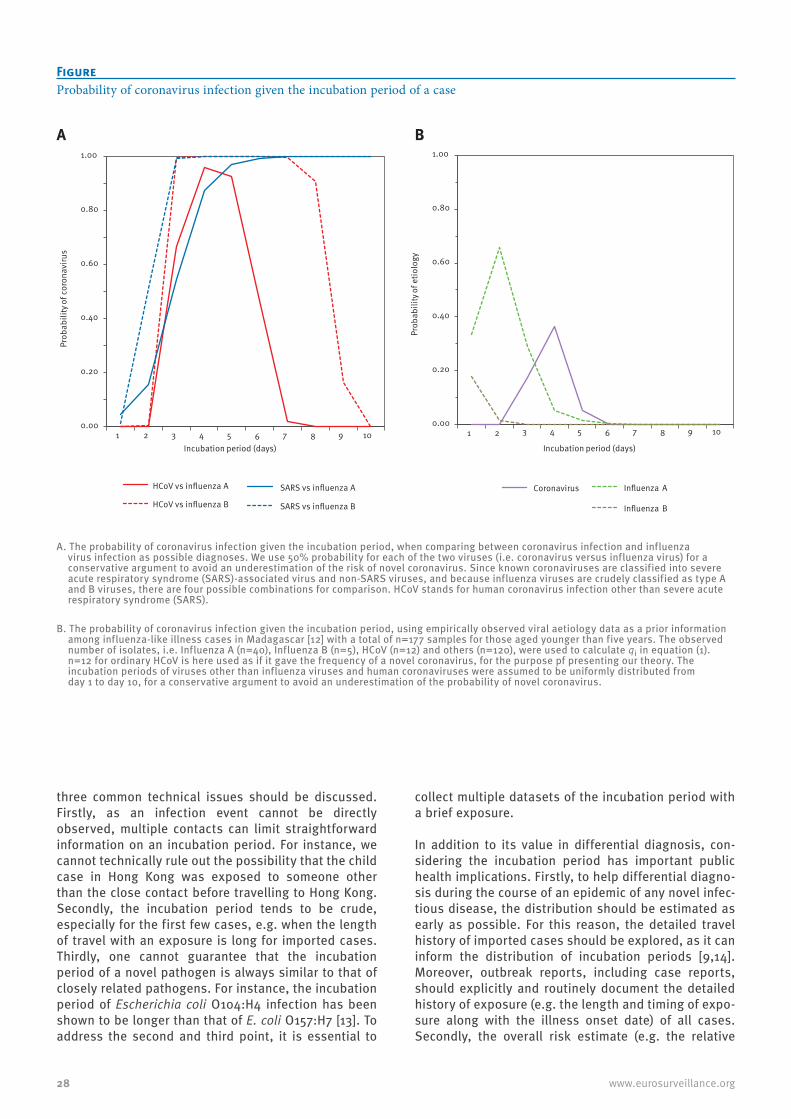
28 www.eurosurveillance.org
three common technical issues should be discussed. Firstly, as an infection event cannot be directly observed, multiple contacts can limit straightforward information on an incubation period. For instance, we cannot technically rule out the possibility that the child case in Hong Kong was exposed to someone other than the close contact before travelling to Hong Kong. Secondly, the incubation period tends to be crude, especially for the first few cases, e.g. when the length of travel with an exposure is long for imported cases. Thirdly, one cannot guarantee that the incubation period of a novel pathogen is always similar to that of closely related pathogens. For instance, the incubation period of Escherichia coli O104:H4 infection has been shown to be longer than that of E. coli O157:H7 [13]. To address the second and third point, it is essential to
collect multiple datasets of the incubation period with a brief exposure.
In addition to its value in differential diagnosis, con-sidering the incubation period has important public health implications. Firstly, to help differential diagno-sis during the course of an epidemic of any novel infec-tious disease, the distribution should be estimated as early as possible. For this reason, the detailed travel history of imported cases should be explored, as it can inform the distribution of incubation periods [9,14]. Moreover, outbreak reports, including case reports, should explicitly and routinely document the detailed history of exposure (e.g. the length and timing of expo-sure along with the illness onset date) of all cases. Secondly, the overall risk estimate (e.g. the relative
FigureProbability of coronavirus infection given the incubation period of a case
0.00
0.20
0.40
0.60
0.80
1.00
1 2 3 4 5 6 7 8 9 10
Prob
abili
ty o
f cor
onav
irus
Incubation period (days)
HCoV vs influenza A HCoV vs influenza B
SARS vs influenza A
SARS vs influenza B
0.00
0.20
0.40
0.60
0.80
1.00
1 2 3 4 5 6 7 8 9 10
Prob
abili
ty o
f etio
logy
Incubation period (days)
Coronavirus
Influenza A
Influenza B
A. The probability of coronavirus infection given the incubation period, when comparing between coronavirus infection and influenza virus infection as possible diagnoses. We use 50% probability for each of the two viruses (i.e. coronavirus versus influenza virus) for a conservative argument to avoid an underestimation of the risk of novel coronavirus. Since known coronaviruses are classified into severe acute respiratory syndrome (SARS)-associated virus and non-SARS viruses, and because influenza viruses are crudely classified as type A and B viruses, there are four possible combinations for comparison. HCoV stands for human coronavirus infection other than severe acute respiratory syndrome (SARS).
B. The probability of coronavirus infection given the incubation period, using empirically observed viral aetiology data as a prior information among influenza-like illness cases in Madagascar [12] with a total of n=177 samples for those aged younger than five years. The observed number of isolates, i.e. Influenza A (n=40), Influenza B (n=5), HCoV (n=12) and others (n=120), were used to calculate qi in equation (1). n=12 for ordinary HCoV is here used as if it gave the frequency of a novel coronavirus, for the purpose pf presenting our theory. The incubation periods of viruses other than influenza viruses and human coronaviruses were assumed to be uniformly distributed from day 1 to day 10, for a conservative argument to avoid an underestimation of the probability of novel coronavirus.
A B
29www.eurosurveillance.org
incidence) would be essential to validate the proposed Bayesian model (1), although in reality, the prior prob-ability varies considerably with time and place. To understand the ongoing risk of infection with a novel virus explicitly, a population-wide serological survey, which allows to infer at least the cumulative incidence, would be a useful method to offer insights into the aeti-ology. Finally, while estimating the relative probabil-ity of alternative aetiologies can help with diagnosis, decisions on possible control measures (such as isola-tion of cases) could also be affected by other concerns including reduction in the risk of larger outbreaks.
Acknowledgments The work of HN was supported by the JST PRESTO program and St Luke’s Life Science Institute Research Grant for Clinical Epidemiology Research 2012. KE received scholar-ship support from the Japan Society for Promotion of Science (JSPS). This work also received financial support from the Harvard Center for Communicable Disease Dynamics from the National Institute of General Medical Sciences (grant no. U54 GM088558).
References1. Danielsson N, On Behalf Of The Ecdc Internal Response
Team C, Catchpole M. Novel coronavirus associated with severe respiratory disease: Case definition and public health measures. Euro Surveill. 2012;17(39):pii=20282. Available from: http://www.eurosurveillance.org/ViewArticle.aspx?ArticleId=20282
2. World Health Organization. Global alert and response (GAR): revised interim case definition—novel coronavirus. Geneva, Switzerland: World Health Organization; 2012. [Accessed 11 Oct 2012]. Available from: http://www.who.int/csr/disease/coronavirus_infections/case_definition/en/index.html
3. Pebody RG, Chand MA, Thomas HL, Green HK, Boddington NL, Carvalho C, et al. The United Kingdom public health response to an imported laboratory confirmed case of a novel coronavirus in September 2012. Euro Surveill. 2012;17(40):pii=20292. Available from: http://www.eurosurveillance.org/ViewArticle.aspx?ArticleId=20292
4. Lessler J, Reich NG, Brookmeyer R, Perl TM, Nelson KE, Cummings DA. Incubation periods of acute respiratory viral infections: a systematic review. Lancet Infect Dis 2009;9(5):291-300.
5. Saudi boy in Hong Kong has flu, not Sars-like virus. Hong Kong: South China Morning Post; 8 Oct 2012. Available from: http://www.scmp.com/news/hong-kong/article/1056321/saudi-boy-tests-negative-sars-virus-hong-kong-health-officials-say
6. Nishiura H, Eichner M. Infectiousness of smallpox relative to disease age: estimates based on transmission network and incubation period. Epidemiol Infect. 2007;135(7):1145-50.
7. Klinkenberg D, Nishiura H. The correlation between infectivity and incubation period of measles, estimated from households with two cases. J Theor Biol. 2011;284(1):52-60.
8. Patient i højrisiko-isolation er ikke smittet med coronavirus. [Patient in high-risk isolation is not infected with coronavirus]. Hvidovre: Hvidovre Hospital. [Accessed 16 Oct 2012]. Danish. Available from: http://www.hvidovrehospital.dk/menu/Presse+og+nyheder/Nyheder+fra+Hvidovre+Hospital/2012/Patient+i+hjrisikoisolation+er+ikke+smittet+med+Coronavirus.htm?wbc_purpose=Basictar
9. Nishiura H, Inaba H. Estimation of the incubation period of influenza A (H1N1-2009) among imported cases: addressing censoring using outbreak data at the origin of importation. J Theor Biol. 2011;272(1):123-30.
10. Nishiura H. Incubation period as a clinical predictor of botulism: analysis of previous izushi-borne outbreaks in Hokkaido, Japan, from 1951 to 1965. Epidemiol Infect. 2007;135(1):126-30.
11. Lessler J, Brookmeyer R, Perl TM. An evaluation of classification rules based on date of symptom onset to identify health-care-associated infections. Am J Epidemiol. 2007;166(10):1220-9.
12. Razanajatovo NH, Richard V, Hoffmann J, Reynes JM, Razafitrimo GM, Randremanana RV, et al. Viral etiology of influenza-like illnesses in Antananarivo, Madagascar, July 2008 to June 2009. PLoS One. 2011;6(3):e17579.
13. Buchholz U, Bernard H, Werber D, Böhmer MM, Remschmidt C, Wilking H, et al. German outbreak of Escherichia coli O104:H4 associated with sprouts. N Engl J Med. 2011;365(19):1763-70.
14. Nishiura H, Lee HW, Cho SH, Lee WG, In TS, Moon SU, et al. Estimates of short- and long-term incubation periods of Plasmodium vivax malaria in the Republic of Korea. Trans R Soc Trop Med Hyg. 2007;101(4):338-43.
30 www.eurosurveillance.org
Rapid communications
Detection of a novel human coronavirus by real-time reverse-transcription polymerase chain reaction
V M Corman1,2, I Eckerle1, T Bleicker1, A Zaki3, O Landt4, M Eschbach-Bludau1, S van Boheemen5, R Gopal6, M Ballhause4, T M Bestebroer5, D Muth1, M A Müller1, J F Drexler1, M Zambon6, A D Osterhaus5, R M Fouchier5, C Drosten ([email protected])1
1. Institute of Virology, University of Bonn Medical Centre, Bonn, Germany2. German Centre for Infection Research (DZIF), Germany3. Virology Laboratory, Dr Soliman Fakeeh Hospital, Jeddah4. TibMolbiol, Berlin, Germany5. Department of Virology and Virosciences, Erasmus Medical Centre, Rotterdam, The Netherlands6. Health Protection Agency (HPA), London, United Kingdom
Citation style for this article: Corman VM, Eckerle I, Bleicker T, Zaki A, Landt O, Eschbach-Bludau M, van Boheemen S, Gopal R, Ballhause M, Bestebroer TM, Muth D, Müller MA, Drexler JF, Zambon M, Osterhaus AD, Fouchier RM, Drosten C. Detection of a novel human coronavirus by real-time reverse-transcription polymerase chain reaction. Euro Surveill. 2012;17(39):pii=20285. Available online: http://www.eurosurveillance.org/ViewArticle.aspx?ArticleId=20285
Article submitted on 27 September 2012 / published on 27 September 2012
We present two real-time reverse-transcription poly-merase chain reaction assays for a novel human coronavirus (CoV), targeting regions upstream of the E gene (upE) or within open reading frame (ORF)1b, respectively. Sensitivity for upE is 3.4 copies per reaction (95% confidence interval (CI): 2.5–6.9 cop-ies) or 291 copies/mL of sample. No cross-reactivity was observed with coronaviruses OC43, NL63, 229E, SARS-CoV, nor with 92 clinical specimens containing common human respiratory viruses. We recommend using upE for screening and ORF1b for confirmation.
IntroductionCoronaviruses (CoV) are large positive-stranded RNA viruses causing mainly respiratory and enteric dis-ease in a range of animals and in humans. Humans are known to maintain circulation of four different human coronaviruses (hCoV) at a global population level. These are part of the spectrum of agents that cause the common cold. The SARS-CoV constitutes a fifth hCoV, which was in circulation for a limited time during 2002 and 2003, when a novel virus appeared in humans and caused an outbreak affecting at least 8,000 people. Mortality was high, at ca. 10% [1]. Symptoms matched the clinical picture of acute primary viral pneumonia, termed severe acute respiratory syndrome (SARS).
During September 2012, health authorities were noti-fied of two cases of severe hCoV infection caused by a novel virus type. Both patients had travelled, or resided, in Saudi Arabia. Laboratories dealing with each of these unlinked cases were situated in Jeddah, Rotterdam and London, respectively.
In a collaborative activity co-ordinated by major European and national epidemic response networks we have developed diagnostic real-time reverse-tran-scription polymerase chain reaction (RT-PCR) assays
suitable for qualitative and quantitative detection of the new agent. Here we summarise the technical evalu-ation and analytical performance of these assays.
Materials and methods
Template for design of assaysA provisional genome sequence as well as an isolate of the new virus were obtained from author RM Fouchier on 24 September 2012, after public notification of the second case case, who was in the United Kingdom (UK), to be most probably infected by the same virus as the first case, yet unrelated. The sequence (GenBank accession number: JX869059 for the Rotterdam virus isolate, termed hCoV-EMC) served as the template for assay design, and the virus was used for initial valida-tion experiments.
Clinical samplesRespiratory swab, sputum, and endotracheal aspirate material was obtained during 2010–2012 from sev-eral hospital wards of the University of Bonn Medical Centre.
Cell cultureVero cells were infected with a the cell culture isolate (unpublished data) at two different doses (multiplici-ties of infection (MOI) of ca. 0.1 and ca. 10 TCID50 per cell) and harvested after 0, 12, 24, and 36 hours for RT-PCR analysis.
RNA extraction RNA was extracted from the samples as described ear-lier [2] by using a viral RNA mini kit (Qiagen). Sputum samples were pretreated with 2× sputum lysis buffer (10 g of N-acetylcysteine/litre, 0.9% sodium chloride) for 30 minutes in a shaking incubator. Swabs were immersed in lysis buffer.
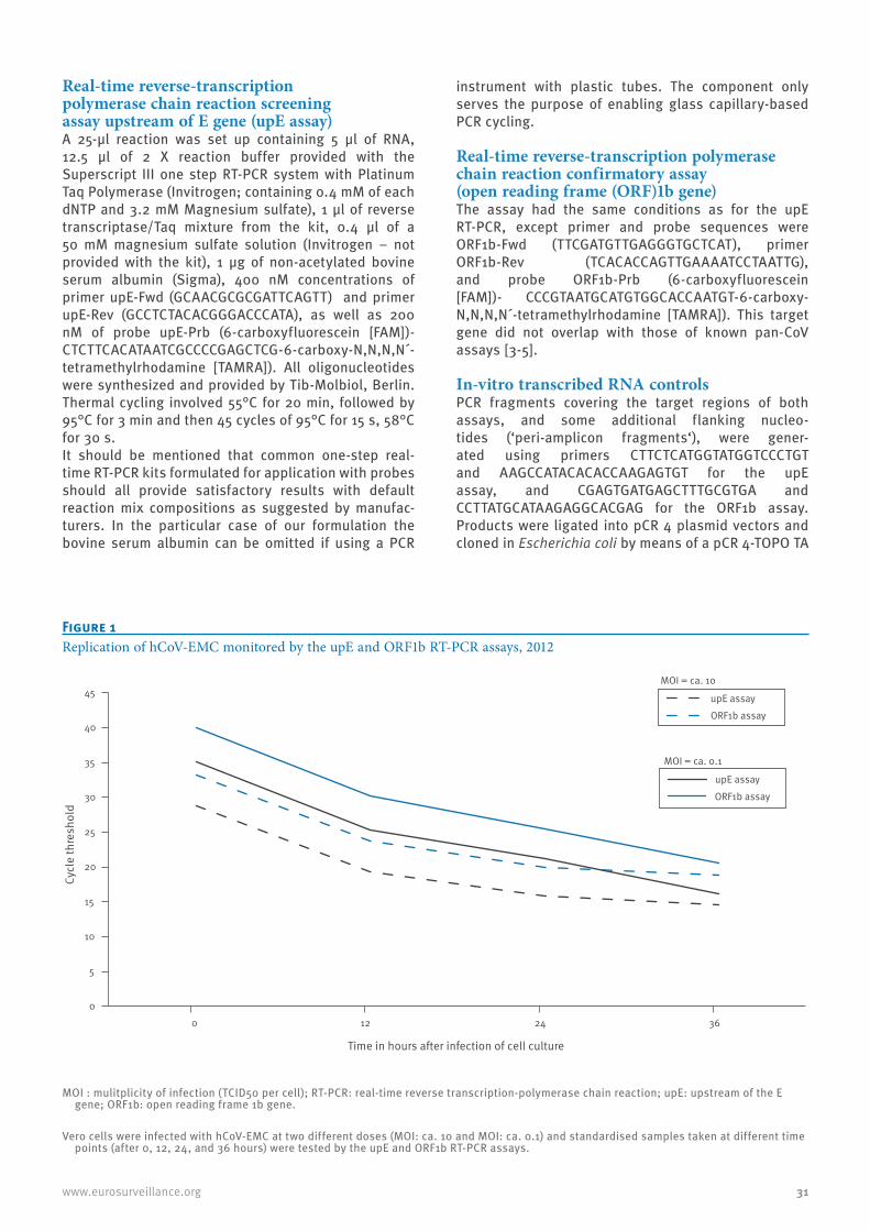
31www.eurosurveillance.org
Real-time reverse-transcription polymerase chain reaction screening assay upstream of E gene (upE assay)A 25-μl reaction was set up containing 5 μl of RNA, 12.5 μl of 2 X reaction buffer provided with the Superscript III one step RT-PCR system with Platinum Taq Polymerase (Invitrogen; containing 0.4 mM of each dNTP and 3.2 mM Magnesium sulfate), 1 μl of reverse transcriptase/Taq mixture from the kit, 0.4 µl of a 50 mM magnesium sulfate solution (Invitrogen – not provided with the kit), 1 μg of non-acetylated bovine serum albumin (Sigma), 400 nM concentrations of primer upE-Fwd (GCAACGCGCGATTCAGTT) and primer upE-Rev (GCCTCTACACGGGACCCATA), as well as 200 nM of probe upE-Prb (6-carboxyfluorescein [FAM])-CTCTTCACATAATCGCCCCGAGCTCG-6-carboxy-N,N,N,N´-tetramethylrhodamine [TAMRA]). All oligonucleotides were synthesized and provided by Tib-Molbiol, Berlin. Thermal cycling involved 55°C for 20 min, followed by 95°C for 3 min and then 45 cycles of 95°C for 15 s, 58°C for 30 s. It should be mentioned that common one-step real-time RT-PCR kits formulated for application with probes should all provide satisfactory results with default reaction mix compositions as suggested by manufac-turers. In the particular case of our formulation the bovine serum albumin can be omitted if using a PCR
instrument with plastic tubes. The component only serves the purpose of enabling glass capillary-based PCR cycling.
Real-time reverse-transcription polymerase chain reaction confirmatory assay (open reading frame (ORF)1b gene)The assay had the same conditions as for the upE RT-PCR, except primer and probe sequences were ORF1b-Fwd (TTCGATGTTGAGGGTGCTCAT), primer ORF1b-Rev (TCACACCAGTTGAAAATCCTAATTG), and probe ORF1b-Prb (6-carboxyfluorescein [FAM])- CCCGTAATGCATGTGGCACCAATGT-6-carboxy-N,N,N,N´-tetramethylrhodamine [TAMRA]). This target gene did not overlap with those of known pan-CoV assays [3-5].
In-vitro transcribed RNA controlsPCR fragments covering the target regions of both assays, and some additional flanking nucleo-tides (‘peri-amplicon fragments‘), were gener-ated using primers CTTCTCATGGTATGGTCCCTGT and AAGCCATACACACCAAGAGTGT for the upE assay, and CGAGTGATGAGCTTTGCGTGA and CCTTATGCATAAGAGGCACGAG for the ORF1b assay. Products were ligated into pCR 4 plasmid vectors and cloned in Escherichia coli by means of a pCR 4-TOPO TA
Figure 1Replication of hCoV-EMC monitored by the upE and ORF1b RT-PCR assays, 2012
0
5
10
15
20
25
30
35
40
45
0 12 24 36
upE assay ORF1b assay
upE assay
ORF1b assay
MOI = ca. 10
MOI = ca. 0.1
Cycl
e th
resh
old
Time in hours after infection of cell culture
MOI : mulitplicity of infection (TCID50 per cell); RT-PCR: real-time reverse transcription-polymerase chain reaction; upE: upstream of the E gene; ORF1b: open reading frame 1b gene.
Vero cells were infected with hCoV-EMC at two different doses (MOI: ca. 10 and MOI: ca. 0.1) and standardised samples taken at different time points (after 0, 12, 24, and 36 hours) were tested by the upE and ORF1b RT-PCR assays.
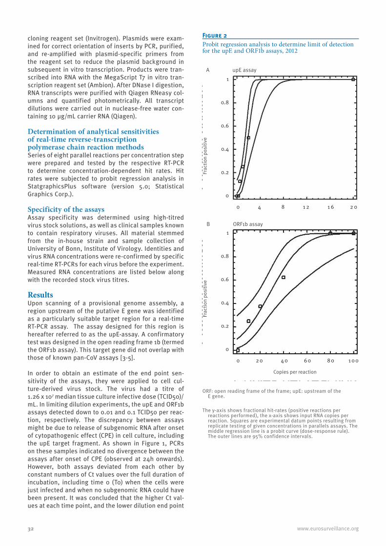
32 www.eurosurveillance.org
cloning reagent set (Invitrogen). Plasmids were exam-ined for correct orientation of inserts by PCR, purified, and re-amplified with plasmid-specific primers from the reagent set to reduce the plasmid background in subsequent in vitro transcription. Products were tran-scribed into RNA with the MegaScript T7 in vitro tran-scription reagent set (Ambion). After DNase I digestion, RNA transcripts were purified with Qiagen RNeasy col-umns and quantified photometrically. All transcript dilutions were carried out in nuclease-free water con-taining 10 µg/mL carrier RNA (Qiagen).
Determination of analytical sensitivities of real-time reverse-transcription polymerase chain reaction methodsSeries of eight parallel reactions per concentration step were prepared and tested by the respective RT-PCR to determine concentration-dependent hit rates. Hit rates were subjected to probit regression analysis in StatgraphicsPlus software (version 5.0; Statistical Graphics Corp.).
Specificity of the assaysAssay specificity was determined using high-titred virus stock solutions, as well as clinical samples known to contain respiratory viruses. All material stemmed from the in-house strain and sample collection of University of Bonn, Institute of Virology. Identities and virus RNA concentrations were re-confirmed by specific real-time RT-PCRs for each virus before the experiment. Measured RNA concentrations are listed below along with the recorded stock virus titres.
ResultsUpon scanning of a provisional genome assembly, a region upstream of the putative E gene was identified as a particularly suitable target region for a real-time RT-PCR assay. The assay designed for this region is hereafter referred to as the upE-assay. A confirmatory test was designed in the open reading frame 1b (termed the ORF1b assay). This target gene did not overlap with those of known pan-CoV assays [3-5].
In order to obtain an estimate of the end point sen-sitivity of the assays, they were applied to cell cul-ture-derived virus stock. The virus had a titre of 1.26 x 10⁷ median tissue culture infective dose (TCID50)/mL. In limiting dilution experiments, the upE and ORF1b assays detected down to 0.01 and 0.1 TCID50 per reac-tion, respectively. The discrepancy between assays might be due to release of subgenomic RNA after onset of cytopathogenic effect (CPE) in cell culture, including the upE target fragment. As shown in Figure 1, PCRs on these samples indicated no divergence between the assays after onset of CPE (observed at 24h onwards). However, both assays deviated from each other by constant numbers of Ct values over the full duration of incubation, including time 0 (T0) when the cells were just infected and when no subgenomic RNA could have been present. It was concluded that the higher Ct val-ues at each time point, and the lower dilution end point
Figure 2Probit regression analysis to determine limit of detection for the upE and ORF1b assays, 2012
A upE assay
Frac
tion
posi
tive
Frac
tion
posi
tive
Copies per reaction
1
0.8
0.6
0.4
0.2
0
1
0.8
0.6
0.4
0.2
0
0 4 8 1 2 1 6 2 0
0 2 0 4 0 6 0 8 0 1 0 0
B ORF1b assay
ORF: open reading frame of the frame; upE: upstream of the E gene.
The y-axis shows fractional hit-rates (positive reactions per reactions performed), the x-axis shows input RNA copies per reaction. Squares are experimental datum points resulting from replicate testing of given concentrations in parallels assays. The middle regression line is a probit curve (dose-response rule). The outer lines are 95% confidence intervals.
33www.eurosurveillance.org
for the ORF1b assay indicated that this assay had a lower sensitivity.
A more detailed assessment of technical sensitivity can be achieved using quantified, in-vitro transcribed RNA derived from the peri-amplicon region of each assay. These transcripts were generated and tested in serial ten-fold dilution experiments. Detection end points were two copies per reaction for the upE assay, and 10 copies per reaction for the confirmatory, ORF1b gene, assay. To obtain a statistically robust assessment of Limit Of Detection (LOD), transcripts were also tested in multiple parallel reactions in smaller dilution inter-vals above and below the end-point PCR limits. The results in terms of the fraction of positive reactions at each concentration were subjected to probit regression analysis and plotted as shown in Figure 2, where panel A shows the upE assay and panel B the ORF1b assay. The resulting LODs are summarised in Table 1. Based on the upE assay with a detection limit of 3.4 copies per reaction, and a cell-culture endpoint equivalent to 0.01 TCID50 per reaction, it was calculated that the RNA/infectious unit ratio of the virus stock must have been ca. 29 (100/3.4).
To exclude non-specific reactivity of oligonucleotides among each other, all formulations were tested 40 times in parallel with assays containing water and no other nucleic acids except the provided oligonucleo-tides. In none of these reactions was any positive sig-nal seen. Cross-reactivity with known, heterospecific human CoVs was excluded by testing high-titred cell culture materials as summarised in Table 1. It should be noted that the unculturable hCoV-HKU1 was not included in these experiments.
To obtain a more clinically relevant figure on assay specificity, the assays were applied on 92 original clinical samples in which other respiratory viruses had already been detected during routine respiratory screening at Bonn University Medical Centre. These samples were prepared using the Qiagen Viral RNA kit, a formulation widely used to extract RNA in clinical lab-oratories. Of note, the tested panel included four sam-ples containing hCoV-HKU1, which was not available as cultured virus stock. In total, none of the 92 original clinical samples as presented in Table 2, containing a wide range of respiratory viruses, gave any detection signal with either assay while positive controls were readily detected. It was concluded that the assay could be reliably applied to clinical samples.
Preliminary testing was also done on a patient hospi-talised with acute infection during preparation of this report (Authors R Gopal and M Zambon, own unpub-lished observations). Both assays provided very clear amplification signal on various clinical samples. The upE assay again appeared more sensitive than the ORF1b assay.
DiscussionHere we provide the technical background data for RT-PCR assays developed in rapid response to the emergence of a novel human CoV (GenBank accession number: JX869059 for the Rotterdam virus isolate, termed hCoV-EMC).
Cell culture-derived virus is a useful source of refer-ence material for the evaluation of molecular detection assays. However, detection end points determined on cell culture-derived virus are difficult to correlate to virus titre. Reasons include the discrepancy between
Experiment upE assay ORF1b assay
Detection end point for cell culture-derived virus 0.01 TCID50/reaction 0.1 TCID50/reaction
Technical LOD 3.4 RNA copies/reaction (95% CI: 2.5–6.9 copies/reaction)
64 RNA copies/reaction(95% CI: 47–126 copies/reaction)
Cross-reactivity with hCoV-229E No reactivity with virus containing 105 PFU/mL (3 x 10⁹ RNA copies/mL)
Cross-reactivity with hCoV-NL63 No reactivity with virus containing 106 PFU/mL(4 x 109 copies/mL)
Cross-reactivity with hCoV-OC43 No reactivity with virus containing 5 X 10⁵ PFU/mL(3 x 1010 copies/mL)
Cross-reactivity with SARS-CoV No reactivity with virus containing 3 x 10⁶ PFU/mL(5 x 1010 copies/mL)
CI: confidence interval CoV: corona virus; LOD: limit of detection; ORF: open reading frame; PFU: plaque forming units; TCID50: median tissue culture infective dose; upE: upstream of the E gene.
Table 1Results of sensitivity and specificity tests for hCoV-EMC assays, 2012*
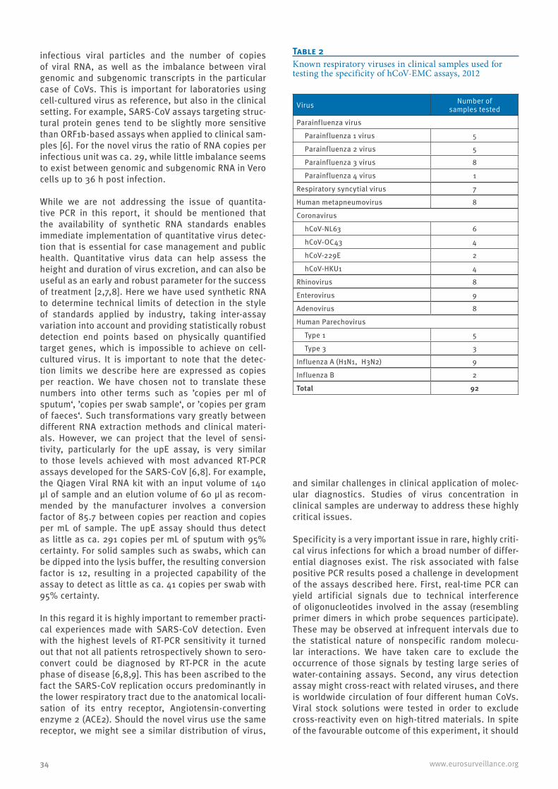
34 www.eurosurveillance.org
infectious viral particles and the number of copies of viral RNA, as well as the imbalance between viral genomic and subgenomic transcripts in the particular case of CoVs. This is important for laboratories using cell-cultured virus as reference, but also in the clinical setting. For example, SARS-CoV assays targeting struc-tural protein genes tend to be slightly more sensitive than ORF1b-based assays when applied to clinical sam-ples [6]. For the novel virus the ratio of RNA copies per infectious unit was ca. 29, while little imbalance seems to exist between genomic and subgenomic RNA in Vero cells up to 36 h post infection.
While we are not addressing the issue of quantita-tive PCR in this report, it should be mentioned that the availability of synthetic RNA standards enables immediate implementation of quantitative virus detec-tion that is essential for case management and public health. Quantitative virus data can help assess the height and duration of virus excretion, and can also be useful as an early and robust parameter for the success of treatment [2,7,8]. Here we have used synthetic RNA to determine technical limits of detection in the style of standards applied by industry, taking inter-assay variation into account and providing statistically robust detection end points based on physically quantified target genes, which is impossible to achieve on cell-cultured virus. It is important to note that the detec-tion limits we describe here are expressed as copies per reaction. We have chosen not to translate these numbers into other terms such as ’copies per ml of sputum‘, ’copies per swab sample‘, or ’copies per gram of faeces‘. Such transformations vary greatly between different RNA extraction methods and clinical materi-als. However, we can project that the level of sensi-tivity, particularly for the upE assay, is very similar to those levels achieved with most advanced RT-PCR assays developed for the SARS-CoV [6,8]. For example, the Qiagen Viral RNA kit with an input volume of 140 µl of sample and an elution volume of 60 µl as recom-mended by the manufacturer involves a conversion factor of 85.7 between copies per reaction and copies per mL of sample. The upE assay should thus detect as little as ca. 291 copies per mL of sputum with 95% certainty. For solid samples such as swabs, which can be dipped into the lysis buffer, the resulting conversion factor is 12, resulting in a projected capability of the assay to detect as little as ca. 41 copies per swab with 95% certainty.
In this regard it is highly important to remember practi-cal experiences made with SARS-CoV detection. Even with the highest levels of RT-PCR sensitivity it turned out that not all patients retrospectively shown to sero-convert could be diagnosed by RT-PCR in the acute phase of disease [6,8,9]. This has been ascribed to the fact the SARS-CoV replication occurs predominantly in the lower respiratory tract due to the anatomical locali-sation of its entry receptor, Angiotensin-converting enzyme 2 (ACE2). Should the novel virus use the same receptor, we might see a similar distribution of virus,
and similar challenges in clinical application of molec-ular diagnostics. Studies of virus concentration in clinical samples are underway to address these highly critical issues. Specificity is a very important issue in rare, highly criti-cal virus infections for which a broad number of differ-ential diagnoses exist. The risk associated with false positive PCR results posed a challenge in development of the assays described here. First, real-time PCR can yield artificial signals due to technical interference of oligonucleotides involved in the assay (resembling primer dimers in which probe sequences participate). These may be observed at infrequent intervals due to the statistical nature of nonspecific random molecu-lar interactions. We have taken care to exclude the occurrence of those signals by testing large series of water-containing assays. Second, any virus detection assay might cross-react with related viruses, and there is worldwide circulation of four different human CoVs. Viral stock solutions were tested in order to exclude cross-reactivity even on high-titred materials. In spite of the favourable outcome of this experiment, it should
Virus Number of samples tested
Parainfluenza virus
Parainfluenza 1 virus 5
Parainfluenza 2 virus 5
Parainfluenza 3 virus 8
Parainfluenza 4 virus 1
Respiratory syncytial virus 7
Human metapneumovirus 8
Coronavirus
hCoV-NL63 6
hCoV-OC43 4
hCoV-229E 2
hCoV-HKU1 4
Rhinovirus 8
Enterovirus 9
Adenovirus 8
Human Parechovirus
Type 1 5
Type 3 3
Influenza A (H1N1, H3N2) 9
Influenza B 2
Total 92
Table 2Known respiratory viruses in clinical samples used for testing the specificity of hCoV-EMC assays, 2012
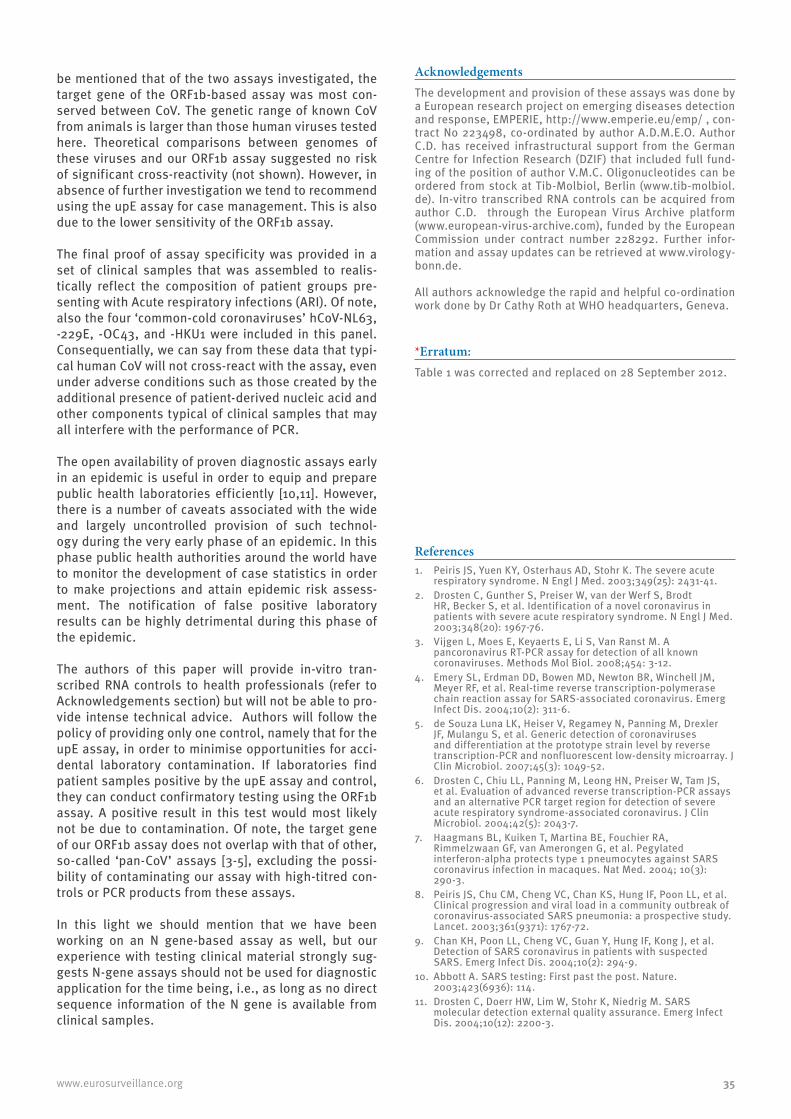
35www.eurosurveillance.org
be mentioned that of the two assays investigated, the target gene of the ORF1b-based assay was most con-served between CoV. The genetic range of known CoV from animals is larger than those human viruses tested here. Theoretical comparisons between genomes of these viruses and our ORF1b assay suggested no risk of significant cross-reactivity (not shown). However, in absence of further investigation we tend to recommend using the upE assay for case management. This is also due to the lower sensitivity of the ORF1b assay.
The final proof of assay specificity was provided in a set of clinical samples that was assembled to realis-tically reflect the composition of patient groups pre-senting with Acute respiratory infections (ARI). Of note, also the four ‘common-cold coronaviruses’ hCoV-NL63, -229E, -OC43, and -HKU1 were included in this panel. Consequentially, we can say from these data that typi-cal human CoV will not cross-react with the assay, even under adverse conditions such as those created by the additional presence of patient-derived nucleic acid and other components typical of clinical samples that may all interfere with the performance of PCR.
The open availability of proven diagnostic assays early in an epidemic is useful in order to equip and prepare public health laboratories efficiently [10,11]. However, there is a number of caveats associated with the wide and largely uncontrolled provision of such technol-ogy during the very early phase of an epidemic. In this phase public health authorities around the world have to monitor the development of case statistics in order to make projections and attain epidemic risk assess-ment. The notification of false positive laboratory results can be highly detrimental during this phase of the epidemic.
The authors of this paper will provide in-vitro tran-scribed RNA controls to health professionals (refer to Acknowledgements section) but will not be able to pro-vide intense technical advice. Authors will follow the policy of providing only one control, namely that for the upE assay, in order to minimise opportunities for acci-dental laboratory contamination. If laboratories find patient samples positive by the upE assay and control, they can conduct confirmatory testing using the ORF1b assay. A positive result in this test would most likely not be due to contamination. Of note, the target gene of our ORF1b assay does not overlap with that of other, so-called ‘pan-CoV’ assays [3-5], excluding the possi-bility of contaminating our assay with high-titred con-trols or PCR products from these assays.
In this light we should mention that we have been working on an N gene-based assay as well, but our experience with testing clinical material strongly sug-gests N-gene assays should not be used for diagnostic application for the time being, i.e., as long as no direct sequence information of the N gene is available from clinical samples.
AcknowledgementsThe development and provision of these assays was done by a European research project on emerging diseases detection and response, EMPERIE, http://www.emperie.eu/emp/ , con-tract No 223498, co-ordinated by author A.D.M.E.O. Author C.D. has received infrastructural support from the German Centre for Infection Research (DZIF) that included full fund-ing of the position of author V.M.C. Oligonucleotides can be ordered from stock at Tib-Molbiol, Berlin (www.tib-molbiol.de). In-vitro transcribed RNA controls can be acquired from author C.D. through the European Virus Archive platform (www.european-virus-archive.com), funded by the European Commission under contract number 228292. Further infor-mation and assay updates can be retrieved at www.virology-bonn.de.
All authors acknowledge the rapid and helpful co-ordination work done by Dr Cathy Roth at WHO headquarters, Geneva.
*Erratum: Table 1 was corrected and replaced on 28 September 2012.
References1. Peiris JS, Yuen KY, Osterhaus AD, Stohr K. The severe acute
respiratory syndrome. N Engl J Med. 2003;349(25): 2431-41. 2. Drosten C, Gunther S, Preiser W, van der Werf S, Brodt
HR, Becker S, et al. Identification of a novel coronavirus in patients with severe acute respiratory syndrome. N Engl J Med. 2003;348(20): 1967-76.
3. Vijgen L, Moes E, Keyaerts E, Li S, Van Ranst M. A pancoronavirus RT-PCR assay for detection of all known coronaviruses. Methods Mol Biol. 2008;454: 3-12.
4. Emery SL, Erdman DD, Bowen MD, Newton BR, Winchell JM, Meyer RF, et al. Real-time reverse transcription-polymerase chain reaction assay for SARS-associated coronavirus. Emerg Infect Dis. 2004;10(2): 311-6.
5. de Souza Luna LK, Heiser V, Regamey N, Panning M, Drexler JF, Mulangu S, et al. Generic detection of coronaviruses and differentiation at the prototype strain level by reverse transcription-PCR and nonfluorescent low-density microarray. J Clin Microbiol. 2007;45(3): 1049-52.
6. Drosten C, Chiu LL, Panning M, Leong HN, Preiser W, Tam JS, et al. Evaluation of advanced reverse transcription-PCR assays and an alternative PCR target region for detection of severe acute respiratory syndrome-associated coronavirus. J Clin Microbiol. 2004;42(5): 2043-7.
7. Haagmans BL, Kuiken T, Martina BE, Fouchier RA, Rimmelzwaan GF, van Amerongen G, et al. Pegylated interferon-alpha protects type 1 pneumocytes against SARS coronavirus infection in macaques. Nat Med. 2004; 10(3): 290-3.
8. Peiris JS, Chu CM, Cheng VC, Chan KS, Hung IF, Poon LL, et al. Clinical progression and viral load in a community outbreak of coronavirus-associated SARS pneumonia: a prospective study. Lancet. 2003;361(9371): 1767-72.
9. Chan KH, Poon LL, Cheng VC, Guan Y, Hung IF, Kong J, et al. Detection of SARS coronavirus in patients with suspected SARS. Emerg Infect Dis. 2004;10(2): 294-9.
10. Abbott A. SARS testing: First past the post. Nature. 2003;423(6936): 114.
11. Drosten C, Doerr HW, Lim W, Stohr K, Niedrig M. SARS molecular detection external quality assurance. Emerg Infect Dis. 2004;10(12): 2200-3.
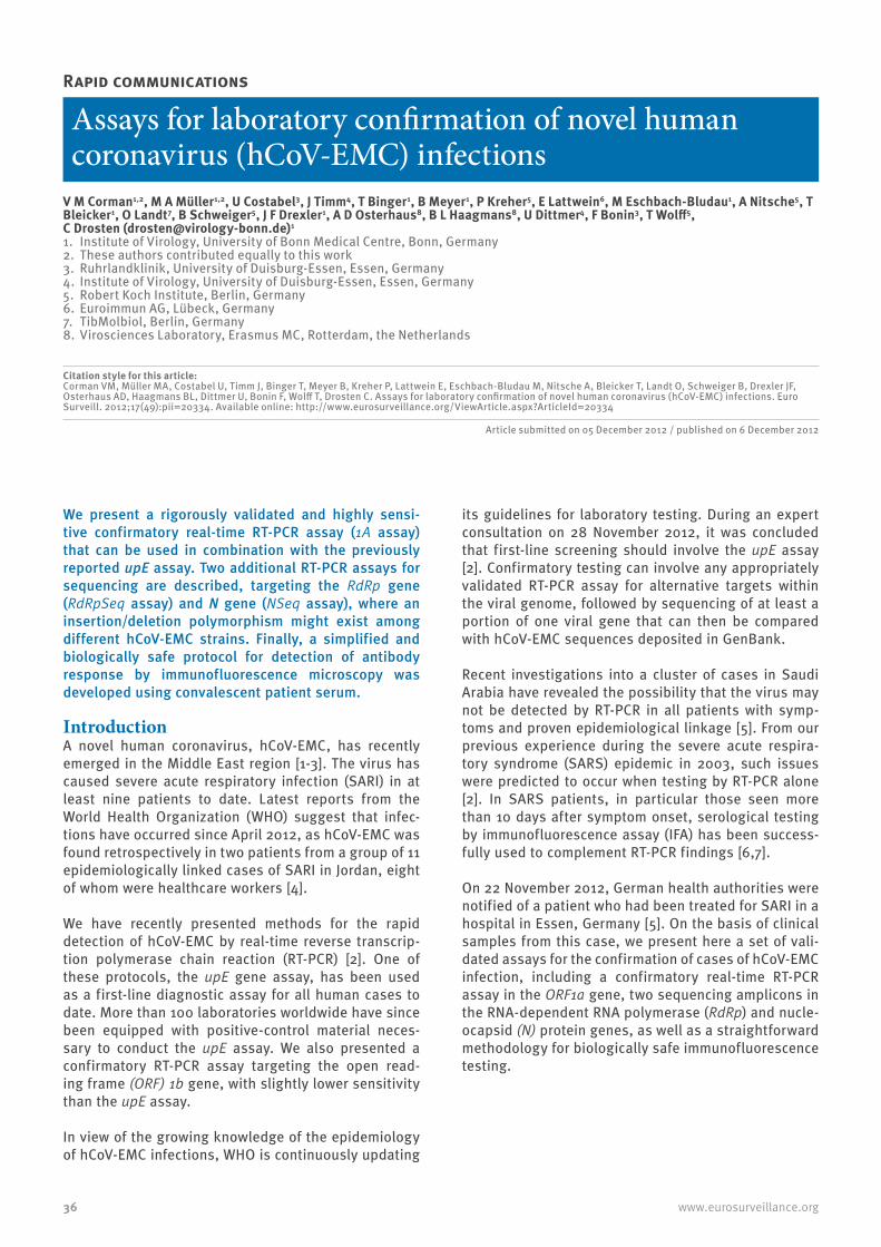
36 www.eurosurveillance.org
Rapid communications
Assays for laboratory confirmation of novel human coronavirus (hCoV-EMC) infections
V M Corman1,2, M A Müller1,2, U Costabel3, J Timm4, T Binger1, B Meyer1, P Kreher5, E Lattwein6, M Eschbach-Bludau1, A Nitsche5, T Bleicker1, O Landt7, B Schweiger5, J F Drexler1, A D Osterhaus8, B L Haagmans8, U Dittmer4, F Bonin3, T Wolff5, C Drosten ([email protected])1
1. Institute of Virology, University of Bonn Medical Centre, Bonn, Germany2. These authors contributed equally to this work3. Ruhrlandklinik, University of Duisburg-Essen, Essen, Germany4. Institute of Virology, University of Duisburg-Essen, Essen, Germany5. Robert Koch Institute, Berlin, Germany6. Euroimmun AG, Lübeck, Germany7. TibMolbiol, Berlin, Germany8. Virosciences Laboratory, Erasmus MC, Rotterdam, the Netherlands
Citation style for this article: Corman VM, Müller MA, Costabel U, Timm J, Binger T, Meyer B, Kreher P, Lattwein E, Eschbach-Bludau M, Nitsche A, Bleicker T, Landt O, Schweiger B, Drexler JF, Osterhaus AD, Haagmans BL, Dittmer U, Bonin F, Wolff T, Drosten C. Assays for laboratory confirmation of novel human coronavirus (hCoV-EMC) infections. Euro Surveill. 2012;17(49):pii=20334. Available online: http://www.eurosurveillance.org/ViewArticle.aspx?ArticleId=20334
Article submitted on 05 December 2012 / published on 6 December 2012
We present a rigorously validated and highly sensi-tive confirmatory real-time RT-PCR assay (1A assay) that can be used in combination with the previously reported upE assay. Two additional RT-PCR assays for sequencing are described, targeting the RdRp gene (RdRpSeq assay) and N gene (NSeq assay), where an insertion/deletion polymorphism might exist among different hCoV-EMC strains. Finally, a simplified and biologically safe protocol for detection of antibody response by immunofluorescence microscopy was developed using convalescent patient serum.
IntroductionA novel human coronavirus, hCoV-EMC, has recently emerged in the Middle East region [1-3]. The virus has caused severe acute respiratory infection (SARI) in at least nine patients to date. Latest reports from the World Health Organization (WHO) suggest that infec-tions have occurred since April 2012, as hCoV-EMC was found retrospectively in two patients from a group of 11 epidemiologically linked cases of SARI in Jordan, eight of whom were healthcare workers [4].
We have recently presented methods for the rapid detection of hCoV-EMC by real-time reverse transcrip-tion polymerase chain reaction (RT-PCR) [2]. One of these protocols, the upE gene assay, has been used as a first-line diagnostic assay for all human cases to date. More than 100 laboratories worldwide have since been equipped with positive-control material neces-sary to conduct the upE assay. We also presented a confirmatory RT-PCR assay targeting the open read-ing frame (ORF) 1b gene, with slightly lower sensitivity than the upE assay.
In view of the growing knowledge of the epidemiology of hCoV-EMC infections, WHO is continuously updating
its guidelines for laboratory testing. During an expert consultation on 28 November 2012, it was concluded that first-line screening should involve the upE assay [2]. Confirmatory testing can involve any appropriately validated RT-PCR assay for alternative targets within the viral genome, followed by sequencing of at least a portion of one viral gene that can then be compared with hCoV-EMC sequences deposited in GenBank.
Recent investigations into a cluster of cases in Saudi Arabia have revealed the possibility that the virus may not be detected by RT-PCR in all patients with symp-toms and proven epidemiological linkage [5]. From our previous experience during the severe acute respira-tory syndrome (SARS) epidemic in 2003, such issues were predicted to occur when testing by RT-PCR alone [2]. In SARS patients, in particular those seen more than 10 days after symptom onset, serological testing by immunofluorescence assay (IFA) has been success-fully used to complement RT-PCR findings [6,7].
On 22 November 2012, German health authorities were notified of a patient who had been treated for SARI in a hospital in Essen, Germany [5]. On the basis of clinical samples from this case, we present here a set of vali-dated assays for the confirmation of cases of hCoV-EMC infection, including a confirmatory real-time RT-PCR assay in the ORF1a gene, two sequencing amplicons in the RNA-dependent RNA polymerase (RdRp) and nucle-ocapsid (N) protein genes, as well as a straightforward methodology for biologically safe immunofluorescence testing.
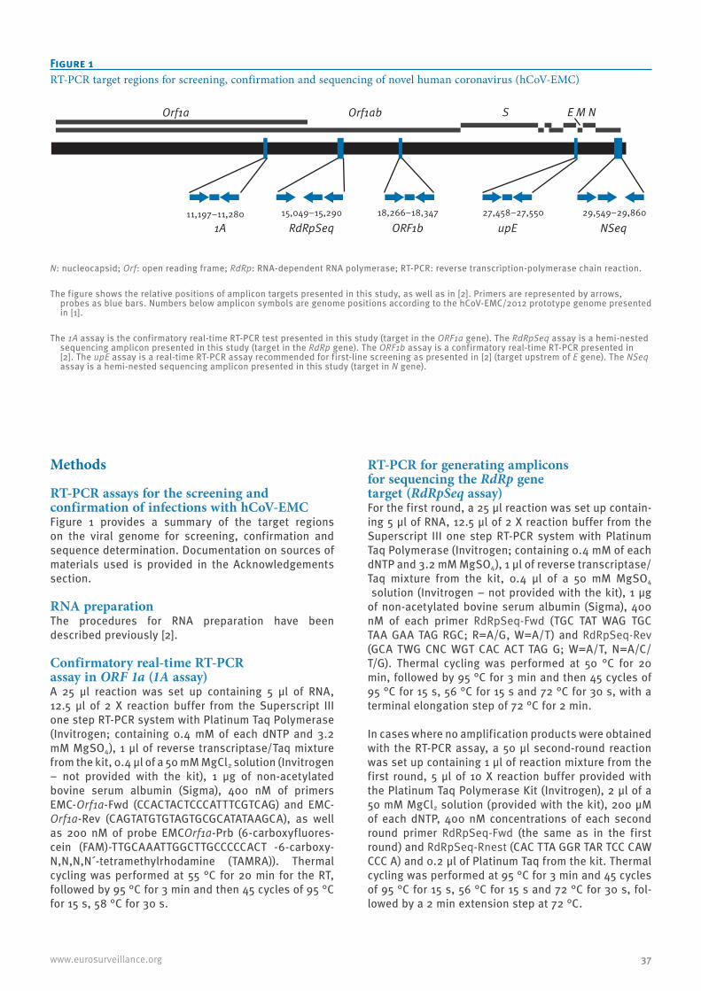
37www.eurosurveillance.org
Methods
RT-PCR assays for the screening and confirmation of infections with hCoV-EMCFigure 1 provides a summary of the target regions on the viral genome for screening, confirmation and sequence determination. Documentation on sources of materials used is provided in the Acknowledgements section.
RNA preparationThe procedures for RNA preparation have been described previously [2].
Confirmatory real-time RT-PCR assay in ORF 1a (1A assay)A 25 µl reaction was set up containing 5 µl of RNA, 12.5 µl of 2 X reaction buffer from the Superscript III one step RT-PCR system with Platinum Taq Polymerase (Invitrogen; containing 0.4 mM of each dNTP and 3.2 mM MgSO4), 1 µl of reverse transcriptase/Taq mixture from the kit, 0.4 µl of a 50 mM MgCl2 solution (Invitrogen – not provided with the kit), 1 μg of non-acetylated bovine serum albumin (Sigma), 400 nM of primers EMC-Orf1a-Fwd (CCACTACTCCCATTTCGTCAG) and EMC-Orf1a-Rev (CAGTATGTGTAGTGCGCATATAAGCA), as well as 200 nM of probe EMCOrf1a-Prb (6-carboxyfluores-cein (FAM)-TTGCAAATTGGCTTGCCCCCACT -6-carboxy-N,N,N,N´-tetramethylrhodamine (TAMRA)). Thermal cycling was performed at 55 °C for 20 min for the RT, followed by 95 °C for 3 min and then 45 cycles of 95 °C for 15 s, 58 °C for 30 s.
RT-PCR for generating amplicons for sequencing the RdRp gene target (RdRpSeq assay)For the first round, a 25 µl reaction was set up contain-ing 5 µl of RNA, 12.5 µl of 2 X reaction buffer from the Superscript III one step RT-PCR system with Platinum Taq Polymerase (Invitrogen; containing 0.4 mM of each dNTP and 3.2 mM MgSO4), 1 µl of reverse transcriptase/Taq mixture from the kit, 0.4 µl of a 50 mM MgSO4
solution (Invitrogen – not provided with the kit), 1 μg of non-acetylated bovine serum albumin (Sigma), 400 nM of each primer RdRpSeq-Fwd (TGC TAT WAG TGC TAA GAA TAG RGC; R=A/G, W=A/T) and RdRpSeq-Rev (GCA TWG CNC WGT CAC ACT TAG G; W=A/T, N=A/C/T/G). Thermal cycling was performed at 50 °C for 20 min, followed by 95 °C for 3 min and then 45 cycles of 95 °C for 15 s, 56 °C for 15 s and 72 °C for 30 s, with a terminal elongation step of 72 °C for 2 min.
In cases where no amplification products were obtained with the RT-PCR assay, a 50 µl second-round reaction was set up containing 1 µl of reaction mixture from the first round, 5 µl of 10 X reaction buffer provided with the Platinum Taq Polymerase Kit (Invitrogen), 2 µl of a 50 mM MgCl2 solution (provided with the kit), 200 µM of each dNTP, 400 nM concentrations of each second round primer RdRpSeq-Fwd (the same as in the first round) and RdRpSeq-Rnest (CAC TTA GGR TAR TCC CAW CCC A) and 0.2 μl of Platinum Taq from the kit. Thermal cycling was performed at 95 °C for 3 min and 45 cycles of 95 °C for 15 s, 56 °C for 15 s and 72 °C for 30 s, fol-lowed by a 2 min extension step at 72 °C.
Figure 1RT-PCR target regions for screening, confirmation and sequencing of novel human coronavirus (hCoV-EMC)
Orf1abOrf1a
15,049−15,290 18,266−18,347 27,458−27,550 29,549−29,860
E M NS
RdRpSeq1A ORF1b upE NSeq11,197−11,280
N: nucleocapsid; Orf: open reading frame; RdRp: RNA-dependent RNA polymerase; RT-PCR: reverse transcription-polymerase chain reaction.
The figure shows the relative positions of amplicon targets presented in this study, as well as in [2]. Primers are represented by arrows, probes as blue bars. Numbers below amplicon symbols are genome positions according to the hCoV-EMC/2012 prototype genome presented in [1].
The 1A assay is the confirmatory real-time RT-PCR test presented in this study (target in the ORF1a gene). The RdRpSeq assay is a hemi-nested sequencing amplicon presented in this study (target in the RdRp gene). The ORF1b assay is a confirmatory real-time RT-PCR presented in [2]. The upE assay is a real-time RT-PCR assay recommended for first-line screening as presented in [2] (target upstrem of E gene). The NSeq assay is a hemi-nested sequencing amplicon presented in this study (target in N gene).
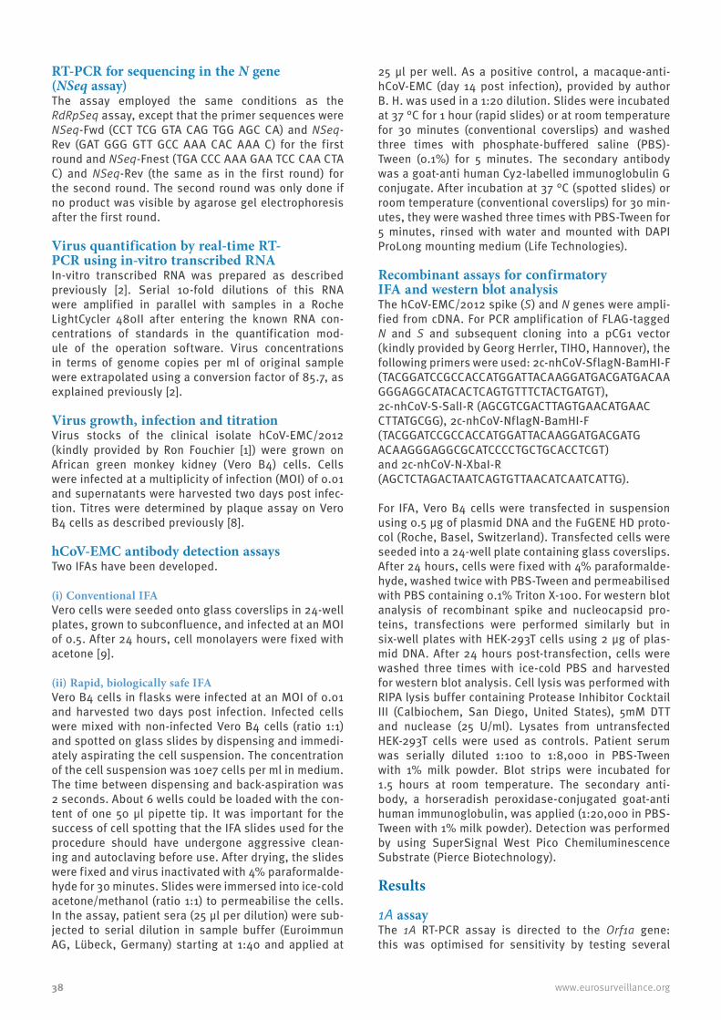
38 www.eurosurveillance.org
RT-PCR for sequencing in the N gene (NSeq assay)The assay employed the same conditions as the RdRpSeq assay, except that the primer sequences were NSeq-Fwd (CCT TCG GTA CAG TGG AGC CA) and NSeq-Rev (GAT GGG GTT GCC AAA CAC AAA C) for the first round and NSeq-Fnest (TGA CCC AAA GAA TCC CAA CTA C) and NSeq-Rev (the same as in the first round) for the second round. The second round was only done if no product was visible by agarose gel electrophoresis after the first round.
Virus quantification by real-time RT-PCR using in-vitro transcribed RNA In-vitro transcribed RNA was prepared as described previously [2]. Serial 10-fold dilutions of this RNA were amplified in parallel with samples in a Roche LightCycler 480II after entering the known RNA con-centrations of standards in the quantification mod-ule of the operation software. Virus concentrations in terms of genome copies per ml of original sample were extrapolated using a conversion factor of 85.7, as explained previously [2].
Virus growth, infection and titrationVirus stocks of the clinical isolate hCoV-EMC/2012 (kindly provided by Ron Fouchier [1]) were grown on African green monkey kidney (Vero B4) cells. Cells were infected at a multiplicity of infection (MOI) of 0.01 and supernatants were harvested two days post infec-tion. Titres were determined by plaque assay on Vero B4 cells as described previously [8].
hCoV-EMC antibody detection assaysTwo IFAs have been developed.
(i) Conventional IFA Vero cells were seeded onto glass coverslips in 24-well plates, grown to subconfluence, and infected at an MOI of 0.5. After 24 hours, cell monolayers were fixed with acetone [9].
(ii) Rapid, biologically safe IFAVero B4 cells in flasks were infected at an MOI of 0.01 and harvested two days post infection. Infected cells were mixed with non-infected Vero B4 cells (ratio 1:1) and spotted on glass slides by dispensing and immedi-ately aspirating the cell suspension. The concentration of the cell suspension was 10e7 cells per ml in medium. The time between dispensing and back-aspiration was 2 seconds. About 6 wells could be loaded with the con-tent of one 50 µl pipette tip. It was important for the success of cell spotting that the IFA slides used for the procedure should have undergone aggressive clean-ing and autoclaving before use. After drying, the slides were fixed and virus inactivated with 4% paraformalde-hyde for 30 minutes. Slides were immersed into ice-cold acetone/methanol (ratio 1:1) to permeabilise the cells. In the assay, patient sera (25 µl per dilution) were sub-jected to serial dilution in sample buffer (Euroimmun AG, Lübeck, Germany) starting at 1:40 and applied at
25 µl per well. As a positive control, a macaque-anti-hCoV-EMC (day 14 post infection), provided by author B. H. was used in a 1:20 dilution. Slides were incubated at 37 °C for 1 hour (rapid slides) or at room temperature for 30 minutes (conventional coverslips) and washed three times with phosphate-buffered saline (PBS)-Tween (0.1%) for 5 minutes. The secondary antibody was a goat-anti human Cy2-labelled immunoglobulin G conjugate. After incubation at 37 °C (spotted slides) or room temperature (conventional coverslips) for 30 min-utes, they were washed three times with PBS-Tween for 5 minutes, rinsed with water and mounted with DAPI ProLong mounting medium (Life Technologies).
Recombinant assays for confirmatory IFA and western blot analysisThe hCoV-EMC/2012 spike (S) and N genes were ampli-fied from cDNA. For PCR amplification of FLAG-tagged N and S and subsequent cloning into a pCG1 vector (kindly provided by Georg Herrler, TIHO, Hannover), the following primers were used: 2c-nhCoV-SflagN-BamHI-F(TACGGATCCGCCACCATGGATTACAAGGATGACGATGACAA GGGAGGCATACACTCAGTGTTTCTACTGATGT), 2c-nhCoV-S-SalI-R (AGCGTCGACTTAGTGAACATGAACCTTATGCGG), 2c-nhCoV-NflagN-BamHI-F (TACGGATCCGCCACCATGGATTACAAGGATGACGATGACAAGGGAGGCGCATCCCCTGCTGCACCTCGT) and 2c-nhCoV-N-XbaI-R (AGCTCTAGACTAATCAGTGTTAACATCAATCATTG).
For IFA, Vero B4 cells were transfected in suspension using 0.5 µg of plasmid DNA and the FuGENE HD proto-col (Roche, Basel, Switzerland). Transfected cells were seeded into a 24-well plate containing glass coverslips. After 24 hours, cells were fixed with 4% paraformalde-hyde, washed twice with PBS-Tween and permeabilised with PBS containing 0.1% Triton X-100. For western blot analysis of recombinant spike and nucleocapsid pro-teins, transfections were performed similarly but in six-well plates with HEK-293T cells using 2 µg of plas-mid DNA. After 24 hours post-transfection, cells were washed three times with ice-cold PBS and harvested for western blot analysis. Cell lysis was performed with RIPA lysis buffer containing Protease Inhibitor Cocktail III (Calbiochem, San Diego, United States), 5mM DTT and nuclease (25 U/ml). Lysates from untransfected HEK-293T cells were used as controls. Patient serum was serially diluted 1:100 to 1:8,000 in PBS-Tween with 1% milk powder. Blot strips were incubated for 1.5 hours at room temperature. The secondary anti-body, a horseradish peroxidase-conjugated goat-anti human immunoglobulin, was applied (1:20,000 in PBS-Tween with 1% milk powder). Detection was performed by using SuperSignal West Pico Chemiluminescence Substrate (Pierce Biotechnology).
Results
1A assayThe 1A RT-PCR assay is directed to the Orf1a gene: this was optimised for sensitivity by testing several
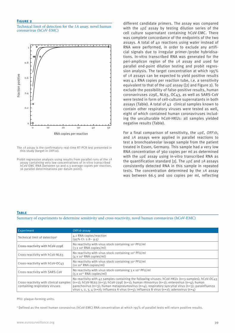
39www.eurosurveillance.org
different candidate primers. The assay was compared with the upE assay by testing dilution series of the cell culture supernatant containing hCoV-EMC. There was complete concordance of the endpoints of the two assays. A total of 40 reactions using water instead of RNA were performed, in order to exclude any artifi-cial signals due to irregular primer-/probe hybridisa-tions. In-vitro transcribed RNA was generated for the peri-amplicon region of the 1A assay and used for parallel end-point dilution testing and probit regres-sion analysis. The target concentration at which >95% of 1A assays can be expected to yield positive results was 4.1 RNA copies per reaction tube, i.e. a sensitivity equivalent to that of the upE assay ([2] and Figure 2). To exclude the possibility of false-positive results, human coronaviruses 229E, NL63, OC43, as well as SARS-CoV were tested in form of cell-culture supernatants in both assays (Table). A total of 42 clinical samples known to contain other respiratory viruses were tested as well, eight of which contained human coronaviruses includ-ing the unculturable hCoV-HKU1: all samples yielded negative results (Table). For a final comparison of sensitivity, the upE, ORF1b, and 1A assays were applied in parallel reactions to test a bronchoalveolar lavage sample from the patient treated in Essen, Germany. This sample had a very low RNA concentration of 360 copies per ml as determined with the upE assay using in-vitro transcribed RNA as the quantification standard [2]. The upE and 1A assays consistently detected RNA in this sample in repeated tests. The concentration determined by the 1A assay was between 66.5 and 100 copies per ml, reflecting
Figure 2Technical limit of detection for the 1A assay, novel human coronavirus (hCoV-EMC)
0
0.2
0.4
0.8
0.6
1
0 10 20 30 40 50
RNA copies per reaction
Frac
tion
posi
tive
The 1A assay is the confirmatory real-time RT-PCR test presented in this study (target in ORF1a).
Probit regression analysis using results from parallel runs of the 1A assay containing very low concentrations of in-vitro transcribed hCoV-EMC RNA (between 50 and 0.3 average copies per reaction, 16 parallel determinations per datum point).
Table Summary of experiments to determine sensitivity and cross-reactivity, novel human coronavirus (hCoV-EMC)
Experiment ORF1b assay
Technical limit of detectiona 4.1 RNA copies/reaction(95% CI: 2.8– 9.5)
Cross-reactivity with hCoV-229E No reactivity with virus stock containing 105 PFU/ml (3 x 10⁹ RNA copies/ml)
Cross-reactivity with hCoV-NL63 No reactivity with virus stock containing 106 PFU/ml (4 x 10⁹ RNA copies/ml)
Cross-reactivity with hCoV-OC43 No reactivity with virus stock containing 104 PFU/ml (1x 10⁸ RNA copies/ml)
Cross-reactivity with SARS-CoV No reactivity with virus stock containing 3 x 10⁶ PFU/ml (5 x 10¹⁰ RNA copies/ml)
Cross-reactivity with clinical samples containing respiratory viruses
No reactivity with 42 samples containing the following viruses: hCoV-HKU1 (n=3 samples); hCoV-OC43 (n=1); hCoV-NL63 (n=3); hCoV-229E (n=1); human rhinovirus (n=2); enterovirus (n=4); human parechovirus (n=3); human metapneumovirus (n=4); respiratory syncytial virus (n=3); parainfluenza virus 1, 2, 3, 4 (n=7); influenza A virus (n=5); influenza B virus (n=2); adenovirus (n=4)
PFU: plaque-forming units.
a Defined as the novel human coronavirus (hCoV-EMC) RNA concentration at which >95% of parallel tests will return positive results.
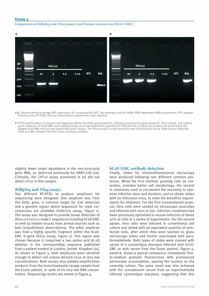
40 www.eurosurveillance.org
slightly lower target abundance in the non-structural gene RNA, as observed previously for SARS-CoV [10]. Critically, the ORF1b assay presented in [2] did not detect virus in this sample.
RdRpSeq and NSeq assaysTwo different RT-PCRs to produce amplicons for sequencing were designed. One amplicon was from the RdRp gene, a common target for CoV detection and a genome region where sequences for most cor-onaviruses are available (RdRpSeq assay, Figure 1). The assay was designed to provide broad detection of Betacoronavirus clade C sequences including hCoV-EMC as well as related viruses from animal sources such as bats (unpublished observations). The other amplicon was from a highly specific fragment within the hCoV-EMC N gene (NSeq assay, Figure 1). This region was chosen because it comprised a two amino acid (6 nt) deletion in the corresponding sequence published from a patient treated in London, United Kingdom [11]. As shown in Figure 3, both amplicons were sensitive enough to detect cell culture-derived virus at very low concentrations. Both assays also yielded amplification products from the bronchoalveolar lavage sample from the Essen patient, in spite of its very low RNA concen-tration. Sequencing results are shown in Figure 4.
hCoV-EMC antibody detectionFinally, slides for immunofluorescence microscopy were produced following two different common pro-tocols. While the first method, growing cells on cov-erslips, provides better cell morphology, the second is commonly used to circumvent the necessity to opti-mise infection dose and duration, and to obtain slides with no infectious virus, to meet the biosafety require-ments for shipment. For the first (conventional) proto-col, Vero cells were seeded on microscope coverslips and infected with virus in situ. Infection conditions had been previously optimised to ensure infection of about 30% of cells in a series of experiments. For the second option, Vero cells were infected in conventional cell culture and mixed with an equivalent quantity of unin-fected cells, after which they were spotted on glass microscope slides and further inactivated with para-formaldehyde. Both types of slides were stained with serum of a cynomolgus macaque infected with hCoV-EMC or with serum from the Essen patient. Figure 5, panel A, shows a typical coronavirus cytoplasmic fine-to-medium granular fluorescence with pronounced perinuclear accumulation, sparing the nucleus on the coverslip culture. The same result was also achieved with the convalescent serum from an experimentally infected cynomolgus macaque, suggesting that this
Figure 3Comparison of RdRpSeq and NSeq assays, novel human coronavirus (hCoV-EMC)
BAL: bronchoalveolar lavage; BP: base pairs; N: nucleocapsid; NTC: No template control; RdRp: RNA-dependent RNA polymerase; PFU: plaque-forming units; RT-PCR: reverse transcription-polymerase chain reaction.
RT-PCR amplification of sequencing fragments within the RdRp gene (panel A, RdRpSeq assay) and N gene (panel B, NSeq assay). Cell culture stock solutions of hCoV-EMC were diluted to the virus concentrations specified (in PFU per ml), of which 50 µl were extracted using the Qiagen Viral RNA mini kit and tested with both assays. The NSeq assay is more sensitive than the RdRpSeq assay. Both assays detected virus in a BAL sample from the Essen, Germany, patient.
300
200
100
BP600
PFU/ml
30 3 0.3 0.03
PFU/ml
NTC BAL 30 3 0.3 0.03 NTC BAL
A B
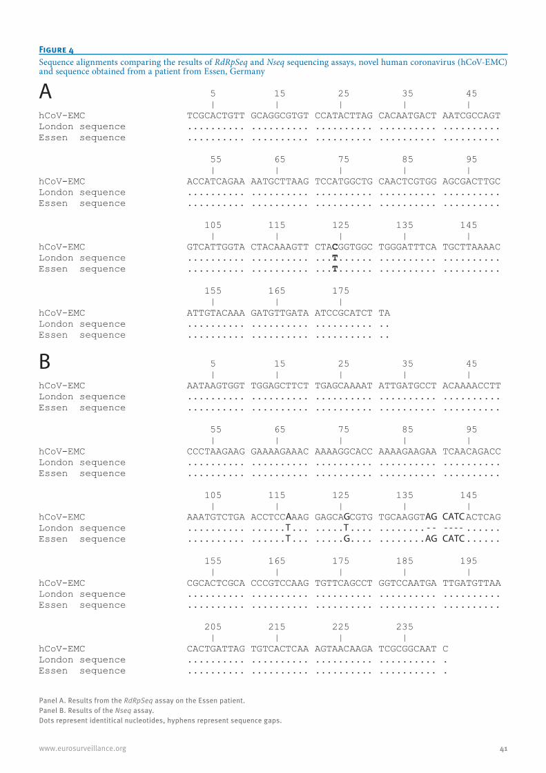
41www.eurosurveillance.org
Figure 4Sequence alignments comparing the results of RdRpSeq and Nseq sequencing assays, novel human coronavirus (hCoV-EMC) and sequence obtained from a patient from Essen, Germany
Panel A. Results from the RdRpSeq assay on the Essen patient. Panel B. Results of the Nseq assay. Dots represent identitical nucleotides, hyphens represent sequence gaps.
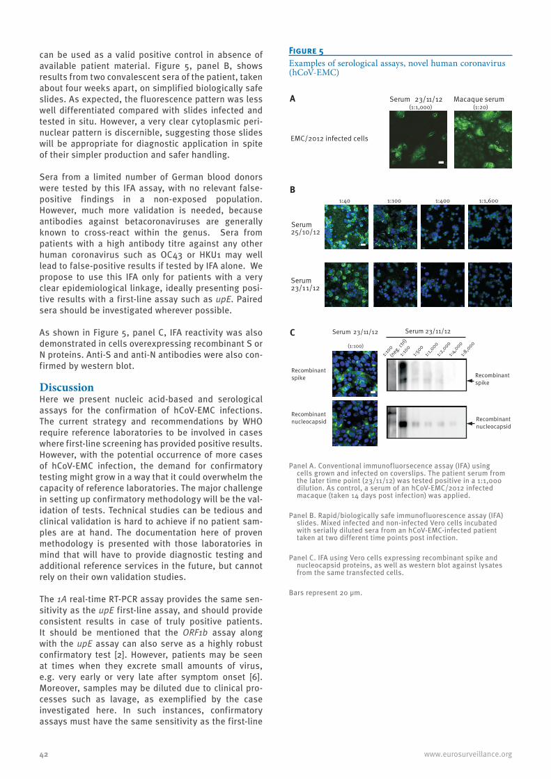
42 www.eurosurveillance.org
can be used as a valid positive control in absence of available patient material. Figure 5, panel B, shows results from two convalescent sera of the patient, taken about four weeks apart, on simplified biologically safe slides. As expected, the fluorescence pattern was less well differentiated compared with slides infected and tested in situ. However, a very clear cytoplasmic peri-nuclear pattern is discernible, suggesting those slides will be appropriate for diagnostic application in spite of their simpler production and safer handling.
Sera from a limited number of German blood donors were tested by this IFA assay, with no relevant false-positive findings in a non-exposed population. However, much more validation is needed, because antibodies against betacoronaviruses are generally known to cross-react within the genus. Sera from patients with a high antibody titre against any other human coronavirus such as OC43 or HKU1 may well lead to false-positive results if tested by IFA alone. We propose to use this IFA only for patients with a very clear epidemiological linkage, ideally presenting posi-tive results with a first-line assay such as upE. Paired sera should be investigated wherever possible.
As shown in Figure 5, panel C, IFA reactivity was also demonstrated in cells overexpressing recombinant S or N proteins. Anti-S and anti-N antibodies were also con-firmed by western blot.
DiscussionHere we present nucleic acid-based and serological assays for the confirmation of hCoV-EMC infections. The current strategy and recommendations by WHO require reference laboratories to be involved in cases where first-line screening has provided positive results. However, with the potential occurrence of more cases of hCoV-EMC infection, the demand for confirmatory testing might grow in a way that it could overwhelm the capacity of reference laboratories. The major challenge in setting up confirmatory methodology will be the val-idation of tests. Technical studies can be tedious and clinical validation is hard to achieve if no patient sam-ples are at hand. The documentation here of proven methodology is presented with those laboratories in mind that will have to provide diagnostic testing and additional reference services in the future, but cannot rely on their own validation studies.
The 1A real-time RT-PCR assay provides the same sen-sitivity as the upE first-line assay, and should provide consistent results in case of truly positive patients. It should be mentioned that the ORF1b assay along with the upE assay can also serve as a highly robust confirmatory test [2]. However, patients may be seen at times when they excrete small amounts of virus, e.g. very early or very late after symptom onset [6]. Moreover, samples may be diluted due to clinical pro-cesses such as lavage, as exemplified by the case investigated here. In such instances, confirmatory assays must have the same sensitivity as the first-line
Figure 5Examples of serological assays, novel human coronavirus (hCoV-EMC)
A Macaque serum(1:20)
1:1,6001:4001:1001:40B
Serum25/10/12
Serum23/11/12
C Serum 23/11/12
(1:100)
1:8,000
1:4,000
1:2,000
1:1,000
1:500
1:100
1:100
(neg.ctr
l)
Recombinantspike
Recombinantnucleocapsid
Recombinantspike
Recombinantnucleocapsid
Serum 23/11/12
EMC/2012 infected cells
Serum 23/11/12(1:1,000)
Panel A. Conventional immunofluorsecence assay (IFA) using cells grown and infected on coverslips. The patient serum from the later time point (23/11/12) was tested positive in a 1:1,000 dilution. As control, a serum of an hCoV-EMC/2012 infected macaque (taken 14 days post infection) was applied.
Panel B. Rapid/biologically safe immunofluorescence assay (IFA) slides. Mixed infected and non-infected Vero cells incubated with serially diluted sera from an hCoV-EMC-infected patient taken at two different time points post infection.
Panel C. IFA using Vero cells expressing recombinant spike and nucleocapsid proteins, as well as western blot against lysates from the same transfected cells.
Bars represent 20 µm.

43www.eurosurveillance.org
test. Such high sensitivity is achieved by the 1A assay, providing an appropriate complement to the upE assay proposed previously [2].
While real-time RT-PCR products can be sequenced, the shortness of their fragments makes DNA preparation inefficient and limits the length of useful sequence information. We present here two different sequencing amplicons (RdRpSeq and NSeq assays) that will yield reasonably large fragments even from samples contain-ing very low virus concentration. We are not proposing to preferentially use either of those two assays, as both have different properties that suggest using them in combination. The RdRpSeq assay provides sequencing results that can be compared with a large database of cognate sequences, as it is commonly used for typing coronaviruses. The amplicon overlaps to a large extent with that proposed earlier by Vijgen et al. for pan-coronavirus detection, ensuring good comparability between laboratory results from different groups [12]. The primers of the RdRpSeq assay are highly conserved and will cross-react with other betacoronaviruses including hCoV-OC43 or -HKU1. Critically, this ampli-con should not be used for screening if not connected with subsequent sequence analysis, as false-positive results are possible in patients infected with other human coronaviruses. In contrast, the NSeq assay pro-vides highly sensitive and specific detection for hCoV-EMC, enabling a sequence-based confirmation even for cases that present with very low virus concentration. Here it is interesting to note that a sequence presented from a patient treated in London has a deletion in the amplified fragment. We should not draw early conclu-sions on virus diversity from these limited data, but it will be interesting to sequence and compare the NSeq fragment from more viruses in the future, in order to determine whether lineages with and without the dele-tion might have formed already. The NSeq assay might be used as a tool for provisional strain classification in the future.
For the augmentation of confirmatory testing by serol-ogy, IFA, ideally in paired sera taken several days apart, proved highly robust during the SARS epidemic [6,7]. In contrast to EIA, IFA provides additional crite-ria for result interpretation via the localisation of sig-nals within cells. False-positive reactivity can thus be circumvented. The data presented here are intended as reference for those laboratories willing to confirm cases of hCoV-EMC infection by IFA. We have shown in this single patient that antibodies were detectable by IFA at a time when the patient still presented severe dis-ease and the virus was not yet eliminated from respira-tory secretions as detectable by RT-PCR (case report to be presented elsewhere). As in many SARS patients, the antibody titre was in the medium range, below 1:1,000, even in convalescence [6]. In SARS patients, IFA seroconversions usually began to show from day 10 of symptoms onward, while virus RNA could not be detected by RT-PCR in respiratory secretions starting from day 15 onward [6,7].
It is important to mention that IFA slides contain virus-infected cells which in theory could retain infec-tious virus. However, it has been shown in a meticu-lous investigation of SARS-CoV that acetone fixation of IFA slides results in the reduction of infectivity to undetectable levels. The extent of reduction of infec-tivity was at least 6.55 log 10 infectious virus doses [9] (greater reductions could not be measured by the assay applied). In the rapid and biologically safe IFA procedure we presented here, further reduction of any conceivable residues of infectivity was achieved by combining acetone fixation with paraformaldehyde treatment. This treatment was shown to confer effi-cient reduction on SARS-CoV [9] and is also effective against other enveloped RNA viruses [13]. No residual infectivity should exist in the rapid and biologically safe IFA slides described here.
We have also shown that there is good correlation between IFA results and western blot against the two major structural proteins, S and N. Western blotting might therefore be an option as a confirmatory diag-nostic for serology. However, in absence of data from a considerably larger number of patients, care must be taken in interpreting the results from western blot alone, as SARS patients were found to vary in their immune responses against single proteins in western blot [14,15]. Not only western blot but also neutrali-sation tests should be evaluated for their capacity to afford a highly specific confirmation of serological results [7]. This is of particular importance because it is unknown to what extent hCoV-EMC antibodies cross-react with those against common human coronaviruses such as OC43 and HKU1. In the present study, we have not investigated cross-reactivity in a larger group of patients, as this requires meticulous counter-testing and selection of samples with high titres against other human coronaviruses, as well as confirmation by addi-tional methods such as differential virus neutralisation tests. The serological data presented here should be regarded as suggestions for confirmatory testing of epidemiologically linked individuals, or of cases under investigation due to positive results in first-line tests.
Acknowledgments The development and provision of these assays was done by a European research project on emerging diseases detection and response, EMPERIE (www.emperie.eu/emp/), contract number 223498, coordinated by author A.D.O. Author C.D. has received infrastructural support from the German Centre for Infection Research (DZIF) that included full funding of the position of author V.M.C.
Oligonucleotides can be ordered from stock at Tib-Molbiol, Berlin (www.tib-molbiol.de). Limited numbers of IFA slides as well as in-vitro transcribed control RNA for the upE and 1A assays can be acquired from author C. D. through the European Virus Archive platform (www.european-virus-archive.com), funded by the European Commission under contract number 228292. Further information and assay up-dates can be obtained from www.virology-bonn.de.
44 www.eurosurveillance.org
References1. Zaki AM, van Boheemen S, Bestebroer TM, Osterhaus
AD, Fouchier RA. Isolation of a novel coronavirus from a man with pneumonia in Saudi Arabia. N Engl J Med. 2012;367(19):1814-20.
2. Corman V, Eckerle I, Bleicker T, Zaki A, Landt O, Eschbach-Bludau M, et al. Detection of a novel human coronavirus by real-time reverse-transcription polymerase chain reaction. Euro Surveill, 2012;17(39):pii=20285. Available from: http://www.eurosurveillance.org/ViewArticle.aspx?ArticleId=20285
3. Bermingham A, Chand M, Brown C, Aarons E, Tong C, Langrish C et al. Severe respiratory illness caused by a novel coronavirus, in a patient transferred to the United Kingdom from the Middle East, September 2012. Euro Surveill. 2012; 17(40): pii=20290. Available from: http://www.eurosurveillance.org/ViewArticle.aspx?ArticleId=20290
4. ProMED mail. Novel coronavirus - Eastern Mediterranean: WHO, Jordan, confirmed, request for information . Archive Number: 20121130.1432498. Available from: http://www.promedmail.org/direct.php?id=20121130.1432498
5. ProMED mail. Novel coronavirus - Saudi Arabia (18): WHO, new cases, cluster, fatality. Archive Number: 20121123.1421664. Available from: http://www.promedmail.org/direct.php?id=20121123.1421664
6. Herzog P, Drosten C, Müller MA. Plaque assay for human coronavirus NL63 using human colon carcinoma cells. Virol J. 2008;5:138.
7. Rabenau HF, Cinatl J, Morgenstern B, Bauer G, Preiser W, Doerr HW. Stability and inactivation of SARS coronavirus. Med Microbiol Immunol. 2005; 194(1-2): 1-6.
8. Kraus AA, Priemer C, Heider H, Kruger DH, Ulrich R, et al. Inactivation of Hantaan virus-containing samples for subsequent investigations outside biosafety level 3 facilities. Intervirology. 2005; 48(4): 255-61.
9. Drosten C Chiu LL, Panning M, Leong HN, Preiser W, Tam JS et al. Evaluation of advanced reverse transcription-PCR assays and an alternative PCR target region for detection of severe acute respiratory syndrome-associated coronavirus. J Clin Microbiol. 2004;42(5): 2043-7.
10. Health Protection Agency (HPA). Genetic sequence information for scientists about the novel coronavirus 2012. London: HPA; 2012. [Accessed 4 Nov 2012]. Available from: http://www.hpa.org.uk/Topics/InfectiousDiseases/InfectionsAZ/NovelCoronavirus2012/respPartialgeneticsequenceofnovelcoronavirus/
11. Peiris JS, Chu CM, Cheng VC, Chan KS, Hung IF, Poon LL, et al. Clinical progression and viral load in a community outbreak of coronavirus-associated SARS pneumonia: a prospective study. Lancet. 2003; 361(9371): 1767-72.
12. Vijgen L, Moës E, Keyaerts E, Li S, Van Ranst M, et al. A pancoronavirus RT-PCR assay for detection of all known coronaviruses. Methods Mol Biol. 2008; 454: 3-12.
13. Peiris JS, Yuen KY, Osterhaus AD, Stöhr K, et al. The severe acute respiratory syndrome. N Engl J Med. 2003; 349(25): 2431-41.
14. He Q, Chong KH, Chng HH, Leung B, Ling AE, Wei T, et al. Development of a Western blot assay for detection of antibodies against coronavirus causing severe acute respiratory syndrome. Clin Diagn Lab Immunol. 2004; 11(2): 417-22.
15. Tan YJ, Goh PY, Fielding BC, Shen S, Chou CF, Fu JL, et al. Profiles of antibody responses against severe acute respiratory syndrome coronavirus recombinant proteins and their potential use as diagnostic markers. Clin Diagn Lab Immunol. 2004; 11(2): 362-71.
45www.eurosurveillance.org
Rapid communications
Specific serology for emerging human coronaviruses by protein microarray
C Reusken ([email protected])1,2, H Mou1,3, G J Godeke1,2, L van der Hoek4, B Meyer5, M A Müller5, B Haagmans6, R de Sousa2, N Schuurman3, U Dittmer7, P Rottier3, A Osterhaus6, C Drosten5, B J Bosch3, M Koopmans2,6
1. These authors contributed equally to this work2. Centre for Infectious Disease Control, Division Virology, National Institute for Public Health and the Environment, Bilthoven,
the Netherlands3. Department of Infectious Diseases and Immunology, Utrecht University, Faculty of Veterinary Medicine, Utrecht, the
Netherlands4. Laboratory of Experimental Virology, Department of Medical Microbiology, Center for Infection and Immunity Amsterdam,
Academic Medical Center, University of Amsterdam, Amsterdam, the Netherlands5. Institute of Virology, University of Bonn Medical Centre, Bonn, Germany6. Department of Viroscience, Erasmus Medical Centre, Rotterdam, the Netherlands7. Institute for Virology, University Hospital Essen, University of Duisburg-Essen, Essen, Germany
Citation style for this article: Reusken C, Mou H, Godeke GJ, van der Hoek L, Meyer B, Müller MA, Haagmans B, de Sousa R, Schuurman N, Dittmer U, Rottier P, Osterhaus A, Drosten C, Bosch BJ, Koopmans M. Specific serology for emerging human coronaviruses by protein microarray. Euro Surveill. 2013;18(14):pii=20441. Available online: http://www.eurosurveillance.org/ViewArticle.aspx?ArticleId=20441
Article submitted on 29 March 2013 / published on 04 April 2013
We present a serological assay for the specific detec-tion of IgM and IgG antibodies against the emerg-ing human coronavirus hCoV-EMC and the SARS-CoV based on protein microarray technology. The assay uses the S1 receptor-binding subunit of the spike protein of hCoV-EMC and SARS-CoV as antigens. The assay has been validated extensively using putative cross-reacting sera of patient cohorts exposed to the four common hCoVs and sera from convalescent patients infected with hCoV-EMC or SARS-CoV.
Background In 2012, a novel human betacoronavirus (hCoV-EMC) emerged in the Middle East [1]. At the end of March 2013, 17 confirmed cases of hCoV-EMC infection had been reported to the World Health Organization (WHO) [2,3]. Person-to-person transmission had been reported twice by the United Kingdom (UK) and may also have occurred in two family clusters in Saudi Arabia (SA) and a hospital cluster in Jordan [2-4]. Fifteen confirmed cases have presented with severe acute respiratory infection (SARI), in some cases accompanied by acute renal failure [5-7]. Eleven patients have died [3]. One confirmed contact case in the UK and one confirmed case in SA presented with mild illness, and the clinical manifestations also appeared milder in unconfirmed but probable cases in the hospital cluster in Jordan [2-4,8]. It is important to understand the full spectrum of illness associated with this new human infection, and to determine how that relates to infectivity and the ability to transmit the virus, as well as to outcomes of diagnostic tests.
The emergence of this novel hCoV lead to an interna-tional collaborative laboratory response resulting in the rapid availability of diagnostic real-time reverse
transcription polymerase chain reaction (RT-PCR) assays [9-11]. Successful use of PCR-based diagnostics relies on timing and technique of sampling, with knowl-edge about kinetics of viraemia and shedding of virus during the course of infection. Investigations into epi-demiologically linked clinical cases in SA and Jordan demonstrated that not in all symptomatic patients within a cluster viral RNA could be detected by RT-PCR, similar to what has been described for SARS and other infectious diseases [2,11]. For diagnosis of hCoV-EMC infection, virus detection by RT-PCR during the acute phase may be less sensitive, as samples from the lower respiratory tracts (tracheal aspirates, bronchoal-veolar lavage) are necessary for optimal detection, and these are not as readily available as upper respiratory tract samples [1,6,12]. Therefore, serological testing is imperative to complement RT-PCR findings for ade-quate diagnosis. In addition serology is essential for the monitoring of the evolution of an outbreak, includ-ing (retrospective) studies of asymptomatic and mild cases and identification of animal reservoirs. [13-16].
Currently an immunofluorescence assay (IFA) using hCoV-EMC-infected cells is available [10]. However, as the authors caution, this assay may generate false-positive results due to the global co-circulation of four hCoVs namely hCoV-NL63, hCoV-OC43, hCoV-229E and hCoV-HKU1. Cross-reactivity to conserved viral pro-teins limits the use of such whole virus-based IFAs, especially as antibodies against coronaviruses within a genus are generally known to cross-react [2,17]. Therefore, the European Centre for Disease Prevention and Control (ECDC) advised not to screen patients by whole virus IFA unless second stage serology is conducted [2]. For confirmation, virus neutralisation assays are the gold standard, but these are difficult to
46 www.eurosurveillance.org
implement and not widely available. Therefore, there is a need for alternative methods.
Here, we describe the use of antigen-microarrays to measure antibodies directed against the receptor-binding spike domain S1 of hCoV- EMC and SARS-CoV. The most variable immunogenic CoV antigen is the amino-terminal S1 subunit of the spike protein, which exhibits at most some 30% amino acid iden-tity between human CoV isolates (data not shown). We describe a specific serological tool, distinguishing cross-reactivity with the four common hCoVs belonging to the same genus as hCoV-EMC and SARS-CoV (genus Betacoronavirus, hCoV-OC43, hCoV-HKU1), and to the genus Alphacoronavirus (hCoV-NL63 and hCoV-229E).
Methods
Protein expressionPlasmids encoding the amino-terminal receptor-bind-ing spike domain S1 of hCoV-EMC and SARS-CoV, fused to the Fc part of human IgG, were expressed in HEK-293T cells, and S1-Fc proteins were purified from the culture supernatant by protein A chromatography as described [18]. Purified S1-Fc was cleaved by throm-bin at the S1-Fc junction. Soluble S1 was subsequently purified by gel-filtration chromatography and concen-trated using Amicon Ultra-0.5 filter (Merck, Darmstadt, Germany).
Preparation and testing of microarraysPurified hCoV-EMC S1 and SARS-CoV S1 were spotted in quadruplicate in two drops of 333 pL each in a two-fold dilution series ranging from 1:2 to 1:8 (starting at 200 µg/mL for undiluted antigen) on 16-pad nitrocellu-lose-coated slides (Fast Slides, Maine Manufacturing, Grand Blanc, US) using a non-contact Piezorray spotter (PerkinElmer, Waltham, US) as described earlier [19]. Slides were pre-treated with Blotto blocking buffer to avoid non-specific binding as described [19]. Dilutions of serum in Blotto containing 0.1 % Surfact-Amps 20 (Thermo Fisher Scientific Inc. Breda, the Netherlands) were transferred in a volume of 90 µL to the slides and incubated for 1 h at 37 ºC in a moist chamber. Sera tested for the presence of IgM were treated with GullSORB (Meridian Bioscience Inc., Cincinnati, US) to eliminate rheumatoid factor and immune IgG, which can interfere with IgM assays. Upon washing, goat anti-human IgG (Fc-fragment specific) or IgM (Fc5µ-fragment specific) conjugated with DyLight649 fluores-cent dye (Jackson Immuno Research, West Grove, US) was incubated for 1 h at 37 ºC in a moist chamber. After washing with buffer and water, the slides were dried. Fluorescence signals were quantified by a ScanArray Gx Plus microarray scanner (PerkinElmer) using an adaptive circle (diameter 80–200 µm) with a saturated signal at 65,535. Median spot fluorescence foreground intensity (background subtracted) was determined using ScanArray Express vs 4.0 software.
SeraFor validation experiments the following serum sam-ples were used. All sera were stored at -20 ºC or 80 ºC prior to testing. •Anonymised serum samples from 72 persons ranging
in age from 0 year to 95 years sampled during 2008. These sera had been sent to the Dutch National Institute for Public Health and the Environment (RIVM) for routine Bordetella pertussis serology and thus represented a cohort biased towards patients with non-influenza-like respiratory symptoms. Anonymised use of serum from RIVM was covered by the rules of the code of conduct for proper use of human tissue of the Dutch Federation of Medical Scientific Associations.
•Anonymised serum samples of 10 children, ages ranging from 9 to 14 months, known to be positive for antibodies to one of the four common hCoVs, as determined by comparative ELISA using N antigen at a dilution factor of 1:200 [20,21]. Samples were obtained in 2001, were stored at -80°C and were cho-sen from this age group because antibodies at this age most likely result from single exposures [21]. Two hCoV-HKU1, two hCoV-OC43, three hCoV-229E and three hCoV-NL63 IgG positive sera were used.
•Three anonymised hCoV-OC43-positive sera (includ-ing one paired sample) from patients with virologi-cally (PCR) and serologically (IgG IFA) confirmed infection, and one hCoV-OC43 IgG positive serum as described in [22].
•Serum samples from two cynomolgus macaques infected with hCoV-EMC (virus stock obtained as described [23]) taken at 28 days post infection, including a pre-infection serum.
•A serum sample from a rabbit immunised with hCoV-EMC S1 taken 28 days post immunisation, including a pre-immunisation serum.
•One serum sample from an hCoV-EMC infected patient who was treated for SARI in a hospital in Essen, Germany taken at day 20 after onset of illness. This serum had an IgG titre of 1:10,000 and an IgM titre of 1:1,000 as determined by IFA on cells infected with hCoV-EMC and an IgM and IgG titre of >1:320 as determined by IFA on cells expressing recombinant S protein [10,22,23].
•Convalescent serum samples from two SARS-CoV infected patients. Serum SARS-1 was taken 3.5 years after disease. It had an IgG titre of 1:160 and no IgM titre as determined by IFA on cells expressing recom-binant S protein [22]. Serum SARS-2 was taken 36 days after onset of illness with an IgG titre of 1:1,000 in IFA and 1:1,600 in ELISA. No IgM titre was found by IFA (personal communication, M. Niedrig, March 2013).
•Convalescent serum samples of three patients with severe respiratory complaints who had travelled to SA, Dubai and Dubai/Qatar within 10 days before onset of illness, and therefore had been tested to exclude hCoV-EMC by RT-PCR, as recommended by WHO.
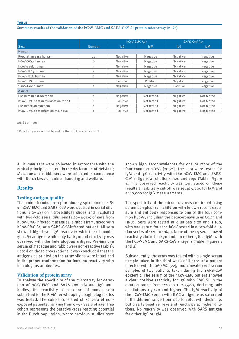
47www.eurosurveillance.org
All human sera were collected in accordance with the ethical principles set out in the declaration of Helsinki; Macaque and rabbit sera were collected in compliance with Dutch laws on animal handling and welfare.
Results
Testing antigen qualityThe amino-terminal receptor-binding spike domains S1 of hCoV-EMC and SARS-CoV were spotted in serial dilu-tions (1:2–1:8) on nitrocellulose slides and incubated with two-fold serial dilutions (1:20–1:640) of sera from hCoV-EMC-infected macaques, a rabbit immunised with hCoV-EMC S1, or a SARS-CoV-infected patient. All sera showed high-level IgG reactivity with their homolo-gous S1 antigen, while only background reactivity was observed with the heterologous antigen. Pre-immune serum of macaque and rabbit were non-reactive (Table). Based on these observations it was concluded that the antigens as printed on the array slides were intact and in the proper conformation for immuno-reactivity with homologous antibodies.
Validation of protein arrayTo analyse the specificity of the microarray for detec-tion of hCoV-EMC and SARS-CoV IgM and IgG anti-bodies, the reactivity of a cohort of human sera submitted to the RIVM for whooping cough diagnostics was tested. The cohort consisted of 72 sera of non-exposed patients, ranging from 0–95 years of age. This cohort represents the putative cross-reacting potential in the Dutch population, where previous studies have
shown high seroprevalences for one or more of the four common hCoVs [20,21]. The sera were tested for IgM and IgG reactivity with the hCoV-EMC and SARS-CoV antigens at dilutions 1:20 and 1:40 (Table, Figure 1). The observed reactivity was low. Based on these results an arbitrary cut-off was set at 5,000 for IgM and at 10,000 for IgG measurements.
The specificity of the microarray was confirmed using serum samples from children with known recent expo-sure and antibody responses to one of the four com-mon hCoVs, including the betacoronaviruses OC43 and HKU1. Sera were tested at dilutions 1:20 and 1:160, with one serum for each hCoV tested in a two-fold dilu-tion series of 1:20 to 1:640. None of the 14 sera showed reactivity above background, for either IgG or IgM, with the hCoV-EMC and SARS-CoV antigens (Table, Figures 1 and 2).
Subsequently, the array was tested with a single serum sample taken in the third week of illness of a patient infected with hCoV-EMC [22], and convalescent serum samples of two patients taken during the SARS-CoV epidemic. The serum of the hCoV-EMC patient showed a clear positive reactivity for IgG with EMC S1 in the dilution range from 1:20 to 1: 20,480, declining only at dilutions 1:5,120 and higher. The IgM reactivity of the hCoV-EMC serum with EMC antigen was saturated in the dilution range from 1:20 to 1:80, with declining, but clearly positive, levels of reactivity at higher dilu-tions. No reactivity was observed with SARS antigen for either IgG or IgM.
Table Summary results of the validation of the hCoV-EMC and SARS-CoV S1 protein microarray (n=94)
hCoV-EMC Aga SARS-CoV Aga
Sera Number IgG IgM IgG IgMHumanPopulation sera human 72 Negative Negative Negative NegativehCoV-OC43 human 6 Negative Negative Negative NegativehCoV-229E human 3 Negative Negative Negative NegativehCoV-NL63 human 3 Negative Negative Negative NegativehCoV-HKU1 human 2 Negative Negative Negative NegativehCoV-EMC human 1 Positive Positive Negative NegativeSARS-CoV human 2 Negative Negative Positive NegativeAnimalPre-immunisation rabbit 1 Negative Not tested Negative Not testedhCoV-EMC post-immunisation rabbit 1 Positive Not tested Negative Not testedPre-infection macaque 1 Negative Not tested Negative Not testedhCoV-EMC post-infection macaque 2 Positive Not tested Negative Not tested
Ag: S1 antigen.
a Reactivity was scored based on the arbitrary set cut-off.
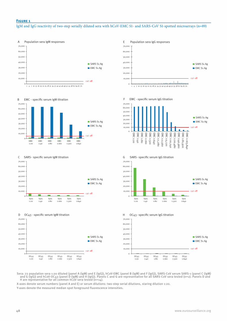
48 www.eurosurveillance.org
Figure 1IgM and IgG reactivity of two-step serially diluted sera with hCoV-EMC S1- and SARS-CoV S1-spotted microarrays (n=89)
Sera: 72 population sera 1:20 diluted (panel A (IgM) and E (IgG)), hCoV-EMC (panel B (IgM) and F (IgG)), SARS-CoV serum SARS-1 (panel C (IgM) and G (IgG)) and hCoV-OC43 (panel D (IgM) and H (IgG)). Panels C and G are representative for all SARS-CoV sera tested (n=2). Panels D and H are representative for all common hCoV sera tested (n=14).
X-axes denote serum numbers (panel A and E) or serum dilutions: two-step serial dilutions, staring dilution 1:20. Y-axes denote the measured median spot foreground fluorescence intensities.
0
10,000
20,000
30,000
40,000
50,000
60,000
70,000
EMC1:20
EMC1:40
EMC1:80
EMC1:160
EMC1:320
EMC1:640
B EMC - specific serum IgM titration
SARS S1 AgEMC S1 Ag
0
10,000
20,000
30,000
40,000
50,000
60,000
70,000
1 4 7 10 13 16 19 22 25 28 31 34 37 40 43 46 49 52 55 58 61 64 67 70
A Population sera IgM responses
SARS S1 AgEMC S1 Ag
0
10,000
20,000
30,000
40,000
50,000
60,000
70,000
Sars1:20
Sars1:40
Sars1:80
Sars1:160
Sars1:320
Sars1:640
C SARS - specific serum IgM titration
SARS S1 AgEMC S1 Ag
OC431:20
OC431:40
OC431:80
OC431:160
OC431:320
OC431:640
H OC43 - specific serum IgG titration
SARS S1 AgEMC S1 Ag
Sars1:20
Sars1:40
Sars1:80
Sars1:160
Sars1:320
Sars1:640
G SARS - specific serum IgG titration
SARS S1 AgEMC S1 Ag
0
10,000
20,000
30,000
40,000
50,000
60,000
70,000
OC431:20
OC431:40
OC431:80
OC431:160
OC431:320
OC431:640
D OC43 - specific serum IgM titration
SARS S1 AgEMC S1 Ag
cut -off cut -off
cut -off
cut -off
cut -off
cut -off
cut -off
cut -off
1 4 7 10 13 16 19 22 25 28 31 34 37 40 43 46 49 52 55 58 61 64 67 70
E Population sera IgG responses
SARS S1 AgEMC S1 Ag
0
10,000
20,000
30,000
40,000
50,000
60,000
70,000
0
10,000
20,000
30,000
40,000
50,000
60,000
70,000
0
10,000
20,000
30,000
40,000
50,000
60,000
70,000
0
10,000
20,000
30,000
40,000
50,000
60,000
70,000
EMC 1:20
EMC 1:40
EMC 1:80
EMC 1:160
EMC 1:320
EMC 1:640
EMC 1:1,280
EMC 1:2,560
EMC 1:5,120
EMC 1:10,240
EMC 1:20,480
EMC 1:40,960
EMC 1:81,920
EMC 1:163,840
F EMC - specific serum IgG titration
SARS S1 AgEMC S1 Ag
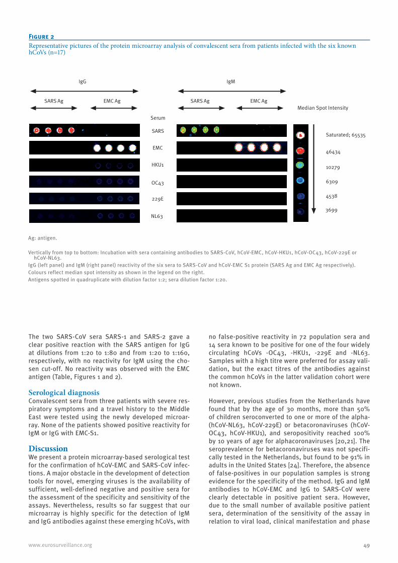
49www.eurosurveillance.org
The two SARS-CoV sera SARS-1 and SARS-2 gave a clear positive reaction with the SARS antigen for IgG at dilutions from 1:20 to 1:80 and from 1:20 to 1:160, respectively, with no reactivity for IgM using the cho-sen cut-off. No reactivity was observed with the EMC antigen (Table, Figures 1 and 2).
Serological diagnosisConvalescent sera from three patients with severe res-piratory symptoms and a travel history to the Middle East were tested using the newly developed microar-ray. None of the patients showed positive reactivity for IgM or IgG with EMC-S1.
DiscussionWe present a protein microarray-based serological test for the confirmation of hCoV-EMC and SARS-CoV infec-tions. A major obstacle in the development of detection tools for novel, emerging viruses is the availability of sufficient, well-defined negative and positive sera for the assessment of the specificity and sensitivity of the assays. Nevertheless, results so far suggest that our microarray is highly specific for the detection of IgM and IgG antibodies against these emerging hCoVs, with
no false-positive reactivity in 72 population sera and 14 sera known to be positive for one of the four widely circulating hCoVs -OC43, -HKU1, -229E and -NL63. Samples with a high titre were preferred for assay vali-dation, but the exact titres of the antibodies against the common hCoVs in the latter validation cohort were not known.
However, previous studies from the Netherlands have found that by the age of 30 months, more than 50% of children seroconverted to one or more of the alpha- (hCoV-NL63, hCoV-229E) or betacoronaviruses (hCoV-OC43, hCoV-HKU1), and seropositivity reached 100% by 10 years of age for alphacoronaviruses [20,21]. The seroprevalence for betacoronaviruses was not specifi-cally tested in the Netherlands, but found to be 91% in adults in the United States [24]. Therefore, the absence of false-positives in our population samples is strong evidence for the specificity of the method. IgG and IgM antibodies to hCoV-EMC and IgG to SARS-CoV were clearly detectable in positive patient sera. However, due to the small number of available positive patient sera, determination of the sensitivity of the assay in relation to viral load, clinical manifestation and phase
Figure 2Representative pictures of the protein microarray analysis of convalescent sera from patients infected with the six known hCoVs (n=17)
SARS Ag EMC Ag SARS Ag EMC Ag
Serum
SARS
EMC
HKU1
OC43
229E
NL63
IgG IgM
Saturated; 65535
46434
10279
6309
4538
3699
Median Spot Intensity
Ag: antigen.
Vertically from top to bottom: Incubation with sera containing antibodies to SARS-CoV, hCoV-EMC, hCoV-HKU1, hCoV-OC43, hCoV-229E or hCoV-NL63.
IgG (left panel) and IgM (right panel) reactivity of the six sera to SARS-CoV and hCoV-EMC S1 protein (SARS Ag and EMC Ag respectively). Colours reflect median spot intensity as shown in the legend on the right. Antigens spotted in quadruplicate with dilution factor 1:2; sera dilution factor 1:20.
50 www.eurosurveillance.org
of infection requires further investigation. For this essential clinical validation, international sharing of positive sera by (national) laboratories in possession of such sera is a prerequisite.
Currently, WHO and ECDC recommend the collection of paired serum samples, preferably from the acute and convalescent phase, of all cases under investigation, as serological testing might be necessary to confirm infection when clinical presentation and epidemiology suggest an infection with hCoV-EMC despite negative PCR results [2,12]. In addition, serology is needed for contact investigations and source tracking. A two-staged serological approach is recommended, which proved effective in a contact investigation of an hCoV-EMC infection treated in Germany. It uses IFA with virus-infected cells for screening, and as second-stage recombinant spike- and nucleocapsid-transfected cells and virus neutralisation tests [22]. Our protein microar-ray enables specific, one-stage, high-throughput test-ing, with the benefit of minimal sample requirement. This technique can use dried blood spots for testing, which greatly facilitates shipping of samples.
The serological assay presented here is available and of great value for human and animal population screen-ing, both of which are necessary to gain insight in the epidemiology of the novel hCoV. The array format can be modified to identify primary and intermediate ani-mal reservoirs by simple adaptation of the conjugate used to visualise reactivity on the array (data not shown). Our assay is available to aid diagnosis in indi-vidual patients, for confirmatory testing of positive tests and for (large-scale) contact studies.
Acknowledgements We are grateful to Matthias Niedrig (Robert Koch Institute, Berlin, Germany) and Jürg Timm (University of Duisburg-Essen, Essen, Germany) for provision of a SARS-CoV se-rum and the hCoV-EMC patient serum. Johan Reimerink is acknowledged for his input in discussions. We thank Dorian Stolk, Joëlle Reijm, Mariëlle Zonneveld, Amber van Broekhoven and Maarten F. Jebbink for hCoV-NL63-, hCoV-229E-, hCoV-HKU1-, and hCoV-OC43-specific sera selection assays. RdS was funded by the European Public Health Microbiology Training Program (EUPHEM), ECDC, Stockholm. LvdH, BH and AO were funded by the European Union FP7 project EMPERIE (contract number 223498).
Conflict of interestNone declared.
Authors’ contributionsCR: assisted in designing the study, coordinated the study, analysed data, wrote manuscript. HM: performed labora-tory testing and serum production. GJG: assisted in design-ing the study, performed laboratory testing, analysed data, read and revised manuscript. LvdH: performed laboratory testing, provided sera, read and revised manuscript. BM: performed laboratory testing, read and revised manuscript.
MM: provided sera, performed laboratory testing, read and revised manuscript. BH: serum production, read and revised manuscript. RdS: assisted in study design, read and revised the manuscript.NS: performed laboratory testing and serum production. UD: provided sera, read and revised manuscript. PR: read and revised the manuscript. AO: provided sera, read and revised the manuscript. CD: provided sera, read and re-vised the manuscript. BJB: produced design antigen, pro-vided antigens and serum, read and revised the manuscript. MK: assisted in designing the study, analysed data, read and revised the manuscript.
51www.eurosurveillance.org
References1. Zaki AM, van Boheemen S, Bestebroer TM, Osterhaus AD,
Fouchier RA. Isolation of a novel coronavirus from a man with pneumonia in Saudi Arabia. N Engl J Med. 2012;367(19):1814-20. http://dx.doi.org/10.1056/NEJMoa1211721. PMid:23075143.
2. European Centre for Disease prevention and Control (ECDC). Severe respiratory disease associated with a novel coronavirus. Rapid Risk Assesment. Stockholm: ECDC; Feb 2013. [Accessed 19 Feb 2013]. Available from: http://www.ecdc.europa.eu/en/publications/Publications/novel-coronavirus-rapid-risk-assessment-update.pdf
3. European Centre for Disease prevention and Control (ECDC). Confirmed cases of novel coronavirus infection (n=17), reported worldwide as of 26 March 2013. Stockholm: ECDC [Accessed 2 Apr 2013]. Available from: http://www.ecdc.europa.eu/en/healthtopics/Documents/Novel-coronavirus-cases-table.pdf
4. Pebody RG. The Health Protection Agency (HPA) UK Novel Coronavirus Investigation team. Evidence of person-to-person transmission within a family cluster of novel coronavirus infections, United Kingdom, February 2013. Euro Surveill. 2013;18(11):pii=20427. Available from: http://www.eurosurveillance.org/ViewArticle.aspx?ArticleId=20427
5. Albarrak AM, Stephens GM, Hewson R, Memish ZA. Recovery from severe novel coronavirus infection. Saudi Med J. 2012;33(12):1265-9. PMid:23232672.
6. Bermingham A, Chand MA, Brown CS, Aarons E, Tong C, Langrish C, et al. Severe respiratory illness caused by a novel coronavirus, in a patient transferred to the United Kingdom from the Middle East, September 2012. Euro Surveill. 2012;17(40): pii=20290. Available from: http://www.eurosurveillance.org/ViewArticle.aspx?ArticleId=20290
7. Pebody RG, Chand MA, Thomas HL, Green HK, Boddington NL, Carvalho C, et al. The United Kingdom public health response to an imported laboratory confirmed case of a novel coronavirus in September 2012. Euro Surveill. 2012;17(40): pii=20292. Available from: http://www.eurosurveillance.org/ViewArticle.aspx?ArticleId=20292
8. ProMED-mail. Novel coronavirus-East.Med. (7): Saudi Arabia, UK, Germany. ProMED mail. Archive number: 20130221.1554109. 21 Feb 2013. Available from: http://www.promedmail.org/?archiveid=20130221
9. Corman VM, Eckerle I, Bleicker T, Zaki A, Landt O, Eschbach-Bludau M, et al. Detection of a novel human coronavirus by real-time reverse-transcription polymerase chain reaction. Euro Surveill. 2012;17(39):pii=20285. Available from: http://www.eurosurveillance.org/ViewArticle.aspx?ArticleId=20285
10. Corman V, Muller M, Costabel U, Timm J, Binger T, Meyer B, et al. Assays for laboratory confirmation of novel human coronavirus (hCoV-EMC) infections. Euro Surveill. 2012;17(49):pii=20334. Available from: http://www.eurosurveillance.org/ViewArticle.aspx?ArticleId=20334
11. Palm D, Pereyaslov D, Vaz J, Broberg E, Zeller H, Gross D, et al. Laboratory capability for molecular detection and confirmation of novel coronavirus in Europe, November 2012. Euro Surveill. 2012;17(49):pii=20335. Available from: http://www.eurosurveillance.org/ViewArticle.aspx?ArticleId=20335
12. World Health Organization (WHO). Laboratory testing for novel coronavirus. Interim recommendations. Geneva: WHO; 21 Dec 2012. [Accessed 14 Mar 2013]. Available from: http://www.who.int/csr/disease/coronavirus_infections/LaboratoryTestingNovelCoronavirus_21Dec12.pdf
13. Ng LF, Wong M, Koh S, Ooi EE, Tang KF, Leong HN, et al. Detection of severe acute respiratory syndrome coronavirus in blood of infected patients. J Clin Microbiol. 2004;42(1):347-50. http://dx.doi.org/10.1128/JCM.42.1.347-350.2004. PMCid:321706.
14. Chen W, Xu Z, Mu J, Yang L, Gan H, Mu F, et al. Antibody response and viraemia during the course of severe acute respiratory syndrome (SARS)-associated coronavirus infection. J Med Microbiol. 2004;53(Pt 5):435-8. Epub 2004/04/21.
15. Guan Y, Zheng BJ, He YQ, Liu XL, Zhuang ZX, Cheung CL, et al. Isolation and characterization of viruses related to the SARS coronavirus from animals in southern China. Science. 2003;302(5643):276-8. http://dx.doi.org/10.1126/science.1087139. PMid:12958366.
16. Prevalence of IgG antibody to SARS-associated coronavirus in animal traders--Guangdong Province, China, 2003. MMWR Morb Mortal Wkly Rep. 2003;52(41):986-7. PMid:14561956.
17. Novel human Coronavirus HCoV-EMC. Recommendations regarding serological testing. Bonn: Institut für Virologie; 2013. [Accessed 21 Feb 2013]. Available from: http://www.virology-bonn.de/index.php?id=40
18. Raj VS, Mou H, Smits SL, Dekkers DH, Muller MA, Dijkman R, et al. Dipeptidyl peptidase 4 is a functional receptor
for the emerging human coronavirus-EMC. Nature. 2013;495(7440):251-4. http://dx.doi.org/10.1038/nature12005. PMid:23486063.
19. Koopmans M, de Bruin E, Godeke GJ, Friesema I, van Gageldonk R, Schipper M, et al. Profiling of humoral immune responses to influenza viruses by using protein microarray. Clin Microbiol Infect. 2012;18(8):797-807. http://dx.doi.org/10.1111/j.1469-0691.2011.03701.x. PMid:22212116.
20. Dijkman R, Jebbink MF, El Idrissi NB, Pyrc K, Muller MA, Kuijpers TW, et al. Human coronavirus NL63 and 229E seroconversion in children. J Clin Microbiol. 2008;46(7):2368-73. http://dx.doi.org/10.1128/JCM.00533-08. PMid:18495857. PMCid:2446899.
21. Dijkman R, Jebbink MF, Gaunt E, Rossen JW, Templeton KE, Kuijpers TW, et al. The dominance of human coronavirus OC43 and NL63 infections in infants. J Clin Virol. 2012;53(2):135-9. http://dx.doi.org/10.1016/j.jcv.2011.11.011. PMid:22188723.
22. Buchholz U, Müller MA, Nitsche A, Sanewski A, Wevering N, Bauer-Balci T, et al. Contact investigation of a case of human novel coronavirus infection treated in a German hospital, October-November 2012. Euro Surveill. 2013;18(8):pii=20406. Available from: http://www.eurosurveillance.org/ViewArticle.aspx?ArticleId=20406. PMid:23449231.
23. van Boheemen S, de Graaf M, Lauber C, Bestebroer TM, Raj VS, Zaki AM, et al. Genomic characterization of a newly discovered coronavirus associated with acute respiratory distress syndrome in humans. MBio. 2012;3(6):pii: e00473-12. http://dx.doi.org/10.1128/mBio.00473-12. PMid:23170002 PMCid:3509437.
24. Severance EG, Bossis I, Dickerson FB, Stallings CR, Origoni AE, Sullens A, et al. Development of a nucleocapsid-based human coronavirus immunoassay and estimates of individuals exposed to coronavirus in a U.S. metropolitan population. Clin Vaccine Immunol. 2008;15(12):1805-10. http://dx.doi.org/10.1128/CVI.00124-08. PMid:18945884 PMCid:2593164.

52 www.eurosurveillance.org
Rapid communications
Laboratory capability for molecular detection and confirmation of novel coronavirus in Europe, November 2012
D Palm1, D Pereyaslov2, J Vaz1, E Broberg1, H Zeller1, D Gross2, C S Brown2, M J Struelens ([email protected])1, on behalf of the Joint ECDC-WHO Regional Office for Europe Novel Coronavirus Laboratory Survey participants; ECDC National Microbiology Focal Points, WHO European Region EuroFlu Network and European Network for Diagnostics of “Imported” Viral Diseases (ENIVD)3
1. European Centre for Disease Prevention and Control (ECDC), Stockholm, Sweden2. World Health Organization (WHO) Regional Office for Europe, Copenhagen, Denmark3. Joint ECDC-WHO Regional Office for Europe Novel Coronavirus Laboratory Survey participants; ECDC National Microbiology
Focal Points, WHO European Region EuroFlu Network and European Network for Diagnostics of “Imported” Viral Diseases (ENIVD)
Citation style for this article: Palm D, Pereyaslov D, Vaz J, Broberg E, Zeller H, Gross D, Brown CS, Struelens MJ, on behalf of the Joint ECDC-WHO Regional Office for Europe Novel Coronavirus Laboratory Survey participants; ECDC National Microbiology Focal Points, WHO European Region EuroFlu Network and European Network for Diagnostics of “Imported” Viral Diseases (ENIVD). Laboratory capability for molecular detection and confirmation of novel coronavirus in Europe, November 2012. Euro Surveill. 2012;17(49):pii=20335. Available online: http://www.eurosurveillance.org/ViewArticle.aspx?ArticleId=20335
Article submitted on 05 December 2012 / published on 6 December 2012
A rapid survey by the European Centre for Disease Prevention and Control (ECDC) and the World Health Organization (WHO) Regional Office for Europe ascer-tained the availability of national reference laboratory testing for a recently detected novel coronavirus as of 28 November 2012. Screening by internal quality controlled upE-RT-PCR assay was available in 23/46 of responding countries in the WHO European Region, of which 19/30 in European Union (EU) and European Economic Area (EEA) countries. Confirmation of posi-tive screened samples by either ORF1b-RT-PCR, or other target RT-PCR assays with sequence analysis or whole-genome sequence analysis was available in 22/46 responding countries of which 18/30 in EU/EEA countries.
In September 2012, a novel coronavirus was first characterised by genome sequencing at the Erasmus Medical Center (EMC) of a viral isolate from a patient in Saudi Arabia with severe pneumonia [1-2]. This virus belongs to the genus beta-coronavirus and is closely related to some bat coronaviruses. Since then, a total of nine confirmed cases of human infection with the novel coronavirus have been reported to public health authorities and WHO [3-6]. These patients developed a severe respiratory disease in Saudi Arabia, Qatar and Jordan over the period April to November 2012 and five had a fatal outcome [1-6]. Two patients were referred to Europe for specialised care [1-6].
Coronaviruses are membrane enveloped viruses with large RNA genomes and a distinctive surface crown causing respiratory and enteric infections in humans and animals. In 2003, zoonotic transmission of SARS-CoV caused a worldwide epidemic associated with more than 8,000 cases of severe acute respiratory syn-drome (SARS) and a case-fatality rate of 10% [7].
Monitoring of novel coronavirusWhile the reservoir and mode of transmission of the novel coronavirus virus are under investigation, WHO and ECDC request that any probable or confirmed case that is diagnosed should rapidly (i) be reported to national authorities to enable them to take appropri-ate public health measures, and (ii) be notified to WHO under the International Health Regulations (2005) and simultaneously through joint reporting system to the Early Warning and Response System (EWRS) for coun-tries in the EU/EEA [3,4,8].
In September 2012, Corman et al described the first diagnostic assays for the novel coronavirus [9]. These involve a two-step screening and confirmation test-ing algorithm using newly developed specific RT-PCR assays that target the regions upstream of the E gene (the upE target; recommended for screening) and open reading frame 1b (ORF1b; recommended for confirma-tion) [9]. Additional testing based on sequence analy-sis of other viral genome targets or whole genome sequence determination from clinical material or cul-ture isolate can also be used to confirm cases [2,6].
A number of pan-coronavirus RT-PCR assays have been described which target the polymerase gene of corona-viruses used for the detection of known and unknown coronaviruses, including coronaviruses currently cir-culating in humans such as hCoV-229E, hCoV-NL63, hCoV-OC43 and SARS-CoV [10-12]. Some of these pan-coronavirus RT-PCR assays may be also used to detect the novel coronavirus. However, a positive result should be confirmed by screening for the specific targets described for hCoV-EMC, sequencing of the RT-PCR product and/or virus isolation [1,2,6].
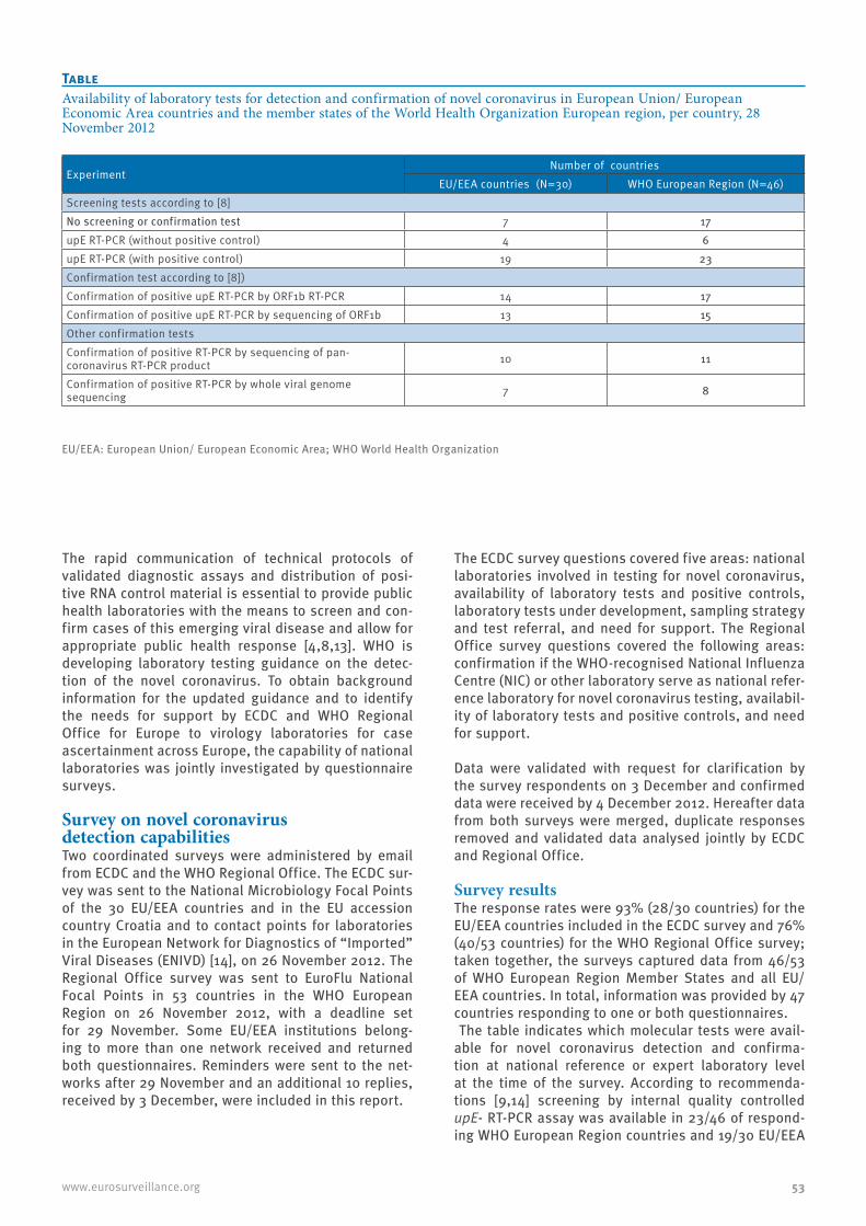
53www.eurosurveillance.org
The rapid communication of technical protocols of validated diagnostic assays and distribution of posi-tive RNA control material is essential to provide public health laboratories with the means to screen and con-firm cases of this emerging viral disease and allow for appropriate public health response [4,8,13]. WHO is developing laboratory testing guidance on the detec-tion of the novel coronavirus. To obtain background information for the updated guidance and to identify the needs for support by ECDC and WHO Regional Office for Europe to virology laboratories for case ascertainment across Europe, the capability of national laboratories was jointly investigated by questionnaire surveys.
Survey on novel coronavirus detection capabilities Two coordinated surveys were administered by email from ECDC and the WHO Regional Office. The ECDC sur-vey was sent to the National Microbiology Focal Points of the 30 EU/EEA countries and in the EU accession country Croatia and to contact points for laboratories in the European Network for Diagnostics of “Imported” Viral Diseases (ENIVD) [14], on 26 November 2012. The Regional Office survey was sent to EuroFlu National Focal Points in 53 countries in the WHO European Region on 26 November 2012, with a deadline set for 29 November. Some EU/EEA institutions belong-ing to more than one network received and returned both questionnaires. Reminders were sent to the net-works after 29 November and an additional 10 replies, received by 3 December, were included in this report.
The ECDC survey questions covered five areas: national laboratories involved in testing for novel coronavirus, availability of laboratory tests and positive controls, laboratory tests under development, sampling strategy and test referral, and need for support. The Regional Office survey questions covered the following areas: confirmation if the WHO-recognised National Influenza Centre (NIC) or other laboratory serve as national refer-ence laboratory for novel coronavirus testing, availabil-ity of laboratory tests and positive controls, and need for support.
Data were validated with request for clarification by the survey respondents on 3 December and confirmed data were received by 4 December 2012. Hereafter data from both surveys were merged, duplicate responses removed and validated data analysed jointly by ECDC and Regional Office.
Survey resultsThe response rates were 93% (28/30 countries) for the EU/EEA countries included in the ECDC survey and 76% (40/53 countries) for the WHO Regional Office survey; taken together, the surveys captured data from 46/53 of WHO European Region Member States and all EU/EEA countries. In total, information was provided by 47 countries responding to one or both questionnaires. The table indicates which molecular tests were avail-able for novel coronavirus detection and confirma-tion at national reference or expert laboratory level at the time of the survey. According to recommenda-tions [9,14] screening by internal quality controlled upE- RT-PCR assay was available in 23/46 of respond-ing WHO European Region countries and 19/30 EU/EEA
Table Availability of laboratory tests for detection and confirmation of novel coronavirus in European Union/ European Economic Area countries and the member states of the World Health Organization European region, per country, 28 November 2012
ExperimentNumber of countries
EU/EEA countries (N=30) WHO European Region (N=46)Screening tests according to [8]No screening or confirmation test 7 17upE RT-PCR (without positive control) 4 6upE RT-PCR (with positive control) 19 23Confirmation test according to [8])Confirmation of positive upE RT-PCR by ORF1b RT-PCR 14 17Confirmation of positive upE RT-PCR by sequencing of ORF1b 13 15 Other confirmation testsConfirmation of positive RT-PCR by sequencing of pan-coronavirus RT-PCR product 10 11
Confirmation of positive RT-PCR by whole viral genome sequencing 7 8
EU/EEA: European Union/ European Economic Area; WHO World Health Organization
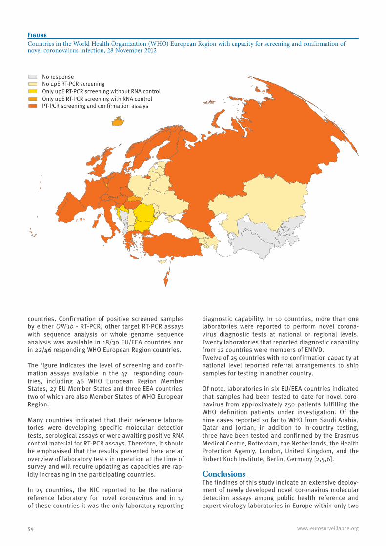
54 www.eurosurveillance.org
countries. Confirmation of positive screened samples by either ORF1b - RT-PCR, other target RT-PCR assays with sequence analysis or whole genome sequence analysis was available in 18/30 EU/EEA countries and in 22/46 responding WHO European Region countries.
The figure indicates the level of screening and confir-mation assays available in the 47 responding coun-tries, including 46 WHO European Region Member States, 27 EU Member States and three EEA countries, two of which are also Member States of WHO European Region.
Many countries indicated that their reference labora-tories were developing specific molecular detection tests, serological assays or were awaiting positive RNA control material for RT-PCR assays. Therefore, it should be emphasised that the results presented here are an overview of laboratory tests in operation at the time of survey and will require updating as capacities are rap-idly increasing in the participating countries.
In 25 countries, the NIC reported to be the national reference laboratory for novel coronavirus and in 17 of these countries it was the only laboratory reporting
diagnostic capability. In 10 countries, more than one laboratories were reported to perform novel corona-virus diagnostic tests at national or regional levels. Twenty laboratories that reported diagnostic capability from 12 countries were members of ENIVD. Twelve of 25 countries with no confirmation capacity at national level reported referral arrangements to ship samples for testing in another country.
Of note, laboratories in six EU/EEA countries indicated that samples had been tested to date for novel coro-navirus from approximately 250 patients fulfilling the WHO definition patients under investigation. Of the nine cases reported so far to WHO from Saudi Arabia, Qatar and Jordan, in addition to in-country testing, three have been tested and confirmed by the Erasmus Medical Centre, Rotterdam, the Netherlands, the Health Protection Agency, London, United Kingdom, and the Robert Koch Institute, Berlin, Germany [2,5,6].
ConclusionsThe findings of this study indicate an extensive deploy-ment of newly developed novel coronavirus molecular detection assays among public health reference and expert virology laboratories in Europe within only two
FigureCountries in the World Health Organization (WHO) European Region with capacity for screening and confirmation of novel coronovairus infection, 28 November 2012
No responseNo upE RT-PCR screeningOnly upE RT-PCR screening without RNA controlOnly upE RT-PCR screening with RNA controlPT-PCR screening and confirmation assays
55www.eurosurveillance.org
months after sequence information on the first reported case was made available. This deployment occurred to a greater extent in EU/EEA countries of the WHO European Region compared with south-east and east-ern European countries. Screening tests were available in nearly half of countries of the WHO European Region and cross-border shipment arrangements were in place in many of those lacking domestic testing capacity.
Our results will allow virologists and public health agencies, including ECDC and the WHO Regional Office, to remedy gaps within their laboratory networks. Such measures may include technical laboratory guidance and collaborative arrangements for cross-border refer-ral testing of clinical materials, technical support such as distribution of reference control materials, assis-tance with development of quality controlled sero-logical assays and, if warranted in the longer term, provision of capacity building courses and external quality assessment schemes.
Acknowledgments We thank Christian Drosten, Ron Fouchier and Maria Zambon for kindly reviewing the survey questionnaire. We are also thankful to the ECDC National Microbiology Focal Points and members of the EURO-FLU and ENIVD networks for dedicated and rapid responses to the surveys.
References1. ProMED-mail. Novel coronavirus - Saudi Arabia: human
isolate. Archive Number: 20120920.1302733. 20 September 2012. Available from: http://www.promedmail.org/direct.php?id=20120920.1302733
2. Van Boheemen S, de Graaf M, Lauber C, Bestebroer TM, Raj VS, Zaki AM, et al .2012. Genomic characterization of a newly discovered coronavirus associated with acute respiratory distress syndrome in humans. mBio. 2012;3(6). pii= e00473-12.
3. World Health Organization (WHO). Global Alert and Response: Background and summary of novel coronavirus infection – as of 30 November 2012. Geneva: WHO; 2012. Available from: http://www.who.int/csr/disease/coronavirus_infections/update_20121130/en/index.html
4. European Centre for Disease Prvention and Control (ECDC). Rapid Risk assessment, update: Severe respiratory disease associated with a novel coronavirus, 26 November 2012. Stockholm: ECDC; 2012. Available from: http://ecdc.europa.eu/en/publications/Publications/Forms/ECDC_DispForm.aspx?ID=1007
5. Robert Koch Institute (RKI). Case of novel Corona virus in Germany 23 November 2012. Berlin: RKI; 2012. [Accessed 25 Nov 2012]. Available from: http://www.rki.de/EN/Home/Corona_Virus.html;jsessionid=77C1C0AF8994434233952DC28C20E642.2_cid290
6. Bermingham A, Chand MA, Brown CS, Aarons E, Tong C, Langrish C, et al. Severe respiratory illness caused by a novel coronavirus, in a patient transferred to the United Kingdom from the Middle East, September 2012. Euro Surveill. 2012;17(40):pii=20290. Available from: http://www.eurosurveillance.org/ViewArticle.aspx?ArticleId=20290
7. World Health Organization (WHO).Summary table of SARS cases by country, 1 November 2002 - 7 August 2003. Geneva: WHO; 2003. Available from: http://www.who.int/csr/sars/country/2003_08_15/en/
8. World Health Organization (WHO). Case definition for case finding severe respiratory disease associated with novel coronavirus. Geneva: WHO; 2012. Available from: http://www.who.int/csr/disease/coronavirus_infections/case_definition/en/index.html
9. Corman VM, Eckerle I, Bleicker T, Zaki A, Landt O, Eschbach-Bludau M, et al. Detection of a novel human coronavirus by real-time reverse-transcription polymerase chain reaction. Euro Surveill. 2012;17(39):pii=20285. Available from: http://www.eurosurveillance.org/ViewArticle.aspx?ArticleId=20285
10. Vijgen L, Moes E, Keyaerts E, Li S, Van Ranst M. A pancoronavirus RT-PCR assay for detection of all known coronaviruses. Methods Mol Biol. 2008;454: 3-12.
11. Drosten C, Günther S, Preiser W, van der Werf S, Brodt HR, Becker S et al. Identification of a novel coronavirus in patients with severe acute respiratory syndrome. N Engl J Med. 2003;348(20):1967-76.
12. Bermingham A, Heinen P, Iturriza-Gómara M, Gray J, Appleton H, Zambon MC. Laboratory diagnosis of SARS. Philos Trans R Soc Lond B Biol Sci. 2004;359(1447):1083-9.
13. Pebody RG, Chand MA, Thomas HL, Green HK, Boddington NL, Carvalho C, et al. The United Kingdom public health response to an imported laboratory confirmed case of a novel coronavirus in September 2012. Euro Surveill. 2012;17(40):pii=20292. Available from: http://www.eurosurveillance.org/ViewArticle.aspx?ArticleId=20292
14. European Network for Diagnostics of “Imported” Viral Diseases (ENIVD). Available from: www.enivd.org
56 www.eurosurveillance.org
Surveillance and outbreak reports
Contact investigation of a case of human novel coronavirus infection treated in a German hospital, October-November 2012
U Buchholz ([email protected])1,2, M A Müller3,2, A Nitsche1,2, A Sanewski4,2, N Wevering5, T Bauer-Balci6, F Bonin5, C Drosten ([email protected])3, B Schweiger1, T Wolff1, D Muth3, B Meyer3, S Buda1, G Krause1, L Schaade1, W Haas1
1. Robert Koch Institute, Berlin, Germany2. These authors contributed equally to this work3. University Bonn, Department of Virology, Bonn, Germany 4. County health department of Essen, Essen, Germany5. Ruhrland hospital, Essen, Germany6. County health department Oberbergischer Kreis, Gummersbach, Germany
Citation style for this article: Buchholz U, Müller MA, Nitsche A, Sanewski A, Wevering N, Bauer-Balci T, Bonin F, Drosten C, Schweiger B, Wolff T, Muth D, Meyer B, Buda S, Krause G, Schaade L, Haas W. Contact investigation of a case of human novel coronavirus infection treated in a German hospital, October-November 2012. Euro Surveill. 2013;18(8):pii=20406. Available online: http://www.eurosurveillance.org/ViewArticle.aspx?ArticleId=20406
Article submitted on 24 January 2013 / published on 21 February 2013
On 24 October 2012, a patient with acute respiratory distress syndrome of unknown origin and symptom onset on 5 October was transferred from Qatar to a specialist lung clinic in Germany. Late diagnosis on 20 November of an infection with the novel Coronavirus (NCoV) resulted in potential exposure of a consider-able number of healthcare workers. Using a question-naire we asked 123 identified contacts (120 hospital and three out-of-hospital contacts) about exposure to the patient. Eighty-five contacts provided blood for a serological test using a two-stage approach with an initial immunofluorescence assay as screening test, followed by recombinant immunofluorescence assays and a NCoV-specific serum neutralisation test. Of 123 identified contacts nine had performed aerosol-gen-erating procedures within the third or fourth week of illness, using personal protective equipment rarely or never, and two of these developed acute respiratory illness. Serology was negative for all nine. Further 76 hospital contacts also tested negative, including two sera initially reactive in the screening test. The con-tact investigation ruled out transmission to contacts after illness day 20. Our two-stage approach for sero-logical testing may be used as a template for similar situations.
IntroductionA novel human coronavirus (NCoV) has recently emerged in the Arabian Peninsula. The first two reported cases infected by the novel agent, then pro-visionally termed hCoV-EMC, occurred in June and September 2012, respectively [1-3]. As of 18 February 2013, a total of 12 cases have been confirmed by WHO [4], including five deaths. Among five cases reported from the Kingdom of Saudi Arabia, three were part of a family cluster. Two further cases were linked to probable exposure in Qatar, and two cases were con-firmed retrospectively, by diagnostic of respiratory
specimens, from Jordan with disease onset in April 2012. The latter were part of a cluster of 11 patients with acute respiratory symptoms linked to a hospital [5]. The most recent three cases identified constitute another cluster that occurred in the United Kingdom (UK) in January to February 2013 [4]. The index case in this cluster is a UK citizen with travel history to Saudi-Arabia and Pakistan before symptom onset [5]. Two of his family members who had not travelled outside the UK and became ill were most likely infected through person-to-person transmission. While one of them had an underlying disease and died, the other presented with milder, influenza-like illness symptoms only.
Because of the long period, 10 months, over which the cases occurred, the source and transmission patterns of the virus remain elusive. Hypotheses include a pre-dominance of zoonotic acquisitions with little poten-tial for human-to-human transmission [5], widespread and unnoticed occurrence of clinically mild infections, and finally the possibility of an early-stage epidemic caused by a highly pathogenic novel human virus.
Because of the potential of human-to-human trans-mission in the hospital outbreak in Jordan and the family clusters, as well as the observed severity of disease, current recommendations regarding protec-tive measures rely on experiences with severe acute respiratory syndrome (SARS) in 2003 [6]. The first of the two Qatari patients was treated in the UK where, under strict isolation measures, no secondary cases occurred. Investigations by polymerase chain reaction (PCR) of 10 healthcare workers (HCW) who had cared for the patient and subsequently developed mild res-piratory disease yielded no evidence of infection [7]. However, to date, published investigations of individu-als with proven exposure to NCoV have not presented
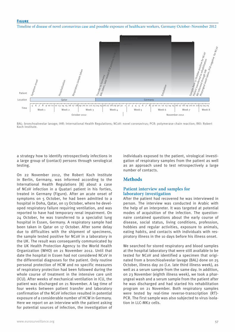
57www.eurosurveillance.org
a strategy how to identify retrospectively infections in a large group of (contact) persons through serological testing.
On 22 November 2012, the Robert Koch Institute in Berlin, Germany, was informed according to the International Health Regulations [8] about a case of NCoV infection in a Quatari patient in his forties, treated in Germany (Figure). After an acute onset of symptoms on 5 October, he had been admitted to a hospital in Doha, Qatar, on 13 October, where he devel-oped respiratory failure requiring ventilation, and was reported to have had temporary renal impairment. On 24 October, he was transferred to a specialist lung hospital in Essen, Germany. A respiratory sample had been taken in Qatar on 17 October. After some delay due to difficulties with the shipment of specimens, the sample tested positive for NCoV in a laboratory in the UK. The result was consequently communicated by the UK Health Protection Agency to the World Health Organization (WHO) on 21 November 2012. Until that date the hospital in Essen had not considered NCoV in the differential diagnoses for the patient. Only routine personal protection of HCW and no specific measures of respiratory protection had been followed during the whole course of treatment in the intensive care unit (ICU). After weeks of mechanical ventilation in ICU, the patient was discharged on 21 November. A lag time of four weeks between patient transfer and laboratory confirmation of the NCoV infection resulted in potential exposure of a considerable number of HCW in Germany. Here we report on an interview with the patient asking for potential sources of infection, the investigation of
individuals exposed to the patient, virological investi-gation of respiratory samples from the patient as well as an approach used to test retrospectively a large number of contacts.
Methods
Patient interview and samples for laboratory investigationAfter the patient had recovered he was interviewed in person. The interview was conducted in Arabic with the help of an interpreter. It was targeted at potential modes of acquisition of the infection. The question-naire contained questions about the early course of disease, social status, living conditions, profession, hobbies and regular activities, exposure to animals, eating habits, and contacts with individuals with res-piratory illness in the 10 days before his illness onset.
We searched for stored respiratory and blood samples at the hospital laboratory that were still available to be tested for NCoV and identified a specimen that origi-nated from a bronchioalveolar lavage (BAL) done on 25 October, illness day 20 (i.e. late third illness week), as well as a serum sample from the same day. In addition, on 23 November (eighth illness week), we took a phar-yngeal wash and a serum sample from the patient after he was discharged and had started his rehabilitation program on 21 November. Both respiratory samples were tested by real-time reverse-transcription (RT)-PCR. The first sample was also subjected to virus isola-tion in LLC-MK2 cells.
FigureTimeline of disease of novel coronavirus case and possible exposure of healthcare workers, Germany October–November 2012
BAL: bronchoalveolar lavage; IHR: International Health Regulations; NCoV: novel coronavirus; PCR: polymerase chain reaction; RKI: Robert Koch Institute.
Sym
ptom
ons
et
Hosp
italis
atio
n
Resp
irato
ry s
ampl
e ta
ken
Tran
sfer
to G
erm
any
BAL t
aken
, lat
er te
sted
PCR
pos
itive
for N
CoV
Extu
batio
n
Sam
ple
from
17 O
ctob
er p
ositi
ve fo
r NCo
V
Patie
nt tr
ansf
erre
d to
reha
bilit
atio
n
RKI i
nfor
med
thro
ugh
IHR
Phar
ynge
al w
ash
take
n, P
CR n
egat
ive
for N
CoV
5 6 7 8 9 10 11 12 13 14 15 16 17 18 19 20 21 22 23 24 25 26 27 28 29 30 31 1 2 3 4 5 6 7 8 9 10 11 12 13 14 15 16 17 18 19 20 21 22 23 24 25
October 2012 November 2012
Week 1 Week 2 Week 3 Week 4 Week 5 Week 6 Week 7 Week 8Time
Location
Patient
Qatar Germany
Barrier nursing
Healthcare workers exposed

58 www.eurosurveillance.org
Contact investigationContact persons were identified based on electronic procedures registration, supplemented by a targeted request to HCW to report contact with the patient while in ICU (and during the transport to the hospital). The electronic registration of procedures requires that any person performing a task in the patient’s room must sign in, sign out and document which procedure was conducted. Using a standardised questionnaire, infor-mation was collected about the time of the first con-tact, types of contact, closest distance to the patient, frequency of using a surgical face mask when in con-tact with the patient, and occurrence of acute respira-tory illness (ARI) up to ten days after the last contact with the patient. No information was collected on the duration or frequency of contact.
Consenting individuals gave blood for serological test-ing on one of three dates (3, 7 or 14 December). The median interval from first patient contact to venipunc-ture was 39 days (range: 13–50 days). Contacts were considered at high risk if they had their first contact with the patient at the beginning of his stay in ICU, i.e. at the end of the patient s third or fourth week of illness, if they had conducted an aerosol-generating procedure, such as suctioning the intubated patient or performing a BAL, and if they had rarely or never used surgical face masks while caring for the patient.
Laboratory methodsNucleic acid detection was performed by RT-PCR as described previously [9,10] after viral RNA was extracted from 300 µl of bronchioalveolar lavage using the MagAttract Viral RNA Kit M48 (Qiagen GmbH, Hilden, Germany).
Serological testing was performed in a two-stage approach. As a first step, screening for antibodies reactive to NCoV was done by indirect immunofluo-rescence assay (IFA) as described previously [10]. Preliminary evaluation of IFA on 50 sera from blood donors yielded no reactivity. For resolution of reac-tive results, IFA was done on Vero B4 cells expressing recombinant spike (S) and nucleocapsid (N) proteins of NCoV, SARS-CoV, hCoV-OC43, and hCoV-NL63. Details of procedures for recombinant IFA are outlined in Corman et al. [10]. For serum neutralisation tests (SNT), Vero B4 cells were grown to subconfluence in 24 well plates. Preincubation involved 25 plaque-forming units of NCoV in 100 µl of medium, mixed 1:1 with patient sera prediluted in medium as indicated. The starting dilution was 1:8. After 1 h incubation at 37 °C, each well was infected for 1 h at 37 °C using the total 200 µl preincubation reaction. Supernatants were removed and overlaid with Avicell resin exactly as described by Herzog et al. [11]. Assays were terminated and stained after three days.
Statistical testsComparison of frequency distributions were done using Fisher’s exact test.
Ethical clearance and data protectionThe contact investigation was carried out based on legal requirements of the Protection against Infection Act of Germany [12] and the International Health Regulations [8], and was led by the local health author-ities. After information about the investigation and its aims, contacts signed a consent form if they agreed with the analysis of blood samples. All questionnaires and samples were fully anonymised before analysis.
Results
Patient interviewThe patient reported to live in Doha, Qatar. He used to be a heavy smoker (2 to 3 packs of cigarettes per day), but denied smoking waterpipe or chewing qat. Disease onset was rapid, with initial symptoms including fever (40 °C), cough, runny nose, and shortness of breath. Subjective weakness was pronounced. After the first two days of illness he improved a little but deterio-rated again, and was finally admitted to hospital on day eight of illness because of increasing dyspnoea. He reported no subjective symptoms of renal impair-ment such as foamy urine, reduced urine output, or back pain. He had not travelled and had no known con-tact with any other reported cases of NCoV infection. The patient owned a camel and goat farm and reported a large number of casual contacts (approx. 50 persons per day) on a regular basis. He remembered that before his disease onset some goats were ill and had fever. He did not have direct contact with the goats or any other animals especially falcons or bats, but said he had eaten goat meat. He also reported to have had contact with one of his animal caretakers who was ill with severe cough and was hospitalised. Other than the animal caretaker, he did not remember persons with severe respiratory illnesses in his wider or closer social environment.
Patient samples Virus detection in the initial sample from illness day 20 and preliminary serological investigations have been described by Corman et al. [10]. Isolation of virus in cell culture failed. Serological testing yielded an IgM titre against NCoV of 1:1,000 and an IgG titre of 1:10,000 at day 20 (week three) of illness. At week eight of ill-ness the IgG titre was still at 1:10,000 while the IgM titre had already decreased to 1:100. SNT titres against NCoV were 1:640 at week three and 1:640 at week eight of illness. The pharyngeal wash sample taken on 23 November 2012 (week eight of illness) tested negative by real-time RT-PCR.
Contact investigationWe identified 120 hospital and three out-of-hospital contacts, including the interpreter of the patient. Protective measures were largely limited to HCW wear-ing gloves and gowns when providing intimate care and use of surgical face masks during suctioning. From 31 October until 4 November (illness weeks five and six), the patient was isolated using barrier nursing due to
59www.eurosurveillance.org
a concurrent Pseudomonas aeruginosa infection. This included use of surgical masks only. Among the 120 hospital contacts the largest group were nurses (n=59; 49%), followed by physicians (n=26; 22%) and labo-ratory technicians (n=15; 13%) (Table 1). Median time from first contact to venipuncture was 39 days (range: 13–50 days).
Eighty-five (69%) of all respondents reported contact at a distance of less than or equal to 2 m, 14 (11%) of more than 2 m, and 24 (20%) of unknown distance to the patient. Frequency of ARI by week of first con-tact differed significantly among the groups (Table 1). However, there was no trend in the ARI proportion over time: eight of 33 contacts with first exposure dur-ing illness weeks three or four experienced ARI within 10 days of last contact; five of nine contacts with first exposure during the patient’s fifth week of illness; and none of 14 with first contact during week six of illness developed ARI.
Among 81 contacts reporting exposure within 2 m, 21 had ARI compared to none of 14 with contact of more than 2 m (p value; 0.04) (Table 1). Among those with first exposure in week three or four of illness of the patient, the proportion of contacts with ARI was not significantly different between those considered to be at high risk and the remaining contacts (p value, 0.87) (Table 1). Thirteen HCW had contact to the patient in weeks three or four of illness, had contact within 2 m to the patient and had worn surgical face masks rarely or never. Among these, nine were high-risk contacts, including one nurse who assisted in a bronchoscopy on 25 October. All nine provided a blood sample. The median time after last contact with the patient for these nine HCW was 32 days (range: 13–46 days). No sample was reactive by IFA.
Of the remaining 76 blood samples, one serum showed reactivity for IgM even at dilutions up to 1:100. This titre could be resolved as a cross-reacting recent infection with hCoV-NL63 by IFA using recombinant S and N pro-teins from major hCoVs (Table 2), as well as absence of NCoV-specific neutralising antibodies. Another serum showed indeterminate IgG-reactivity in a 1:10 dilution. Specific anti-NCoV antibodies were ruled out by recom-binant IFA, indicating earlier infection with hCoV-OC43 and hCoV-NL63, as well as absence of any significant titre in SNT (Table 2).
DiscussionHere we describe a case and contact investigation of a laboratory-confirmed patient with NCoV infection for whom the suspicion of this possible aetiology had not been discussed with the treating hospital upon admis-sion of the patient. The patient still tested PCR-positive late in his third week of illness. Despite this we con-cluded from the laboratory findings that his infectious-ness was then absent or very low. While at that time no consistent dedicated personal protective measures had been applied by HCW caring for the patient, our public
health investigation did not show infection in any of the 85 serologically tested contact persons, mainly HCW. The conducted serological two-stage approach was an effective method of screening a large number of contact persons for infection.
For initial risk assessment, after the information in November about the cause of the patient’s disease, it was important to know if he had been potentially infec-tious at the time of arrival at the hospital in Germany in October. One stored respiratory sample taken at the time of admission, yielded clear, albeit very low quan-tities of NCoV RNA in the range of 66.5 to 100 copies per mL [10]. Attempts to isolate virus from this sample were unsuccessful. Even though the sample had been stored for prolonged time under less-than-optimal conditions, these combined RT-PCR and cell culture data suggested absent or very low infectiousness at the time of admission. Negative RT-PCR four weeks later, just after discharge from hospital, suggested the patient had cleared the virus, and no further respira-tory precautions were necessary upon admission to the rehabilitation centre.
Nevertheless, anxiety and lack of any other epidemi-ological data made it necessary to gauge rapidly the significance of some cases of ARI experienced in HCW who had been in contact with the patient. Our data yielded no direct correlation of ARI rates with time of exposure. In particular, those contacts considered at highest risk had no more ARI than other contacts who also had their first contact with the patient during the third or fourth illness week.
In the context of a retrospective contact investigation, our two-staged serological approach proved effec-tive in ruling out any NCoV infections among contacts including those who developed acute respiratory dis-ease. Preliminary screening using a generic serologi-cal test provides a reliable result for negative samples. Hereafter only positive or indeterminate results need to be further scrutinised using the described methods.
During two interviews that the patient kindly agreed to, we explored a wide spectrum of factors that he might have been exposed to. Even though NCoV is geneti-cally similar to bat coronaviruses [1,13,14], other ani-mals may serve as (intermediate) host as well. While our patient denied contact to bats, he remembered ill goats among the animals on his farm. Albarrak et al. reported that the first Saudi case was exposed to farm animals, but the first Qatari patient and the sec-ond Saudi patient were not [15]. Although our patient reported no direct contact with his animals, one ani-mal caretaker working for him was ill with cough and might have been an intermediate link in the chain of infection.
Coronaviruses do infect ruminants such as goats [16] and thus goats could be considered as a possi-ble source of origin for the novel virus, particularly in
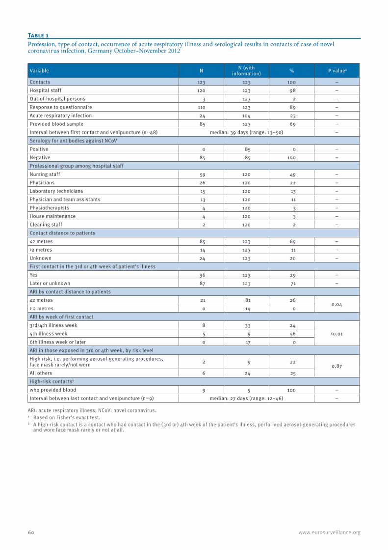
60 www.eurosurveillance.org
Table 1Profession, type of contact, occurrence of acute respiratory illness and serological results in contacts of case of novel coronavirus infection, Germany October–November 2012
Variable N N (with information) % P valuea
Contacts 123 123 100 –Hospital staff 120 123 98 –Out-of-hospital persons 3 123 2 –Response to questionnaire 110 123 89 –Acute respiratory infection 24 104 23 –Provided blood sample 85 123 69 –Interval between first contact and venipuncture (n=48) median: 39 days (range: 13–50) –Serology for antibodies against NCoVPositive 0 85 0 –Negative 85 85 100 –Professional group among hospital staffNursing staff 59 120 49 –Physicians 26 120 22 –Laboratory technicians 15 120 13 –Physician and team assistants 13 120 11 –Physiotherapists 4 120 3 –House maintenance 4 120 3 –Cleaning staff 2 120 2 –Contact distance to patients≤2 metres 85 123 69 –>2 metres 14 123 11 –Unknown 24 123 20 –First contact in the 3rd or 4th week of patient’s illnessYes 36 123 29 –Later or unknown 87 123 71 –ARI by contact distance to patients≤2 metres 21 81 26
0.04> 2 metres 0 14 0ARI by week of first contact3rd/4th illness week 8 33 24
<0.015th illness week 5 9 566th illness week or later 0 17 0ARI in those exposed in 3rd or 4th week, by risk levelHigh risk, i.e. performing aerosol-generating procedures,face mask rarely/not worn 2 9 22
0.87All others 6 24 25High-risk contactsb
who provided blood 9 9 100 –Interval between last contact and venipuncture (n=9) median: 27 days (range: 12–46) –
ARI: acute respiratory illness; NCoV: novel coronavirus.a Based on Fisher’s exact test.b A high-risk contact is a contact who had contact in the (3rd or) 4th week of the patient’s illness, performed aerosol-generating procedures
and wore face mask rarely or not at all.
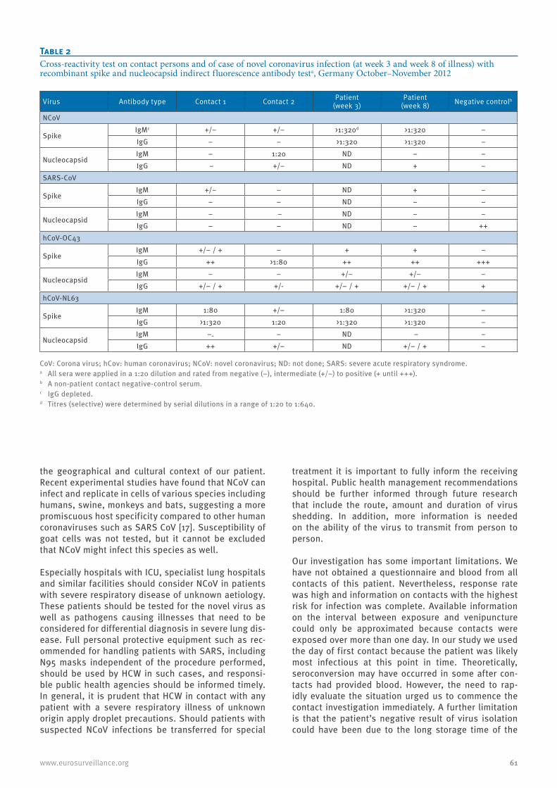
61www.eurosurveillance.org
the geographical and cultural context of our patient. Recent experimental studies have found that NCoV can infect and replicate in cells of various species including humans, swine, monkeys and bats, suggesting a more promiscuous host specificity compared to other human coronaviruses such as SARS CoV [17]. Susceptibility of goat cells was not tested, but it cannot be excluded that NCoV might infect this species as well.
Especially hospitals with ICU, specialist lung hospitals and similar facilities should consider NCoV in patients with severe respiratory disease of unknown aetiology. These patients should be tested for the novel virus as well as pathogens causing illnesses that need to be considered for differential diagnosis in severe lung dis-ease. Full personal protective equipment such as rec-ommended for handling patients with SARS, including N95 masks independent of the procedure performed, should be used by HCW in such cases, and responsi-ble public health agencies should be informed timely. In general, it is prudent that HCW in contact with any patient with a severe respiratory illness of unknown origin apply droplet precautions. Should patients with suspected NCoV infections be transferred for special
treatment it is important to fully inform the receiving hospital. Public health management recommendations should be further informed through future research that include the route, amount and duration of virus shedding. In addition, more information is needed on the ability of the virus to transmit from person to person.
Our investigation has some important limitations. We have not obtained a questionnaire and blood from all contacts of this patient. Nevertheless, response rate was high and information on contacts with the highest risk for infection was complete. Available information on the interval between exposure and venipuncture could only be approximated because contacts were exposed over more than one day. In our study we used the day of first contact because the patient was likely most infectious at this point in time. Theoretically, seroconversion may have occurred in some after con-tacts had provided blood. However, the need to rap-idly evaluate the situation urged us to commence the contact investigation immediately. A further limitation is that the patient’s negative result of virus isolation could have been due to the long storage time of the
Table 2Cross-reactivity test on contact persons and of case of novel coronavirus infection (at week 3 and week 8 of illness) with recombinant spike and nucleocapsid indirect fluorescence antibody testa, Germany October–November 2012
Virus Antibody type Contact 1 Contact 2 Patient(week 3)
Patient(week 8) Negative controlb
NCoV
SpikeIgMc +/− +/− >1:320d >1:320 −IgG − − >1:320 >1:320 −
NucleocapsidIgM − 1:20 ND − −IgG – +/− ND + −
SARS-CoV
SpikeIgM +/− − ND + −IgG − − ND − −
NucleocapsidIgM − − ND − −IgG − − ND − ++
hCoV-OC43
SpikeIgM +/− / + − + + −IgG ++ >1:80 ++ ++ +++
NucleocapsidIgM − − +/− +/− −IgG +/− / + +/- +/− / + +/− / + +
hCoV-NL63
SpikeIgM 1:80 +/− 1:80 >1:320 −IgG >1:320 1:20 >1:320 >1:320 −
NucleocapsidIgM −. − ND − −IgG ++ +/− ND +/− / + −
CoV: Corona virus; hCov: human coronavirus; NCoV: novel coronavirus; ND: not done; SARS: severe acute respiratory syndrome.a All sera were applied in a 1:20 dilution and rated from negative (−), intermediate (+/−) to positive (+ until +++).b A non-patient contact negative-control serum.c IgG depleted.d Titres (selective) were determined by serial dilutions in a range of 1:20 to 1:640.
62 www.eurosurveillance.org
sample – in contrast to our favoured hypothesis of low RNA concentration.
In spite of this, we believe that it is fair to conclude the patient’s infectiousness on illness day 20 was absent or very low. Our contact investigation has found no evidence of infection among hospital or out-of-hospi-tal contacts. Our two-staged approach to serological screening where a first-line testing is done by full-virus IFA and supplemented by confirmatory recombinant IFA and SNT should provide a template for similar investi-gations in the future. Finally, if patients suspected to be infected with NCoV are to be transferred for special-ised treatment, receiving hospitals need to be informed so that appropriate infection control measures can be implemented.
Acknowledgements We thank Kerstin Prahm and Aryna Zanuzdana for support with data entry and validation, Sami Marzougi and Ghassan Moussa for their kind interpreting assistance, Annette Jurke for being the contact point at the state health depart-ment, Mareen Adam, Susi Hafemann, Ingrid Zadow and Julia Hinzmann for excellent technical assistance regarding PCR analyses and Petra Kreher and Anette Teichmann for serol-ogy test development and testing of sera. We thank Ron Fouchier of Erasmus Medical Centre for providing the virus isolate.
Work at the University of Bonn was supported by the European Union FP7 project EMPERIE (contract number 223498), ANTIGONE (contract number 278976), the German Research Foundation (DFG grant DR 772/3-1, KA1241/18-1), as well as the German Ministry of Education and Research (BMBF SARS II).
Conflict of interest None declared.
Authors’ contributionsUB: assisted in designing the study, analysed data, wrote manuscript. MAM: performed laboratory testing, read and revised manuscript. AN: performed laboratory testing, read and revised manuscript. AS: collected data, read and revised manuscript. NW: collected data, read and revised manu-script. TBB: collected data, read and revised manuscript. FB: collected data, read and revised manuscript. CD: assisted in designing the study, performed laboratory testing, read and revised manuscript. BS: performed laboratory testing, read and revised manuscript. TW: performed laboratory testing, read and revised manuscript. DM: performed laboratory test-ing, read and revised manuscript. BM: performed laboratory testing, read and revised manuscript. SB: assisted in design-ing the study, read and revised the manuscript. GK: assisted in designing the study, read and revised the manuscript. LS: assisted in designing the study, read and revised the manu-script. WH: assisted in designing the study, co-ordinated the study, read and revised the manuscript.
References1. Zaki AM, van Boheemen S, Bestebroer TM, Osterhaus AD,
Fouchier RA. Isolation of a novel coronavirus from a man with pneumonia in Saudi Arabia. N Engl J Med. 2012;367(19):1814-20. http://dx.doi.org/10.1056/NEJMoa1211721. PMid:23075143.
2. Centers for Disease Control and Prevention. Severe respiratory illness associated with a novel coronavirus--Saudi Arabia and Qatar, 2012. MMWR Morbidity and mortality weekly report 2012;61:820. PMid:23051613.
3. Bermingham A, Chand M, Brown C, Aarons E, Tong C, Langrish C et al. Severe respiratory illness caused by a novel coronavirus, in a patient transferred to the United Kingdom from the Middle East, September 2012. Euro Surveill. 2012; 17(40): pii=20290. Available from: http://www.eurosurveillance.org/ViewArticle.aspx?ArticleId=20290
4. World Health Organization (WHO). Novel coronavirus infection - update. Geneva: (WHO); 2013. Accessed on February 18, 2012. Available from: http://www.who.int/csr/don/2013_02_16/en/index.html
5. European Center for Disease Prevention and Control (ECDC). Rapid risk assessment: Update: Severe respiratory disease associated with a novel coronavirus. Update 19 February 2013. Stockholm:ECDC;2013. Accessed on: February 19, 2012. Available from: http://www.ecdc.europa.eu/en/publications/Publications/novel-coronavirus-rapid-risk-assessment-update.pdf
6. World Health Organization (WHO). Infection prevention and control of epidemic- and pandemic-prone acute respiratory diseases in health care - WHO interim guidelines. Geneva: (WHO); 2007. Accessed on: December 17, 2012. Available from: http://www.who.int/csr/resources/publications/WHO_CDS_EPR_2007_6c.pdf
7. Pebody RG, Chand MA, Thomas HL, Green HK, Boddington NL, Carvalho C, et al. The United Kingdom public health response to an imported laboratory confirmed case of a novel coronavirus in September 2012. Euro Surveill. 2012;17(40):pii=20292. Available from: http://www.eurosurveillance.org/ViewArticle.aspx?ArticleId=20292
8. World Health Organization (WHO). International health regulations (2005). Second edition. Geneva: WHO; 2008. Available from: http://whqlibdoc.who.int/publications/2008/9789241580410_eng.pdf
9. Corman V, Eckerle I, Bleicker T, Zaki A, Landt O, Eschbach-Bludau M, et al. Detection of a novel human coronavirus by real-time reverse-transcription polymerase chain reaction. Euro Surveill, 2012;17(39):pii=20285. Available from: http://www.eurosurveillance.org/ViewArticle.aspx?ArticleId=20285
10. Corman VM, Müller MA, Costabel U, Timm J, Binger T, Meyer B, et al. Assays for laboratory confirmation of novel human coronavirus (hCoV-EMC) infections. Euro Surveill. 2012;17(49):pii=20334. Available from: http://www.eurosurveillance.org/ViewArticle.aspx?ArticleId=20334
11. Herzog P, Drosten C, Müller MA. Plaque assay for human coronavirus NL63 using human colon carcinoma cells. Virol J. 2008;5:138. http://dx.doi.org/10.1186/1743-422X-5-138. PMid:19014487. PMCid:2603006.
12. Federal Ministry of Justice. Gesetz zur Verhütung und Bekämpfung von Infektionskrankheiten beim Menschen (Infektionsschutzgesetz - IfSG). [Regulation on preventing and control of infectious diseases in humans (Act on protection against infection)]. 20 Jul 2000. German. Available from: http://bundesrecht.juris.de/bundesrecht/ifsg/gesamt.pdf
13. van Boheemen S, de Graaf M, Lauber C, Bestebroer TM, Raj VS, Zaki AM, et al. Genomic characterization of a newly discovered coronavirus associated with acute respiratory distress syndrome in humans. MBio. 2012;3. pii: e00473-12. http://dx.doi.org/10.1128/mBio.00473-12
14. Annan A, Baldwin HJ, Corman VM, Klose SM, Owusu M, Nkrumah EE, et al. Human betacoronavirus 2c EMC/2012¬related viruses in bats, Ghana and Europe. Emerg Infect Dis 2013 Mar (epub ahead of print). http://dx.doi.org/10.3201/eid1903.121503
15. Albarrak AM, Stephens GM, Hewson R, Memish ZA. Recovery from severe novel coronavirus infection. Saudi medical journal 2012;33:1265-9. PMid:23232672.
16. Saif LJ. Bovine respiratory coronavirus. Vet Clin North Am Food Anim Pract. 2010;26:349-64. http://dx.doi.org/10.1016/j.cvfa.2010.04.005
17. Muller MA, Raj VS, Muth D, Meyer B, Kallies S, Smits SL, et al. Human Coronavirus EMC Does Not Require the SARS-Coronavirus Receptor and Maintains Broad Replicative Capability in Mammalian Cell Lines. mBio 3:e00515-12. http://dx.doi.org/10.1128/mBio.00515-12
63www.eurosurveillance.org
Perspectives
Transmission scenarios for Middle East Respiratory Syndrome Coronavirus (MERS-CoV) and how to tell them apart
S Cauchemez ([email protected])1, M D Van Kerkhove1, S Riley1, C A Donnelly1, C Fraser1, N M Ferguson1
1. MRC Centre for Outbreak Analysis and Modelling, Department of Infectious Disease Epidemiology, Imperial College London, London, United Kingdom
Citation style for this article: Cauchemez S, Van Kerkhove MD, Riley S, Donnelly CA, Fraser C, Ferguson NM. Transmission scenarios for Middle East Respiratory Syndrome Coronavirus (MERS-CoV) and how to tell them apart . Euro Surveill. 2013;18(24):pii=20503. Available online: http://www.eurosurveillance.org/ViewArticle.aspx?ArticleId=20503
Article submitted on 02 May 2013 / published on 13 June 2013
Detection of human cases of Middle East Respiratory Syndrome Coronavirus (MERS-CoV) infection interna-tionally is a global public health concern. Rigorous risk assessment is particularly challenging in a con-text where surveillance may be subject to under-ascer-tainment and a selection bias towards more severe cases. We would like to assess whether the virus is capable of causing widespread human epidemics, and whether self-sustaining transmission is already under way. Here we review possible transmission scenarios for MERS-CoV and their implications for risk assess-ment and control. We discuss how existing data, future investigations and analyses may help in reducing uncertainty and refining the public health risk assess-ment and present analytical approaches that allow robust assessment of epidemiological characteristics, even from partial and biased surveillance data. Finally, we urge that adequate data be collected on future cases to permit rigorous assessment of the transmis-sion characteristics and severity of MERS-CoV, and the public health threat it may pose. Going beyond mini-mal case reporting, open international collaboration, under the guidance of the World Health Organization and the International Health Regulations, will impact on how this potential epidemic unfolds and prospects for control.
As of 30 May 2013, 50 laboratory-confirmed cases of Middle East Respiratory Syndrome Coronavirus (MERS-CoV) infection have occurred worldwide [1]. An appar-ently high case-fatality ratio (60%; 30 deaths as of 30 May 2013 [1]) and growing evidence that human-to-human transmission is occurring [2] make MERS-CoV a threat to global health. The current situation has already been compared to the early stages of the severe acute respiratory syndrome (SARS) epidemic in 2003 [3,4].
No animal reservoir has yet been identified for MERS-CoV, and yet human cases, mostly severe, have been detected over a wide geographical area in the Middle East and Europe. If most human cases to date have
arisen from animal exposure, this implies a large but as yet uncharacterised zoonotic epidemic is under way in animal species to which humans have frequent expo-sure (Figure 1A). In this scenario, we might expect rela-tively small numbers of human cases overall, though with the limited surveillance data available to date, we cannot rule out the possibility that substantial num-bers of human cases, with milder disease, have gone undetected.
Even if most human cases to date have been infected through zoonotic exposure, is it possible that MERS-CoV already has the potential to support sustained human-to-human transmission but has by chance so far failed to do so?
Alternatively, how feasible is it that most of the severe MERS-CoV cases detected to date were in fact infected via human-to-human transmission and that the epi-demic is already self-sustaining in human populations (Figure 1B)? Under this transmission scenario, substan-tial numbers of human infections may have already occurred, with only a small proportion of them being detected. But is it feasible that such an epidemic would not have been recognised?
Each of these scenarios has very different implications for the assessment of severity, relevance of reservoir-targeted strategies and potential impact of MERS-CoV globally. Although it may not be possible to completely rule out any of the scenarios with the data currently available, it is timely to consider the priorities for data collection and analysis as cases accrue, so as to best be able to reduce uncertainty and refine the public health risk assessment.
Transmission scenarios for an emerging infectionThe human-to-human transmissibility (and thus epi-demic potential) of an emerging pathogen is quantified by the (effective) reproduction number, R, the average number of secondary infections caused by an index
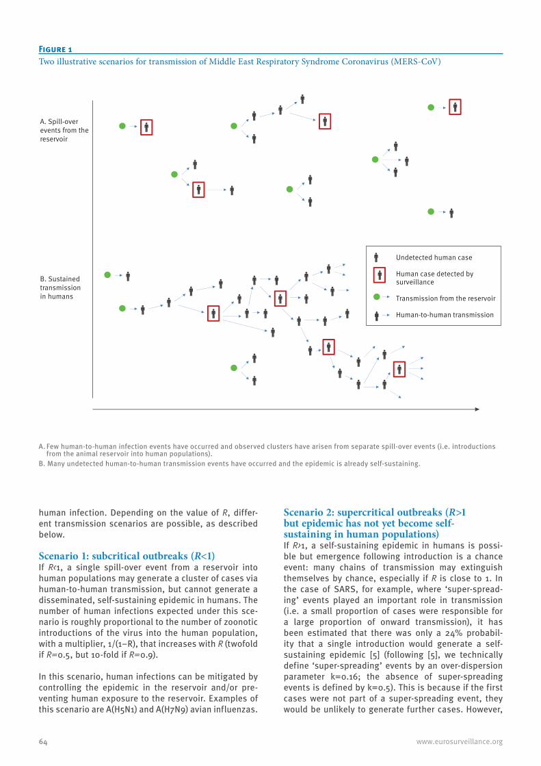
64 www.eurosurveillance.org
human infection. Depending on the value of R, differ-ent transmission scenarios are possible, as described below.
Scenario 1: subcritical outbreaks (R<1)If R<1, a single spill-over event from a reservoir into human populations may generate a cluster of cases via human-to-human transmission, but cannot generate a disseminated, self-sustaining epidemic in humans. The number of human infections expected under this sce-nario is roughly proportional to the number of zoonotic introductions of the virus into the human population, with a multiplier, 1/(1−R), that increases with R (twofold if R=0.5, but 10-fold if R=0.9).
In this scenario, human infections can be mitigated by controlling the epidemic in the reservoir and/or pre-venting human exposure to the reservoir. Examples of this scenario are A(H5N1) and A(H7N9) avian influenzas.
Scenario 2: supercritical outbreaks (R>1 but epidemic has not yet become self-sustaining in human populations)If R>1, a self-sustaining epidemic in humans is possi-ble but emergence following introduction is a chance event: many chains of transmission may extinguish themselves by chance, especially if R is close to 1. In the case of SARS, for example, where ‘super-spread-ing’ events played an important role in transmission (i.e. a small proportion of cases were responsible for a large proportion of onward transmission), it has been estimated that there was only a 24% probabil-ity that a single introduction would generate a self-sustaining epidemic [5] (following [5], we technically define ‘super-spreading’ events by an over-dispersion parameter k=0.16; the absence of super-spreading events is defined by k=0.5). This is because if the first cases were not part of a super-spreading event, they would be unlikely to generate further cases. However,
Figure 1Two illustrative scenarios for transmission of Middle East Respiratory Syndrome Coronavirus (MERS-CoV)
A. Few human-to-human infection events have occurred and observed clusters have arisen from separate spill-over events (i.e. introductions from the animal reservoir into human populations).
B. Many undetected human-to-human transmission events have occurred and the epidemic is already self-sustaining.
A. Spill-over events from the reservoir
B. Sustained transmissionin humans
Undetected human case
Human case detected by surveillance
Transmission from the reservoir
Human-to-human transmission
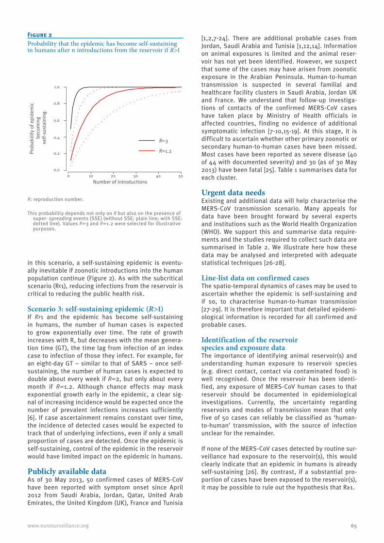
65www.eurosurveillance.org
in this scenario, a self-sustaining epidemic is eventu-ally inevitable if zoonotic introductions into the human population continue (Figure 2). As with the subcritical scenario (R<1), reducing infections from the reservoir is critical to reducing the public health risk.
Scenario 3: self-sustaining epidemic (R>1)If R>1 and the epidemic has become self-sustaining in humans, the number of human cases is expected to grow exponentially over time. The rate of growth increases with R, but decreases with the mean genera-tion time (GT), the time lag from infection of an index case to infection of those they infect. For example, for an eight-day GT – similar to that of SARS – once self-sustaining, the number of human cases is expected to double about every week if R=2, but only about every month if R=1.2. Although chance effects may mask exponential growth early in the epidemic, a clear sig-nal of increasing incidence would be expected once the number of prevalent infections increases sufficiently [6]. If case ascertainment remains constant over time, the incidence of detected cases would be expected to track that of underlying infections, even if only a small proportion of cases are detected. Once the epidemic is self-sustaining, control of the epidemic in the reservoir would have limited impact on the epidemic in humans.
Publicly available dataAs of 30 May 2013, 50 confirmed cases of MERS-CoV have been reported with symptom onset since April 2012 from Saudi Arabia, Jordan, Qatar, United Arab Emirates, the United Kingdom (UK), France and Tunisia
[1,2,7-24]. There are additional probable cases from Jordan, Saudi Arabia and Tunisia [1,12,14]. Information on animal exposures is limited and the animal reser-voir has not yet been identified. However, we suspect that some of the cases may have arisen from zoonotic exposure in the Arabian Peninsula. Human-to-human transmission is suspected in several familial and healthcare facility clusters in Saudi Arabia, Jordan UK and France. We understand that follow-up investiga-tions of contacts of the confirmed MERS-CoV cases have taken place by Ministry of Health officials in affected countries, finding no evidence of additional symptomatic infection [7-10,15-19]. At this stage, it is difficult to ascertain whether other primary zoonotic or secondary human-to-human cases have been missed. Most cases have been reported as severe disease (40 of 44 with documented severity) and 30 (as of 30 May 2013) have been fatal [25]. Table 1 summarises data for each cluster.
Urgent data needsExisting and additional data will help characterise the MERS-CoV transmission scenario. Many appeals for data have been brought forward by several experts and institutions such as the World Health Organization (WHO). We support this and summarise data require-ments and the studies required to collect such data are summarised in Table 2. We illustrate here how these data may be analysed and interpreted with adequate statistical techniques [26-28].
Line-list data on confirmed casesThe spatio-temporal dynamics of cases may be used to ascertain whether the epidemic is self-sustaining and if so, to characterise human-to-human transmission [27-29]. It is therefore important that detailed epidemi-ological information is recorded for all confirmed and probable cases.
Identification of the reservoir species and exposure data The importance of identifying animal reservoir(s) and understanding human exposure to reservoir species (e.g. direct contact, contact via contaminated food) is well recognised. Once the reservoir has been identi-fied, any exposure of MERS-CoV human cases to that reservoir should be documented in epidemiological investigations. Currently, the uncertainty regarding reservoirs and modes of transmission mean that only five of 50 cases can reliably be classified as ‘human-to-human’ transmission, with the source of infection unclear for the remainder.
If none of the MERS-CoV cases detected by routine sur-veillance had exposure to the reservoir(s), this would clearly indicate that an epidemic in humans is already self-sustaining [26]. By contrast, if a substantial pro-portion of cases have been exposed to the reservoir(s), it may be possible to rule out the hypothesis that R≥1.
Figure 2Probability that the epidemic has become self-sustaining in humans after n introductions from the reservoir if R>1
R: reproduction number.
This probability depends not only on R but also on the presence of super- spreading events (SSE) (without SSE: plain line; with SSE: dotted line). Values R=3 and R=1.2 were selected for illustrative purposes.
0 10 50403020Number of introductions
0.0
0.2
1.0
0.8
0.6
0.4
Prob
abili
ty o
f epi
dem
ic
beco
min
gse
lf-su
stai
ning
R=3
R=1.2
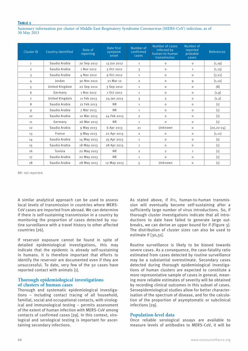
66 www.eurosurveillance.org
A similar analytical approach can be used to assess local levels of transmission in countries where MERS-CoV cases are imported from abroad. We can determine if there is self-sustaining transmission in a country by monitoring the proportion of cases detected by rou-tine surveillance with a travel history to other affected countries [26].
If reservoir exposure cannot be found in spite of detailed epidemiological investigations, this may indicate that the epidemic is already self-sustaining in humans. It is therefore important that efforts to identify the reservoir are documented even if they are unsuccessful. To date, very few of the 50 cases have reported contact with animals [1].
Thorough epidemiological investigations of clusters of human casesThorough and systematic epidemiological investiga-tions – including contact tracing of all household, familial, social and occupational contacts, with virolog-ical and immunological testing – permits assessment of the extent of human infection with MERS-CoV among contacts of confirmed cases [29]. In this context, viro-logical and serological testing is important for ascer-taining secondary infections.
As stated above, if R>1, human-to-human transmis-sion will eventually become self-sustaining after a sufficiently large number of virus introductions. So, if thorough cluster investigations indicate that all intro-ductions to date have failed to generate large out-breaks, we can derive an upper bound for R (Figure 3). The distribution of cluster sizes can also be used to estimate R [30,31].
Routine surveillance is likely to be biased towards severe cases. As a consequence, the case-fatality ratio estimated from cases detected by routine surveillance may be a substantial overestimate. Secondary cases detected during thorough epidemiological investiga-tions of human clusters are expected to constitute a more representative sample of cases in general, mean-ing more reliable estimates of severity will be obtained by recording clinical outcomes in this subset of cases. Seroepidemiological studies allow for better character-isation of the spectrum of disease, and for the calcula-tion of the proportion of asymptomatic or subclinical infections [29].
Population-level dataOnce reliable serological assays are available to measure levels of antibodies to MERS-CoV, it will be
Table 1Summary information per cluster of Middle East Respiratory Syndrome Coronavirus (MERS-CoV) infection, as of 30 May 2013
Cluster ID Country identified Date of reporting
Date first symptom
onset
Number of confirmed
cases
Number of cases infected by
human-to-human transmission
Number of reported probable
cases
References
1 Saudia Arabia 20 Sep 2012 13 Jun 2012 1 0 0 [1,19]
2 Saudia Arabia 1 Nov 2012 5 Oct 2012 3 0 1 [1,13]
3 Saudia Arabia 4 Nov 2012 9 Oct 2012 1 0 0 [7,21]
4 Jordan 30 Nov 2012 21 Mar 12 2 0 9 [1,12]
5 United Kingdom 22 Sep 2012 3 Sep 2012 1 0 0 [8]
6 Germany 1 Nov 2012 1 Oct 2012 1 0 0 [1,9]
7 United Kingdom 11 Feb 2013 24 Jan 2013 3 2 0 [1,2]
8 Saudia Arabia 21 Feb 2013 NR 1 0 0 [1]
9 Saudia Arabia 7 Mar 2013 NR 1 0 0 [1]
10 Saudia Arabia 12 Mar 2013 24 Feb 2013 2 0 0 [1]
11 Germany 26 Mar 2013 NR 1 0 0 [1]
12 Saudia Arabia 9 May 2013 6 Apr 2013 21 Unknown 0 [20,22-24]
13 France 9 May 2013 22 Apr 2013 2 0 0 [1,11]
14 Saudia Arabia 14 May 2013 25 Apr 2013 1 0 0 [1]
15 Saudia Arabia 18 May 2013 28 Apr 2013 1 0 0 [1]
16 Tunisia 22 May 2013 NR 2 2 1 [1]
17 Saudia Arabia 22 May 2013 NR 1 0 0 [1]
18 Saudia Arabia 28 May 2013 12 May 2013 5 Unknown 0 [1]
NR: not reported.
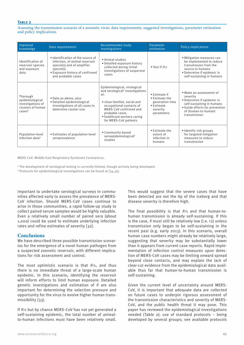
67www.eurosurveillance.org
important to undertake serological surveys in commu-nities affected early to assess the prevalence of MERS-CoV infection. Should MERS-CoV cases continue to arise in those communities, a rapid follow-up study to collect paired serum samples would be highly valuable. Even a relatively small number of paired sera (about 1,000) could be used to estimate underlying infection rates and refine estimates of severity [32].
ConclusionsWe have described three possible transmission scenar-ios for the emergence of a novel human pathogen from a suspected zoonotic reservoir, with different implica-tions for risk assessment and control.
The most optimistic scenario is that R<1, and thus there is no immediate threat of a large-scale human epidemic. In this scenario, identifying the reservoir will inform efforts to limit human exposure. Detailed genetic investigations and estimation of R are also important for determining the selection pressure and opportunity for the virus to evolve higher human trans-missibility [33].
If R>1 but by chance MERS-CoV has not yet generated a self-sustaining epidemic, the total number of animal-to-human infections must have been relatively small.
This would suggest that the severe cases that have been detected are not the tip of the iceberg and that disease severity is therefore high.
The final possibility is that R>1 and that human-to-human transmission is already self-sustaining. If this is the case, R must still be relatively low (i.e. <2) unless transmission only began to be self-sustaining in the recent past (e.g. early 2013). In this scenario, overall human case numbers might already be relatively large, suggesting that severity may be substantially lower than it appears from current case reports. Rapid imple-mentation of infection control measures upon detec-tion of MERS-CoV cases may be limiting onward spread beyond close contacts, and may explain the lack of clear-cut evidence from the epidemiological data avail-able thus far that human-to-human transmission is self-sustaining.
Given the current level of uncertainty around MERS-CoV, it is important that adequate data are collected on future cases to underpin rigorous assessment of the transmission characteristics and severity of MERS-CoV, and the public health threat it may pose. This paper has reviewed the epidemiological investigations needed (Table 2); use of standard protocols – being developed by several groups; see available protocols
Table 2Assessing the transmission scenario of a zoonotic virus: data requirements, suggested investigations, parameter estimation and policy implications
Improved knowledge Data requirements Recommended study
investigationsParameter estimation Policy implications
Identification of reservoir species and exposure data
•Identificationofthesourceofinfection, of animal reservoir specie(s) and of amplifier specie(s)
•Exposurehistoryofconfirmedand probable cases
•Animalstudies•Detailedexposurehistory
collected during initial investigations of suspected cases
•TestifR>1
•Mitigationmeasurescanbe implemented to reduce transmission from the source to humans
•Determineifepidemicisself-sustaining in humans
Thorough epidemiological investigations of clusters of human casesb
•Dataasabove,plus•Detailedepidemiological
investigations of all cases to determine cluster size
Epidemiological, virological and serologicala investigations of:
•closefamilial,socialandoccupational contacts of MERS-CoV confirmed and probable cases
•healthcareworkerscaringfor MERS-CoV patients
•EstimateR•Estimatethe
generation time•Estimate
severity parameters
•Makeanassessmentofseverity
•Determineifepidemicisself-sustaining in humans
•Guideeffortsforpreventionof (human-to-human) transmission
Population-level infection datab
•Estimatesofpopulation-levelseroprevalence
•Community-basedseroepidemiologicala studies
•Estimatetheextent of infection in humans
•Identifyriskgroupsfor targeted mitigation measures to reduce transmission
MERS-CoV: Middle East Respiratory Syndrome Coronavirus.
a The development of serological testing is currently limited, though actively being developed. b Protocols for epidemiological investigations can be found at [34,35].

68 www.eurosurveillance.org
from WHO [34], the Consortium for the Standardization of Influenza Seroepidemiology (CONSISE) [35] and International Severe Acute Respiratory and Emerging Infection Consortium (ISARIC) [36]) – where possi-ble, would be beneficial. Going beyond minimal case reporting, open international collaboration, guided by the International Health Regulations, will impact how this potential epidemic unfolds and prospects for control.
Acknowledgements The research leading to these results received funding from the European Union Seventh Framework Programme (FP7/2007-2013) under Grant Agreement nu278433-PREDEM-ICS, the European Union FP7 EMPERIE project, the NIGMS MIDAS initiative, the Medical Research Council, and the Bill & Melinda Gates Foundation. The funders had no role in study design, data collection and analysis, decision to pub-lish, or preparation of the manuscript.
Conflict of interestSC received consulting fees from Sanofi Pasteur MSD for a project on the modelling of varicella zoster virus transmis-sion. The authors declare no other competing interests.
Authors’ contributionsSC, MVK, SR, SAD, CF, NMF planned the analysis; MVK compiled the data; SC developed the methods and ran the analysis; SC wrote the first draft; SC, MVK, SR, SAD, CF, NMF edited the paper.
Figure 3Upper bound for the reproduction number R as a function of the number of introductions from the reservoir that failed to generate self-sustaining epidemics
0 20 40 60 80 100
0.0
0.5
1.0
1.5
2.0
2.5
3.0
Number of introductions that failedto generate self-sustaining epidemics
Uppe
r bou
nd fo
r R
With super spreading
Without super spreading
69www.eurosurveillance.org
References1. World Health Organization (WHO). Coronavirus infections.
Geneva: WHO. [Accessed 7 Jun 2013]. Available from: http://www.who.int/csr/don/archive/disease/coronavirus_infections/en/index.html
2. The Health Protection Agency (HPA) UK Novel Coronavirus Investigation team. Evidence of person-to-person transmission within a family cluster of novel coronavirus infections, United Kingdom, February 2013. Euro Surveill. 2013;18(11):pii=20427. Available from: http://www.eurosurveillance.org/ViewArticle.aspx?ArticleId=20427
3. McCloskey B, Zumla A, Stephens G, Heymann DL, Memish ZA. Applying lessons from SARS to a newly identified coronavirus. Lancet Infect Dis. 2013;13(5):384-5. http://dx.doi.org/10.1016/S1473-3099(13)70082-3
4. Pebody R, Zambon M, Watson JM. Novel coronavirus: how much of a threat? We know the questions to ask; we don’t yet have many answers. BMJ. 2013;346:f1301. http://dx.doi.org/10.1136/bmj.f1301 PMid:23447352
5. Lloyd-Smith JO, Schreiber SJ, Kopp PE, Getz WM. Superspreading and the effect of individual variation on disease emergence. Nature. 2005;438(7066):355-9. http://dx.doi.org/10.1038/nature04153 PMid:16292310
6. Feng D, de Vlas SJ, Fang LQ, Han XN, Zhao WJ, Sheng S, et al. The SARS epidemic in mainland China: bringing together all epidemiological data. Trop Med Int Health. 2009;14 Suppl 1:4-13. http://dx.doi.org/10.1111/j.1365-3156.2008.02145.x PMid:19508441
7. AlBarrak AM, Stephens GM, Hewson R, Memish ZA. Recovery from severe novel coronavirus infection. Saudi Med J. 2012;33(12):1265-9. PMid:23232672
8. Bermingham A, Chand MA, Brown CS, Aarons E, Tong C, Langrish C, et al. Severe respiratory illness caused by a novel coronavirus, in a patient transferred to the United Kingdom from the Middle East, September 2012. Euro Surveill. 2012;17(40):pii=20290. Available from: http://www.eurosurveillance.org/ViewArticle.aspx?ArticleId=20290
9. Buchholz U, Müller MA, Nitsche A, Sanewski A, Wevering N, Bauer-Balci, et al. Contact investigation of a case of human novel coronavirus infection treated in a German hospital, October-November 2012. Euro Surveill. 2013;18(8):pii=20406. Available from: http://wwweurosurveillanceorg/ViewArticleaspx?ArticleId=20406
10. European Centre for Disease Prevention and Control (ECDC). What’s new. Stockholm: ECDC. Novel coronavirus updates. [Accessed 13 Mar 2013]. Available from: http://www.ecdc.europa.eu/en/healthtopics/coronavirus-infections/whats-new/Pages/whats_new.aspx
11. Guery B, Poissy J, El Mansouf L, Séjourné C, Ettahar N, Lemaire X, et al. Clinical features and viral diagnosis of two cases of infection with Middle East Respiratory Syndrome coronavirus: a report of nosocomial transmission. Lancet. 2013. pii: S0140-6736(13)60982-4.
12. Hijawi B, Abdallat M, Sayaydeh A, Alqasrawi S, Haddadin A, Jaarour N, et al. Novel coronavirus infections in Jordan, April 2012: epidemiological findings from a retrospective investigation. East Mediterr Health J. 2013;19 Suppl 1:S12-8.
13. Memish ZA, Alhakeem R, Stephens GM. Saudi Arabia and the emergence of a novel coronavirus. East Mediterr Health J. 2013;19 Suppl 1:S7-11.
14. Memish ZA, Zumla AI, Al-Hakeem RF, Al-Rabeeah AA, Stephens GM. Family cluster of Middle East Respiratory Syndrome Coronavirus infections. N Engl J Med. 2013 May 29. [Epub ahead of print]. http://dx.doi.org/10.1056/NEJMoa1303729 PMid:23718156
15. Pebody RG, Chand MA, Thomas HL, Green HK, Boddington NL, Carvalho C, et al. The United Kingdom public health response to an imported laboratory confirmed case of a novel coronavirus in September 2012. Euro Surveill. 2012;17(40):pii=20292. Available from: http://www.eurosurveillance.org/ViewArticle.aspx?ArticleId=20292
16. Health Protection Agency (HPA). Third case of novel coronavirus infection identified in family cluster. 15 Feb 2013. London: HPA; 2013. Press release. Available from: http://www.hpa.org.uk/NewsCentre/NationalPressReleases/2013PressReleases/1302153rdcaseofcoronavirus/
17. Health Protection Agency (HPA). Case of novel coronavirus identified in the UK. 11 Feb 2013. London: HPA; 2013. Press release. Available from: : http://www.hpa.org.uk/ NewsCentre/NationalPressReleases/2013PressReleases/ 130211statementonlatestcoronaviruspatient/
18. Health Protection Agency (HPA). Further UK case of novel coronavirus. 13 Feb 2013. London: HPA; 2013. Press release. Available from: http://www.hpa.org.uk/
NewsCentre/NationalPressReleases/2013PressReleases 130213statementonlatestcoronaviruspatient/
19. Zaki AM, van Boheemen S, Bestebroer TM, Osterhaus AD, Fouchier RA. Isolation of a novel coronavirus from a man with pneumonia in Saudi Arabia. N Engl J Med. 2012;367(19):1814-20. http://dx.doi.org/10.1056/NEJMoa1211721 PMid:23075143
20. Saudi Arabia Ministry of Health (MOH). A new case of coronavirus recorded in Al-Ahsa and three cases passed away in the Eastern Region. 29 May 2013. Riyadh: MOH; 2013. Available from: http://www.moh.gov.sa/en/HealthAwareness/Corona/PressReleases/Pages/PressStatement-2013-05-29-001.aspx
21. ProMED-mail. Novel coronavirus - Saudi Arabia (15): new case. Archive Number 20121104.1391285. 11 Apr 2012. Available from: http://www.promedmail.org/direct.php?id=20121104.1391285
22. ProMED-mail. Novel coronavirus - Eastern Mediterranean (17): Saudi Arabia. Archive Number 20130503.1688355. 3 May 2013. Available from: http://www.promedmail.org/direct.php?id=20130503.1688355
23. ProMED-mail. Novel coronavirus - Eastern Mediterranean (18): Saudi Arabia. Archive Number 20130505.1693290. 5 May 2013. Available from: http://www.promedmail.org/direct.php?id=20130505.1693290
24. ProMED-mail. Novel coronavirus - Eastern Mediterranean (21): Saudi Arabia. Archive Number 20130509.1701527. 9 May 2013. Available from: http://www.promedmail.org/direct.php?id=20130509.1701527
25. World Health Organization (WHO). Middle East respiratory syndrome- coronavirus - update. 29 May 2013. Geneva: WHO; 2013. Available from: http://www.who.int/csr/don/2013_05_29_ncov/en/index.html
26. Cauchemez S, Epperson S, Biggerstaff M, Swerdlow D, Finelli L, Ferguson NM. Using routine surveillance data to estimate the epidemic potential of emerging zoonoses: application to the emergence of US swine origin influenza A H3N2v virus. PLoS Med. 2013;10(3):e1001399. http://dx.doi.org/10.1371/journal.pmed.1001399 PMid:23472057 PMCid:3589342
27. Mills CE, Robins JM, Lipsitch M. Transmissibility of 1918 pandemic influenza. Nature. 2004;432(7019):904-6. http://dx.doi.org/10.1038/nature03063 PMid:15602562
28. Wallinga J, Teunis P. Different epidemic curves for severe acute respiratory syndrome reveal similar impacts of control measures. Am J Epidemiol. 2004;160(6):509-16. http://dx.doi.org/10.1093/aje/kwh255 PMid:15353409
29. Van Kerkhove MD, Broberg E, Engelhardt OG, Wood J, Nicoll A; CONSISE steering committee. The consortium for the standardization of influenza seroepidemiology (CONSISE): a global partnership to standardize influenza seroepidemiology and develop influenza investigation protocols to inform public health policy. Influenza Other Respi Viruses. 2013;7(3):231-4. http://dx.doi.org/10.1111/irv.12068 PMid:23280042
30. Jansen VA, Stollenwerk N, Jensen HJ, Ramsay ME, Edmunds WJ, Rhodes CJ. Measles outbreaks in a population with declining vaccine uptake. Science. 2003;301(5634):804. http://dx.doi.org/10.1126/science.1086726 PMid:12907792
31. Ferguson NM, Fraser C, Donnelly CA, Ghani AC, Anderson RM. Public health. Public health risk from the avian H5N1 influenza epidemic. Science. 2004;304(5673):968-9. http://dx.doi.org/10.1126/science.1096898 PMid:15143265
32. Riley S, Kwok KO, Wu KM, Ning DY, Cowling BJ, Wu JT, et al. Epidemiological characteristics of 2009 (H1N1) pandemic influenza based on paired sera from a longitudinal community cohort study. PLoS Med. 2011;8:e1000442. http://dx.doi.org/10.1371/journal.pmed.1000442 PMid:21713000 PMCid:3119689
33. Antia R, Regoes RR, Koella JC, Bergstrom CT. The role of evolution in the emergence of infectious diseases. Nature. 2003;426(6967):658-61. http://dx.doi.org/10.1038/nature02104 PMid:14668863
34. World Health Organization (WHO). Case-control study to assess potential risk factors related to human illness caused by novel coronavirus. Geneva: WHO: 2013. Available from: http://www.who.int/csr/disease/coronavirus_infections/NovelCoronaCaseControlStudyPotentialRiskFactors_17May13.pdf
35. The CONSISE Steering Committee. Novel coronavirus (MERS-CoV). The Consortium for the Standardization of Influenza Seroepidemiology (CONSISE). CONSISE. [Accessed 5 Jun 2013]. Available from: http://consise.tghn.org/articles/novel-coronavirus-ncov/
36. International Severe Acute Respiratory and Emerging Infection Consortium (ISARIC). Resources. ISARIC. [Accessed 5 Jun 2013]. Available from: http://isaric.tghn.org/articles/
70 www.eurosurveillance.org
National Bulletins
AustriaMitteilungen der SanitätsverwaltungBundesministerium für Gesundheit Familie und Jugend, ViennaMonthly, print only. In German.http://www.bmgfj.gv.at/cms/site/thema.html?channel=CH0951
BelgiumVlaams Infectieziektebulletin Department of Infectious Diseases Control, FlandersQuarterly, print and online. In Dutch, summaries in English. http://www.infectieziektebulletin.be
Bulletin d’information de la section d’EpidémiologieInstitut Scientifique de la Santé Publique, BrusselsMonthly, online. In French.http://www.iph.fgov.be/epidemio/epifr/episcoop/episcoop.htm
BulgariaBulletin of the National Centre of Infectious and Parasitic Diseases, Sofia Print version. In Bulgarian.http://www.ncipd.org/
CyprusNewsletter of the Network for Surveillance and Control of Communicable Diseases in CyprusMedical and Public Health Services, Ministry of Health, NicosiaBiannual, print and online. In Greek. http://www.moh.gov.cy
Czech Republic Zpravy CEM (Bulletin of the Centre ofEpidemiology and Microbiology)Centrum Epidemiologie a Mikrobiologie StátníhoZdravotního Ústavu, PragueMonthly, print and online. In Czech, titles in English. http://www.szu.cz/cema/adefaultt.htm
EPIDAT (Notifications of infectious diseases in the Czech Republic) http://www.szu.cz/cema/epidat/epidat.htm
Denmark EPI-NEWSDepartment of Epidemiology, Statens Serum Institut, CopenhagenWeekly, print and online. In Danish and English.http://www.ssi.dk
Finland KansanterveyslaitosDepartment of Infectious Disease Epidemiology, National Public Health Institute, HelsinkiMonthly, print and online. In Finnish.http://www.ktl.fi/portal/suomi/osastot/infe/tutkimus/tartuntatautien_seuranta/tartuntatautilaakarin_kommentit/
FranceBulletin épidémiologique hebdomadaireInstitut de veille sanitaire, Saint-Maurice CedexWeekly, print and online. In French.http://www.invs.sante.fr/beh/default.htm
GermanyEpidemiologisches BulletinRobert Koch-Institut, Berlin Weekly, print and online. In German.http://www.rki.de/DE/Content/Infekt/EpidBull/epid__bull__node.html
Greece HCDCP Newsletter Hellenic Centre for Disease Control and Prevention (HCDCP/KEELPNO), Athens Monthly, online. In English and Greek. http://www2.keelpno.gr/blog/?lang=en
Hungary Epinfo (az Országos Epidemiológiai Központ epidemiológiai információs hetilapja) National Center For Epidemiology, BudapestWeekly, online. In Hungarian.http://www.oek.hu/oek.web?to=839&nid=41&pid=7&lang=hun
IcelandEPI-ICELandlæknisembættiðDirectorate Of Health, Seltjarnarnes Monthly, online. In Icelandic and English.http://www.landlaeknir.is
IrelandEPI-INSIGHTHealth Protection Surveillance Centre, DublinMonthly, print and online. In English.http://www.hpsc.ie/hpsc/EPI-Insight
Italy Notiziario dell’Istituto Superiore di SanitàIstituto Superiore di Sanità, Reparto di Malattie Infettive, RomeMonthly, online. In Italian. http://www.iss.it/publ/noti/index.php?lang=1&tipo=4
Bolletino Epidemiologico Nazionale (BEN)Istituto Superiore di Sanità, Reparto di Malattie Infettive, RomeMonthly, online. In Italian.http://www.epicentro.iss.it/ben
Latvia Epidemiologijas BileteniSabiedribas veselibas agentura Public Health Agency, RigaOnline. In Latvian.http://www.sva.lv/epidemiologija/bileteni
Lithuania Epidemiologijos žiniosUžkreciamuju ligu profilaktikos ir kontroles centrasCenter for Communicable Disease Prevention and Control, VilniusOnline. In Lithuanian.http://www.ulac.lt/index.php?pl=26
NetherlandsInfectieziekten BulletinRijksinstituut voor Volksgezondheid en MilieuNational Institute of Public Health and the Environment, Bilthoven Monthly, print and online. In Dutch.http://www.rivm.nl/infectieziektenbulletin
NorwayMSIS-rapportFolkehelseinstituttet, OsloWeekly, print and online. In Norwegian. http://www.folkehelsa.no/nyhetsbrev/msis
71www.eurosurveillance.org
PolandMeldunki o zachorowaniach na choroby zakazne i zatruciach w Polsce Panstwowy Zaklad Higieny, National Institute of Hygiene, WarsawFortnightly, online. In Polish and English. http://www.pzh.gov.pl
PortugalSaúde em NúmerosMinistério da Saúde,Direcção-Geral da Saúde, LisbonSporadic, print only. In Portuguese. http://www.dgs.pt
SloveniaCNB Novice Inštitut za varovanje zdravja, Center za nalezljive bolezni, Institute of Public Health, Center for Infectious Diseases, LjubljanaMonthly, online. In Slovene. http://www.ivz.si
RomaniaInfo EpidemiologiaCentrul pentru Prevenirea si Controlul Bolilor Transmisibile, National Centre of Communicable Diseases Prevention and Control, Institute of Public Health, BucharestSporadic, print only. In Romanian.Sporadic, print only. In Romanian. http://www.insp.gov.ro/cnscbt/index.php?option=com_docman&Itemid=12
SpainBoletín Epidemiológico SemanalCentro Nacional de Epidemiología, Instituto de Salud Carlos III, MadridFortnightly, print and online. In Spanish.http://revista.isciii.es
SwedenSmittskyddsinstitutets nyhetsbrevSmittskyddsinstitutet, StockholmWeekly, online. In Swedish. http://www.smittskyddsinstitutet.se
United KingdomEngland and Wales Health Protection Report Health Protection Agency, LondonWeekly, online only. In English.http://www.hpa.org.uk/hpr
Northern IrelandCommunicable Diseases Monthly Report Communicable Disease Surveillance Centre, Northern Ireland, BelfastMonthly, print and online. In English.http://www.cdscni.org.uk/publications
ScotlandHealth Protection Scotland Weekly Report Health Protection Scotland, GlasgowWeekly, print and online. In English. http://www.hps.scot.nhs.uk/ewr/
Other journalsEpiNorth journalNorwegian Institute of Public Health, Folkehelseinstituttet, Oslo, NorwayPublished four times a year in English and Russian.http://www.epinorth.org
European Union“Europa” is the official portal of the European Union. It provides up-to-date coverage of main events and information on activities and institutions of the European Union.http://europa.eu
European Commission - Public HealthThe website of European Commission Directorate General for Health and Consumer Protection (DG SANCO).http://ec.europa.eu/health/
Health-EU PortalThe Health-EU Portal (the official public health portal of the European Union) includes a wide range of information and data on health-related issues and activities at both European and international level.http://ec.europa.eu/health-eu/

72 www.eurosurveillance.org

73www.eurosurveillance.org

74 www.eurosurveillance.org

All material in Eurosurveillance is in the public domain and may be used and reprinted without special permission. However, the source should be cited properly and we suggest adding a link to the exact page on the Eurosurveillance website.
Articles published in Eurosurveillance are indexed in PubMed/Medline.
The Eurosurveillance print edition is a selection of short and long articles previously published on the Eurosurveillance website. The full listing of all Eurosurveillance articles can be found in the Archives section of the website.
The opinions expressed by authors contributing to Eurosurveillance do not necessarily reflect the opinions of the European Centre for Disease Prevention and Control (ECDC) or the Editorial team or the institutions with which the authors are affiliated. Neither the ECDC nor any person acting on behalf of the ECDC is respon-sible for the use which might be made of the information in this journal.
TQ-AD-13-002-EN
-C
ISSN 1025 496X4,500 copiesGraphic design © ECDC, Stockholm
Contributions to Eurosurveillance are welcomed. Full instructions to authors are available at our website www.eurosurveillance.org
Visit our website at www.eurosurveillance.org
The Eurosurveillance print edition is a compilation of short and long articles that have previously been published on our website.
All the articles in this issue are available online: you can print each page separately or download the whole quarterly in pdf format.
The website archives all articles since 1995, and offers a search facility.
To receive Eurosurveillance’s free electronic releases and e-alerts by e-mail, please subscribe on our website.
Papers published in the former monthly release are indexed for MEDLINE since January 2001, and papers published in the weekly release from January 2005 (with the exception of short, non-scientific notices) are also indexed for MEDLINE. The Index Medicus abbreviation for Eurosurveillance is Euro Surveill.
Follow us on Twitter! https://twitter.com/#!/eurosurveillanc