Embed Size (px)
Citation preview

Arch. Dis. Childh., 1969, 44, 203.
Sotos' Syndrome of Cerebral GigantismJ. M. ABRAHAM and G. J. A. I. SNODGRASS
From the General Hospital, Ashton-under-Lyne, and the Department of Paediatrics,Charing Cross Group of Hospitals, London
Excessive secretion of growth hormone afterepiphysial fusion will produce the clinical featuresof acromegaly, which is not in itself associated withincreased stature. Before closure of the epiphysesthis situation will lead to gigantism, which isconfined almost exclusively to the adolescentage-group in its first appearance. Though con-comitant enlargement of some of the extremitiesis not uncommon in true or even constitutionalgigantism, it is almost unknown in pre-adolescentchildren. Recently Sotos et al. (1964) describeda condition characterized by excessively rapidgrowth dating from infancy, acromegalic features,and a non-progressive neurological dysfunctionmanifested by clumsiness and a dull intelligence.The affected patients bore a remarkable resemblanceto each other. The authors coined the term'cerebral gigantism' for this syndrome. Otherrelatively constant clinical features noted in theircases included dolicocephaly, with macrocrania,hypertelorism, a high arched palate, acceleratedskeletal maturation, and the absence of obviousendocrine dysfunction. Pneumoencephalographyin such cases usually reveals dilated cerebralventricles without an obstructive lesion. Con-vulsions are frequent in this syndrome but electro-encephalography shows only non-specific ab-normalities.A patient conforming closely to the criteria of
Sotos and co-workers had been described earlier(Mikulowski, Stopyrowa, and Medvey, 1962).41 examples of this syndrome are now known,and details of 2 further cases with full endocrino-logical findings are presented in this paper.
Case ReportsCase 1. The patient was the only child of unrelated
19-year-old parents. The mother's general healthhad been good, and the only drugs taken during thepregnancy were promethazine and iron. Labour was
Received September 11, 1968.
induced surgically for moderate pre-eclamptic toxaemiaat 37 weeks' gestation. She was delivered normallyby the vertex, the birthweight being 2466 g. (1 SDbelow the mean). Length and head circumferencewere not recorded at birth, but she seemed long andhad long fingers and toes, a possible diagnosis ofMarfan's syndrome being entertained at this time.Initially, she fed poorly and was moderately jaundiced,the serum bilirubin reaching 19 mg./100 ml. on thesixth day.At 2 weeks of age, she developed bronchopneumonia
with cyanosis and convulsions which responded toantibiotics, anticonvulsants, and oxygen. Four weekslater, she was readmitted with vomiting which subsidedafter administration of methylscopolamine nitrate,though the cause was not ascertained. Scatteredcavernous haemangiomata were noticed at this time andseveral fresh lesions appeared over the next 6 months.The patient was admitted on two further occasionson account of febrile convulsions secondary to otitismedia.On examination at the age of 14 months her acro-
megalic features and increased stature were obvious(Fig. 1). She had a dolicocephalic skull, a prominentforehead with recession of the hair line (Fig. 2), plethoriccheeks, prognathism, a high arched palate, and largehands and feet. There was a marked kyphosis. Thefeet were extremely flat, and there was limitation ofabduction at both hips. Multiple haemangiomata,many of them fading, ranging in size from 2 mm. to4 cm. in diameter, covered part ofthe trunk and buttocks.The subcutaneous tissues seemed generally thickened.She exceeded the 97th centile for height (Fig. 3) andweight (Fig. 4), and had 12 teeth.
She was noticed to rock herself frequently, screamfor no apparent cause, and drool incessantly. At thisage, she could sit without support, stand with supportonly, and speak six meaningful words. Neurologicalexamination failed to reveal any specific deficiencythough she was notably clumsy. The skull circum-ference was 50 8 cm. (Fig. 5), with a cephalic indexof 0*73.There was some facial resemblance to the mother
and both had reddish-gold hair. The only relevantdisease in the family was diabetes mellitus in a maternalgreat aunt. The father was 174 cm. and the mother165 cm. tall.
203
copyright. on F
ebruary 26, 2020 by guest. Protected by
http://adc.bmj.com
/A
rch Dis C
hild: first published as 10.1136/adc.44.234.203 on 1 April 1969. D
ownloaded from

Abraham and Snodgrass:;
FIG. 2.-Profile of Case 1 to compare with Fig. 8.FIG. L.-Case 1 aged 16 months with normal child of
2 years.
97 ---
_ -, I
/3
x-x Case 1*-.* Case 2
2 3
FIG. 4.-Weight chart of Cases 1 and 2.
20100'
90-
- 80E
crJ 70-
50
50
10
5
00 1 2Age (years)
FIG. 3.-Height chart of Cases 1 and 2.
0Age (years)
copyright. on F
ebruary 26, 2020 by guest. Protected by
http://adc.bmj.com
/A
rch Dis C
hild: first published as 10.1136/adc.44.234.203 on 1 April 1969. D
ownloaded from
Sotos' Syndrome of Cerebral Gigantism
55
50
E
c
FaE
u
-va
45.
40-
30 I b
Age (months)
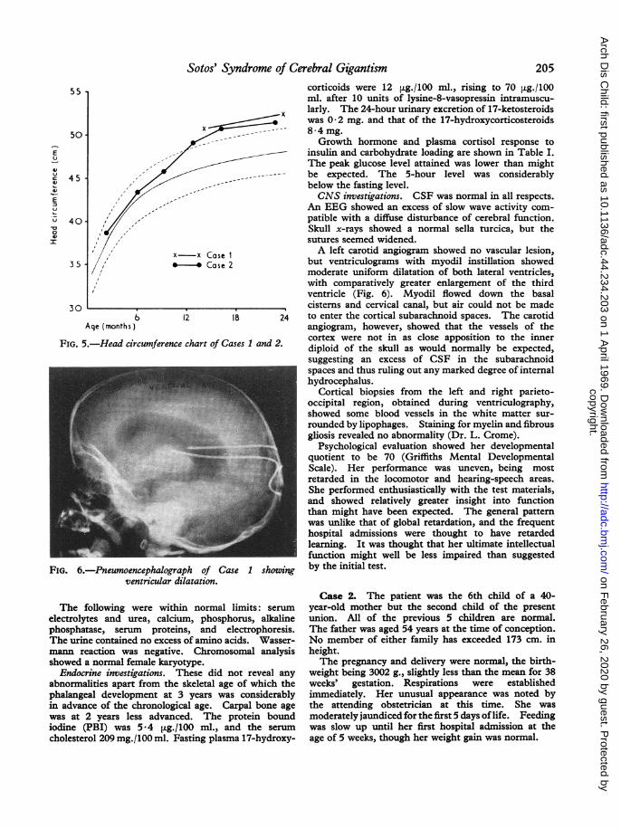
FIG. 5.-Head circumference
FIG. 6.-Pneumoencephalograventricular di
corticoids were 12 [tg./100 ml., rising to 70 [±g./100ml. after 10 units of lysine-8-vasopressin intramuscu-
x larly. The 24-hour urinary excretion of 17-ketosteroidswas 0-2 mg. and that of the 17-hydroxycorticosteroids
x 8-4mg.Growth hormone and plasma cortisol response to
f insulin and carbohydrate loading are shown in Table I.The peak glucose level attained was lower than might
------ be expected. The 5-hour level was considerablybelow the fasting level.CNS investigations. CSF was normal in all respects.
An EEG showed an excess of slow wave activity com-patible with a diffuse disturbance of cerebral function.Skull x-rays showed a normal sella turcica, but thesutures seemed widened.
- x Case 1 A left carotid angiogram showed no vascular lesion,* Case 2 but ventriculograms with myodil instillation showed
moderate uniform dilatation of both lateral ventricles,with comparatively greater enlargement of the thirdventricle (Fig. 6). Myodil flowed down the basalcisterns and cervical canal, but air could not be made
12 18 24 to enter the cortical subarachnoid spaces. The carotidangiogram, however, showed that the vessels of thecortex were not in as close apposition to the inner
chart of*Cases 1 and 2. diploid of the skull as would normally be expected,suggesting an excess of CSF in the subarachnoidspaces and thus ruling out any marked degree of internalhydrocephalus.
Cortical biopsies from the left and right parieto-occipital region, obtained during ventriculography,showed some blood vessels in the white matter sur-rounded by lipophages. Staining for myelin and fibrousgliosis revealed no abnormality (Dr. L. Crome).
Psychological evaluation showed her developmentalquotient to be 70 (Griffiths Mental DevelopmentalScale). Her performance was uneven, being mostretarded in the locomotor and hearing-speech areas.She performed enthusiastically with the test materials,and showed relatively greater insight into functionthan might have been expected. The general patternwas unlike that of global retardation, and the frequent
4 11 * | | uiSl _ hospital admissions were thought to have retardedlearning. It was thought that her ultimate intellectualfunction might well be less impaired than suggested
z-h of Case I showinag by the initial test.'ilatation.
The following were within normal limits: serum
electrolytes and urea, calcium, phosphorus, alkalinephosphatase, serum proteins, and electrophoresis.The urine contained no excess of amino acids. Wasser-mann reaction was negative. Chromosomal analysisshowed a normal female karyotype.
Endocrine investigations. These did not reveal any
abnormalities apart from the skeletal age of which thephalangeal development at 3 years was considerablyin advance of the chronological age. Carpal bone age
was at 2 years less advanced. The protein boundiodine (PBI) was 5*4 [g./100 ml., and the serumcholesterol 209 mg./100 ml. Fasting plasma 17-hydroxy-
Case 2. The patient was the 6th child of a 40-year-old mother but the second child of the presentunion. All of the previous 5 children are normal.The father was aged 54 years at the time of conception.No member of either family has exceeded 173 cm. inheight.The pregnancy and delivery were normal, the birth-
weight being 3002 g., slightly less than the mean for 38weeks' gestation. Respirations were establishedimmediately. Her unusual appearance was noted bythe attending obstetrician at this time. She wasmoderately jaundiced for the first 5 days of life. Feedingwas slow up until her first hospital admission at theage of 5 weeks, though her weight gain was normal.
205
x-
copyright. on F
ebruary 26, 2020 by guest. Protected by
http://adc.bmj.com
/A
rch Dis C
hild: first published as 10.1136/adc.44.234.203 on 1 April 1969. D
ownloaded from
Abraham and Snodgrass
M, .." N
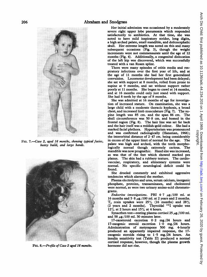
FIG. 7.-Case 2, aged 16 months, showing typical facies,heavy build, and large hands.
FIG. 8.-Profile ofCase 2 aged 16 months.
Her initial admission was occasioned by a moderatelysevere right upper lobe pneumonia which respondedsatisfactorily to antibiotics. At that time, she wasnoted to have mild inspiratory stridor, long digits,a high arched palate, small mandible, and dolicocephalicskull. Her extreme length was noted on this and manysubsequent occasions (Fig. 3), though the weightincrements were not commensurate until the age of 12months (Fig. 4). Additionally, a congenital dislocationof the left hip was discovered, which was successfullytreated with a van Rosen splint.
There were many episodes of otitis media and res-piratory infections over the first year of life, and atthe age of 11 months she had her first generalizedconvulsion. Locomotor development had been delayed;she sat with support at 8 months, rolled from prone tosupine at 9 months, and sat without support ratherpoorly at 11 months. She began to crawl at 14 months,and at 16 months could only just stand with support.She had 8 teeth by the age of 8 months.She was admitted at 16 months of age for investiga-
tion of increased stature. On examination, she was alarge child with a moderate thoracic kyphosis, a broadchest, and increased limb musculature (Fig. 7). The su-pine length was 85 cm. and the span 86 cm. Theskull circumference was 50-8 cm. and bossed in thefrontal region (Fig. 8). The hair line was set far backand the hair itself was a reddish-gold colour. She had amarked facial plethora. Hypertelorism was pronouncedand was confirmed radiologically (Hansman, 1966);the interorbital distance of 2 * 47 cm. being considerablyin excess of the upper limit of normal for the age. Thepalate was high and arched, with the teeth morpho-logically normal though extremely carious. Themandible was now prognathic. Hand size was increased,as was that of the feet which showed marked pesplanus. The skin had a rubbery texture. The cardio-vascular, respiratory, and alimentary systems werenormal. No specific neurological deficit could befound.
She drooled constantly and exhibited aggressivetendencies which alarmed the mother.Plasma electrolytes and urea, serum calcium, inorganic
phosphate, proteins, transaminases, and cholesterolwere normal, as were two urinary amino-acid chromato-grams.
Endocrine investigations. PBI 4-7 hg./100 ml. at16 months and 5 8 ltg./100 ml. at 2 years and 2 months.T3 resin uptakes were 25% (16 months) and 28%(2 years and 2 months). Thyroidal 132I uptake was12% at 2 hours and 15% at 4 hours.Synacthen test-resting plasma cortisol 25 pg.f100 ml.
and 58 ig./100 ml. 30 minutes later.17-oxosteroid excretion 0*2 mg./24 hours and
17-oxogenic steroid excretion 1*0 mg.124 hours.Administration of metyrapone 500 mg. 4-hourlyproduced an apparently impaired response, the 17-oxogenic steroids rising to 1*5 mg./24 hours. Aninsulin sensitivity test (Table II) produced a normalcortisol response, however, though the plasma growthhormone did not rise.
206
copyright. on F
ebruary 26, 2020 by guest. Protected by
http://adc.bmj.com
/A
rch Dis C
hild: first published as 10.1136/adc.44.234.203 on 1 April 1969. D
ownloaded from

Sotos' Syndrome of Cerebral GigantismTABLE I
Insulin and Glucose Tolerance Tests in Case 1
207
Time (min.) After Insulin Time (min.) After Oral GlucoseValue I-1~
0 20 30 40 | 60 80 0 30 60 120 180 240 300
Blood glucose (mg./100 ml.) .. .. 52 <10 19 27 49 47 91 107 91 98 72 67 72Plasma growth hormone (ng./ml.) .. 2-0 2-5 14-5 9 5 5 0 3-5 2-0 2-0 2-5 3 0 2-5 2-0 2-0Plasma 'cortisol' (,ug./100 ml.) .. .. 39 - 37 - 29 24 - _ _ _
TABLE IIAInsulin and Oral Glucose Tolerance Tests in Case 2 at 16 Months
Time (min.) After Insulin Time (min.) After Oral GlucoseValue
0 20 30 40 60 80 0 20 30 60 90 120 150
Blood glucose (mg./100 ml.) .. .. 95 40 40 - 90 - 55 136 155 189 82 64 59Plasma growth hormone (ng./ml.) .. 25 2*5 2*5 - 2*5 - _ - - -_ _Plasma 'cortisol' (,ag./100 ml.) .. .. 38 37 45 - 55 z- - -
TABLE IIBInsulin and Intravenous Glucose Tolerance Tests in Case 2 at 2 years 2 months
Time (min.) After Insulin Time (min.) After Intravenous Glucose*Value
0 20 30 40 60 80 0 3 15 30 60 120 1180
Blood glucose (mg./100 ml.) .. .. 59 28 - 29 29 39 75 215 170 120 75 80 80Plasmagrowthhormone(ng./ml.) | 30 40 - 3 5 2*5 30- - | -|
* Rate of clearance (k) glucose, 1 *82% per minute.
The carpal bone age at 16 months was compatible withchronological age, but the phalangeal developmentwas at the 2a-year-old level.
Carbohydrate tolerance tests. These are givenin Table I and are shown to be normal.CNS investigations. The CSF was normal. Two
EEGs were within normal limits for the age.An air encephalogram at this time showed generalized
dilatation of the lateral and third ventricles, with noevidence of displacement. The fourth ventricle andaqueduct were central. The basal, pontine, andchiasmatic cisterns were well demonstrated and noabnormalities were seen. The pituitary fossa was ofnormal size.
Psychological evaluation showed her developmentalquotient to be 86 (Griffiths Mental DevelopmentalScale). Retardation was most marked in the locomotorarea and in hearing and speech. She enjoyed themanipulative tests and performed best in this sphere.
Clinical features of the SyndromeThe main clinical features are summarized in
Table III. Complete information is not obtain-able for all cases reported. Only those findingsknown to be definitely present or absent are there-
fore included in this Table. For the purpose ofclarification some of these are discussed below.
Neonatal features. The birthweight tendsto be high, with a mean of 4002 g. for 39 patients,1 SD above the mean for full-term infants in theUnited Kingdom. The affected patients tend tobe born near or beyond term. Only 3 patientsare recorded as being less than 38 weeks' gestation,including Feingold's (1966) remarkable patientof 26 weeks' gestation who weighed 1728 g. atbirth. Hypoxia at the time of birth and respiratoryproblems during the first week of life are notinfrequent and may play a part in the aetiologyof the cortical atrophy found in some cases.Several patients were cyanosed after birth (Sotoset al., 1964; Turner and Sloan, 1965; Sizonenkoet al., 1968) or required immediate resuscitation.The feeding problems encountered in some infantsseem to be related to poor sucking and slowness infeeding rather than to failure to gain weight nor-mally.
Developmental features. Though many patients
copyright. on F
ebruary 26, 2020 by guest. Protected by
http://adc.bmj.com
/A
rch Dis C
hild: first published as 10.1136/adc.44.234.203 on 1 April 1969. D
ownloaded from

Abraham and SnodgrassTABLE III
Analysis of Findings in 43 Cases of Sotos' Syndrome
Feature Case 1 Case 2 Observed Frequency
NeonatalSex .F F 26 M 17 FBirthweight (g.) 2466 3002 Mean 4002 (39 cases)Birth length (cm.) .. ? Mean 55-4 (21 cases)Gestation (wk.) 37 38 Mean 39 (17 cases)Respiratory problems + - 10/30Jaundice .. + + 7/22Feeding problems . + + 7/14
DevelopmentalEarly dentition .. + + 7/9Delayed walking .. + + 22/31Delayed speech .. + + 18/25Aggressiveness .. _ + 5/7IQ or DQ <90 or
severely retarded + + 34/42
Cranio-facialMacrocrania for age + + 41/41Dolicocephaly .. + + 23/26Hypertelorism .. + + 22/30Antimongoloid slant
of palpebralfissures .. + _ 10/18
High arched palate.. + + 28/32Prognathism .. + + 24/28Facial plethora .. + + 4/4Marked macro-
glossia .. .. _ Twice reported
SkeletalKyphosis or
scoliosis .. .. + + 8/24Spina bifida occulta - - Twice reportedLarge hands and feet + + 23/27Syndactyly of toes.. _ _ 8/30
CNSConvulsions .. + + 13/39Clumsiness.. .. + + 26/26Drooling .. .. + + 4/7
InvestigationsAbnormal EEG .. + - 14/36Abnormal AEG .. + + 23/27Normal pituitary
fossa .. .. + + 36/40Growth hormonenormal .. .. + + 23/33
Elevated 17-ketosteroids .. _ - Frequent finding
Advanced bone age + + 33/39
Source of material for Table IIICohen, 1 case; Hook and Reynolds 6 cases; Kjellman, 1 case;Kowlessar, 1 case; Ludwig, Chaykin, and Escueta, 1 case; Marieet al., 4 cases; Mikulowski, 1 case; Milunsky, 2 cases; Poznanski andStephenson, 10 cases; Sizonenko et al., 6 cases; Sotos et al., 5 cases;Turner and Sloan, 2 cases; and Feingold, 1 case.
sat unsupported at the normal age, delay in walkingis frequent, and few have walked before the ageof 15 months. Invariably there is no specificexplanation for this on neurological examinationapart from the generalized clumsiness noted inall cases. Delay in speaking is usual; manycases are unable to say more than a few words by the
age of 21 years. The IQ or DQ is less than 90in most cases, but the retardation is not necessarilysevere. 5 children have been in the 80-90 range
and 8 patients in excess of 90 have been reported.2 patients reported by Sizonenko et al. (1968) were
considered mentally normal, and IQ's of 110(Cohen, 1964), 115, and 109 (Hook and Reynolds,1967) have been reported.
Cranio-facial features. As can be seen fromTables I and II and from the illustrations in thisreport, the patients tend to resemble each other inappearance. Prognathism is not a feature atbirth and tends to become more prominent withage. The facial plethora in our cases was strikingbut only Milunsky, Cowie and Donoghue (1967)have commented on this previously. A furthercase observed elsewhere by one of us also had thisfeature.
Skeletal features. Apart from the enlargedhands and feet, the most frequent finding is thatof kyphosis or kypho-scoliosis. Kyphosis is,interestingly, a frequent finding in acromegaly(Kellgren, Ball, and Tutton, 1952). The accelera-tion of skeletal maturation typically affects thephalangeal centres more than the carpal centres(Poznanski and Stephenson, 1967). This disparitywas found in both our cases.
Central nervous system features. Convul-sions have occurred in a third of the reportedcases, but the EEG findings have often been normal,while the abnormal EEG findings, typically diffuseand non-specific, have not been confined to thosepatients having convulsions.The typical clumsiness of these patients seems
partially to be related to the difficulty of the develop-ing locomotor system in coping with the unusualsize. The gait is deliberate and limb movementsponderous. Continuous drooling together withthe facial plethora, frequent respiratory infections,kypho-scoliosis, and relative insensitivity to pain,suggested a similarity to some of the features offamilial dysautonomia. An intradermal histaminetest on Case 2 gave a normal response, and no
pupillary constriction was observed on administra-tion of methacholine eye drops.Marked aggressiveness, a feature of Case 2, has
been commented upon previously (Kjellman,1965; Milunsky et al., 1967).The pneumoencephalograms in affected patients
have generally shown a non-specific ventricular dila-tation without a block (Poznanski and Stephenson,1967). Cortical atrophy is not uncommon. Three
208
copyright. on F
ebruary 26, 2020 by guest. Protected by
http://adc.bmj.com
/A
rch Dis C
hild: first published as 10.1136/adc.44.234.203 on 1 April 1969. D
ownloaded from

Sotos' Syndrome of Cerebral Gigantismcases have had a cavum septum pellucidum demon-strated (Marie et al., 1965; Poznanski and Stephen-son, 1967).
Dermatoglyphic features. Professor L. S.Penrose analysed the dermatoglyphs of our patientsand compared them with those of the two cases
published by Milunsky et al. (1967). The unusualfeatures noted by these authors included a thenarexit of the A-line, an increased a-b ridge distance,and a high a-b ridge count. Case 1 in this reporthad a normal exit of the A-line, a low a-b ridgecount (66), and a total ridge count of 141. Case 2had a thenar exit of the A-line on the left hand,a normal a-b ridge count (88), and a rather hightotal ridge count of 174. Of two further cases
to which the authors had access, one had a thenarexit of the A-line on one hand and a total ridgecount of 172, and the other had only an increaseda-b distance. The general dermatoglyphic patternsin these cases are not therefore particularly unusual,with the exception of a relatively increased a-bdistance and a considerable increase in the mean
ridge breadth. The latter would be expected ingigantism (L. S. Penrose, 1968, personal com-
munication), but interestingly this parametermeasured 0 434 mm. in Case 1 and 0 400 mm. inCase 2, being appropriate to controls of 5 and 4years, respectively; the prints were taken when theheight age was 3 years for Case 1 and 21 years forCase 2. These findings suggest that the handsin these cases were disproportionately large, even
when taking into account the excessive stature.
DiscussionThe two cases reported here bear a striking
resemblance to each other and conform closelywith the criteria proposed by Sotos and his co-
workers (1964). Other causes of large stature inthis age-group, such as Marfan's syndrome,neurofibromatosis, McCune-Albright's syndrome,familial gigantism, and various virilizing conditions,could be readily excluded on clinical and laboratorygrounds. Patients suffering from these disordersdo not resemble each other, and, with the exceptionof Marfan's syndrome, enlargement of the ex-
tremities is not found.That the rapid growth is not mediated by excessive
growth hormone secretion is probably borne outby the normal levels found in Case 1 and the sub-normal levels of Case 2. Similar findings havebeen reported by all other authors where this hadbeen measured (Hook and Reynolds, 1967; Milunskyet al., 1967; Stephenson, Mellinger, and Manson1968). Likewise it is probably not due to a hyper-
sensitivity to this hormone, as there is a normalrise in plasma non-esterified fatty acids after itsexogenous administration (Stephenson et al., 1968).The 24-hour total secretion of growth hormonemight conceivably be greater in these cases thanin the normal, but this is impossible to demon-strate at this time. Against this theory, however,is the acceleration of skeletal maturation and theabsence of visceromegaly. In a number of reportedcases the gigantism has been present at birth, asinstanced by two cases being 63-5 cm. in lengthat this time (Turner and Sloan, 1965; Hook andReynolds, 1967). Possibly a separate growthfactor, fetal or maternal, may operate over theinitial period of high growth velocity occurringover the last trimester of intrauterine life and forthe first three months of life. Intrauterine growthretardation was not observed in 16 cases of isolatedgrowth hormone deficiency recently described(Goodman, Grumbach, and Kaplan, 1968), andnone of these exhibited growth failure before theage of 4 months. The continuance of the excessivegrowth beyond this time cannot, however, beexplained by such a factor.The acceleration of skeletal maturation has been
ascribed to the increase of urinary 17-ketosteroidsseen in some cases (Kowlessar, 1965; Stephensonet al., 1968) where these have been considerablyin excess of the normal for either chronologicalor height age. But most cases, including ours, showno such increase. Case 1 in this report, however,showed a disproportionate increase in urinary17-hydroxycorticosteroids, noted also in Kowlessar'scase.The hypothalamic region, being a link between
the higher cerebral centres and the endocrineglands, has been incriminated as the main seat ofpathology in this syndrome. Only the non-specificenlargement of the third ventricle found in manycases might support this. Lesions in this regionare known to be associated with early increasein stature and enlargement of the extremities inat least three other conditions. The diencephalicsyndrome of emaciation may initially show thispattern, but this is not maintained because ofextension of the neoplasm into the anteriorhypothalamus (Russell, 1951; Gamstorp, Kjellman,and Palmgren, 1967). Likewise, this situationis found in generalized lipodystrophy (Berardinelli,1954; Seip, 1959), though the total absence of bodyfat is obviously the most striking feature.
Increased stature is also found in the syndrome ofexomphalos with macroglossia (Wiedemann, 1964;Beckwith et al., 1964; Irving, 1967), and increasedbone age, prognathism, and high arched palate
209,
copyright. on F
ebruary 26, 2020 by guest. Protected by
http://adc.bmj.com
/A
rch Dis C
hild: first published as 10.1136/adc.44.234.203 on 1 April 1969. D
ownloaded from

210 Abraham and Snodgrassare further clinical features shared with Sotos'syndrome. Features absent, however, from Sotos'syndrome include relative microcephaly, exom-phalos, neonatal hypoglycaemia, and a tendencyto familial incidence.As far as we are aware, there has been no report
of cerebral biopsy or post-mortem study of thebrain in Sotos' syndrome. The cortical biopsyin Case 1, performed at the time of ventriculographyfor which a burr hole was required, showed onlya non-specific abnormality. The absence ofdemonstrable endocrine abnormalities, the resem-blance of the patients to each other, sometimesat birth, and the neurological features suggest acongenital malformation syndrome.
SummaryTwo cases of Sotos' syndrome are described and
the 43 recorded cases reviewed. The conditionis characterized by: (1) high birthweight for gesta-tional maturity, (2) an unusual appearance withmacrocrania, dolicocephaly, hypertelorism, prog-nathism, a high arched palate, and facial plethora,(3) accelerated growth and accelerated skeletalmaturation from early infancy, with some featuresof acromegaly (4) clumsiness without neurologicaldeficit, (5) normal endocrine functions. The aetio-logy remains obscure.
We wish to thank Dr. L. Crome who performed theneuropathological examination on the biopsy materialin Case 1, Dr. H. Barrie for permission to publish Case 2,and Mr. H. Maslowski for the ventriculographicstudies in Case 1.
RBFERENCES
Beckwith, J. B., Wang, C. I., Donnell, G. N., and Gwinn, J. L.(1964). Hyperplastic fetal visceromegaly with macroglossia,omphalocele, cytomegaly of adrenal fetal cortex, postnatalsomatic gigantism, and other abnormalities: a newly recognizedsyndrome. Transactions of the American Pediatric Society,74th Annual Meeting, Seattle, Washington. Abstr., no. 41.
Berardinelli, W.. (1954). An undiagnosed endocrinometabolicsyndrome: report of two cases. J. clin. Endocr., 14, 193.
Cohen, M. I. (1964). Cerebral gigantism in childhood. NewEngl. J. Med., 271, 635.
Feingold, M. (1966). Personal communication. Quoted byMilunsky et al., 1967.
Gamstorp, I., Kjellman, B., and Palmgren, B. (1967). Dience-phalic syndromes of infancy. Report of three children withemaciation syndrome and disproportionately large hands andfeet. J. Pediat., 70, 383.
Goodman, H. G., Grumbach, M. M., and Kaplan, S. L. (1968).Growth and growth hormone. II. A comparison of isolatedgrowth-hormone deficiency and multiple pituitary-hormonedeficiencies in 35 patients with idiopathic hypopituitarydwarfism. New Engi. J. Med., 278, 57.
Hansman, C. F. (1966). Growth of interorbital distance and skullthickness as observed in roentgenographic measurements.Radiology, 86, 87.
Hook, E. B., and Reynolds, J. W. (1967). Cerebral gigantism:endocrinological and clinical observations of six patientsincluding a congenital giant, concordant monozygotic twinsand a child who achieved adult gigantic size. J. Pediat., 70, 900.
Irving, I. M. (1967). Exomphalos with macroglossia: a study ofeleven cases. J. pediat. Surg., 2, 499.
Kellgren, J. H., Ball, J., and Tutton, G. K. (1952). The articularand other limb changes in acromegaly. Quart. . Med., 21, 405.
Kjellman, B. (1965). Cerebral gigantism. Acta paediat. scand.,54, 603.
Kowlessar, M. (1965). Cerebral gigantism. Abnormal urinarycorticoid excretion. Minn. Med., 48, 1610.
Ludwig, G. D., Chaykin, L. B., and Escueta, A. V. (1967). Cere-bral gigantism with intermittent fractional hypopituitarismand normal sella turcica. Ann. intern. Med., 67, 123.
Marie, J., Royer, P., Leveque, B., Debauchez, C., and Rappaport, R.(1965). Gigantisme avec encephalopathie et dysmorphiecranio-faciale. Ann. PJdiat., 12, 682.
Mikulowski, W., Stopyrowa, J., and Medvey, K. (1962). Gigantismand acromegaly in a 6-year-old boy. (In Polish.) Endokr.pol., 13, 407.
Milunsky, A., Cowie, V. A., and Donoghue, E. C. (1967). Cerebralgigantism in childhood. Pediatrics, 40, 395.
Poznanski, A. K., and Stephenson, J. M. (1967). Radiographicfindings in hypothalamic acceleration of growth associated withcerebral atrophy and mental retardation (cerebral gigantism).Radiology, 88, 446.
Russell, A. (1951). A diencephalic syndrome of emaciation ininfancy and childhood. Arch. Dis. Childh., 26, 274.
Seip, M. (1959). Lipodystrophy and gigantism with associatedendocrine manifestation. A new diencephalic syndrome ?Acta. paediat. (Uppsala)., 48, 555.
Sizonenko, P. C., Job, J. C., Sebouk, S., and Rossier, A. (1968).Gigantisme cerebral de l'enfant: dosage de l'hormone decroissance dans le plasma. Arch. franc. Pldiat., 25, 151.
Sotos, J. F., Dodge, P. R., Muirhead, D., Crawford, J. D., andTalbot, N. B. (1964). Cerebral gigantism in childhood.A syndrome of excessively rapid growth with acromegalicfeatures and a nonprogressive neurologic disorder. NewEngl. J. Med., 271, 109.
Stephenson, J. N., Mellinger, R. C., and Manson, G. (1968).Cerebral gigantism. Paediatrics, 41, 130.
Tumer, E. K., and Sloan, L. E. G. (1965). Cerebral gigantismin childhood. Aust. paediat. J., 1, 243.
Wiedemann, H. R. (1964). Complexe malformatif familial avechernie ombilicale et macroglossie-'un syndrome nouveau' ?J. GOnit. hum., 13, 223.
Correspondence to: Dr. G. J. A. I. Snodgrass,Department of Paediatrics, Guy's Hospital MedicalSchool, St. Thomas Street, London S.E.1.
copyright. on F
ebruary 26, 2020 by guest. Protected by
http://adc.bmj.com
/A
rch Dis C
hild: first published as 10.1136/adc.44.234.203 on 1 April 1969. D
ownloaded from